Fluid, Electrolyte, and Acid-Base Disorders Darren Dreyfus, D.O. Associates In Nephrology, P.C. St....
156
Fluid, Fluid, Electrolyte, and Electrolyte, and Acid-Base Acid-Base Disorders Disorders Darren Dreyfus, D.O. Associates In Nephrology, P.C. St. Vincent’s Health System
-
Upload
suzan-caldwell -
Category
Documents
-
view
214 -
download
2
Transcript of Fluid, Electrolyte, and Acid-Base Disorders Darren Dreyfus, D.O. Associates In Nephrology, P.C. St....

Fluid, Electrolyte, and Fluid, Electrolyte, and Acid-Base DisordersAcid-Base Disorders
Darren Dreyfus, D.O.Associates In Nephrology, P.C.St. Vincent’s Health System

Question 1Question 1

Question 1Question 1
A 71-year-old man is evaluated because of intermittent cough and increasing dyspnea on exertion. He has smoked one pack of cigarettes per day for 49 years. He has no other symptoms.
On physical examination, distant breath sounds are audible in both lungs and there are scattered rhonchi. Chest radiograph shows a perihilar mass. Abnormal laboratory results include hemoglobin of 10.5 g/dL and a serum sodium of 127 meq/L. Endobronchial biopsy reveals small-cell lung cancer.

Question 1Question 1
Which of the following is most appropriate treatment for this patient’s hyponatremia?
(A) Chemotherapy(B) Hypertonic saline(C) Fluid restriction(D) Fluid restriction and demeclocycline

AnswerAnswer

Question 1 - AnswerQuestion 1 - Answer
Answer: AThis patient with mild hyponatremia is newly
diagnosed with small-cell lung cancer. He has no symptoms that are clearly attributed to the hyponatremia and there initiating non-specific therapy such as fluid restriction with or without demeclocycline therapy, or aggressively treating the hyponatremia with hypertonic saline is not warranted. The most important therapeutic intervention is to treat the underlying cancer with chemotherapy.

Question 2Question 2

Question 2Question 2
A 78-year-old woman with metastatic breast cancer involving the bones and soft tissues is evaluated in the emergency department because of lethargy and weakness, nausea, thirst, and dizziness. She has a history of congestive heart failure that has been controlled with medications. On physical examination she has orthostatic hypotension associated with dizziness and dry mucous membranes. Her lungs are clear and she has no dependent edema. Blood urea nitrogen – 42 mg/dL Total serum calcium – 11.4 mg /dL Serum creatinine – 1.6 mg/dL Serum albumin – 3.0 g/dL

Question 2Question 2
Administration of which of the following is the most appropriate treatment?
(A) A biophosphonate, intravenously(B) Corticosteroids, intravenously(C) Half-normal saline(D) Normal saline(E) Furosemide

AnswerAnswer

Question 2 - Answer Question 2 - Answer
Answer: DThe patient has hypercalcemia, the most
common metabolic complication of malignancy. She is volume depleted as evidenced by her orthostatic hypotension, dizziness and dry mucous membranes. Despite her history of congestive heart failure, the most appropriate initial therapy is to replace vascular volume as vigorously as possible with normal saline.

Question 2 - AnswerQuestion 2 - Answer
Furosemide must not be given until the patient has first been adequately rehydrated. Administration of a bisphosphonate is not an appropriate initial therapeutic intervention in this patient because its delayed onset of effect and her more urgent need for volume repletion. Although corticosteroids can be considered as an adjunctive therapy in a patient with a potentially hormone-responsive tumor such as breast cancer, the top priority is volume repletion.

Question 3Question 3

Question 3Question 3
A 56-year-old man with a 25-pack-year smoking history, distant cerebrovascular accident, and a 10-year history of hypertension treated with hydrochlorothiazide is evaluated because of generalized fatigue. Blood pressure is 110/70 mm Hg. Serum sodium – 128 meq/L Serum potassium – 3.3 meq/L Serum chloride – 79 meq/L Serum biocarbonate – 38 meq/L Arterial blood gases on room air:
►pH – 7.50; PCO2 50 mm Hg, PO2 74 mm Hg

Question 3Question 3
Which of the following best explains the patient’s acid-base disturbance?
(A) Metabolic alkalosis(B) Respiratory acidosis(C) Respiratory alkalosis(D) Metabolic acidosis

AnswerAnswer

Question 3 - AnswerQuestion 3 - Answer
Answer: AThe low blood pressure and hypochloremia
suggest volume-sensitive metabolic alkalosis due to diuretic therapy. Metabolic alkalosis is indicated by the high serum bicarbonate level and pH greater than 7.4 Respiratory compensation for the metabolic alkalosis is appropriate. The PCO2 is 50 mm Hg, showing the expected 10-mm Hg increase that compensates for the 14-meq/L increase in serum bicarbonate level.

Question 3 - AnswerQuestion 3 - Answer
Metabolic acidosis is not a possible diagnosis since the bicarbonate level and pH are elevated. The normal arterial-to-alveolar oxygen gradient makes underlying chronic obstructive pulmonary disease unlikely, and blood gas values do not indicate concurrent respiratory acidosis.

Question 4Question 4

Question 4Question 4
A 65-year-old man with chronic alcoholism is evaluated because of a seizure 1 hour ago. In the emergency department, he is lethargic but conversant and oriented. He reports a several-day history of diarrhea and has muscle cramps. He has no history of trauma or previous seizures. He is taking no medication but has smoked 1 pack of cigarettes daily for the past 30 years.

Question 4Question 4
On physical examination, blood pressure is 11/075 mm Hg, pulse rate is 100/min, and respiration rate is 18/min. The neck is supple. Carpopedal spasm and Chvostek’s signs are present. The remainder of the physical examination is normal.

Question 4Question 4
Serum creatinine – 1.4 mg/dLSerum sodium – 136 meq/LSerum potassium 2.7 meq/LSerum chloride – 98 meq/LSerum bicarbonate – 23 meq/LSerum calcium – 7.6 mg/dLSerum magnesium – 0.5 mg /dLSerum phosphorus – 3.0 mg/dL

Question 4Question 4
Which of the following will best correct the underlying deficiency responsible for this patient’s electrolyte disorders?
(A) Calcium(B) Magnesium sulfate(C) Magnesium sulfate and potassium
chloride(D) Potassium chloride

AnswerAnswer

Question 4 - AnswerQuestion 4 - Answer
Answer: CHypomagnesemia is frequently seen in
alcoholics and in persons with severe diarrhea. If the serum magnesium level is less than 1.0 mg/dL and carpopedal spasm, Chvostek’s or Trousseau’s sign, or seizures are present, intravenous treatment with magnesium is indicated.

Question 4 - AnswerQuestion 4 - Answer
The hypokalemia is due to diarrhea and volume depletion (volume depletion triggers aldosterone release causing sodium conservation at the expense of potassium loss) and to the renal potassium wasting induced by hypomagnesemia.
Attempts at correction of the hypokalemia will be ineffective unless hypomagnesemia and volume are also corrected.
Question 4 - AnswerQuestion 4 - Answer
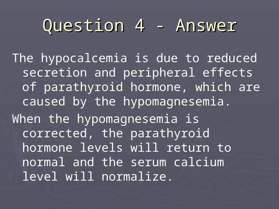
The hypocalcemia is due to reduced secretion and peripheral effects of parathyroid hormone, which are caused by the hypomagnesemia.
When the hypomagnesemia is corrected, the parathyroid hormone levels will return to normal and the serum calcium level will normalize.

Question 5Question 5

Question 5Question 5
A 35-year-old man is evaluated because of recurrent nepholithiasis and is found to have a normal serum calcium concentration and hypercalciuria.

Question 5Question 5
Which of the following diets should this man avoid in order to reduce the risk of recurrent calcium stones?
(A) Low-sodium diet(B) Low-calcium diet(C) Low-oxalate diet(D) Low-protein diet(E) Low-purine diet

AnswerAnswer

Question 5 - AnswerQuestion 5 - Answer
Answer: BRisk factors for calcium nephrolithiasis include
low daily fluid intake, high dietary sodium and oxalate, hyperoxaluria, hypercalciuria, hypocitrauria, and hyperuricosuria.
Several studies in men and women have shown that a high dietary calcium intake is associated with reduced risk for calcium stone disease.
In a recent study, men with hypercalciuric nephrolithiasis placed on a low-calcium versus a high-calcium diet had greater risk of subsequent stone formation.

Question 5 - AnswerQuestion 5 - Answer
Although not proven, it is believed that the risk for stone formation is lower with high dietary calcium because higher amounts of dietary calcium bind with dietary oxalate and lessen oxalate absorption and urinary excretion. For this same reason, oral magnesium therapy has also been used.
A low sodium diet can reduce renal calcium excretion and may reduce calcium stone formation. A diet high in animal protein increases urinary excretion of calcium and uric acid while it decreases excretion of citrate, and is associated with increased risk of calcium stone formation.
A diet low in animal protein may minimize this risk.
Question 5 - AnswerQuestion 5 - Answer
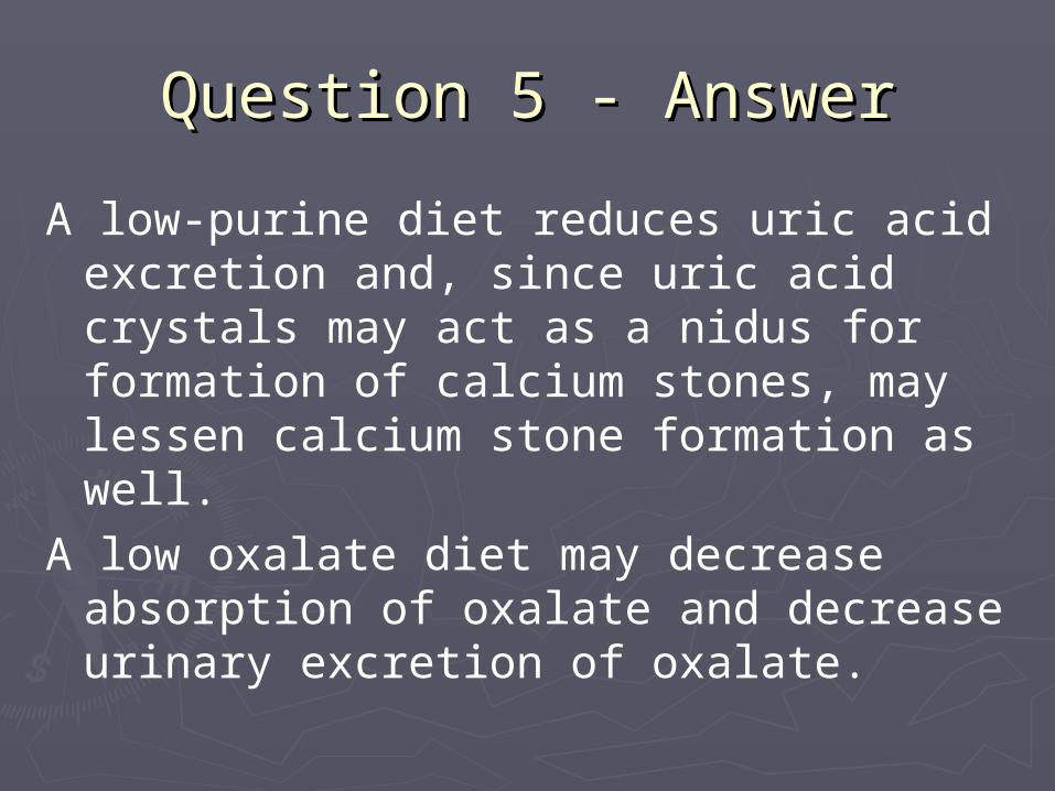
A low-purine diet reduces uric acid excretion and, since uric acid crystals may act as a nidus for formation of calcium stones, may lessen calcium stone formation as well.
A low oxalate diet may decrease absorption of oxalate and decrease urinary excretion of oxalate.

Question 6Question 6

Question 6Question 6
A 26-year-old woman with type 1 diabetes mellitus is evaluated in the emergency department because of a serum glucose concentration of 450 mg/DL and abdominal pain for the past 24 hours. Her temperature is 38 oC (101 oF). Serum sodium – 133 meq/L Serum potassium – 3.9 meq/L Serum chloride – 97 meq/L Serum bicarbonate – 10 meq/L Arterial blood gases – pH 7.2; PCO2, 23 mm Hg Whole blood lactacte - 0.6 mmol/L

Question 6Question 6
Which of the following best explains the patient’s acid-base status?
(A) Diabetic ketoacidosis(B) Diabetic ketoacidosis and proximal renal
tubular acidosis(C) Diabetic ketoacidosis and respiratory
acidosis(D) Diabetic ketoacidosis and metabolic
alkalosis

AnswerAnswer

Question 6 - AnswerQuestion 6 - Answer
Answer: AThe patient has an anion gap metabolic acidosis with
appropriate respiratory compensation consistent with a diabetic ketoacidosis. Metabolic acidosis is indicated by a low serum bicarbonate level and an arterial blood pH less than 7.4. The respiratory compensation is appropriate; thus, there is not a concomitant respiratory disorder accompanying this metabolic acidosis.
The patient has an anion gap of 26. The change in the anion gap is 14. The change in the bicarbonate level is 14, calculated as the normal bicarbonate concentration minus the measured bicarbonate concentration.
Question 6 - AnswerQuestion 6 - Answer
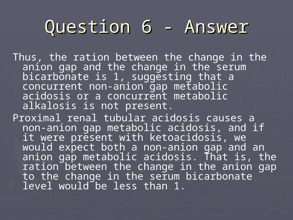
Thus, the ration between the change in the anion gap and the change in the serum bicarbonate is 1, suggesting that a concurrent non-anion gap metabolic acidosis or a concurrent metabolic alkalosis is not present.
Proximal renal tubular acidosis causes a non-anion gap metabolic acidosis, and if it were present with ketoacidosis, we would expect both a non-anion gap and an anion gap metabolic acidosis. That is, the ration between the change in the anion gap to the change in the serum bicarbonate level would be less than 1.

Question 7Question 7

Question 7Question 7
A 23-year-old woman with type 1 diabetes mellitus is evaluated in the emergency department because of a 2-day history of dysuria and urinary frequency. Three years ago she had “cystitis” twice in 6 months; in both occasions, she was treated with antibiotics. She uses insulin to control her diabetes and takes 1 or 2 ibuprofen tablets daily for headaches.

Question 7Question 7
On physical examination, the blood pressure is 115/80 mm Hg, pulse rate is 80/min, and temp is 37.4 oC (99.3 oF). Optic fundoscopy reveals microaneurysms and the neurological examination demonstrates diminished sensitivity to pinprick and light touch in the lower extremities. The remainder of the examination is unremarkable.
Question 7Question 7
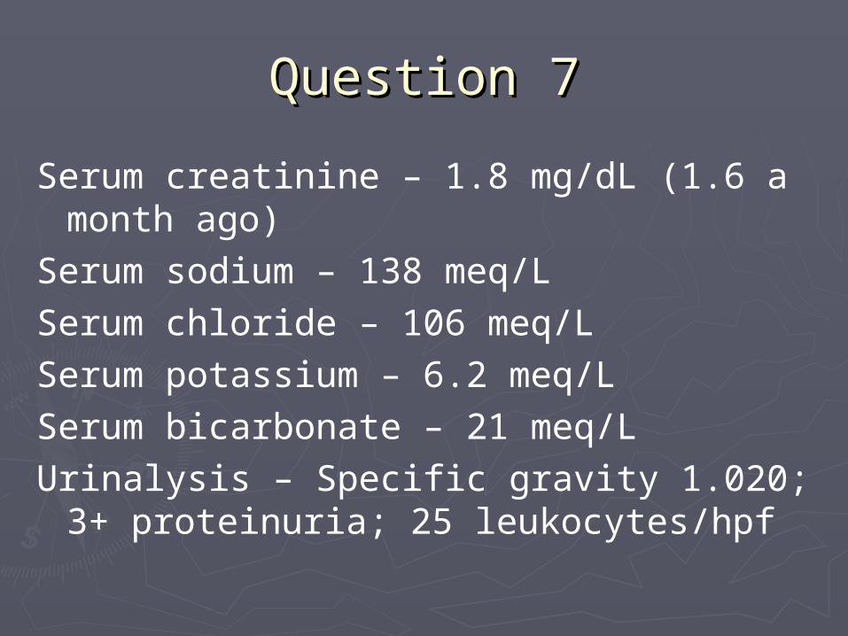
Serum creatinine – 1.8 mg/dL (1.6 a month ago)
Serum sodium – 138 meq/LSerum chloride – 106 meq/LSerum potassium – 6.2 meq/LSerum bicarbonate – 21 meq/LUrinalysis – Specific gravity 1.020; 3+
proteinuria; 25 leukocytes/hpf

Question 7Question 7
On renal ultrasonography, the right kidney is 11.0 cm and the left kidney is 10.9 cm. No hydronephrosis or stones are present.
Which of the following is the most likely cause of her hyperkalemia?
(A) Acute renal failure(B) Diabetic ketoacidosis(C) High sodium diet(D) Hyporeninemic hypoaldosteronism

AnswerAnswer

Question 7 - AnswerQuestion 7 - Answer
Answer: DThe most likely cause of hyperkalemia is hyporeninemic
hypoaldosteronism, a condition that occurs in 20% to 30% of diabetic persons.
Hyperkalemia occurs most often in diabetic patients who have mild to moderate renal failure (as in this case) superimposed on the hyporenin hypoaldosterone state.
The later condition seems to be due to unresponsiveness of the adrenal zona glomerulosa to stimulation by angiotensin II, renin, and hyperkalemia itself.
Many diabetic patients with hyperkalemia have low renin levels, caused by impaired conversion of prorenin to renin.

Question 7 - AnswerQuestion 7 - Answer
Since prostaglandins stimulate renin, use of ibuprofen may contribute to the hypoaldosterone state, but by itself rarely causes hyperkalemia.
Diabetic ketoacidosis is not present.A change in serum creatinine concentration from
1.6 to 1.8 would not cause hyperkalemia in a patient who had normal renin-aldosterone function.
A high-sodium diet does not cause hyperkalemia.

Question 8Question 8

Question 8Question 8
A 63-year-old man is evaluated because of upper and lower extremity cramps and diffuse muscle weakness of 2 weeks’ duration. He has been taking aspirin for 6 weeks because of low back pain. The physical examination is normal. Serum sodium – 135 meq/L Serum potassium – 2.6 meq/L Serum chloride – 117 meq/L Serum bicarbonate – 15 meq/L Arterial blood gases – pH, 7.30; PCO2, 31 mm Hg Urine pH – 5.0

Question 8Question 8
Which of the following best explains his acid-base status?
(A) Ethylene glycol toxicity(B) Lactic acidosis(C) Proximal renal tubular acidosis(D) Salicylate toxicity

AnswerAnswer

Question 8 - AnswerQuestion 8 - Answer
Answer: CThe electrolyte and arterial blood gas patterns are
consistent with a non-anion gap metabolic acidosis. Metabolic acidosis is indicated by a low serum bicarbonate
level and an arterial blood pH less than 7.4.The anion gap, calculated as 135 – (117 + 15)=3,
indicates a non-anion gap acidosis.A low anion gap (defined as less than 7) can be caused by
an increase in unmeasured cation such as calcium, magnesium, lithium, or a positively charged paraprotein produced by a myeloma.
A low anion gap can also be cause by a decrease in unmeasured anions such as albumin.

Question 8 - AnswerQuestion 8 - Answer
The urine pH of 5.0 is consistent with a proximal renal tubular acidosis in which distal tubular function is normal. In these patients, when the serum bicarbonate falls below the threshold for complete proximal reabsorption, the distal tubule is able to lower the pH normally. The low urine pH rules out distal renal tubular acidosis. Proximal renal tubular acidosis often causes hypokalemia and a non-anion gap metabolic acidosis.

Question 8 - AnswerQuestion 8 - Answer
One cause of renal tubular acidosis is the presence of a monoclonal light chain.
Proximal renal tubular acidosis due to a monoclonal light chain may occur before multiple myeloma is diagnoses.
Evaluation for multiple myeloma is warranted.
Salicylate toxicity, lactic acidosis, and ethylene glycol toxicity all cause an anion gap metabolic acidosis.

Question 9Question 9

Question 9Question 9
A 67-year-old woman is evaluated because of a 6-month history of gradual-onset dementia. She has smoked 1 to 2 packs per day of cigarettes for 40 years and she drinks 1 ounce of alcohol weekly.
On examination, she is disoriented to time and place. Her blood pressure is 142/88 mm Hg, without orthostatic change, pulse rate is 68/min, respiration rate is 12/mi, and temperature 37 oC (98.6 oF). There is no neck vein distention, the lungs are clear, the cardiac and neurological examinations are normal, and there is no peripheral edema.

Question 9Question 9
Plasma glucose – 84 mg/dLBlood urea nitrogen – 6 mg/dLSerum creatine – 0.5 mg/dLSerum sodium – 124 meq/LSerum potassium – 4.2 meq/LSerum chloride – 89 meq/LSerum bicarbonate – 24 meq/L

Question 9Question 9
Serumn cholesterol – 182 mg/dLSerum triglyceride – 60 mg/dLSerum total protein – 7.5 g/dLSerum osmolality – 255 mosmol/kg Urine osmolality – 400 mosmo/kg

Question 9Question 9
Which of the following is the most likely cause of her hyponatremia?
(A) Cirrhosis(B) Pseudohyponatremia(C) Psychogenic polydipsia(D) Syndrome of inappropriate
antidiuretic hormone secretion

AnswerAnswer

Question 9 - AnswerQuestion 9 - Answer
Answer: DThis patient has the syndrome of inappropriate
antidiuretic hormone secretion. The physical examination; normal serum potassium level; and low concentrations of blood urea nitrogen, serum creatinine, and serum uric acid are consistent with euvolemic hyponatremia and not with cirrhosis which is associated with a decreased circulating vascular volume.
The normal levels of cholesterol, triglycerides, and total proteins rule out pseudohyponatremia.

Question 9 - AnswerQuestion 9 - Answer
In a patient with psychogenic polydipsia, ingestion of large amounts of water would result in hyponatremia. However, the appropriate compensatory response would be suppression of antidiuretic hormone, which results in a dilute urine (excretion of free water); this phenomenon is not present in this patient, as evidences by the inappropriately high urine osmolality.

Question 10Question 10

Question 10Question 10
A 39-year-old man is evaluated in the emergency department because of a severe left flank pain and hematuria after playing softball. The pain is sharp and radiates to the groin. He vomited eight times before presentation. He has nonobstructing, calcium-containing kidney stone in the uretopelvic junction on the left side.

Question 10Question 10
On initial evaluation, his blood pressure was 130/90 mm Hg and pulse rate was 110/min. Serum sodum – 141 meq/L Serum potassium- 4.0 meq/L Serum chloride – 100 meq/L Serum bicarbonate – 34 meq/L Arterial blood gases – pH, 7.61; PCO2, 36
mg Hg

Question 10Question 10
Which of the following best describes this patient’s acid-base disorder?
(A) Metabolic acidosis and respiratory alkalosis
(B) Metabolic alkalosis(C) Metabolic alkalosis and respiratory
acidosis(D) Metabolic and respiratory alkalosis(E) Respiratory alkalosis

AnswerAnswer
Question 10 - AnswerQuestion 10 - Answer
Answer: DArterial blood gas values demonstrate a mixed
metabolic and respiratory alkalosis. Metabolic alkalosis is indicated by the high serum bicarbonate level and a pH greater than 7.4
Respiratory compensation for the metabolic alkalosis is not appropriate; the PCO2 would be expected to increase in compensation for the elevated serum bicarbonate level, but instead, the PCO2 has decreased to 36 mm Hg, indicated the presence of a respiratory alkalosis.

Question 10 - AnswerQuestion 10 - Answer
The anion gap is 7, calculated as (141 – [100 + 34]); thus, there is no hidden anion-gap metabolic acidosis. The respiratory alkalosis is most likely due to pain induced hyperventilation from the kidney stone, and metabolic alkalosis is probably a result of vomiting.

Question 11Question 11

Question 11Question 11
The preceding patient is given intravenous infusion of 0.9% normal saline at 200 mL/h. Two days later, his flank pain worsens dramatically, but nausea and vomiting have resolved. Blood pressure and pulse rate are unchanged.

Question 11Question 11
BUN – 8 mg/dLSerum creatinine – 0.9 mg/dLSerum sodum – 138 meq/LSerum sodium – 138 meq/LSerum potassium – 4.0 meq/LSerum chloride – 105 meq/LSerum bicarbonate – 22 meq/LArterial blood gases – ph, 7.48; PCO2, 30 mm
Hg

Question 11Question 11
Which of the following best describes his acid-base status?
(A) Metabolic acidosis with respiratory alkalosis(B) Metabolic alkalosis and respiratory alkalosis(C) Respiratory acidosis and metabolic alkalosis(D) Respiratory alkalosis

AnswerAnswer

Question 11 - AnswerQuestion 11 - Answer
Answer: DThe patient has persistent respiratory alkalosis,
as indicate by the low PCO2, and a pH greater than 7.4. The condition is most likely due to hyperventilation in response to pain from the kidney stone.
For each 10 mm Hg drop in the PCO2, the serum bicarbonate will decrease by 2. to 4.5 meq/L. Since the serum bicarbonate level decreased by 2 meq/L, this confirms the diagnosis of a simple, compensated respiratory alkalosis.

Question 12Question 12

Question 12Question 12
A 46-year-old man is evaluated because of a 20-year history of difficult to control hypertension and hypokalemia treated with amlodipine. He also has irritable bowel syndrome and produces two semi-formed stools daily. On examination, the blood pressure is 150/88 mg Hg, and pulse rate is 74/min. The lungs are clear, and the heart rhythm is regular, with no gallop or murmur. Abdominal examination is normal. Pulses in the lower extremities are normal, and no edema is present.

Question 12Question 12
Serum sodium – 142 meq/LSerum potassium – 2.7 meq/LSerum chloride – 105 meq/LSerum bicarbonate – 30 meq/LUrinalysis – pH 5.0; specific gravity 1.020;
dipstick negative for protein and blood24-hour urine results:
Sodum – 100 meq Potassium – 82 meq Calcium – 200 mg

Question 12Question 12
Which of the following is the most likely cause of his hypokalemia?
(A) Diarrhea(B) Distal renal tubular acidosis(C) Gitelman’s syndrome(D) Primary hyperaldosteronism

AnswerAnswer

Question 12 - AnswerQuestion 12 - Answer
Answer: DPrimary hyperaldosteronism is associated with
hypertension, hypkalemia, and renal potassium wasting. It is usually associated with a 24-hour urinary potassium excretion greater then 30 meq in the setting of a serum potassium concentration less than 3.0 meq/L.
All of these features are present in this patient. Distal renal tubular acidosis is incorrect because the patient is not acidotic and has an acid urine pH.

Question 12 - AnswerQuestion 12 - Answer
Gitelman’s syndrome is associated with hypokalemia, renal potassium wasting, and hypocalciuria (which is not present in this patient). And normotension.
Diarrhea would cause metabolic acidosis and may cause hypokalemia but would not be associated with renal potassium unless the patient is hypovolemic resulting in stimulation of the renin-angiotensin-aldosterone system.

Question 13Question 13

Question 13Question 13
A 60-year-old woman with a history of essential hypertension is admitted to the hospital after 7 years of vomiting. On physical examination, she appears ill. The systolic blood pressure is 110 mg Hg seated and 70 mg Hg standing. The pulse rate while seated is 120/min. The abdominal examination reveals rebound tenderness and no bowel sounds.

Question 13Question 13
Serum sodium – 140 meq/LSerum potassium – 3.2 meq/LSerum chloride – 80 meq/LSerum bicarbonate – 11 meq/LArterial blood gases – pH, 7.29; PCO2, 24
mm Hg

Question 13Question 13
Which of the following best describes her acid-base status?
(A) Anion gap metabolic acidosis(B) Anion gap metabolic acidosis and
metabolic alkalosis(C) Non-anion gap metabolic acidosis and
metabolic alkalosis(D) Non-anion gap metabolic acidosis

AnswerAnswer

Question 13 - AnswerQuestion 13 - Answer
Answer: BThe patient has an anion gap metabolic
acidosis and a con-current metabolic alkalosis. Metabolic acidosis is indicated by the low serum bicarbonate level and a pH less than 7.4. The distinctly abnormal anion gap of 49, calculated as 140 – (80 + 11), suggests the presence of an anion gap metabolic acidosis, perhaps due to ischemic bowel and resultant lactic acidosis.

Question 13 - AnswerQuestion 13 - Answer
The change in the anion gap is 37, calculated as the anion gap (49) minus the expected normal anion gap (12). The change in serum bicarbonate level is 13, calculated as the normal serum bicarbonate level (24 meq/L) minus the actual serum bicarbonate level (11 meq/L).
The ratio between the change in the anion gap and the change in the serum bicarbonate level, or the delta-delta, is greater than 2, thus suggesting a concurrent metabolic alkalosis.

Question 13 - AnswerQuestion 13 - Answer
Vomiting causes hydrogen ion loss and is the most likely cause of the metabolic alkalosis.
Respiratory compensation for the metabolic acidosis is appropriate.

Question 14Question 14

Question 14Question 14
An 18-year-old male high school student is evaluated in the emergency department because of confusion, nausea, headache, and decreased vision after a camping trip. The patient’s friends state that he became ill 14 hours ago.

Question 14Question 14
BUN – 14 mg/dLSerum creatinine – 1.0 mg/dLSerum sodium – 140 meq/LSerum chloride – 100 meq/LSerum potassium – 4 meq/LSerum bicarbonate – 12 meq/LSerum glucose – 108 mg/dLMeasured serum osmolality – 326Serum ketones – NegativeSerum lactate – 0.7 meq/LArterial blood gases – pH, 7.29; PCO2, 26 mm Hg

Question 14Question 14
Ingestion of which of the following best explains the acid-base abnormalities?
(A) Ethanol(B) Isopropyl alcohol(C) Methanol(D) Salicylate

AnswerAnswer

Question 14 - AnswerQuestion 14 - Answer
Answer: CThe patient has an anion gap metabolic
acidosis. The differential diagnosis of an anion gap metabolic acidosis can be narrowed by calculating the osmolar gap.
Methanol causes both an anion gap metabolic acidosis an an elevated osmolar gap.

Question 14 - AnswerQuestion 14 - Answer
Ethanol ingestion increases the osmolar gap but does not cause this degree of metabolic acidosis unless it is accompanied by concomitant lactic acidosis or alcoholic ketoacidosis.
Ingestion of isopropyl alcohol also increases the osmolar gap but does not produce anion gap metabolic acidosis.
Use of salicylate causes anion gap metabolic acidosis but does not increase the osmolar gap.
Finally, the clinical clue of decreased vision strongly supports methanol and not ethanol as the poison; methanol is toxic to the optic nerve.

Question 15Question 15

Question 15Question 15
A 28-year-old woman is evaluated because of recurrent calcium-containing kidney stones. She currently has no symptoms of renal colic. For several years, she has had dry eyes, dry mouth, and Raynaud’s phenomenon. Crohn’s disease was diagnosed 10 years ago; she is currently asymptomatic and passes one formed stool daily.

Question 15Question 15
She takes no medications. On examination, the blood pressure is 115/74 mm Hg, pulse rate is 72/min, and temperature is 37 oC (98.6 oF). The physical examination is unremarkable. Plain abdominal radiography shows multiple calcifications within the renal parenchyma bilaterally.

Question 15Question 15
Serum sodium – 138 meq/LSerum potassium – 2.8 meq/LSerum chloride – 109 meq/LSerum bicarbonate – 19 meq/LSerum calcium – 9.1 mg/dLSerum phosphorus – 3.2 mg/dLUrinalysis – pH 6.0; specific gravity 1.020;
trace hematuria, no proteinuriaArterial blood pH – 7.29

Question 15Question 15
Which of the following is the most likely etiology of her renal stone disease?
(A) Distal renal tubular acidosis(B) Enteric hyperoxaluria(C) Idiopathic hypercalciuria(D) Primary hyperparathyroidism

AnswerAnswer

Question 15 - AnswerQuestion 15 - Answer
Answer: AThis patient has renal tubular acidosis, as evidenced
by hypokalemia and inappropriately alkaline urine in the presence of a metabolic acidosis.
Type I or distal renal tubular acidosis is associated with increased urinary calcium excretion and an increased incidence of nephrolithiasis and nephrocalcinosis.
Distal renal tubular acidosis can result from the tubulointerstitial renal disease that occurs with Sjogren’s syndrome, which is suggested in this case by the patient’s Raynaud’s phenomenon and symptoms of dry eyes and mouth.

Question 15 - AnswerQuestion 15 - Answer
The normal calcium and phosphate values make hyperparathyroidism unlikely.
Enteric hyperoxaluria may be seen in inflammatory bowel diseases, but usually only in the setting of significant gastrointestinal malabsorption and diarrhea.
Idiopathic hypercalciuria does not cause metabolic acidosis or hypokalemia.

Question 16Question 16
Question 16Question 16
A 39-year-old man is admitted for elective right inguinal hernia repair. He has bipolar disorder, for which he takes lithium carbonate. In preparation for surgery, he has received nothing by mouth for the previous 12 hours.
On examination, the patient is alert and in no distress, but is thirsty. Blood pressure is 110/70 mm Hg seated and standing, pulse rate 90/min, respiratory rate 12/min, temperature 36.9 oC (98.4 oF). No neck vein distension is present. Except for a right inguinal hernia, the remainder of the examination is normal.

Question 16Question 16
BUN – 18 mg/dLSerum creatinine – 1.1 mg/dLSerum sodium – 150 meq/LSerum potassium – 4.5 meq/LSerum chloride – 112 meq/LSerum bicarbonate – 26 meq/LUrinalysis – Specific gravity 1.006

Question 16Question 16
Which of the following is the most likely cause of the elevated serum sodium level?
(A) Fluid restriction(B) High dietary sodium intake(C) Renal concentrating defect(D) Syndrome of inappropriate antidiuretic
hormone secretion

AnswerAnswer

Question 16 - AnswerQuestion 16 - Answer
The patient probably has hypernatremia due to a renal concentrating defect caused by lithium therapy. The urine specific gravity is low despite an increased serum sodium level, indicating a renal concentrating defect.
In healthy persons, 12 or more hours of fluid restriction would result in renal water conservation and high specific gravity of the urine and would not result in hypernatremia.
High dietary sodium intake does not cause hypernatemia, and the syndrome of inappropriate antidiuretic hormone secretion results in hyponatremia.

Question 17Question 17
Question 17Question 17
A 19-year-old man with epilepsy previously well controlled with phenytoin therapy is brought to the hospital by emergency medical personnel because of a generalized seizure. The patient had stopped taking phenytoin about 3 weeks earlier. A second seizure occurred shortly after arrival at the emergency department and was successfully treated with lorazepam.

Question 17Question 17
On examination, he is confused but his physical examination is otherwise normal. Serum sodium – 140 meq/L Serum potassium – 4.0 meq/L Serum chloride – 104 meq/L Serum bicarbonate – 10 meq/L Serum creatine kinase – 45 U/L Arterial blood gasses – pH, 7.05; PCO2, 38
mm Hg
Question 17Question 17
For the acid-base disorder, which of the following is the most appropriate course of action?
(A) Acetazolamide(B) Intravenous bicarbonate(C) Mechanical ventilation(D) 0.45% normal saline(E) Observe

AnswerAnswer

Question 17 - AnswerQuestion 17 - Answer
Answer: EThe patient has an anion gap metabolic acidosis with
concurrent respiratory acidosis. Lactic acidosis associated with the seizure is the most likely explanation for the metabolic acidosis. Since this state quickly reverses with cessation of the seizure, observation alone is warranted.
Mechanical ventilation to correct the concurrent respiratory acidosis is not needed if the patient is awake and breathing normally.
Acetazolamide may cause a proximal renal tubular acidosis with bicarbonate wasting and thus worsen the acidosis.

Question 17 - AnswerQuestion 17 - Answer
Intravenous fluids are not contraindicated, but neither are they are necessary to correct the acid-base disturbance.
Although the patient has an arterial blood pH less than 7.2, the transient nature of the acidosis along with the ability to regenerate bicarbonate from lactate makes bicarbonate therapy unnecessary.

Question 18Question 18
Question 18Question 18
A 64-year-old man with previously diagnosed alcoholic cirrhosis is admitted to the hospital with a 5-day history of lethargy and confusion. Blood pressure is 110/70 mm Hg, pulse rate is 87/min, and temperature is 36 oC (96.8 oF). The neck veins are not distended. The lungs are clear, with decreased breath sounds at both bases. There is no S3. Ascites and 1+ ankle edema are present.

Question 18Question 18
BUN – 15 mg/dLSerum creatinine – 1.0 mg/dLSerum sodium – 114 meq/LSerum potassium – 4.1 meq/LSerum chloride – 80 meq/LSerum bicarbonate – 28 meq/LSerum total protein – 6.9 g/dLCholesterol – 186 mg/dLSerum osmolality – 241 mosmol/kgUrine osmolaity – 400 mosmol/kgSpot urine sodium – 10 meq/L

Question 18Question 18
What is the cause of this patient’s hyponatremia?
(A) Nonosmotic stimulation of antidiuretic hormone
(B) Hepatorenal syndrome(C) Low-sodium diet(D) Reset osmostat(E) Pseudohyponatremia

AnswerAnswer

Question 18 - AnswerQuestion 18 - Answer
Answer: AThis patient with hyponatremia and excess
extracellular fluid volume has nonosmotic stimulation of antidiuretic hormone due to decreased arterial blood volume (decreased effective circulating volume) from splanchnic vasodilation.
Hyponatremia may be seen in the hepatorenal syndrome, but his renal function is normal and it is not the cause of the hyponatremia.

Question 18 - AnswerQuestion 18 - Answer
A low-sodium diet is almost never associated with hyponatremia ; this patient’s urine is inappropriately concentrated for the level of serum hypotonicity. Pseudo-hyponatremia occurs in patients with a normal osmolality and very high serum levels of either proteins or lipids.

Question 19Question 19

Question 19Question 19
A 48-year-old woman is found to have a serum calcium concentration of 10.9 mg/dL on routine screening.
Previously, a bone densitometry showed T scores at the lumbar spine and left proximal femur of –2.14 and –2.64, respectively. There is no history or evidence of renal stones, bone fracture, cognitive impairment, or fatigue. The intact serum parathyroid hormone level is 115 pg/mL (normal range, 10 to 65 pg/mL).

Question 19Question 19
What is the most appropriate next step in the treatment of this patient?
(A) Biphosphonate therapy(B) Estrogen replacement therapy(C) Low-calcium diet(D) Parathyroid surgery

AnswerAnswer

Question 19 - AnswerQuestion 19 - Answer
Answer: DThis patient has mild hypercalcemia and
osteoporosis of the hip. The most common cause of this presentation is primary hyperparathyroidism, which is confirmed in this patient on the basis of an elevated serum parathyroid hormone concentration.
According to the National Institutes of Health’s recommendations for treatment of otherwise asymptomatic patients with primary hyperparathyroidism, this patient’s bone loss is itself an indication for surgery.

Question 19 - AnswerQuestion 19 - Answer
She should be referred to an experienced parathyroid surgeon for resection of what is likely to be a single parathyroid adenoma.
The calcium metabolism should normalize postoperatively, and bone density is like to improve.
Estrogen replacement therapy or a biphosphonate will reduce bone turnover and may stabilize bone density, but patient with a recognized secondary cause of bone loss, correction of the underlying abnormality is more logical.

Question 19 - AnswerQuestion 19 - Answer
However, if surgery were considered too risky, or if declined by the patient, medical antiresorptive therapy may have a short-term salutary effect on the skeleton in older women with hyperparathyroidism.
Recent studies, however, have also demonstrated a significant increase in the risk of breast cancer and cardiovascular disease in women treated with post-menopausal estrogen/progestin regimens.

Question 19 - AnswerQuestion 19 - Answer
A low-calcium diet will probably do little to correct hypercalcemia, may increase the parathyroid hormone concentration further, and may increase the risk of further bone loss.

Question 20Question 20

Question 20Question 20
A 44-year-old man comes to the emergency department with polyuria and polydipsia. He has a 2-year history of a brain tumor involving the pons and middle cerebral peduncles. A bone scan has shown multiple areas of abnormal uptake and has been receiving high-dose dexamethasone. Over the past 2 weeks, he has noted increased urination with nearly constant thirst.

Question 20Question 20
Physical examination reveals orthostatic hypotension, cerebellar ataxia, and diffuse muscle weakness. Admission laboratory results included a serum sodium of 155 meq/L, plasma glucose of 150 mg/dL, and urine osmolality of 117 mosm/kg. He has significant increase in urine osmolality (greater) than 50%) within 1 to 2 hours after injection of arginine vasopression.

Question 20Question 20
What is the most likely cause of the hypernatremia?
(A) Central diabetes insipidus(B) Diabetes mellitus(C) Diabetes insipidus, type
undetermined(D) Nephrogenic diabetes insipidus(E) Primary polydipsia

AnswerAnswer

Question 20 - AnswerQuestion 20 - Answer
Answer: AThis patient is clearly hyperosmolar, as estimated by
multiplying the serum sodium level by 2. The appropriate renal response to hyperosmolality is to maximally concentrate the urine. This response is not seen in this patient. Thus, he has either diabetes insipidus or a solute diuresis. A solute diuresis is most often caused by hyperglycemia. This patient does have a plasma glucose level of 180 mg/dL; however this degree of elevation is unlikely to cause significant solute diuresis because the renal threshold for glucose reabsorption is most persons is 200 to 225 mg/dL.

Question 20 - AnswerQuestion 20 - Answer
Furthermore, solute diuresis is usually characterized by isotonicity of the urine, whereas this patient has a markedly hypotonic urine. Consequently, diabetes mellitus is unlikely.
Hyperosmolar patients without glucosuria who have submaximally concentrated urine have diabetes insipidus by definition.
Given his known brain tumor, central diabetes insipidus is clinically more like than nephrogenic diabetes insipidus.

Question 21Question 21

Question 21Question 21
A 22-year-old woman is evaluated in the emergency department because of severe dizziness, weakness, nausea, and vomiting of 1 week’s duration. She has noted fatigue and moderate weight loss over the preceding 2 months. She has a history of hypothyroidism and takes levothyroxine, 100 ug/d.

Question 21Question 21
On physical examination, the patient is 168 cm (66 in.) tall and weighs 53 kg (116 lb.) Blood pressure is 90/60 mm Hg supine and 80/50 mm Hg standing, and pulse rate is 84/min supine and 96/min standing. The skin is well tanned, and there is markedly increased pigmentation of the gums and palmar creases.

Question 21Question 21
Hematocrit – 40%Serum creatinine – 1.2 mg/dLBlood urea nitrogen – 39 mg/dLSerum sodium – 124 meq/LSerum potassium – 6.8 meq/LPlasma glucose – 61 mg/dL

Question 21Question 21
What is the most likely underlying cause of the hyponatremia?
(A) Acute adrenal hemorrhage(B) Autoimmune adrenalitis (Addison’s
disease)(C) Fulminant meningococcemia(D) Pituitary apoplexy(E) Tuberculosis

AnswerAnswer

Question 21 - AnswerQuestion 21 - Answer
Answer: BThis patient presents with an acute adrenal crisis with
volume depletion, hyponatremia, and hyperkalemia, all of which strongly suggest primary adrenal insufficiency.
Central adrenal insufficiency due to pituitary apoplexy can present with an acute crisis, but headaches and visual field abnormalities are usually prominent features and hyperkalemia does not generally occur.
Primary adrenal insufficiency can result from all of the other causes shown as options, but autoimmune adrenalitis (Addison’s disease) is the most common of these in the United States.

Question 21 - AnswerQuestion 21 - Answer
The presence of hypothyroidism in this patient makes autoimmune adrenalitis even more likely, since these two disorders occur together in patients with autoimmune polyglandular syndromes.
Fulminent meningococcemia is an unlikely cause of adrenal insufficiency considering the protracted time course of the patient’s illness, dark skin, and the absence of a sepsis syndrome.

Question 22Question 22

Question 22Question 22
An 82-year-old man who is in the intensive care unit with multiple system failure is evaluated for hypocalcemia. He wad admitted 2 months ago with multilobar pneumococcal pneumonia dn respiratory failure. His course has been complicated by difficulty in weaning from the ventilator, myocardial infarction, prolonged ileus, line sepsis, and sacral decubiti. The serum calcium level has gradually decreased from 9.2 mg/dL on admission to 7.6 mg/dL today.

Question 22Question 22
What is the first piece of information that is needed to evaluate the hypocalcemia?
(A) Serum albumin concentration(B) Urine calcium concentration(C) Serum parathyroid hormone
concentration(D) Serum phosphate concentration

AnswerAnswer

Question 22 - AnswerQuestion 22 - Answer
Answer: AThe measured serum calcium concentration
combines bound and free fractions. Because calcium is bound to plasma proteins, chiefly albumin, hypoalbuminemia invariably leads to a decreased total serum calcium level, yet free or ionized calcium levels will remain normal. Therefore, before a diagnostic search is begun for the cause of the hypocalcemia, it is necessary to determine whether the ionized calcium level is actually low.

Question 22 - AnswerQuestion 22 - Answer
In this patient with severe illness and multiple complications affecting his nutritional status, substantial hypoalbuminemia would be expected.
The serum phosphate level is elevated in patients with hypoparathyroidism but is likely to be low-normal in this patient (unless renal failure coexists).
For unclear reasons, parathyroid hormone levels tend to be lower than expected in critically ill patients.
Large volume blood transfusions may decrease the serum calcium level by complexing with citrate, the anticoagulant in banked blood. This matter may be more pertinent if the ionized calcium level is low.

End of LectureEnd of Lecture
Thank you for your attendance.