Flow cytometric analysis of T lymphocytes and cytokines in ...
10
RESEARCH ARTICLE Open Access Flow cytometric analysis of T lymphocytes and cytokines in aqueous humor of patients with varicella zoster virus- mediated acute retinal necrosis Hao Kang 1 , Yunbo Wei 2 , Ming Liu 4 , Di Yu 2,3 and Yong Tao 1* Abstract Background: The purpose of this study is to investigate the aqueous humor (AH) T lymphocyte subsets and cytokines of acute retinal necrosis (ARN) to elucidate the immunologic inflammatory features of this disorder. Methods: Three patients with ARN infected with varicella zoster virus (VZV) who underwent multiple intravitreal injections of ganciclovir were enrolled in this study. The control group consisted of four non-infectious patients with acute anterior uveitis (AAU). Flow cytometric analysis was performed on the lymphocyte subsets from the AH and peripheral blood (PB) samples during the active phase of intraocular inflammation. Five inflammatory cytokines were measured in each AH sample and various clinical characteristics were also assessed. Results: VZV deoxyribonucleic acid (DNA) was detected by real-time polymerase chain reaction (PCR) in AH from all the ARN patients, who showed higher CD8+ T lymphocytes population in AH than the AAU patients (p = 0.006). CD4/CD8 ratios of T lymphocytes and the percentage of CD8 + CD25+ T lymphocytes in AH were significantly lower in ARN than in AAU (p = 0.006; p = 0.012). In the ARN patients, the percentages of CD4+ and CD8+ T lymphocytes in AH were higher than those found in PB. The percentage of CD4 + CD25+ T lymphocytes in AH was significantly higher than the proportion in PB in the AAU patients (p = 0.001). Immunoregulatory cytokine Interleukin-10 in AH was significantly elevated in the ARN patients in comparison with the case of the AAU patients (p = 0.036). In ARN, the copy number of VZV DNA in AH positively correlated with the percentage of CD8+ T lymphocytes in AH and negatively correlated with the CD4/CD8 ratio in AH during the course of disease treatment (p = 0.009, r = 0.92; p = 0.039, r = - 0.834). Conclusion: The ARN patients caused by VZV had different intraocular T lymphocyte subsets and cytokines profile than those of the non-infectious patients. High percentages of CD8+ T lymphocytes and low CD4/CD8 T cell ratios may be a potential biomarker for diagnosis of viral-infectious uveitis. T lymphocytes examination at the inflammatory sites has the potential to become a useful research tool for differentiating viral and non-viral uveitis. Keywords: Acute retinal necrosis (ARN), T lymphocytes, Cytokine, Varicella zoster virus (VZV), Aqueous humor (AH) © The Author(s). 2021 Open Access This article is licensed under a Creative Commons Attribution 4.0 International License, which permits use, sharing, adaptation, distribution and reproduction in any medium or format, as long as you give appropriate credit to the original author(s) and the source, provide a link to the Creative Commons licence, and indicate if changes were made. The images or other third party material in this article are included in the article's Creative Commons licence, unless indicated otherwise in a credit line to the material. If material is not included in the article's Creative Commons licence and your intended use is not permitted by statutory regulation or exceeds the permitted use, you will need to obtain permission directly from the copyright holder. To view a copy of this licence, visit http://creativecommons.org/licenses/by/4.0/. The Creative Commons Public Domain Dedication waiver (http://creativecommons.org/publicdomain/zero/1.0/) applies to the data made available in this article, unless otherwise stated in a credit line to the data. * Correspondence: [email protected] 1 Department of Ophthalmology, Beijing Chaoyang Hospital, Capital Medical University, Beijing, China Full list of author information is available at the end of the article Kang et al. BMC Ophthalmology (2021) 21:193 https://doi.org/10.1186/s12886-021-01951-1
Transcript of Flow cytometric analysis of T lymphocytes and cytokines in ...

RESEARCH ARTICLE Open Access
Flow cytometric analysis of T lymphocytesand cytokines in aqueous humor ofpatients with varicella zoster virus-mediated acute retinal necrosisHao Kang1, Yunbo Wei2, Ming Liu4, Di Yu2,3 and Yong Tao1*
Abstract
Background: The purpose of this study is to investigate the aqueous humor (AH) T lymphocyte subsets andcytokines of acute retinal necrosis (ARN) to elucidate the immunologic inflammatory features of this disorder.
Methods: Three patients with ARN infected with varicella zoster virus (VZV) who underwent multiple intravitrealinjections of ganciclovir were enrolled in this study. The control group consisted of four non-infectious patientswith acute anterior uveitis (AAU). Flow cytometric analysis was performed on the lymphocyte subsets from the AHand peripheral blood (PB) samples during the active phase of intraocular inflammation. Five inflammatory cytokineswere measured in each AH sample and various clinical characteristics were also assessed.
Results: VZV deoxyribonucleic acid (DNA) was detected by real-time polymerase chain reaction (PCR) in AH from allthe ARN patients, who showed higher CD8+ T lymphocytes population in AH than the AAU patients (p = 0.006).CD4/CD8 ratios of T lymphocytes and the percentage of CD8 + CD25+ T lymphocytes in AH were significantlylower in ARN than in AAU (p = 0.006; p = 0.012). In the ARN patients, the percentages of CD4+ and CD8+ Tlymphocytes in AH were higher than those found in PB. The percentage of CD4 + CD25+ T lymphocytes in AH wassignificantly higher than the proportion in PB in the AAU patients (p = 0.001). Immunoregulatory cytokineInterleukin-10 in AH was significantly elevated in the ARN patients in comparison with the case of the AAU patients(p = 0.036). In ARN, the copy number of VZV DNA in AH positively correlated with the percentage of CD8+ Tlymphocytes in AH and negatively correlated with the CD4/CD8 ratio in AH during the course of disease treatment(p = 0.009, r = 0.92; p = 0.039, r = − 0.834).
Conclusion: The ARN patients caused by VZV had different intraocular T lymphocyte subsets and cytokines profilethan those of the non-infectious patients. High percentages of CD8+ T lymphocytes and low CD4/CD8 T cell ratiosmay be a potential biomarker for diagnosis of viral-infectious uveitis. T lymphocytes examination at theinflammatory sites has the potential to become a useful research tool for differentiating viral and non-viral uveitis.
Keywords: Acute retinal necrosis (ARN), T lymphocytes, Cytokine, Varicella zoster virus (VZV), Aqueous humor (AH)
© The Author(s). 2021 Open Access This article is licensed under a Creative Commons Attribution 4.0 International License,which permits use, sharing, adaptation, distribution and reproduction in any medium or format, as long as you giveappropriate credit to the original author(s) and the source, provide a link to the Creative Commons licence, and indicate ifchanges were made. The images or other third party material in this article are included in the article's Creative Commonslicence, unless indicated otherwise in a credit line to the material. If material is not included in the article's Creative Commonslicence and your intended use is not permitted by statutory regulation or exceeds the permitted use, you will need to obtainpermission directly from the copyright holder. To view a copy of this licence, visit http://creativecommons.org/licenses/by/4.0/.The Creative Commons Public Domain Dedication waiver (http://creativecommons.org/publicdomain/zero/1.0/) applies to thedata made available in this article, unless otherwise stated in a credit line to the data.
* Correspondence: [email protected] of Ophthalmology, Beijing Chaoyang Hospital, Capital MedicalUniversity, Beijing, ChinaFull list of author information is available at the end of the article
Kang et al. BMC Ophthalmology (2021) 21:193 https://doi.org/10.1186/s12886-021-01951-1

BackgroundIt keeps difficult to find out the cause of uveitis at earlystage for clinicians [1]. However, more and more evi-dence showed that viruses may be the causes for someuveitis with unknown causes previously, such as Posner-Schlossman syndrome [2–4] and Fuchs uveitis syndrome[5, 6]. Since the intraocular cytokine environment andmolecular mechanisms and the infiltrating intraocularinflammatory cells or cytokines varied remarkably be-tween viral infectious uveitis and non-viral infectiousuveitis [7], it may be a potential way to analyzing the in-traocular inflammatory cells and cytokines for cliniciansto partially confirm the presumed viral-induced uveitis,even when the real-time polymerase chain reaction(PCR) of specific viruses is negative.Acute retinal necrosis (ARN) is an infectious viral uve-
itis syndrome which presents itself as a necrotizing retin-itis and may lead to a fatal visual outcome. ARN is mostcommonly caused by varicella zoster virus (VZV), herpessimplex 1 and 2 (HSV-1, HSV-2), cytomegalovirus(CMV), and infrequently, Epstein-Barr virus (EBV) [8].VZV is the most common cause, followed by HSV (types1 and 2) [9].Apart from the direct cytopathic effect of the patho-
genic virus on infected retinal cells, the intraocular Tcell response to the triggering virus has been indicatedto play a part in the ARN’s local inflammatory response[10, 11]. A previous study reported a high number ofCD4+ and CD8+ cells in the vitreous cell population ineyes with ARN mediated by HSV-1 or VZV. CD4+ andcytolytic CD8+ T cell responses make great contribu-tions to restraining virus replication [11], but the im-mune responses that occur in infectious eyes during theacute infection phase have not been analyzed in detail.The purpose of this study was to undertake a compre-
hensive detailed investigation of the types and distribu-tion of T lymphocytes present in aqueous humor (AH)and peripheral blood (PB) in patients with confirmedvirus-induced uveitis, i.e., ARN, and provide new in-sights into the immunological mechanisms that may beimportant in regulating ocular inflammation followingVZV infection. We also compared the differences in thecytokine profiles in the AH from both viral infectionsand non-infectious patients to aid in the elucidation ofmechanisms of the disease. The results may be helpfulfor clinic to find out a new diagnostic way to differenti-ate the uveitis between viral causes and autoimmunecauses.
MethodsPatientsThree consecutive human immunodeficiency virus(HIV)-negative, non-immunosuppressed patients withARN and four acute anterior uveitis (AAU) patients
were recruited between November 2018 and February2019 from the Ophthalmology Department of BeijingChaoyang Hospital, which is affiliated to the CapitalMedical University. The diagnosis of ARN was con-firmed using diagnostic criteria put forward by theAmerican Uveitis Society in 1994 [12]. The etiology ofARN was confirmed based on PCR results for the viralgenome of HSV or VZV. The diagnosis of AAU wasbased on clinical features, including unilateral involve-ment, acute eye pain, redness, intense photophobia, thepresence of keratic precipitates (KPs), aqueous flare,aqueous cells, distorted pupil or posterior synechiae, andelevated intraocular pressure (IOP).Detailed clinical data, including age, gender, medical
history, disease duration from onset to etiological diag-nosis, viral species, clinical presentations, and ocularcomplications were collected from each patient. Thepurpose of the research and the invasive and therapeuticprotocols were explained in detail to all of the patients.All patients underwent anterior chamber paracentesisand AH and PB samples of all the patients were ob-tained only after informed consent and Ethics Commit-tee approval. This study was conducted according to theprinciples of the Declaration of Helsinki.In the ARN group, all the patients received intraven-
ous antiviral treatment and oral antiviral therapy(acyclovir 10 mg/kg thrice daily followed by valacyclovir1 g 3 times a day or acyclovir 800 mg 5 times a day). Sys-temic corticosteroids were administered in all patients48–72 h after the initiation of the antiviral treatment.Samples of AAU patients were taken before the start ofthe corticosteroids, topical non-steroidal anti-inflammatory drugs (NSAIDs) or topical mydriatictreatment.
Real-time PCR analysis of AH samplesGenomic DNA of the VZV in the AH was measuredusing the chickenpox-zoster virus nucleic acid assay kit(Liferiver, Shanghai), and PCR was performed usingMulticolor Real-time PCR Detection System (LineGene9600 Plus, BIOER TECHNOLOGY, Hangzhou). Whenmore than 3000 copies/mL were detected, the value ofthe viral copy number in the sample was consideredsignificant.
Immunologic analysisAll of the obtained samples were promptly stored at 4 °Cand then brought to the laboratory for cell analysis. AH(100–200 μl) was aspirated and immediately aliquotedinto microfuge tube containing cell culture medium toprevent the cells from clumping. The AH samples werecentrifuged at 350 g for 5 min. All the samples were thenwashed and resuspended with staining buffer. Lympho-cytes subpopulations were studied by BD FACSCantoII
Kang et al. BMC Ophthalmology (2021) 21:193 Page 2 of 10
flow cytometry in the AH and PB by using Becton Dick-inson monoclonal CD45+, CD3+, CD4+, CD8+, CD15+,CD19+, CD11b+, CD16+, and CD14+ antibodies. Ac-cording to the high heterogeneity of various lympho-cytes, white blood cell regions and each T cell subgroupwere circled. Cytokines (IL-8, IL-6, IL-10, vascular endo-thelial growth factor (VEGF) and IL-1β) were measuredby means of CBA with BD FACSCantoII flow cytometry.
Statistical analysisStatistical analysis was performed by using SPSS forWindows, version 21.0. Statistical comparisons betweenthe two groups for the CD4/CD8 ratios, CD4+, CD8+and CD25+ populations were performed by unpaired t-tests or Mann-Whitney U-tests. A p value of < 0.05 wasconsidered statistically significant. The Spearman rankcorrelation coefficient was employed to statisticallyanalyze correlations between cytokines, and between thecopy number of VZV DNA and T lymphocytes in theARN group.
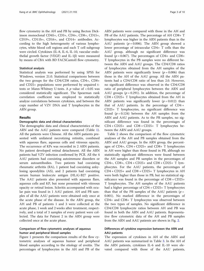
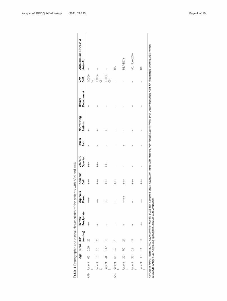
ResultsDemographic data and clinical characteristicsThe demographic data and clinical characteristics of theARN and the AAU patients were compared (Table 1).All the patients were Chinese. All the ARN patients pre-sented with unilateral necrotizing retinitis, combinedwith aqueous flare, aqueous cells and vitreous opacity.The occurrence of KPs was recorded in 2 ARN patients.No patient developed retinal detachment. All the ARNpatients had VZV infection confirmed by PCR. All the 4AAU patients had coexisting autoimmune disorders orserum autoantibodies. Two patients had coexistingrheumatic arthritis (RA), 1 patient had coexisting anky-losing spondylitis (AS), and 2 patients had coexistingserum human leukocyte antigen (HLA)-B27 positive.The AAU patients also presented with aqueous flare,aqueous cells and KP, but none presented with vitreousopacity or retinal lesion. Scleritis accompanied with ocu-lar pain was found in 1 AAU patient. AH and PB sam-ples of all the AAU patients were obtained only once atthe acute phase of the disease. In the ARN group, theAH and PB of patients 1 and 3 were collected at theacute phase, 1 week and 2 weeks after treatment, respect-ively, and a total of 3 samples of every patient were col-lected. The data for Patient 2 in the ARN group werecollected once at the acute phase.
Comparison of flow cytometric analyses of aqueoushumor and peripheral blood samplesFigure 1 presents the comparison results of the flow cy-tometric analyses of aqueous humor and peripheralblood samples according to the etiology of uveitis. Thepercentages of lymphocytes in the AH and PB of the
ARN patients were compared with those in the AH andPB of the AAU patients. The percentage of AH CD8+ Tlymphocytes was higher in the ARN patients than in theAAU patients (p = 0.006). The ARN group showed alower percentage of intraocular CD4+ T cells than theAAU group, although no significant difference wasfound (p = 0.067). The percentages of CD4+ and CD8+T lymphocytes in the PB samples were no different be-tween the ARN and AAU groups. The CD4/CD8 ratiosof lymphocytes obtained from the AH samples of theARN patients were significantly lower (p = 0.006) thanthose in the AH of the AAU group. All the ARN pa-tients had a CD4/CD8 ratio of less than 2.0. However,no significant difference was observed in the CD4/CD8ratio of peripheral lymphocytes between the ARN andAAU groups (p = 0.291). In addition, the percentage ofCD8 + CD25+ T lymphocytes obtained from the AH inARN patients was significantly lower (p = 0.012) thanthat of AAU patients. In the percentage of CD4 +CD25+ T lymphocytes, no significant difference wasfound (p = 0.124) between the AH samples from theARN and AAU patients. As to the PB samples, no sig-nificant difference was found in the percentages ofCD4 + CD25+ and CD8 + CD25+ T lymphocytes be-tween the ARN and AAU groups.Table 2 shows the comparison of the flow cytometric
analyses of the AH and PB samples obtained from theARN and AAU groups. In the ARN group, the percent-ages of CD4+, CD4 + CD25+ and CD8+ T lymphocytesin AH were higher than those found in PB, although nostatistically significant differences were found betweenthe AH samples and PB samples in the percentages ofCD4+, CD8+, CD4 + CD25+ and CD8 + CD25+ T lym-phocytes. For the AAU patients, the percentages ofCD4 + CD25+ and CD8 + CD25+ T lymphocytes in AHwere both higher than those in PB, but no statistical sig-nificance was found in the percentage of CD8 + CD25+T lymphocytes. The AH samples of the AAU patientshad a higher percentage of CD4 + CD25+ T lymphocytesthan that of the PB samples of the AAU patients (p =0.001). No marked difference in the percentages ofCD4+ and CD8+ T lymphocytes was observed betweenthe two types of samples. No significant difference inCD4/CD8 lymphocyte ratios between AH and PB wasfound in both the ARN and AAU patients. Representa-tive flow cytometric data of the AH and PB samplesfrom the ARN and AAU patients are shown in Fig. 2.
Differences of cytokine expression between the ARN andAAU patientsThe expression of cytokines in AH of the ARN andAAU patients was summarized in Table 3. In the AH ofthe ARN patients, cytokines IL-6 and IL-10 were ele-vated compared with those of the AAU patients,
Kang et al. BMC Ophthalmology (2021) 21:193 Page 3 of 10
Table
1Dem
ograph
icandclinicalcharacteristicsof
thepatientswith
ARN
andAAU
Age
BCVA
IOP
(mmHg)
Keratic
Precipitate
Aque
ous
Flare
Aque
ous
Cell
Vitreou
sOpacity
Ocu
lar
Pain
Necrotizing
Retinits
Retina
lDetachm
ent
VZV
DNA
Autoimmun
eDisea
se&
Auto-Ab
ARN
Patient
145
0.09
25++
+++
+++
+++
–+
–1.06E+
07–
Patient
218
0.6
20+
++
+++
+++
–+
–1.21E+
05–
Patient
341
0.12
15–
++
+++
+++
–+
–1.10E+
06–
AAU
Patient
454
0.2
7–
++++
––
––
–RA
Patient
532
FC27
+++++
+++
–+
––
–HLA
-B27+
Patient
638
0.2
17+
++++
––
––
–AS,HLA
-B27+
Patient
730
0.4
11++
++
+++
––
––
–RA
ARN
Acute
Retin
alNecrosis,AAUAcute
AnteriorUveitis,BC
VABe
stCorrected
Visual
Acuity
,IOPIntrao
cularPressure,V
ZVVa
ricella
Zoster
Virus,DNADeo
xyrib
onucleicAcid,
RARh
eumatoidArthritis,HLA
Hum
anLeuk
ocyteAntigen
,ASAnk
ylosingSp
ondy
litis,A
uto-AbAuto-Antibod
ies
Kang et al. BMC Ophthalmology (2021) 21:193 Page 4 of 10

although no significant difference was found in the in-crease of IL-6 (IL-10: p = 0.036, IL-6: p = 0.727). Therewere no significant differences between the two groupsas to AH concentration of IL-8, VEGF and IL-1β.
Correlation between the copy number of VZV DNA andthe T lymphocytes in AHWe then analyzed the potential correlations between thedecreased copy number of VZV DNA after antiviraltreatment and the percentages of CD4+, CD8+, CD4 +CD25+, and CD8 + CD25+ T lymphocytes and CD4/CD8 ratio. Table 4 shows the significant decrease ofaqueous viral load in 2 ARN patients. One week afterantiviral treatment, the aqueous viral load of Patient 1decreased from 1.06*107 copies/mL to 3.72*106 copies/
mL, and decreased to 1.64*106 copies/mL after 2 weeksof treatment. As to Patient 3, the aqueous viral load de-creased from 1.10*106 copies/mL to 1.65*105 copies/mLafter 1 week of treatment, and slightly increased to6.14*105 copies/mL after 2 weeks of treatment. Furthercorrelation analyses showed that the copy number ofVZV DNA in AH positively correlated well with the per-centage of the CD8+ T lymphocytes in AH (p = 0.009,r = 0.92; Fig. 3). Although the percentage of CD4+ Tlymphocytes in AH increased after antiviral treatment,the tendency of CD4+ T cells was not significantly cor-related to the aqueous VZV load (p = 0.286). Further-more, it was found that the CD4/CD8 ratio of AHnegatively correlated with the copy number of VZVDNA in the ARN patients (p = 0.039, r = − 0.834; Fig. 3).
Fig. 1 Comparison of percentage of CD4+, CD8+, CD4 + CD25+ and CD8 + CD25+ T lymphocytes and ratio of CD4/CD8 in AH between ARN andAAU patients. The comparisons between the ARN and AAU groups for the CD4/CD8 ratios, CD4+, CD8+ and CD25+ populations were performedby Mann-Whitney U-tests
Table 2 Comparison of percentages of T lymphocyte subsets of the ARN and AAU patients
ARN AAU
AH PB p Value† AH PB p Value†
CD4+ (%), median (P25, P75) 50.0 (41.0, 61.0) 46.0 (40.0, 50.0) 0.364 61.5 (54.0, 69.8) 56.5 (32.0, 66.0) 0.354
CD4 + CD25+ (%), median (P25, P75) 14.0 (11.0, 19.0) 7.9 (4.1, 13.0) 0.074 22.0 (14.5, 25.0) 3.3 (1.95, 5.3) 0.001*
CD8+ (%), median (P25, P75) 46.0 (33.0 52.0) 43.0 (41.0, 47.0) 0.662 23.5 (20.3, 27.5) 36.5 (26.8, 55.3) 0.091
CD8 + CD25+ (%), median (P25, P75) 1.2 (0.4, 3.7) 1.5 (1.4, 2.3) 0.753 14.0 (6.4, 23.8) 1.9 (0.85, 10.9) 0.125
CD4+/CD8+, median (P25, P75) 1.1 (0.8, 1.8) 1.1 (0.9, 1.2) 0.703 2.45 (2.25, 3.25) 1.6 (0.63, 2.5) 0.106
ARN Acute Retinal Necrosis, AAU Acute Anterior Uveitis, AH Aqueous Humor, PB Peripheral Blood*P < 0.05†Mann–Whitney U-test
Kang et al. BMC Ophthalmology (2021) 21:193 Page 5 of 10

No significant correlation between the aqueous VZVload and the percentages of CD4 + CD25+ and CD8 +CD25+ T lymphocytes was observed (CD4 + CD25+: p =0.414; CD8 + CD25+: p = 0.551). These findings collect-ively imply that the percentage of CD8+ T lymphocytesand the ratio of CD4/CD8 in AH may be a candidatebiomarker for active intraocular viral infectious diseases,especially in ARN.
DiscussionThis study described the immunological features of theAH and PB T lymphocytes population in patients withviral infections or non-infectious uveitis. It also analyzed
the mainly inflammatory cytokines of the AH samples ofthose patients.The present study reported that there are intrinsic dif-
ferences in both the T lymphocytes subsets and cyto-kines within the AH when comparing the VZV mediatedARN patients and the AAU patients. VZV is a double-stranded DNA virus belonging to the herpesviridae fam-ily. Among the human herpes viruses, HSV-1 and 2 andVZV are neurotropic, i.e., they establish latent infectionin the peripheral nervous system and the viral genome isretained in peripheral sensory ganglia throughout thehost’s lifespan [13, 14]. Most ARN patients are immuno-competent, and typical cases of ARN are uncommon in
Fig. 2 Representative dot plot diagrams showing the percentage of CD4+ and CD8+ T cell subsets among the PB and AH cells in patients withacute retinal necrosis (ARN) and acute anterior uveitis (AAU). The percentage of CD8+ T cell in the AH was elevated in ARN patients
Table 3 Cytokines levels in the AH of ARN and AAU patients
ARN AAU P Value†
IL-8 (pg/ml), median (P25, P75) 1118.4 (467.9, 2057.3) 2287.3 (294.6, 4457.8) 0.373
IL-6 (pg/ml), median (P25, P75) 7673.3 (2071.5, 26,725.2) 3963.3 (3037.8, 8815.8) 0.727
IL-10 (pg/ml), median (P25, P75) 66.5 (43.9, 178.8) 11.7 (7.9, 21.3) 0.036*
VEGF (pg/ml), median (P25, P75) 234.8 (93.0, 604.4) 719.2 (442.4, 994.9) 0.286
IL-1β (pg/ml), median (P25, P75) 4.7 (0.0, 6.9) 11.0 (0.0, 39.6) 0.397
ARN Acute Retinal Necrosis, AAU Acute Anterior Uveitis, AH Aqueous Humor, PB Peripheral Blood, IL-8 Interleukin-8, IL-6 Interleukin-6, IL-10 Interleukin-10, VEGFVascular Endothelial Cell Growth Factor, IL-1β Interleukin-1β*P < 0.05†Mann–Whitney U-test
Kang et al. BMC Ophthalmology (2021) 21:193 Page 6 of 10

immunocompromised individuals [14]. VZV infectionprovides stimuli to the antigen-specific cell-mediatedimmunity essential to recovering from primary infection,to containing VZV reactivation, and to protectingagainst virus. It was reported that T cells predominateamong the inflammatory cells located in AH of ARN pa-tients [15]. The present study described the immuno-logic profile of AH T lymphocytes in ARN patientscharacterized by a high percentage of CD8+ T lympho-cytes and low CD4/CD8 ratio. It was found that CD8+ Tlymphocytes infiltration into the AH was higher in pa-tients with ARN than patients with AAU, and this isconsistent with previous reports [16, 17]. In comparisonwith the non-infectious patients, significant increase ofCD8+ cell population and decrease of CD4/CD8 ratio inAH during the acute phase of viral infection were ob-served in the viral infectious patients. Previous studiesfound that large immune infiltrates consisted of CD8+ Tcells following VZV activation in human ganglia [14,18]. These results were similar to those found in previ-ous research, providing further support to the hypothesisthat changes in T lymphocytes subpopulations, includinga higher percentage of CD8+ T cells and a low CD4/CD8 ratio, are associated with viral activation. After anti-viral treatment, the symptoms of the ARN patients re-lieved, manifesting in the form of reduced ocular
inflammation and improved visual acuity. The copynumber of VZV DNA in AH in the ARN patients signifi-cantly decreased after treatment. Correlation analysesshowed that the copy number of VZV DNA in AH posi-tively correlated with the percentage of CD8+ T lympho-cytes in AH and negatively correlated with the CD4/CD8 ratio in AH. Importantly, this study also showedthat after treatment the percentage of CD8+ lympho-cytes in the ARN patients was even higher than that inthe AAU patients. Similarly, even after antiviral treat-ment, the ratio of CD4/CD8 was lower in the ARNgroup than that in the AAU group. Therefore, a highpercentage of CD8+ T lymphocytes and a low CD4/CD8T cell ratio may be a potential biomarker, serving asclinically useful indicators to evaluate the severity of in-traocular immune inflammatory response by which theprognosis of ARN can be forecasted. The identificationand analysis of T lymphocytes from sites of intraocularinflammation can help investigate the roles of infiltratingT lymphocytes in the pathogenesis of uveitis with differ-ent etiologies.In the uveitis associated with systemic and intraocular
infections, it is unclear whether the T lymphocytes enterthe eye as part of a peripheral immune response, orthere is a passive extravasation of T lymphocytes enterthe eye during a breakdown of the blood-ocular barrier.
Table 4 Changes of the T lymphocyte subsets and the copy number of VZV DNA in AH after treatment
CD4+ (%) CD4 + CD25+ (%) CD8+ (%) CD8 + CD25+ (%) CD4/CD8 VZV DNA (copies/mL)
Patient 1 Acute Phase 31 19 63 0.9 0.5 1.06E+ 07
1w after treatment 41 14 51 0.4 0.8 3.72E+ 06
2w after treatment 20 11 46 0.1 1.1 1.64E+ 06
Patient 3 Acute Phase 52 13 41 3.7 1.3 1.10E+ 06
1w after treatment 61 16 33 1.2 1.8 1.65E+ 05
2w after treatment 61 19 32 2.0 1.9 6.14E+ 05
ARN Acute Retinal Necrosis, AAU Acute Anterior Uveitis, VZV Varicella Zoster Virus, DNA Deoxyribonucleic Acid, DNA Deoxyribonucleic Acid
Fig. 3 The correlation between T lymphocytes and VZV DNZ load in AH. A graph showing the copy number of VZV DNA in AH positivelycorrelated well with the percentage of the CD8+ T lymphocytes in AH (p = 0.009, r = 0.92). B graph showing the CD4/CD8 ratio of AH negativelycorrelated with the copy number of VZV DNA in the ARN patients (p = 0.039, r = − 0.834). The Sperman’s correlation and the p value are indicatedat the figure
Kang et al. BMC Ophthalmology (2021) 21:193 Page 7 of 10

In line with previously published data, the percentagesof CD4+ and CD8+ T lymphocytes in AH were higherthan those found in PB [11]. In this study, comparedwith the non-infectious patients, ARN manifests pecu-liar focal lymphocytosis characterized by an elevatedpercentage of CD8+ T lymphocytes and a correspond-ing decreased CD4/CD8 ratio in AH. However, thechanges of CD8+ T lymphocytes and CD4/CD8 pat-terns were observed only in AH but not in PB. The re-sults suggest that there may be a particular mechanismattracting T lymphocytes into the eye, or the blood-aqueous barrier may channel the selective infiltration ofactivated T cells into uveitis patients’ ocular tissues andAH. Another possible explanation for this is that theARN patients with ocular inflammation only, but with-out other systemic symptoms, are too constrained toenable detection of whatever alterations in the blood’scell phenotypes. PB is not the perfect sample for re-search on immune abnormalities in intraocular inflam-mation in that it cannot represent the immuneinflammatory processes taking place within the eye.Further detailed studies are necessary to investigate theimmune function of intraocular T lymphocyte subsetsand immunologic abnormalities in viral infectious uve-itis, or to study whether there is a characteristic Tlymphocyte population in the AH of viral infectionpatients.CD4 + CD25+ T cells have been shown to be one dis-
tinct subset of regulatory T cells for the prevention ofautoimmunity [19]. Several studies have shown thatregulatory CD4 + CD25+ T cells contribute much tocontrolling immune homeostasis, to maintaining self-tolerance, and to preventing autoimmunity, as impairedCD4 + CD25+ T cell activity can cause autoimmune dis-ease [20, 21]. There was predominant CD4 + CD25+ Tcell enrichment in the inflammatory sites during the dis-ease course and their accumulation correlated with dis-ease resolution [22]. In this study, the CD25+ Tlymphocytes comprised 20.5% of the CD4+ T lympho-cytes in AH in the AAU patients, who had significantlyincreased CD4 + CD25+ T lymphocytes in AH comparedwith PB. All the AAU patients had coexisting auto-immune disorder or serum autoantibodies, and the an-terior uveitis of those patients was occurring inassociation with systemic diseases. In the AAU patients,the enrichment of regulatory CD4 + CD25+ T lympho-cytes in AH may suppress the activation and prolifera-tion of T cells to regulate local inflammatory responses[23]. However, there were collaborative roles for severalmechanisms requiring both cell-cell contact and cyto-kines production in the identification of the quantityand function of CD4 + CD25+ T cells in the inflamma-tory sites and peripheral immune sites [24]. Furthermechanism research is needed to investigate the
regulatory mechanism and immunosuppressive activityof regulatory CD4 + CD25+ T lymphocytes.Chemokines are unnegligible inflammatory mediators
excreted by inflammatory and immune cells during ocu-lar inflammation [25]. The intraocular concentration ofIL-10 is very useful for etiology diagnosis of uveitis, es-pecially in intraocular lymphoma (IOL) [26]. Previousstudies have also reported that IL-10 was highly elevatedin herpetic and noninfectious uveitis [7, 27]. IL-10 is animmunosuppressive regulatory cytokine and can be pro-duced by T lymphocytes. It has been suggested that IL-10 downregulates the immune responses in order tomoderate T cell mediated immune reaction and regulatethe extent of tissue damage [28, 29]. The high expressionof intraocular IL-10 in the ARN patients was in keepingwith previous research [7, 26]. However, the high IL-10level in AH of the ARN patients seems paradoxical re-garding the severe course of the intraocular inflamma-tion. One possible reason is that the expression of IL-10is not sufficient to suppress the harmful inflammatoryimmune response. Alternatively, in addition to the im-mune responses downregulated by IL-10, other potentialinflammatory response mechanisms may contribute incombination to the damage. Because the cytokine andchemokine expression patterns are constituents of a verycomplex regulatory network that depends on many en-vironmental stimuli, they are likely to vary remarkablyover the progression of the disease. Therefore, the effectof cytokines on the T lymphocytes necessitates thoroughinvestigation if cytokine immunology therapy is to beemployed in future. Combined with changes of intraocu-lar cytokines, immune cells profiles in AH seem to pro-vide insights into the severity and stage of theinflammatory process. Our results show that through cy-tometric analysis of AH, the immunologic informationof this disease was useful for the differential diagnosis ofintraocular viral infection. It helps improve our ability toclassify viral infection from other uveitis, which couldenable an early and less-invasive diagnosis.Undeniably, this study has its limitations. First, the
number of the ARN and AAU patients enrolled in thestudy was not sufficient, and therefore future researchneeds to further enlarge the sample size to analyze thechanges of T lymphocyte subsets in these diseases. Fur-ther studies will provide detailed information on immu-nopathologic mechanisms of lymphocytes in AH fromdifferent types of uveitis. Second, this study focused onthe distribution of T lymphocytes subsets in AH and PBof ARN and AAU patients, but did not analyze the func-tion of those cells. Further thorough studies are necessi-tated to investigate the detailed T lymphocyte subsetsand functional properties of intraocular infiltrating Tlymphocytes in uveitis patients in order to elucidateunderlying immunopathogenic mechanisms of this
Kang et al. BMC Ophthalmology (2021) 21:193 Page 8 of 10

disorder. Third, the follow-up period for the patientswith antiviral treatment was not long enough, but futurelongitudinal follow-up studies in ARN patients are ex-pected to overcome this shortcoming, and hopefully,they may provide precious insights into the progressionof immune inflammation in ARN.
ConclusionsIn conclusion, the patients with ARN caused by VZV inthis study had a higher percentage of CD8+ T lympho-cytes and a lower CD4/CD8 ratio in AH compared withthe control subjects suffering non-infectious uveitis. Thepercentage of the CD8+ T lymphocyte subset and theratio of the CD4/CD8 in AH from the ARN patients sig-nificantly correlated to the aqueous VZV load during thecourse of disease treatment. AH cell profiles might bediscriminative for the diagnosis of viral-infectious andimmune-mediated uveitis, and AH cell examination hasthe potential to become a useful research tool for inves-tigating various uveitis entities.
AbbreviationsAH: Aqueous humor; ARN: Acute retinal necrosis; VZV: Varicella zoster virus;AAU: Acute anterior uveitis; PB: Peripheral blood; PCR: Polymerase chainreaction; IL: Interleukin; HSV: Herpes simplex virus; CMV: Cytomegalovirus;EBV: Epstein-Barr virus; KPs: Keratic precipitates; IOP: Intraocular pressure
AcknowledgementsNot Applicable.
Authors’ contributionsDesign and conduct of the study by DY and YT. Collection, management,analysis and interpretation of the data by HK, YW and ML. Preparation of themanuscript by HK and YT. Review and final approval of the manuscript by allthe authors.
FundingThis work was supported by the Beijing Chaoyang 1351 talent trainingprogram (No. CYXX-2017-21), the Taishan Scholars Program of ShandongProvince (ZR2016YL013), the Shandong Provincial Natural Science Founda-tion (2018YYSP022), and the Key lab foundation of Shandong Academy ofSciences (2017GSF19111).
Availability of data and materialsThe datasets used during the current study are available from thecorresponding author on reasonable request.
Declarations
Ethics approval and consent to participateWe adhered to the tenets of the Declaration of Helsinki. Ethics approval wasobtained from the the ethics committee of Beijing Chaoyang hospital. Allparticipants involved were informed of the purpose of this study and awritten consent was obtained from themselves.
Consent for publicationWritten informed consent was obtained from all patients for the publicationof this report and any accompanying images. A copy of the writtenconsentis available for review by the editor of this journal.
Competing interestsNo conflict of interest exits in the submission of this manuscript, and thismanuscript was approved by all authors for publication. We declare that thework described was original research that has not been published previouslyand is not under consideration for publication elsewhere, in whole or in part.
Author details1Department of Ophthalmology, Beijing Chaoyang Hospital, Capital MedicalUniversity, Beijing, China. 2Laboratory of Immunology for Environment andHealth, Shandong Analysis and Test Center, Qilu University of Technology(Shandong Academy of Sciences), Jinan, Shandong, China. 3Department ofImmunology and Infectious Disease, The John Curtin School of MedicalResearch, The Australian National University, Canberra, ACT, Australia. 4BeijingGiantMed Diagnostics Co., LTD, Beijing, China.
Received: 10 August 2020 Accepted: 16 April 2021
References1. Zhao XY, Xia S, Chen YX. Role of diagnostic pars Plana vitrectomy in
determining the etiology of uveitis initially unknown. Retina.2020;40(2):359–69.
2. Chan NS, Chee SP, Caspers L, Bodaghi B. Clinical features of CMV-associatedanterior uveitis. Ocul Immunol Inflamm. 2018;26(1):107–15.
3. Keorochana N, Treesit I, Funarunart P. Characteristics and clinical outcomesof hypertensive anterior uveitis. Ocul Immunol Inflamm. 2019;538:1–11.
4. Megaw R, Agarwal PK. Posner-Schlossman syndrome. Surv Ophthalmol.2017;62(3):277–85.
5. Chan NS, Chee SP. Demystifying viral anterior uveitis: a review. Clin ExpOphthalmol. 2019;47(3):320–33.
6. Fazil Z, Ten Berge JC, Langerak AW, Rothova A, Dik WA. An intraocularinflammatory profile of rubella associated uveitis. Ocul Immunol Inflamm.2019;27(3):418–23.
7. de Visser L, de Visser L, de Boer JH, Rijkers GT, Wiertz K, van den Ham H-J,et al. Cytokines and chemokines involved in acute retinal necrosis. InvestOphthalmol Vis Sci. 2017;58(4):2139–51.
8. Schoenberger SD, Kim SJ, Thorne JE, Mruthyunjaya P, Yeh S, Bakri SJ, et al.Diagnosis and treatment of acute retinal necrosis: a report by the AmericanAcademy of ophthalmology. Ophthalmology. 2017;124(3):382–92.
9. Hillenkamp J, Nolle B, Bruns C, Rautenberg P, Fickenscher H, Roider J. Acuteretinal necrosis: clinical features, early vitrectomy, and outcomes.Ophthalmology. 2009;116(10):1971–5 e1972.
10. Verjans GM, Dings ME, McLauchlan J, van Der Kooi A, Hoogerhout P,Brugghe HF, et al. Intraocular T cells of patients with herpes simplex virus(HSV)-induced acute retinal necrosis recognize HSV tegument proteinsVP11/12 and VP13/14. J Infect Dis. 2000;182(3):923–7.
11. Verjans GM, Feron EJ, Dings ME, Cornelissen JG, Van der Lelij A, Baarsma GS,et al. T cells specific for the triggering virus infiltrate the eye in patients withherpes simplex virus-mediated acute retinal necrosis. J Infect Dis. 1998;178(1):27–34.
12. Holland GN. Standard diagnostic criteria for the acute retinal necrosissyndrome. Executive Committee of the American Uveitis Society. Am JOphthalmol. 1994;117(5):663–7.
13. Steain M, Sutherland JP, Rodriguez M, Cunningham AL, Slobedman B,Abendroth A. Analysis of T cell responses during active varicella-zoster virusreactivation in human ganglia. J Virol. 2014;88(5):2704–16.
14. Lee JH, Agarwal A, Mahendradas P, Lee CS, Gupta V, Pavesio CE, et al. Viralposterior uveitis. Surv Ophthalmol. 2017;62(4):404–45.
15. Wang XC, Norose K, Yano A, Ohta K, Segawa K. Two-color flow cytometricanalysis of activated T lymphocytes in aqueous humor of patients withendogenous vs. exogenous uveitis. Curr Eye Res. 1995;14(6):425–33.
16. Maruyama K, Inaba T, Sugita S, Ichinohasama R, Nagata K, Kinoshita S, et al.Comprehensive analysis of vitreous specimens for uveitis classification: aprospective multicentre observational study. BMJ Open. 2017;7(11):e014549.
17. Sant AJ, McMichael A. Revealing the role of CD4(+) T cells in viral immunity.J Exp Med. 2012;209(8):1391–5.
18. Gowrishankar K, Steain M, Cunningham AL, Rodriguez M, Blumbergs P,Slobedman B, et al. Characterization of the host immune response inhuman ganglia after herpes zoster. J Virol. 2010;84(17):8861–70.
19. Sakaguchi S, Sakaguchi N, Asano M, Itoh M, Toda M. Immunologic self-tolerance maintained by activated T cells expressing IL-2 receptor alpha-chains (CD25). Breakdown of a single mechanism of self-tolerance causesvarious autoimmune diseases. J Immunol. 1995;155(3):1151–64.
20. Schiopu A, Wood KJ. Regulatory T cells: hypes and limitations. Curr OpinOrgan Transplant. 2008;13(4):333–8.
21. Hall BM. CD4+CD25+ T regulatory cells in transplantation tolerance: 25years on. Transplantation. 2016;100(12):2533–47.
Kang et al. BMC Ophthalmology (2021) 21:193 Page 9 of 10

22. McGeachy MJ, Stephens LA, Anderton SM. Natural recovery and protectionfrom autoimmune encephalomyelitis: contribution of CD4+CD25+regulatory cells within the central nervous system. J Immunol. 2005;175(5):3025–32.
23. Kursar M, Bonhagen K, Fensterle J, Kohler A, Hurwitz R, Kamradt T, et al.Regulatory CD4+CD25+ T cells restrict memory CD8+ T cell responses. JExp Med. 2002;196(12):1585–92.
24. Shi HZ, Qin XJ. CD4CD25 regulatory T lymphocytes in allergy and asthma.Allergy. 2005;60(8):986–95.
25. Weinstein JE, Pepple KL. Cytokines in uveitis. Curr Opin Ophthalmol. 2018;29(3):267–74.
26. Fukunaga H, Kaburaki T, Shirahama S, Tanaka R, Murata H, Sato T, et al.Analysis of inflammatory mediators in the vitreous humor of eyes with pan-uveitis according to aetiological classification. Sci Rep. 2020;10(1):2783–95.
27. Lahmar I, Abou-Bacar A, Abdelrahman T, Guinard M, Babba H, Ben Yahia S,et al. Cytokine profiles in toxoplasmic and viral uveitis. J Infect Dis. 2009;199(8):1239–49.
28. Awasthi A, Carrier Y, Peron JP, Bettelli E, Kamanaka M, Flavell RA, et al. Adominant function for interleukin 27 in generating interleukin 10-producinganti-inflammatory T cells. Nat Immunol. 2007;8(12):1380–9.
29. Trinchieri G. Interleukin-10 production by effector T cells: Th1 cells show selfcontrol. J Exp Med. 2007;204(2):239–43.
Publisher’s NoteSpringer Nature remains neutral with regard to jurisdictional claims inpublished maps and institutional affiliations.
Kang et al. BMC Ophthalmology (2021) 21:193 Page 10 of 10