FLOPPY MITRAL VALVE, MITRAL VALVE …anatomedunesa.weebly.com/uploads/1/8/7/1/1871495/floppy...Part...
25
Copyright ©2001 Lippincott Williams & Wilkins Allen, Hugh D., Gutgesell, Howard P., Clark, Edward B., Driscoll, David J. Moss & Adams' Heart Disease in Infants, Children & Adolescents: Including the Fetus and Young Adults, 6th Edition FLOPPY MITRAL VALVE, MITRAL VALVE PROLAPSE, AND THE MITRAL VALVE PROLAPSE SYNDROME Part of "47 - THE FLOPPY MITRAL VALVE, MITRAL VALVE PROLAPSE, AND MITRAL VALVULAR REGURGITATION" Definition It is apparent today that the FMV is the central issue in the FMV/MVP/MVR triad and the MVP syndrome (MVPS) (1,2,3,4 and 5). The FMV is a common mitral valve abnormality with a broad spectrum of structural and functional changes (Fig. 47.1) (1,2,3 and 4,6). Whereas the pathobiology of the FMV has been and still is being reexamined in contemporary terms, we are still dealing with gross structural and morphologic characteristics at the clinical level. Distinguishing between the normal mitral valve with its minor variants and a mitral valve with an intrinsic structural derangement, especially in the pediatric population, remains a difficult matter. The FMV may be an inherited lesion, either as an isolated event or as part of the recognized or incompletely defined heritable disorders of connective tissue. The FMV has gradually become recognized as the most common cardiac lesion in many heritable systemic disorders. In this chapter, inheritance, its association with other lesions, complications, the high-risk patient, neurohumoral abnormalities, natural history, and clinical presentation in pediatric FIGURE 47.1. The FMV/MVP/MVR triad. The floppy mitral valve occupies the central role in the triad. Postural auscultatory phonocardiographic changes are illustrated at the top of the figure. (From Wooley CF, Sparks EA, Boudoulas H. The floppy mitral valve-mitral valve prolapse-mitral valvular regurgitation triad. ACC Current Journal Review 1994;July/August:25–26, with permission). P.950 Página 1 de 25 Ovid: Moss & Adams' Heart Disease in Infants, Children & Adolescents: Including the ... 06/04/05 http://gateway.ut.ovid.com/gw2/ovidweb.cgi
Transcript of FLOPPY MITRAL VALVE, MITRAL VALVE …anatomedunesa.weebly.com/uploads/1/8/7/1/1871495/floppy...Part...
Copyright ©2001 Lippincott Wil l iams & Wilkins Allen, Hugh D., Gutgesell, Howard P., Clark, Edward B., Driscoll, David J. Moss & Adams' Heart Disease in Infants, Children & Adolescents: Including the Fetus and Young Adults, 6th Edit ion
FLOPPY MITRAL VALVE, MITRAL VALVE PROLAPSE, AND THE MITRAL VALVE PROLAPSE SYNDROME
Part of "47 - THE FLOPPY MITRAL VALVE, MITRAL VALVE PROLAPSE, AND MITRAL VALVULAR REGURGITATION"
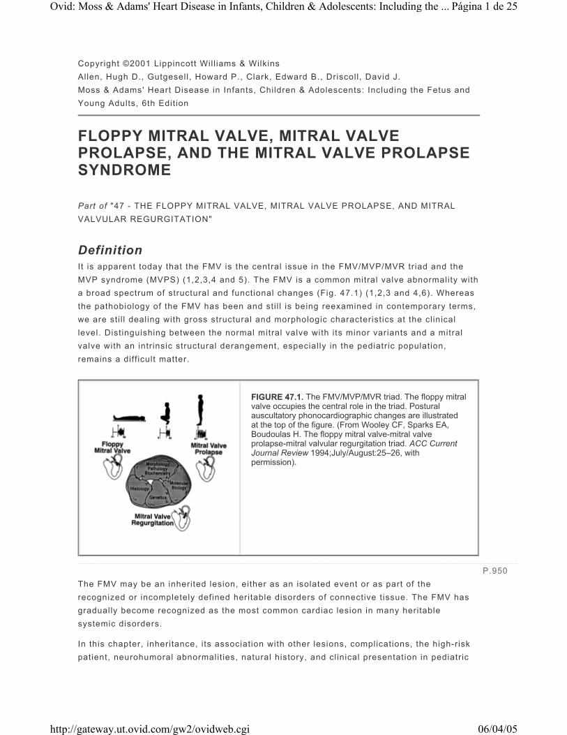
Definition I t is apparent today that the FMV is the central issue in the FMV/MVP/MVR triad and the MVP syndrome (MVPS) (1,2,3,4 and 5). The FMV is a common mitral valve abnormality with a broad spectrum of structural and functional changes (Fig. 47.1) (1,2,3 and 4,6). Whereas the pathobiology of the FMV has been and sti l l is being reexamined in contemporary terms, we are sti l l dealing with gross structural and morphologic characteristics at the cl inical level. Distinguishing between the normal mitral valve with its minor variants and a mitral valve with an intrinsic structural derangement, especially in the pediatric population, remains a diff icult matter.
The FMV may be an inherited lesion, either as an isolated event or as part of the recognized or incompletely defined heritable disorders of connective t issue. The FMV has gradually become recognized as the most common cardiac lesion in many heritable systemic disorders.
In this chapter, inheritance, its association with other lesions, complications, the high-risk patient, neurohumoral abnormalit ies, natural history, and cl inical presentation in pediatric
FIGURE 47.1. The FMV/MVP/MVR triad. The floppy mitral valve occupies the central role in the triad. Postural auscultatory phonocardiographic changes are illustrated at the top of the figure. (From Wooley CF, Sparks EA, Boudoulas H. The floppy mitral valve-mitral valve prolapse-mitral valvular regurgitation triad. ACC Current Journal Review 1994;July/August:25–26, with permission).
P.950
Página 1 de 25Ovid: Moss & Adams' Heart Disease in Infants, Children & Adolescents: Including the ...
06/04/05http://gateway.ut.ovid.com/gw2/ovidweb.cgi

population are presented. Further, because children often involved in athletics, FMV/MVP/MVR and athletics also are discussed.
FMV/MVP Inheritance As yet, genetic diagnostic testing for FMV/MVP has not been entered in cl inical practice. It is l ikely that genetically FMV/MVP is a heterogeneous group; in this case, degeneration of the mitral valve may be the f inal pathway for several protein defects (1). At present, i t appears that at least two forms of inheritance exist. One form is transmitted by an autosomal dominant manner with a different degree of penetration (the most common form); the other form (less common) is X-chromosome linked valvular disease with myxomatous degeneration. Further, definit ion of genetic defects wil l al low better classif ication of this complex valvular abnormality. Such approaches open up diagnostic pathways that are not part of our current perspectives but wil l be part of the armamentarium of twenty-f irst century cardiologists.
In addit ion, a large proportion of patients with FMV/MVP/MVR have systemic features similar to those that occur in patients with heritable disorders of connective t issue (see later). FMV/MVP/MVR has been documented in a number of recognized heritable connective t issue disorders; for this reason, committee experts classif ied FMV/MVP/MVR as one of the heritable disorders of connective t issue. Because of i ts high frequency in the general population, FMV/MVP/MVR designates the largest group of patients with the connective t issue abnormality of the heart (1,2).
Association with Other Abnormalities The FMV/MVP/MVR triad may be associated with several other cardiovascular and noncardiovascular abnormalit ies (Table 47.1). Earl ier studies suggested that the incidence of FMV/MVP is greater in patients with hypertrophic cardiomyopathy compared with the general population. Other studies, however, in a large group of patients with hypertrophic cardiomyopathy, indicated that the incidence of FMV/MVP in patients with hypertrophic cardiomyopathy was not greater than expected in the general population; the presence of FMV/MVP, however, in patients with hypertrophic cardiomyopathy appeared to predispose such patients to atrial f ibri l lation. In one study, the incidence of congenital abnormalit ies of the coronary arteries in patients with FMV/MVP was reported to be slightly greater compared with patients without FMV/MVP (1,2).
TABLE 47.1. FLOPPY MITRAL VALVE, MITRAL VALVE PROLAPSE, AND ASSOCIATED ABNORMALITIES
Página 2 de 25Ovid: Moss & Adams' Heart Disease in Infants, Children & Adolescents: Including the ...
06/04/05http://gateway.ut.ovid.com/gw2/ovidweb.cgi
Heritable Disorders of Connective Tissue In several of the most common heritable disorders of connective t issue, such as Marfan's syndrome, Stickler's syndrome, Ehlers-Danlos syndrome, and polycystic kidney disease in adults, FMV/MVP occurs as a pleiotropic manifestation (1,2,7,8 and 9) (Table 47.1). The precise relationship between FMV/MVP and heritable connective t issue disorders is not clear. Until basic genetic, molecular, and biochemical defects are defined, assumptions of a common pathophysiology are purely speculative. Understanding the pathophysiology and natural history of FMV/MVP in these syndromes may improve understanding of the natural history of famil ial FMV/MVP.
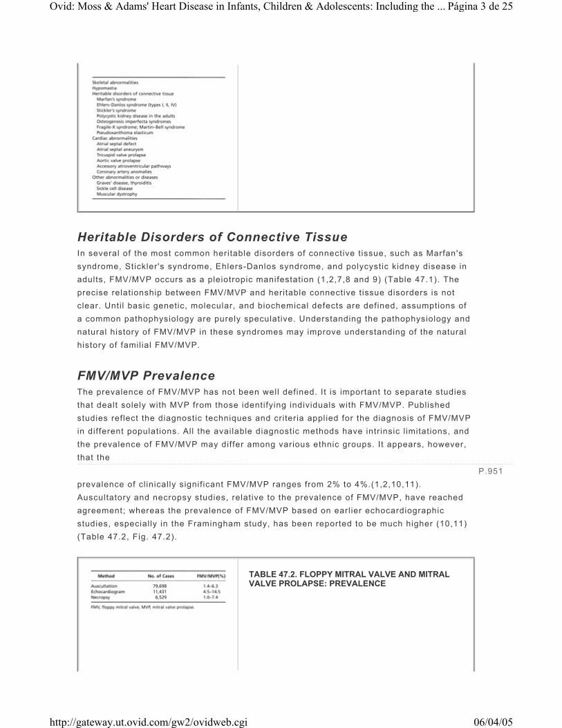
FMV/MVP Prevalence The prevalence of FMV/MVP has not been well defined. It is important to separate studies that dealt solely with MVP from those identifying individuals with FMV/MVP. Published studies reflect the diagnostic techniques and criteria applied for the diagnosis of FMV/MVP in different populations. All the available diagnostic methods have intrinsic l imitations, and the prevalence of FMV/MVP may differ among various ethnic groups. It appears, however, that the prevalence of clinically signif icant FMV/MVP ranges from 2% to 4%.(1,2,10,11). Auscultatory and necropsy studies, relative to the prevalence of FMV/MVP, have reached agreement; whereas the prevalence of FMV/MVP based on earl ier echocardiographic studies, especially in the Framingham study, has been reported to be much higher (10,11) (Table 47.2, Fig. 47.2).
P.951
TABLE 47.2. FLOPPY MITRAL VALVE AND MITRAL VALVE PROLAPSE: PREVALENCE
Página 3 de 25Ovid: Moss & Adams' Heart Disease in Infants, Children & Adolescents: Including the ...
06/04/05http://gateway.ut.ovid.com/gw2/ovidweb.cgi

The prevalence of FMV/MVP increases with the age. Sakamoto, in a phonocardiographic study, found an incidence of MVP of 0.753% in elementary school children and 1.325% in the middle school children (1). Hickey and Wilcken (12) investigated the incidence of MVP in 6,887 consecutive patients referred for echocardiography and in 206 first-degree relatives of 65 patients with MVP. The incidence of MVP from ages 0 to 19 years was 0.3%; from 20 to 39 years, 2.0%; from 40 to 59 years, 2.7%; and from 60 to 79 years, 2.3%. The incidence of MVP in the f irst-degree relatives group from ages 0 to 19 years was 3%; from 20 to 39 years, 15%; from 40 to 59 years, 11%; and from 60 to 79, 9%. Ohara et al. (13) studied the incidence of MVP by two-dimensional echocardiography in 4,328 children from 1 day to 15 years of age. MVP was not seen in any of the 198 children who were 1 to 28 days of age; the incidence of MVP was 0.25% in 391 children who were from 6 to 18 months of age, 2.1% in 2,801 children 6 to 7 years of age, and 5.1% in 938 children aged 12 to 15 years. Signif icant MVR was present in six children (two 6–7 years of age, four 12–15 years of age). Greenwood (14) evaluated 3,100 children whose age ranged from 1 month to 18 years and diagnosed MVP by auscultation in 154 children (4.97%). In another cl inical auscultatory study, Greenwood (15) evaluated 6,168 children ages from 2 months to 21 years; MVP was present in 331 (6.37%), 175 boys and 156 girls (1,2).
Diagnostic Evaluation Family history provides important information because FMV/MVP may be inherited, i t may be part of an inherited connective-tissue syndrome, and family members may have similar complications (1,2) (Table 47.3).
FIGURE 47.2. Upper panel: Echocardiographic incidence of mitral valve prolapse (FMV/MVP) in female subjects from the Framingham study. (Modified from Savage DD, Garrison RJ, Devereux RB, et al. Mitral valve prolapse in the general population: epidemiologic features: the Framingham study. Am Heart J 1983;106:571–576, with permission.) Lower panel: Incidence of FMV/MVP in female subjects from a routine necropsy study. Grade 1 is mild, grade 2 is moderate, grade 3 is moderately severe, and grade 4 is severe FMV/MVP. The total represents all patients with grades 1, 2, 3, and 4 FMV/MVP. From Davies MJ, Moore BP, Brainbridge MV. The floppy mitral valve: study of incidence, pathology, and complications in surgical, necropsy and forensic material. Br Heart J 1978;40:468–481, with permission.)
TABLE 47.3. FLOPPY MITRAL VALVE AND MITRAL
Página 4 de 25Ovid: Moss & Adams' Heart Disease in Infants, Children & Adolescents: Including the ...
06/04/05http://gateway.ut.ovid.com/gw2/ovidweb.cgi
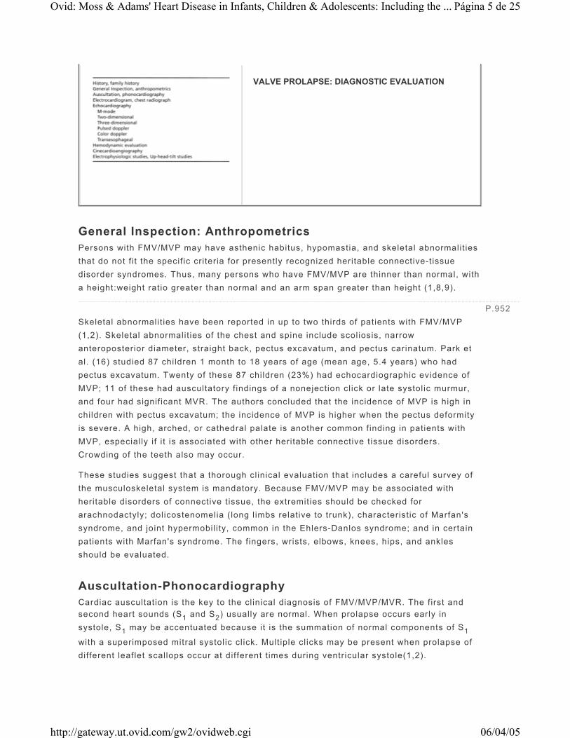
General Inspection: Anthropometrics Persons with FMV/MVP may have asthenic habitus, hypomastia, and skeletal abnormalit ies that do not f it the specif ic criteria for presently recognized heritable connective-tissue disorder syndromes. Thus, many persons who have FMV/MVP are thinner than normal, with a height:weight ratio greater than normal and an arm span greater than height (1,8,9).
Skeletal abnormalit ies have been reported in up to two thirds of patients with FMV/MVP (1,2). Skeletal abnormalit ies of the chest and spine include scoliosis, narrow anteroposterior diameter, straight back, pectus excavatum, and pectus carinatum. Park et al. (16) studied 87 children 1 month to 18 years of age (mean age, 5.4 years) who had pectus excavatum. Twenty of these 87 children (23%) had echocardiographic evidence of MVP; 11 of these had auscultatory f indings of a nonejection cl ick or late systolic murmur, and four had signif icant MVR. The authors concluded that the incidence of MVP is high in children with pectus excavatum; the incidence of MVP is higher when the pectus deformity is severe. A high, arched, or cathedral palate is another common finding in patients with MVP, especially if i t is associated with other heritable connective t issue disorders. Crowding of the teeth also may occur.
These studies suggest that a thorough clinical evaluation that includes a careful survey of the musculoskeletal system is mandatory. Because FMV/MVP may be associated with heritable disorders of connective t issue, the extremities should be checked for arachnodactyly; dolicostenomelia (long l imbs relative to trunk), characteristic of Marfan's syndrome, and joint hypermobil ity, common in the Ehlers-Danlos syndrome; and in certain patients with Marfan's syndrome. The fingers, wrists, elbows, knees, hips, and ankles should be evaluated.
Auscultation-Phonocardiography Cardiac auscultation is the key to the cl inical diagnosis of FMV/MVP/MVR. The f irst and second heart sounds (S1 and S2) usually are normal. When prolapse occurs early in systole, S1 may be accentuated because it is the summation of normal components of S1
with a superimposed mitral systolic cl ick. Mult iple cl icks may be present when prolapse of different leaflet scallops occur at different t imes during ventricular systole(1,2).
VALVE PROLAPSE: DIAGNOSTIC EVALUATION
P.952
Página 5 de 25Ovid: Moss & Adams' Heart Disease in Infants, Children & Adolescents: Including the ...
06/04/05http://gateway.ut.ovid.com/gw2/ovidweb.cgi

The presence of nonejection systolic cl ick(s) with or without late mitral systolic murmur constitutes the auscultatory criteria for the diagnosis of FMV/MVP. Mult iple systolic cl icks that occur in succession may sound l ike a pericardial fr ict ion rub or resemble the scratching sound produced by sandpaper on the wood. The high-pitched mid- to late systolic murmur of MVR often is introduced by a cl ick or may occur alone; the murmur may crescendo into the S2. When the posterior mitral valve leaflet prolapses, the regurgitant
f low may be directed anteriorly toward the aortic root; in this case, the MVR murmur is heard at the apex, may radiate along the left sternal border to the aortic area, and may mimic left ventricular outf low tract murmurs. In contrast, when the anterior mitral valve leaflet prolapses, the MVR murmur may radiated to the axil la and to the spine. Phonocardiography is an important adjunct to cl inical auscultation and in certain cases may be used to confirm the auscultatory f indings. Patients with redundant FMV and signif icant MVR may have holosystolic murmurs even in the supine posit ion, and cl ick(s) may not be audible (Fig. 47.3). Holosystolic murmurs attr ibutable to FMV/MVP/MVR are rare in the pediatric population.
Dynamic Auscultation The most specif ic physical diagnostic criteria for the diagnosis of FMV/MVP/MVR are the characteristic changes of the systolic cl ick(s) or the MVR murmur that occur with postural changes. The systolic clicks move toward the S1 with upright posture, often merging with the S1 if marked postural tachycardia occurs, and new clicks may appear. The systolic MVR
murmur becomes longer in duration, often louder, and may become holosystolic i f an exaggerated heart rate response occurs (1,9) (Fig. 47.4). An MVR murmur may be present only with the patient in the upright posit ion. Rarely, a systolic precordial “honk” or whooping sound may be heard with the MVR murmur; most of the t ime, these sounds are heard only in the sitt ing or standing posit ions. Precordial honks may be transient and heard for only a few beats immediately after standing.
P.953
FIGURE 47.3. Phonocardiogram from a patient with FMV/MVP. A: Sitting position. First and second heart sounds (S1, S2) are shown. Note an early systolic click (SC). B: Standing position. A systolic murmur (SM) is present. C: Squatting position. The systolic murmur disappears and the click becomes late systolic. D: Phonocardiogram from another patient with FMV/MVP, significant mitral regurgitation and thick-“floppy” mitral valve (see echocardiogram in Fig. 47.5). Holosystolic murmur is present in the supine position. Phono, phonocardiogram; ECG, electrocardiogram.
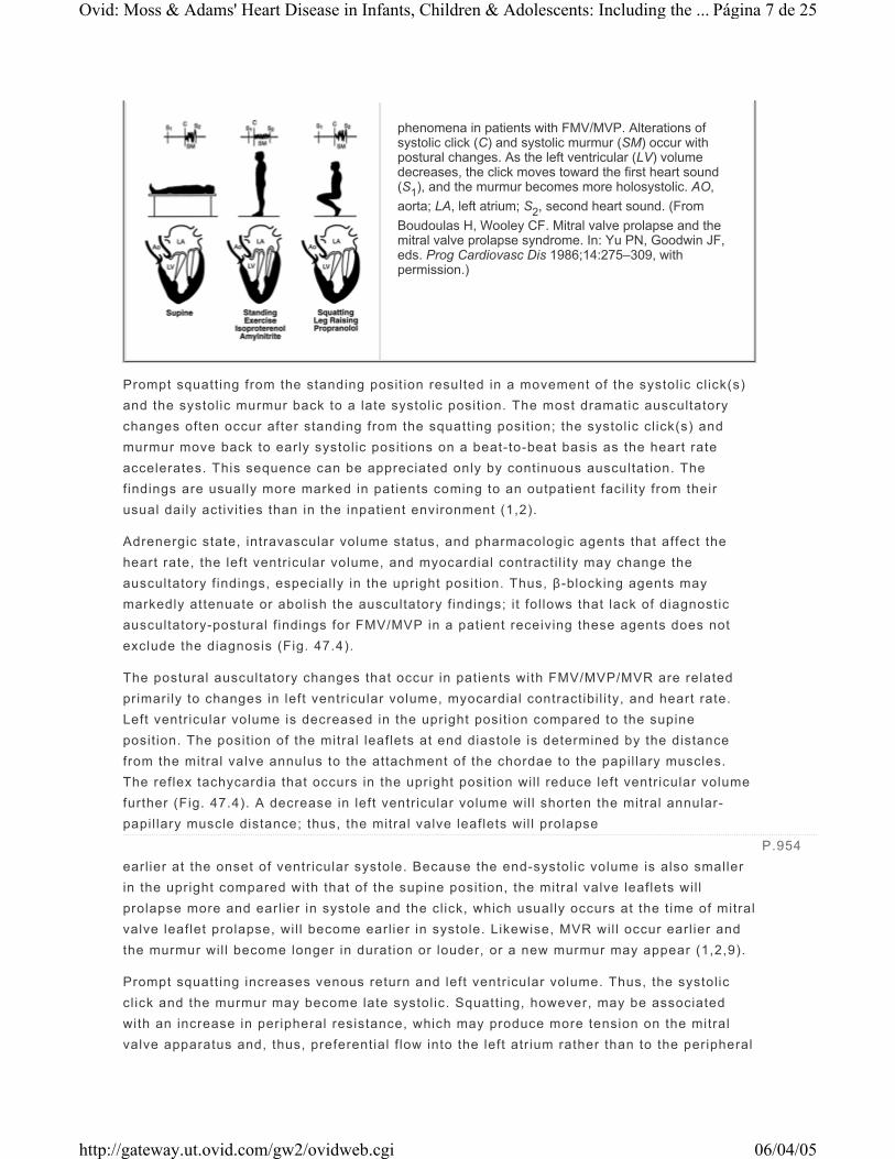
FIGURE 47.4. Postural changes and auscultatory
Página 6 de 25Ovid: Moss & Adams' Heart Disease in Infants, Children & Adolescents: Including the ...
06/04/05http://gateway.ut.ovid.com/gw2/ovidweb.cgi
Prompt squatting from the standing posit ion resulted in a movement of the systolic cl ick(s) and the systolic murmur back to a late systolic posit ion. The most dramatic auscultatory changes often occur after standing from the squatting posit ion; the systolic cl ick(s) and murmur move back to early systolic posit ions on a beat-to-beat basis as the heart rate accelerates. This sequence can be appreciated only by continuous auscultation. The findings are usually more marked in patients coming to an outpatient facil ity from their usual daily activit ies than in the inpatient environment (1,2).
Adrenergic state, intravascular volume status, and pharmacologic agents that affect the heart rate, the left ventricular volume, and myocardial contracti l i ty may change the auscultatory f indings, especially in the upright posit ion. Thus, β-blocking agents may markedly attenuate or abolish the auscultatory f indings; i t fol lows that lack of diagnostic auscultatory-postural f indings for FMV/MVP in a patient receiving these agents does not exclude the diagnosis (Fig. 47.4).
The postural auscultatory changes that occur in patients with FMV/MVP/MVR are related primari ly to changes in left ventricular volume, myocardial contractibi l i ty, and heart rate. Left ventricular volume is decreased in the upright posit ion compared to the supine posit ion. The posit ion of the mitral leaflets at end diastole is determined by the distance from the mitral valve annulus to the attachment of the chordae to the papil lary muscles. The reflex tachycardia that occurs in the upright posit ion wil l reduce left ventricular volume further (Fig. 47.4). A decrease in left ventricular volume wil l shorten the mitral annular-papil lary muscle distance; thus, the mitral valve leaflets wil l prolapse earl ier at the onset of ventricular systole. Because the end-systolic volume is also smaller in the upright compared with that of the supine posit ion, the mitral valve leaflets will prolapse more and earl ier in systole and the click, which usually occurs at the t ime of mitral valve leaflet prolapse, wil l become earlier in systole. Likewise, MVR wil l occur earl ier and the murmur wil l become longer in duration or louder, or a new murmur may appear (1,2,9).
Prompt squatting increases venous return and left ventricular volume. Thus, the systolic cl ick and the murmur may become late systolic. Squatting, however, may be associated with an increase in peripheral resistance, which may produce more tension on the mitral valve apparatus and, thus, preferential f low into the left atrium rather than to the peripheral
phenomena in patients with FMV/MVP. Alterations of systolic click (C) and systolic murmur (SM) occur with postural changes. As the left ventricular (LV) volume decreases, the click moves toward the first heart sound (S1), and the murmur becomes more holosystolic. AO, aorta; LA, left atrium; S2, second heart sound. (From Boudoulas H, Wooley CF. Mitral valve prolapse and the mitral valve prolapse syndrome. In: Yu PN, Goodwin JF, eds. Prog Cardiovasc Dis 1986;14:275–309, with permission.)
P.954
Página 7 de 25Ovid: Moss & Adams' Heart Disease in Infants, Children & Adolescents: Including the ...
06/04/05http://gateway.ut.ovid.com/gw2/ovidweb.cgi
circulation; in this case, the late systolic cl ick and murmur may become accentuated in the squatting posit ion.
Other maneuvers that change left ventricular volume can be used in the diagnosis of FMV/MVP/MVR, such as elevating the legs, isometric handgrip exercise, tourniquets on the extremities, lower-body negative pressure, Valsalva's maneuver, or amyl nitr ite inhalation. None of these interventions, however, is as practical and helpful as a systematically performed postural dynamic auscultation.
Conditions with Auscultatory Findings that May Mimic FMV/MVP Floppy tr icuspid valve with tr icuspid valve prolapse may coexist with FMV/MVP. Separation of the auscultatory phenomena of FMV/MVP from tricuspid valve prolapse by physical examination may be diff icult. Systolic cl ick(s) and systolic murmur moving later in systole immediately on deep inspiration, with leg raising, or with squatting may suggest tr icuspid valve origin because FMV/MVP click(s) and MVR murmur require several beats after the intervention to change timing. The jugular venous pulse generally is normal unless a suff icient degree of tr icuspid regurgitation is present to produce a prominent C-V wave; such patients wil l have holosystolic murmur of tr icuspid regurgitation. Confirming the diagnosis of tr icuspid valve prolapse as the cause of the tr icuspid regurgitation requires imaging techniques.
Hypertrophic cardiomyopathy with dynamic left ventricular outf low tract pressure gradient may produce dynamic auscultatory changes that mimic the FMV/MVP/MVR postural auscultatory changes. Apical nonejection systol ic cl icks are not generally present in hypertrophic cardiomyopathy. Further, double or tr iple apex impulse and the “spike and dome” carotid arterial pulse favor the diagnosis of hypertrophic cardiomyopathy (1,2,9).
Electrocardiogram The vast majority of patients with FMV/MVP have normal electrocardiogram. Nonspecif ic ST and T wave changes and T wave inversion, especially in the inferior leads, have been described in certain patients with FMV/MVP; certainly, these findings are not diagnostic for FMV/MVP. The pathophysiologic mechanisms for these changes are not clear. ST and T wave changes, however, may improve with exercise or the administration of β-blockers in certain patients with FMV/MVP (1,2,9).
Chest Roentgenograms Posteroanterior and left lateral chest roentgenograms usually show normal heart and lung findings. Skeletal abnormalit ies, however, i f present, wil l be seen in a routine chest roentgenographic examination (1,2,9).
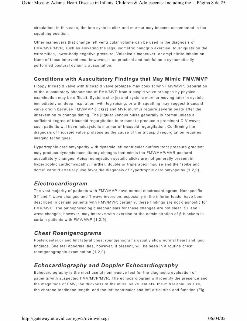
Echocardiography and Doppler Echocardiography Echocardiography is the most useful noninvasive test for the diagnostic evaluation of patients with suspected FMV/MVP/MVR. The echocardiogram will identify the presence and the magnitude of FMV, the thickness of the mitral valve leaflets, the mitral annulus size, the chordae tendineae length, and the left ventricular and left atrial size and function (Fig.
Página 8 de 25Ovid: Moss & Adams' Heart Disease in Infants, Children & Adolescents: Including the ...
06/04/05http://gateway.ut.ovid.com/gw2/ovidweb.cgi
47.5). Further, associated cardiac abnormalit ies (1,2) (Table 47.1) can be defined. It should be emphasized, however, that echocardiography is a tomographic cross-sectional technique, and no single view should be considered diagnostic. The parasternal long-axis view permits visualization of medial aspect of the anterior and middle scallop of the posterior mitral valve leaflets. If MVP, however, is localized to the lateral scallop of the posterior mitral valve leaflet, i t would be best visualized by the apical four-chamber view. Because FMV/MVP may be focal, this view should not be ignored. All available echocardiographic views should be used with the provision that the anterior mitral valve leaflet sagging alone in the four-chamber view is not an evidence of FMV/MVP. A displacement of the posterior leaflet or the coaptation point in any view, however, including the apical views, should suggest the diagnosis of FMV/MVP. The echocardiographic criteria for FMV/MVP must be based on firm structural changes, such as leaflet thickening, redundancy, increasing surface area, annular di lation, and chordal elongation. The diagnostic accuracy of echocardiography wil l improve by using a constellation of f indings, which include structural as well as functional changes. It should also be emphasized that the FMV pathologic process also may involve more than one valve (1,2,17). The introduction of three-dimensional echocardiography in cl inical practice provides addit ional and precise information related to the structure and function of FMV. Echocardio graphy can define potential embolic sources on the mitral valve or in cardiac chambers in patients with focal neurologic signs and symptoms.
Doppler and color Doppler echocardiography are useful for the detection and quantitation of mitral regurgitation. Mitral regurgitation jet, magnitude, and direction also can be defined. Patients with late systolic murmurs usually have no more than mild or mild to moderate MVR by Doppler echocardiography. Postural echo-Doppler studies will be of great value in patients with the FMV/MVP/MVR tr iad. To date, however, no detailed reports have appeared (1,2).
Clinical-Echocardiographic Correlates Patients with FMV who have echocardiographic criteria of MVP without evidence of
FIGURE 47.5. Echocardiogram from a patient with FMV/MVP, significant mitral valvular regurgitation; thick “floppy” mitral valve (MV) is shown. A: Diastolic frame. Note the thickness of the MV. B: Systolic frame: MV is prolapsing into the left atrium (LA). LV, left ventricle; Ao, aorta; ECG, electrocardiogram.
P.955
Página 9 de 25Ovid: Moss & Adams' Heart Disease in Infants, Children & Adolescents: Including the ...
06/04/05http://gateway.ut.ovid.com/gw2/ovidweb.cgi
thickened redundant leaflets require thoughtful analysis. If such patients have typical auscultatory f indings, the echocardiogram usually confirms the cl inical diagnosis. A patient with typical auscultatory f indings and phonocardiographic confirmation but with a negative echocardiogram probably has FMV/MVP but is at one end of the spectrum not detectable by routinely performed echocardiography. One explanation for this discrepancy could be that echocardiography is routinely done in the supine and left lateral posit ions, when ventricular volume is maximal, whereas the auscultatory f indings of FMV/MVP are more evident in the upright posit ions (see discussion of dynamic auscultation).
The l ikelihood of f inding FMV/MVP using echocardiography in patients with or without symptoms who have negative, carefully performed dynamic auscultation is extremely low. The issue is whether patients with symptoms, negative auscultatory f indings, and nonspecif ic FMV/MVP findings on echocardiogram should be labeled as having FMV/MVP and their symptoms ascribed to FMV/MVP. We favor not labeling these patients with a diagnosis of FMV/MVP (1,17). In certain cases, repeat examination or echocardiograms over a couple of years may be necessary. Family history may be useful in such cases because FMV/MVP may be inherited.
Generally, the diagnosis of FMV/MVP is reliable when it is based on the auscultatory postural complex with confirmatory echo-phonocardiographic and Doppler f indings (1,2). Diagnosis based on a subjective interpretation of auscultatory systolic click without echocardiographic confirmation or on nonspecif ic echocardiographic f indings without other cl inical correlates contributed to exaggeration of the incidence of FMV/MVP and resulted in an epidemic of FMV/MVP as a result of overdiagnosis, as Allen stated (18). FMV/MVP on occasion, however, may be present without auscultatory f indings; thus, typical FMV/MVP on echocardiogram should not be ignored (1).
Cardiac Catheterization/Ventriculography The diagnosis of FMV/MVP/MVR can be made by auscultation and echocardiographic definit ion of the FMV anatomy and degree of MVR. Thus, the indications for cardiac catheterization have changed with the development of increasingly sophisticated imaging techniques. Cardiac catheterization studies are useful in patients with protracted symptoms unresponsive to simple therapeutic measures, where hemodynamic measurements may be of benefit, and to evaluate associated condit ions (e.g., atrial septal defect, coronary artery anomalies). Cineangiography and coronary arteriography may be important prior to decisions regarding intervention in symptomatic FMV/MVP patients with signif icant MVR (1).
Electrophysiologic Testing The indications for electrophysiologic testing in patients with FMV/MVP are similar to those in general cl inical practice. Electrophysiologic studies are necessary in patients with supraventricular tachycardia because accessory atrioventricular pathways are more common in patients with FMV/MVP compared with the general population. Electrophysiologic studies in this subset of patients is important because radiofrequency ablation can be used during the same procedure for management of such patients. Upright
P.956
Página 10 de 25Ovid: Moss & Adams' Heart Disease in Infants, Children & Adolescents: Including t...
06/04/05http://gateway.ut.ovid.com/gw2/ovidweb.cgi
t i l t studies with monitoring blood pressure and cardiac rhythm may be valuable in patients with l ightheadedness or syncope when vasodepressor-vasovagal reaction is suspected (1,19).
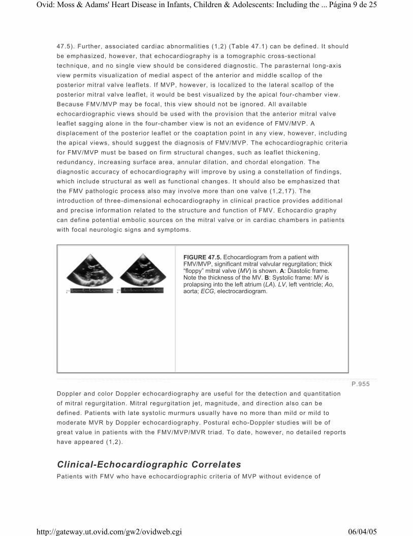
FMV/MVP: Severity of Mitral Valve Abnormalities Because FMV/MVP occurs in a heterogeneous group of patients with a wide spectrum of mitral valve involvement and hemodynamic abnormalit ies, i t is important in each case to establish not only the diagnosis of FMV/MVP but also to define a place within the spectrum of mitral valve abnormalit ies and to describe associated f indings (Table 47.4) (17,19). Patients with FMV/MVP also should be classif ied according to their symptoms. Auscultatory f indings should be described in detail (e.g., cl ick, multiple cl icks, murmur); phonocardiographic confirmation of auscultatory f indings may be valuable in certain circumstances and may contribute to more precise auscultatory conclusions. The echocardiographic interpretation should include the type of prolapse (e.g., late systolic, holosystolic, anterior, posterior leaflet), the thickness of mitral valve leaflets, the size of the mitral annulus, and the left ventricular and left atrial size and function (1). Doppler analysis should include the presence, severity, and timing of MVR. Jet direction and magnitude also should be defined.
Pathology and Histology Mitral valves excised from patients with severe FMV/MVP/MVR have large mitral valve surface areas (9,11) (Fig. 47.6), and the morphology differs signif icantly from normal mitral valves (2) (Fig. 47.7). The most specif ic, fundamental, and characteristic histologic change is collagen dissolution and disruption in the pars f ibrosa of the mitral valve leaflets. There is also a replacement of the dense collagenous fibrosa by loose myxomatous connective t issue with high acid mucopolysaccharide content. Similar histologic abnormalit ies have been observed in chordae tendineae (1,2).
P.957
TABLE 47.4. FLOPPY MITRAL VALVE AND MITRAL VALVE PROLAPSE: SEVERITY OF MITRAL VALVE ABNORMALITIES
Página 11 de 25Ovid: Moss & Adams' Heart Disease in Infants, Children & Adolescents: Including t...
06/04/05http://gateway.ut.ovid.com/gw2/ovidweb.cgi

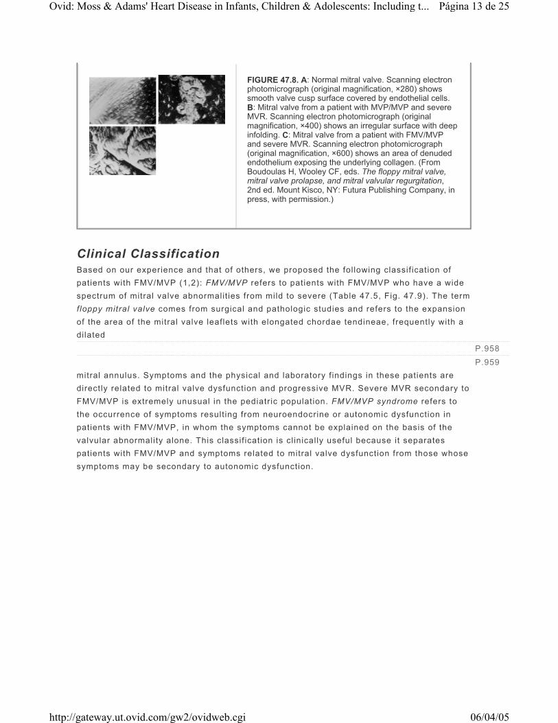
Scanning electron photomicrographs demonstrated surface folds and focal loss of endothelial cells on mitral valve leaflets obtained from patients with severe FMV/MVP and signif icant MVR1 (Fig. 47.8). These surface abnormalit ies may predispose to thromboembolic complications or infective endocardit is. Continuous pressure and stress due to left ventricular systole on the mitral valve leaflets and chordae tendineae contribute to gradual progression of these histologic changes.
FIGURE 47.6. FMV, Atrial view, from a patient with severe MVR. The surface area of the valve is increased, with increased folding of the valve surface. The widths of the anterior of the anterior leaflet (AL) and the the posterior leaflet (PL) are almost equal. Individual scallops of posterior leaflet are enlarged and redundant. B: Comparison of an excised FMV from a patient with FMV/MVP and severe MVR (top) with a normal mitral valve from a patient who died of noncardiac cause (bottom) showing the increased surface area of both anterior leaflets (AL) and PL of the FMV with enlarged and redundant posterior leaflet scallops, enlarged mitral annulus, elongated chordae tendineae. PCS, posteromedia commissural scallop; MS, middle scallop; ACS, anterolateral commissural scallop. (From Boudoulas H, Wooley CF. Mitral valve prolapse and the mitral valve prolapse syndrome. In: Yu PN, Goodwin JF, eds. Prog Cardiovasc Dis 1986;14:275–309, with permission.)
FIGURE 47.7. A: A normal mitral valve cusp. The histologic zones are represented in a cross-section. The atrialis is a thin zone of dense collagen immediately below the inflow surface (between arrowheads). The next zone, the spongiosa (S), consists of loose connective tissue, and the remainder of the cusp, composed of dense collagen, is the fibrosa (F). (Jones' silver stain; original magnification, ×25.) B: Floppy mitral valve cusp. The cusp has a large expanded central zone of myxomatous connective tissue with focal thinning and disruption of the fibrosa (arrow). The dense layer seen at the top just below the inflow surface is the markedly thickened atrialis (A). Fibrous connective tissue “pads” are seen on the ventricular surface. (Jones' silver stain; original magnification, ×4.) B inset: A high-magnification view of the myxomatous area showing disoriented, separated collagen bundles. A Mowry's colloidal iron stain demonstrated abundant accumulation of acid mucopolysaccharides in this area. (Jones' silver stain; original magnification, ×100.) (From Boudoulas H, Kolibash AJ, Baker P, et al. Mitral valve prolapse and the mitral valve prolapse syndrome: a diagnostic classification and pathogenesis of symptoms. Am Heart J 1989;118:796–818, with permission.)
Página 12 de 25Ovid: Moss & Adams' Heart Disease in Infants, Children & Adolescents: Including t...
06/04/05http://gateway.ut.ovid.com/gw2/ovidweb.cgi
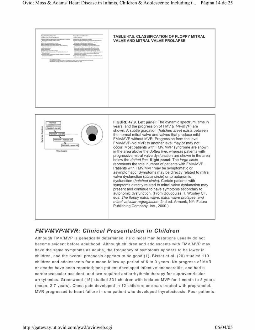
Clinical Classification Based on our experience and that of others, we proposed the fol lowing classif ication of patients with FMV/MVP (1,2): FMV/MVP refers to patients with FMV/MVP who have a wide spectrum of mitral valve abnormalit ies from mild to severe (Table 47.5, Fig. 47.9). The term f loppy mitral valve comes from surgical and pathologic studies and refers to the expansion of the area of the mitral valve leaflets with elongated chordae tendineae, frequently with a dilated mitral annulus. Symptoms and the physical and laboratory f indings in these patients are directly related to mitral valve dysfunction and progressive MVR. Severe MVR secondary to FMV/MVP is extremely unusual in the pediatric population. FMV/MVP syndrome refers to the occurrence of symptoms result ing from neuroendocrine or autonomic dysfunction in patients with FMV/MVP, in whom the symptoms cannot be explained on the basis of the valvular abnormality alone. This classif ication is cl inically useful because it separates patients with FMV/MVP and symptoms related to mitral valve dysfunction from those whose symptoms may be secondary to autonomic dysfunction.
FIGURE 47.8. A: Normal mitral valve. Scanning electron photomicrograph (original magnification, ×280) shows smooth valve cusp surface covered by endothelial cells. B: Mitral valve from a patient with MVP/MVP and severe MVR. Scanning electron photomicrograph (original magnification, ×400) shows an irregular surface with deep infolding. C: Mitral valve from a patient with FMV/MVP and severe MVR. Scanning electron photomicrograph (original magnification, ×600) shows an area of denuded endothelium exposing the underlying collagen. (From Boudoulas H, Wooley CF, eds. The floppy mitral valve, mitral valve prolapse, and mitral valvular regurgitation, 2nd ed. Mount Kisco, NY: Futura Publishing Company, in press, with permission.)
P.958P.959
Página 13 de 25Ovid: Moss & Adams' Heart Disease in Infants, Children & Adolescents: Including t...
06/04/05http://gateway.ut.ovid.com/gw2/ovidweb.cgi
FMV/MVP/MVR: Clinical Presentation in Children Although FMV/MVP is genetically determined, its cl inical manifestations usually do not become evident before adulthood. Although children and adolescents with FMV/MVP may have the same symptoms as adults, the frequency of symptoms appears to be lower in children, and the overall prognosis appears to be good (1). Bisset et al. (20) studied 119 children and adolescents for a mean follow-up period of 6 to 9 years. No progress of MVR or deaths have been reported; one patient developed infective endocardit is, one had a cerebrovascular accident, and two required antiarrhythmic therapy for supraventricular arrhythmias. Greenwood (15) studied 331 children with isolated MVP for 1 month to 8 years (mean, 2.7 years). Chest pain developed in 12 children; one was treated with propranolol. MVR progressed to heart fai lure in one patient who developed thyrotoxicosis. Four patients
TABLE 47.5. CLASSIFICATION OF FLOPPY MITRAL VALVE AND MITRAL VALVE PROLAPSE
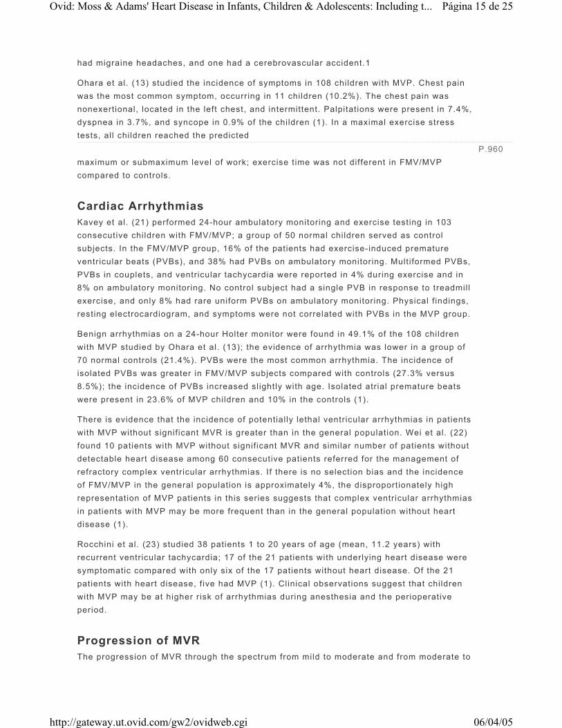
FIGURE 47.9. Left panel: The dynamic spectrum, time in years, and the progression of FMV (FMV/MVP) are shown. A subtle gradation (hatched area) exists between the normal mitral valve and valves that produce mild FMV/MVP without MVR. Progression from the level FMV/MVP-No MVR to another level may or may not occur. Most patients with FMV/MVP syndrome are shown in the area above the dotted line, whereas patients with progressive mitral valve dysfunction are shown in the area below the dotted line. Right panel: The large circle represents the total number of patients with FMV/MVP. Patients with FMV/MVP may be symptomatic or asymptomatic. Symptoms may be directly related to mitral valve dysfunction (black circle) or to autonomic dysfunction (hatched circle). Certain patients with symptoms directly related to mitral valve dysfunction may present and continue to have symptoms secondary to autonomic dysfunction. (From Boudoulas H, Wooley CF, eds. The floppy mitral valve, mitral valve prolapse, and mitral valvular regurgitation, 2nd ed. Armonk, NY: Futura Publishing Company, Inc., 2000.)
Página 14 de 25Ovid: Moss & Adams' Heart Disease in Infants, Children & Adolescents: Including t...
06/04/05http://gateway.ut.ovid.com/gw2/ovidweb.cgi
had migraine headaches, and one had a cerebrovascular accident.1
Ohara et al. (13) studied the incidence of symptoms in 108 children with MVP. Chest pain was the most common symptom, occurring in 11 children (10.2%). The chest pain was nonexertional, located in the left chest, and intermittent. Palpitations were present in 7.4%, dyspnea in 3.7%, and syncope in 0.9% of the children (1). In a maximal exercise stress tests, al l children reached the predicted maximum or submaximum level of work; exercise t ime was not different in FMV/MVP compared to controls.
Cardiac Arrhythmias Kavey et al. (21) performed 24-hour ambulatory monitoring and exercise testing in 103 consecutive children with FMV/MVP; a group of 50 normal children served as control subjects. In the FMV/MVP group, 16% of the patients had exercise-induced premature ventricular beats (PVBs), and 38% had PVBs on ambulatory monitoring. Mult i formed PVBs, PVBs in couplets, and ventricular tachycardia were reported in 4% during exercise and in 8% on ambulatory monitoring. No control subject had a single PVB in response to treadmil l exercise, and only 8% had rare uniform PVBs on ambulatory monitoring. Physical f indings, resting electrocardiogram, and symptoms were not correlated with PVBs in the MVP group.
Benign arrhythmias on a 24-hour Holter monitor were found in 49.1% of the 108 children with MVP studied by Ohara et al. (13); the evidence of arrhythmia was lower in a group of 70 normal controls (21.4%). PVBs were the most common arrhythmia. The incidence of isolated PVBs was greater in FMV/MVP subjects compared with controls (27.3% versus 8.5%); the incidence of PVBs increased slightly with age. Isolated atrial premature beats were present in 23.6% of MVP children and 10% in the controls (1).
There is evidence that the incidence of potential ly lethal ventricular arrhythmias in patients with MVP without signif icant MVR is greater than in the general population. Wei et al. (22) found 10 patients with MVP without signif icant MVR and similar number of patients without detectable heart disease among 60 consecutive patients referred for the management of refractory complex ventricular arrhythmias. If there is no selection bias and the incidence of FMV/MVP in the general population is approximately 4%, the disproportionately high representation of MVP patients in this series suggests that complex ventricular arrhythmias in patients with MVP may be more frequent than in the general population without heart disease (1).
Rocchini et al. (23) studied 38 patients 1 to 20 years of age (mean, 11.2 years) with recurrent ventricular tachycardia; 17 of the 21 patients with underlying heart disease were symptomatic compared with only six of the 17 patients without heart disease. Of the 21 patients with heart disease, f ive had MVP (1). Clinical observations suggest that children with MVP may be at higher risk of arrhythmias during anesthesia and the perioperative period.
Progression of MVR The progression of MVR through the spectrum from mild to moderate and from moderate to
P.960
Página 15 de 25Ovid: Moss & Adams' Heart Disease in Infants, Children & Adolescents: Including t...
06/04/05http://gateway.ut.ovid.com/gw2/ovidweb.cgi
severe in patients with FMV/MVP is gradual, and the entire process accelerates after a prolonged asymptomatic interval. Symptoms and complications in FMV/MVP related to mitral dysfunction include infective endocardit is, thromboembolic phenomena, cardiac arrhythmias, sudden death, progressive MVR, ruptured chordae tendineae, and congestive heart fai lure (1,2,19,24,25 and 26). Because of the slow gradual progression, signif icant MVR usually occurs after the fourth or f if th decades of l i fe and therefore usually is not seen in the pediatric population (Fig. 47.10) (26). Infective endocardit is, cardiac arrhythmias, and thromboembolic phenomena, however, although rare, may occur in children.
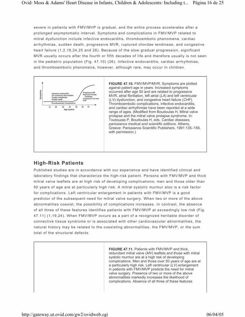
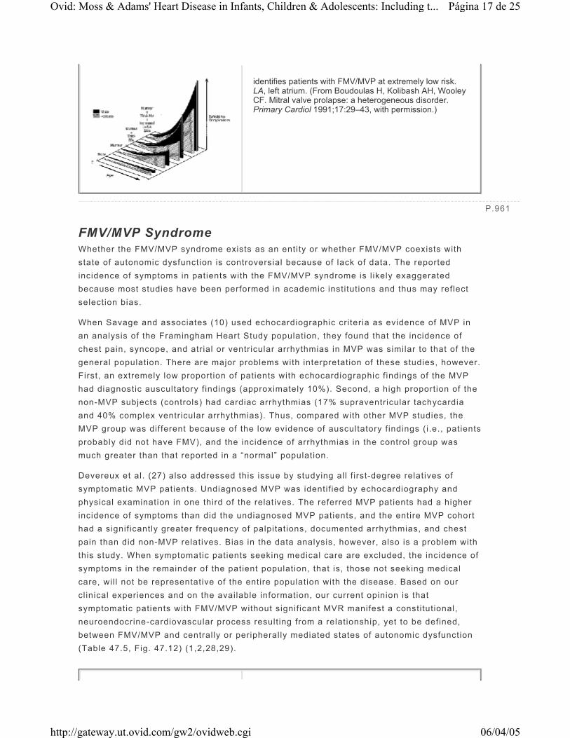
High-Risk Patients Published studies are in accordance with our experience and have identif ied cl inical and laboratory f indings that characterize the high-risk patient. Persons with FMV/MVP and thick mitral valve leaflets are at high risk of developing complications; men and those older than 50 years of age are at particularly high risk. A mitral systolic murmur also is a risk factor for complications. Left ventricular enlargement in patients with FMV/MVP is a good predictor of the subsequent need for mitral valve surgery. When two or more of the above abnormalit ies coexist, the possibil i ty of complicat ions increases. In contrast, the absence of al l three of these features identif ies patients with FMV/MVP at exceedingly low risk (Fig. 47.11) (1,19,24). When FMV/MVP occurs as a part of a recognized heritable disorder of connective t issue syndrome or is associated with other cardiovascular abnormalit ies, the natural history may be related to the coexisting abnormalit ies, the FMV/MVP, or the sum total of the structural defects.
FIGURE 47.10. FMV/MVP/MVR. Symptoms are plotted against patient age in years. Increased symptoms occurred after age 50 and are related to progressive MVR, atrial fibrillation, left atrial (LA) and left ventricular (LV) dysfunction, and congestive heart failure (CHF). Thromboembolic complications, infective endocarditis, and cardiac arrhythmias have been reported at a wide range of ages. (Modified from Boudoulas H. Mitral valve prolapse and the mitral valve prolapse syndrome. In: Toutouzas P, Boudoulas H, eds. Cardiac diseases, parissianos medical and scientific editions. Athens, Greece: Parissianos Scientific Publishers, 1991:135–156, with permission.)
FIGURE 47.11. Patients with FMV/MVP and thick, redundant mitral valve (MV) leaflets and those with mitral systolic murmur are at a high risk of developing complications. Men and those over 50 years of age are at a particularly high risk. Left ventricular (LV) enlargement in patients with FMV/MVP predicts the need for mitral valve surgery. Presence of two or more of the above abnormalities markedly increases the likelihood of complications. Absence of all three of these features
Página 16 de 25Ovid: Moss & Adams' Heart Disease in Infants, Children & Adolescents: Including t...
06/04/05http://gateway.ut.ovid.com/gw2/ovidweb.cgi
FMV/MVP Syndrome Whether the FMV/MVP syndrome exists as an entity or whether FMV/MVP coexists with state of autonomic dysfunction is controversial because of lack of data. The reported incidence of symptoms in patients with the FMV/MVP syndrome is l ikely exaggerated because most studies have been performed in academic institutions and thus may reflect selection bias.
When Savage and associates (10) used echocardiographic criteria as evidence of MVP in an analysis of the Framingham Heart Study population, they found that the incidence of chest pain, syncope, and atrial or ventricular arrhythmias in MVP was similar to that of the general population. There are major problems with interpretation of these studies, however. First, an extremely low proportion of patients with echocardiographic f indings of the MVP had diagnostic auscultatory f indings (approximately 10%). Second, a high proportion of the non-MVP subjects (controls) had cardiac arrhythmias (17% supraventricular tachycardia and 40% complex ventricular arrhythmias). Thus, compared with other MVP studies, the MVP group was different because of the low evidence of auscultatory f indings (i.e., patients probably did not have FMV), and the incidence of arrhythmias in the control group was much greater than that reported in a “normal” population.
Devereux et al. (27) also addressed this issue by studying all f irst-degree relatives of symptomatic MVP patients. Undiagnosed MVP was identif ied by echocardiography and physical examination in one third of the relatives. The referred MVP patients had a higher incidence of symptoms than did the undiagnosed MVP patients, and the entire MVP cohort had a signif icantly greater frequency of palpitations, documented arrhythmias, and chest pain than did non-MVP relatives. Bias in the data analysis, however, also is a problem with this study. When symptomatic patients seeking medical care are excluded, the incidence of symptoms in the remainder of the patient population, that is, those not seeking medical care, wil l not be representative of the entire population with the disease. Based on our cl inical experiences and on the available information, our current opinion is that symptomatic patients with FMV/MVP without signif icant MVR manifest a constitutional, neuroendocrine-cardiovascular process result ing from a relationship, yet to be defined, between FMV/MVP and centrally or peripherally mediated states of autonomic dysfunction (Table 47.5, Fig. 47.12) (1,2,28,29).
identifies patients with FMV/MVP at extremely low risk. LA, left atrium. (From Boudoulas H, Kolibash AH, Wooley CF. Mitral valve prolapse: a heterogeneous disorder. Primary Cardiol 1991;17:29–43, with permission.)
P.961
Página 17 de 25Ovid: Moss & Adams' Heart Disease in Infants, Children & Adolescents: Including t...
06/04/05http://gateway.ut.ovid.com/gw2/ovidweb.cgi
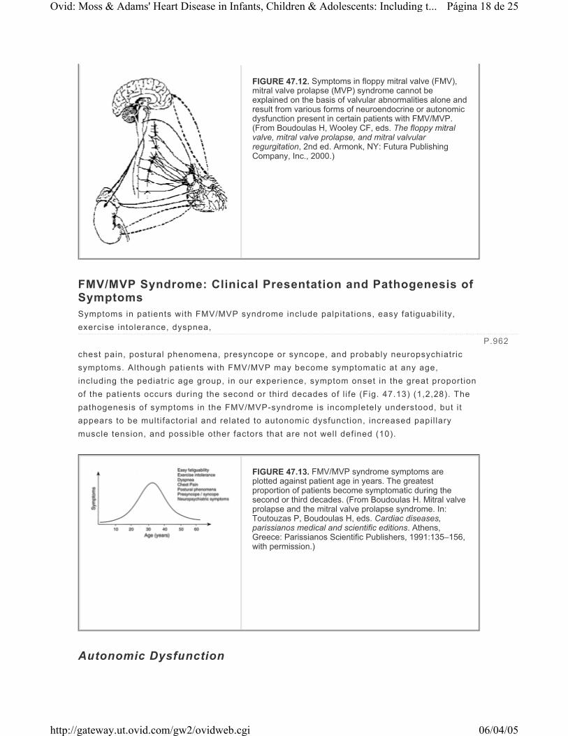
FMV/MVP Syndrome: Clinical Presentation and Pathogenesis of Symptoms Symptoms in patients with FMV/MVP syndrome include palpitations, easy fatiguabil i ty, exercise intolerance, dyspnea, chest pain, postural phenomena, presyncope or syncope, and probably neuropsychiatric symptoms. Although patients with FMV/MVP may become symptomatic at any age, including the pediatric age group, in our experience, symptom onset in the great proportion of the patients occurs during the second or third decades of l i fe (Fig. 47.13) (1,2,28). The pathogenesis of symptoms in the FMV/MVP-syndrome is incompletely understood, but i t appears to be multifactorial and related to autonomic dysfunction, increased papil lary muscle tension, and possible other factors that are not well defined (10).
Autonomic Dysfunction
FIGURE 47.12. Symptoms in floppy mitral valve (FMV), mitral valve prolapse (MVP) syndrome cannot be explained on the basis of valvular abnormalities alone and result from various forms of neuroendocrine or autonomic dysfunction present in certain patients with FMV/MVP. (From Boudoulas H, Wooley CF, eds. The floppy mitral valve, mitral valve prolapse, and mitral valvular regurgitation, 2nd ed. Armonk, NY: Futura Publishing Company, Inc., 2000.)
P.962
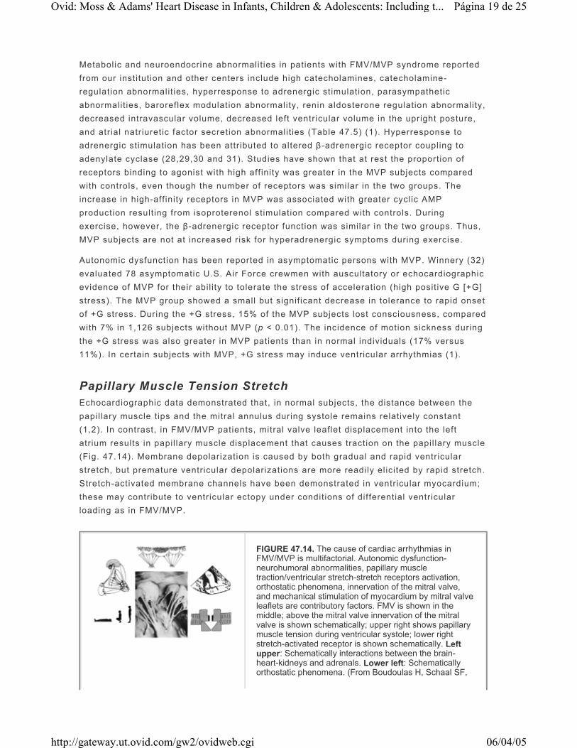
FIGURE 47.13. FMV/MVP syndrome symptoms are plotted against patient age in years. The greatest proportion of patients become symptomatic during the second or third decades. (From Boudoulas H. Mitral valve prolapse and the mitral valve prolapse syndrome. In: Toutouzas P, Boudoulas H, eds. Cardiac diseases, parissianos medical and scientific editions. Athens, Greece: Parissianos Scientific Publishers, 1991:135–156, with permission.)
Página 18 de 25Ovid: Moss & Adams' Heart Disease in Infants, Children & Adolescents: Including t...
06/04/05http://gateway.ut.ovid.com/gw2/ovidweb.cgi
Metabolic and neuroendocrine abnormalit ies in patients with FMV/MVP syndrome reported from our institution and other centers include high catecholamines, catecholamine-regulation abnormalit ies, hyperresponse to adrenergic stimulation, parasympathetic abnormalit ies, baroreflex modulat ion abnormality, renin aldosterone regulation abnormality, decreased intravascular volume, decreased left ventricular volume in the upright posture, and atrial natriuretic factor secretion abnormalit ies (Table 47.5) (1). Hyperresponse to adrenergic stimulation has been attr ibuted to altered β-adrenergic receptor coupling to adenylate cyclase (28,29,30 and 31). Studies have shown that at rest the proportion of receptors binding to agonist with high aff inity was greater in the MVP subjects compared with controls, even though the number of receptors was similar in the two groups. The increase in high-aff inity receptors in MVP was associated with greater cyclic AMP production result ing from isoproterenol stimulation compared with controls. During exercise, however, the β-adrenergic receptor function was similar in the two groups. Thus, MVP subjects are not at increased risk for hyperadrenergic symptoms during exercise.
Autonomic dysfunction has been reported in asymptomatic persons with MVP. Winnery (32) evaluated 78 asymptomatic U.S. Air Force crewmen with auscultatory or echocardiographic evidence of MVP for their abil ity to tolerate the stress of acceleration (high posit ive G [+G] stress). The MVP group showed a small but significant decrease in tolerance to rapid onset of +G stress. During the +G stress, 15% of the MVP subjects lost consciousness, compared with 7% in 1,126 subjects without MVP (p < 0.01). The incidence of motion sickness during the +G stress was also greater in MVP patients than in normal individuals (17% versus 11%). In certain subjects with MVP, +G stress may induce ventricular arrhythmias (1).
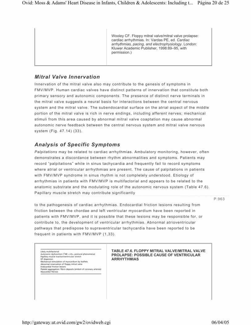
Papillary Muscle Tension Stretch Echocardiographic data demonstrated that, in normal subjects, the distance between the papil lary muscle t ips and the mitral annulus during systole remains relatively constant (1,2). In contrast, in FMV/MVP patients, mitral valve leaflet displacement into the left atrium results in papil lary muscle displacement that causes traction on the papil lary muscle (Fig. 47.14). Membrane depolarization is caused by both gradual and rapid ventricular stretch, but premature ventricular depolarizations are more readily el icited by rapid stretch. Stretch-activated membrane channels have been demonstrated in ventricular myocardium; these may contribute to ventricular ectopy under condit ions of differential ventricular loading as in FMV/MVP.
FIGURE 47.14. The cause of cardiac arrhythmias in FMV/MVP is multifactorial. Autonomic dysfunction-neurohumoral abnormalities, papillary muscle traction/ventricular stretch-stretch receptors activation, orthostatic phenomena, innervation of the mitral valve, and mechanical stimulation of myocardium by mitral valve leaflets are contributory factors. FMV is shown in the middle; above the mitral valve innervation of the mitral valve is shown schematically; upper right shows papillary muscle tension during ventricular systole; lower right stretch-activated receptor is shown schematically. Left upper: Schematically interactions between the brain-heart-kidneys and adrenals. Lower left: Schematically orthostatic phenomena. (From Boudoulas H, Schaal SF,
Página 19 de 25Ovid: Moss & Adams' Heart Disease in Infants, Children & Adolescents: Including t...
06/04/05http://gateway.ut.ovid.com/gw2/ovidweb.cgi
Mitral Valve Innervation Innervation of the mitral valve also may contribute to the genesis of symptoms in FMV/MVP. Human cardiac valves have distinct patterns of innervation that constitute both primary sensory and autonomic components. The presence of distinct nerve terminals in the mitral valve suggests a neural basis for interactions between the central nervous system and the mitral valve. The subendocardial surface on the atrial aspect of the middle portion of the mitral valve is r ich in nerve endings, including afferent nerves; mechanical stimuli from this area caused by abnormal mitral valve coaptation may cause abnormal autonomic nerve feedback between the central nervous system and mitral valve nervous system (Fig. 47.14) (33).
Analysis of Specific Symptoms Palpitations may be related to cardiac arrhythmias. Ambulatory monitoring, however, often demonstrates a discordance between rhythm abnormalit ies and symptoms. Patients may record “palpitations” while in sinus tachycardia and frequently fail to record symptoms where atrial or ventricular arrhythmias are present. The cause of palpitations in patients with FMV/MVP syndrome in sinus rhythm is not completely understood. Etiology of arrhythmias in patients with FMV/MVP is multi factorial and appears to be related to the anatomic substrate and the modulating role of the autonomic nervous system (Table 47.6). Papil lary muscle stretch may contribute signif icantly to the pathogenesis of cardiac arrhythmias. Endocardial fr ict ion lesions result ing from frict ion between the chordae and left ventricular myocardium have been reported in patients with FMV/MVP, and it is possible that these lesions may be responsible for, or contribute to, the development of ventricular arrhythmias. Abnormal atrioventricular pathways that predispose to supraventricular tachycardia have been reported to be frequent in patients with FMV/MVP (1,33).
Wooley CF. Floppy mitral valve/mitral valve prolapse: cardiac arrhythmias. In: Vardas PE, ed. Cardiac arrhythmias, pacing, and electrophysiology. London: Kluwer Academic Publisher, 1998:89–95, with permission.)
P.963
TABLE 47.6. FLOPPY MITRAL VALVE/MITRAL VALVE PROLAPSE: POSSIBLE CAUSE OF VENTRICULAR ARRHYTHMIAS
Página 20 de 25Ovid: Moss & Adams' Heart Disease in Infants, Children & Adolescents: Including t...
06/04/05http://gateway.ut.ovid.com/gw2/ovidweb.cgi
Autonomic dysfunction may init iate, precipitate, or contribute to arrhythmias in patients with FMV/MVP syndrome. Increased sympathetic activity may intensify ventricular and supraventricular arrhythmias. Extreme vasovagal reactions may be responsible for bradyarrhythmias in patients with or without sinoatrial or atrioventricular node disease. Inappropriate postural tachycardia secondary to autonomic dysfunction may result in subendocardial ischemia and ventricular arrhythmias. Electrolyte abnormalit ies or pharmacological agents also may be contributory factors in the pathogenesis of cardiac arrhythmias.
Fatigue is a common and nonspecif ic symptom of an underlying physical or emotional disorder and can be conceptualized as one of two neurobiologic emergency systems used by most higher organisms for self-preservation: the f ight-or-f l ight response mediated through the sympathetic neuroendocrine system and the conservation-withdrawal response characterized by a general dampening of metabolic and physical activity (1). Isoproterenol infusion in patients with FMV/MVP syndrome produced fatigue that lasted for several hours following infusion. Thus, fatigue is more than a state of mind, but usually i t is associated with physiologic alterations and in patients with FMV/MVP-syndrome may have a basis in disturbances of autonomic function (29).
Dyspnea in FMV/MVP syndrome patients cannot be explained on the basis of clear-cut cardiac or pulmonary abnormalit ies. Pulmonary-function abnormalit ies have been described in patients with MVP but are not severe enough to explain the dyspnea. Further, discrete pulmonary function abnormalit ies were not demonstrated in patients with MVP syndrome and dyspnea. Left ventricular function and central hemodynamics are usually normal in FMV/MVP-syndrome patients with dyspnea. The respiratory awareness and symptoms in patients with the FMV/MVP-syndrome represent alterations in centrally modulated breathing cycle control (1,2).
The cause of chest pain in patients with FMV/MVP syndrome may be multifactorial. Excessive stretching of the chordae tendineae presumably causes forceful traction on the papil lary muscles and the adjacent left ventricular wall, which may produce variations in papil lary muscle and subendocardial blood flow and oxygen demand with resultant papil lary muscle ischemia and chest pain. Platelet aggregation, hemorrhage, and fibrin deposits have been observed in the angle between the left atrium and the posterior mitral valve leaflet; microembolism from these deposits may involve the coronary circulation with subsequent myocardial ischemia.
Subendocardial blood flow is totally diastolic and thus depends on the duration of diastole. Extremely fast heart rate, inappropriate sinus tachycardia with excessive postural changes, and physical and emotional stresses may occur in patients with FMV/MVP syndrome. Sudden heart rate increases wil l produce disproportionately greater decreases in the diastolic t ime necessary for subendocardial f low than in systolic t ime because of a
P.964
Página 21 de 25Ovid: Moss & Adams' Heart Disease in Infants, Children & Adolescents: Including t...
06/04/05http://gateway.ut.ovid.com/gw2/ovidweb.cgi
nonlinear relationship between heart rate and diastolic t ime. The presence of a hyperadrenergic state in certain patients with FMV/MVP syndrome further increases myocardial oxygen consumption. Coronary artery vasoregulatory abnormalit ies may be present in certain patients with FMV/MVP syndrome and may contribute to the pathogenesis of subendocardial or papil lary muscle ischemia. Coronary artery spasm has been documented in a few patients with FMV/MVP syndrome. Myocardial or subendocardial ischemia may be secondary to a combination of the aforementioned factors. Indeed, studies that use rapid atrial pacing have shown myocardial lactate production with chest pain and ischemic electrocardiographic changes in patients with FMV/MVP syndrome and normal coronary arteries.
Patients with FMV/MVP syndrome with exercise intolerance and normal coronary arteries may have exercise-induced ischemic electrocardiographic changes. The precise mechanisms for these ischemic changes are not well understood and may be related to autonomic dysfunction, hyperadrenergic state, and in some cases may represent myocardial ischemia. β-blockade therapy in patients with the MVP syndrome and ischemic response to exercise abolished the ischemic electrocardiographic changes; this effect of β-blocking drugs on the exercise electrocardiogram is not specif ic, and similar responses may be seen in patients with coronary artery disease.
Patients with FMV/MVP often present with postural phenomena such as orthostatic decreases in cardiac output, orthostatic hypotension, tachycardia, arrhythmias, and symptoms related to alterations in heart rate, blood pressure, and cardiac output. Orthostatic phenomena are multifactorial in origin. A decreased intravascular volume, an abnormal renin-aldosterone response to volume depletion, a baroreflex modulation abnormality, a hyperadrenergic state, or a parasympathetic abnormality may account in part for these phenomena (1,2,31). Further, the inabil ity of patients with FMV/MVP syndrome to maintain normal left ventricular diastolic volume in the upright posture wil l result in a greater FMV/MVP and papil lary muscle tension; these changes in left ventricular size and mitral valve apparatus are also important factors that may contribute to orthostatic changes (1,2).
Syncope and presyncope are relatively common symptoms in patients with FMV/MVP syndrome; the causes are mult ifactorial. Arrhythmias definitely play some role in certain patients with FMV/MVP syndrome. Syncope or presyncope, l ike palpitations, often correlate poorly with cardiac arrhythmias. A given arrhythmia, however, may not always produce symptoms, depending on the sett ing in which it occurs. Neurocardiogenic syncope secondary to autonomic dysfunction certainly also plays an important role (see also discussion of athletics) (1,2).
Neuropsychiatric Symptoms A consistent, yet controversial f inding in many cl inical studies of patients with FMV/MVP syndrome has been the incidence of anxiety, panic attacks, and other complaints that are considered to be neuropsychiatric symptoms. Further, the incidence of FMV/MVP is greater in patients with condit ions considered to be related to autonomic dysfunction. The relationship between FMV/MVP and anxiety disorders remains something of an enigma: Other studies showed no association of FMV/MVP and neuropsychiatric symptoms.
Página 22 de 25Ovid: Moss & Adams' Heart Disease in Infants, Children & Adolescents: Including t...
06/04/05http://gateway.ut.ovid.com/gw2/ovidweb.cgi
Although panic disorder is not common in children, it appears that some association exists between FMV/MVP and panic disorders in the pediatric population (1,2).
FMV/MVP Sudden Death Sudden death in patients with FMV/MVP in the absence of hemodynamically signif icant MVR is rare but may occur (1,33). Autopsy studies have shown that mitral annular circumference, anterior and posterior mitral valve leaflet lengths, posterior mitral valve thickness, and endocardial fr ict ion lesions were greater in hearts from patients with FMV/MVP and sudden death compared with hearts from those with other cause of death and incidental FMV/MVP.
Patients with FMV/MVP who die suddenly without other underlying pathology tend to be relatively young women without MVR. Patients with a history of recurrent syncope or a history of sustained supraventricular or complex ventricular arrhythmias or a family history of cardiac sudden death appear to be at higher risk for sudden death (1,33).
In a study from the Ohio State University Hospitals, we reported nine cases of resuscitated survivors of cardiac arrest in patients with FMV/MVP, only one of whom had signif icant MVR. All patients but one were symptomatic before cardiac arrest; eight had long histories of palpitations with documented ventricular arrhythmias, and three of these eight patients had recurrent syncope. Sudden death in patients with FMV/MVP without signif icant MVR most often is due to ventricular f ibril lation. Among patients with FMV/MVP cardiac arrest reported from the Ohio State University Hospitals, ventricular f ibri l lation was documented in eight patients. The prognosis of patients with FMV/MVP who have been successfully resuscitated from cardiac arrest appears to be good; β-blockade therapy may be beneficial in this subset of patients (1).
FMV/MVP: Athletics Careful studies in athletes with FMV/MVP are lacking; thus, hard data on which to base a judgment for recommendations are not yet available. A few general statements, however, can be made (1,34).
Despite the high prevalence of FMV/MVP in the general population, FMV/MVP is an extremely rare cause of sudden death in competit ive athletes. Patients with FMV/MVP with myxomatous and collagen changes in the mitral valve may be in danger of further prolapse and increased chordal tension by strenuous activity, but no information is available at present.
No data exist to demonstrate that strenuous exercise in patients with FMV/MVP predisposes to death that otherwise would not have occurred, or the opposite, that withdrawal from competit ive athletics wil l prolong li fe. Without hard data, at present, the best approach for recommendations to athletes should be based on common sense and good clinical judgment. Recommendations and advice need to be balanced between restrict ing activity unduly and reducing chance of death or injury from the participation in athletics.
P.965
Página 23 de 25Ovid: Moss & Adams' Heart Disease in Infants, Children & Adolescents: Including t...
06/04/05http://gateway.ut.ovid.com/gw2/ovidweb.cgi
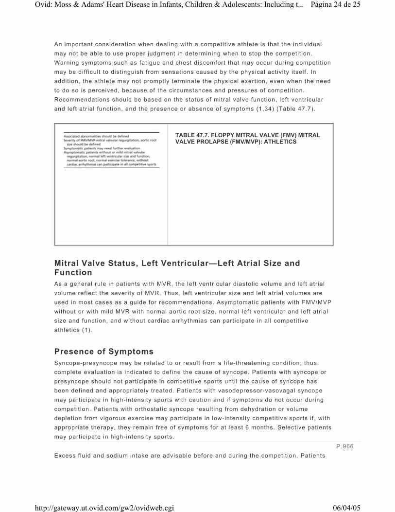
An important consideration when dealing with a competit ive athlete is that the individual may not be able to use proper judgment in determining when to stop the competit ion. Warning symptoms such as fatigue and chest discomfort that may occur during competit ion may be diff icult to distinguish from sensations caused by the physical activity itself. In addit ion, the athlete may not promptly terminate the physical exertion, even when the need to do so is perceived, because of the circumstances and pressures of competit ion. Recommendations should be based on the status of mitral valve function, left ventricular and left atrial function, and the presence or absence of symptoms (1,34) (Table 47.7).
Mitral Valve Status, Left Ventricular—Left Atrial Size and Function As a general rule in patients with MVR, the left ventricular diastolic volume and left atrial volume reflect the severity of MVR. Thus, left ventricular size and left atrial volumes are used in most cases as a guide for recommendations. Asymptomatic patients with FMV/MVP without or with mild MVR with normal aortic root size, normal left ventricular and left atrial size and function, and without cardiac arrhythmias can participate in all competit ive athletics (1).
Presence of Symptoms Syncope-presyncope may be related to or result from a l ife-threatening condit ion; thus, complete evaluation is indicated to define the cause of syncope. Patients with syncope or presyncope should not participate in competit ive sports until the cause of syncope has been defined and appropriately treated. Patients with vasodepressor-vasovagal syncope may participate in high-intensity sports with caution and if symptoms do not occur during competit ion. Patients with orthostatic syncope result ing from dehydration or volume depletion from vigorous exercise may participate in low-intensity competit ive sports if, with appropriate therapy, they remain free of symptoms for at least 6 months. Selective patients may participate in high-intensity sports. Excess f luid and sodium intake are advisable before and during the competit ion. Patients
TABLE 47.7. FLOPPY MITRAL VALVE (FMV) MITRAL VALVE PROLAPSE (FMV/MVP): ATHLETICS
P.966
Página 24 de 25Ovid: Moss & Adams' Heart Disease in Infants, Children & Adolescents: Including t...
06/04/05http://gateway.ut.ovid.com/gw2/ovidweb.cgi
with arrhythmic syncope may participate in low-intensity competit ive sports if, with appropriate therapy, they remain free of symptoms for at least 6 months. Patients with a history of syncope regardless of the etiology should not participate in sports with a danger of body coll ision with increased risk when syncope occurs.
Palpitations and Arrhythmias Patients with a history of palpitations should be carefully evaluated to exclude any signif icant arrhythmia before permission to participate in competit ive sports. The prognosis of ventricular or supraventricular arrhythmias detected with exercise testing or ambulatory monitoring in patients with FMV/MVP and normal left ventricular size and function appears to be good, although it is not completely defined. Patients with suspected cardiac arrhythmias being considered for competit ive athletics should have long-term ambulatory monitoring, i f possible, during the type of exercise the athlete performs as well as an exercise test. Arrhythmias may not be reproducible during a routine exercise test, and exercise may need to be adapted specif ically for the athlete (e.g., begin exercise at peak level in a sprinter rather than with slowly increasing workload). Patients with arrhythmias should be reevaluated at intervals after they have trained to determine the effect of training on arrhythmia.
Younger patients with ventricular preexcitation or concealed atrioventricular pathways should have an in-depth evaluation before recommendation for participation in competit ive athletics. Arrhythmias that result from abnormal atrioventricular pathways may be abolished with radiofrequency ablation.
Coelho et al. (35) studied 19 young athletes aged 14 to 32 years with documented symptomatic tachyarrhythmias; f ive had paroxysmal atrial f ibri l lation, f ive paroxysmal supraventricular tachycardia, eight paroxysmal ventricular tachycardia, and one ventricular f ibri l lation. Nine of these patients (47%) had MVP, and five (26%) had an abnormal atrioventricular pathway. Tachyarrhythmias started during strenuous exercise in 13 patients (68%); tachyarrhythmia that closely resembled the spontaneous arrhythmia induced by programmed cardiac stimulation began in 13 patients (68%) and was reproducibly provoked by treadmill exercise testing in 8 patients (48%). In four of the eight patients with ventricular tachycardia, tachycardia was provoked with isoproterenol infusion (1).
Chest Pain Coronary artery anomaly or other underlying cardiac pathology in patients with chest pain should be excluded before permission is given to participate in sports. An exercise test should be performed, and, if cl inically indicated, further diagnostic studies, including coronary arteriography, should be undertaken. It has been suggested that patients with a family history of sudden death from FMV/MVP should not participate in competit ive sports. No data are available, however, to support this thesis.
Página 25 de 25Ovid: Moss & Adams' Heart Disease in Infants, Children & Adolescents: Including t...
06/04/05http://gateway.ut.ovid.com/gw2/ovidweb.cgi