Flea-borne Rickettsia species in fleas, Caldas department ...
11
Original Article Flea-borne Rickettsia species in fleas, Caldas department, Colombia Carol B Colonia 1 # , Alejandro Ramírez-Hernández 2,3 # , Juliana Gil-Mora 1 , Juan C Agudelo 4 , Gabriel Jaime Castaño Villa 5 , Camilo Pino 6 , Paola Betancourt-Ruiz 1 , Jorge E Pérez Cárdenas 7 , Lucas S Blanton 8 , Marylin Hidalgo 1 1 Grupo de Enfermedades Infecciosas, Facultad de Ciencias, Pontificia Universidad Javeriana, Bogotá D.C., Colombia 2 Grupo Parasitología Veterinaria, Facultad de Medicina Veterinaria y de Zootecnia, Universidad Nacional de Colombia, Bogotá D.C., Colombia 3 Rickettsial and Ehrlichial Disease Research, Department of Pathology, University of Texas Medical Branch, Galveston, TX, United States 4 Facultad de Ciencias Agropecuarias, Universidad de Caldas, Manizales, Caldas, Colombia 5 Grupo de investigación GEBIOME, Departamento de Desarrollo Rural y Recursos Naturales, Facultad de Ciencias Agropecuarias, Universidad de Caldas, Manizales, Caldas, Colombia 6 Laboratorio de Investigación en Sistemas Inteligentes, Facultad de Ingeniería, Universidad Nacional de Colombia, Bogotá D.C., Colombia 7 Facultad de Ciencias para la Salud, Universidad de Caldas, Manizales, Caldas, Colombia 8 Division of Infectious Diseases, Department of Internal Medicine, University of Texas Medical Branch, Galveston, TX, United States # Authors contributed equally to this work. Abstract Introduction: Rickettsioses are zoonotic diseases caused by pathogenic bacteria of the genus Rickettsia and transmitted to man by means of arthropod vectors such as ticks, fleas, mites and lice. Historically, Caldas Department has reported a significant number of cases of murine typhus to the Colombian national health surveillance system, and consequent studies of flea-borne rickettsiosis identified the circulation of Rickettsia typhi and Rickettsia felis in multiple municipalities. Our aim was to genotype species of Rickettsia detected in fleas collected from domestic and wild mammals in Caldas. Methodology: Flea samples were taken by convenience sampling from dogs, cats and wild mammals (rodents and marsupials) in 26 municipalities. Specimens were classified by current taxonomic keys and pooled for DNA extraction and molecular screening for Rickettsia spp. by PCR amplification of gltA, htrA and sca5 genes. Positive samples were genotyped by enzyme digestion (htrA) and sequencing. Results: A total of 1388 flea samples were collected. Rickettsia DNA was amplified in 818 (gltA), 883 (htrA) and 424 (sca5) flea pools. Alignment analysis with available Rickettsia DNA sequences showed greater similarity with R. asembonensis (gltA) and with R. felis (sca5 and htrA). Restriction pattern was compatible with R. felis. R. typhi was not identified. Conclusion: The present study confirms the presence and high prevalence of R. asembonensis and R. felis in fleas from domestic and wild animals in different municipalities from Caldas Department. Key words: Rickettsia felis; Rickettsia asembonensis; RFLP; vector-borne diseases; zoonotic diseases.. J Infect Dev Ctries 2020; 14(10):1155-1163. doi:10.3855/jidc.12524 (Received 07 February 2020 – Accepted 17 May 2020) Copyright © 2020 Colonia et al. This is an open-access article distributed under the Creative Commons Attribution License, which permits unrestricted use, distribution, and reproduction in any medium, provided the original work is properly cited. Introduction Rickettsia spp. are obligately intracellular bacteria belonging to the family Rickettsiaceae (order Rickettsiales), which can cause mild to severe diseases in humans and other animals [1]. Historically, two flea- borne rickettsial species have been recognized (i.e., Rickettsia typhi and Rickettsia felis) [2], but recently, new R. felis-related species (i.e., Rickettsia asembonensis sp. nov. and ‘Candidatus Rickettsia senegalensis’) and others have been described [3-5]. Rickettsia typhi is the etiologic agent of murine or endemic typhus, a febrile zoonotic disease which involves the Oriental rat flea (Xenopsylla cheopis) and different rodents (e.g., Rattus spp.) in its enzootic cycle [6]. Murine typhus has a wide distribution in tropical regions throughout the world and is currently
Transcript of Flea-borne Rickettsia species in fleas, Caldas department ...

Original Article Flea-borne Rickettsia species in fleas, Caldas department, Colombia Carol B Colonia1 #, Alejandro Ramírez-Hernández2,3 #, Juliana Gil-Mora1, Juan C Agudelo4, Gabriel Jaime Castaño Villa5, Camilo Pino6, Paola Betancourt-Ruiz1, Jorge E Pérez Cárdenas7, Lucas S Blanton8, Marylin Hidalgo1
1 Grupo de Enfermedades Infecciosas, Facultad de Ciencias, Pontificia Universidad Javeriana, Bogotá D.C., Colombia 2 Grupo Parasitología Veterinaria, Facultad de Medicina Veterinaria y de Zootecnia, Universidad Nacional de Colombia, Bogotá D.C., Colombia 3 Rickettsial and Ehrlichial Disease Research, Department of Pathology, University of Texas Medical Branch, Galveston, TX, United States 4 Facultad de Ciencias Agropecuarias, Universidad de Caldas, Manizales, Caldas, Colombia 5 Grupo de investigación GEBIOME, Departamento de Desarrollo Rural y Recursos Naturales, Facultad de Ciencias Agropecuarias, Universidad de Caldas, Manizales, Caldas, Colombia 6 Laboratorio de Investigación en Sistemas Inteligentes, Facultad de Ingeniería, Universidad Nacional de Colombia, Bogotá D.C., Colombia 7 Facultad de Ciencias para la Salud, Universidad de Caldas, Manizales, Caldas, Colombia 8 Division of Infectious Diseases, Department of Internal Medicine, University of Texas Medical Branch, Galveston, TX, United States # Authors contributed equally to this work. Abstract Introduction: Rickettsioses are zoonotic diseases caused by pathogenic bacteria of the genus Rickettsia and transmitted to man by means of arthropod vectors such as ticks, fleas, mites and lice. Historically, Caldas Department has reported a significant number of cases of murine typhus to the Colombian national health surveillance system, and consequent studies of flea-borne rickettsiosis identified the circulation of Rickettsia typhi and Rickettsia felis in multiple municipalities. Our aim was to genotype species of Rickettsia detected in fleas collected from domestic and wild mammals in Caldas. Methodology: Flea samples were taken by convenience sampling from dogs, cats and wild mammals (rodents and marsupials) in 26 municipalities. Specimens were classified by current taxonomic keys and pooled for DNA extraction and molecular screening for Rickettsia spp. by PCR amplification of gltA, htrA and sca5 genes. Positive samples were genotyped by enzyme digestion (htrA) and sequencing. Results: A total of 1388 flea samples were collected. Rickettsia DNA was amplified in 818 (gltA), 883 (htrA) and 424 (sca5) flea pools. Alignment analysis with available Rickettsia DNA sequences showed greater similarity with R. asembonensis (gltA) and with R. felis (sca5 and htrA). Restriction pattern was compatible with R. felis. R. typhi was not identified. Conclusion: The present study confirms the presence and high prevalence of R. asembonensis and R. felis in fleas from domestic and wild animals in different municipalities from Caldas Department. Key words: Rickettsia felis; Rickettsia asembonensis; RFLP; vector-borne diseases; zoonotic diseases.. J Infect Dev Ctries 2020; 14(10):1155-1163. doi:10.3855/jidc.12524 (Received 07 February 2020 – Accepted 17 May 2020) Copyright © 2020 Colonia et al. This is an open-access article distributed under the Creative Commons Attribution License, which permits unrestricted use, distribution, and reproduction in any medium, provided the original work is properly cited. Introduction
Rickettsia spp. are obligately intracellular bacteria belonging to the family Rickettsiaceae (order Rickettsiales), which can cause mild to severe diseases in humans and other animals [1]. Historically, two flea-borne rickettsial species have been recognized (i.e., Rickettsia typhi and Rickettsia felis) [2], but recently, new R. felis-related species (i.e., Rickettsia
asembonensis sp. nov. and ‘Candidatus Rickettsia senegalensis’) and others have been described [3-5].
Rickettsia typhi is the etiologic agent of murine or endemic typhus, a febrile zoonotic disease which involves the Oriental rat flea (Xenopsylla cheopis) and different rodents (e.g., Rattus spp.) in its enzootic cycle [6]. Murine typhus has a wide distribution in tropical regions throughout the world and is currently

Colonia et al. – Flea-borne Rickettsia in fleas from Colombia J Infect Dev Ctries 2020; 14(10):1155-1163.
1156
recognized as endemic in parts of South America, Australia, Asia and southeastern Europe [7-11]. The disease is also endemic in California and Texas (USA), where an alternate suburban transmission cycle, apparently involving opossums and cat fleas (Ctenocephalides felis), has been described [12,13].
Rickettsia felis, a species discovered within a C. felis laboratory colony in 1990 [14], has been molecularly detected in a diversity of arthropods including other flea species, ticks, mites, booklice and even mosquitoes [15]; nonetheless, the cat flea is the only recognized vector and reservoir [2]. Reports of human infections have increased in recent years, but the role of R. felis as a cause of disease has been scrutinized [2,16,17].
Several studies have suggested that flea-borne Rickettsia species (R. typhi and R. felis) can share vectors such as the cat flea (C. felis) and the dog flea (C. canis); however, R. felis is the most frequently detected species [18,19]. Importantly, the role of cats, dogs and other animals as possible reservoirs of these Rickettsia species remains unknown [20-22].
In Colombia, Caldas Department has been considered an endemic area for murine typhus since 1942 [23]. Recent studies in this region have established a seroprevalence of 25.5% and 17.8% for R. typhi and R. felis, respectively, and 28.7% for both species [24]. Likewise, another report confirmed, by seroconversion, the diagnosis of 12 patients with signs and symptoms suggestive of murine typhus [25]. In 2013, the presence of R. felis in fleas collected from animals was reported, for the first time, in the same area in Colombia [26]. Despite this, the circulation of other flea-borne Rickettsia species in Caldas remains unknown.
The aim of this work was to detect and genotype rickettsial DNA from flea samples collected from domestic and wild animals in order to contribute to the knowledge of flea-borne Rickettsia species in this endemic region.
Methodology Flea sampling
This cross-sectional study was conducted between November 2015 and January 2017 and was approved by the ethics committees of the Pontificia Universidad Javeriana and Universidad de Caldas (Act 8th, June 9th, 2014; and CBCS-016-14, May 28th, 2014, respectively).
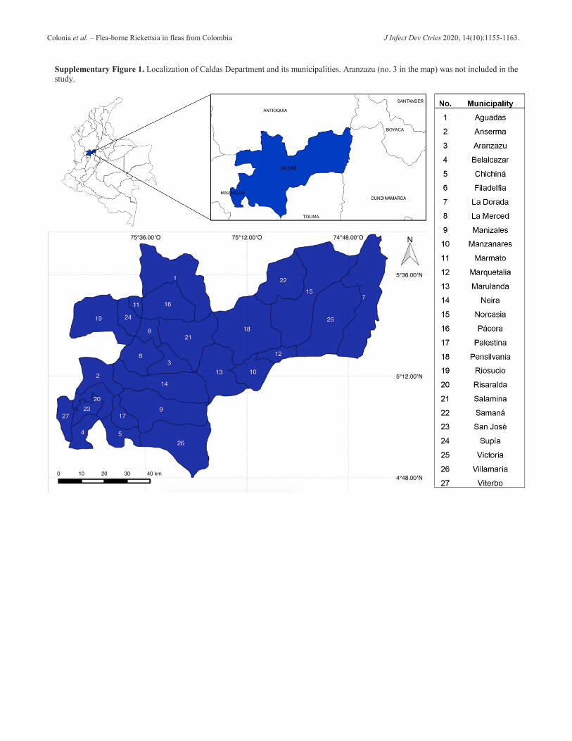
The area of study was the Department of Caldas, located in the midwestern area of Colombia (central branch of the Andes). Sampling was performed in urban and rural zones of 26 municipalities as listed in Table 1;
a map and geographic coordinates are presented as supplemental material (Supplementary Figure 1 and Supplementary Table 1).
Domestic animals (dogs and cats) and wild mammals (synanthropic and non-synanthropic species) were included in the study for ectoparasite sampling. Dogs and cats, in urban and rural households, were sampled after the owner’s consent and proper manual restraint. Wild mammals were captured by live trapping. For these, Sherman and Tomahawk traps were distributed in parallel transects of variable longitude and separated between them by 20 to 50 meters, depending on the local topography. Traps were placed 10 meters apart and at two alternate heights (ground-level and two-meter level). A mixture of banana, cereal, vanilla essence and sardines were used as bait during three nights of sampling with a total sampling effort of 2148 trap-nights. Captured animals were anaesthetized with isoflurane within a hermetic plastic box (3 to 5-minute exposure depending on body size) and submitted to morphometric and photographic measures for further taxonomic classification. All individuals were released to nature after recovery.
Fleas were collected manually or by hair combing from domestic dogs, cats and wild mammals. All specimens were stored in 70% ethanol, further classified by current morphological keys [27-29] and preserved at -20 ºC prior to DNA extraction. For this, specimens were grouped into pools (2-3 fleas/pool), considering the following criteria: flea species, animal host and geographic origin.
DNA extraction
Flea samples were submitted to a dry bath (56 ºC for 30 minutes) to eliminate any ethanol trace and DNA was further extracted with a modified protocol using guanidine thiocyanate (DNAzol; InvitrogenTM, Life Technologies Corp., Grand Island, NY, USA) and the DNeasy Blood and Tissue Kit (Qiagen TM, Germantown, MD, USA), as previously reported [26]. Subsequently, DNA purity and concentration were measured using a calibrated spectrophotometer (West Tune NanoGenius series, Hangzhou, China).
Rickettsia DNA amplification
Before performing PCR reactions for Rickettsia, we assessed the presence of amplifiable DNA and the absence of inhibitors in the extracted flea samples by amplification of cytochrome oxidase subunit II (COII) gene (primers COII-F-Leu and COII-F-Lys) [30].
The detection of Rickettsia DNA in flea samples was performed by the amplification of gltA (primers CS

Colonia et al. – Flea-borne Rickettsia in fleas from Colombia J Infect Dev Ctries 2020; 14(10):1155-1163.
1157
-78-CS323 and CS5-CS6) [31], htrA (17kD1-17kD2) [31] and sca5 (also known as ompB) genes (120.M59-120.807 and 120.607 F-120.1497R) [32]. Rickettsia slovaca DNA was used as a positive control and water as a negative control in all reactions. For positive flea pools, Minimal Infection Rates (MIR) were calculated for each of the three genes evaluated, as previously reported [33].
RFLP
Amplified htrA products were submitted to endonuclease digestion using the enzymes AluI and XbaI [13]. Further analysis of the restriction sites was carried out with the NEBcutter V2.0 program (New England Biolabs, Inc., Ipswich, MA, USA) in order to
identify fragment patterns and genotype of the Rickettsia species.
DNA Sequencing
With the aim to identify Rickettsia species circulating in fleas, some PCR positive samples were reamplified with a proof-reading Taq enzyme PCR protocol and purified with ExoSAP-IT Express PCR Cleanup kit (Thermo Fisher Scientific, Waltham, MA, USA). Thereafter, they were submitted for Sanger automatized capillary sequencing (ABI-3500XL Genetic Analyzer, Applied Biosystems, Waltham, MA, USA). Flea pools samples positive for the three genes evaluated were submitted for sequencing, excepting those from Xenopsylla cheopis which could be positive for at least one gene.
Table 1. Flea species collected in domestic animals and wild mammals between November 2015 and January 2017, in 26 municipalities from Caldas Department, Colombia.
Municipality Flea species
Ctenocephalides felis Ctenocephalides canis Other flea speciesb Doga Cat WM Dog Cat WM
Aguadas - - - - - - Anserma - 43 (3.9) - 4 (1.6) - -
Belalcazar 29 (2.6) 32 (2.9) - - 2 (0.8) - Pulex irritans (Dog) (2) Chinchiná 8 (0.7) 43 (3.9) - - 1 (0.4) - Xenopsylla cheopis (Didelphis marsupialis) (1) Filadelfia 5 (0.5) - - - - - La Dorada - 10 (0.9) - - - - La Merced - - - - - -
Manizales 29 (2.6) 40 (3.6) - 3 (1.2) 8 (3.1.) -
Leptopsylla segnis (Thomasomys cf. baeops) (1), Leptopsylla sp. (Thomasomys cf. baeops) (2), Ctenophtalmus sp. (Nephelomys childi, Thomasomys cf. baeops) (4), Nosopsyllus sp. (Thomasomys cf. baeops) (2)
Manzanares 56 (5.1) 106 (9.7) - 6 (2.3) 2 (0.8) -
Marmato 23 (2.1) - 10 (0.9) 16 (6.2) - 3 (1.2) Leptopsylla segnis (Didelphis marsupialis) (1); X. cheopis (Didelphis marsupialis) (3)
Marquetalia 20 (1.8) 104 (9.5) - 4 (1.6) 2 (0.8) - Marulanda 4 (0.4) 18 (1.6) - 7 (2.7) 2 (0.8) -
Neira - 5 (0.5) - - - - Norcasia 1 (0.1) 40 (3.6) - 42 (16.3) - -
Pácora - - - - - - Leptopsylla segnis (Didelphis marsupialis) (6); X. cheopis (Didelphis marsupialis) (1)
Palestina 45 (4.1) 23 (2.1) - - - - Rhopalopsyllus spp. (Didelphis marsupialis) (1) Pensilvania 6 (0.6) 21 (1.9) - - - -
Riosucio 2 (0.2) 46 (4.2) - - - - P. irritans (Cat) (2) Risaralda 25 (2.3) - - 23 (8.9) - - Salamina - - - - - - Samaná 18 (1.6) 14 (1.3) - - - - San José 28 (2.6) 42 (3.8) - - - -
Supía 1 (0.1) 44 (4.0) - 40 (15.5) - - X. cheopis (Cat) (1) Victoria 14 (1.3) 36 (3.3) - 62 (24) - - X. cheopis (Proechymis spp.) (2)
Villamaría 15 (1.4) 17 (1.6) - 24 (9.3) 3 (1.2) - Leptopsylla segnis (Nephelomys childi, Riphidomys cf. latimanus) (2) Viterbo 20 (1.8) 56 (5.1) - - - -
Total 349 (31.8) 740 (67.3) 10 (0.9) 235 (91.1) 20 (7.8) 3 (1.2) 31 (0.02)
1099 (79.4) 258 (18.6) 1388
WM: Wild mammals; a. Data are presented as total number (%) b. Data are presented as flea species (host) (number collected).

Colonia et al. – Flea-borne Rickettsia in fleas from Colombia J Infect Dev Ctries 2020; 14(10):1155-1163.
1158
Figure 1. Restriction patterns with AluI for the htrA gene in 8 pools of samples.
Lanes: ladder 50 bp molecular weight marker (ZYMO RESEARCH); lanes 1-8, samples; lane 9, positive control (Rickettsia slovaca). The restriction pattern corresponds to Rickettsia felis according to the size of the fragments generated by the digestion (177, 119 and 109 bp).
Figure 2. Restriction patterns with XbaI for the htrA gene in 8 pools of samples.
Lanes: ladder 50 bp molecular weight marker (PROMEGA); lanes 1-8, samples; lane 9, positive control (Rickettsia slovaca); ladder 50 bp. Rickettsia felis and R. slovaca have no restriction site for this enzyme (fragment size: 433 bp).
Table 2. Flea samples collected between November 2015 and January 2017, in different municipalities from Caldas Department (Colombia), positive for Rickettsia DNA by gene and host.
Municipalityb
Flea samplesa gltA sca5 htrA
Dogs Cats WM Dogs Cats WM Dogs Cats WM Anserma - 30/47 (0.6) - - 29/47 (0.6) - - 28/47 (0.6) -
Belalcazar 11/31 (0.4) 25/34 (0.7) - 15/31 (0.5) 3/34 (0.1) - 16/31 (0.5) 28/34 (0.8) - Chinchiná 2/12 (0.2) 28/44 (0.6) 1/1 (1.0) 5/12 (0.4) 27/44 (0.6) 0/1 (0) 6/12 (0.5) 29/44 (0.7) 1/1 (1.0) Filadelfia 3/5 (0.6) - - 3/5 (0.6) - - 3/5 (0.6) - - La Dorada - 4/10 (0.4) - - 0/10 (0) - - 4/10 (0.4) - Manizales 21/32 (0.7) 21/48 (0.4) 0/6 (0) 19/32 (0.6) 12/48 (0.3) 6/6 (1.0) 22/32 (0.7) 29/48 (0.6) 0/6 (0)
Manzanares 28/62 (0.5) 54/108 (0.5) - 17/62 (0.3) 50/108 (0.5) - 31/62 (0.5) 58/108 (0.5) - Marmato 30/39 (0.8) - 9/17 (0.5) 10/39 (0.3) - 12/17 (0.7) 30/39 (0.8) - 17/17 (1.0)
Marquetalia 12/24 (0.5) 54/106 (0.5) - 8/24 (0.3) 34/106 (0.3) - 12/24 (0.5) 58/106 (0.6) - Marulanda 8/11 (0.7) 8/20 (0.4) - 8/11 (0.7) 8/20 (0.4) - 9/11 (0.8) 8/20 (0.4) -
Neira - 3/5 (0.6) - - 1/5 (0.2) - - 3/5 (0.6) - Norcasia 30/43 (0.7) 29/40 (0.7) - - 11/40 (0.3) - 30/43 (0.7) 29/40 (0.7) - Pácora - - 0/7 (0) - - 1/7 (0.1) - - 3/7 (0.4)
Palestina 30/45 (0.7) 18/23 (0.8) 0/1 (0) - - 1/1 (1.0) 30/45 (0.7) 18/23 (0.8) 0/1 (0) Pensilvania 6/6 (1.0) 13/21 (0.6) - - 0/21 (0.0) - 6/6 (1.0) 14/21 (0.7) -
Riosucio 1/2 (0.5) 28/48 (0.6) - 1/2 (0.5) 28/48 (0.6) - 1/2 (0.5) 28/48 (0.6) - Risaralda 42/48 (0.9) - - - - - 42/48 (0.9) - - Samaná 18/18 (1.0) 11/14 (0.8) - - 11/14 (0.8) - 18/18 (1.0) 11/14 (0.8) - San José 14/28 (0.5) 26/42 (0.6) - 14/28 (0.5) 25/42 (0.6) - 14/28 (0.5) 26/42 (0.6) -
Supía 30/41 (0.7) 31/45 (0.7) - - - - 30/41 (0.7) 31/45 (0.7) - Victoria 30/76 (0.4) 30/36 (0.8) 0/2 (0) - - 0/2 (0) 30/76 (0.4) 30/36 (0.8) 2/2 (1.0)
Villamaría 29/39 (0.7) 1/20 (0.1) 0/2 (0) 12/39 (0.3) 6/20 (0.3) 0/2 (0) 29/39 (0.7) 15/20 (0.8) 2/2 (1.0) Viterbo 15/20 (0.8) 34/56 (0.6) - 14/20 (0.7) 33/56 (0.6) - 15/20 (0.8) 37/56 (0.7) - Total 360/582 (0.6) 448/767 (0.6) 10/36 (0.3) 126/305 (0.4) 278/663 (0.4) 20/36 (0.6) 374/582 (0.6) 484/767 (0.6) 25/36 (0.7)
WM: Wild mammals; a. Data are presented as No. of positive pools/Total number of fleas tested (Minimum Infection Rate-MIR); b. Municipalities without flea samples (i.e. Aguadas, La Merced and Salamina) were not included.
Colonia et al. – Flea-borne Rickettsia in fleas from Colombia J Infect Dev Ctries 2020; 14(10):1155-1163.
1159
Bioinformatic analysis The pre-processing of the sequence data was
performed using Trace Tuner [34] and CAP3 [35] programs. Nucleotide-nucleotide alignment analysis was performed using FASTA files with a rickettsial genome database obtained from NCBI assembly [36], through NCBI Taxonomy [37], including sequences from this study and those obtained in the work published by Ramírez-Hernández et al. [26]. The ClustalW program [38] was used for refining alignments prior to construction of phylogenetic trees. These were built using the Maximum Likelihood method based on the Tamura-Nei model [39]. Branch support was tested by bootstrap analysis using 1000 replicates. Rickettsia canadensis was used as an outgroup (accession numbers: MH595545.1 and CP000409.1). Trees were constructed and analyzed with MEGA 7 software [40].
Results Rickettsia detection in flea samples
In total, 1388 fleas were collected in 23 out of 26 municipalities (none were collected in Aguadas, La Merced and Salamina). 1344 (96.8%) were from domestic animals (584; 43.5% from dogs and 760; 56.6% from cats) and 44 (3.2%) from wild mammals (Table 1). C. felis was the most prevalent species (1099; 79.4%) followed by C. canis (258; 18.6%). Other flea species included Leptopsylla segnis (10; 0.7%), X.
cheopis (8; 0.6%), Pulex irritans (4; 0.3%) Ctenophthalmus sp. (4; 0.3%), Nosopsyllus sp. (2; 0.1%), Leptopsylla sp. (2, 0.1%) and Rhopalopsyllus sp. (1; 0.07%) (Table 1).
In total, 911 pools were grouped using the criteria mentioned. The cytochrome oxidase (COII) gene was amplified from all flea pools. Globally, 818 (89.8%), 424 (46.5%) and 883 (96.9%) flea pools were positive for gltA, sca5 and htrA genes, respectively (Table 2). As presented in Table 2, total MIR ranged from 0.3 (gltA) to 0.7 (htrA) with values by municipalities that achieved up to 1.0 (i.e. 100% of infection in Chinchiná, Manizales, Marmato, Pensilvania, Samaná, Victoria and Villamaria).
Amplification of the three Rickettsia genes was achieved in 382 flea pools, which were considered for RFLP analysis. By species, 367 (96.1%) and 15 (3.9%) pools from C. felis and C. canis, respectively, were positive for the three genes. In contrast, none of the other flea species amplified all genes.
Rickettsia species identification by RFLP in flea samples
A sample of 169 of the amplified products for the htrA gene, were regrouped into 79 pools according to the municipality of origin, host and flea species. Each pool for RFLP analysis had a maximum of 5 amplified products positive for htrA (randomly chosen); and were further divided into two aliquots to be digested with AluI and XbaI. The obtained results were consistent with R. felis restriction patterns, previously generated in silico for both endonucleases (Figures 1 and 2).
Rickettsia species identification by sequencing in flea samples
For sequencing, we randomly selected 28 different flea pools as follows: 8 positives for gltA, 8 positives for sca5 and 12 positives for htrA, respectively. The criteria for inclusion were based on municipality of origin, host and flea species. All electropherograms obtained were of good quality for editing and alignment analysis. By gltA sequence alignment, R. asembonensis (1 pool, identity > 99%) and R. felis (7 pools, > 98%) were identified; by sca5 the same species and number of pools were identified (R. asembonensis, 1, > 99%; R. felis, 7, > 98%), and, finally, by htrA sequence alignment, R. felis (8 pools, > 98%) and R. asembonensis (4 pools; > 98%) were identified. Phylogenetic trees were constructed with some sequences from sca5 and htrA genes. In the phylogenetic tree constructed for sca5, 7 sequences grouped within an R. felis clade and 1 sequence within
Figure 3. Molecular phylogenetic analysis of Rickettsia sca5 gene.
The evolutionary history was inferred by using the Maximum Likelihood method based on the Tamura-Nei model. The percentage of trees in which the associated taxa clustered together is shown next to the branches. The tree is drawn to scale, with branch lengths measured in the number of substitutions per site. Sequences obtained in the present study are marked with black diamonds. The accession number for each sequence is indicated.

Colonia et al. – Flea-borne Rickettsia in fleas from Colombia J Infect Dev Ctries 2020; 14(10):1155-1163.
1160
an R. asembonensis clade (Figure 3). Similarly, in the htrA tree, 6 sequences grouped within an R. felis clade and 3 sequences within an R. asembonensis clade (Figure 4).
Discussion
Ctenocephalides felis was the main flea species collected in the present study, from both domestic animals and wild mammals, demonstrating that it is a multi-host and ubiquitous ectoparasite that can serve as a vector for Rickettsia, among other pathogens, and represents a risk of exposure for human populations due to the close contact with domestic and synanthropic hosts [41]. These results are in accordance with a previous study in seven municipalities of Caldas Department, in which C. felis was the dominant species, collected particularly from dogs and cats [26]. Additionally, in agreement with the latter, C. canis specimens were the second most prevalent species on domestic animals. In contrast, a smaller number of P. irritans (35 vs. 4 fleas) and X. cheopis (16 vs. 8) were obtained. Finally, there was significant diversity of flea species collected from wild mammals. Specimens from the genus Leptopsylla, Nosopsyllus and Ctenophthalmus were identified parasitizing rodents and opossums. Previous reports have detected different pathogenic Rickettsia and Bartonella species within these fleas in different countries of Africa, Asia and Europe [42-49].
In the present study, Minimum Infection Rates (MIR%) ranged between 10 and 100%, as determined by the three genes amplified, which are higher in comparison to those reported by Ramírez-Hernández et al. [26] (2.7-50%) in this department. This variance could be due to the lower number of flea specimens included in each pool in the present study (maximum: 3 fleas/pool) compared with the former (max.: 7 fleas/pool); and also due to the small number of fleas collected in some localities and hosts (i.e. wild mammals). Nonetheless, it confirms high variable infection rates in fleas collected from domestic animals as reported elsewhere [50]. Besides, negative amplification in other flea species (those different from C. felis and C. canis) could be related to the small number of specimens collected, variability in PCR sensitivity and consequent reduced likelihood of detection.
PCR-RFLP and sequencing of DNA obtained from flea samples confirmed the presence of R. felis and R. asembonensis. The results obtained by PCR-RFLP were validated by performing the in-silico digestion of the htrA gene, obtained from the complete genome
published in RefSeq [51], with the two endonucleases, AluI and XbaI, used in this study. However, the sizes of the restriction patterns differ in very few base pairs with those obtained for R. felis and R. asembonensis, making them difficult to interpret. The sizes of the restriction fragments obtained are consistent with results obtained using similar methodologies in different studies [52-55]. A study drawback was that different samples for each gene were included in sequencing reactions, which probably reduced species identification accuracy. Nonetheless, we consider that species identification is well supported through the number of sequences obtained and phylogenetic trees constructed.
Rickettsia felis, a widely distributed species reported in different arthropods from all continents except Antarctica [41], has been previously detected in Colombia in several flea species from Caldas [26] and Cundinamarca [56]. In the former, sequences were obtained from C. felis, C. canis and P. irritans collected from domestic animals from six municipalities (Aguadas, Aranzazu, Filadelfia, Neira, Pácora and Salamina); and, in the latter, sequences were acquired from a C. felis flea obtained in a human bed from Villeta. On the other hand, R. asembonensis, a flea-borne species originally described in fleas collected in 2009 in Asembo (Kenya) [51,57,58], which has also been identified in different South American countries including Brazil [59,60], Ecuador [61] and Peru [62,63], has only been identified in Colombia in C. felis
Figure 4. Molecular phylogenetic analysis of Rickettsia htrA gene.
The evolutionary history was inferred by using the Maximum Likelihood method based on the Tamura-Nei model. The percentage of trees in which the associated taxa clustered together is shown next to the branches. The tree is drawn to scale, with branch lengths measured in the number of substitutions per site. Sequences obtained in the present study are marked with black diamonds. The accession number for each sequence is indicated.

Colonia et al. – Flea-borne Rickettsia in fleas from Colombia J Infect Dev Ctries 2020; 14(10):1155-1163.
1161
from Villeta (Cundinamarca) [56]. The pathogenicity of this R. felis-like species in vertebrates is unknown and must be clarified by further ecologic and experimental studies [2].
The Department of Caldas has been recognized as an endemic area for murine typhus since the first cases were recognized and reported in 1940 [23]. Further studies corroborated the disease in febrile patients [24,25] and the active circulation of flea-borne Rickettsia species, with seven municipalities demonstrating a high seroprevalence (71.7%) [24]. Furthermore, a subsequent ecologic study examining fleas detected R. felis in the same localities [26]. Although the specific etiologic cause of the febrile syndrome compatible with murine typhus is unknown in this region, the results herein obtained suggest the circulation of flea-borne Rickettsia species (e.g., R. asembonensis and R. felis) in fleas from domestic (i.e. dogs and cats) and wild mammals in a higher number of municipalities than previously recognized. We cannot discard the circulation of R. typhi in this territory. A small number of rodents captured and a subsequent small number of X. cheopis collected, which is recognized as the primary vector [64], could explain why it was not detected.
It is worthy to note that presented phylogenetic trees for sca5 and htrA included sequences obtained from a previous study of fleas collected in the Caldas department between 2010 and 2011 [26]. Although in this work they were identified as R. felis, here, some sequences (i.e. sequences identified as “Caldas 2013” in figures 3 and 4) grouped within the R. asembonensis clade (3 and 2 sequences with sca5 and htrA, respectively). Those results, and those obtained with flea samples from the present study, corroborate that both Rickettsia species have been circulating in Caldas Department since, at least, 2010.
Conclusion
In conclusion, we found two flea-borne rickettsiae (i.e., R. felis and R. asembonensis) in fleas from pets and synanthropic animals in close contact with the human population. Even though many epidemiological, ecological and pathogenic questions must be resolved, healthcare providers should be aware of flea-borne rickettsioses as a potential diagnosis in patients with acute febrile illness.
Acknowledgements This work was funded by Colciencias (Code: 112765740609) and was supported by a grant from the Fogarty International
Center of the National Institutes of Health, “Research Training Program on the Impact of Zoonotic and Vector-borne Viruses, Rickettsiae, and Leptospira in Acute Undifferentiated Febrile Illnesses” (grant D43TW010331). We must thank Alejandra Cedeño, Stefanny Díaz and Claudia Cuervo for their contributions during different study phases. We acknowledge Dr. David H. Walker for their valuable contributions through manuscript editing. Finally, we acknowledge Pontificia Universidad Javeriana and University of Caldas for physical and human resources provided. References 1. Merhej V, Raoult D (2011) Rickettsial evolution in the light of
comparative genomics. Biol Rev Camb Philos Soc 86: 379-405.
2. Blanton LS, Walker DH (2017) Flea-borne rickettsioses and rickettsiae. Am J Trop Med Hyg 96: 53-56.
3. Maina AN, Luce-Fedrow A, Omulo S, Hang J, Chan TC, Ade F, Jima DD, Ogola E, Ge H, Breiman RF, Njenga MK, Richards AL (2016) Isolation and characterization of a novel Rickettsia species (Rickettsia asembonensis sp. nov.) obtained from cat fleas (Ctenocephalides felis). Int J Syst Evol Microbiol 66: 4512-4517.
4. Kho KL, Tay ST (2018) Identification of rickettsial infections (Rickettsia sp. TH2014) in Ctenocephalides orientis fleas (Siphonaptera: Pulicidae). J Med Entomol 56: 526-532.
5. Mediannikov O, Aubadie-Ladrix M, Raoult D (2015) Candidatus 'Rickettsia senegalensis' in cat fleas in Senegal. New Microbes New Infect 3: 24-8.
6. Badiaga S, Benkouiten S, Hajji H, Raoult D, Brouqui P (2012) Murine typhus in the homeless. Comp Immunol Microbiol Infect Dis 35: 39-43.
7. Bolanos-Rivero M, Santana-Rodriguez E, Angel-Moreno A, Hernandez-Cabrera M, Liminana-Canal JM, Carranza-Rodriguez C, Martin-Sanchez AM, Perez-Arellano JL (2011) Seroprevalence of Rickettsia typhi and Rickettsia conorii infections in the Canary Islands (Spain). Int J Infect Dis 15: e481-485.
8. Gikas A, Kokkini S, Tsioutis C, Athenessopoulos D, Balomenaki E, Blasak S, Matheou C, Tselentis Y, Psaroulaki A (2009) Murine typhus in children: clinical and laboratory features from 41 cases in Crete, Greece. Clin Microbiol Infect 15 Suppl 2: 211-212.
9. Chaliotis G, Kritsotakis EI, Psaroulaki A, Tselentis Y, Gikas A (2012) Murine typhus in central Greece: epidemiological, clinical, laboratory, and therapeutic-response features of 90 cases. Int J Infect Dis 16: e591-596.
10. Aung AK, Spelman DW, Murray RJ, Graves S (2014) Rickettsial infections in Southeast Asia: implications for local populace and febrile returned travelers. Am J Trop Med Hyg 91: 451-460.
11. Jones SL, Athan E, O'Brien D, Graves SR, Nguyen C, Stenos J (2004) Murine typhus: the first reported case from Victoria. Med J Aust 180: 482.
12. Adams WH, Emmons RW, Brooks JE (1970) The changing ecology of murine (endemic) typhus in Southern California. Am J Trop Med Hyg 19: 311-318.
13. Blanton LS, Vohra RF, Bouyer DH, Walker DH (2013) Reemergence of murine typhus in Galveston, Texas, USA. Emerg Infect Dis 21: 484-486.
Colonia et al. – Flea-borne Rickettsia in fleas from Colombia J Infect Dev Ctries 2020; 14(10):1155-1163.
1162
14. Adams JR, Schmidtmann ET, Azad AF (1990) Infection of colonized cat fleas, Ctenocephalides felis (Bouche), with a rickettsia-like microorganism. Am J Trop Med Hyg 43: 400-409.
15. Brown LD, Macaluso KR (2016) Rickettsia felis, an emerging flea-borne rickettsiosis. Curr Trop Med Rep 3: 27-39.
16. Billeter SA, Metzger ME (2017) Limited evidence for Rickettsia felis as a cause of zoonotic flea-borne rickettsiosis in Southern California. J Med Entomol 54: 4-7.
17. Legendre K, Macaluso K (2017) Rickettsia felis: a review of transmission mechanisms of an emerging pathogen. Trop Med Infect Dis 2: 64.
18. Capelli G, Montarsi F, Porcellato E, Maioli G, Furnari C, Rinaldi L, Oliva G, Otranto D (2009) Occurrence of Rickettsia felis in dog and cat fleas (Ctenocephalides felis) from Italy. Parasit Vectors 2 Suppl 1: 8.
19. Gillespie JJ, Driscoll TP, Verhoeve VI, Utsuki T, Husseneder C, Chouljenko VN, Azad AF, Macaluso KR (2014) Genomic diversification in strains of Rickettsia felis isolated from different arthropods. Genome Biol Evol 7: 35-56.
20. Wedincamp J, Jr., Foil LD (2002) Vertical transmission of Rickettsia felis in the cat flea (Ctenocephalides felis Bouche). J Vector Ecol 27: 96-101.
21. Reif KE, Macaluso KR (2009) Ecology of Rickettsia felis: a review. J Med Entomol 46: 723-736.
22. Eisen RJ, Gage KL (2012) Transmission of flea-borne zoonotic agents. Annu Rev Entomol 57: 61-82.
23. Patiño-Camargo L (1942) Exanthematic typhus in the valley of the river cauca (Colombia). Revista de la Facultad de Medicina 11: 13-21.[Article in Spanish].
24. Hidalgo M, Montoya V, Martinez A, Mercado M, De la Ossa A, Velez C, Estrada G, Perez JE, Faccini-Martinez AA, Labruna MB, Valbuena G (2013) Flea-borne rickettsioses in the north of Caldas province, Colombia. Vector Borne Zoonotic Dis 13: 289-294.
25. Hidalgo M, Salguero E, de la Ossa A, Sanchez R, Vesga JF, Orejuela L, Valbuena G (2008). Murine typhus in Caldas, Colombia. Am J Trop Med Hyg 78: 321-322.
26. Ramirez-Hernandez A, Montoya V, Martinez A, Perez JE, Mercado M, de la Ossa A, Velez C, Estrada G, Correa MI, Duque L, Ariza JS, Henao C, Valbuena G, Hidalgo M (2013) Molecular detection of Rickettsia felis in different flea species from Caldas, Colombia. Am J Trop Med Hyg 89: 453-459.
27. Acosta R, Morrone J (2003) Illustrated key for identification of supraspecific taxons of Siphonaptera from Mexico. Acta Zool. Mex 89:39-53.[Article in Spanish].
28. Wall R, Shearer D (2001) Veterinary ectoparasites: biology, pathology and control. Second edition. Oxford: Blackwell Science 262 p.
29. Bicho CL, Ribeiro PB (1998) Illustrated key for the main Siphonaptera species of medical and veterinary importance in Brasil. Rev Bras Parasitol Vet 7: 47-51.[Article in Portuguese].
30. Whiting MF (2002) Mecoptera is paraphyletic: multiple genes and phylogeny of Mecoptera and Siphonaptera. Zool Scr 31: 93-104.
31. Labruna MB, Whitworth T, Horta MC, Bouyer DH, McBride JW, Pinter A, Popov V, Gennari SM, Walker DH (2004) Rickettsia species infecting Amblyomma cooperi ticks from an area in the state of Sao Paulo, Brazil, where Brazilian spotted fever is endemic. J Clin Microbiol 42: 90-98.
32. Roux V, Raoult D (2000) Phylogenetic analysis of members of the genus Rickettsia using the gene encoding the outer-
membrane protein rOmpB (ompB). Int J Syst Evol Microbiol 50: 1449-1455.
33. Abramowicz KF, Wekesa JW, Nwadike CN, Zambrano ML, Karpathy SE, Cecil D, Burns J, Hu R, Eremeeva ME (2012) Rickettsia felis in cat fleas, Ctenocephalides felis parasitizing opossums, San Bernardino County, California. Med Vet Entomol 26: 458-462
34. Denisov G, Arehart A, Curtin M (2004) System and method for improving the accuracy of DNA sequencing and error probability estimation through application of a mathematical model to the analysis of electropherograms. US Patent No. 6,681,186 B1.
35. Huang X, Madan A (1999) CAP3: A DNA sequence assembly program. Genome research 9: 868-877.
36. Kitts PA, Church DM, Thibaud-Nissen F, Choi J, Hem V, Sapojnikov V, Smith RG, Tatusova T, Xiang C, Zherikov A, DiCuccio M, Murphy TD, Pruitt KD, Kimchi A (2015) Assembly: a resource for assembled genomes at NCBI. Nucleic Acids Res 44: D73-D80.
37. Federhen S (2012) The NCBI Taxonomy database. Nucleic Acids Res 40 (Database issue) D136-43.
38. Thompson JD, Higgins DG, Gibson TJ (1994) CLUSTAL W: improving the sensitivity of progressive multiple sequence alignment through sequence weighting, position-specific gap penalties and weight matrix choice. Nucleic Acids Res 22: 4673-4680.
39. Tamura K, Nei M (1993) Estimation of the number of nucleotide substitutions in the control region of mitochondrial DNA in humans and chimpanzees. Mol Biol Evol 10: 512-526.
40. Kumar S, Stecher G, Tamura K (2016) MEGA7: Molecular evolutionary genetics analysis version 7.0 for bigger datasets. Mol Biol Evol 33: 1870-1874.
41. Parola P (2011) Rickettsia felis: from a rare disease in the USA to a common cause of fever in sub-Saharan Africa. Clin Microbiol Infect 17: 996-1000.
42. Loftis AD, Reeves WK, Szumlas DE, Abbassy MM, Helmy IM, Moriarity JR, Dasch GA (2006) Surveillance of Egyptian fleas for agents of public health significance: Anaplasma, Bartonella, Coxiella, Ehrlichia, Rickettsia, and Yersinia pestis. Am J Trop Med Hyg 75: 41-48.
43. Bitam I, Dittmar K, Parola P, Whiting MF, Raoult D (2010) Fleas and flea-borne diseases. Int J Infect Dis 14: e667-676.
44. Leulmi H, Socolovschi C, Laudisoit A, Houemenou G, Davoust B, Bitam I, Raoult D, Parola P (2014) Detection of Rickettsia felis, Rickettsia typhi, Bartonella species and Yersinia pestis in fleas (Siphonaptera) from Africa. PLoS Negl Trop Dis 8: e3152.
45. Christou C, Psaroulaki A, Antoniou M, Toumazos P, Ioannou I, Mazeris A, Chochlakis D, Tselentis Y (2010) Rickettsia typhi and Rickettsia felis in Xenopsylla cheopis and Leptopsylla segnis parasitizing rats in Cyprus. Am J Trop Med Hyg 83: 1301-1304.
46. De Sousa R, Edouard-Fournier P, Santos-Silva M, Amaro F, Bacellar F, Raoult D (2006) Molecular detection of Rickettsia felis, Rickettsia typhi and two genotypes closely related to Bartonella elizabethae. Am J Trop Med Hyg 75: 727-731.
47. Spitalska E, Boldis V, Mosansky L, Sparagano O, Stanko M (2015) Rickettsia species in fleas collected from small mammals in Slovakia. Parasitol Res 114: 4333-4339.
48. Kim BJ, Kim SJ, Kang JG, Ko S, Won S, Kim H, Chong ST, Klein TA, Lee S, Chae JS (2013) First report for the seasonal and annual prevalence of flea-borne bartonella from rodents

Colonia et al. – Flea-borne Rickettsia in fleas from Colombia J Infect Dev Ctries 2020; 14(10):1155-1163.
1163
and soricomorphs in the Republic of Korea. Vector Borne Zoonotic Dis 13: 457-467.
49. Kamani J, Morick D, Mumcuoglu KY, Harrus S (2013) Prevalence and diversity of Bartonella species in commensal rodents and ectoparasites from Nigeria, West Africa. PLoS Negl Trop Dis 7: e2246.
50. Horta MC, Ogrzewalska M, Azevedo MC, Costa FB, Ferreira F, Labruna MB (2014) Rickettsia felis in Ctenocephalides felis felis from five geographic regions of Brazil. Am J Trop Med Hyg 91: 96-100.
51. Jima DD, Luce-Fedrow A, Yang Y, Maina AN, Snesrud EC, Otiang E, Njenga K, Jarman RG, Richards AL, Hang J (2015) Whole-genome sequence of "Candidatus Rickettsia asemboensis" Strain NMRCii, isolated from fleas of Western Kenya. Genome Announc 3: e00018-15.
52. Reeves WK, Nelder MP, Korecki JA (2005) Bartonella and Rickettsia in fleas and lice from mammals in South Carolina, U.S.A. J Vector Ecol 30: 310-315.
53. Blanton LS, Idowu BM, Tatsch TN, Henderson JM, Bouyer DH, Walker DH (2016) Opossums and cat fleas: new insights in the ecology of murine typhus in Galveston, Texas. Am J Trop Med Hyg 95: 457-461.
54. Boostrom A, Beier MS, Macaluso JA, Macaluso KR, Sprenger D, Hayes J, Radulovic S, Azad AF (2002) Geographic association of Rickettsia felis-infected opossums with human murine typhus, Texas. Emerg Infect Dis 8: 549-554.
55. Blanton LS, Quade BR, Bouyer DH (2019) Differentiation of Rickettsia felis and Rickettsia felis-like organisms via restriction fragment length polymorphism analysis. Vector Borne Zoonotic Dis 19: 637-639.
56. Faccini-Martinez AA, Ramirez-Hernandez A, Forero-Becerra E, Cortes-Vecino JA, Escandon P, Rodas JD, Palomar AM, Portillo A, Oteo JA, Hidalgo M (2016) Molecular evidence of different Rickettsia species in Villeta, Colombia. Vector Borne Zoonotic Dis 16: 85-87.
57. Luce-Fedrow A, Maina AN, Otiang E, Ade F, Omulo S, Ogola E, Ochieng L, Njenga MK, Richards AL (2015) Isolation of Candidatus Rickettsia asemboensis from Ctenocephalides fleas. Vector Borne Zoonotic Dis 15: 268-277.
58. Jiang J, Maina AN, Knobel DL, Cleaveland S, Laudisoit A, Wamburu K, Ogola E, Parola P, Breiman RF, Njenga MK,
Richards AL (2013) Molecular detection of Rickettsia felis and Candidatus Rickettsia asemboensis in fleas from human habitats, Asembo, Kenya. Vector Borne Zoonotic Dis 13: 550-558.
59. Silva AB, Vizzoni VF, Costa AP, Costa FB, Moraes-Filho J, Labruna MB, Gazeta GS, de Maria Seabra Nogueira R (2017) First report of a Rickettsia asembonensis related infecting fleas in Brazil. Acta Trop 172: 44-49.
60. Dall'Agnol B, Souza U, Webster A, Weck B, Stenzel B, Labruna M, Klafke G, Martins JR, Ferreira CA, Reck J (2017) "Candidatus Rickettsia asemboensis" in Rhipicephalus sanguineus ticks, Brazil. Acta Trop 167: 18-20.
61. Oteo JA, Portillo A, Portero F, Zavala-Castro J, Venzal JM, Labruna MB (2014) 'Candidatus Rickettsia asemboensis' and Wolbachia spp. in Ctenocephalides felis and Pulex irritans fleas removed from dogs in Ecuador. Parasit Vectors 7: 455.
62. Loyola S, Flores-Mendoza C, Torre A, Kocher C, Melendrez M, Luce-Fedrow A, Maina AN, Richards AL, Leguia M (2018) Rickettsia asembonensis characterization by multilocus sequence typing of complete genes, Peru. Emerg Infect Dis 24: 931-933.
63. Kocher C, Morrison AC, Leguia M, Loyola S, Castillo RM, Galvez HA, Astete H, Flores-Mendoza C, Ampuero JS, Bausch DG, Halsey ES, Cespedes M, Zevallos K, Jiang J, Richards AL (2016) Rickettsial disease in the Peruvian Amazon Basin. PLoS Negl Trop Dis 10: e0004843
64. Azad AF, Radulovic S, Higgins JA, Noden BH, Troyer JM (1997) Flea-borne rickettsioses: ecologic considerations. Emerg Infect Dis 3: 319-327.
Corresponding author Marylin Hidalgo PhD. Departamento de Microbiología. Facultad de Ciencias Cra 7a Nº 43-82. Edificio 53. Laboratorio 401 Bogotá D.C. Colombia Tel 57+1+3208320 ext 4155 Email: [email protected] Conflict of interests: No conflict of interests is declared.

Colonia et al. – Flea-borne Rickettsia in fleas from Colombia J Infect Dev Ctries 2020; 14(10):1155-1163.
Annex – Supplementary Items Supplementary Table 1. Geographical coordinates of municipalities included in the study.
Municipality Geographical coordinates
Latitude Longitude Aguadas 5.612458 -75.457548 Anserma 5.232087 -75.78527 Belalcazar 4.994312 -75.812254 Chichiná 4.984308 -75.604848 Filadelfia 5.298106 -75.560822 La Dorada 5.453972 -74.667146 La Merced 5.399049 -75.546934 Manizales 5.06768 -75.509819 Manzanares 5.253492 -75.153634 Marmato 5.474164 -75.598926 Marquetalia 5.296501 -75.053844 Marulanda 5.284023 -75.259838 Neira 5.165727 -75.520327 Norcasia 5.574879 -74.888868 Pácora 5.526423 -75.459719 Palestina 5.020476 -75.623254 Pensilvania 5.383393 -75.16118 Riosucio 5.420838 -75.703171 Risaralda 5.167189 -75.76587 Salamina 5.406654 -75.487369 Samaná 5.412465 -74.99264 San José 5.081215 -75.791367 Supía 5.451751 -75.651113 Victoria 5.317354 -74.912025 Villamaría 5.044416 -75.51431 Viterbo 5.062456 -75.87224
Colonia et al. – Flea-borne Rickettsia in fleas from Colombia J Infect Dev Ctries 2020; 14(10):1155-1163.
Supplementary Figure 1. Localization of Caldas Department and its municipalities. Aranzazu (no. 3 in the map) was not included in the study.