Flaxseed oil as a protective agent against bisphenol-A ...
9
RESEARCH Open Access Flaxseed oil as a protective agent against bisphenol-A deleterious effects in male mice Aida El Makawy 1* , Fawzy Eissa 2 , Mahmoud EL-Bamby 2 and Osama Elhamalawy 2 Abstract Background: Flaxseed oil is one of the most vital oils that contain a high content of omega-3 polyunsaturated fatty acids (PUFAs) recognized as a high-quality nutrition for health benefits. In addition, a source of vitamin E and β-carotene has a positive health effect in a variety of pathologies. The goal of the present work was to investigate the protecting character of flaxseed oil against bisphenol-A deleterious effects in male mice. Animals were gavaged with BPA for 28 successive days. They were orally administered flaxseed oil either before, with, or after treatment of BPA. DNA damage was evaluated in the liver, testes, and bone marrow cells using comet and micronucleus assays. Liver and testes histopathological examination as well as sperm physical characters were also investigated. Results: The results showed that BPA induced a significant raise in DNA damage that obviously appear in the increase of tail length, tail DNA percentage, and tail moment in the liver and testes. In addition, there was an increase in the frequency of micronuclei in bone marrow cells. Liver and testes histopathological alterations and sperm count and motility significant comedown were seen in male mice exposed to BPA as well. Conversely, flaxseed oil oral administration through the three regimens of treatment effectively attenuated the abovementioned effects. Moreover, administration of flaxseed oil before BPA treatment was the best protocol in the attenuation of BPA toxic effects. Conclusions: Flaxseed oil successfully attenuated the BPA genotoxicity, sperm defects, and histological alterations in male mice that may be referred to its antioxidant property. Keywords: Flaxseed oil, Bisphenol-A, DNA damage, Histopathological alterations, Sperm character Background Bisphenol-A (BPA) is an important manufacturing chemical that is intensively used in many consumer products, such as polycarbonate plastics, epoxy resins, medical instruments, baby toys, water pipe, dental sealants, food packaging liners, and coatings for tins (Huang et al. 2012; Makris et al. 2014). It has known that BPA annual production more than 4 million tons (Michalowicz 2014). Numerous studies established its occurrence in different ecological samples (Fu and Kawamura 2010; Rocha et al. 2013). Human BPA exposure sources including atmosphere, water, sewage, food, and dust (Michalowicz 2014). However, food is mainly the vital resource of common population con- tact to BPA (Vandenberg et al. 2007). BPA exposure was linked with sperm DNA damage increase and a decline in semen quality among men (Meeker et al. 2010). Moreover, it has been revealed that BPA was able to cause oxidative stress by forming reactive oxygen spe- cies (ROS) (Yi et al. 2011) that may cause genetic toxicity (Tiwari et al. 2012). Consequently, there is a critical need to find a pro- tective agent, which has possible roles in intra- and extracellular defense against oxygen radicals and lipid peroxides induced by oxidative stress. Among natural products, flaxseed oil (FXO) became a matter of choice for our investigation. It is obtained from Linum usitatissimum that belongs to the family Lina- ceae, and it is commonly known as Flax or Linseed. It is one of the most vital oils, which contain high content of omega-3 polyunsaturated fatty acids (PUFAs), specific- ally α-linolenic acid recognized as a high-quality nutrition for health benefits (Barcelo-Coblijn and Murphy 2009). Recently, studies report that a high intake of saturated fats was inversely associated with sperm physical characters. The higher intake of omega-3 polyunsaturated fats * Correspondence: [email protected] 1 Department of Cell Biology, National Research Center, 33 El-Bohouth St.- Dokki, P.O.12622, Giza, Egypt Full list of author information is available at the end of the article Bulletin of the National Research Centre © The Author(s). 2018 Open Access This article is distributed under the terms of the Creative Commons Attribution 4.0 International License (http://creativecommons.org/licenses/by/4.0/), which permits unrestricted use, distribution, and reproduction in any medium, provided you give appropriate credit to the original author(s) and the source, provide a link to the Creative Commons license, and indicate if changes were made. El Makawy et al. Bulletin of the National Research Centre (2018) 42:5 https://doi.org/10.1186/s42269-018-0007-4
Transcript of Flaxseed oil as a protective agent against bisphenol-A ...

RESEARCH Open Access
Flaxseed oil as a protective agent againstbisphenol-A deleterious effects in male miceAida El Makawy1* , Fawzy Eissa2, Mahmoud EL-Bamby2 and Osama Elhamalawy2
Abstract
Background: Flaxseed oil is one of the most vital oils that contain a high content of omega-3 polyunsaturatedfatty acids (PUFAs) recognized as a high-quality nutrition for health benefits. In addition, a source of vitamin Eand β-carotene has a positive health effect in a variety of pathologies. The goal of the present work was to investigatethe protecting character of flaxseed oil against bisphenol-A deleterious effects in male mice. Animals were gavaged withBPA for 28 successive days. They were orally administered flaxseed oil either before, with, or after treatment of BPA. DNAdamage was evaluated in the liver, testes, and bone marrow cells using comet and micronucleus assays. Liver and testeshistopathological examination as well as sperm physical characters were also investigated.
Results: The results showed that BPA induced a significant raise in DNA damage that obviously appear in the increase oftail length, tail DNA percentage, and tail moment in the liver and testes. In addition, there was an increase inthe frequency of micronuclei in bone marrow cells. Liver and testes histopathological alterations and spermcount and motility significant comedown were seen in male mice exposed to BPA as well. Conversely, flaxseed oil oraladministration through the three regimens of treatment effectively attenuated the abovementioned effects. Moreover,administration of flaxseed oil before BPA treatment was the best protocol in the attenuation of BPA toxic effects.
Conclusions: Flaxseed oil successfully attenuated the BPA genotoxicity, sperm defects, and histological alterations inmale mice that may be referred to its antioxidant property.
Keywords: Flaxseed oil, Bisphenol-A, DNA damage, Histopathological alterations, Sperm character
BackgroundBisphenol-A (BPA) is an important manufacturingchemical that is intensively used in many consumerproducts, such as polycarbonate plastics, epoxy resins,medical instruments, baby toys, water pipe, dentalsealants, food packaging liners, and coatings for tins(Huang et al. 2012; Makris et al. 2014). It has knownthat BPA annual production more than 4 million tons(Michalowicz 2014). Numerous studies established itsoccurrence in different ecological samples (Fu andKawamura 2010; Rocha et al. 2013). Human BPAexposure sources including atmosphere, water, sewage,food, and dust (Michalowicz 2014). However, food ismainly the vital resource of common population con-tact to BPA (Vandenberg et al. 2007). BPA exposurewas linked with sperm DNA damage increase and a
decline in semen quality among men (Meeker et al. 2010).Moreover, it has been revealed that BPA was able tocause oxidative stress by forming reactive oxygen spe-cies (ROS) (Yi et al. 2011) that may cause genetictoxicity (Tiwari et al. 2012).Consequently, there is a critical need to find a pro-
tective agent, which has possible roles in intra- andextracellular defense against oxygen radicals and lipidperoxides induced by oxidative stress. Among naturalproducts, flaxseed oil (FXO) became a matter ofchoice for our investigation. It is obtained fromLinum usitatissimum that belongs to the family Lina-ceae, and it is commonly known as Flax or Linseed.It is one of the most vital oils, which contain high contentof omega-3 polyunsaturated fatty acids (PUFAs), specific-ally α-linolenic acid recognized as a high-quality nutritionfor health benefits (Barcelo-Coblijn and Murphy 2009).Recently, studies report that a high intake of saturated fatswas inversely associated with sperm physical characters.The higher intake of omega-3 polyunsaturated fats
* Correspondence: [email protected] of Cell Biology, National Research Center, 33 El-Bohouth St.-Dokki, P.O.12622, Giza, EgyptFull list of author information is available at the end of the article
Bulletin of the NationalResearch Centre
© The Author(s). 2018 Open Access This article is distributed under the terms of the Creative Commons Attribution 4.0International License (http://creativecommons.org/licenses/by/4.0/), which permits unrestricted use, distribution, andreproduction in any medium, provided you give appropriate credit to the original author(s) and the source, provide a link tothe Creative Commons license, and indicate if changes were made.
El Makawy et al. Bulletin of the National Research Centre (2018) 42:5 https://doi.org/10.1186/s42269-018-0007-4

(PUFAs) was positively associated with sperm morphology(Safarinejad 2011; Attaman et al. 2012; Olsen andRamlau-Hansen 2012). It is also a source of vitamin E andβ-carotene (Hana and Saed 2013). Sargi et al. (2013) re-vealed that flaxseed is an excellent source of alpha-lino-lenic acid that is a precursor of long-chain PUFAsmetabolically synthesized in the human body. It has ahigher level of omega-3 and omega-6 and showed higherantioxidant capacity. Earlier studies have reported thatFXO-supplemented diets have a positive health effectin a variety of pathologies (Lin et al. 2002; Newairy andAbdou 2009).DNA damage in single cells can be determined by nu-
merous standard methods like micronucleus (MN) testor the comet assay. The rodent erythrocyte MN assay inthe bone marrow is considered as a primary assay toassess the genotoxic potential. The alkaline comet assayis being proposed by testing and regulatory agencies as asecond in vivo genotoxicity bioassay to complement theMN assay, since it can detect DNA repair and a broadspectrum of DNA damage (Kang et al. 2013).Hence, the in-progress work aimed to investigate the
deleterious effects of BPA on DNA integrity in liver,testis and femoral bone marrow; sperm count and motil-ity; and histopathological alterations in the liver andtestis of male mice and the protecting effects of FXO onthese parameters.
MethodsChemicalsBisphenol-A (BPA) (≥ 99%) was purchased fromSigma–Aldrich Company (St. Louis, MO, USA). Flax-seed commercial oil (FXO) was purchased from ELCaptin Company (Al Obour City, Cairo, Egypt). Allother chemicals were of analytical grade and pur-chased from standard commercial suppliers.
Dose preparationBPA was dissolved in absolute ethyl alcohol (95%) anddiluted with corn oil [1:20 alcohol to corn oil (vehicle)]to obtain a final concentration of BPA 50 mg/kg bw(Tyl et al. 2008).
AnimalsMale Swiss albino mice (26 ± 5 g) were purchased fromTheodor Bilharz Research Institute, Giza, Egypt, at 10–12 weeks of age. All animals were housed in polycarbon-ate cages with stainless steel covers in an air-conditionedroom (12-h light/dark cycle with a temperature of 23 ±0.5 °C and a relative humidity of 55 ± 5%). All animalsreceived humane care in compliance with the guidelinesof the Animal Care and Use Committee of the NationalResearch Centre and the National Institutes of Health(NIH publication 86-23 revised 1985).
Experimental designSeventy male Swiss albino mice were randomly dividedinto seven groups (n = 10), according to approximatelyequal mean body weight. Animals were administeredorally with BPA (50 mg/kg bw) and/or FXO (1 mL/kg bw) according to Kaithwas and Majumdar (2012)either before, with, or after BPA administration for 28successive days as follows: group 1 (control), group 2(vehicle), group 3 (FXO), group 4 (BPA), group 5 (FXObefore BPA), group 6 (FXO with BPA), and group 7(FXO after BPA).
Oil analysisFatty acids analysisThe fatty acids profile was determined as fatty acid me-thyl esters by Thermo Scientific TRACE 1310 GasChromatograph attached with ISQ LT single quadru-pole mass spectrometer (GC–MS). Preparation of fattyacid methyl esters (FAMEs) was performed accordingto the procedure of AOAC (2000). A sample of oils(50 mg) and 1% H2SO4 in absolute methanol was putin a screw-cap vial (2 ml). The vial was covered under astream of nitrogen before heating in an oven at 90 °C.Finally, 1 μl of the solution thus obtained was injectedinto the GC–MS system after addition of the internalstandard. FAME composition analysis was performed ina Thermo Scientific TRACE 1310 Gas Chromatographattached with ISQ LT single quadrupole mass spec-trometer with DB1, 15 m; 0.25 mm ID (J&W scientific)capillary column. Helium was used as a carrier gas withflow rate at 1.5 mL min−1, and the injector temperaturewas maintained at 200 °C. Oven temperature was pro-grammed with an initial temperature of 115 °C, holdingfor 1 min, and then increasing to 280 °C by 7.5 °C min−1,holding for 3 min.
Total phenolic contentTotal phenolic content (TPC) was determined using theFolin–Ciocalteau’s reagent according to the methodreported by Lin and Tang (2007) at 760 nm with a spec-trophotometer (UV-1601; Shimadzu, Tokyo, Japan), andthe quantification was done on the basis of the standardcurve of gallic acid concentration ranging between 10 to80 mg/mL (r2 = 0.99).
Determination of total tocopherol (vitamin E)High-performance liquid chromatography (HPLC) sys-tem (1100 series, Agilent Technologies) was used for thequantification of vitamin E based on a method describedby Gimeno et al. (2000). The oil sample was diluted inhexane (1:10). Thereafter, 200 μl was transferred to ascrew-capped tube, where 600 ml of methanol and200 ml of the internal standard solution (300 μg/ml ofα-tocopherol acetate in ethanol) were added. After being
El Makawy et al. Bulletin of the National Research Centre (2018) 42:5 Page 2 of 9

vortex-mixed and centrifuged (3000g, 5 min), the sam-ples were filtered through a 0.45-mm pore size filter andan aliquot of the overlay was directly injected into thechromatograph.
Measurement of antioxidant activityThe ability of FXO at 200 μL to scavenge 2.9 mL of1,1′-diphenyl-1–2-picrylhydrazyl (DPPH) free radicalswas estimated by the method of Singh et al. (2002).
DNA damage evaluationComet assay in cells of the liver and testesThe comet assay was performed in liver and testes cellsaccording to Bandyopadhyaya et al. (2008). Briefly, 50 μLof cell suspension was mixed with 100 μL of 1% lowmelting point (LMP) agarose and added to fully frostedslides coated with 80 μL of 1% normal melting point(NMP) agarose. The cells were then incubated in a lysissolution (2.5 mol L−1 NaCl, 100 mmol L−1 EDTA,10 mmol L−1 Tris-HCL, 1% Triton X-100, pH 10) at 4 °Cfor at least 2 h, at which the slides were placed into an al-kaline solution (300 mmol L−1 NaOH, 1 mmol L−1 EDTA,pH 13) at 4 °C for 20 min so as to allow DNA unwinding,and electrophoresed at 25 V (300 mA) for 20 min. Finally,the slides were neutralized in a 400 mmol L−1 Tris buffer(pH 7.5) for 15 min and stained with ethidium bromide(5 μg mL−1). Images of 50 randomly selected nuclei perexperimental group were captured using a fluorescencemicroscope (Eclipse 800, Nikon, Tokyo, Japan) and ana-lyzed with image analysis software (Comet Assay IV, Per-ceptive Instruments, Suffolk, UK). Scored parametersincluded tail length, DNA percentage in tail, and olive tailmoment (OTM). Tail length is the maximum distance thatthe damaged DNA migrates from the center of the cellnucleus. DNA percentage in tail is the DNA content thatmigrates from the nucleus into the comet tail. OTM is theproduct of the tail length and percentage of DNA, whichgives a more integrated measurement of overall DNAdamage in the cell.
Micronucleus assayThe micronucleus test was carried out in mice femoralbone marrow cells according to Chauhan et al. (2000).Numbers of normochromatic and polychromatic eryth-rocytes and micronuclei were evaluated in the controland treated groups. For micronuclei evaluation, 2000polychromatic erythrocytes were scored per animal.Both normochromatic erythrocytes (NCEs) and poly-chromatic erythrocytes (PCEs) were scored in 500erythrocytes for determination of the PCEs to NCEsratio according to the OECD No. 474 guideline ofmammalian erythrocyte micronucleus test for chemicaltesting (OECD 1997).
Sperm collection and analysisThe epididymides from each mouse were removed, andsperm was collected as quickly as possible after dissec-tion. Epididymides was excised and minced in 1 mL ofphosphate-buffered saline (pH 7.2) to obtain sperm sus-pension that was filtered through a nylon mesh. Thesperm count was assessed from the right cauda epididy-mides while sperm motility was analyzed from the leftone. Sperm count was performed according to Narayanaet al. (2002) using a Neubauer hemocytometric chamber.Sperm motility was assessed by counting the progressivemotile sperms. Approximately 10 μL of sperm suspen-sion was layered onto a warmed microscope slide. Ineach semen sample, at least 10 microscopic fields wereexamined with at least 100 sperm/field counted. Thetotal number divided by the number of motile spermcells in each field and the average of the fields was calcu-lated. The percentage of motile sperm was determinedas described by Kvist and Bjorndahl (2002).
Histopathological examinationThe liver and testes from each sacrificed mouse weredissected out and trimmed of excess fat tissues. Tissueswere fixed in 10% buffered formalin and processed forparaffin sectioning by dehydration in different concen-trations of alcohol, cleared with xylol and embedded inparaffin blocks. Sections of about 5 μm thickness werestained with Harris hematoxylin and eosin (H&E) forhistological study (Delafield 1984).
Statistical analysesStatistical analyses were performed with SPSS 16 soft-ware. Experimental data were analyzed using one-wayanalysis of variance (ANOVA). Duncan’s multiple rangetest was used to determine the significant differencesbetween means. All values were expressed as mean ±SD, and the significance level was set at p ≤ 0.05.
ResultsFlaxseed oil analysesAs summarized in (Table 1), the main fatty acids in FXOare stearic (2.30 ± 0.26), oleic (20.60 ± 1.49), linoleic (26.90± 3.30), linolenic (43.26 ± 5.55), and palmitic (4.70 ± 0.65).Besides, some traces of myristic (0.47 ± 0.15), pentadecanoic(0.37 ± 0.15), heptadecanoic (0.53 ± 0.15), and arachidic(0.87 ± 0.81) are found. In addition, data of oil analysis illus-trate that the FXO’s phenolic and vitamin E content was(18 ± 1.0) and (20.63 ± 0.31), respectively. In addition, FXOat 200 μL gave an antioxidant activity of (69.17 ± 0.42).
Genetic investigationBPA DNA damageThe toxic effects of BPA on a variety of comet assay pa-rameters are tabulated in (Table 2). Compared to the
El Makawy et al. Bulletin of the National Research Centre (2018) 42:5 Page 3 of 9
control and vehicle, the mean values of tail length, tailDNA percentage, and olive tail moment in liver and tes-tes cells appeared at a significant increase (P ≤ 0.05) fol-lowing the treatment of male mice with BPA. Regardingthe micronucleus test, the micronucleated polychromaticerythrocyte (MNPCE) frequencies were significantly in-creased (P ≤ 0.05) in male mice treated with BPA(50 mg/kg) for 28 successive days. The PCEs to NCEsratio was significantly decreased as compared to that incontrol mice (Table 3).
The protective role of FXO against BPA’s DNA damageAs illustrated in Table 2, administration of FXOthrough the three regimens of treatment produced asignificant reduction (P ≤ 0.05) in the mean values oftail length, DNA tail percentage, and olive tail momentthan the treatment with BPA in liver and testes cells ofmale mice. Moreover, treatment with the three regi-mens of FXO significantly alleviated the deleteriouseffects of BPA on the frequency of PCEs and NCEs,and MNPCEs and PCEs to NCEs ratio (Table 3). Inaddition to that, data clearly illustrated that the
treatment of mice with FXO before BPA was the bestregimen in restoring the observed DNA alterations invarious tested cells followed by administration of FXOafter BPA whereas the co-administration of both FXOand BPA was the least.
Sperm qualityBPA effect on sperm physical charactersBPA showed a significant decrease (P ≤ 0.05) in the meanvalues of sperm count (281.33 ± 32.01) and motilitypercentage (55.00 ± 5.00) of treated mice compared tocontrol (385.00 ± 32.18 and 90.00 ± 5.00, respectively) asin (Table 4).
The protective role of FXO against BPA sperm defectsThe means of the caudal epididymal sperm count andmotility percentage of BPA- and/or FXO-treated malemice are illustrated in (Table 4). Results showed thatsupplementation of FXO through the three regimens oftreatment caused a significant increase (P ≤ 0.05) in themean values of sperm count and motility compared withthe treatment with BPA in mice. Meanwhile, administra-tion of FXO before BPA was the best regimen in theelevation of sperm count and motility.
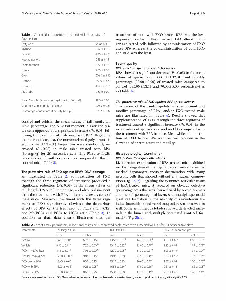
Histopathological examinationBPA histopathological alterationsLiver section examination of BPA-treated mice exhibitedmarked congestion of the hepatic blood vessels as well asmarked hepatocytes vacuolar degeneration with manynecrotic cells that showed without any nuclear compos-ition (Fig. 1b, c). Regarding the examined testicular tissueof BPA-treated mice, it revealed an obvious defectivespermatogenesis that was characterized by severe necrosisand loss of spermatogonial layers with multiple spermatidgiant cell formation in the majority of seminiferous tu-bules. Interstitial blood vessel congestion was observed aswell. Some seminiferous tubules showed destructed mate-rials in the lumen with multiple spermatid giant cell for-mation (Fig. 2b, c).
Table 1 Chemical composition and antioxidant activity offlaxseed oil
Fatty acids Value (%)
Myristic 0.47 ± 0.15
Palmitic 4.70 ± 0.65
Heptadecanoic 0.53 ± 0.15
Pentadecanoic 0.37 ± 0.15
Stearic 2.30 ± 0.26
Oleic 20.60 ± 1.49
Linoleic 26.90 ± 3.30
Linolenic 43.26 ± 5.55
Arachidic 0.87 ± 0.20
Total Phenolic Content (mg gallic acid/100 g oil) 18.0 ± 1.00
Vitamin E Concentration (μg/mL) 20.63 ± 0.31
Percentage of antioxidant activity (200 μL) 69.17 ± 0.42
Table 2 Comet assay parameters in liver and testes cells of treated male mice with BPA and/or FXO for 28 consecutive days
Treatments Tail length (μm) Tail DNA (%) Olive tail moment (μm)
Liver Testes Liver Testes Liver Testes
Control 7.66 ± 0.80e 6.73 ± 0.46e 13.53 ± 0.51d 14.26 ± 0.20d 1.03 ± 0.08d 0.98 ± 0.11e
Vehicle 8.56 ± 0.41e 7.26 ± 0.30de 13.15 ± 0.22d 15.00 ± 0.50d 1.12 ± 0.04cd 1.09 ± 0.08e
FXO (1 mL/kg bw) 8.16 ± 1.04e 7.06 ± 0.20de 12.70 ± 0.95d 14.30 ± 0.51d 1.03 ± 0.14d 1.01 ± 0.04e
BPA (50 mg/kg bw) 17.30 ± 1.08a 9.83 ± 0.15a 19.93 ± 0.58a 23.56 ± 0.45a 3.63 ± 0.52a 2.37 ± 0.05a
FXO before BPA 12.43 ± 0.45d 8.33 ± 0.15c 15.13 ± 0.23c 16.43 ± 0.35c 1.87 ± 0.04c 1.36 ± 0.02d
FXO with BPA 15.23 ± 0.55b 9.23 ± 0.25b 16.56 ± 0.64b 17.80 ± 0.26b 2.51 ± 0.16b 1.65 ± 0.05b
FXO after BPA 13.90 ± 0.26c 8.60 ± 0.26c 15.33 ± 0.30c 17.26 ± 0.49b 2.09 ± 0.40c 1.48 ± 0.01c
Data are expressed as means ± SD. Mean values in the same column within each parameter bearing superscript do not differ significantly (P ≤ 0.05)
El Makawy et al. Bulletin of the National Research Centre (2018) 42:5 Page 4 of 9

The protective role of FXO against BPA histopathologicalalterationsAdministration of FXO through the three regimens oftreatment with BPA obviously attenuated the liver andtestes histopathological alterations. The examination ofliver of FXO-treated mice for 28 successive days beforeBPA treatment revealed mild degenerative changes ofthe hepatocytes with few necrotic cells (Fig. 1d). Liversections of co-treated mice with FXO and BPA revealeda mild degree of restoration of a large number of hepaticcells with mild vacuolar degeneration and dispersed nec-rotic cells (Fig. 1e). In addition, liver histoarchitecture ofFXO-treated mice for 28 successive days after BPA treat-ment showed moderate hepatocellular degeneration andnecrosis especially in the centrilobular area (Fig. 1f ).Concerning examination of the testes tissues, mice givenFXO before BPA treatment revealed a moderate degreeof preservation of a large number of the spermatogonialcells and mild interstitial edema (Fig. 2d). In regard tothe testes, examination of FXO and BPA co-treated miceshowed marked defective spermatogenesis and presenceof extensive debris in the lumen of the seminiferous tu-bules (Fig. 2e). In addition, examination of the testes tis-sues of mice given orally FXO after BPA treatmentdisplayed congested blood vessels, nuclear pyknosis,and destruction of many spermatogonial cells withslightly active spermatogenesis in scanty seminiferoustubules (Fig. 2f ).
DiscussionFlaxseed is abundant in many nutrients, such as polyun-saturated fatty acid, protein, and lignans, and it is emi-nent by α-linolenic acid in high content, which recentlyhas been found as chiefly vital for human organism(Wang et al. 2007). The results revealed that the mainfatty acids in FXO are stearic, oleic, linoleic, linolenic,and palmitic, in addition to its own high content of vita-min E and phenolic compounds. These results were con-firmed by Kakilashvili et al. (2014) whose investigationshowed that in flaxseed oil linoleic and linolenic acidswere predominant and together constitute the majorbasis of research composition, while palmitic and ste-aric acids in less quantity. In addition, flaxseed oil isa source of many vitamins, and the main plentifulvitamins that constitute flaxseeds are tocopherols (α,β, and γ forms). Vitamin E as an antioxidant nutrientkeeps cell constituents from free radical damagingeffects that might lead to cancer development (Winterand Vitamin 2013).DNA damage is an important initial event in carcino-
gensis. DNA lesions can change in nucleotide sequence,causing mutagenesis and other cellular mechanisms(Lord and Ashworth 2012). In the current study, liverand testes comet assay and bone marrow micronucleusassay evaluated DNA damage. The alkaline comet assaywas used as a quantitative and illustrative technique todetermine DNA strand breaks (Gedik et al. 1998). Theresults of the present study indicated that BPA signifi-cantly increased the DNA damage in the tested tissues.These results are in coincidence with the findings ofTiwari et al. (2012) who found a significant elevation inthe incidence of micronuclei and chromosomal abnor-mality in bone marrow and lymphocyte DNA damage inBPA-exposed rats. In addition, Xin et al. (2015) foundsignificant increases in the content of DNA damage,frequencies of micronucleus, and conventional chromo-some aberrations in CHO cells exposed to BPA. Grow-ing evidence indicated that the oxidative stress causedby BPA could be its mechanisms to cause genetic tox-icity (Tiwari et al. 2012; Meeker et al. 2010). In addition,the induction of micronuclei could be owing to BPA
Table 3 Frequencies of MN PCEs and PCEs/ NCEs ratio in bone marrow cells BPA and/or FXO treated male mice for 28 consecutive days
Treatments MNPCEs PCEs NCEs PCEs/NCEs ratio
Control 10.40 ± 2.96d 1438.00 ± 32.95a 562.00 ± 32.95c 5.13 ± 0.43a
Vehicle 10.00 ± 4.00d 1434.40 ± 57.36 a 565.60 ± 57.36c 5.13 ± 0.75a
FXO (1 mL/kg bw) 9.20 ± 2.28d 1451.60 ± 47.01a 548.40 ± 47.01c 5.33 ± 0.65a
BPA (50 mg/kg bw) 66.40 ± 9.94a 1149.60 ± 56.80c 850.40 ± 56.80a 2.71 ± 0.31c
FXO before BPA 22.40 ± 4.56c 1363.60 ± 57.64b 636.40 ± 57.64b 4.31 ± 0.57b
FXO with BPA 34.40 ± 8.05b 1302.80 ± 54.38b 697.20 ± 54.38b 3.75 ± 0.45b
FXO after BPA 26.00 ± 7.07c 1344.40 ± 62.79b 655.60 ± 62.79b 4.14 ± 0.59b
Data are expressed as means ± SD. Mean values in the same column within each parameter bearing the same superscript do not differ significantly (P ≤ 0.05)
Table 4 Mean values of sperm count and motility in treated malemice with BPA and/or FXO for 28 consecutive days
Treatments Sperm count (106/ml) Sperm motility (%)
Control 385.00 ± 32.18ab 90.00 ± 5.00ab
Vehicle 382.00 ± 37.80ab 93.00 ± 2.64a
FXO (1 mL/kg bw) 385.00 ± 33.42ab 88.33 ± 2.88ab
BPA (50 mg/kg bw) 281.33 ± 32.01c 55.00 ± 5.00d
FXO before BPA 387.67 ± 7.63a 88.00 ± 2.64ab
FXO with BPA 340.00 ± 5.00b 79.00 ± 1.00c
FXO after BPA 365.00 ± 6.08ab 84.00 ± 1.00bc
Data are expressed as means ± SD. Mean values in the same column parameterbearing the same superscript do not differ significantly (P ≤ 0.05)
El Makawy et al. Bulletin of the National Research Centre (2018) 42:5 Page 5 of 9
aneugenic effect (Quick et al. 2008). Moreover, treat-ment with the three regimens of FXO significantly allevi-ated the deleterious effects of BPA on DNA integrity.Several studies are in agreement with these results; AbdEl-Rahim and Hafiz (2009) revealed that pretreatmentwith FXO (0.1 mL/kg bw) for 14 days prior to an IP in-jection of cyclophosphamide (25 mg/kg bw) significantlydiminished the cyclophosphamide chromosomal aberra-tions and DNA fragmentation percentage in mice. Inaddition, the protecting effect of FXO on lead acetate-in-duced DNA fragmentation in the hepatic tissue of ratswas reported by Abdel-Moneim et al. (2011). The pro-tection afforded by FXO may be due to its intrinsicantioxidant and/or free radical scavenging propertiesassociated with its constituent bioactive components asomega-3 and lignans (Burdge and Calder 2005; Newairyand Abdou 2009). FXO’s lignans exhibited strong anti-oxidant and protective effects in quenching the free
radical and inhibiting peroxyl-radical-mediated damageof plasmid DNA (Hu et al. 2007). Furthermore, thepresence of polyphenols and vitamin E in FXO mayhave contributed to its therapeutic role against BPA’sDNA damage. Polyphenols were revealed to amelioratecell injury and protect DNA from lesion induced by re-active oxygen species (ROS) owing to their capacity toscavenge free radicals (Urquiaga and Leighton 2000). Inaddition, vitamin E has antioxidant and enzymatic ac-tivities that have the ability to prevent DNA damage(Songthaveesin et al. 2004).In addition, BPA exhibited reduction in the count
and motility of sperms. These results are in coincidencewith the findings of Chitra et al. (2002) who reportedthat BPA at dose levels of 0.2–20 μg/kg bw showed adose-dependent decline in rats’ epididymal spermmotility and count. BPA is an estrogenic endocrinedisruptor, which interfere with processes related to
Fig. 1 a Liver of control mice showing normal hepatic parenchyma. b Liver of BPA-treated mice showing congestion (C) of the hepatic bloodvessels and marked vacuolar degeneration of the hepatocytes with many necrotic cells (arrow). c Liver of BPA-treated mice showing congestionof the central vein (C) and portal blood vessels (P) with marked hepatocellular vacuolar degeneration and necrosis (arrow). d Liver of FXO-treatedmice before BPA treatment showing mild degenerative changes of the hepatocytes with few necrotic cells (arrow). e Liver of FXO-treated miceconcurrently with BPA showing a mild degree of restoration of a large number of hepatic cells with mild vacuolar degeneration (short arrow) andscattered necrotic cells (long arrow). f Liver of FXO-treated mice after BPA treatment showing moderate hepatocellular vacuolation (arrow) andnecrosis especially in the centrilobular area. (H&E, × 400)
El Makawy et al. Bulletin of the National Research Centre (2018) 42:5 Page 6 of 9

spermatogenesis, such as androgen production and Sertolicell activity (Akingbemi et al. 2004; Salian et al. 2009). Inaddition, it has been reported that BPA generate ROS thatcause oxidative damage and the oxidative stress thatcauses sperm damage (Chitra et al. 2003). FXO supple-mentation through the three regimens of treatmentcaused a significant increase (P ≤ 0.05) in the mean valuesof sperm count and motility compared with the BPA treat-ment in mice. These findings are in accordance with thosereported by Abd El-Rahim and Hafiz (2009) who foundthat pretreatment with FXO for 14 days prior to an IPdose of cyclophosphamide improved the sperm count per-centage in treated mice. The increase in sperm count andmotility may be attributed to the antioxidant nature ofvitamin E present in FXO, which is believed to be theprimary component of the spermatozoa antioxidantsystem and the major membrane protectants against ROSand lipid peroxidation attack (Hsu and Guo 2002). Fur-thermore, FXO has high content of linolenic acid. Com-haire and Mahmoud (2003) reported that linolenic acid
deficiency is related with impaired sperm motility andcauses sperm abnormalities.BPA treated mice exhibited marked histopathological
alteration s in the tissue of liver and testes. These resultsare in concurrence with Korkmaz et al. (2010) whonoticed hepatic necrosis and congestion in the liver ofmale rats treated with BPA at the dose of 25 mg/kg/daythree times a week for 50 days. In addition, Kalb et al.(2016) found that BPA (3000 μg/kg bw) via breast milkcaused testicular erosion and complete aplasia in someseminiferous tubules. The observed histopathological al-terations in liver and testes tissues may be related to DNAdamage induced by BPA in these tissues. Moreover, Sangaiand Verma (2012) found that BPA caused changes in theactivities of ATPase in the liver and kidney of micethereby causing a reduction in ATP that cause necrosis.Moreover, it has reported that BPA oxidative damage as aresult of ROS generation (Kabuto et al. 2004). In addition,the great histopathological alterations in testes tissuescaused by BPA may be due to its estrogen-mimicking
Fig. 2 a Testis of control mice showing normal spermatogenesis within the seminiferous tubules (ST). b Testis of BPA-treated mice showing severenecrosis and loss of the spermatogonial layers with multiple spermtide giant cells formation (arrow) in most of the seminiferous tubules. c Testis ofBPA-treated mice showing congestion of the interstitial blood vessel, destructed and abnormal spermatid formation (thin arrow), spermatid giant cells(thick arrow), and loss of most of the spermatogonial cell layers. d Testis of FXO-treated mice before BPA treatment showing a moderate degree ofpreservation of a large number of the spermatogonial cells (arrow) and mild interstitial edema (E). e Testis of FXO-treated mice concurrently with BPAshowing marked defective spermatogenesis and presence of extensive debris (arrow) in the lumen of the seminiferous tubules. f Testis of FXO-treatedmice after BPA treatment showing congested blood vessels (C) nuclear pyknosis (P) and destruction of many spermatogonial cells with slightly activespermatogenesis in scanty seminiferous tubules (arrow). (H&E, × 400)
El Makawy et al. Bulletin of the National Research Centre (2018) 42:5 Page 7 of 9

(Korkmaz et al. 2010). Meanwhile, administration of FXOthrough the three regimens of treatment with BPA obvi-ously attenuated the liver and testes histopathological al-terations. These observations are in accord with resultsobtained by Abdel-Moneim et al. (2011) who indicatedthat treatment of rats with FXO largely preventedlead-induced liver histopathological changes as indicatedby a reduction in inflammatory cellular infiltration andhepatocytic damages. In addition, Karaca and Eraslan(2013) reported that FXO attenuated cadmium-inducedtesticular oxidative damage in male rats. The improve-ments in histopathological alterations afforded by FXOmay be due to the polyunsaturated fatty acids (PUFAs)that may reduce cellular tendency to lipid peroxidationand alter membrane fluidity (Best et al. 2003). Inaddition, the presence of oleic acid, a monounsaturatedfatty acid in FXO, reduces the susceptibility of the tes-tes to lipid peroxidation (Bourre et al. 2004). Moreover,flaxseed is a high-quality supply of dietary fiber andphytoestrogenic lignans that have antioxidant proper-ties (Zanwar et al. 2010).
ConclusionsFlaxseed has dietary and functional properties. The contentof compounds such as polyunsaturated fatty acids, essentialamino acids, vitamin E, lignans, and dietary fibers makesflaxseed a supply to satisfy essential needs in human dietand health maintenance. As mentioned above, FXO admin-istration through the three regimens of treatment with BPAsuccessfully attenuated the genotoxicity, sperm defects, andhistological alterations induced by BPA in male mice thatmay be referred to its antioxidant property. In addition, thetreatment of mice with FXO before BPA was the best regi-men in restoring bisphenol-A deleterious effects. Flaxseedantioxidant may have prospective application in the foodand health industry as a food stabilizer and nutraceutical.
FundingThe research was financed by authors.
Availability of data and materialsThe datasets used and/or analyzed during the current study are available fromthe corresponding author on reasonable request.
Authors’ contributionsAE, FE, and OE developed the concepts of the study, design, and materialpreparation and collected the literature research. AE, FE, OE, and MEanalyzed and interpreted the data and manuscript preparation. AE, FE,and OE wrote the manuscript. AE and FE revised the manuscript. Allauthors read and approved the manuscript.
Ethics approval and consent to participateNot applicable.
Consent for publicationNot applicable.
Competing interestsThe authors declare that they have no competing interests.
Publisher’s NoteSpringer Nature remains neutral with regard to jurisdictional claims in publishedmaps and institutional affiliations.
Author details1Department of Cell Biology, National Research Center, 33 El-Bohouth St.-Dokki, P.O.12622, Giza, Egypt. 2Department of Environment andBio-Agriculture, Faculty of Agriculture, Al-Azhar University, Cairo, Egypt.
Received: 9 June 2018 Accepted: 16 August 2018
ReferencesAbd El-Rahim AH, Hafiz NA (2009) Investigation on the protective effect of grape
seed and linseed oils against cyclophosphamide induced genotoxicity in mice.Global Veterinaria 3(5):377–382
Abdel-Moneim AE, Dkhil MA, Al-Quraishy S (2011) The redox status in rats treatedwith flaxseed oil and lead-induced hepatotoxicity. Biol Trace Element Res143:457–467
Akingbemi B, Sottas C, Koulova A, Klinefelter G, Hardy M (2004) Inhibition oftesticular steroidogenesis by the xenoestrogen bisphenol A is associatedwith reduced pituitary luteinizing hormone secretion and decreased steroidogenicenzyme gene expression in rat Leydig cells. Endocrinol 145:592–603
AOAC (2000) Association of Official Analytical Chemists. Official methods of analysis.17thed. Gaithersburg, M.D. In: U.S.A
Attaman JA, Toth TL, Furtado J, Campos H, Hauser R et al (2012) Dietary fat andsemen quality among men attending a fertility clinic. Hum Reprod 27:1466–1474
Bandyopadhyaya G, Sinha S, Chattopadhyay BD, Chakraborty A (2008) Protectiverole of curcumin against nicotine-induced genotoxicity on rat liver underrestricted dietary protein. Euro J Pharmacol 588:151–157
Barcelo-Coblijn G, Murphy EJ (2009) Alpha-linolenic acid and its conversion to longerchain n-3 fatty acids: benefits for human health and a role in maintaining tissuen-3 fatty acid levels. Prog in Lipid Res 48:355–374
Best CA, Cluette-Brown JE, Teruya M, Teruya A, Laposata M (2003) Red blood cellfatty acid ethyl esters: a significant component of fatty acid ethyl esters in theblood. J Lipid Res 44:612–620
Bourre JM, Dumont O, Durand G (2004) Dose-effect of dietary oleic acid: oleic acid isconditionally essential for some organs. Reprod Nutr Dev 44(4):371–380
Burdge GC, Calder PC (2005) Conversion of alpha-linolenic acid to longer-chainpolyunsaturated fatty acids in human adults. Reprod Nutr Dev 45:581–589
Chauhan LK, Pant N, Gupta SK, Srivastava SP (2000) Induction of chromosomeaberrations, micronucleus formation and sperm abnormalities in mousefollowing carbofuran exposure. Mut Res 465:123–129
Chitra KC, Latchoumycandane C, Mathur PP (2002) Effect of nonylphenol on theantioxidant system in epididymal sperm of rats. Arch Toxicol 76:545–551
Chitra KC, Latchoumycandane C, Mathur PP (2003) Induction of oxidative stressby bisphenol A in the epididymal sperm of rats. Toxicol 185:119–127
Comhaire FH, Mahmoud A (2003) The role of food supplements in the treatmentof the infertile man. Reprod BioMed Online 7:385–391
Delafield F (1984) Haematoxylin and eosin for general staining. Staining of the animaltissues practical and theoretical. Oxford University Press, London
Fu P, Kawamura K (2010) Ubiquity of bisphenol A in the atmosphere. Environ Poll158:3138–3143
Gedik CM, Wood SG, Collins AR (1998) Measuring oxidative damage to DNA: HPLCand the comet assay compared. Free Radic Res 29:609–615
Gimeno E, Castellote AI, Lamuela-Raventos RM, Torre MC, Lopez-Sabater MC(2000) Rapid determination of vitamin E in vegetable oils by reversedphase high-performance liquid chromatography. J Chromatography A881:251–254
Hana RS, Saed N (2013) Alteration in oxidants, antioxidants and cytokines levelsin blood of malathion exposed human and animal groups and the effect offlaxseed oil in alleviating malathion toxic effects. Euro J Biotechnol Biosci 1(1):8–19
Hsu PC, Guo YL (2002) Antioxidant nutrients and lead toxicity. Toxicol 180:33–44Hu C, Yuan YV, Kitts DD (2007) Antioxidant activities of the flaxseed lignan
secoisolariciresinol diglucoside, its aglycone secoisolariciresinol and themammalian lignans enterodiol and enterolactone in vitro. Food ChemToxicol 45(11):2219–2227
Huang Y, Wong C, Zheng J, Bouwman H, Barra R, Wahlstrom B, Neretin L, HongM (2012) Bisphenol A (BPA) in China: a review of sources, environmental levels,and potential human impacts. Environ Int 42:91–99
El Makawy et al. Bulletin of the National Research Centre (2018) 42:5 Page 8 of 9

Kabuto H, Amakawa M, Shishibori T (2004) Exposure to bisphenol A duringembryonic/fetal life and infancy increases oxidative injury and causesunderdevelopment of the brain and testis in mice. Life Sci 74:2931–2940
Kaithwas G, Majumdar DK (2012) In vitro antioxidant and in vivo antidiabetic,antihyperlipidemic activity of linseed oil against streptozotocin-inducedtoxicity in albino rats. Eur J Lipid Sci Tech 114:12–45
Kakilashvili BI, Zurabashvili DZ, Turabelidze DG, Shanidze LA, Parulava GK (2014)The fatty acid composition of ordinary flax seed oil (Linum usitatissimum L.)cultivated in Georgia and its byological activity. Georgian Med News 227:86–88
Kalb AC, Kalb AL, Cardoso FT, Fernandes CG, Corcini CD, Junior ASV, Martine PE(2016) Maternal transfer of bisphenol a during nursing causes spermimpairment in male offspring. Arch Environ Contam Toxicol 70:793–801
Kang HS, KwonYJ LKJ, Seo RY (2013) Recent advances in in vivo genotoxicitytesting: prediction of carcinogenic potential using comet and micronucleusassay in animal models. J Cancer Prevent 18(4):277–288
Karaca S, Eraslan G (2013) The effects of flaxseed oil on cadmium-induced oxidativestress in rats. Biol Trace Elem Res 155:423–430
Korkmaz A, Ahbab MA, Kolankaya D, Barlas N (2010) Influence of vitamin C onbisphenol A, nonylphenol and octylphenol induced oxidative damages inliver of male rats. Food Chem Toxicol 48(10):2865–2871
Kvist U, Bjorndahl L (2002) Manual on basic semen analysis. In: ESHRE Monographs,vol 2. Oxford University Press, Oxford
Lin JY, Tang CY (2007) Determination of total phenolic and flavonoid contents inselected fruits and vegetables, as well as their stimulatory effects on mousesplenocyte proliferation. Food Chem 101:140–147
Lin X, Gingrich JR, Bao W, Li J, Haroon ZA, Demark-Wahnefried W (2002) Effect offlaxseed supplementation on prostatic carcinoma in transgenic mice. Urol 60:919–924
Lord CJ, Ashworth A (2012) The DNA damage response and cancer therapy. Nature481:287–294
Makris KC, Andra SS, Jia A, Herrick L, Christophi CA, Snyder SA, Hauser R (2014)Association between water consumption from polycarbonate containers andbisphenol A intake during harsh environmental conditions in summer. Environ SciTechnol 47:3333–3343
Meeker J, Calafat A, Hauser R (2010) Urinary bisphenol A concentration in relationto serum thyroid and reproductive hormones in men from an infertilityclinic. Environ Sci Technol 44:1458–1463
Michalowicz J (2014) Bisphenol A-sources, toxicity and biotransformation. EnvironToxicol Pharmacol 37:738–758
Narayana K, D’Souza UJA, Rao KPS (2002) Ribavirin induced sperm shape abnormalitiesin Wistar rat. Mut Res 513:193–196
Newairy AS, Abdou HM (2009) Protective role of flax lignans against lead acetateinduced oxidative damage and hyperlipidemia in rats. Food ChemToxicol 47(4):813–818
OECD (1997) Test No. 474: Mammalian Erythrocyte Micronucleus Test.OECD Publishing, Paris
Olsen J, Ramlau-Hansen HC (2012) Dietary fats may impact semen quantity andquality. Asian J Androl 14(4):511–512
Quick EL, Parry EM, Parry JM (2008) Do oestrogens induce chromosome specificaneuploidy in vitro, similar to the pattern of aneuploidy seen in breastcancer. Mut Res 651:46–55
Rocha S, Domingues V, Pinho C, Fernandes V, Delerue-Matos C, Gameiro P, MansilhaC (2013) Occurrence of bisphenol A,estrone, 17β-estradiol and 17α-ethinylestradiol in Portugalese rivers. Bulletin Environ Contam Toxicol 90:73–78
Safarinejad MR (2011) Effect of omega-3 polyunsaturated fatty acid supplementationon semen profile and enzymatic anti-oxidant capacity of seminal plasma ininfertile men with idiopathic oligoasthenoteratospermia: a double blind,placebo-controlled, randomized study. Andrologia 43:38–47
Salian S, Doshi T, Vanage G (2009) Neonatal exposure of male rats to bisphenol Aimpairs fertility and expression of sertoli cell junctional proteins in the testis.Toxicology 265:56–67
Sangai NP, Verma RJ (2012) Effect of quercetin on bisphenol A-caused alterationsin succinate dehydrogenase and adenosine triphosphatase activities in liverand kidney of mice. Acta Poloniae Pharmaceutica-Drug Res 69(6):1189–1194
Sargi CS, Silva CB, Santos CMH, Montanher FP, Boeing SJ, Junior SOO, Souza EN,Visentainer VJ (2013) Antioxidant capacity and chemical composition inseeds rich in omega-3: chia, flax, and perilla. Food Sci Technol Campinas33(3):541–548
Singh RP, Murthy KN, Jayaprakasha GK (2002) Studies on the antioxidant activityof pomegranate (Punica granatum) peel and seed extracts using in vitromodels. J Agric Food Chem 50:81–86
Songthaveesin C, Saikhun J, Kitiyananta Y, Pavasuthipaisit K (2004) Radio protectiveeffect of vitamin E on spermatogenesis in mice exposed to gamma-irradiation:a flow cytometric study. Asian J Androl 6:331–336
Tiwari D, Kamble J, Chilgunde S, Patil P, Maru G, Kawle D, Bhartiya U, Joseph L,Vanage G (2012) Clastogenic and mutagenic effects of bisphenol A: anendocrine disruptor. Mut Res 743:83–90
Tyl RW, Myers CB, Marr MC, Sloan CS, Castillo NP, Veselica MM, Seely JC, DimondSS, Van Miller JP, Shiotsuka RN, Beyer D, Hentges SG, Waechter JM Jr (2008)Two-generation reproductive toxicity study of dietary bisphenol A in CD-1(Swiss) mice. Toxicol Sci 104(2):362–384
Urquiaga I, Leighton F (2000) Plant polyphenol antioxidants and oxidative stress.Biol Res 33:55–64
Vandenberg LN, Hauser R, Marcus M, Olea N, Welshons WV (2007) Humanexposure to bisphenol A (BPA). Reprod Toxicol 24:139–177
Wang B, Li D, Wang L, Huang Z, Zhang L, Chen XD, Mao Z (2007) Effect ofmoisture content on the physical properties of fibered flaxseed. Int J FoodEng 3(5):1–11
Winter R. Vitamin E: Your protection against exercise fatigue, weakenedimmunity, heart disease, cancer, aging, diabetic damage, environmentaltoxins. Crown Publishing Group, 2013
Xin L, Lin Y, Wang A, Zhu W, Liang Y, Su X, Hong C, Wan J, Wang Y, Tian H(2015) Cytogenetic evaluation for the genotoxicity of bisphenol-A in Chinesehamster ovary cells. Environ Toxicol Pharmacol 40:524–529
Yi B, Kasai H, Lee HS, Kang Y, Park JY, Yang M (2011) Inhibition by wheat sprout(Triticum aestivum) juice of bisphenol A-induced oxidative stress in youngwomen. Mut Res/Genet Toxicol Environ Mutagen 724:64–68
Zanwar AA, Hegde MV, Bodhankar SL (2010) In vitro antioxidant activity of ethanolicextract of Linum usitatissimum. Pharmacol Online 1:683–696
El Makawy et al. Bulletin of the National Research Centre (2018) 42:5 Page 9 of 9

![Bisphenol A Diglycidyl Ether of Bisphenol A Method · PDF file4 of 18 Diglycidyl Ether of Bisphenol A13 synonyms: 2,2-bis[4-(glycidyloxy)phenyl]propane, 4,4′-isopropylidenediphenol](https://static.fdocuments.us/doc/165x107/5a76e9947f8b9a93088d7abf/bisphenol-a-diglycidyl-ether-of-bisphenol-a-method-4-of-18-diglycidyl-ether.jpg)