Flagella and cilia: Motility at low Reynolds...
14
University of Ljubljana Faculty of Mathematics and Physics Department of Physics Biophysics First seminar at subject Seminar 1, first year of second cycle degrees Flagella and cilia: Motility at low Reynolds numbers Author: Nina Lopič Mentor: dr. Mojca Vilfan Ljubljana, January 2012 Abstract In this seminar I’m presenting the movement of micro-organisms with flagella and cilia. They swim in a world of very low Reynolds numbers. As a consequence, flagella and cilia need to move in a nonreciprocal way in order to generate fluid flow. The swimming of these small organisms is explained by the structure and function of cilia and flagella. At the end I mention artificial cilia, which can be used in microfluidics.
Transcript of Flagella and cilia: Motility at low Reynolds...

University of Ljubljana Faculty of Mathematics and Physics
Department of Physics Biophysics
First seminar at subject Seminar 1, first year of second cycle degrees
Flagella and cilia: Motility at low Reynolds numbers
Author: Nina Lopič
Mentor: dr. Mojca Vilfan Ljubljana, January 2012
Abstract
In this seminar I’m presenting the movement of micro-organisms with flagella and cilia. They swim in a world of very low Reynolds numbers. As a consequence, flagella and cilia need to move in a nonreciprocal way in order to generate fluid flow. The swimming of these small organisms is explained by the structure and function of cilia and flagella. At the end I mention artificial cilia, which can be used in microfluidics.

FLAGELLA AND CILIA: MOTILITY AT LOW REYNOLDS NUMBER
2
Contents
Abstract ................................................................................................................................................... 1
Contents .................................................................................................................................................. 2
1. INTRODUCTION ............................................................................................................................... 2
2. EXAMPLES OF FLAGELLA AND CILIA ................................................................................................ 2
2.1. Micro-organisms ...................................................................................................................... 3
2.2. Human body ............................................................................................................................ 3
3. REYNOLDS NUMBER ........................................................................................................................ 4
4. MOTILITY OF FLAGELLA AND CILIA .................................................................................................. 5
4.1. The prokaryotic flagella ........................................................................................................... 6
4.1.1. Rotary motor and proton flow ........................................................................................ 6
4.2. The eukaryotic flagella and cilia .............................................................................................. 7
4.2.1. Microtubules and molecular motor dynein .................................................................... 8
5. COMPARISONS OF MOVEMENT .................................................................................................... 10
6. ARTIFICIAL CILIA............................................................................................................................. 11
6.1. Formation of cilia ................................................................................................................... 11
6.2. Pumping of fluid .................................................................................................................... 11
7. CONCLUSION ................................................................................................................................. 13
References ............................................................................................................................................. 14
1. INTRODUCTION
Nature has devised many different ways of creating fluid flow that is required for animal propulsion such as flying or swimming. Examples are flapping wings of birds or waving fins of fish. Going smaller, flapping wings are also found in insects. At very small scale, typically for sub-mm sizes, a fluid flow manipulation mechanism used by nature is by flagella or cilia. In this seminar I am presenting a world, which we almost never think about. This is the world of very low Reynolds numbers – a world that is inhabited by the overwhelming majority of the micro-organisms. This world is quite different from the one that we have developed our intuitions in. I will present how microorganisms swim and generate fluid flow using flagella or cilia.
2. EXAMPLES OF FLAGELLA AND CILIA
Flagella and cilia can be viewed as small hair. Flagella are rather long, their typical length is between 20
and 100 m, while cilia are shorter, their length is typically between 2 and 15 m. Their most important
FLAGELLA AND CILIA: MOTILITY AT LOW REYNOLDS NUMBER
3
function is generation of fluid flow for movement or for bringing food. Some of the cilia also move cargo, others serve as sensors.
2.1. Micro-organisms
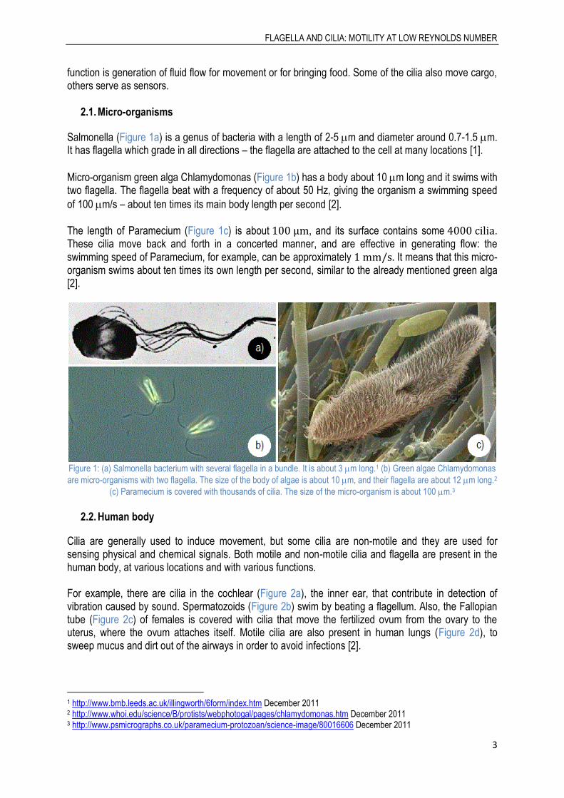
Salmonella (Figure 1a) is a genus of bacteria with a length of 2-5 m and diameter around 0.7-1.5 m. It has flagella which grade in all directions – the flagella are attached to the cell at many locations [1].
Micro-organism green alga Chlamydomonas (Figure 1b) has a body about 10 m long and it swims with two flagella. The flagella beat with a frequency of about 50 Hz, giving the organism a swimming speed
of 100 m/s – about ten times its main body length per second [2].
The length of Paramecium (Figure 1c) is about , and its surface contains some . These cilia move back and forth in a concerted manner, and are effective in generating flow: the swimming speed of Paramecium, for example, can be approximately It means that this micro-organism swims about ten times its own length per second, similar to the already mentioned green alga [2].
Figure 1: (a) Salmonella bacterium with several flagella in a bundle. It is about 3 m long.1 (b) Green algae Chlamydomonas
are micro-organisms with two flagella. The size of the body of algae is about 10 m, and their flagella are about 12 m long.2
(c) Paramecium is covered with thousands of cilia. The size of the micro-organism is about 100 m.3
2.2. Human body
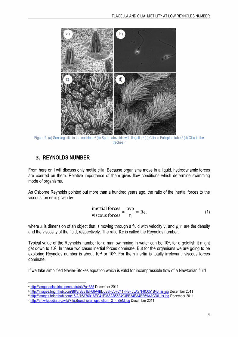
Cilia are generally used to induce movement, but some cilia are non-motile and they are used for sensing physical and chemical signals. Both motile and non-motile cilia and flagella are present in the human body, at various locations and with various functions. For example, there are cilia in the cochlear (Figure 2a), the inner ear, that contribute in detection of vibration caused by sound. Spermatozoids (Figure 2b) swim by beating a flagellum. Also, the Fallopian tube (Figure 2c) of females is covered with cilia that move the fertilized ovum from the ovary to the uterus, where the ovum attaches itself. Motile cilia are also present in human lungs (Figure 2d), to sweep mucus and dirt out of the airways in order to avoid infections [2].
1 http://www.bmb.leeds.ac.uk/illingworth/6form/index.htm December 2011 2 http://www.whoi.edu/science/B/protists/webphotogal/pages/chlamydomonas.htm December 2011 3 http://www.psmicrographs.co.uk/paramecium-protozoan/science-image/80016606 December 2011
FLAGELLA AND CILIA: MOTILITY AT LOW REYNOLDS NUMBER
4
Figure 2: (a) Sensing cilia in the cochlear.4 (b) Spermatozoids with flagella.5 (c) Cilia in Fallopian tube.6 (d) Cilia in the
trachea.7
3. REYNOLDS NUMBER
From here on I will discuss only motile cilia. Because organisms move in a liquid, hydrodynamic forces are exerted on them. Relative importance of them gives flow conditions which determine swimming mode of organisms. As Osborne Reynolds pointed out more than a hundred years ago, the ratio of the inertial forces to the viscous forces is given by
(1)
where is dimension of an object that is moving through a fluid with velocity , and are the density and the viscosity of the fluid, respectively. The ratio is called the Reynolds number. Typical value of the Reynolds number for a man swimming in water can be 104, for a goldfish it might get down to 102. In these two cases inertial forces dominate. But for the organisms we are going to be exploring Reynolds number is about 10-4 or 10-5. For them inertia is totally irrelevant, viscous forces dominate. If we take simplified Navier-Stokes equation which is valid for incompressible flow of a Newtonian fluid
4 http://languagelog.ldc.upenn.edu/nll/?p=555 December 2011 5 http://images.brighthub.com/B8/8/B881EF68A4BD5B8FC07C41FFBF55A97F8C051B43_lis.jpg December 2011 6 http://images.brighthub.com/15/A/15A7601AEC41F368AB56F493BB34EA4BF69AACD0_lis.jpg December 2011 7 http://en.wikipedia.org/wiki/File:Bronchiolar_epithelium_3_-_SEM.jpg December 2011

FLAGELLA AND CILIA: MOTILITY AT LOW REYNOLDS NUMBER
5
(
( ) ) (2)
and if we neglect the inertia terms, we obtain
(3)
where is the pressure. From equation (3), two characteristics of movement follow: How microorganisms are moving is entirely determined by the forces that are exerted on them at a
given moment, and not by the past. They move around with a typical speed 30 m/s. If we push that organism to move it, and suddenly we stop pushing, it drifts about 0.01 nm before it slows down. This is a characteristic of low Reynolds numbers, where inertia plays no significant role [3]. The other characteristic is the requirement for non-reciprocal motion. It means that in order to move, the organism has to change its body to a certain shape and return to the original shape by going through a different sequence. This is because at low Reynolds numbers, everything reverses. Time makes no difference – only configuration. If we change quickly or slowly, the pattern of motion is exactly the same. If the animal tries to swim by a reciprocal motion, it does not move. Fast or slow, it exactly retraces its trajectory and it's back where it started [3]. An example of reciprocal motion from nature is a scallop. A scallop opens its shell slowly and closes it fast, squirting out water. It only has one hinge, and with only one degree of freedom in configuration space, the motion is bound to be reciprocal. So, the scallop at a low Reynolds number cannot swim. Another example of time reversal at low Reynolds numbers is an experiment with dyes and glycerol:
Figure 3: Experiment with dyes in glycerol.8 (a) We put yellow, blue and red dyes in glycerol. (b) We turn transparent cylinder four times. Dyes mix. (c) We turn the cylinder four times in another direction as the first time. Colours are in the same order
as at the beginning.
4. MOTILITY OF FLAGELLA AND CILIA Organisms with flagella and cilia solve the problem of motility at low Reynolds numbers with nonreciprocal movement. Motility of prokaryotic and eukaryotic organisms is different. Eukaryotic flagella and cilia have the same internal structure, while structure of prokaryotic flagella is different.
8 http://www.youtube.com/watch?feature=endscreen&v=_dbnH-BBSNo&NR=1 December 2011

FLAGELLA AND CILIA: MOTILITY AT LOW REYNOLDS NUMBER
6
4.1. The prokaryotic flagella
Many species of bacteria move using long flagella that rotate, similar to a propeller on a motor boat. The flagella spin at more than 100 rotations per second [1]. A bacterium can have just one flagellum, or several. In the latter case, the flagella are bundled together. Bacterial flagella can be unidirectional or reversible. If the bundle of flagella is rotating counter-clockwise, propelling the bacterium forward – the cell swims in a smooth line called a “run”. Reversing the direction of the rotation, the bundle unravels and the bacterium “tumbles” and turns. Rotating the flagella again counter-clockwise, the bacterium propels itself in a new direction. By controlling rotation of flagella, bacteria can move toward optimal regions of their environment.
Figure 4: If the flagellum rotates counter clockwise, the cell swims in a smooth line called a "run". During "tumbles" the
flagellum reverses direction and the cell stops and changes its course.9
4.1.1. Rotary motor and proton flow
Each prokaryotic flagellum is comprised of three units: the basal body, the hook and the filament.
Figure 5: A model of the flagellar motor. [1]
9 http://www.pc.maricopa.edu/Biology/rcotter/BIO%20205/LessonBuilders/Chapter%204%20LB/Ch4Lessonbuilder4.html December 2011
FLAGELLA AND CILIA: MOTILITY AT LOW REYNOLDS NUMBER
7
The filament of bacterial flagella is composed of subunits of a protein flagellin, and is helically shaped. The base of the flagellum is different in structure from that of the filament. There is a wider region at the base of the flagellum called the hook. The hook consists of a single type of protein and connects the filament to the motor portion of the flagellum [1]. In the basal body, the motor is anchored in the cytoplasmic membrane and cell wall. The motor consists of the rotor (C, MS, P rings) and the stator (Mot proteins). Proton movement across the cytoplasmic membrane through the stator drives rotation of the flagellum. The rotation by the molecular motor can be explained by a proton turbine model. In this model, protons flowing through channels in the stator exert electrostatic forces on helically arranged charges on the rotor proteins. Attractions between positive and negative charges generate torque. It causes the basal body to rotate as protons flow through the stator. Special proteins function as the motor switch, reversing the direction of rotation of the flagella in response to intracellular signals. The energy required for rotation of the flagellum comes from the proton motive force ( ) or sodium-
motive force ( ) in motors driven by H+ and Na+, respectively. Forces come from transmembrane ion fluxes that are driven by an electrochemical gradient. The ion-motive force consists of two components, and may be written as [4]
(
) (4)
where is the transmembrane voltage, and are the concentrations of the ions inside and outside the cell respectively, is the thermal energy and the charge of the ion. The electrical component is the voltage, ; the chemical component is due to different ion concentrations on either side of the membrane and is represented by the second term on the right-hand side of the equation. The chemical and electrical components of are equivalent in terms of torque generation [4].
4.2. The eukaryotic flagella and cilia
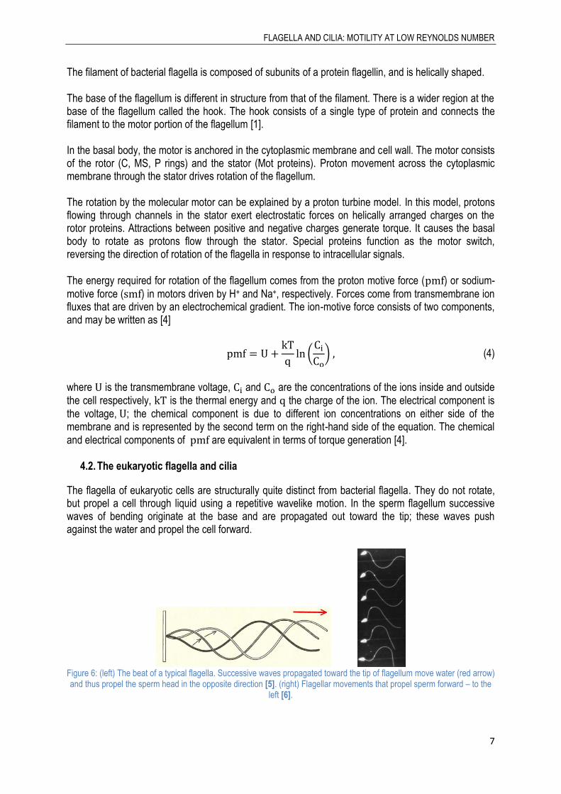
The flagella of eukaryotic cells are structurally quite distinct from bacterial flagella. They do not rotate, but propel a cell through liquid using a repetitive wavelike motion. In the sperm flagellum successive waves of bending originate at the base and are propagated out toward the tip; these waves push against the water and propel the cell forward.
Figure 6: (left) The beat of a typical flagella. Successive waves propagated toward the tip of flagellum move water (red arrow) and thus propel the sperm head in the opposite direction [5]. (right) Flagellar movements that propel sperm forward – to the
left [6].
FLAGELLA AND CILIA: MOTILITY AT LOW REYNOLDS NUMBER
8
Beating of cilia and flagella occurs in two stages, called the effective stroke (Figure 7a-c) and the recovery stroke (Figure 7d-h). The effective stroke pulls the organism through the water. During the recovery stroke, a different wave of bending moves outward, from the bases of the flagella, pushing the flagella along the surface of the cell, until they reach the position to initiate another effective stroke. Beating commonly occurs 5-10 times per second [6].
Figure 7: Flagellar movements that propel Chlamydomonas forward - up [6].
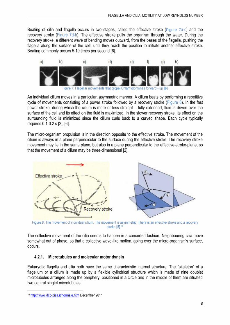
An individual cilium moves in a particular, asymmetric manner. A cilium beats by performing a repetitive cycle of movements consisting of a power stroke followed by a recovery stroke (Figure 8). In the fast power stroke, during which the cilium is more or less straight – fully extended, fluid is driven over the surface of the cell and its effect on the fluid is maximized. In the slower recovery stroke, its effect on the surrounding fluid is minimized since the cilium curls back to a curved shape. Each cycle typically requires 0.1-0.2 s [2], [6]. The micro-organism propulsion is in the direction opposite to the effective stroke. The movement of the cilium is always in a plane perpendicular to the surface during the effective stroke. The recovery stroke movement may lie in the same plane, but also in a plane perpendicular to the effective-stroke-plane, so that the movement of a cilium may be three-dimensional [2].
Figure 8: The movement of individual cilium. The movement is asymmetric. There is an effective stroke and a recovery
stroke [5].10
The collective movement of the cilia seems to happen in a concerted fashion. Neighbouring cilia move somewhat out of phase, so that a collective wave-like motion, going over the micro-organism's surface, occurs.
4.2.1. Microtubules and molecular motor dynein
Eukaryotic flagella and cilia both have the same characteristic internal structure. The “skeleton” of a flagellum or a cilium is made up by a flexible cylindrical structure which is made of nine doublet microtubules arranged along the periphery, positioned in a circle and in the middle of them are situated two central singlet microtubules.
10 http://www.dcp-pisa.it/normale.htm December 2011
FLAGELLA AND CILIA: MOTILITY AT LOW REYNOLDS NUMBER
9
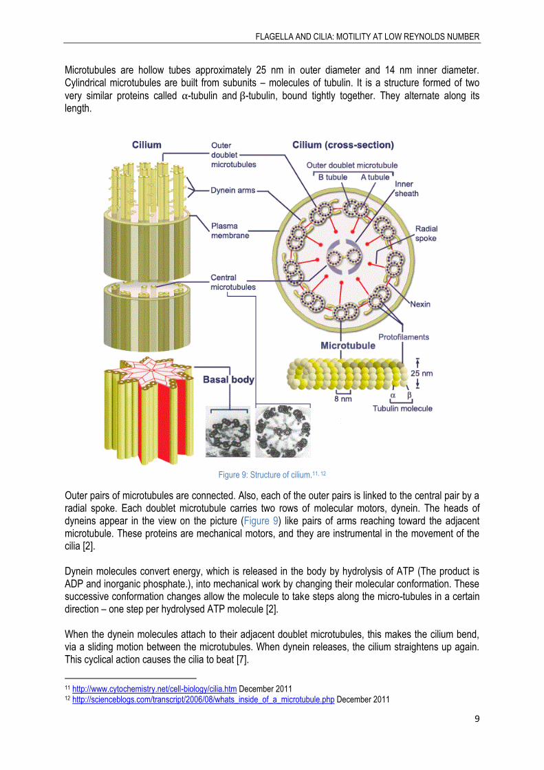
Microtubules are hollow tubes approximately 25 nm in outer diameter and 14 nm inner diameter. Cylindrical microtubules are built from subunits – molecules of tubulin. It is a structure formed of two
very similar proteins called -tubulin and -tubulin, bound tightly together. They alternate along its length.
Figure 9: Structure of cilium.11, 12
Outer pairs of microtubules are connected. Also, each of the outer pairs is linked to the central pair by a radial spoke. Each doublet microtubule carries two rows of molecular motors, dynein. The heads of dyneins appear in the view on the picture (Figure 9) like pairs of arms reaching toward the adjacent microtubule. These proteins are mechanical motors, and they are instrumental in the movement of the cilia [2]. Dynein molecules convert energy, which is released in the body by hydrolysis of ATP (The product is ADP and inorganic phosphate.), into mechanical work by changing their molecular conformation. These successive conformation changes allow the molecule to take steps along the micro-tubules in a certain direction – one step per hydrolysed ATP molecule [2]. When the dynein molecules attach to their adjacent doublet microtubules, this makes the cilium bend, via a sliding motion between the microtubules. When dynein releases, the cilium straightens up again. This cyclical action causes the cilia to beat [7].
11 http://www.cytochemistry.net/cell-biology/cilia.htm December 2011 12 http://scienceblogs.com/transcript/2006/08/whats_inside_of_a_microtubule.php December 2011

FLAGELLA AND CILIA: MOTILITY AT LOW REYNOLDS NUMBER
10
Figure 10: (a) In isolated doublet microtubules dynein produces microtubule sliding. (b) In normal flagellum dynein causes
microtubule bending [8]. (c) Sliding of microtubules causes bending.13
Flagellum bend when dyneins on one side are active (red) (Figure 11) while those on the other side are inactive (yellow). Dyneins on the active side ‘walk into the page’. On the inactive side, dyneins move passively in the opposite direction. To propagate a beat down the flagellum, activity states of the dyneins on the two sides must switch in a spatially and temporally controlled manner [9].
Figure 11: Cross section of a sperm tail [9].
The details of the control of beating of cilia are still not completely understood and are a topic of on-going research. It is believed to be a combination of the properties of the molecular motors, their interaction with the elastic properties of all microtubules, as well as the hydrodynamic coupling through the fluid, that determines the cilia movement: their asymmetry in motion, their frequency, the direction of beating, and the wave characteristics [2].
5. COMPARISONS OF MOVEMENT Comparison between motility with flagella or cilia and swimming of higher organisms: Flagella rotation can move bacteria through liquid media at speeds of up to 60 cell lengths/s. Although this is only about 0.00017 km/h, when comparing this speed with that of higher organisms in terms of
13 http://web.virginia.edu/Heidi/chapter17/chp17.htm December 2011

FLAGELLA AND CILIA: MOTILITY AT LOW REYNOLDS NUMBER
11
the number of lengths moved per second, it is extremely fast. A human Olympic swimming champion swims at a speed of about one times his body length per second [2], [1]. If a man would swim at the same conditions as his own sperm, with the same Reynolds number, he could be in a swimming pool which is full of molasses, and he should not move any part of his body faster than 1 cm/s [3].
6. ARTIFICIAL CILIA The mechanisms of flow generation based on cilia have recently attracted considerable attention. Not only because the cilia are of fundamental importance in biology and physiology but also for practical microfluidic applications [10]. A group of Slovenian researchers14 tries to mimic a ciliated surface and imitates its motion to generate fluid flow.
6.1. Formation of cilia
The artificial cilia were constructed with spherical superparamagnetic micro particles. They were assembled in two different ways: the first was to individually trap the beads by optical tweezers and arrange them into a chain (Figure 12a), whereas the second was to let the beads self-assemble that was parallel to the direction of the field (Figure 12b). External magnetic field (Figure 13a) was used to induce magnetic dipole moments in the beads. Attractive forces between the magnetic dipoles resulted in the formation of stable, long colloidal chains.
Figure 12: (a) Superparamagnetic beads were individually trapped by tweezers and assembled into chains. (b) The beads
self-assembled into chains in an external magnetic field [11].
6.2. Pumping of fluid
Once the colloidal chains were formed, their orientation followed the direction of the external magnetic field. By changing the magnetic field, researchers were able to drive the cilia in a nonreciprocal asymmetric manner. The simplest yet effective example of asymmetric nonreciprocal motion is a tilted conical path.
14
IJS, LPKF, FMF/UL: Mojca Vilfan, Anton Potočnik, Blaž Kavčič, Natan Osterman, Igor Poberaj, Andrej Vilfan, Dušan Babič, Gašper Kokot.

FLAGELLA AND CILIA: MOTILITY AT LOW REYNOLDS NUMBER
12
Figure 13: (a) A model of the experiment. (b) The cilium was rotated along a tilted conical path with the angular frequency
[11].
Theoretical calculations of the motion based on the resistive force approximation show that the pumping velocity is proportional to:
(5)
with being the length of cilium and the angular velocity of rotation, yielding the maximum velocity for and [11]. By varying angles, researchers were able to change the pumping performance of the artificial cilia.
An important parameter in the pumping performance of the ciliated surface is the cone tilt angle . Consistent with the theoretical predictions, the net pumping velocity was zero if there was no tilt, as in this case no asymmetry was introduced into the system.
Figure 14: (a) Visualization of the fluid flow generated by the artificial cillia. There are paths of four tracer particles. (b)
Schematic view of the numerical simulation that was done for exactly the same configuration as in the experiment and for the same parameter values. Tracer particles were randomly distributed through the sample. The arrow denotes the direction of
the external magnetic field [11].
Artificial cilia that are controlled by external fields can be used in microfluidic devices as micro-scale pumps and as mixers. Several different techniques have been proposed so far for manufacturing artificial cilia with different degrees of success regarding the generation of fluid flow [10]. Researchers made also numerical model of the system of artificial cilia, assumed exactly the same conditions as the experiment. Simulation successfully reproduced the experimentally obtained data.

FLAGELLA AND CILIA: MOTILITY AT LOW REYNOLDS NUMBER
13
Figure 15: Simulation of metachronal waves.15
7. CONCLUSION
Micro-organisms have devised different ways to successfully move at low Reynolds numbers. The motility of prokaryotic flagella is different from motility of eukaryotic flagella or cilia. Prokaryotic organism rotates its flagellum, similar to a propeller on a motor boat, while eukaryotic organisms create fluid flow by a complex beating pattern of flagella or cilia. Compared to human swimming, micro-organisms with flagella or cilia move many times faster comparing their lengths per second. Nowadays researchers mimic mechanisms from nature to create artificial cilia that can be used in microfluidic devices as micro-scale pumps and as mixers.
15 http://svizec.ijs.si/avilfan/metachron.gif January 2012
FLAGELLA AND CILIA: MOTILITY AT LOW REYNOLDS NUMBER
14
References
[1] Michael T. Madigan, John M. Martinko, Biology of microorganisms, Eleventh Edition, Southern
Illinois, 2006.
[2] “Cilia in nature,” August 2007. [Online]. Available: http://www.hitech-
projects.com/euprojects/artic/index/Cilia%20in%20nature.pdf. [Accessed December 2011].
[3] E. M. Purcell, “Life at Low Reynolds Number,” American Journal of Physics, vol. 45, pp. 3-11, June
1977.
[4] “Molecular Motors, Chapter 17,” [Online]. Available:
http://web.virginia.edu/Heidi/chapter17/chp17.htm#boxpage554. [Accessed December 2011].
[5] P. Satir, “How Cilia Move,” Scientific American, pp. 45-52, October 1974.
[6] Harvey Lodish, Arnold Berk, others, Molecular Cell Biology, Sixth Edition, New York, 2007.
[7] “The Histology Guide - Epithelia: Specialisations - Cilia,” Faculty of Biological Sciences, University
of Leeds, [Online]. Available:
http://histology.leeds.ac.uk/tissue_types/epithelia/epi_specialisations.php. [Accessed
December 2011].
[8] Bruce Alberts, Dennis Bray, Karen Hopkin, others, Essential Cell Biology, Second Edition, New
York and London: Garland Science, Taylor & Francis Group, 2004.
[9] T. J. Mitchison, H. M. Mitchison, “Nature, International weekly journal of science, Cell Biology:
How cilia beat,” 20 January 2010. [Online]. Available:
http://www.nature.com/nature/journal/v463/n7279/fig_tab/463308a_F1.html. [Accessed
January 2012].
[10] Gašper Kokot, Mojca Vilfan, Ntan Osterman, Andrej Vilfan, Blaž Kavčič, Igor Poberaj, Dušan
Babič, “Measurement of fluid flow generated by artificial cilia,” Biomicrofluidics, 2011.
[11] Mojca Vilfan, Anton Potočnik, Blaž Kavčič, Natan Osterman, Igor Poberaj, Andrej Vilfan, Dušan
Babič, “Self-assembled artificial cilia,” PNAS, 2010.
[12] “Salmonella,” [Online]. Available: http://en.wikipedia.org/wiki/Salmonella. [Accessed December
2011].
[13] M. Schliwa, Molecular Motors, Weinheim, 2003.
[14] Jeremy M. Berg, John L. Tymoczko, Lubert Stryer, Biochemistry, Sixth Edition, New York: W. H.
Freeman and Company, 2006.