Fit-for-purpose based testing and validation of antibodies ...
24
Vol.:(0123456789) 1 3 Histochemistry and Cell Biology (2021) 156:479–502 https://doi.org/10.1007/s00418-021-02025-5 ORIGINAL PAPER Fit‑for‑purpose based testing and validation of antibodies to amino‑ and carboxy‑terminal domains of cannabinoid receptor 1 Leyre Echeazarra 1,9 · Gontzal García del Caño 2,8 · Sergio Barrondo 3,7,8 · Imanol González‑Burguera 2,8 · Miquel Saumell‑Esnaola 3,8 · Xabier Aretxabala 2 · Maider López de Jesús 3,8 · Leire Borrega‑Román 3,8 · Susana Mato 4,10,11,12 · Catherine Ledent 6 · Carlos Matute 4,10,11 · María Aranzazu Goicolea 5 · Joan Sallés 3,7,8 Accepted: 17 August 2021 / Published online: 27 August 2021 © The Author(s) 2021 Abstract Specific and selective anti-CB 1 antibodies are among the most powerful research tools to unravel the complex biological processes mediated by the CB 1 receptor in both physiological and pathological conditions. However, low performance of antibodies remains a major source of inconsistency between results from different laboratories. Using a variety of techniques, including some of the most commonly accepted ones for antibody specificity testing, we identified three of five commer- cial antibodies against different regions of CB 1 receptor as the best choice for specific end-use purposes. Specifically, an antibody against a long fragment of the extracellular amino tail of CB 1 receptor (but not one against a short sequence of the extreme amino-terminus) detected strong surface staining when applied to live cells, whereas two different antibodies against an identical fragment of the extreme carboxy-terminus of CB 1 receptor (but not one against an upstream peptide) showed acceptable performance on all platforms, although they behaved differently in immunohistochemical assays depending on the tissue fixation procedure used and showed different specificity in Western blot assays, which made each of them particularly suitable for one of those techniques. Our results provide a framework to interpret past and future results derived from the use of different anti-CB 1 antibodies in the context of current knowledge about the CB 1 receptor at the molecular level, and highlight the need for an adequate validation for specific purposes, not only before antibodies are placed on the market, but also before the decision to discontinue them is made. Keywords Antibody specificity · CB 1 receptor · Carboxy-terminus · Amino-terminus · Antigen retrieval · CB 1 -knockout mice * Gontzal García del Caño [email protected] * Joan Sallés [email protected] 1 Departament of Physiology, Faculty of Pharmacy, University of the Basque Country UPV/EHU, Vitoria-Gasteiz, Spain 2 Department of Neurosciences, Faculty of Pharmacy, University of the Basque Country UPV/EHU, Vitoria-Gasteiz, Spain 3 Department of Pharmacology, Faculty of Pharmacy, University of the Basque Country UPV/EHU, Vitoria-Gasteiz, Spain 4 Department of Neurosciences, Faculty of Medicine and Nursing, University of the Basque Country UPV/EHU, Leioa, Spain 5 Department of Analytical Chemistry, Faculty of Pharmacy, University of the Basque Country UPV/EHU, Vitoria-Gasteiz, Spain 6 6IRIBHN, Universite Libre de Bruxelles, Brussels, Belgium 7 Centro de Investigación Biomédica en Red de Salud Mental (CIBERSAM), 28029 Madrid, Spain 8 Bioaraba, Neurofarmacología Celular y Molecular, 01008 Vitoria-Gasteiz, Spain 9 Bioaraba, Dispositivos Móviles para el Control de Enfermedades Crónicas, 01008 Vitoria-Gasteiz, Spain 10 Centro de Investigación Biomédica en Red sobre Enfermedades Neurodegenerativas (CIBERNED), Madrid, Spain 11 Achucarro Basque Center for Neuroscience, Leioa, Spain 12 Multiple Sclerosis and Other Demyelinating Diseases Unit, Biocruces Bizkaia, Barakaldo, Spain
Transcript of Fit-for-purpose based testing and validation of antibodies ...

Vol.:(0123456789)1 3
Histochemistry and Cell Biology (2021) 156:479–502 https://doi.org/10.1007/s00418-021-02025-5
ORIGINAL PAPER
Fit‑for‑purpose based testing and validation of antibodies to amino‑ and carboxy‑terminal domains of cannabinoid receptor 1
Leyre Echeazarra1,9 · Gontzal García del Caño2,8 · Sergio Barrondo3,7,8 · Imanol González‑Burguera2,8 · Miquel Saumell‑Esnaola3,8 · Xabier Aretxabala2 · Maider López de Jesús3,8 · Leire Borrega‑Román3,8 · Susana Mato4,10,11,12 · Catherine Ledent6 · Carlos Matute4,10,11 · María Aranzazu Goicolea5 · Joan Sallés3,7,8
Accepted: 17 August 2021 / Published online: 27 August 2021 © The Author(s) 2021
AbstractSpecific and selective anti-CB1 antibodies are among the most powerful research tools to unravel the complex biological processes mediated by the CB1 receptor in both physiological and pathological conditions. However, low performance of antibodies remains a major source of inconsistency between results from different laboratories. Using a variety of techniques, including some of the most commonly accepted ones for antibody specificity testing, we identified three of five commer-cial antibodies against different regions of CB1 receptor as the best choice for specific end-use purposes. Specifically, an antibody against a long fragment of the extracellular amino tail of CB1 receptor (but not one against a short sequence of the extreme amino-terminus) detected strong surface staining when applied to live cells, whereas two different antibodies against an identical fragment of the extreme carboxy-terminus of CB1 receptor (but not one against an upstream peptide) showed acceptable performance on all platforms, although they behaved differently in immunohistochemical assays depending on the tissue fixation procedure used and showed different specificity in Western blot assays, which made each of them particularly suitable for one of those techniques. Our results provide a framework to interpret past and future results derived from the use of different anti-CB1 antibodies in the context of current knowledge about the CB1 receptor at the molecular level, and highlight the need for an adequate validation for specific purposes, not only before antibodies are placed on the market, but also before the decision to discontinue them is made.
Keywords Antibody specificity · CB1 receptor · Carboxy-terminus · Amino-terminus · Antigen retrieval · CB1-knockout mice
* Gontzal García del Caño [email protected]
* Joan Sallés [email protected]
1 Departament of Physiology, Faculty of Pharmacy, University of the Basque Country UPV/EHU, Vitoria-Gasteiz, Spain
2 Department of Neurosciences, Faculty of Pharmacy, University of the Basque Country UPV/EHU, Vitoria-Gasteiz, Spain
3 Department of Pharmacology, Faculty of Pharmacy, University of the Basque Country UPV/EHU, Vitoria-Gasteiz, Spain
4 Department of Neurosciences, Faculty of Medicine and Nursing, University of the Basque Country UPV/EHU, Leioa, Spain
5 Department of Analytical Chemistry, Faculty of Pharmacy, University of the Basque Country UPV/EHU, Vitoria-Gasteiz, Spain
6 6IRIBHN, Universite Libre de Bruxelles, Brussels, Belgium7 Centro de Investigación Biomédica en Red de Salud Mental
(CIBERSAM), 28029 Madrid, Spain8 Bioaraba, Neurofarmacología Celular y Molecular,
01008 Vitoria-Gasteiz, Spain9 Bioaraba, Dispositivos Móviles para el Control de
Enfermedades Crónicas, 01008 Vitoria-Gasteiz, Spain10 Centro de Investigación Biomédica en Red sobre
Enfermedades Neurodegenerativas (CIBERNED), Madrid, Spain
11 Achucarro Basque Center for Neuroscience, Leioa, Spain12 Multiple Sclerosis and Other Demyelinating Diseases Unit,
Biocruces Bizkaia, Barakaldo, Spain

480 Histochemistry and Cell Biology (2021) 156:479–502
1 3
Introduction
The endogenous cannabinoid system is composed of endogenous ligands (endocannabinoids), such as anan-damide (AEA) and 2-arachidonoylglycerol (2-AG), the enzymes responsible for their turnover and the inhibi-tory G-protein-coupled receptors (GPCRs) CB1 and CB2 (Piomelli 2003; Kano et al. 2009). CB1 receptor is the most abundant GPCR in the central nervous system (Herkenham 1991; Piomelli 2003) and is densely expressed in brain (Herkenham 1991; Mailleux and Vanderhaeghen 1992; Matsuda et al. 1993; Dove Pettit et al. 1998; Tsou et al. 1998; Marsicano and Lutz 1999; Egertová and Elphick 2000; Howlett et al. 2002; McPartland et al. 2007). It is now known that brain CB1 receptor plays key roles in regulating a variety of behavioural responses and primary physiological processes, such as memory and cognitive processes, motor activity, pain perception, temperature regulation, feeding behaviour, energy balance and stress responses (Maldonado et al. 2020), while dysregulation of CB1 receptor-mediated signalling underlies a plethora of pathological conditions, including neuropsychiatric and neurodegenerative diseases among others (Cristino et al. 2020). Thus, CB1 receptor has emerged as a promising therapeutic target for a variety of diseases (Chicca et al. 2017; Di Marzo 2018; Cristino et al. 2020; Fernández-Ruiz et al. 2020), and consequently, research towards the development of synthetic CB1 and natural ligands as potential therapeutic drugs for brain disorders underwent a rapid expansion (An et al. 2020; Cinar et al. 2020), in parallel with a growing effort of basic scientists towards unravelling the complex molecular mechanisms of CB1 receptor-mediated signalling. The expression of brain CB1 receptors in a variety of cell phenotypes and subcellular compartments, the pleiotropic effects of exogenous CB1 receptor ligands and the dynamic processes governing CB1 receptor trafficking (Busquets-Garcia et al. 2018) consti-tute additional sources of complexity that require the use of reliable research tools, of which specific and selective anti-CB1 antibodies are among the most powerful ones.
An important caveat for the use of antibodies is that they may provide poorly reproducible and inaccurate results, and therefore, antibody testing and validation are essential before being used in research. Development of reliable antibodies against GPCRs is especially challeng-ing (Saper 2005; Jositsch et al. 2009; Kirkpatrick 2009; Talmont et al. 2012; Baker 2015), and serious doubts had been raised about the usefulness of a variety of anti-GPCR antibodies (O’Connell et al. 2006; Rhodes and Trimmer 2006; Pradidarcheep et al. 2008; Jositsch et al. 2009; Michel et al. 2009). Obviously, all these caveats are equally applicable to antibodies against CB1 receptor, and proper
validation is a fundamental pre-requisite before studies using these antibodies are conducted. However, there are only two research papers devoted entirely to the study of the specificity of anti-CB1 antibodies. In one of these studies (Grimsey et al. 2008), five antibodies generated against different sequences of the amino- and carboxy-tails of the CB1 receptor were tested for specificity by immu-nohistochemistry, in tissue sections of mouse brain and transfected HEK cells, and by Western blot, in transfected cells and brain lysates. The authors reported good results for two antibodies developed by Ken Mackie’s research group (Hájos et al. 2000; Wager-Miller et al. 2002) against carboxy-terminal (C-terminal) cytosolic regions of the CB1 receptor, but poor specificity for three commercial antibodies against amino-terminal (N-terminal) extracel-lular regions of CB1 receptor in all end uses assayed. In a more recent study using two commercial N-terminal and two C-terminal antibodies, authors focused on establish-ing the appropriate conditions for Western blot detection and immunoprecipitation of CB1 receptor in samples from brain and cortical neuron cultures (Esteban et al. 2020). This study emphasized the importance of temperature and detergents for the final result and proposed a new inter-pretation of Western blot and immunoprecipitation data based on the folding and packing state of CB1 and the detergent used.
Notably, antibody testing and validation must consider their end-use application, and a recently proposed guide for antibody validation included explicit recommenda-tions on the suitability experimental approaches for such a purpose (Uhlen et al. 2016). Circumventing this aspect can lead companies to discontinue production of antibod-ies that would otherwise be very useful for a given plat-form, and indeed, several companies have incorporated the fit-for-purpose (F4P) concept for antibody development (Voskuil 2014). Here we performed a F4P-based analysis of the specificity of five representative commercial anti-CB1 antibodies designed against N- and C-terminal regions of CB1 receptor (hereinafter referred to as N- and C-termi-nal antibodies) and selected on the basis of the sequences against which they were generated, which can determine the final outcome in different end-use applications. This included two N-terminal and one C-terminal antibodies from Santa Cruz Biotechnology, which have been discon-tinued and replaced by other antibodies probably due to low demand, and two polyclonal antibodies raised in goat and rabbit against the 31 amino acids at the extreme car-boxy-terminus (C-terminus) of CB1 receptor, which have been widely used in the last decade (Yoneda et al. 2013; Rivera et al. 2015; Rodríguez-Cueto et al. 2016; Mateo et al. 2017; Puighermanal et al. 2017; Rhomberg et al. 2018; Diniz et al. 2019; Puente et al. 2019; Uchigashima et al. 2020; Exposito-Alonso et al. 2020; Peñasco et al.

481Histochemistry and Cell Biology (2021) 156:479–502
1 3
2020; Egaña-Huguet et al. 2021; Fuerte-Hortigón et al. 2021) and validated for some applications using differ-ent transgenic mice models lacking CB1 receptor, either completely or in specific cell phenotypes or subcellular compartments (Hebert-Chatelain et al. 2014a; Remmers et al. 2017; Gutiérrez-Rodríguez et al. 2018). To this end, our workflow combined commonly accepted testing and validation approaches along with pharmacological assays to confirm or rule out the presence of CB1 receptor in sam-ples yielding CB1-like immunoreactive bands on Western blot. Of the five antibodies analysed, only the two raised against the extreme C-terminus of CB1 were suitable for detection of CB1 receptor in all the applications tested. However, although the other three antibodies analysed were unable to detect CB1 receptor by Western blot and by immunohistochemistry in tissue sections, two of them recognized CB1 receptor in CB1-transfected cells HEK-293 cells, and moreover, one of them raised against a large fragment of the extracellular N-terminal region of CB1 receptor yielded strong specific immunofluorescence at the plasma membrane under non-permeabilizing conditions in live cells. Our results provide robust data on the suitability for different applications of the anti-CB1 antibodies tested, and highlight the importance of choosing the platform that best fits the end use of a given antibody before discard-ing it for any use on the basis of an inaccurate validation approach.
Materials and methods
Animals
Sprague–Dawley rats were obtained from SGIker facilities (University of the Basque Country, UPV/EHU, Spain). CB1 receptor null mutant (CB1-KO) and wild-type (CB1-WT) mice were either bred from the Spanish colony estab-lished at the University of the Basque Country (Ledent’s CB1-KO mice) or kindly provided by Dr. Giovanni Mar-sicano (Institute François Magendie, Bordeaux, France) (Marsicano’s CB1-KO mice) and genotyped as described before (Ledent et al. 1999; Marsicano et al. 2002). Both mice and rats were kept in a controlled environment (12 h light–dark cycle, 22 ± 2 °C and 55 ± 5% relative humid-ity) with food and water provided ad libitum, for at least 7 days until they were sacrificed at 10–12 weeks of age. All experiments involving animals were approved by the Committee of Ethics for Animal Welfare of the University of the Basque Country (UPV/EHU; CEBA/146/2010 and CEBA/61/2010) and performed following guidelines of the Directive of the European Commission (2010/63/EU) and
Spanish regulations (RD 53/2013) for care and manage-ment of experimental animals.
Perfusion and preparation of tissue sections for immunohistochemistry
Six adult Sprague–Dawley rats, four adult mice from the Ledent’s line (2 CB1-WT and 2 CB1-KO) and four adult mice from the Marsicano’s line (2 CB1-WT and 2 CB1-KO) were used for immunohistochemistry. Animals were anaesthetized intraperitoneally with an overdose of choral hydrate (1 g/kg i.p.; Panreac Química S.A., Castellar del Vallés, Barcelona, Spain) before perfusion. Rats were transcardially perfused with either 0.1 M phosphate-buffered saline pH 7.4 (PBS) or a 0.37% (w/v) sulphide solution (three animals each) for 4 min, followed by 4% (w/v) paraformaldehyde (Sigma, St. Louis, MO, USA) in 0.1 M phosphate buffer for 4 min at a constant flow of 30 ml/min (Heidolph Instruments GmbH & Co. KG, Pumpdrive PD 5106, Schwabach, Germany). All four CB1-WT and CB1-KO mice were transcardially per-fused with a 0.37% (w/v) sulphide solution for 4 min, fol-lowed by 4% (w/v) paraformaldehyde for 4 min at a constant flow of 10 ml/min. After that, brains were removed and kept immersed in the same fixative medium during 4 h. Next, brains were transferred to phosphate buffer 0.1 M, pH 7.4 (PB) containing 30% sucrose and kept at 4 °C and constant stirring until they sank. Brains were cryosectioned using a microtome (Leitz-Wetzlar 1310, Wetzlar, Germany) pro-vided with a specific sensor to control temperature (5MP BFS-Physitemp Controller, Clifton, New Jersey, USA). Twelve (rats) or six (mice) separate representative series of free-floating 40-µm-thick coronal sections were obtained from each brain and collected in PBS. Sections were cryo-protected by incubations in increasing concentrations (5%, 10% and 20% v/v) of dimethyl sulphoxide (Sigma, St. Louis, MO, USA) in PB. Section series were then separately placed in the bottom of Eppendorf tubes, subjected to a permeabi-lization protocol, consisting of three freeze–thaw cycles in isopentane at −80 °C, and stored frozen until use.
Cell culture and transfection of HEK‑293 cells
Human embryonic kidney 293 (HEK-293) from the Amer-ican Type Culture Collection (ATCC; CRL-1573™) were grown in 75 cm2 cell culture flasks (430,725; Corning, Barcelona, Spain) in DMEM culture medium (ATCC, 30–2002), supplemented with 10% fetal bovine serum (Sigma-Aldrich) and antibiotics (100 U/mL penicillin and 100 µg/mL streptomycin, Gibco, Life Technologies S.A., Madrid, Spain). When approaching 70–80% conflu-ence, cells were harvested using trypsin–EDTA solution (25,300–054, Gibco, Barcelona, Spain) and transferred to 12-well plates containing poly-d-lysine coated glass

482 Histochemistry and Cell Biology (2021) 156:479–502
1 3
coverslips. When they reached 70–80% confluence, cells were transfected with pcDNA3.0 plasmid containing a cDNA insert encoding the human cannabinoid receptor 1 (pCDNA-CB1; 1 µg DNA/well) using Lipofectamine 3000 (L3000001; Invitrogen S.A., Spain). Cells were pro-cessed for single or double immunofluorescence 48 h after transfection.
Isolation of enriched subcellular fractions
A total of ten adult Sprague–Dawley rats and ten adult mice from the Ledent’s line (5 CB1-WT and 5 CB1-KO) were used for isolation of subcellular fractions intended to be used in Western blot and pharmacological assays. After sacrificing animals by decapitation, brains were immedi-ately removed and cerebral cortices were dissected out on ice and stored at −80 °C. P1, P2 and cytosolic (Cyt) sub-cellular fractions from five rat and four mouse (5 CB1-WT and 5 CB1-KO) cerebral cortex samples were obtained essentially as previously described for rat and human brain tissues (Garro et al. 2001; Sallés et al. 2001; Montaña et al. 2012; García del Caño et al. 2015). To isolate highly purified intact nuclei (N fraction) used for both immuno-fluorescence and Western blot, we followed the procedure described by Thompson and colleagues (Thompson 1973) with slight modifications (Montaña et al. 2012; García del Caño et al. 2015) (Supplementary Material and Methods for details).
Immunohistochemistry and double immunofluorescence
Brain sections were incubated free-floating with the same amount of freshly prepared reaction solutions in all cases. Sections were treated for 20 min with 1% H2O2 in phos-phate-buffered saline 0.1 M, pH 7.4 (PBS) to inactivate endogenous peroxidase. Thereafter, they were incubated at 20–25 °C for 1 h in blocking solution, consisting of PBS containing 1% serum albumin bovine (BSA; Sigma, St. Louis, MO, USA) and 1% normal goat or rabbit serum (Vector Laboratories, Burlingame, CA, USA) for anti-CB1 antibodies raised in rabbit (H150 and Af380) or goat (N15, K15 and Af450), respectively (see details for anti-CB1 receptor antibodies in Table 1). Subsequently, tissue sections were incubated overnight at 4 °C in the corre-sponding anti-CB1 primary antibody diluted in blocking solution. Sections were then incubated for 1 h at 20–25 °C with affinity-purified biotinylated secondary antibodies goat anti-rabbit (BA-1000; Vector Laboratories) or rab-bit anti-goat (BA-5000; Vector Laboratories), both diluted 1:200 in blocking solution. Sections were then processed by the avidin–biotin-peroxidase method using the Vec-tastain kit (Vector Laboratories) and reacted with 0.05% 3,3′-diaminobenzidine tetrahydrochloride and 0.01% H2O2 in 50 mM Tris-HCl, pH 7.6. Finally, the sections were mounted onto gelatine-coated slides, air-dried, dehydrated and coverslipped using DPX (Fluka, Buchs, Switzerland).
Table 1 Anti-CB1 receptor primary antibodies
Antibody manufacturers: Santa Cruz Biotechnology, Santa Cruz, CA, USA; Frontier Science Co. Ltd., Hokkaido, JapanIHC immunohistochemistry, IF immunofluorescence, WB Western blot
Short name Dilution (IHC/IF) Dilution (WB) Host and clonality Isotype and purity Immunizing antigen Source, cat. no.
N15 1:100 1:500 Goat polyclonal Affinity-purified IgG Peptide derived from within residues 1–50 of the human CB1 receptor
Santa Cruz Biotech.,CB1 (N-15): sc-10066
H150 1:200 1:250 Rabbit polyclonal Affinity-purified IgG Peptide correspond-ing to amino acids 1–150 of human CB1 receptor
Santa Cruz Biotech.,CB1 (H-150): sc-20754
K15 1:200 1:250 Goat polyclonal Affinity-purified IgG Peptide sequence from within residues 397–447 of the human CB1 receptor
Santa Cruz Biotech.,CB1 (K-15): sc-10068
Af380 1:200 1:1000 Rabbit polyclonal Immunogen affinity-purified IgG
Peptide corresponding to the carboxy-termi-nal 31 amino acids of mouse CB1 receptor
Frontier Institute Co., Ltd.,
CB1-Rb-Af380
Af450 1:200 1:500 Goat polyclonal Immunogen affinity-purified IgG
Peptide corresponding to the carboxy-termi-nal 31 amino acids of mouse CB1 receptor
Frontier Institute Co., Ltd.,
CB1-Go-Af450
483Histochemistry and Cell Biology (2021) 156:479–502
1 3
For double immunofluorescence in tissue, sections were preincubated at 20–25 °C for 1 h in blocking solution, con-sisting of 1% serum albumin bovine (BSA; Sigma, St. Louis, MO, USA) and 1% normal donkey serum (Jackson Immu-noresearch Laboratories, Inc.; West Grove, PA, USA), fol-lowed by overnight incubation at 4 °C with a combination of goat anti-CB1 Af450 and mouse monoclonal anti-LaminB1 (sc-56144; Santa Cruz Biotechnology) primary antibodies, diluted in blocking solution, both at a final concentration of 2 µg/ml. Thereafter, sections were incubated at 20–25 °C temperature for 1 h in Alexa Fluor 488 donkey anti-goat IgG (A11055; Invitrogen S.A.) and DyLight 549 donkey anti-mouse F(ab′)2 fragment (715-506-151; Jackson Immu-noresearch Laboratories, Inc.) both diluted 1:400 in block-ing solution. Finally, sections were mounted onto gelatine-coated slides with Mowiol reagent (Calbiochem, Bad Soden am Taunus, Germany). When the immunizing peptide was available (N15, K15, Af380 and Af450), negative controls were performed by using antibodies preabsorbed overnight at 4 °C with excess immunizing antigen (IgG-to-peptide mass ratios 1:5 in all cases).
Cells processed for single or double immunofluorescence against CB1 receptor under permeabilizing conditions were fixed for 10 min at 20–25 °C with 4% phosphate-buffered paraformaldehyde, washed extensively with wash buffer (PBS containing 0.22% gelatine) and incubated with per-meabilizing blocking buffer (PERM; wash buffer contain-ing 0.066% saponin, 1% bovine serum albumin, 1% normal donkey serum) for 1 h at 20–25 °C. Thereafter, cells were incubated overnight at 4 °C with rabbit (H150 and Af380) or goat (N15, K15 and Af450) anti-CB1 primary antisera (see Table 1 for details), either alone (for single immuno-fluorescence) or in the following combinations (for double immunofluorescence): Af380/N15 Af450/H150, Af380/K15 or Af450/Af380. To test the ability of N-terminal antibod-ies to detect surface CB1 receptor in live HEK-293 cells, culture medium was replaced with cold serum-free Opti-MEM (51,985–034; Life Technologies, Barcelona, Spain), and goat N15 or rabbit H150 antibodies were added directly to cell cultures, followed by incubation for 30 min at 4 °C. After three washes with cold PBS, cells were fixed, washed and blocked as above and incubated overnight at 4 °C with Af380 or Af450 antibodies, respectively. After three washes (10 min each) at 20–25 °C with washing buffer, cells were incubated with the appropriate fluorescent dye-conjugated secondary antibodies diluted 1:400 in PERM, for 1 h at 20–25 °C. Thus, Dylight 549 Donkey anti-Rabbit F(ab′)2 fragment (711-506-152, Jackson Immunoresearch Labora-tories, Inc.; West Grove, PA, USA,) and Alexa Fluor 488 Donkey anti-Goat IgG (A-11055; Invitrogen S.A.) were used either alone, for single immunofluorescence, or com-bined, for double immunofluorescence. After the secondary antibody incubation, cells were washed twice with wash
buffer for 10 min at 20–25 °C, and cell nuclei were coun-terstained with 0.1 µg/mL Hoechst 33,342 (Sigma-Aldrich) in wash buffer, for 10 min at 20–25 °C. After two addi-tional washes (10 min each) at 20–25 °C with PBS, cells were mounted onto glass slides using homemade Mowiol (Calbiochem, Madrid, Spain) mounting medium, contain-ing anti-fade reagent 1,4-phenylene-diamine dihydrochloride (Sigma-Aldrich).
Double immunofluorescence in intact nuclei with anti-CB1 Af450 antibody combined with mouse monoclonal antibodies to the nuclear components Lamin-B1, Histone H1, NeuN-Fox-3 or SC35 (see Table S1 for details) was performed as described for fixed HEK-293 cells, except that Alexa Fluor 488 donkey anti-goat IgG (A-11055; Invitrogen S.A.) and DyLight 549 donkey anti-mouse F(ab′)2 fragment (715-506-151; Jackson Immunoresearch Laboratories, Inc.) were used as secondary antibodies.
To establish dilutions of the primary antibodies used in the immunohistochemistry and double immunofluorescence experiments (Table 1), preliminary tests were carried out at three dilutions in transfected HEK-293 cells and the one that provided the best signal-to-noise ratio was chosen. The concentrations tested for all antibodies were 1: 100, 1: 200 and 1: 400, which were based on the range recommended by the manufacturer in the case of Santa Cruz antibodies N15, H150 and K15 (1: 50–1: 500) and on the data available in the literature in the case of Frontier antibodies. Institute Co. Af380 and Af450.
Microscope imaging and co‑localization analysis
Immunostained brain sections were examined with an Olym-pus BX50F optic microscope (Olympus, Tokyo, Japan) equipped with a high-resolution digital camera (Olympus and Soft Imaging Systems, Tokyo, Japan). Images were digitized using CellA software for image acquisition with automatic or manual exposure control (Olympus and Soft Imaging Systems, Tokyo, Japan). Images of double immuno-fluorescence-stained tissue sections were captured sequen-tially on an Olympus Fluoview FV500 confocal microscope (Olympus, Tokyo, Japan) equipped with a diode laser line of 405 nm, an Argon laser line of 457, 488 and 514 nm, and HeNe laser line of 543 nm and 633 nm. Alexa Fluor 488 was viewed using 505/525 nm BP filters and Alexa Fluor 568 using 560–600 nm BP filters. Images were acquired using a pinhole of one airy unit and objective 60× (1.40 NA, Plan Apochromat). Viewing of Z-stacks and minor despeckling was performed on the Fluoview Image Browser software, version 5.0 (Olympus, Tokyo, Japan). Images were subse-quently exported to TIFF format. Boundaries of cortical layers were determined on the basis of variations in the intensity of the immunohistochemical reaction and Nissl

484 Histochemistry and Cell Biology (2021) 156:479–502
1 3
staining distribution in neighbouring sections. All figures were compiled and labelled using Adobe Photoshop CS3.
Fluorescence imaging of CB1-transfected HEK-293 cells was performed on a Carl Zeiss Axio Observer.Z1 epifluores-cence microscope (Carl Zeiss MicroImaging, Inc, Gottigen, Germany), equipped with a HXP 120 C metal halide light source. Micrographs were acquired using a AxioCam MRm (1388 × 1040 pixels) monochromatic camera (Carl Zeiss MicroImaging, Inc.) and a 63× Plan-Apochromat objec-tive (NA 1.4) with exposure time set to levels just below saturation for each dye. The ApoTome structured illumi-nation module and a computer-controlled XYZ motorized stage (both from Carl Zeiss MicroImaging, Inc.) were used to obtain optical sections in the Z-axis, with camera set-tings adjusted to obtain images with a pixel size of 0.01 µm2. Bandpass filters used were 49 DAPI (Ex G 365/Em 445/50) for Hoechst’s staining, 38 HE eGFP (Ex 470/40, Em 525/50) for Alexa Fluor 488, and 43 HE Cy3 shift free (Ex 550/25, Em 605/70) for DyLight 549. Images were digitized using Zeiss Axio Vision 4.8 software (Carl Zeiss MicroImaging, Inc). Minor despeckling was performed on ImageJ (NIH, Bethesda, MD, USA) software. Images were exported to TIFF format, and compiled and labelled using Adobe Pho-toshop CS3 (San Jose, CA, USA).
Co-localization analysis was performed on 10–14 rep-resentative images for each of the four antibody combina-tions (Af380/N15, n = 10; Af450/H150, n = 11; Af380/K15, n = 14; Af380/Af450, n = 11) from at least two independ-ent experiments. To calculate Mander’s overlap (M1 and M2) and Pearson’s intensity correlation (RP) coefficients, all Z-stacks acquired from the top to the bottom of cells at 0.24 µm intervals were analysed using JACoP plugin (Bolte and Cordelières, 2006) in Fiji-ImageJ Software (National Institute of Health, Bethesda, MA, USA). A Kruskal–Wallis analysis of variance was used to identify statistically sig-nificant differences in Mander’s and Pearson’s coefficients among the four antibody combinations. Dunn’s multiple comparison post hoc test was used to find significant differ-ences between selected pairs of antibody combinations. The level of significance was set at P < 0.05 for both analyses.
Western blotting
Western blot studies were performed as previously reported with minor modifications (Garro et al., 2001; López de Jesús et al., 2006; Ruiz de Azúa et al. 2006). Briefly, known amounts of total protein from P1, P2, Cyt or N fractions were heated for 5 min at 60 °C in urea-denaturing buffer (20 mM Tris-HCl, pH 8.0, 12% glycerol, 12% urea, 5% dithi-othreitol, 2% sodium dodecyl sulphate, 0.01% bromophe-nol blue) and resolved by electrophoresis in 5–12% gradi-ent SDS-polyacrylamide gels (SDS-PAGE) using the Mini Protean II gel apparatus (Bio-Rad; Hercules, CA, USA).
Proteins were transferred to polyvinylidene fluoride (PVDF) membranes (Amersham Biosciences, Piscataway, NJ, USA), using the Mini TransBlot transfer unit (Bio-Rad; Hercules, CA, USA) at 30 V constant overnight at 4 °C and processed for immunoblot analysis (see Supplementary Material and Methods for details).
Binding assays
Radioligand binding assays were carried out as previously described with minor modifications (López-Rodríguez et al. 2002; Barrondo and Sallés 2009; Casadó et al. 2010). The affinity (KD) and the maximal number of sites (Bmax) for the selective CB1 receptor antagonist [3H]SR141716A were measured by saturation binding experiments in P1, P2 and N subcellular fractions of the adult rat brain cortex (see Sup-plementary Material and Methods for details). For assays of [35S]GTPγS binding stimulated by increasing concentra-tions of the CB1 receptor agonist WIN 55,212-2, we fol-lowed the procedure described elsewhere for human and rat brain membranes (González-Maeso et al. 2000; Barrondo and Sallés, 2009) to obtain the efficacy (Emax) and potency (EC50) values.
Radioligand and [35S]GTPγS binding assays were per-formed in triplicate and duplicate, respectively, and the results were obtained from at least three independent experi-ments. Data are expressed as mean ± SEM. Experimental data were analysed using computerized iterative procedure (GraphPad Prism version 4.0) by directly fitting the data to the suitable mathematical models, as described previously (Barrondo and Sallés 2009; Casadó et al. 2010). For the statistical significance (set at P < 0.05) of the differences between affinity and potency constant values, these param-eters were logarithmically transformed because it has been demonstrated that parameters like affinity and EC50 con-stants obtained experimentally are log-normally distributed, and, therefore, statistical analysis should be performed as such (Christopoulos 1998) (see Supplementary Material and Methods for further details).
Results
Five commercial antibodies designed against different sequences of the CB1 receptor highly conserved across human mouse and rat were assayed for specificity. Goat polyclonal antibodies N15 and H150 were designed against two different sequences of the N-terminal region of the human CB1 receptor. Rabbit polyclonal antibody K15 was raised against an internal sequence near the C-terminus of CB1 receptor. Af380 and Af450 antibodies were both raised against the C-terminal 31 amino acids of CB1 receptor, and produced in rabbit and goat, respectively (Fig. 1; Table 1).

485Histochemistry and Cell Biology (2021) 156:479–502
1 3
The ability of antibodies to specifically bind their target antigen was first tested by immunofluorescence in paraform-aldehyde fixed and permeabilized HEK-293 cells transiently transfected with an expression plasmid encoding for the full-length human CB1 receptor. Under these conditions, all the five anti-CB1 antibodies clearly detected CB1-transfected cells, although N15 and K15 antibodies required longer exposure times to capture immunofluorescence signals to the point just below saturation, and N15 antibody produced a diffuse background staining in non-transfected cells. Intense intracellular immunostaining was seen with all five antibod-ies, whereas plasma membrane staining was more variable (Fig. 2). As a more accurate indicator of the variable abil-ity of the different antibodies to recognize their target anti-gens in CB1 overexpressing HEK-293 cells, we combined anti-CB1 antibodies raised in different species for double immunofluorescence in fixed and permeabilized cells. As expected for couples of antibodies against the same target protein, all combinations resulted in double staining of CB1 receptor-transfected cells, but with differences in the extent of co-localization. Thus, stainings produced by N15 and Af380 antibodies were highly co-localized in the cyto-plasm surrounding the cell nucleus but not in the periphery of the cell, including the plasma membrane, where Af450
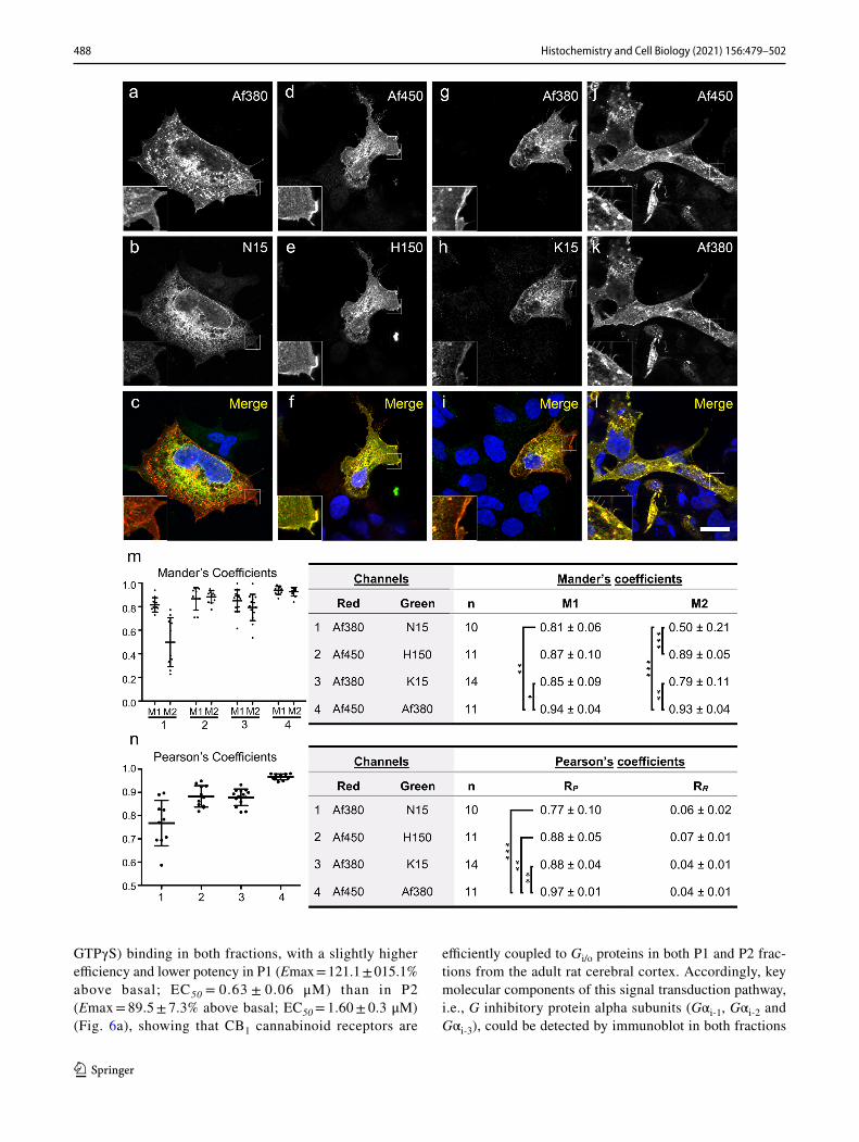
antibody produced a considerably stronger immunoreactiv-ity than N15 (Fig. 2a–c). Combination of antibody couples H150/Af450 (Fig. 3d–f) and Af380/Af450 (Fig. 3j–l) led to an almost complete co-localization throughout the entire cell, whereas K15/Af380 combination produced a reddish-coloured plasma membrane in merged images as a conse-quence of a considerably weaker surface labelling with K15 antibody compared with Af450 (Fig. 3g–i). These findings were quantitatively analysed by measuring the percent pixel overlap (Mander’s M1 and M2 coefficients) and the pixel intensity correlation (Pearson’s RP coefficient) between the two immunofluorescence signals of doubly immunostained cells. Kruskal–Wallis ANOVA revealed statistically signifi-cant differences for both M1 (P < 0.005) and M2 coefficients (P < 0.0001), whereas Dunn’s multiple comparison test found significant differences between selected pairs of anti-body combinations (Fig. 3m). Thus, according to qualitative observations, both M1 and M2 coefficients were close to 1.0 for the Af450/Af380 combination (M1, 0.94 ± 0.04 SD, M2, 0.93 ± 0.04 SD) and slightly lower, but not statistically different, for the Af450/H150 combination (M1, 0.87 ± 0.10 SD; M2, 0.89 ± 0.05 SD). Lower Mander’s coefficients, par-ticularly M2, were obtained for the Af380/K15 pair (M1, 0.85 ± 0.09 SD; M2, 0.79 ± 0.11 SD), reaching statistically
Fig. 1 Linear scale representation of the sequence of the rat CB1 receptor (Uniprot ID P20272; available at http:// www. unipr ot. org/) and amino acid sequence alignment of human, rat and mouse CB1 receptor using UniProt Align (https:// www. unipr ot. org/ align/). The position of the antigenic sequences used to produce the anti-CB1 rab-bit (H150, Af380) and goat (N15, K15, Af450) polyclonal antibod-
ies tested are indicated in both (see Table 1 for further details on anti-CB1 antibodies). Numbers on the linear representation of CB1 receptor refer to the amino acid residues in the sequence. Asterisks, two dots and one dot below the sequence alignment indicate fully conserved, highly conserved and weakly conserved residues across human, rat and mouse.

486 Histochemistry and Cell Biology (2021) 156:479–502
1 3
significant differences compared with the Af450/Af380 pair. As expected, the lowest coefficients were observed for the Af380/N15 combination, with a remarkably low mean value of M2 (M1, 0.81 ± 0.06 SD; M2, 0.50 ± 0.21 SD) (Fig. 3m), consistent with the observation that immunostaining with the Af380 antibody involved a considerably larger cell area than with N15, which was predominantly intracellu-lar (Fig. 3a–c). In consequence, the resulting M1 and M2 coefficients were statistically lower compared with those obtained for the Af450/Af380 pair, whereas the value of M2 was statistically different also for the comparison between the Af380-N15 and Af450-H150 combinations. In addition to yielding the highest Mander’s overlap coefficients when combined, the Af380, Af450 and H150 antibodies produced the most intense immunofluorescence staining. Thus, Af380 showed the shortest exposure time to achieve immunofluo-rescence signal just below saturation, closely followed by Af450 and H150 antibodies (2.4 ± 1.2 SD and 3.5 ± 1.2 SD fold longer exposure times, respectively). By contrast, much longer exposure times were observed with N15 and K15 antibodies (13.3 ± 4.7 SD and 21.4 ± 6.9 SD fold longer, respectively). Measurement of Pearson’s RP coefficients revealed a high degree of positive pixel intensity spatial
correlation between immunofluorescence signals generated by all couples of antibodies. Coste’s randomization analysis yielded RR values close to 0 with a confidence limit above 99% for all individual images, showing that Pearson’s RP coefficients did not originate by random chance (Fig. 3n). Despite the significant positive correlation found in all anti-body combinations, there were marked and statistically sig-nificant differences in RP values (Kruskal–Wallis ANOVA, P < 0.0001). Thus, the highest and lowest mean RP values for the co-localization within the dual colour fluorescence images corresponded to the Af450/Af380 and Af380/N15 combinations (0.97 ± 0.01 SD and 0.77 ± 0.10 SD, respec-tively), whereas Af450/H150 and Af380/K15 combinations yielded very high RP values (0.88 ± 0.05 SD and 0.88 ± 0.04 SD, respectively), but significantly lower in comparison with Af450/Af380 combination as revealed by Dunn’s multiple comparison test (P < 0.01). Again, consistent with qualita-tive findings, the highest significance was detected between Af450/Af380 and Af380/N15 combinations (P < 0.001) (Fig. 3n).
To test the ability of anti-CB1 receptor N-terminal antibodies to bind their target antigens in native condi-tions, double immunofluorescence assays were performed
Fig. 2 Immunofluorescence labelling of CB1-transfected HEK293 cells (pseudocoloured green) with anti-CB1 polyclonal antibodies N15 (a–c), H150 (d–f), K15 (g–i), Af380 (j–l) and Af450 (m–o), combined with Hoechst’s chromatin staining (pseudocoloured red). Filled and empty arrowheads correspond to transfected and non-
transfected cells, respectively. Micrographs are maximum inten-sity projections of three consecutive optical sections separated by 0.24 μm, obtained by structured illumination microscopy. Scale bar: 20 µm (applies to a–o)

487Histochemistry and Cell Biology (2021) 156:479–502
1 3
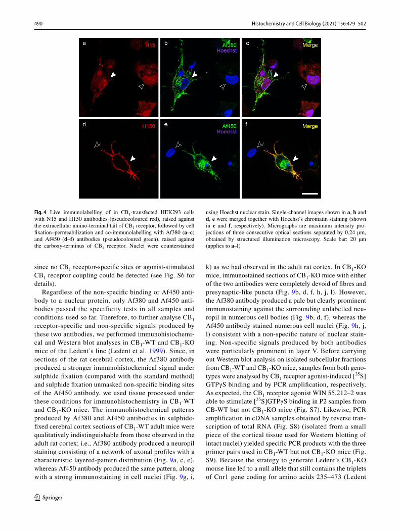
by incubation of live cells with either goat N15 or rabbit H150 polyclonal antibodies followed by paraformaldehyde fixation and incubation with rabbit Af380 and goat Af450 antibodies, respectively. In these conditions, plasma mem-brane staining with N15 antibody was variable among the CB1-transfected cell. Thus, N15 antibody detected clear plasma membrane staining after long exposure times in a subset of cells, causing a marked intracellular background autofluorescence to emerge (Fig. S1a–f). On the contrary, faint or no membrane staining was observed using similar acquisition settings in other cells (Fig. 4a–c). By contrast, H150 antibody systematically produced strong plasma membrane immunofluorescence staining, with virtu-ally no background, which was highly co-localized with plasma membrane staining produced by Af450 antibody (Fig. 4d–f). These results indicate that antigen masking caused by paraformaldehyde fixation does not account for the different ability of N15 and H150 antibodies to detect CB1 receptors localized at the plasma membrane.
Next, the anti-CB1 antibodies were assayed by immu-nohistochemistry for their ability to specifically bind CB1 receptor on histological sections from paraformal-dehyde-fixed adult rat brain cortex. Under these condi-tions, all the five antibodies led to an unevenly distributed immunostaining pattern throughout cortical layers I–VI. However, the dorsoventral distribution of immunolabel-ling and the types of detected cellular and subcellular structures varied considerably with the different antibod-ies used (Fig. 5a–j). Goat polyclonal antibodies N15 and K15, raised against the N-terminal region and against a sequence within the cytosolic C-terminal tail of CB1 receptor, respectively (Fig. 1), yielded a marked somatic immunostaining throughout cortical layers II–VI that was considerably more intense in layer V than in the rest (Fig. 5a, c). Immunolabelling with these two antibodies clearly delineated neuronal perikarya of variable mor-phology (Fig. 5f, h). A similar but more diffuse distribu-tion pattern could be observed with the rabbit polyclonal antibody H150 (Fig. 5b, g), which was designed against a large peptide encompassing the extracellular N-termi-nal tail, the first transmembrane domain and most of the intracellular loop 1 of CB1 receptor (Fig. 1). By contrast, the anti-CB1 rabbit polyclonal Af380 and goat polyclonal Af450 antibodies, raised in rabbit against the C-terminal 31 amino acids of CB1 receptor, produced a neuropil stain-ing throughout the cortical depth, being more intense in layers II/III than in the rest (Fig. 5d, e). As seen at high magnification, this neuropil staining consisted of fibre profiles decorated with intensely stained presynaptic-like boutons (Fig. 5i, j). In summary, only Af380 and Af450 produced an immunostaining pattern consistent with the laminar and subcellular distribution previously described
in the rodent brain cortex (Egertová and Elphick 2000; Bodor et al. 2005; Deshmukh et al. 2007).
To test the ability of the different antibodies to recog-nize denatured CB1 receptor from brain tissue, Western blot assays were performed in samples of P1, P2 and Cyt frac-tions obtained from adult rat brain cortex. Both N15 and K15 antibodies detected several bands migrating slightly below the 70 kDa standard in P1 and P2 fractions, which were more intense in P2 than in P1 fraction (Fig. 5k, m). Additionally, N15 antibody detected a band migrating between the 75 and 100 kDa standards in Cyt fraction (Fig. 5K) and K15 anti-body yielded a net band of similar intensity P1 and P2 frac-tions and migrating above the 100 kDa standard (Fig. 5m). In the three fractions analysed, intense bands were detected above the 50 kDa standard with H150 antibody, with no clear differences in the signal intensity seen among the dif-ferent fractions (Fig. 5l). Both Af380 and Af450 antibodies detected a major band around the 50 kDa standard in P1 and P2 but not in Cyt fraction (Fig. 5n–o). As a test of the speci-ficity of immunohistochemical and Western blot staining, we performed immnohistochemical and Western blot assays before and after preadsorption of antibodies with the corre-sponding antigenic peptides, which were available for N15, K15, Af380 and Af450 antibodies, but not H150. Antigen-preabsorbed and non-preabsorbed N15 and K15 antibodies produced a similar pattern of immunohistochemical staining and detected similar bands in Western blot analysis (Fig. S2a, b), indicating that the signals observed resulted from non-specific binding of antibodies. By contrast, both immu-nohistochemical signals and the major immunoreactive band found in P1 and P2 fractions at ~ 50 kDa with both Af380 and Af450 antibodies were virtually undetectable following preadsorption of the primary antibodies with the specific blocking peptide (Fig. S2a, b). These findings suggested that both the immunohistochemical staining pattern observed and the ~ 50 kDa immunoreactive band detected in P1 fraction and P2 membranes resulted from specific binding of Af380 and Af450 antibodies to CB1 receptor.
The presence of an antigen preadsorption-sensi-tive ~ 50 kDa strong immunoreactivity band for anti-CB1 Af380 and Af450 antibodies in P1 samples (likely enriched in cell nuclei) prompted us to analyse the density of CB1 receptor-specific ligand binding sites in P1 fraction and to compare it with that in P2 membranes (known to be enriched in plasma membrane). To this end, we performed satura-tion binding experiments with the selective CB1 receptor radioligand antagonist [3H]SR141716A (0.1–10 nM), show-ing that this compound labelled a single and homogeneous population of binding sites with a similar maximal density in both P1 and P2 samples (Fig. 6a), further demonstrat-ing the presence of CB1 receptors in both fractions. Moreo-ver, the CB1 receptor agonist WIN 55,212-2 was able to stimulate guanosine-5′-O-(3−[35S]thio)-triphosphate ([35S]
488 Histochemistry and Cell Biology (2021) 156:479–502
1 3
GTPγS) binding in both fractions, with a slightly higher efficiency and lower potency in P1 (Emax = 121.1 ± 015.1% above basal; EC50 = 0.63 ± 0.06 μM) than in P2 (Emax = 89.5 ± 7.3% above basal; EC50 = 1.60 ± 0.3 μM) (Fig. 6a), showing that CB1 cannabinoid receptors are
efficiently coupled to Gi/o proteins in both P1 and P2 frac-tions from the adult rat cerebral cortex. Accordingly, key molecular components of this signal transduction pathway, i.e., G inhibitory protein alpha subunits (Gαi-1, Gαi-2 and Gαi-3), could be detected by immunoblot in both fractions

489Histochemistry and Cell Biology (2021) 156:479–502
1 3
by using an antibody raised against a peptide common to three major G inhibitory protein alpha subunits (Gαi-1, Gαi-2 and Gαi-3) (Fig. 6b). To ascertain whether the concurrence of CB1-immunoreactivity, CB1 receptor-specific ligand binding and CB1 receptor agonist-stimulated [35S]GTPγS binding in P1 nuclear fractions could be explained by the presence of cell membranes pelleted during the first cen-trifugation step after tissue homogenization, we performed Western blot experiments in P1, P2 and Cyt fractions using specific markers of subcellular compartments (see Table S1 for details). Although P1 membranes displayed immuno-reactivity for NPCx and histone H1 proteins, revealing the enrichment of P1 in cell nuclei, strong immunoreactivity was also observed for the plasma membrane markers Na+/K+ ATPase, NMDAR1 and SNAP25, whereas no signal was detected for the cytosolic marker β-tubulin (Fig. 6c). Therefore, the concurrence of CB1 immunoreactivity, CB1 receptor-specific ligand binding sites and CB1 receptor cou-pling to Gi/o proteins in P1 fractions was very likely due to the presence of cell membranes in these samples.
The results shown so far demonstrate that all the five antibodies tested are able to bind CB1 receptors in tran-siently transfected HEK-293 cells, although with variable capacity to detect discrete subcellular pools of CB1 receptor (Figs. 2–4, S1). However, only Af380 and Af450 antibodies clearly labelled axons and presynaptic-like boutons in tissue sections of the adult rat cortex, consistent with the expected distribution of CB1 receptor (Figs. 5, S2). Therefore, we attempted to rescue (N15, H150 and K15 antibodies) and/or enhance (Af380 and Af450) specific immunostaining with all the five antibodies in sections of adult rat brain cortex, using a fixation method, consisting of a brief perfusion with sodium sulphide buffer before aldehyde fixation, which has been previously shown to improve antibody sensitivity without compromising specificity for a variety of antigens (Mitchell et al. 1993; Montaña et al. 2012; García del Caño et al. 2015). Under these conditions, N15, H150 and K15
antibodies produced a similar pattern as compared with standard conditions, and immunostaining was insensitive to preadsorption of N15 and K15 antibodies with their corre-sponding blocking peptides. By contrast, under sulphide fix-ation, Af380 immunostaining intensity increased considera-bly in axonal profiles and presynaptic-like puncta throughout the depth of the cortex, particularly in layers II/III (Fig. S3 and Supplementary results for further details). Strikingly, in cortical tissue subjected to sodium sulphide fixation, Af450 antibody produced a somatic immunostaining composed of round profiles that resembled cell nuclei, along with the axonal and presynaptic-like immunostaining pattern already observed under standard fixation (Fig. S4 and Supplemen-tary results for further details). Combination of Af450 and anti-lamin B1 antibodies in double immunofluorescence assays revealed the presence of Af450 immunoreactivity in large and medium sized nuclei of the rat cortex (Fig. 7b, c), and this particular labelling pattern was mostly internal to the nuclear lamina (Fig. 7a–c, Insets). Of note, preadsorption of the primary antibody with the specific blocking peptide abolished both presynaptic and nuclear staining produced by Af450 antibody (Fig. 7d–f), showing that sulphide fixa-tion unmasks binding sites of Af450 antibody within the nucleus without affecting the presynaptic staining observed in sections from brain tissue fixed by the standard method.
To analyse more in depth the origin of the nuclear signal observed with Af450 antibody, we isolated highly purified intact nuclei (N fraction) from the adult rat cortex (Fig. 8a) for Western blot and immunofluorescence analysis. Immu-noblot on N samples using the Af450 antibody yielded a net band at ~ 60 kDa clearly above the theoretical 52 kDa molecular mass of rat CB1 receptor observed in P2 samples (Fig. 8b). Consistent with that observed in tissue sections under sulphide fixation, Af450 antibody produced strong immunoreactivity in intact nuclei isolated from the adult rat cortex. Notably, co-immunolabelling with the neuronal marker NeuN/Fox-3 revealed that only neuronal nuclei were Af450 positive (Fig. 8c–e). Again, Af450 immunoreactiv-ity in N samples was virtually abolished by preadsorption of the primary antibody with the immunizing peptide, both in Western blot (Fig. 8b) and immunofluorescence assays (Fig. 8f–g). High-resolution images showed that Af450 immunoreactivity was distributed throughout the nucleo-plasm in subdomains poor in chromatin (Fig. 8h, S5a–c), internal to the nuclear lamina (Fig. S5d–f) and partially overlapping with components of the nuclear matrix NeuN/Fox-3 (Fig. S5g–i) and SC35 (Fig. S5j–l), which is inconsist-ent with the expected localization of an integral membrane protein, which is predicted to partition into cell membranes. These results, pointing strongly to the conclusion that the signal detected by the Af380 antibody in nuclei is non-spe-cific, were confirmed by saturation radioligand binding and agonist-stimulated [35S]GTPγS binding assays in N samples,
Fig. 3 Double immunofluorescence labelling of CB1-transfected HEK293 cells (a–l) with combined goat and rabbit anti-CB1 receptor antibody couples: Af380/N15 (a–c), Af450/ H150 (d–f), Af380/K15 (g–i) and Af450/Af380 (j–l). Single-channel images shown in a, d, g, j and in b, e, h, i were pseudocoloured red and green, respectively, and together with Hoechst’s chromatin staining to generate images shown in c, f, i, l. Scale bar: 20 µm (applies to a–l). Micrographs are maximum intensity projections of three consecutive optical sections separated by 0.24 μm, obtained by structured illumination micros-copy (m–n). Measurement of the Mander’s pixel overlap M1 and M2 and Pearson’s RP pixel intensity correlation coefficients between the two immunofluorescence signals of doubly immunostained cells. Graphs in m–n show plots and mean ± SD values of the Mander’s and Pearson’s coefficients. Kruskal–Wallis ANOVA yielded statisti-cally significant differences between groups for M1 (P < 0.005) and M2 coefficients (P < 0.0001) and RP (P < 0.0001) values. Asterisks in tables refer to significant differences from Dunn’s multiple compari-son test (*P < 0.05; **P < 0.01; ***P < 0.001)
◂
490 Histochemistry and Cell Biology (2021) 156:479–502
1 3
since no CB1 receptor-specific sites or agonist-stimulated CB1 receptor coupling could be detected (see Fig. S6 for details).
Regardless of the non-specific binding or Af450 anti-body to a nuclear protein, only Af380 and Af450 anti-bodies passed the specificity tests in all samples and conditions used so far. Therefore, to further analyse CB1 receptor-specific and non-specific signals produced by these two antibodies, we performed immunohistochemi-cal and Western blot analyses in CB1-WT and CB1-KO mice of the Ledent’s line (Ledent et al. 1999). Since, in sections of the rat cerebral cortex, the Af380 antibody produced a stronger immunohistochemical signal under sulphide fixation (compared with the standard method) and sulphide fixation unmasked non-specific binding sites of the Af450 antibody, we used tissue processed under these conditions for immunohistochemistry in CB1-WT and CB1-KO mice. The immunohistochemical patterns produced by Af380 and Af450 antibodies in sulphide-fixed cerebral cortex sections of CB1-WT adult mice were qualitatively indistinguishable from those observed in the adult rat cortex; i.e., Af380 antibody produced a neuropil staining consisting of a network of axonal profiles with a characteristic layered-pattern distribution (Fig. 9a, c, e), whereas Af450 antibody produced the same pattern, along with a strong immunostaining in cell nuclei (Fig. 9g, i,
k) as we had observed in the adult rat cortex. In CB1-KO mice, immunostained sections of CB1-KO mice with either of the two antibodies were completely devoid of fibres and presynaptic-like puncta (Fig. 9b, d, f, h, j, l). However, the Af380 antibody produced a pale but clearly prominent immunostaining against the surrounding unlabelled neu-ropil in numerous cell bodies (Fig. 9b, d, f), whereas the Af450 antibody stained numerous cell nuclei (Fig. 9h, j, l) consistent with a non-specific nature of nuclear stain-ing. Non-specific signals produced by both antibodies were particularly prominent in layer V. Before carrying out Western blot analysis on isolated subcellular fractions from CB1-WT and CB1-KO mice, samples from both geno-types were analysed by CB1 receptor agonist-induced [35S]GTPγS binding and by PCR amplification, respectively. As expected, the CB1 receptor agonist WIN 55,212–2 was able to stimulate [35S]GTPγS binding in P2 samples from CB-WT but not CB1-KO mice (Fig. S7). Likewise, PCR amplification in cDNA samples obtained by reverse tran-scription of total RNA (Fig. S8) (isolated from a small piece of the cortical tissue used for Western blotting of intact nuclei) yielded specific PCR products with the three primer pairs used in CB1-WT but not CB1-KO mice (Fig. S9). Because the strategy to generate Ledent’s CB1-KO mouse line led to a null allele that still contains the triplets of Cnr1 gene coding for amino acids 235–473 (Ledent
Fig. 4 Live immunolabelling of in CB1-transfected HEK293 cells with N15 and H150 antibodies (pseudocoloured red), raised against the extracellular amino-terminal tail of CB1 receptor, followed by cell fixation–permeabilization and co-immunolabelling with Af380 (a–c) and Af450 (d–f) antibodies (pseudocoloured green), raised against the carboxy-terminus of CB1 receptor. Nuclei were counterstained
using Hoechst nuclear stain. Single-channel images shown in a, b and d, e were merged together with Hoechst’s chromatin staining (shown in c and f, respectively). Micrographs are maximum intensity pro-jections of three consecutive optical sections separated by 0.24 μm, obtained by structured illumination microscopy. Scale bar: 20 µm (applies to a–l)

491Histochemistry and Cell Biology (2021) 156:479–502
1 3
et al. 1999) (Fig. S9a), results of PCR amplification also ruled out the possibility that a transcript containing the coding sequence for the immunizing peptide (residues 443–473 of mouse CB1 receptor) could be still expressed in CB1-KO mice, thus hindering the interpretation of non-specific signals observed with Af380 and Af450 (Fig. S9 and Supplementary results for further details). Western blot analysis of P2 membranes from CB1-WT and CB1-KO mice showed that Af380 antibody produced a major band at ~ 50 kDa, which was absent in P2 samples from CB1-KO mice, and a second, less intense but clearly positive band around the 20 kDa standard, which still remained in samples from CB1-KO mice. By contrast, a single weak band was observed in N samples, from both CB1-WT and CB1-KO mice, immunoblotted with Af380 antibody. As observed in P2 samples from the adult rat brain, the Af450 antibody produced a single specific band at ~ 50 kDa only in P2 fractions from CB1-WT but not CB1-KO animals, whereas it detected a single intense band at ~ 60 kDa in N samples obtained from either phenotype (Fig. 9o). Overall, these results show that Af380 and Af450 antibodies bind, both in tissue sections and in denatured samples resolved
by SDS-PAGE, not only to the CB1 receptor but also to non-CB1 receptor targets.
To confirm immunohistochemical results obtained in Ledent’s CB1-WT and CB1-KO mice, Af380 and Af450 antibodies were assayed in brain sections of CB1-WT and CB1-KO animals kindly provided by Dr. Giovanni Marsi-cano (Marsicano et al. 2002), which were generated using a Cre/loxP-based gene-targeting strategy that involves removal of the entire Cnr1 coding region. The immunohistochemi-cal patterns produced by Af380 and Af450 in sulphide-fixed cerebral cortical sections of CB-1WT and CB1-KO littermates from this line were virtually identical to those observed in Ledent's model (Fig. S10).
Discussion
It is well known that the ability of antibodies to recognize their target antigen is highly influenced by the experimental context in which they are used and, therefore, not all antibod-ies are suitable for all end-use applications (Bordeaux et al. 2010; Voskuil 2014, 2017; Uhlen et al. 2016). The analysis
Fig. 5 (a–j) Micrographs of coronal sections of the adult rat parietal cortex immunostained with the different antibodies designed against CB1 receptor tested here. (a–e) Low-magnification micrographs show the overall distribution of immunoreactivity throughout the depth of the cortex using the different antibodies. (f–j) Higher-magnification micrographs show details of the immunostaining pattern in cortical
layer V. Scale bars: 200 µm in e (applies to a–e); 50 µm in j (applies to f–j). (k–o) Western blot analysis using the different antibodies against the CB1 receptor tested in this study. Equivalent amounts of protein (20 µg/lane) from P1, P2 and Cyt fractions obtained from homogenates of adult rat brain cortex were loaded in duplicate and run in parallel

492 Histochemistry and Cell Biology (2021) 156:479–502
1 3
performed here focused on the ability of five anti-CB1 recep-tor antibodies of commercial source, designed against the N-terminal (N15 and H150) or C-terminal (K15, Af380 and
Af450) regions of the CB1 receptor, to specifically recognize their target in cells transfected with plasmids coding for the native human CB1 receptor, in fixed sections from rat and
Fig. 6 a Radioligand and [35S]GTPγS binding assays in P1 and P2 fractions from the adult rat brain cortex. Representative saturation binding curve for [3H]SR141716A (0.01–10 nM) and representative dose–response curve of WIN 55,212-2 (0.1 nM–10 μM) stimulated [35S]GTPγS binding (right), both in P1 fraction from the adult rat cortex. Non-specific binding in stimulated [35S]GTPγS binding assays was determined in the presence of 10 μM unlabelled GTPγS. Each point in both curves corresponds to the mean ± SEM value of one representative experiment performed in triplicate and duplicate for radioligand and [35S]GTPγS binding assays, respectively. The accom-panying table shows maximal number of sites (Bmax) and affinity (KD) values for the selective CB1 receptor antagonist [3H]SR141716A and the maximal effect (Emax) and potency (EC50) of the CB1 cannabi-
noid receptor agonist WIN 55,212-2 to stimulate [35S]GTPγS bind-ing assays in P1 and P2 subcellular fractions of the adult rat cortex. Values shown in the table are mean ± SEM of at least three independ-ent experiments. b Western blot analysis against CB1 receptor and G inhibitory protein alpha subunits (Gαi-1,2,3) in both P1 and P2 subcel-lular fractions from adult rat brain cortex (20 µg/lane). c Western blot analysis of P1, P2 and Cyt fractions from the adult rat cortex (10 µg/lane) with antibodies against subcellular fraction-specific antigens: NPCx (62 kDa component of the nuclear pore complex), Histone H1, Na+/K+ ATPase (α1 subunit of Na+/K+ ATPase); NMDAR1 (NR1 subunit of the NMDA receptor), SNAP25 (synaptosome-associated protein 25) and β-tubulin (see Supplementary Table S1 for further details)
Fig. 7 a–c Double immuno-fluorescence labelling in the adult rat cortex under sodium sulphide fixation combining a mouse monoclonal antibody recognizing the nuclear protein lamin B1 (pseudocoloured red) and the goat polyclonal anti-CB1 antibody Af450 (pseudocoloured green). The anti-CB1 antibody immu-nostained presynaptic-like boutons (arrowheads) as well as large and medium sized (filled arrows) but not small nuclei (empty arrows), as seen by co-staining with lamin B1. d–f The same experiment depicted in a–c was performed after preadsorption of the anti-CB1 with the immunizing peptide. Images are single 0.5-µm-thick confocal optical sections. Scale bar: 50 µm in f (applies to a–f)

493Histochemistry and Cell Biology (2021) 156:479–502
1 3
mouse cerebral cortex and in subcellular fractions obtained from rat and mouse brain cortex homogenates. In addition, N15 and H150 antibodies were tested for their ability to detect surface CB1 receptor in live cells.
In HEK-293 cells fixed with paraformaldehyde, all the five antibodies tested made it possible to readily distinguish cells transfected with the CB1 receptor from non-transfected ones. However, whereas the N-terminal H150 antibody and the C-terminal Af380 and Af450 antibodies produced bright signals that were highly co-localized with each other both intracellularly and on the plasma membrane, the N-termi-nal N15 and C-terminal K15 antibodies barely and weakly detected surface receptors, respectively. Noteworthy, the H150 antibody was also highly specific and sensitive in detecting surface CB1 receptors in live cells, which con-trasts with the uselessness of the N15 antibody for this pur-pose. The distinct peptides used as antigen to generate N15 and H150 antibodies could explain this difference. Indeed,
the N15 antibody was raised against an unspecified short sequence near the end of the N-terminal extracellular tail of the human CB1 receptor, whereas the H150 antibody was raised against a long peptide spanning residues 1–150 of the human CB1 receptor and, therefore, likely to contain more immunogenic sequences. However, this would only explain the differences in sensitivity but not the different ability to detect the surface receptor. Interestingly, it has been demon-strated that a large fraction of CB1 receptor are truncated at their N-terminal at early stages in the secretory pathway just before being translocated to the endoplasmic reticulum (ER) (Nordström and Andersson 2006), whereas the untruncated receptors appear to be inefficiently translocated across the ER membrane, leading to high levels of misfolded recep-tor that are subsequently degraded (Andersson et al. 2003). Although the extent of this truncation has not yet been deter-mined, endogenous truncation of N-terminally c-myc-tagged CB1 receptors overexpressed in baby hamster kidney cells
Fig. 8 Analysis of immunoreactivity of the goat polyclonal Af450 antibody in intact nuclei (N) isolated from adult rat brain cortex. a Representative image of phase-contrast microscopy of nuclei isolated from a homogenate of adult rat cerebral cortex showing no debris or contamination by other organelles. Scale bar: 20 µm. b Western blot analysis of N, P2 and Cyt from the adult rat brain cortex using Af450 antibody. Bands of ~ 62 kDa and ~ 50 kDa detected in N and P2 sam-ples, respectively, disappeared after preadsorption of the antibody with the immunizing peptide. Equal amounts of protein (10 µg/lane) were loaded and run in parallel. c–e Double immunofluorescence labelling with Af450 (pseudocoloured green) and anti-NeuN/Fox-3 (NeuN, pseudocoloured green) antibodies combined with Hoechst’s
staining (pseudocoloured blue). The Af450 antibody detected a strong signal in every NeuN-immunopositive nucleus (filled arrowheads), whereas very weak or no staining was observed in nuclei devoid of NeuN/Fox-3 (empty arrowheads). Scale bar: 20 µm in e (applies to c–d). f–h Combined immunofluorescence with the Af450 antibody (pseudocoloured green) and Hoechst’s chromatin staining (pseudoc-oloured red) in isolated cell nuclei from the adult rat cortex before (f) and after (g) incubation of the primary antibody with the specific blocking peptide. The higher-magnification micrograph (h) of the nuclei shown in f depicts the non-overlapping pattern between Af380 immunofluorescence and patches of intense Hoechst’s staining. Scale bars: 10 µm in g (applies to f–g); 5 µm in h

494 Histochemistry and Cell Biology (2021) 156:479–502
1 3
Fig. 9 Anti-CB1 receptor immunohistochemical staining in sulphide-fixed parietal cortex sections and Western blot analysis of P2 and N fractions from CB1-WT and CB1-KO mice generated by the Ledent’s lab (Ledent et al. 1999) using the rabbit polyclonal Af380 and the goat polyclonal Af450 antibodies. a–f Micrographs showing the dis-tribution of immunoreactivity produced by the Af380 antibody in the parietal cortex of CB1-WT (a, c, e) and CB1-KO (b, d, f) mice. Framed areas in panoramic images a and b are shown at higher mag-nification in c, d and e, f respectively. g–l Micrographs showing the distribution of immunoreactivity produced by the Af450 antibody in the parietal cortex of CB1-WT (g, i, k) and CB1-KO (h, j, l) mice.
Framed areas in panoramic images g and h are shown at higher magnification in i–k and j–l, respectively. m–n Low-magnification micrographs showing the absence of immunostaining in the parietal cortex of CB1-WT (m) and CB1-KO (n) when Af450 antibody was used after being preabsorbed with the immunizing peptide. Scale bars: 200 µm in n (applies to a, b, g, h, m, n), 50 µm in l (applies to c–f, i–l). o Western blot analysis of P2 and N fractions isolated from the adult mouse brain cortex of Ledent’s CB1-WT and CB1-KO mice using either the Af380 (left immunoblot) or Go-Af380 (right immunoblot) antibodies. Double amount of protein was loaded from CB1-KO mice samples

495Histochemistry and Cell Biology (2021) 156:479–502
1 3
has been estimated to cause a mobility shift of about 4 kDa on SDS-PAGE compared with non-truncated ones (Nor-dström and Andersson 2006). Therefore, considering the 1.2 kDa size of the c-myc tag, it is expected that CB1 recep-tors are cleaved at around residue 26 of the CB1 N-tail, prob-ably resulting in the deletion of the N15 antibody epitope. Consequently, the N15 antibody would primarily recognize the newly synthesized N-terminal tail of CB1 receptors prior to their translocation to the ER membrane via the secretory pathway used by most GPCRs lacking a cleavable signal peptide, with the first transmembrane domain of the mature receptor functioning as a signal anchor sequence (Wallin and Von Heijne 1995). In addition, N15 antibody could recog-nize incorrectly folded untruncated CB1 receptors retained and subjected to quality control in the ER as well as untrun-cated CB1 receptors that had passed ER quality control (Nordström and Andersson 2006). This is consistent with the observed immunostaining pattern of the N15 antibody, which was distributed mainly around the cell nucleus and showed a weak variable or no staining in the periphery and the plasma membrane of the transfected HEK-293 cells. The fact that the untruncated CB1 receptor probably represents only a small fraction of the entire population (Nordström and Andersson 2006) could explain the apparent low sensitivity of the N15 antibody, which in combination with markers of intracellular organelles could be very useful to study spe-cific intracellular species of CB1 receptors. Indeed, retained GPCRs have been shown to display enhanced interaction with the ER luminal chaperone BiP, as the primary regula-tor of endoplasmic reticulum translocation, as well as with carbohydrate-binding chaperones, which are key elements for recycling misfolded receptors through N-linked glycan recognition and processing (Moremen and Molinari 2006; Achour et al. 2008).
When tested on histological sections from the rat cer-ebral cortex, neither of the two N-terminal antibodies pro-duced a distribution pattern of CB1 receptor in fibres and presynaptic-like varicosities widely reported in previous studies (Egertová and Elphick 2000; Bodor et al. 2005; Deshmukh et al. 2007). Although this could be expected for the N15 antibody, it was surprising for the H150 antibody in view of the high specificity and sensitivity that it showed for detecting the CB1 receptor in HEK-293 cells. This con-trasts with results obtained by other authors showing the ability of antibodies against large fragments of the amino end of CB1 receptor (Tsou et al. 1998; Eggan and Lewis 2007) or against a short 15-amino-acid peptide far from the amino end of CB1 receptor (Dove Pettit et al. 1998), but not antibodies against a short 14-amino-acid peptide mapping at the extreme end of the N-terminus (Mukhopadhyay and Howlett 2001; Matias et al. 2002), to produce immunohis-tochemical signals in brain tissue sections consistent with the accepted gross anatomical and fine distribution patterns
for the CB1 receptor (Dove Pettit et al. 1998; Tsou et al. 1998; Eggan and Lewis 2007). Intriguingly, in one of the few studies devoted to the analysis of the specificity of anti-CB1 antibodies (Grimsey et al. 2008), authors reported that two commercial antibodies produced against residues 1–77 (PA1-745, Affinity BioReagents) and 1–99 (C1108, Sigma-Aldrich) of the rat and human CB1 receptors, respectively, failed to detect the CB1 receptor in histological sections of rodent brain, not even in HEK-293 cells transfected with a HA-tagged human CB1 receptor, and neither in live cells nor in paraformaldehyde-fixed and permeabilized cells. In agreement with these negative results, other commercial antibody against rat CB1 receptor N-terminal amino acids 1–77 (C1233, Sigma-Aldrich) and 1–99 (C1108, Sigma-Aldrich) produced immunohistochemical patterns in the cortex, striatum and hippocampus (Fusco et al. 2004) that were inconsistent with the widely accepted distribution of CB1 receptor. Differences in immunization and/or purifica-tion procedures relative to similar “homemade” antibodies could explain these discrepancies. Coming back to our pre-sent data, a possible reason for the lack of specificity of the H150 antibody in rat brain tissue sections could be that this antibody was generated using an immunogen corresponding to a sequence of human origin, since there are eight amino acid discrepancies between the human and rat CB1 recep-tor within a portion of the extracellular N-tail encompass-ing residues 68–110 and 68–111 of human and rat proteins, respectively. A second possible explanation is that N-linked glycans at asparagine residues 78 and 84 of the rat CB1 receptor (Song and Howlet 1995) could affect the recog-nition of the epitope by the H150 antibody, although this is unlikely since N-glycosylation should have also affected epitope recognition in HEK-293 cells transfected with the human CB1 receptor, which is known to be extensively N-glycosylated (Nordström and Andersson 2006; Ruehle et al. 2017).
Again, N15 and H150 antibodies did not detect CB1 receptors under denaturing conditions by SDS-PAGE and immunoblotting, as concluded from the analysis of the molecular mass of the immunoreactive bands (both anti-bodies), antibody–antigen preadsorption control (N15) and the presence of immunoreactive bands of identical size in the fractions of the rat cerebral showing (P1 and P2) and lacking (Cyt) CB1 receptor-specific ligand binding sites and CB1 receptor coupling to Gi/o proteins. Similar to what has been discussed about the inability of these N-terminal antibodies to detect the brain CB1 receptor by immunohis-tochemistry, the uselessness of N15 and H150 for Western blotting could also be due to the constitutive N-terminal truncation of the CB1 receptor and to differences in the primary sequence between human versus rat CB1 recep-tor, respectively. Another possibility is that the large size of the immunogen used to generate the H150 antibody could

496 Histochemistry and Cell Biology (2021) 156:479–502
1 3
contain conformational antigenic determinants that would no longer be detectable after denaturation.
Of the three C-terminal antibodies analysed here, the Af450 and Af380 antibodies, raised against 31 amino acids at the extreme carboxy-terminus of mouse CB1 receptor were by far the ones that provided the best results in all the final applications tested, although they were not exempt from some issues related to sample processing and end-use application. Double immunofluorescence assays demon-strated a virtual complete co-localization between Af450, and Af380-immunoreactivities, but more importantly, both antibodies produced intense immunoreactivity largely restricted to axonal fibres and presynaptic-like puncta with an excellent signal-to-noise relationship in rat brain cortical sections, and the signals were virtually abolished by peptide preadsorption. This is consistent with several studies that showed a similar CB1 immunoreactivity distribution profile using antibodies raised against C-terminal end fragments of variable length, including peptides comprising the last 13 (Egertová and Elphick 2000), 15 (Bodor et al. 2005; Desh-mukh et al. 2007; Eggan and Lewis 2007; Eggan et al. 2010) and 73 (Hájos et al. 2000; Wager-Miller et al. 2002; Harkany et al. 2003; Monory et al. 2006; Eggan and Lewis 2007) C-terminal end residues, as well as a number of reports using Af380 (Lafourcade et al. 2007; Yoneda et al. 2013; Diniz et al. 2019; Fuerte-Hortigón et al. 2021) and Af450 (Yoneda et al. 2013; Rivera et al. 2015; Exposito-Alonso et al. 2020) antibodies in the rodent brain cortex. Moreover, both Af380 (Mateo et al. 2017) and Af450 (Lafourcade et al. 2007; Peñasco et al. 2020; Egaña-Huguet et al. 2021) antibodies have proven to be adequate to describe the ultrastructural distribution of CB1 receptors and have been validated for specificity in transgenic mice lacking CB1 receptor (Hebert-Chatelain et al. 2014b; Remmers et al. 2017; Gutiérrez-Rodríguez et al. 2018). Remarkably, it has been observed that preadsorption of an antibody generated against the 73 C-terminal residues of the CB1 receptor with a peptide that spans only the last 15 amino acids removed most but not all of the CB1-specific axonal labelling, indicating that the 15 residues at the C-terminus of CB1 receptor contained most of the antigenic determinants (Eggan and Lewis 2007). This could explain why the K15 antibody against an unspecified sequence upstream of the last 15 residues was able to rec-ognize the CB1 receptor under overexpression conditions in HEK-293, although with a low sensitivity that may not be sufficient to detect physiological levels in brain tissue.
It is well known that the fixation procedure used affects immunohistochemical staining, increasing or reducing specific and non-specific signals or masking antigens to a greater or lesser extent (Fritschy 2008). For instance, Egertová and colleagues reported that the use of Bouin’s fixative for immunohistochemical staining of rodent brain sections with C-terminally directed anti-CB1 antibodies
abolishes the background staining that still remains when immunogen-preadsorbed antibodies were used in 4% par-aformaldehyde-fixed tissue (Egertová et al. 1998; Egertová and Elphick 2000) and preserves only specific immunostain-ing produced by the same antibodies as seen by comparing tissue sections from CB1-WT and CB1-KO mice (Egertová et al. 2003; Monory et al. 2006). In addition, certain fixation procedures can prevent epitope masking that occurs as a result of conformational changes in antigens caused by para-formaldehyde fixation. Here, we examined the immunohisto-chemical staining profile obtained with the different antibod-ies in histological sections of the adult rat cortex obtained from sulphide-fixed brains. Although this procedure was first developed to reveal zinc-rich terminals using Timm’s staining method (Hassler and Söremark 1968; Sloviter 1982) and, more particularly, as a marker of hippocampal mossy fibre sprouting in animal models of epilepsy (Nadler et al. 1980) and human epilepsy (Sutula et al. 1989), it has been described as a good method to improve immunoreactivity without compromising the immunostaining profile of dif-ferent proteins (Mitchell et al. 1993); compared with other antigen-retrieval methods, it offers an important advantage for our particular purposes. Specifically, we have previously shown that sulphide fixation improves immunofluorescence signal intensity for phospholipase C beta 1 (PLCβ1) (Mon-taña et al. 2012) and diacylglycerol lipase α (DAGLα) (García del Caño et al. 2015) and allows their detection in subcellular compartments where they do not appear stained or do so very weakly after standard fixation. Moreover, sul-phide fixation did not affect the immunostaining profile of various GABAergic neuronal phenotype markers and glial markers (Montaña et al. 2012) and even increased the immu-nostaining intensity of subcellular markers such as lamin B1 (García del Caño et al. 2015). Thus, enhancement or maintenance of CB1 receptor immunoreactivity after sul-phide fixation could allow simultaneous immunostaining of CB1 with key components of the brain endocannabinoid system such as PLCβ1 and DAGLα, which are key enzymes for on-demand the synthesis (Shonesy et al. 2015) of the most abundant endogenous CB1 agonist (Stella et al. 1997) 2-arachidonoylglycerol (2-AG), as well as with phenotype and subcellular markers. Sulphide fixation did not rescue CB1-specific immunolabelling with N15, H150 and K15 antibodies, whereas it increased CB1-immunoreactivity with Af380 antibody and led to the emergence of a nuclear stain-ing with Af450 antibody (which was sensitive to peptide preadsorption) with no changes in typical axonal labelling. Interestingly, double immunofluorescence assays in intact nuclei (N fraction) isolated from rat cerebral cortex lack-ing plasma membrane protein contaminants revealed that the nuclear staining produced by anti-CB1 antibody over-lapped with components of the nuclear matrix, similar to that previously observed for PLCβ1 and DAGLα (Montaña

497Histochemistry and Cell Biology (2021) 156:479–502
1 3
et al. 2012; García del Caño et al. 2014, 2015). In addition, Western blot analysis on isolated intact nuclei from rat cer-ebral cortex produced a clean and strong band at ~ 60 kDa that was sensitive to preadsorption of the antibody with the immunogen. Although this signal was clearly above the theoretical 52 kDa molecular mass of the rat CB1 receptor, suggesting that it was not related to the CB1 receptor, other authors have interpreted the slower migration of these bands on SDS-PAGE as a consequence of N-linked glycosylation at the N-terminal region of CB1 receptor (Song and Howlett 1995; Egertová and Elphick 2000; De Jesús et al. 2006). Because such post-translational modifications could lead to a slower mobility of CB1 receptors on SDS-PAGE, our find-ings in intact nuclei still left open the unlikely but feasible and attractive possibility that the signal found in nuclei of neuronal cells with the Af450 antibody could be specific. However, neither specific CB1 receptor binding sites nor CB1 receptor coupling to inhibitory G proteins could be detected in intact nuclei samples by saturation radioligand binding assays and agonist-stimulated [35S]GTPγS binding. In view of these surprising and apparently contradictory results, we performed immunohistochemical assays on cerebral cortex sections of sulphide-fixed brains from a CB1-KO mouse line (Ledent et al., 1999). As expected, the Af450 antibody pro-duced the typical fibre staining and presynaptic-like profiles in tissue sections from CB1-WT mice but not from CB1-KO littermates, whereas a strong nuclear signal was observed in both. Furthermore, Western blot analysis on N samples from CB1-WT and CB1-KO mice showed that Af450 detected the strong ~ 60 kDa band mentioned above in both genotypes, virtually demonstrating that the nuclear staining produced by the Af450 antibody was not related to the CB1 receptor. It is likely that the strong immunoreactivity in cell nuclei observed with the Af450 antibody in both sulphide-fixed tis-sue sections and N samples resolved by SDS-PAGE results from the binding of IgGs to a sequence of the same CB1 receptor-unrelated protein. If that was the case, the observed artefact, rather than be caused by the appearance of “false epitopes” as a consequence of chemical modifications due to sulphide fixation, would be produced by an unmasking effect of a site inaccessible in tissue fixed under standard conditions but for which the Af450 antibody has high affin-ity when the protein is denatured for Western blot, thus reinforcing our previous evidence showing that sulphide fixation is a good antigen-retrieval method (Montaña et al. 2012; García del Caño et al. 2015) for immunohistochemis-try and, furthermore, compatible with electron microscopy (Danscher 1981). Specificity tests with the Af380 antibody in tissue sections of CB1-WT and CB1-KO littermates sub-jected to sulphide fixation revealed that, as we had observed in rat tissue sections, this antibody is highly sensitive and specific under these conditions and produced only negligi-ble background staining in brain cortex sections of CB1-KO
mice, which makes it the best option for use in combination with the immunostaining of antigens that are retrieved by sulphide fixation.
Transgenic animals in which the antigen of interest have been eliminated are considered to be the gold standard for validation of antibody specificity in immunohistochemistry (Saper and Sawchenko 2003; Saper 2005; Bordeaux et al. 2010). However, these models are not free from pitfalls, as exemplified in a recent study on the specificity of four antibodies raised against synthetic peptide sequences cor-responding to different parts of the murine CB2 cannabi-noid receptor (Zhang et al. 2019). In this study, two different CB2-KO models generated by homologous recombination that replaced genetic sequences near the 3′ (Zimmer et al. 1999) and 5′ ends (CB2-KO strain from Deltagen Inc.) of mouse Cnr2 exon 3 (which contains the entire mouse CB2 receptor encoding region) with the neomycin gene were used. Results of this study indicated that the undeleted Cnr2 gene sequences still encodes mutant or truncated CB2 recep-tor proteins or fragments that could be detected by anti-CB2 antibodies. Of note, the strategy to generate the Ledent’s CB1-KO line led to null allele that still contains the triplets of Cnr1 gene coding for amino acids 235–473 (Ledent et al. 1999). Although Ledent’s mice have been well characterized for the correct transgene insertion and on the basis of a vari-ety of responses to drugs, functional responses and behav-ioural tests, the evidence in CB2-KO models discussed above makes feasible the possibility that a transcript containing the coding sequence for the immunizing peptides for Af380 and Af450 antibodies (residues 443–473 of mouse CB1 receptor) could be still expressed in these animals. However, three primer pairs targeting either the deleted or the remaining sequences of the Cnr1 gene in the Ledent’s CB1-KO mouse were unable to amplify any specific mRNA in CB1-KO samples by RT-PCR, whereas all the three probes detected CB1-specific mRNA sequences in CB1-WT littermates. To our knowledge, this is the first time that the possibility that transcripts containing sequences encoding anti-CB1 anti-body epitopes may still be expressed in Ledent’s CB1-KO mice has been addressed. Finally, immunohistochemical staining in histological sections of the cerebral cortex of CB1-KO mice in which the entire coding region of the Cnr1 gene was eliminated (Marsicano et al. 2002) cleared up any doubt, since the Af380 and Af450 antibodies produced spe-cific and non-specific signals identical to those observed in Ledent’s CB1-KO mice, ruling out any possibility that the nuclear staining observed in Ledent’s CB1-KO mice could be due to the presence of CB1 receptor protein fragments.
Of the three C-terminal antibodies used, the K15 anti-body failed to detect CB1 receptor-specific signals by West-ern blot, since it produced immunoreactive bands in the P1 and P2 fractions that migrated clearly above the theoreti-cal 52 kDa molecular mass of rat CB1 receptor, which in
498 Histochemistry and Cell Biology (2021) 156:479–502
1 3
addition were not eliminated by preadsorption of antibod-ies with an excess of the immunizing peptide. As discussed in relation to the futility of this antibody for immunohis-tochemical detection of the CB1 receptor, the inability of the K15 antibody to detect CB1 under denaturing conditions could be due to the fact that most of the antigenic determi-nants of the C-terminal end of the CB1 receptor are located downstream of the antigen used. By contrast, both Af380 and Af450 antibodies were shown to detect a major band migrating slightly below the theoretical 52 kDa molecular mass of rat CB1 only in P1 and P2, which contained CB1 receptor-specific binding sites and displayed agonist-stim-ulated CB1 receptor coupling to Gi/o proteins as shown by saturation radioligand binding and agonist-stimulated [35S]GTPγS binding assays and, in addition, this ~ 50 kDa band was undetectable when the antibodies were preabsorbed with the immunizing peptide. Af380 antibody detected sev-eral other, weaker but clear bands with considerably higher and lower molecular weight in P1 and P2 fractions, whereas extra bands were faint or undetectable with Af450 antibody. The molecular weight of the ~ 50 kDa specific band detected by Western blot in our study agrees with previous reports using the same rabbit Af380 (Fukudome et al. 2004; Yoneda et al. 2013; Rodríguez-Cueto et al. 2016) and goat polyclonal Af450 (Yoneda et al. 2013) antibodies, including two stud-ies from our lab (Peñasco et al. 2020; Egaña-Huguet et al. 2021). Despite the consistency of our data, results obtained by other authors reporting the detection of a major band of about 60 kDa or higher using different anti-CB1 receptor antibodies deserve consideration (Song and Howlett 1995; Egertová and Elphick 2000; Mukhopadhyay and Howl-ett 2001; Wager-Miller et al. 2002; De Jesús et al. 2006; Diniz et al. 2019). Although this slower mobility has been explained as a result of glycosylation of the CB1 terminal tail (Song and Howlett 1995; Egertová and Elphick 2000; De Jesús et al. 2006), deglycosylation experiments cause also a considerably mobility shift of the ~ 50 kDa detected with antibodies against large fragments of the N-terminal region or the C-terminal end of CB1 receptor (Nordström and Andersson 2006; Esteban et al. 2020), and therefore, these discrepancies must be due to other factors. On the one hand, the antibodies used in some cases were raised against a small fragment of the amino terminal end of the CB1 receptor (Song and Howlett 1995; Mukhopadhyay and Howlett 2001), and as discussed extensively above, it is pos-sible that truncation of a short N-terminal fragment of the CB1 receptor in the early stages of maturation could cause these antibodies to recognize only untruncated CB1 recep-tors of higher molecular weight, which appear to represent a small fraction of the total receptor population (Nordström and Andersson 2006). On the other hand, heating samples at high temperature causes CB1 receptor aggregates with considerably lower mobility in SDS-PAGE (Wager-Miller
and Mackie 2016), and it has been recently reported that temperatures above 65 °C favour the formation of high-molecular-weight aggregates (Esteban et al. 2020), which could explain the high-molecular-weight bands migrating considerably above 60 kDa described before (Matias et al. 2002; Wager-Miller et al. 2002). Among the studies focused on the specificity of anti-CB1 antibodies intended for West-ern blotting, it is worth highlighting a recent work devoted to the analysis of the mobility of the CB1 receptor on SDS-PAGE using different N-terminal and C-terminal antibod-ies and CB1-KO mice as negative control (Esteban et al. 2020). Noteworthily, only one of the three antibodies used by these authors designed against the first 14 residues of the N-terminal end of the CB1 receptor recognized a single CB1 receptor-specific band at ~ 64 kDa, whereas two antibodies generated against the C-terminal end of the CB1 receptor and one against residues 84–99 of the N-terminal tail (and, there-fore, far from the amino end of CB1 receptor) detected a sin-gle specific band at ~ 50 kDa. Although the authors proposed an interpretation based on the folding and packing state of the protein and the detergent used, their data could be also interpreted as a result of the ability of antibodies against the N-terminal end of the CB1 receptor to recognize solely a minor fraction of high-molecular-weight untruncated CB1 receptors, which would be below the detection threshold when antibodies are designed against peptides correspond-ing to sequences located far from the N-terminal extreme. In favour of this interpretation, two of the three antibodies that recognized a specific band of ~ 50 kDa in mouse cerebral cortex and cerebellum could not detect the band of ~ 64 kDa in mouse brain immunoprecipitates prepared with the anti-body against the end of the N-tail of CB1 receptor, whereas they detected the ~ 50 kDa band in immunoprecipitates pre-pared with the other two antibodies, probably due to the extremely low density of the putatively untruncated ~ 64 kDa CB1 receptors compared to ~ 50 kDa species (Esteban et al. 2020). On the contrary, the antibody against the N-terminal end of the CB1 receptor produced a single band at ~ 64 kDa of the same intensity regardless of the antibody used for immunoprecipitation, strongly suggesting that this anti-body detects with high sensitivity a minor species of the untruncated receptor. In any case, the results described by Esteban and colleagues (2020) using anti-CB1 antibodies designed either against the C-terminus or against residues 84–99 of the N-tail of the CB1 receptor are in good agree-ment with our finding that both Af380 and Af450 recognize a unique CB1 receptor-specific band migrating at ~ 50 kDa on SDS-PAGE as seen by analysing immunoreactive bands in P2 membranes from the cerebral cortex of CB1-WT and CB1-KO littermates. The use of CB1-KO animals as nega-tive controls also allowed us to establish Af450 antibody as the best choice for Western blot assays in cortical P2 membranes, since Af380 antibody produced a number of
499Histochemistry and Cell Biology (2021) 156:479–502
1 3
non-specific bands of variable intensity and migrating above and below the ~ 50 kDa CB1 receptor-specific band, whereas Af450 led to a single ~ 50 kDa specific band.
In conclusion, of the five antibodies tested for specificity, three of them were identified as the best choice for specific end-use applications. Thus, the ability of the H150 antibody to detect surface receptors in HEK-293 cells overexpressing CB1 receptors makes it an excellent unique tool of particular interest to study the dynamics of processes such as inter-nalization, recycling or trafficking to the plasma membrane of CB1 receptors. Antibodies Af380 and Af450 provided excellent results for immunodetection of CB1 receptors in permeabilized HEK-293 cells and in fixed brain tissue, although only the Af380 antibody is suitable under sulphide fixation when this method is used to increase immunoreac-tivity for other antigens such as components of the endocan-nabinoid system or cell phenotype markers. Both Af380 and Af450 antibodies are useful for detection of CB1 receptor by Western blot, although the Af380 antibody detects several non-specific bands while Af450 does not, making the latter option the best for Western blot analysis. Our results further highlight the need for a F4P approach for validation of anti-bodies before they are placed on the market or discontinued, and we urge suppliers to leave final validation of antibodies for specific end uses to basic researchers.
Supplementary Information The online version contains supplemen-tary material available at https:// doi. org/ 10. 1007/ s00418- 021- 02025-5.
Acknowledgements The authors thank Giovanni Marsicano (INSERM U1215, NeuroCentre Magendie, Bordeaux, France) for kindly supply-ing CB1-KO mice. The authors wish to thank Ricardo Andrade (Ser-vicio de Microscopía Analítica y de Alta Resolución en Biomedicina, SGIker) for skilful technical assistance with confocal microscopy experiments. Joan Sallés is a member of the Societat Catalana de Bio-logia, a subsidiary society of the Institut d’ Estudis Catalans (Barce-lona, Catalonia).
Author contributions L.E. and G.G.C. contributed equally to experi-mental work.
Funding Open Access funding provided thanks to the CRUE-CSIC agreement with Springer Nature. This work was funded by Basque Government (IT1230-19, to J.S.), MINECO CTQ2017-85686-R (Span-ish Ministry of Economy and Competitiveness, to J.S. and M.A.G.), FEDER and ISCIII (AES 2018—PI18/00513, to S.M.), MINECO SAF2016-75292-R MINECO, to C.M.) and Centro de Investigación Biomédica en Red de Salud Mental (CIBERSAM, to J.S. and S.B.).
Data availability The authors confirm that the data supporting the find-ings of this study are available within the article and the associated Electronic supplementary material.
Declarations
Conflict of interest The authors have no conflict of interests to declare.
Ethical approval All experiments involving animals were approved by the Committee of Ethics for Animal Welfare of the University of the Basque Country (UPV/EHU; CEBA/146/2010 and CEBA/61/2010) and performed following guidelines of the Directive of the European Commission (2010/63/EU) and Spanish regulations (RD 53/2013) for care and management of experimental animals.
Open Access This article is licensed under a Creative Commons Attri-bution 4.0 International License, which permits use, sharing, adapta-tion, distribution and reproduction in any medium or format, as long as you give appropriate credit to the original author(s) and the source, provide a link to the Creative Commons licence, and indicate if changes were made. The images or other third party material in this article are included in the article's Creative Commons licence, unless indicated otherwise in a credit line to the material. If material is not included in the article's Creative Commons licence and your intended use is not permitted by statutory regulation or exceeds the permitted use, you will need to obtain permission directly from the copyright holder. To view a copy of this licence, visit http:// creat iveco mmons. org/ licen ses/ by/4. 0/.
References
Achour L, Labbé-Jullié C, Scott MGH, Marullo S (2008) An escort for GPCRs: implications for regulation of receptor density at the cell surface. Trends Pharmacol Sci 29:528–535. https:// doi. org/ 10. 1016/j. tips. 2008. 07. 009
An D, Peigneur S, Hendrickx LA, Tytgat J (2020) Targeting cannabi-noid receptors: current status and prospects of natural products. Int J Mol Sci 21:1–33. https:// doi. org/ 10. 3390/ ijms2 11450 64
Andersson H, D’Antona AM, Kendall DA et al (2003) Membrane assembly of the cannabinoid receptor 1: impact of a long N-ter-minal tail. Mol Pharmacol 64:570–577. https:// doi. org/ 10. 1124/ mol. 64.3. 570
Baker M (2015) Blame it on the antibodies. Nature 521:274–276. https:// doi. org/ 10. 1038/ 52127 4a
Barrondo S, Sallés J (2009) Allosteric modulation of 5-HT1A recep-tors by zinc: binding studies. Neuropharmacology 56:455–462. https:// doi. org/ 10. 1016/j. neuro pharm. 2008. 09. 018
Bodor ÁL, Katona I, Nyíri G et al (2005) Endocannabinoid signaling in rat somatosensory cortex: laminar differences and involvement of specific interneuron types. J Neurosci 25:6845–6856. https:// doi. org/ 10. 1523/ JNEUR OSCI. 0442- 05. 2005
Bordeaux J, Welsh AW, Agarwal S et al (2010) Antibody validation. Biotechniques 48:197–209. https:// doi. org/ 10. 2144/ 00011 3382
Busquets-Garcia A, Bains J, Marsicano G (2018) CB 1 receptor signal-ing in the brain: extracting specificity from ubiquity. Neuropsy-chopharmacology 43:4–20. https:// doi. org/ 10. 1038/ npp. 2017. 206
Casadó V, Barrondo S, Spasic M et al (2010) Gi protein coupling to adenosine A1–A2A receptor heteromers in human brain caudate nucleus. J Neurochem 114:972–980. https:// doi. org/ 10. 1111/j. 1471- 4159. 2010. 06810.x
Chicca A, Nicolussi S, Bartholomäus R et al (2017) Chemical probes to potently and selectively inhibit endocannabinoid cellular reup-take. Proc Natl Acad Sci U S A 114:E5006–E5015. https:// doi. org/ 10. 1073/ pnas. 17040 65114
Christopoulos A (1998) Assessing the distribution of parameters in models of ligand-receptor interaction: to log or not to log. Trends Pharmacol Sci 19:351–357. https:// doi. org/ 10. 1016/ S0165- 6147(98) 01240-1
Cinar R, Iyer MR, Kunos G (2020) The therapeutic potential of sec-ond and third generation CB1R antagonists. Pharmacol Ther 208:107477. https:// doi. org/ 10. 1016/j. pharm thera. 2020. 107477

500 Histochemistry and Cell Biology (2021) 156:479–502
1 3
Cristino L, Bisogno T, Di Marzo V (2020) Cannabinoids and the expanded endocannabinoid system in neurological disorders. Nat Rev Neurol 16:9–29. https:// doi. org/ 10. 1038/ s41582- 019- 0284-z
Danscher G (1981) Light and electron microscopic localization of sil-ver in biological tissue. Histochemistry 71:177–186. https:// doi. org/ 10. 1007/ BF005 07822
De Jesús ML, Sallés J, Meana JJ, Callado LF (2006) Characterization of CB1 cannabinoid receptor immunoreactivity in postmortem human brain homogenates. Neuroscience 140:635–643. https:// doi. org/ 10. 1016/j. neuro scien ce. 2006. 02. 024
Deshmukh S, Onozuka K, Bender KJ et al (2007) Postnatal develop-ment of cannabinoid receptor type 1 expression in rodent soma-tosensory cortex. Neuroscience 145:279–287. https:// doi. org/ 10. 1016/j. neuro scien ce. 2006. 11. 033
Di Marzo V (2018) New approaches and challenges to targeting the endocannabinoid system. Nat Rev Drug Discov 17:623–639. https:// doi. org/ 10. 1038/ nrd. 2018. 115
Diniz CRAF, Biojone C, Joca SRL et al (2019) Dual mechanism of TRKB activation by anandamide through CB1 and TRPV1 receptors. Peer J 2019:1–21. https:// doi. org/ 10. 7717/ peerj. 6493
Dove Pettit DA, Harrison MP, Olson JM et al (1998) Immunohisto-chemical localization of the neural cannabinoid receptor in rat brain. J Neurosci Res 51:391–402. https:// doi. org/ 10. 1002/ (SICI) 1097- 4547(19980 201) 51:3% 3c391:: AID- JNR12% 3e3.0. CO;2-A
Egaña-Huguet J, Bonilla-Del Río I, Gómez-Urquijo SM et al (2021) The absence of the transient receptor potential vanilloid 1 directly impacts on the expression and localization of the endo-cannabinoid system in the mouse hippocampus. Front Neuroanat 15:1–17. https:// doi. org/ 10. 3389/ fnana. 2021. 645940
Egertová M, Cravatt BF, Elphick MR (2003) Comparative analysis of fatty acid amide hydrolase and cb(1) cannabinoid receptor expression in the mouse brain: evidence of a widespread role for fatty acid amide hydrolase in regulation of endocannabinoid signaling. Neuroscience 119:481–496. https:// doi. org/ 10. 1016/ s0306- 4522(03) 00145-3
Egertová M, Elphick MR (2000) Localisation of cannabinoid receptors in the rat brain using antibodies to the intracellular C-terminal tail of CB1. J Comp Neurol 422:159–171. https:// doi. org/ 10. 1002/ (SICI) 1096- 9861(20000 626) 422:2% 3c159:: AID- CNE1% 3e3.0. CO;2-1
Egertová M, Giang DK, Cravatt BF, Elphick MR (1998) A new per-spective on cannabinoid signalling: complementary localization of fatty acid amide hydrolase and the CB1 receptor in rat brain. Proc Biol Sci 265:2081–2085. https:// doi. org/ 10. 1098/ rspb. 1998. 0543
Eggan SM, Lewis DA (2007) Immunocytochemical distribution of the cannabinoid CB1 receptor in the primate neocortex: a regional and laminar analysis. Cereb Cortex 17:175–191. https:// doi. org/ 10. 1093/ cercor/ bhj136
Eggan SM, Mizoguchi Y, Stoyak SR, Lewis DA (2010) Development of cannabinoid 1 receptor protein and messenger RNA in mon-key dorsolateral prefrontal cortex. Cereb Cortex 20:1164–1174. https:// doi. org/ 10. 1093/ cercor/ bhp179
Esteban PF, Garcia-Ovejero D, Paniagua-Torija B et al (2020) Revisit-ing CB1 cannabinoid receptor detection and the exploration of its interacting partners. J Neurosci Methods 337:108680. https:// doi. org/ 10. 1016/j. jneum eth. 2020. 108680
Exposito-Alonso D, Osório C, Bernard C et al (2020) Subcellular sort-ing of neuregulins controls the assembly of excitatory-inhibitory cortical circuits. eLife 9:1–28. https:// doi. org/ 10. 7554/ eLife. 57000
Fernández-Ruiz J, Galve-Roperh I, Sagredo O, Guzmán M (2020) Pos-sible therapeutic applications of cannabis in the neuropsychop-harmacology field. Eur Neuropsychopharmacol 36:217–234. https:// doi. org/ 10. 1016/j. euron euro. 2020. 01. 013
Fritschy JM (2008) Is my antibody-staining specific? How to deal with pitfalls of immunohistochemistry. Eur J Neurosci 28:2365–2370. https:// doi. org/ 10. 1111/j. 1460- 9568. 2008. 06552.x
Fuerte-Hortigón A, Gonçalves J, Zeballos L et al (2021) Distribution of the cannabinoid receptor type 1 in the brain of the geneti-cally audiogenic seizure-prone hamster GASH/sal. Front Behav Neurosci 15:1–20. https:// doi. org/ 10. 3389/ fnbeh. 2021. 613798
Fukudome Y, Ohno-shosaku ÃT, Matsui ÃM et al (2004) Two distinct classes of muscarinic action on hippocampal inhibitory synapses. Eur J Neurosci 19:2682–2692. https:// doi. org/ 10. 1111/j. 1460- 9568. 2004. 03384.x
Fusco FR, Martorana A, Giampà C et al (2004) Immunolocalization of CB1 receptor in rat striatal neurons: a confocal microscopy study. Synapse 53:159–167. https:// doi. org/ 10. 1002/ syn. 20047
García del Caño G, Aretxabala X, González-Burguera I et al (2015) Nuclear diacylglycerol lipase-α in rat brain cortical neurons: evidence of 2-arachidonoylglycerol production in concert with phospholipase C-β activity. J Neurochem 132:489–503. https:// doi. org/ 10. 1111/ jnc. 12963
García del Caño G, Montaña M, Aretxabala X et al (2014) Nuclear phospholipase C-β1 and diacylglycerol LIPASE-α in brain corti-cal neurons. Adv Biol Regul 54:12–23. https:// doi. org/ 10. 1016/j. jbior. 2013. 09. 003
Garro MA, De Jesús ML, De Azúa IR et al (2001) Regulation of phospholipase Cβ activity by muscarinic acetylcholine and 5-HT2 receptors in crude and synaptosomal membranes from human cerebral cortex. Neuropharmacology 40:686–695. https:// doi. org/ 10. 1016/ S0028- 3908(00) 00206-9
González-Maeso J, Rodríguez-Puertas R, Gabilondo AM, Meana JJ (2000) Characterization of receptor-mediated [35S]GTPγS bind-ing to cortical membranes from postmortem human brain. Eur J Pharmacol 390:25–36. https:// doi. org/ 10. 1016/ s0014- 2999(99) 00827-4
Grimsey NL, Goodfellow CE, Scotter EL et al (2008) Specific detec-tion of CB1 receptors; cannabinoid CB1 receptor antibodies are not all created equal! J Neurosci Methods 171:78–86. https:// doi. org/ 10. 1016/j. jneum eth. 2008. 02. 014
Gutiérrez-Rodríguez A, Bonilla-Del Río I, Puente N et al (2018) Localization of the cannabinoid type-1 receptor in subcellular astrocyte compartments of mutant mouse hippocampus. Glia 66:1417–1431. https:// doi. org/ 10. 1002/ glia. 23314
Hájos N, Katona I, Naiem SS et al (2000) Cannabinoids inhibit hip-pocampal GABAergic transmission and network oscillations. Eur J Neurosci 12:3239–3249. https:// doi. org/ 10. 1046/j. 1460- 9568. 2000. 00217.x
Harkany T, Härtig W, Berghuis P et al (2003) Complementary dis-tribution of type 1 cannabinoid receptors and vesicular gluta-mate transporter 3 in basal forebrain suggests input-specific retrograde signalling by cholinergic neurons. Eur J Neurosci 18:1979–1992. https:// doi. org/ 10. 1046/j. 1460- 9568. 2003. 02898.x
Hassler O, Söremark R (1968) Accumulation of zinc in mouse brain. An autoradiographic study with 65Zn. Arch Neurol 19:117–120. https:// doi. org/ 10. 1001/ archn eur. 1968. 00480 01013 5011
Hebert-Chatelain E, Reguero L, Puente N et al (2014a) Studying mito-chondrial CB1 receptors: yes we can. Mol Metab 3:339. https:// doi. org/ 10. 1016/j. molmet. 2014. 03. 008
Hebert-Chatelain E, Reguero L, Puente N et al (2014b) Cannabinoid control of brain bioenergetics: exploring the subcellular localiza-tion of the CB1 receptor. Mol Metab 3:495–504. https:// doi. org/ 10. 1016/j. molmet. 2014. 03. 007
Herkenham M (1991) Characterization and localization of cannabinoid receptors in brain: an in vitro technique using slide-mounted tis-sue sections. NIDA Res Monogr Ser 7:129–145

501Histochemistry and Cell Biology (2021) 156:479–502
1 3
Howlett AC, Barth F, Bonner TI et al (2002) International Union of Pharmacology. XXVII Classification of cannabinoid receptors. Pharmacol Rev 54:161–202. https:// doi. org/ 10. 1124/ pr. 54.2. 161
Jositsch G, Papadakis T, Haberberger RV et al (2009) Suitability of muscarinic acetylcholine receptor antibodies for immunohisto-chemistry evaluated on tissue sections of receptor gene-deficient mice. Naunyn Schmiedebergs Arch Pharmacol 379:389–395. https:// doi. org/ 10. 1007/ s00210- 008- 0365-9
Kano M, Ohno-Shosaku T, Hashimotodani Y et al (2009) Endocan-nabinoid-mediated control of synaptic transmission. Physiol Rev 89:309–380. https:// doi. org/ 10. 1152/ physr ev. 00019. 2008
Kirkpatrick P (2009) Specificity concerns with antibodies for receptor mapping. Nat Rev Drug Discov 8:278. https:// doi. org/ 10. 1038/ nrd28 54
Lafourcade M, Elezgarai I, Mato S et al (2007) Molecular components and functions of the endocannabinoid system in mouse prefrontal cortex. PLoS ONE 2:1–11. https:// doi. org/ 10. 1371/ journ al. pone. 00007 09
Ledent C, Valverde O, Cossu G et al (1999) Unresponsiveness to cannabinoids and reduced addictive effects of opiates in CB1 receptor knockout mice. Science 283:401–404. https:// doi. org/ 10. 1126/ scien ce. 283. 5400. 401
López-Rodríguez ML, Vicente B, Deupi X et al (2002) Design, synthe-sis and pharmacological evaluation of 5-hydroxytryptamine(1a) receptor ligands to explore the three-dimensional structure of the receptor. Mol Pharmacol 62:15–21. https:// doi. org/ 10. 1124/ mol. 62.1. 15
Mailleux P, Vanderhaeghen JJ (1992) Distribution of neuronal can-nabinoid receptor in the adult rat brain: a comparative receptor binding radioautography and in situ hybridization histochem-istry. Neuroscience 48:655–668. https:// doi. org/ 10. 1016/ 0306- 4522(92) 90409-U
Maldonado R, Cabañero D, Martín-García E (2020) The endocan-nabinoid system in modulating fear, anxiety, and stress. Dia-logues Clin Neurosci 22:229–239. https:// doi. org/ 10. 31887/ DCNS. 2020. 22.3/ RMALD ONADO
Marsicano G, Lutz B (1999) Expression of the cannabinoid receptor CB1 in distinct neuronal subpopulations in the adult mouse forebrain. Eur J Neurosci 11:4213–4225. https:// doi. org/ 10. 1046/j. 1460- 9568. 1999. 00847.x
Marsicano G, Wotjak CT, Azad SC et al (2002) The endogenous cannabinoid system controls extinction of aversive memories. Nature 418:530–534. https:// doi. org/ 10. 1038/ natur e00839
Mateo Y, Johnson KA, Covey DP et al (2017) Endocannabinoid actions on cortical terminals orchestrate local modulation of dopamine release in the nucleus accumbens. Neuron 96:1112-1126.e5. https:// doi. org/ 10. 1016/j. neuron. 2017. 11. 012
Matias I, Pochard P, Orlando P et al (2002) Presence and regulation of the endocannabinoid system in human dendritic cells. Eur J Biochem 269:3771–3778. https:// doi. org/ 10. 1046/j. 1432- 1033. 2002. 03078.x
Matsuda LA, Bonner TI, Lolait SJ (1993) Localization of cannabi-noid receptor mRNA in rat brain. J Comp Neurol 327:535–550. https:// doi. org/ 10. 1002/ cne. 90327 0406
McPartland JM, Glass M, Pertwee RG (2007) Meta-analysis of cannabinoid ligand binding affinity and receptor distribution: interspecies differences. Br J Pharmacol 152:583–593. https:// doi. org/ 10. 1038/ sj. bjp. 07073 99
Michel MC, Wieland T, Tsujimoto G (2009) How reliable are G-protein-coupled receptor antibodies? Naunyn Schmiede-bergs Arch Pharmacol 379:385–388. https:// doi. org/ 10. 1007/ s00210- 009- 0395-y
Mitchell J, Best N, Sundstrom LE, Wheal HV (1993) The use of sodium sulphide-fixed brain tissue for immunocytochemical staining of activated microglia and reactive astrocytes. His-tochemistry 99:91–94. https:// doi. org/ 10. 1007/ BF002 68026
Monory K, Massa F, Egertová M et al (2006) The endocannabinoid system controls key epileptogenic circuits in the hippocampus. Neuron 51:455–466. https:// doi. org/ 10. 1016/j. neuron. 2006. 07. 006
Montaña M, García del Caño G, López de Jesús M et al (2012) Cel-lular neurochemical characterization and subcellular localization of phospholipase C β1 in rat brain. Neuroscience 222:239–268. https:// doi. org/ 10. 1016/j. neuro scien ce. 2012. 06. 039
Moremen KW, Molinari M (2006) N-linked glycan recognition and processing: the molecular basis of endoplasmic reticulum quality control. Curr Opin Struct Biol 16:592–599. https:// doi. org/ 10. 1016/j. sbi. 2006. 08. 005
Mukhopadhyay S, Howlett AC (2001) CB1 receptor-G protein associa-tion: subtype selectivity is determined by distinct intracellular domains. Eur J Biochem 268:499–505. https:// doi. org/ 10. 1046/j. 1432- 1327. 2001. 01810.x
Nadler JV, Perry BW, Cotman CW (1980) Selective reinnervation of hippocampal area CA1 and the fascia dentata after destruction of CA3-CA4 afferents with kainic acid. Brain Res 182:1–9. https:// doi. org/ 10. 1016/ 0006- 8993(80) 90825-2
Nordström R, Andersson H (2006) Amino-terminal processing of the human cannabinoid receptor 1. J Recept Signal Transduct 26:259–267. https:// doi. org/ 10. 1080/ 10799 89060 07582 52
O’Connell TD, Swigart PM, Rodrigo MC et al (2006) Alpha1-adrener-gic receptors prevent a maladaptive cardiac response to pressure overload. J Clin Invest 116:1005–1015. https:// doi. org/ 10. 1172/ JCI22 811
Peñasco S, Rico-Barrio I, Puente N et al (2020) Intermittent ethanol exposure during adolescence impairs cannabinoid type 1 recep-tor-dependent long-term depression and recognition memory in adult mice. Neuropsychopharmacology 45:309–318. https:// doi. org/ 10. 1038/ s41386- 019- 0530-5
Piomelli D (2003) The molecular logic of endocannabinoid signalling. Nat Rev Neurosci 4:873–884. https:// doi. org/ 10. 1038/ nrn12 47
Pradidarcheep W, Labruyère WT, Dabhoiwala NF, Lamers WH (2008) Lack of specificity of commercially available antisera: better specifications needed. J Histochem Cytochem 56:1099–1111. https:// doi. org/ 10. 1369/ jhc. 2008. 952101
Puente N, Bonilla-Del Río I, Achicallende S et al (2019) High-reso-lution immunoelectron microscopy techniques for revealing dis-tinct subcellular type 1 cannabinoid receptor domains in brain. Bio Protoc. https:// doi. org/ 10. 21769/ biopr otoc. 3145
Puighermanal E, Cutando L, Boubaker-Vitre J et al (2017) Anatomi-cal and molecular characterization of dopamine D1 receptor-expressing neurons of the mouse CA1 dorsal hippocampus. Brain Struct Funct 222:1897–1911. https:// doi. org/ 10. 1007/ s00429- 016- 1314-x
Remmers F, Lange MD, Hamann M et al (2017) Addressing suffi-ciency of the CB1 receptor for endocannabinoid-mediated func-tions through conditional genetic rescue in forebrain GABAergic neurons. Brain Struct Funct 222:3431–3452. https:// doi. org/ 10. 1007/ s00429- 017- 1411-5
Rhodes KJ, Trimmer JS (2006) Antibodies as valuable neuroscience research tools versus reagents of mass distraction. J Neurosci 26:8017–8020. https:// doi. org/ 10. 1523/ JNEUR OSCI. 2728- 06. 2006
Rhomberg T, Rovira-Esteban L, Vikór A et al (2018) Vasoactive intes-tinal polypeptide-immunoreactive interneurons within circuits of the mouse basolateral amygdala. J Neurosci 38:6983–7003. https:// doi. org/ 10. 1523/ JNEUR OSCI. 2063- 17. 2018
Rivera P, Bindila L, Pastor A et al (2015) Pharmacological blockade of the fatty acid amide hydrolase (FAAH) alters neural prolifera-tion, apoptosis and gliosis in the rat hippocampus, hypothalamus and striatum in a negative energy context. Front Cell Neurosci 9:1–17. https:// doi. org/ 10. 3389/ fncel. 2015. 00098
502 Histochemistry and Cell Biology (2021) 156:479–502
1 3
Rodríguez-Cueto C, Hernández-Gálvez M, Hillard CJ et al (2016) Dysregulation of the endocannabinoid signaling system in the cerebellum and brainstem in a transgenic mouse model of spi-nocerebellar ataxia type-3. Neuroscience 339:191–209. https:// doi. org/ 10. 1016/j. neuro scien ce. 2016. 09. 046
Ruehle S, Wager-Miller J, Straiker A et al (2017) Discovery and char-acterization of two novel CB1 receptor splice variants with modi-fied N-termini in mouse. J Neurochem 142:521–533. https:// doi. org/ 10. 1111/ jnc. 14099
Ruiz de Azúa I, del Olmo E, Pazos A, Sallés J (2006) Transmembrane signaling through phospholipase C-β in the developing human prefrontal cortex. J Neurosci Res 84:13–26. https:// doi. org/ 10. 1002/ jnr. 20858
Sallés J, López de Jesús M, Goñi O et al (2001) Transmembrane signal-ing through phospholipase C in cortical and hippocampal mem-branes of psychogenetically selected rat lines. Psychopharmacol-ogy 154:115–125. https:// doi. org/ 10. 1007/ s0021 30000 621
Saper CB (2005) An open letter to our readers on the use of antibodies. J Comp Neurol 493:477–478. https:// doi. org/ 10. 1002/ cne. 20839
Saper CB, Sawchenko PE (2003) Magic peptides, magic antibodies: guidelines for appropriate controls for immunohistochemistry. J Comp Neurol 465:161–163. https:// doi. org/ 10. 1002/ cne. 10858
Shonesy BC, Winder DG, Patel S, Colbran RJ (2015) The initiation of synaptic 2-AG mobilization requires both an increased supply of diacylglycerol precursor and increased postsynaptic calcium. Neuropharmacology 91:57–62. https:// doi. org/ 10. 1016/j. neuro pharm. 2014. 11. 026
Sloviter RS (1982) A simplified Timm stain procedure compatible with formaldehyde fixation and routine paraffin embedding of rat brain. Brain Res Bull 8:771–774. https:// doi. org/ 10. 1016/ 0361- 9230(82) 90104-6
Song C, Howlett AC (1995) Rat brain cannabinoid receptors are N-linked glycosylated proteins. Life Sci 56:1983–1989. https:// doi. org/ 10. 1016/ 0024- 3205(95) 00179-A
Stella N, Schweitzer P, Plomelli D (1997) A second endogenous’ can-nabinoid that modulates long-term potentiation. Nature 388:773–778. https:// doi. org/ 10. 1038/ 42015
Sutula T, Cascino G, Cavazos J et al (1989) Mossy fiber synaptic reor-ganization in the epileptic human temporal lobe. Ann Neurol 26:321–330. https:// doi. org/ 10. 1002/ ana. 41026 0303
Talmont F, Moulédous L, Boué J et al (2012) Denatured G-protein coupled receptors as immunogens to generate highly specific antibodies. PLoS ONE. https:// doi. org/ 10. 1371/ journ al. pone. 00463 48
Thompson RJ (1973) Studies on RNA synthesis in two populations of nuclei from the mammalian cerebral cortex. J Neurochem 21:19–40. https:// doi. org/ 10. 1111/j. 1471- 4159. 1973. tb042 22.x
Tsou K, Brown S, Sañudo-Peña MC et al (1998) Immunohistochemi-cal distribution of cannabinoid CB1 receptors in the rat central nervous system. Neuroscience 83:393–411. https:// doi. org/ 10. 1016/ S0306- 4522(97) 00436-3
Uchigashima M, Konno K, Demchak E et al (2020) Specific Neuroligin3-αNeurexin1 signaling regulates GABAergic syn-aptic function in mouse hippocampus. eLife 9:1–31. https:// doi. org/ 10. 7554/ eLife. 59545
Uhlen M, Bandrowski A, Carr S et al (2016) A proposal for validation of antibodies. Nat Methods 13:823–827. https:// doi. org/ 10. 1038/ nmeth. 3995
Voskuil J (2014) Commercial antibodies and their validation. F1000Res 3:232. https:// doi. org/ 10. 12688/ f1000 resea rch. 4966.1
Voskuil JLA (2017) The challenges with the validation of research antibodies. F1000Res 6:1–9. https:// doi. org/ 10. 12688/ f1000 resea rch. 10851.1
Wager-Miller J, Mackie K (2016) Western blotting of the endocan-nabinoid system. Methods Mol Biol 1412:247–254. https:// doi. org/ 10. 1007/ 978-1- 4939- 3539-0_ 25
Wager-Miller J, Westenbroek R, Mackie K (2002) Dimerization of G protein-coupled receptors: CB1 cannabinoid receptors as an example. Chem Phys Lipids 121:83–89. https:// doi. org/ 10. 1016/ S0009- 3084(02) 00151-2
Wallin E, Von Heijne G (1995) Properties of N-terminal tails in G-pro-tein coupled receptors: a statistical study. Protein Eng Des Sel 8:693–698. https:// doi. org/ 10. 1093/ prote in/8. 7. 693
Yoneda T, Kameyama K, Esumi K et al (2013) Developmental and visual input-dependent regulation of the CB1 cannabinoid recep-tor in the mouse visual cortex. PLoS ONE. https:// doi. org/ 10. 1371/ journ al. pone. 00530 82
Zhang H-Y, Shen H, Jordan CJ et al (2019) CB2 receptor antibody sig-nal specificity: correlations with the use of partial CB2-knockout mice and anti-rat CB2 receptor antibodies. Acta Pharmacol Sin 40:398–409. https:// doi. org/ 10. 1038/ s41401- 018- 0037-3
Zimmer A, Zimmer AM, Hohmann AG et al (1999) Increased mortal-ity, hypoactivity, and hypoalgesia in cannabinoid CB1 receptor knockout mice. Proc Natl Acad Sci U S A 96:5780–5785. https:// doi. org/ 10. 1073/ pnas. 96. 10. 5780
Publisher’s Note Springer Nature remains neutral with regard to jurisdictional claims in published maps and institutional affiliations.