
First records of Aspergillus porphyreostipitatus and ...During a survey of phyllosphere and...
16
First records of Aspergillus porphyreostipitatus and Aspergillus carlsbadensis since their original descriptions ABDEL-AAL H. MOUBASHER 1,2 *, MOHAMED A. ABDEL-SATER 1,2 ,ZEINAB S.M. SOLIMAN 2 1 Department of Botany and Microbiology, Faculty of Science, Assiut University, P.O. Box 71526, Assiut, Egypt 2 Assiut University Mycological Centre, Assiut University, P.O.Box 71526, Assiut, Egypt *corresponding author: [email protected] Moubasher A.H., Abdel-Sater M.A., Soliman Z.S.M. (2018): First records of Asper- gillus porphyreostipitatus and Aspergillus carlsbadensis since their original de- scriptions. – Czech Mycol. 70(1): 67–82. During a survey of phyllosphere and non-rhizosphere soil fungi of orange plantations in the Assiut area, Egypt, several isolates of species of Aspergillus belonging to the section Usti were iso- lated at 25 °C. These were identified using phenotypic and genotypic characters as Aspergillus porphyreostipitatus and Aspergillus carlsbadensis. To the best of our knowledge, these are the first global records since their original descriptions and indicate their probable wide distribution. The strains of both species could grow at 37 °C (a character contrasting to that of the original de- scription of A. carlsbadensis), but both were not able to grow on CYA at 5 °C or 45 °C or to produce acid on creatine. It is interesting to report that both strains produced the urease enzyme (however weakly in A. porphyreostipitatus) and failed to grow on G25N at 25 °C, characters not examined in the original descriptions. Key words: Aspergillus, section Usti, orange plantations, Assiut, Egypt, phenotypic and genotypic characterisation. Article history: submitted 23 November 2017, revised 25 February 2018, accepted 3 May 2018, pub- lished online 29 May 2018. Moubasher A.H., Abdel-Sater M.A., Soliman Z.S.M. (2018): První nálezy Aspergillus porphyreostipitatus a Aspergillus carlsbadensis od jejich originálního popisu. – Czech Mycol. 70(1): 67–82. Během výzkumu společenstev půdních hub mimo rhizosféru a fylosférních hub v pomerančo- vých plantážích v okolí Asijútu (Egypt) byly při 25 °C izolovány dva druhy rodu Aspergillus, patřící do sekce Usti. Na základě fenotypových a genotypových znaků byly určeny jako Aspergillus porphy- reostipitatus a Aspergillus carlsbadensis, přičemž podle našich poznatků se jedná o první záznamy o výskytu těchto druhů od doby, kdy byly popsány; tyto záznamy svědčí o jejich širším rozšíření. U kmenů obou druhů byl zjistěn růst při 37 °C (oproti původnímu popisu A. carlsbadensis), ale ani jeden nebyl schopný růst na CYA při 5 °C a 45 °C ani nebyla zaznamenána tvorba kyseliny na kreatinovém agaru. Zajímavé je, že oba druhy produkují ureázu (i když A. porphyreostipitatus jen slabě) a nerostly na G25N při 25 °C, což jsou znaky, které nebyly zjištěny v rámci originálního popisu. 67 CZECH MYCOLOGY 70(1): 67–82, MAY 29, 2018 (ONLINE VERSION, ISSN 1805-1421)
Transcript of First records of Aspergillus porphyreostipitatus and ...During a survey of phyllosphere and...

First records of Aspergillus porphyreostipitatus and
Aspergillus carlsbadensis since their original descriptions
ABDEL-AAL H. MOUBASHER1,2*, MOHAMED A. ABDEL-SATER
1,2, ZEINAB S.M. SOLIMAN2
1 Department of Botany and Microbiology, Faculty of Science, Assiut University, P.O. Box 71526,Assiut, Egypt
2 Assiut University Mycological Centre, Assiut University, P.O.Box 71526, Assiut, Egypt*corresponding author: [email protected]
Moubasher A.H., Abdel-Sater M.A., Soliman Z.S.M. (2018): First records of Asper-
gillus porphyreostipitatus and Aspergillus carlsbadensis since their original de-scriptions. – Czech Mycol. 70(1): 67–82.
During a survey of phyllosphere and non-rhizosphere soil fungi of orange plantations in theAssiut area, Egypt, several isolates of species of Aspergillus belonging to the section Usti were iso-lated at 25 °C. These were identified using phenotypic and genotypic characters as Aspergillus
porphyreostipitatus and Aspergillus carlsbadensis. To the best of our knowledge, these are the firstglobal records since their original descriptions and indicate their probable wide distribution.
The strains of both species could grow at 37 °C (a character contrasting to that of the original de-scription of A. carlsbadensis), but both were not able to grow on CYA at 5 °C or 45 °C or to produceacid on creatine. It is interesting to report that both strains produced the urease enzyme (howeverweakly in A. porphyreostipitatus) and failed to grow on G25N at 25 °C, characters not examined inthe original descriptions.
Key words: Aspergillus, section Usti, orange plantations, Assiut, Egypt, phenotypic and genotypiccharacterisation.
Article history: submitted 23 November 2017, revised 25 February 2018, accepted 3 May 2018, pub-lished online 29 May 2018.
Moubasher A.H., Abdel-Sater M.A., Soliman Z.S.M. (2018): První nálezy Aspergillus
porphyreostipitatus a Aspergillus carlsbadensis od jejich originálního popisu. –Czech Mycol. 70(1): 67–82.
Během výzkumu společenstev půdních hub mimo rhizosféru a fylosférních hub v pomerančo-vých plantážích v okolí Asijútu (Egypt) byly při 25 °C izolovány dva druhy rodu Aspergillus, patřícído sekce Usti. Na základě fenotypových a genotypových znaků byly určeny jako Aspergillus porphy-
reostipitatus a Aspergillus carlsbadensis, přičemž podle našich poznatků se jedná o první záznamyo výskytu těchto druhů od doby, kdy byly popsány; tyto záznamy svědčí o jejich širším rozšíření.
U kmenů obou druhů byl zjistěn růst při 37 °C (oproti původnímu popisu A. carlsbadensis), aleani jeden nebyl schopný růst na CYA při 5 °C a 45 °C ani nebyla zaznamenána tvorba kyseliny nakreatinovém agaru. Zajímavé je, že oba druhy produkují ureázu (i když A. porphyreostipitatus jenslabě) a nerostly na G25N při 25 °C, což jsou znaky, které nebyly zjištěny v rámci originálního popisu.
67
CZECH MYCOLOGY 70(1): 67–82, MAY 29, 2018 (ONLINE VERSION, ISSN 1805-1421)

INTRODUCTION
Raper & Fennell (1965) classified Aspergillus ustus (together with A. conjunc-
tus, A. deflectus, A. panamensis and A. puniceus) to the Aspergillus ustus spe-cies group (Aspergillus section Usti according to Gams et al. 1985). Later,Kozakiewicz (1989) revised the group, and included A. ustus, A. conjunctus,A. granulosus, A. panamensis, A. pseudodeflectus and A. puniceus in the A. ustus
species group, and established the A. deflectus species group including A. de-
flectus, A. pulvinus and A. silvaticus, based on morphological studies. Klich(1993) treated A. granulosus as a member of section Versicolores, and found thatA. pseudodeflectus is only weakly related to this section based on a morphologi-cal treatment of section Versicolores. Peterson (2000) transferred A. conjunctus,A. funiculosus, A. panamensis, A. silvaticus and A. anthodesmis to sectionSparsi. More recently, Peterson (2008) examined the relationships of the Asper-
gillus genus using a phylogenetic analysis of sequences of four loci, and assigned15 species to this section. In 2011, Samson et al. described, based on a phylogen-etic analysis of sequence data, five new species, proposed one new combination,and included 21 species in section Usti, at least two of which are able to repro-duce sexually: Aspergillus heterothallicus (� Emericella heterothallica) andAspergillus monodii (� Fennellia monodii). On 2012, Nováková et al. describedtwo more species, namely A. baeticus and A. thesauricus in section Usti, fromSpanish caves. On 2014, Visagie et al. added another novel species, A. porphyreo-
stipitatus and on 2016, Jurjević & Peterson described two new species A. asper
and A. collinsii in the section. In 2016, Hubka et al. showed that sect. Usti is notmonophyletic and designated four Usti members “incertae sedis”. As a conse-quence, Chen et al. (2016) introduced the new section Cavernicolus for the fourmembers (A. cavernicola, A. egyptiacus, A. kassunensis, A. subsessilis) thatwere designated “incertae sedis” by Hubka et al. (2016) in addition to A. cali-
fornicus and accepted 23 species in sect. Usti. Recently, a new member was de-scribed, A. contaminans, by Crous et al. (2017), therewith increasing the numberof species in the section to 24.
Species of Aspergillus section Usti are common in foods, stored maize, soil,dung and indoor air environments (Moubasher 1993, Samson et al. 2004, 2011).However, a species like A. calidoustus is considered a rare human pathogenwhich can cause invasive infection in immunocompromised hosts (Houbraken etal. 2007, Varga et al. 2008, Balajee et al. 2009, Peláez et al. 2013), A. granulosus
has been demonstrated to cause disseminated infection in a cardiac transplantpatient (Fakih et al. 1995), and A. deflectus can cause disseminated mycosis indogs (Jang et al. 1986, Robinson et al. 2000, Schultz et al. 2008, Krockenberger etal. 2011).
68
CZECH MYCOLOGY 70(1): 67–82, MAY 29, 2018 (ONLINE VERSION, ISSN 1805-1421)

Various molecular methods have been used for genotypic studies of aspergilli(Rinyu et al. 2000, Varga et al. 2000). The internal transcribed spacer (ITS) region,located between the 18S and 28S rRNA genes, is an area of particular importancein discriminating between closely related species or at intraspecific level and hasbeen used to identify Aspergillus species (Henry et al. 2000). However, many au-thors (Varga et al. 2011, Visagie et al. 2014, Hubka et al. 2014, 2016, Chen et al.2016, 2017) have revealed that ITS has only limited discriminatory power in thegenus Aspergillus in contrast to beta-tubulin, calmodulin and RPB2 loci.
Several Aspergillus isolates were obtained from orange plantations in theAssiut area. Our research aimed at identifying some of these isolates to specieslevel, using phenotypic and molecular methods and this work also provides inter-esting records of two rare species, contributing to the knowledge of their globaldistribution. These two species are described in detail and their features and vari-ous growth characteristics are compared with related species.
MATERIAL AND METHODS
S t r a i n s e x a m i n e d. During the course of a survey of mycobiota of Citrus
sinensis (L.) Osbeck (orange) plantations in the town of Sahel-Saleem approxi-mately 25 km south-east of the city of Assiut, Egypt, several isolates of species ofAspergillus were isolated at 25 °C on plates with dichloran rose Bengalchloramphenicol agar, DRBC (King et al. 1979) and dichloran yeast extract maltextract agar, DYM (Wickerham 1951 and modified by Moubasher et al. 2016). Thestrains examined were isolated from the phyllosphere in October 2008 and non-rhizosphere soil of the orange plantation in August 2008. They were isolated byZeinab Soliman in a laboratory of Assiut University Mycological Centre (AUMC),Assiut, Egypt. The macro- and micro-morphological characteristics of the iso-lates proved the species to be related to section Usti.
M o r p h o l o g y. For macromorphological observations, the strain was grownin the dark on the following standard media: Czapek yeast extract agar (CYA;Samson & Pitt 1985), Czapek’s agar (CZ; Raper & Thom 1949), Czapek’s agar with20% sucrose (CZ20S; Raper & Fennell 1965), malt extract agar (MEA; Blakeslee1915), malt yeast with 40% sucrose agar (M40Y; Raper & Fennell 1965), glycerol25% nitrate agar (G25N; Pitt 1973), mannitol agar (MAN; Brayford & Bridge 1989),tannin sucrose agar (TAN; Thrane 1986), creatine sucrose agar (CREA; Frisvad1985) and Christensen’s urea agar (UREA; Christensen 1946). Three replicateplates of 3-pointed inoculation of all media were incubated at 25 °C, but CYAplates were incubated at 5 °C, 25 °C, 37 °C and 45 °C for 7 days. Growth rateswere recorded on CYA, CZ and MEA after 7 days of incubation. Assessment of
69
MOUBASHER A.H., ABDEL-SATER M.A., SOLIMAN Z.S.M.: FIRST RECORDS OF ASPERGILLI

growth on media with reduced water activity (CZ20S, G25N and M40Y) was alsocarried out. The change of colour to pink on the UREA medium was assessed asurease positive. Results of MAN were assessed by growth and acid production,turning the phenol red pH indicator from red to yellow. Growth and base produc-tion on CREA were also recorded by visible colour change of the medium frompurple to yellow. Colony colours were identified according to Kornerup &Wanscher (1978). A Sony Cybershot DSCW5 5.1MP Digital Camera with 3× Opti-cal Zoom was used for plate photography.
For micromorphological observations, microscopic mounts were made inlactophenol cotton blue from CYA colonies after 7–10 days of cultivation. A CarlZeiss, Axiostar Plus microscope (Microimaging GmbH, Göttingen, Germany),magnification up to 1000× connected with a Canon Powershot G6 7.1MP DigitalCamera was used for examination and microscopic photography.
G r o w t h o f t h e f u n g u s a n d D N A e x t r a c t i o n a n d s e q u e n c i n g.The fungus was grown on CYA plates and incubated at 25 °C for 7 days. A smallamount of fungal biomass was scraped off and resuspended in 100 μl of distilledwater and boiled at 100 °C for 15 minutes, then sent to SolGent Co., Ltd.(Daejeon, South Korea) for DNA extraction and sequencing. The DNA was ex-tracted using SolGent purification bead. Internal transcribed spacer (ITS) se-quences of nuclear rDNA were amplified using universal primers ITS1 (5'- TCCGTA GGT GAA CCT GCG G -3') and ITS4 (5'- TCC TCC GCT TAT TGA TAT GC -3').Then amplification was performed using the polymerase chain reaction (PCR)(GeneAmp® PCR System 9700 thermal cycler, Applied Biosystems, Foster City,California, USA). The PCR reaction mixtures were prepared using SolGent EF-Taq as follows: 10X EF-Taq buffer 2.5 μl, 10 mM dNTP (T) 0.5 μl, forward primer(10 pmol/μl) 1.0 μl, reverse primer (10 pmol/μl) 1.0 μl, EF-Taq (2.5 U) 0.25 μl, tem-plate 1.0 μl, distilled water up to 25 μl. Then the amplification was carried out us-ing the following PCR reaction conditions: one round of amplification consistingof denaturation at 95 °C for 15 min. followed by 30 cycles of denaturation at 95 °Cfor 20 s, annealing at 50 °C for 40 s and extension at 72 °C for 1 min., with a finalextension step of 72 °C for 5 min. The PCR products were then purified with theSolGent PCR Purification Kit-Ultra prior to sequencing. After that, the purifiedPCR products were reconfirmed (using size markers) by electrophoreses of thePCR products on 1% agarose gel. These bands were then eluted and sequenced.Each sample was sequenced in sense and antisense direction. Contigs were cre-ated from the sequence data using the CLCBio Main Workbench program. The se-quence obtained from each isolate was further analysed using BLAST from theNational Center of Biotechnology Information (NCBI) website. Sequences ob-tained together with those retrieved from the GenBank database were subjectedto the Clustal W analysis using MegAlign software version 5.05 (DNASTAR Inc.,
70
CZECH MYCOLOGY 70(1): 67–82, MAY 29, 2018 (ONLINE VERSION, ISSN 1805-1421)

Madison, Wisconsin, USA) for phylogenetic analysis (Thompson et al. 1994). Thephylogenetic tree was constructed based on the neighbour-joining method withinthe DNASTAR software package. The bar below the tree indicates the number ofsubstitutions per site. The sequences of other Aspergillus species used for com-parison were retrieved from the GenBank database (http://www.ncbi.nlm.nih.gov).
RESULTS AND DISCUSSION
Aspergillus porphyreostipitatus Visagie, Hirooka & Samson 2014
This species was isolated infrequently from a phyllosphere sample of orangeplantations on DRBC at 25 °C. The strain was first identified as Aspergillus ustus
(Soliman 2012). The strain was deposited at the culture collection of Assiut Uni-versity Mycological Centre (AUMC) and assigned to AUMC 6930. The ITS genesequence of the strain is registered under GenBank accession number JQ425378(Tab. 1, Fig. 1).
G r o w t h c h a r a c t e r i s t i c s. Colony diameters (range and mean ± SD) after7 days at 25 °C on CYA, CZ, MEA, CREA, UREA, MAN, TAN and low water activitymedia CZ20S, M40Y and G25N, and at 5 °C, 37 °C and 45 °C on CYA are shown inTab. 2. No growth was detected on CYA at 5 °C or at 45 °C nor on G25N at 25 °C, andno acid was produced on CREA or MAN agar at 25 °C, but urease was produced
71
MOUBASHER A.H., ABDEL-SATER M.A., SOLIMAN Z.S.M.: FIRST RECORDS OF ASPERGILLI
Fig. 1. Phylogenetic tree of Aspergillus porphyreostipitatus AUMC 6930 and A. carlsbadensis
AUMC 6717 together with closely related species.

72
CZECH MYCOLOGY 70(1): 67–82, MAY 29, 2018 (ONLINE VERSION, ISSN 1805-1421)
Tab
.1
.G
enet
icsi
mil
arit
ies
bet
wee
nex
amin
edis
ola
tes
and
the
ex-t
ype
iso
late
so
fA
sp
ergil
lus
sect
ion
Usti
mem
ber
sb
ased
on
BL
AS
Tsi
mil
arit
yse
arch
es.
AU
MC
nu
mb
erG
enB
an
k
acc
ess
ion
nu
mb
er
Len
gth
(bp
)C
lose
st
Gen
Ba
nk
ma
tch
#IT
S
Cu
ltu
re
coll
ect
ion
cod
e
Seq
uen
cin
g
sim
ila
rity
(%
)
Sp
ecie
sR
efe
ren
ces
6930
JQ42
5378
573
NR
_13
5461
NR
_13
1284
NR
_13
5431
NR
_10
3579
NR
_13
5368
NR
_13
5348
CB
S13
8203
T
CB
S26
1.67
T
CC
F42
26T
CB
S49
5.65
T
NR
RL
5096
T
NR
RL
1932
T
526/
527
(99.
81)
509/
514
(99.
20)
550/
556
(98.
92)
559/
577
(96.
88)
547/
567
(96.
47)
543/
562
(96.
61)
A.
po
rph
yre
ost
ipit
atu
s
A.
ust
us
A.
ba
etic
us
A.
pu
nic
eus
A.
het
ero
tha
llic
us
A.
gra
nu
losu
s
Vis
agie
etal
.20
14
Varg
aet
al.
2008
Nov
ákov
áet
al.
2012
Rak
eman
etal
.20
05
Pet
erso
n20
08
Pet
erso
n20
08
6717
JQ42
5406
816
NR
_13
7522
KT
6988
40
NR
_13
7507
NR
_13
5435
NR
_13
5432
NR
_13
5372
NR
_13
1292
NR
_13
5348
NR
_13
5368
NR
_10
3579
NR
_13
5431
NR
_13
5461
NR
_13
7492
NR
_13
1284
IBT
1449
3T
NR
RL
3591
0T
CB
S12
3887
T
CB
S12
1601
T
CC
F41
66T
NR
RL
6135
T
NR
RL
279T
NR
RL
1932
T
NR
RL
5096
T
CB
S49
5.65
T
CC
F42
26T
CB
S13
8203
T
CB
S20
9.92
T
CB
S26
1.67
T
464/
464
(100
)
518/
526
(98.
47)
454/
464
(97.
84)
454/
464
(97.
84)
541/
554
(97.
65)
545/
559
(97.
49)
544/
559
(97.
31)
540/
562
(96.
81)
542/
567
(95.
59)
541/
568
(95.
25)
535/
561
(95.
36)
511/
531
(96.
23)
495/
510
(97.
05)
498/
518
(96.
13)
A.
carl
sba
den
sis
A.
asp
er
A.
ger
ma
nic
us
A.
cali
do
ust
us
A.
thes
au
ricu
s
A.
pse
ud
od
efle
ctu
s
A.
insu
etu
s
A.
gra
nu
losu
s
A.
het
ero
tha
llic
us
A.
pu
nic
eus
A.
ba
etic
us
A.
po
rph
yre
ost
ipit
atu
s
A.
kev
eii
A.
ust
us
Sam
son
etal
.20
11
Jurj
ević
&P
eter
son
2016
Sam
son
etal
.20
11
Nov
ákov
áet
al.
2012
Nov
ákov
áet
al.
2012
Pet
erso
n20
08
Pet
erso
n20
08
Pet
erso
n20
08
Pet
erso
n20
08
Rak
eman
etal
.20
05
Nov
ákov
áet
al.
2012
Vis
agie
etal
.20
14
Hou
bra
ken
etal
.20
07
Varg
aet
al.
2008

73
MOUBASHER A.H., ABDEL-SATER M.A., SOLIMAN Z.S.M.: FIRST RECORDS OF ASPERGILLI
Tab
.2
.G
row
thm
easu
rem
ents
ofA
sp
ergil
lus
porp
hy
reosti
pit
atu
sA
UM
C69
30an
dA
.ca
rls
ba
den
sis
AU
MC
6717
.Th
eva
lues
rep
rese
ntc
olo
ny
dia
me-
ter
inm
m(r
ange
and
mea
n±
SD
of
9re
adin
gs).
Gro
wth
tem
per
atu
re2
5°C
Cu
ltiv
ati
on
med
ium
CYA
CZ
ME
AC
RE
AU
RE
AM
AN
TA
NC
Z2
0S
M4
0Y
G2
5N
A.
po
rph
yre
ost
ipit
atu
s34
–42
24–
3525
–40
25–
3234
–43
20–
2815
–25
25–
3218
–24
0
38.5
0±
2.01
27.3
9±
3.38
31.3
9±
3.76
26.6
7±
2.72
37.6
7±
2.72
23.3
9±
2.25
20.3
9±
2.62
28.7
8±
2.05
19.5
6±
1.76
A.
carl
sba
den
sis
26–
3812
–22
14–
2720
–28
25–
3214
–22
8–28
19–
2618
–22
0
31.1
1±
3.31
17.9
4±
2.99
21.3
3±
4.07
24.2
8±
2.69
29.0
6±
2.53
18.0
6±
2.07
17.6
7±
7.52
22.6
1±
2.28
20.2
2±
1.31
Gro
wth
tem
per
atu
re5
°C3
7°C
45
°C
Cu
ltiv
ati
on
med
ium
CYA
CYA
CYA
A.
po
rph
yre
ost
ipit
atu
s0
16–
450
26.7
2±
11.3
4
A.
carl
sba
den
sis
08–
340
22.0
0±
7.87
Tab
.3
.M
icro
sco
pic
mea
sure
men
ts(r
ange
so
fat
leas
t10
read
ings
,in
μm
)an
dp
hys
iolo
gica
lch
arac
teri
stic
so
fA
sp
ergil
lus
porp
hy
reosti
pit
atu
s
AU
MC
6930
and
A.
ca
rls
ba
den
sis
AU
MC
6717
.
Sp
ecie
sS
tip
ele
ngt
hS
tip
ew
idth
Vesi
cle
dia
m.
Con
idia
size
Hü
lle
cell
size
Aci
dfr
om
crea
tin
e
Ure
ase
A.
po
rph
yre
ost
ipit
atu
s(2
0)50
–12
5(3
)3.5
–5.
5(5
)7–
153–
4×
3–4
26–
92×
10–
13N
oac
idw
A.
carl
sba
den
sis
105–
245
5–6
(6)1
0–15
3.5–
4×
3–3.
520
–33
×16
–27
No
acid
+
Nu
mb
ers
inp
aren
thes
esre
pre
sen
tth
elo
wes
tan
din
freq
uen
tva
lues
.S
ymb
ols
:w=
wea
k,+
=p
osi
tive
.

74
CZECH MYCOLOGY 70(1): 67–82, MAY 29, 2018 (ONLINE VERSION, ISSN 1805-1421)
weakly just beneath the colonies (Fig. 2). The current strain is able to grow at37 °C just as those in the original description (Visagie et al. 2014).
C o l o n y f e a t u r e s . C o l o u r: on CYA yellowish grey to brownish grey inthe centre (4B–E2), mixed with orange grey to greyish orange (5B2–3), on CZorange grey to greyish orange (6B2–3) with edge white, on MEA grey to brownishgrey (3–4C–E1–2, with some sectors formed), on CZ20S grey to brownish grey inthe centre (5C–D1–2) to brownish grey to light brown in the medium (6C–D2–4,with edge white), on MAN grey to dull green (28D–E1–3).
R e v e r s e: white at the edge and orange yellow to olive brown in the centre(4–5B–D7–8) on CYA, white at the edge and pale to dull yellow in the centre(3A–B3–4) on CZ, white at the edge and brownish orange (6–7C4–6) to darkbrown (7F4–6) in the centre on CZ20S, orange yellow to light brown (4–5B–D7–8)on MEA. No reverse colouration on MAN agar (Fig. 2).
75
MOUBASHER A.H., ABDEL-SATER M.A., SOLIMAN Z.S.M.: FIRST RECORDS OF ASPERGILLI
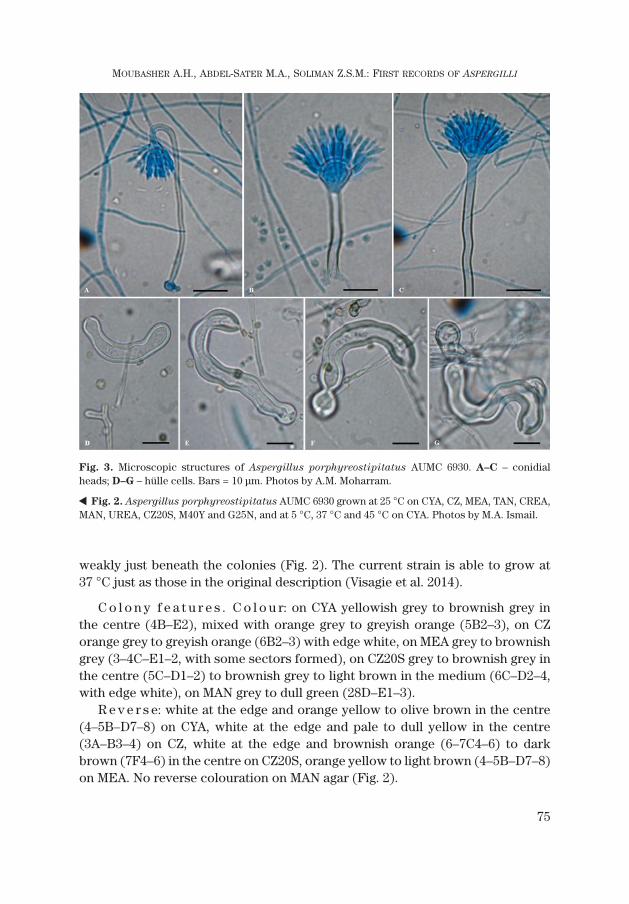
Fig. 3. Microscopic structures of Aspergillus porphyreostipitatus AUMC 6930. A–C – conidialheads; D–G – hülle cells. Bars = 10 μm. Photos by A.M. Moharram.
� Fig. 2. Aspergillus porphyreostipitatus AUMC 6930 grown at 25 °C on CYA, CZ, MEA, TAN, CREA,MAN, UREA, CZ20S, M40Y and G25N, and at 5 °C, 37 °C and 45 °C on CYA. Photos by M.A. Ismail.

Te x t u r e: floccose on CYA, CZ, CZ20S, CREA, MAN and UREA at 25 °C,slightly floccose on MEA at 25 °C and on CYA at 37 °C, slightly floccose with whit-ish powdery patches on TAN.
M i c r o s c o p i c f e a t u r e s (Tab. 3, Fig. 3). Conidial heads radiate to shortcolumnar; conidiophores biseriate; stipes sinuate, hyaline to brownish, mostlysmooth, but some areas show roughness, (20)50–125 μm long, (3)3.5–5.5 μm wide;vesicles globose, sometimes slightly elongated, (5)7–15 μm; metulae 7 × 2.5 μm,covering 75% of head; phialides ampulliform, 6–10 × 2.5–3.0 μm; conidia globoseto subglobose, 3–4 × 3–4 μm, spiny to tuberculate. Hülle cells on CYA, thick-walled,predominantly elongate, twisted, 26–92 μm long × 10–13 μm wide. Sclerotia absent,sexual stage not observed on any growth media incubated for up to 2 months.
M o l e c u l a r i d e n t i f i c a t i o n. Phylogenetically our current strain AUMC6930 matched with the type strains of A. porphyreostipitatus (99.81% sequencesimilarity), A. ustus (99.20%), A. baeticus (98.92%), A. puniceus (96.88%), A. gra-
nulosus (96.61%) and A. heterothallicus (96.47%) (Tab. 1, Fig. 1).
N o t e s. This species was first isolated in 2010 from dust from a church,Sayulita, Mexico (type CBS 138203T = DTO 266D9), and from house dust, Song-khla, Thailand (CBS 138202 = DTO 132D1) by Ed Whitfield & Kalima Mwange,and described as a new species (A. porphyreostipitatus) by Visagie et al. (2014).
The micro-morphological features of A. porphyreostipitatus are similar tothose of A. baeticus, A. ustus, A. puniceus, and A. pseudoustus. These speciesare similar in producing brownish colours in colonies (Visagie et al. 2014). Vesicleshapes and diameter both show significant differences between A. porphyreo-
stipitatus and A. ustus (hemispherical to subglobose, 7–15 μm in diameter) onthe one hand and A. baeticus (elliptical to elongate, 9.8–15.4 × 16.8–19.6 μm indiameter), A. pseudoustus (globose, 10–14 μm) and A. puniceus (subglobose,8–16 μm in diam. to elliptical, 15–18 × 13–15 μm) on the other. Additionally, thewidth of the stipes is smaller in A. porphyreostipitatus (3.5–6.5 μm), A. ustus
(3–6 μm) and A. pseudoustus (3.5–5 μm) than in A. baeticus (5–8 μm) andA. puniceus (5.5–8 μm). The ability of A. porphyreostipitatus to grow on CYA at37 °C easily distinguishes it from its morphologically similar relatives (A. baeti-
cus, A. ustus, A. puniceus, and A. pseudoustus).
Aspergillus carlsbadensis Frisvad, Varga & Samson 2011
In 2008–2009 and during a survey of filamentous fungi of orange and grape-vine plantations in the Assiut area, several isolates of Aspergillus related to sec-tion Usti were recovered from non-rhizosphere soil, in high frequency from or-ange and in moderate and low frequency from grapevine plantations on DRBC(King et al. 1979) and DYM (Moubasher et al. 2016). These isolates were initially
76
CZECH MYCOLOGY 70(1): 67–82, MAY 29, 2018 (ONLINE VERSION, ISSN 1805-1421)

identified as Aspergillus calidoustus (Soliman 2012, Abdel-Sater et al. 2016).A representative isolate was deposited at the culture collection of Assiut Univer-sity Mycological Centre and assigned code AUMC 6717. The ITS gene sequence ofthe strain is registered under GenBank accession number JQ425406 (Tab. 1, Fig. 1).The strain was also deposited in the Culture Collection of Fungi at the Depart-ment of Botany, Prague under no. CCF 5180.
G r o w t h c h a r a c t e r i s t i c s. Colony diameters (range and mean ± SD) af-ter 7 days at 25 °C on CYA, CZ, MEA, CREA, UREA, MAN, TAN and the low wateractivity media CZ20S, M40Y and G25N, and at 5 °C, 37 °C and 45 °C on CYA areshown in Tab. 2. No growth was detected on CYA at 5 °C or at 45 °C nor on G25Nat 25 °C, and no acid was produced on CREA or MAN agar at 25 °C, but the ureaseenzyme was produced (Fig. 4). Growth of the current strain at 37 °C is in contrastto that of the original description (Samson et al. 2011).
C o l o n y f e a t u r e s . C o l o u r: whitish on CYA, CZ, CREA (3A1–2), pale yel-low (3A1–3) to yellowish grey in the centre (2B2) on CZ20S, white to pale yellow(3–4A1–3) on UREA, dull green to greyish green (27E4–5) on M40Y, grey to greenishgrey (26B–C1–2) on MEA and greenish grey to dull green (27C–E2–3) on MAN.
R e v e r s e: edge and centre pale yellow (3A4–5) with a brown ring near edge(6F4–6) on CYA, pale yellow edge (3A3–4), yellow brown centre (5F5–8), witha yellowish brown ring in between (5F5–8) on CZ, brownish orange to goldenbrown (5B–D5–7) on MEA, pale yellow edge (3A2–3) to yellowish brown centre(5D–F5–8) on CZ20S at 25 °C, and pale to light yellow edge (3A3–5) to greyish yel-low to amber yellow in the centre (4B6–7) on CYA at 37 °C. No reverse colourationon MY40S, CREA, MAN and TAN.
Te x t u r e: velutinous on CYA, MEA, M40Y, TAN, CREA, MAN, UREA at 25 °Cand on CYA at 37 °C, slightly floccose on CZ and CZ20S at 25 °C.
M i c r o s c o p i c f e a t u r e s (Tab. 3, Fig. 5). Conidiophores biseriate with typ-ical smooth-walled, brown stipes 105–245 μm long, 5–6 μm wide, sometimes con-stricted below the vesicle; vesicles globose (6)10–15 μm in diam.; conidia dis-tinctly ornamented with spines or echinulations, ellipsoidal 3.5–4.0 × 3.0–3.5 μm.Hülle cells hyaline, thick-walled, globose to broadly ellipsoidal, 20–33 μm long to16–27 μm wide. Sclerotia not observed after 7 days on any growth media incu-bated at different temperatures; sexual stage not observed on any growth mediaincubated for up to 2 months.
M o l e c u l a r i d e n t i f i c a t i o n. The ribosomal DNA sequence of our strainmatched with the type strains of A. carlsbadensis (100% sequence similarity),A. asper (98.47%), A. germanicus (97.84%), A. calidoustus (97.84%), A. thesauricus
(97.65%), A. pseudodeflectus (97.49%), A. insuetus (97.31%) and A. keveii (97.05%)(Tab. 1, Fig. 1).
77
MOUBASHER A.H., ABDEL-SATER M.A., SOLIMAN Z.S.M.: FIRST RECORDS OF ASPERGILLI

78
CZECH MYCOLOGY 70(1): 67–82, MAY 29, 2018 (ONLINE VERSION, ISSN 1805-1421)

N o t e s. This species was first isolated in 1992 from soil, Lechuguilla Cave,Carlsbad Caverns National Park, New Mexico, USA, by D.E. Northup, soil fromGalapagos Islands, Ecuador, and soil from Carthage, Tunesia (Samson et al.2011).
Samson et al. (2011) revealed that A. carlsbadensis is related to but clearlydistinct from a clade including A. calidoustus, A. pseudodeflectus, A. insuetus
and A. keveii in all three phylogenetic trees presented, and it is unable to grow at37 °C, while acid production was not observed on CREA. However, our straincould grow at 37 °C, but does not produce organic acid on CREA.
The micro-morphological features of A. carlsbadensis are similar to those ofA. contaminans, A. calidoustus, A. keveii, A. pseudodeflectus, A. insuetus andA. granulosus. The vesicle shapes and diameter of these species (A. carlsbadensis
globose, 10–14 μm, A. ustus hemispherical to subglobose 7–15 μm diam.,A. insuetus hemispherical to subglobose, 11–16 μm diam., A. keveii pyriform,9–13 μm diam., A. pseudodeflectus globose to elevate, 4–12 μm and A. calidoustus
79
MOUBASHER A.H., ABDEL-SATER M.A., SOLIMAN Z.S.M.: FIRST RECORDS OF ASPERGILLI
Fig. 5. Microscopic structures of Aspergillus carlsbadensis AUMC 6717. A–C – conidial heads;D–G – hülle cells. Bars = 10 μm (A–C), 20 μm (D–G). Photos by A.M. Moharram.
� Fig. 4. Aspergillus carlsbadensis AUMC 6717 grown at 25 °C on CYA, CZ, MEA, TAN, CREA, MAN,UREA, CZ20S, M40Y and G25N, and at 5 °C, 37 °C and 45 °C on CYA. Photos by M.A. Ismail.

pyriform to broadly spathulate, 7–20 μm) are significantly different from those ofA. contaminans (globose or pyriform, 12–28 μm diam.) and A. granulosus (ovoidto elliptical, 15–25 × 12–18 μm). Additionally, the width of the stipes is smaller inA. carlsbadensis (4–5 μm), A. pseudodeflectus (2.5–3.5 μm), A. ustus (3–6 μm),and A. keveii (4–6 μm) than in A. contaminans, A. insuetus (4–8 μm), A. granu-
losus (5.5–8 μm) and A. calidoustus (4–7 μm). The conidia sizes can be used asa reliable feature for distinguishing three groups of species – conidia less 3 μm:A keveii; conidia exceeding 3 μm but less than 5 μm: A. carlsbadensis, A. calido-
ustus, A. contaminans, A. insuetus, A. ustus; up to 5–5.5 μm: A. pseudodeflectus
and A. granulosus. Aspergillus carlsbadensis strain AUMC 6717 (contrasting tothe originally described strains) shares with A. calidoustus, A. peseudodeflecus
and A. granulosus the ability to grow on CYA at 37 °C, which easily distinguishesthese species from morphologically similar relatives (A. contaminans, A. keveii,A. insuetus and A. ustus) (Houbraken et al. 2007, Samson et al. 2011, Crous et al.2017).
An added value of the current study was the use of media such as UREA,G25N, TAN and MAN, which enabled us to find more cultural or physiologicalcharacters that might be beneficial for differentiating these or other related spe-cies (ability of urease production, growing on G25N and TAN, growing and acidproduction on mannitol).
REFERENCES
ABDEL-SATER M.A., MOUBASHER A.H., SOLIMAN Z.S.M. (2016): Biodiversity of filamentous and yeastfungi in soil of citrus and grapevine plantations in Assiut area, Egypt. – Czech Mycology 68(2):183–214.
BALAJEE S.A., KANO R., BADDLEY J.W., MOSER S.A., MARR K.A., ALEXANDER B.D., ANDES D., KONTO-
YIANNIS D.P., PERRONE G., PETERSON S., BRANDT M.E., PAPPAS P.G., CHILLER T. (2009): Molecularidentification of Aspergillus species collected for the transplant-associated infection surveil-lance network. – Journal of Clinical Microbiology 47: 3138–3141. DOI: 10.1128/JCM.01070-09.
BLAKESLEE A. (1915): Lindner’s roll tube method of separation cultures. – Phytopathology 5: 68–69.BRAYFORD D., BRIDGE P.D. (1989): Differentiation of Fusarium oxysporum from Fusarium solani
by growth and pigmentation on media containing sugar alcohols. – Letters in Applied Microbiol-ogy 9(1): 9–12.
CHEN A.J., FRISVAD J.C., SUN B.D., VARGA J., KOCSUBE S., DIJKSTERHUIS J., KIM D.H., HONG S.-B.,HOUBRAKEN J., SAMSON R.A. (2016): Aspergillus section Nidulantes (formerly Emericella):polyphasic taxonomy, chemistry and biology. – Studies in Mycology 84: 1–118.DOI: 10.1016/j.simyco.2016.10.001.
CHEN A.J., HUBKA V., FRISVAD J.C., VISAGIE C.M., HOUBRAKEN J., MEIJER M., VARGA J., DEMIRE R.,JURJEVIĆ Ž., KUBÁTOVÁ A., SKLENÁŘ F., ZHOU Y.G., SAMSON R.A. (2017): Polyphasic taxonomy ofAspergillus section Aspergillus (formerly Eurotium), and its occurrence in indoor environ-ments and food. – Studies in Mycology 88: 37–135. DOI: 10.1016/j.simyco.2017.07.001.
80
CZECH MYCOLOGY 70(1): 67–82, MAY 29, 2018 (ONLINE VERSION, ISSN 1805-1421)

CHRISTENSEN W.B. (1946): Urea decomposition as a means of differentiating Proteus and paracoloncultures from each other and from Salmonella and Shigella types. – Journal of Bacteriology52(4): 461–466.
CROUS P.W., WINGFIELD M.J., BURGESS T.I., CARNEGIE A.J., HARDY G.E.St.J., SMITH D., SUMMERELL B.A.,CANO-LIRA J.F., GUARRO J., HOUBRAKEN J., LOMBARD L., MARTÍN M.P., SANDOVAL-DENIS M. et al.(2017): Fungal Planet description sheets: 625–715. – Persoonia 39: 270–467.DOI: 10.3767/persoonia.2017.39.11.
FAKIH M.G., BARDEN G.E., OAKES C.A., BERENSON C.S. (1995): First reported case of Aspergillus
granulosus infection in a cardiac transplant patient. – Journal of Clinical Microbiology 33:471–473.
FRISVAD J.F. (1985): Creatine sucrose agar, a differential medium for mycotoxin producing terverti-cillate Penicillium species. – Letters in Applied Microbiology 1: 109-113.
GAMS W., CHRISTENSEN M., ONIONS A.H., PITT J.I., SAMSON R.A. (1985): Infrageneric taxa of Asper-
gillus. – In: Samson R.A., Pitt J.I., eds., Advances in Penicillium and Aspergillus Systematics,pp. 55–62. Plenum Press, New York.
HENRY T., IWEN P.C., HINRICHS S.H. (2000): Identification of Aspergillus species using internal tran-scribed spacer regions 1 and 2. – Journal of Clinical Microbiology 38: 1510–1515.
HOUBRAKEN J., DUE M., VARGA J., MEIJER M., FRISVAD J.C., SAMSON R.A. (2007): Polyphasic taxonomyof Aspergillus section Usti. – Studies in Mycology 59: 107–128. DOI: 10.3114/sim.2007.59.12.
HUBKA V., LYSKOVÁ P., FRISVAD J.C., PETERSON S.W., SKOŘEPOVÁ M., KOLAŘÍK M. (2014): Aspergillus
pragensis sp. nov. discovered during molecular re-identification of clinical isolates belonging toAspergillus section Candidi. – Medical Mycology 52: 565–576. DOI: 10.1093/mmy/myu022.
HUBKA V., NOVÁKOVÁ A., PETERSON S.W., FRISVAD J.C., SKLENÁŘ F., MATSUZAWA T., KUBÁTOVÁ A.,KOLAŘÍK M. (2016): A reappraisal of Aspergillus section Nidulantes with descriptions of two newsterigmatocystin-producing species. – Plant Systematics and Evolution 302: 1267–1299.DOI: 10.1007/s00606-016-1331-5.
JANG S.S., DORR T.E., BIBERSTEIN E.L., WONG A. (1986): Aspergillus deflectus infection in four dogs. –Journal of Medical and Veterinary Mycology 24: 95–104.
JURJEVIĆ Ž., PETERSON S.W. (2016): Aspergillus asper sp. nov. and Asperillus collinsii sp. nov., fromAspergillus secion Usti. – International Journal of Systematic and Evolutionary Microbiology 66:2566–2572. DOI: 10.1099/ijsem.0.001094.
KING D.A., HOCKING A.D., PITT J.I. (1979): Dichloran rose Bengal medium for enumeration and isola-tion of molds from foods. – Applied and Environmental Microbiology 37: 959–964.
KLICH M.A. (1993): Morphological studies of Aspergillus section Versicolores and related species. –Mycologia 85: 100–107.
KORNERUP A., WANSCHER J.H. (1978): Methuen Handbook of Colour, 3rd ed. – 252 pp., London.KOZAKIEWICZ Z. (1989): Aspergillus species in stored products. – Mycological Papers 161: 1–188.KROCKENBERGER M.B., SWINNEY G., MARTIN P., ROTHWELL T.R., MALIK R. (2011): Sequential opportu-
nistic infections in two German Shepherd dogs. – Australian Veterinary Journal 89: 9–14.DOI: 10.1111/j.1751-0813.2010.00666.x.
MOUBASHER A.H. (1993): Soil fungi in Qatar and other Arab countries. – 566 pp., Scientific and Ap-plied Research Center, Qatar University, Doha.
MOUBASHER A.H., ABDEL-SATER M.A., SOLIMAN Z.S.M. (2016): Biodiversity and molecular character-ization of yeast and filamentous fungi in the air of citrus and grapevine plantations in Assiut area,Egypt. – Mycosphere 7(3): 236–261. DOI: 10.5943/mycosphere/7/3/1.
NOVÁKOVÁ A., HUBKA V., SAIZ-JIMENEZ C., KOLAŘÍK M. (2012): Aspergillus baeticus sp. nov. and Asper-
gillus thesauricus sp. nov., two species in section Usti from Spanish caves. – International Jour-nal of Systematic and Evolutionary Microbiology 62: 2778–2785. DOI: 10.1099/ijs.0.041004-0.
PELÁEZ T., ÁLVAREZ-PÉREZ S., MELLADO E., SERRANO D., VALERIO M., BLANCO J.L., GARCIA M.E.,MUŃOZ P., CUENCA-ESTRELLA M., BOUZA E. (2013): Invasive aspergillosis caused by cryptic
81
MOUBASHER A.H., ABDEL-SATER M.A., SOLIMAN Z.S.M.: FIRST RECORDS OF ASPERGILLI

Aspergillus species: a report of two consecutive episodes in a patient with leukaemia. – Journalof Medical Microbiology 62: 474–478. DOI: 10.1099/jmm.0.044867-0.
PETERSON S.W. (2000): Phylogenetic relationships in Aspergillus based on rDNA sequence analysis. –In: Samson R.A., Pitt J.I., eds., Integration of modern taxonomic methods for Penicillium andAspergillus classification, pp. 323–355. Harwood Academic Publishers, Amsterdam.
PETERSON S.W. (2008): Phylogenetic analysis of Aspergillus species using DNA sequences from fourloci. – Mycologia 100: 205–226.
PITT J.I. (1973): An appraisal of identification methods for Penicillium species: novel taxonomic cri-teria based on temperature and water relations. – Mycologia 65: 1135–1157.DOI: 10.1002/jobm.19810210822.
RAKEMAN J.L., BUI U., LAFE K., CHEN Y.-C., HONEYCUTT R.J., COOKSON B.T. (2005): Multilocus DNAsequence comparisons rapidly identify pathogenic molds. – Journal of Clinical Microbiology43(7): 3324–3333. DOI: 10.1128/JCM.43.7.3324-3333.2005.
RAPER K.B., FENNELL D.I. (1965): The genus Aspergillus. – 686 pp., Williams & Wilkins, Baltimore.RAPER K.B., THOM C. (1949): A manual of the Penicillia. – 878 pp., Williams & Wilkins, Baltimore.RINYU E., VARGA J., FERENCZY L., KOZAKIEWICZ Z. (2000): Phenotypic and genotypic variability within
Aspergillus section Fumigati. – In: Samson R.A., Pitt J.I., eds., Integration of modern taxonomicmethods for Penicillium and Aspergillus classification, pp. 483–490. Harwood Academic Pub-lishers, Amsterdam.
ROBINSON W.F., CONNOLE M.D., KING T.J., PITT J.I., MOSS S.M. (2000): Systemic mycosis due toAspergillus deflectus in a dog. – Australian Veterinary Journal 78: 600–602.
SAMSON R.A., PITT J.I. (1985): General recommendations. – In: Samson R.A., Pitt J.I., eds., Advancesin Penicillium and Aspergillus systematics, pp. 455–460. Plenum Press, New York.
SAMSON R.A., HOEKSTRA E.S., FRISVAD J.C., eds. (2004): Introduction to food and airborne fungi. 7th ed.– Centraal Bureau voor Schimmelcultures, Utrecht.
SAMSON R.A., VARGA J., MEIJER M., FRISVAD J.C. (2011): New taxa in Aspergillus section Usti. – Stud-ies in Mycology 69: 81–97. DOI: 10.3114/sim.2011.69.06.
SCHULTZ R.M., JOHNSON E.G., WISNER E.R., BROWN N.A., BYRNE B.A., SYKES J.E. (2008): Clinico-pathologic and diagnostic imaging characteristics of systemic aspergillosis in 30 dogs. – Journalof Veterinary Internal Medicine 22: 851–859.
SOLIMAN Z.S.M. (2012): Studies on yeast and filamentous fungi in citrus and grapevine plantations. –M.Sc. Thesis [depon. in Faculty of Science, Assiut University, Assiut, Egypt].
THOMPSON J.D., HIGGINS D.G., GIBSON T.J. (1994): CLUSTAL W: improving the sensitivity of progres-sive multiple sequence alignment through sequence weighting, position-specific gap penaltiesand weight matrix choice. – Nucleic Acids Research 22: 4673–4680. DOI: 10.1093/nar/22.22.4673.
THRANE U. (1986): The ability of common Fusarium species to grow on tannin-sucrose agar. – Let-ters in Applied Microbiology 2: 33–36.
VARGA J., HOUBRAKEN J., VAN DER LEE H.A., VERWEIJ P.E., SAMSON R.A. (2008): Aspergillus calidoustus
sp. nov., causative agent of human infections previously assigned to Aspergillus ustus. – EukaryoticCell 7: 630–638.
VARGA J., TÓTH B., RIGÓ K., TÉREN J., HOEKSTRA R.F., KOZAKIEWICZ Z. (2000): Phylogenetic analysis ofAspergillus section Circumdati based on sequences of the internal transcribed spacer regions ofthe 5.8 S rRNA gene. – Fungal Genetics and Biology 30: 71–80. DOI: 10.1006/fgbi.2000.1204.
VARGA J., FRISVAD J.C., SAMSON R.A. (2011): Two new aflatoxin producing species, and an overviewof Aspergillus section Flavi. – Studies in Mycology 69: 57–80. DOI: 10.3114/sim.2011.69.05.
VISAGIE C.M., HIROOKA Y., TANNEY J.B., WHITFIELD E., MWANGE K., MEIJER M., AMEND A.S., SEIFERT K.A.,SAMSON R.A. (2014): Aspergillus, Penicillium and Talaromyces isolated from house dust sam-ples collected around the world. – Studies in Mycology 78: 63–139.
WICKERHAM L.J. (1951): Taxonomy of yeasts. – Technical Bulletin No. 1029, U.S. Department of Agri-culture, Washington D.C.
82
CZECH MYCOLOGY 70(1): 67–82, MAY 29, 2018 (ONLINE VERSION, ISSN 1805-1421)


![[] Microbial Ecology of the Rhizosphere(BookFi.org)](https://static.fdocuments.us/doc/165x107/55cf944c550346f57ba106f1/-microbial-ecology-of-the-rhizospherebookfiorg.jpg)


