
Copyright OASIS, 2002 OASIS - LISA Global e-Business Survey.
Upload
truongkhanhCategory
view
239download
0
Finding An Oasis in OASIS‐C
Melanie Bowe RN, BSN, CWOCN
2
Home Health Agency Challenges
Under PPS (Prospective Payment System) the goal of Home Health Care providers is to provide quality, evidence-based care to patients in a cost-efficient manner, which may lead to improved outcomes.
Accurate completion of the integumentary related OASIS-C items provides data to ensure Home Health Care providers are achieving this goal.
3
Finding an oasis in OASIS‐C
Knowledge will provide you the fertile ground
needed to successfully
complete OASIS-C!

4
• OASIS‐C Integumentary Status Section
• WOCN Guidance on OASIS‐C Integumentary Items
• Clarification of the assessment and documentation of the types of wounds OASIS‐C is presently tracking:
– Pressure Ulcers
– Venous Stasis Ulcers
– Surgical Wounds
This program is not intended to provide OASIS coding advice or guidance on specific patients.
Please refer all coding questions to CMS or your agency coding specialist.
This presentation is designed to provide information on:
5
OASIS‐C Outcome Indicators‐Integmentary
Outcome Based Quality Improvement (OBQI) 1Measures changes in a patient’s health status between two or more time points
•Improvement in the Number of Surgical Wounds•Improvement in the Status of Surgical Wounds
Outcome Based Quality Management (OBQM)2 Adverse events•Increase in Number of Pressure Ulcers•Emergent Care for Wound Infection, Deteriorating Wound Status
Home Health Compare3 Publicly reported measures available on Medicare’s consumer-focused web site www.medicare.gov
•Improvement in the Status of Surgical Wounds•Percentage of patients whose wounds improved or healed after an operation
•Emergent Care for Wound Infection, Deteriorating Wound Status• Percentage of patients who need unplanned medical care related to a wound that is new, is worse, or has become infected.
Non Routine Supply (NRS) Payment4
• Medicare requires home health agencies to furnish the medical supplies (routine and non‐routine) required by a patient under a home health plan of care
• Medicare added a non‐routine supply (NRS) component to be paid in addition to the 60‐day episodic payment for certain supplies.
• The Non Routine Supply payment is based on a cumulative point system, determined by the response to specific OASIS questions. The specific areas are targeted:– Pressure Ulcer – number and stage of pressure ulcers– Stasis Ulcer ‐ number of stasis ulcers and most problematic– Surgical Wounds – status of most problematic healing and non healing
surgical wound– Ostomy – immediate post‐op or pre‐existing– Urinary Incontinence – requiring catheter– Bowel Incontinence– IV/Infusion therapy
Non‐Routine Supply Weights/Payment20105
Federal Register / Vol. 74, No. 216 / Tuesday, November 10, 2009 / Rules and Regulations, Page 58108
$561.4299+6
$326.4349‐985
$211.6928‐484
$142.4815‐273
$51.961‐142
$14.3901
PaymentPointsSeverity Level
8
Clinical Impact of Non‐Routine Supply Component
Severity‐adjusted payment will empower you to:
• Develop a dressing formulary that supports use of quality wound products
• Provide ostomy supplies that are appropriate to patient needs
The only catch is…
• You must now document supply use in the medical record for tracking/reporting of non‐routine supply costs5
9
WOCN Guidance on OASIS‐C Integumentary Items
• 2001 CMS (Centers for Medicare and Medicaid Services) collaborated with WOCN to clarify definitions for:6
– “Fully granulating”
– “Early/partial granulation”
– “Not healing”
These terms are used to describe the condition of wounds being tracked by the OASIS document.
• December 2009, The system for wound classification uses terms that lack universal definition and clinicians have verbalized concerns that they may be interpreting these terms incorrectly. The WOCN Society has therefore developed the new guidelines for the classification of wounds. These items were developed by consensus among the WOCN Society panel of content experts.7
10
OASIS Wound Assessment Definitions7
• Avascular: Lacking in blood supply; synonyms are dead, devitalized, necrotic and nonviable. Specific types include slough and eschar.
• Clean wound:Wound free of devitalized tissue, purulent drainage, foreign material or debris.
• Dead space: A defect or cavity.
• Dehisced: Separation of surgical incision; loss of approximation of wound edges.
• Full Thickness: Tissue damage involving total loss of epidermis and dermis and extending into the subcutaneous tissue and possibly into the muscle or bone.
• Partial Thickness: Confined to the skin layers; damage does not penetrate below the dermis and may be limited to the epidermal layers only.
11
Wound Bed Tissue Type
Granulation: The pink/red moist tissue comprised of new blood vessels, connective tissue, fibroblasts and inflammatory cells, which fills an open wound when it starts to heal; typically appears deep pink or red with an irregular, “berry‐like” surface7
Pink
Deep Pink
Red
12
Wound Bed Tissue Type
Clean but non‐granulating: Absence of granulation tissue: wound surface appears smooth as opposed to granular. For example, in a wound that is clean but non‐granulating, the wound surface appears smooth and red as opposed to berry‐like. 7
13
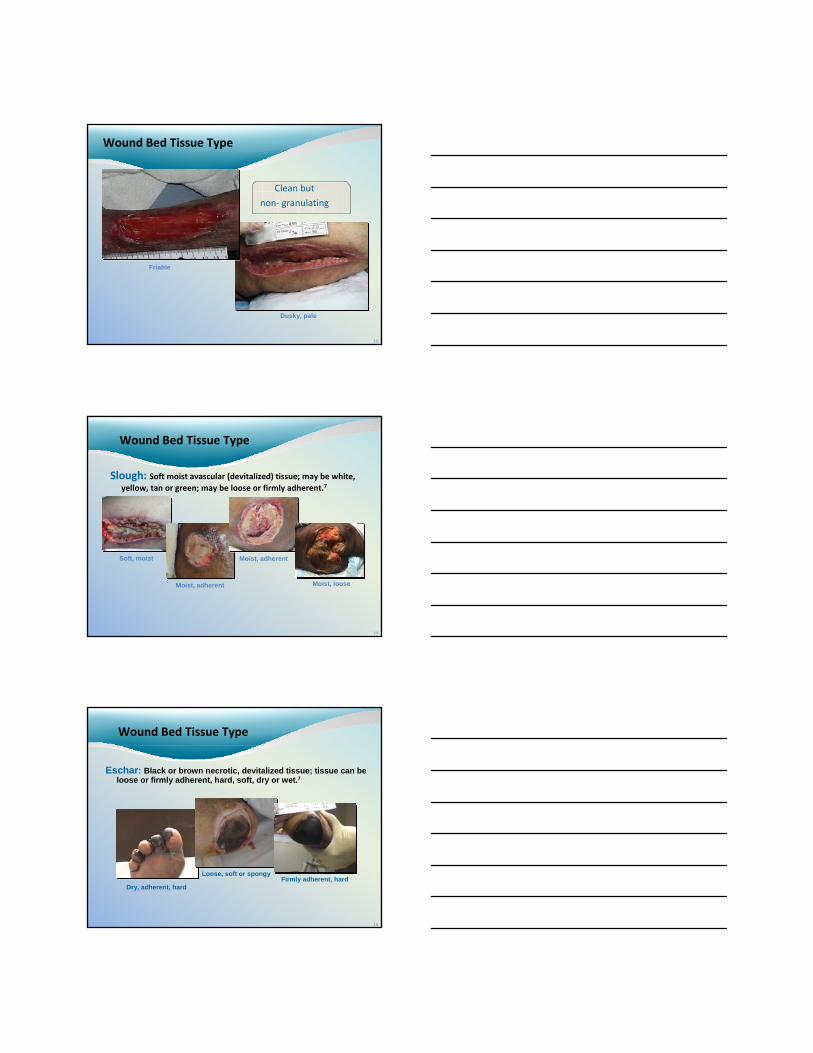
Wound Bed Tissue Type
Dusky, pale
Clean but
non‐ granulating
Friable
14
Wound Bed Tissue Type
Slough: Soft moist avascular (devitalized) tissue; may be white, yellow, tan or green; may be loose or firmly adherent.7
Soft, moist
Moist, adherent
Moist, adherent
Moist, loose
15
Wound Bed Tissue Type
Eschar: Black or brown necrotic, devitalized tissue; tissue can be loose or firmly adherent, hard, soft, dry or wet.7
Dry, adherent, hard
Loose, soft or spongyFirmly adherent, hard
16
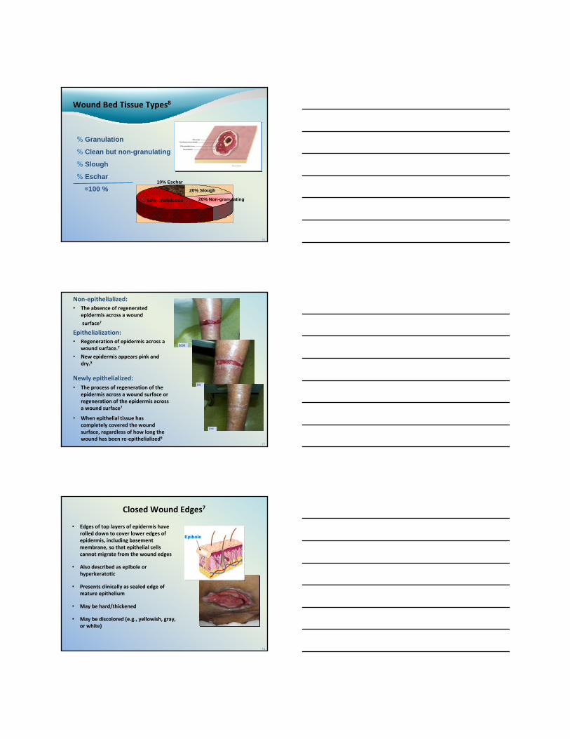
Wound Bed Tissue Types8
% Granulation
% Clean but non-granulating
% Slough
% Eschar
=100 %50% Granulation
20% Slough
20% Non-granulating
10% Eschar
17
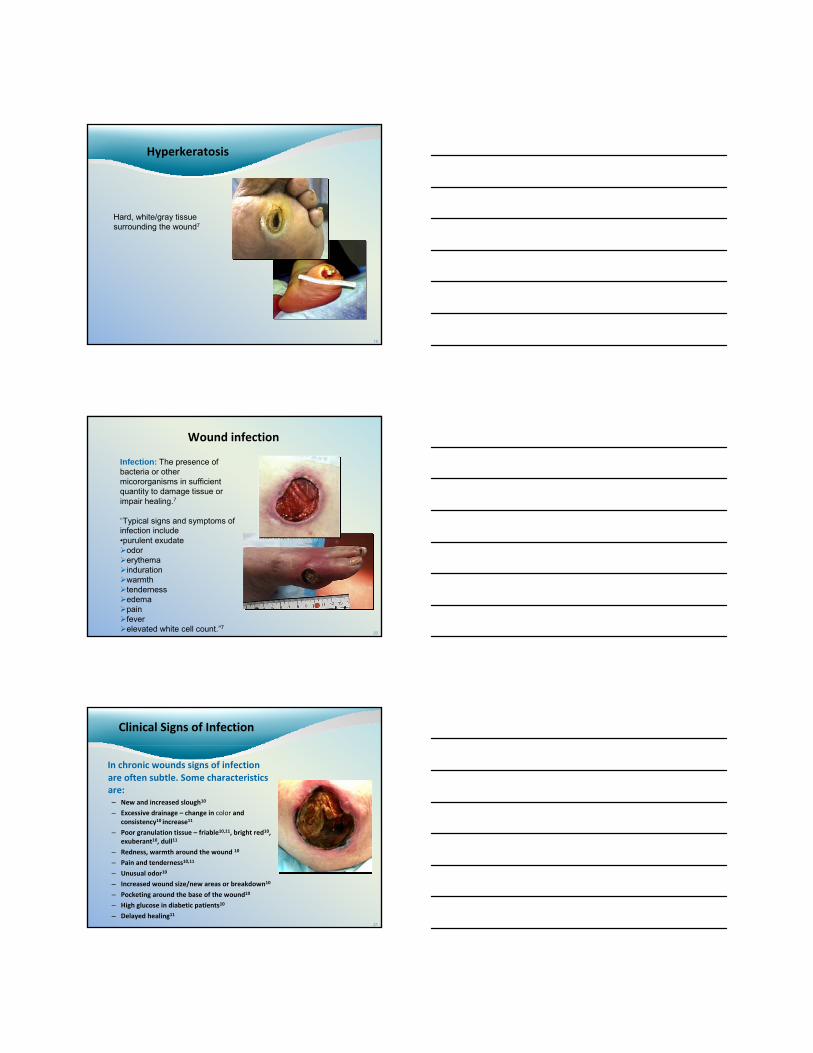
Non‐epithelialized:• The absence of regenerated
epidermis across a wound
surface7
Epithelialization:• Regeneration of epidermis across a
wound surface.7
• New epidermis appears pink and dry.8
Newly epithelialized:• The process of regeneration of the
epidermis across a wound surface or regeneration of the epidermis across a wound surface7
• When epithelial tissue has completely covered the wound surface, regardless of how long the wound has been re‐epithelialized9
18
Closed Wound Edges7
• Edges of top layers of epidermis have rolled down to cover lower edges of epidermis, including basement membrane, so that epithelial cells cannot migrate from the wound edges
• Also described as epibole or hyperkeratotic
• Presents clinically as sealed edge of mature epithelium
• May be hard/thickened
• May be discolored (e.g., yellowish, gray, or white)
Epibole
19
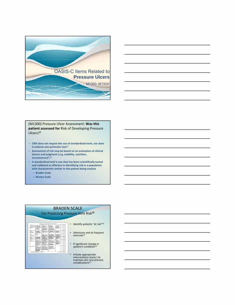
Hyperkeratosis
Hard, white/gray tissue surrounding the wound7
20
Wound infection
Infection: The presence of bacteria or other micororganisms in sufficient quantity to damage tissue or impair healing.7
“Typical signs and symptoms of infection include •purulent exudate
odor erythemaindurationwarmth tenderness edemapain feverelevated white cell count.”7
21
Clinical Signs of Infection
In chronic wounds signs of infection are often subtle. Some characteristics are:– New and increased slough10
– Excessive drainage – change in color and consistency10 increase11
– Poor granulation tissue – friable10,11, bright red10, exuberant10, dull11
– Redness, warmth around the wound 10
– Pain and tenderness10,11
– Unusual odor10
– Increased wound size/new areas or breakdown10
– Pocketing around the base of the wound10
– High glucose in diabetic patients10
– Delayed healing11
OASIS-C Items Related to Pressure Ulcers
M1300- M13249
(M1300) Pressure Ulcer Assessment: Was this patient assessed for Risk of Developing Pressure Ulcers?9
• CMS does not require the use of standardized tools, nor does it endorse one particular tool.9
• Assessment of risk may be based on an evaluation of clinical factors and judgment ( e.g. mobility, nutrition, incontinence)9,12
• A standardized tool is one that has been scientifically tested and validated as effective in identifying risk in a population with characteristic similar to the patient being evaluat
– Braden Scale
– Norton Scale
• Identify patients “at risk”14
• Admission and at frequent intervals14
• If significant change in patient’s condition14
• Initiate appropriate interventions (early ) to maintain skin and prevent complications14
BRADEN SCALEFor Predicting Pressure Sore Risk¹3
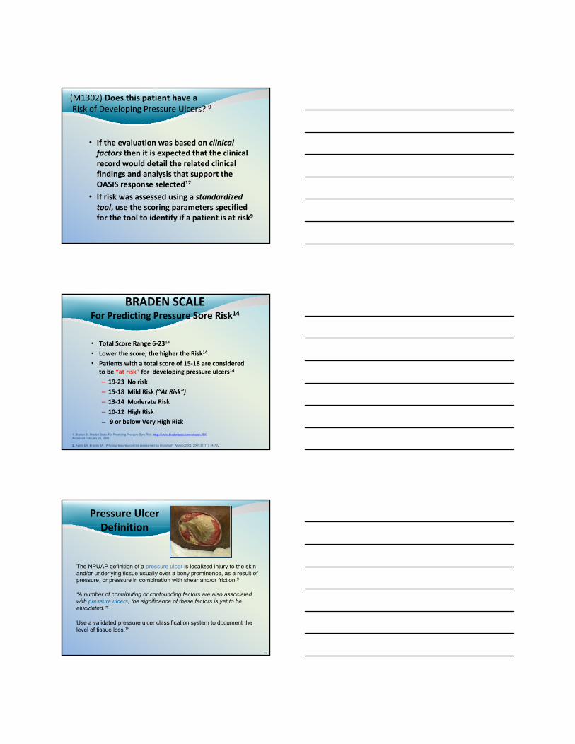
(M1302) Does this patient have aRisk of Developing Pressure Ulcers? 9
• If the evaluation was based on clinical factors then it is expected that the clinical record would detail the related clinical findings and analysis that support the OASIS response selected12
• If risk was assessed using a standardized tool, use the scoring parameters specified for the tool to identify if a patient is at risk9
• Total Score Range 6‐2314
• Lower the score, the higher the Risk14
• Patients with a total score of 15‐18 are considered to be “at risk” for developing pressure ulcers14
– 19‐23 No risk
– 15‐18 Mild Risk (“At Risk”)
– 13‐14 Moderate Risk
– 10‐12 High Risk
– 9 or below Very High Risk
BRADEN SCALEFor Predicting Pressure Sore Risk14
2. Ayello EA, Braden BA. Why is pressure ulcer risk assessment so important? Nursing2001. 2001;31(11): 74-79.
1. Braden B. Braden Scale For Predicting Pressure Sore Risk. http://www.bradenscale.com/braden.PDF. Accessed February 20, 2008.
27
Pressure UlcerDefinition
The NPUAP definition of a pressure ulcer is localized injury to the skin and/or underlying tissue usually over a bony prominence, as a result of pressure, or pressure in combination with shear and/or friction.9
“A number of contributing or confounding factors are also associated with pressure ulcers; the significance of these factors is yet to be elucidated.”7
Use a validated pressure ulcer classification system to document the level of tissue loss.15
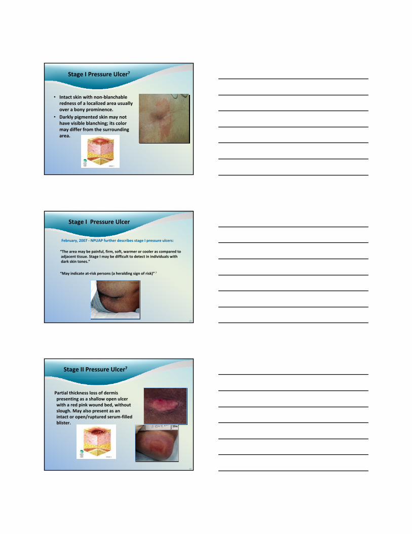
Stage I Pressure Ulcer7
• Intact skin with non‐blanchableredness of a localized area usually over a bony prominence.
• Darkly pigmented skin may not have visible blanching; its color may differ from the surrounding area.
29
Stage I Pressure Ulcer
February, 2007 ‐ NPUAP further describes stage I pressure ulcers:
“The area may be painful, firm, soft, warmer or cooler as compared to adjacent tissue. Stage I may be difficult to detect in individuals with dark skin tones.”
“May indicate at‐risk persons (a heralding sign of risk)” 7
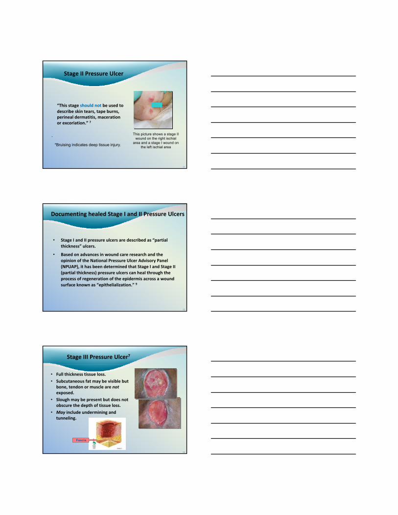
Stage II Pressure Ulcer7
Partial thickness loss of dermis presenting as a shallow open ulcer with a red pink wound bed, without slough. May also present as an intact or open/ruptured serum‐filled blister.
30
Stage II Pressure Ulcer
“This stage should not be used to describe skin tears, tape burns, perineal dermatitis, maceration or excoriation.” 7
.
*Bruising indicates deep tissue injury.
31
This picture shows a stage II wound on the right ischial
area and a stage I wound on the left ischial area
32
Documenting healed Stage I and II Pressure Ulcers
• Stage I and II pressure ulcers are described as “partial thickness” ulcers.
• Based on advances in wound care research and the opinion of the National Pressure Ulcer Advisory Panel (NPUAP), it has been determined that Stage I and Stage II (partial thickness) pressure ulcers can heal through the process of regeneration of the epidermis across a wound surface known as “epithelialization.” 9
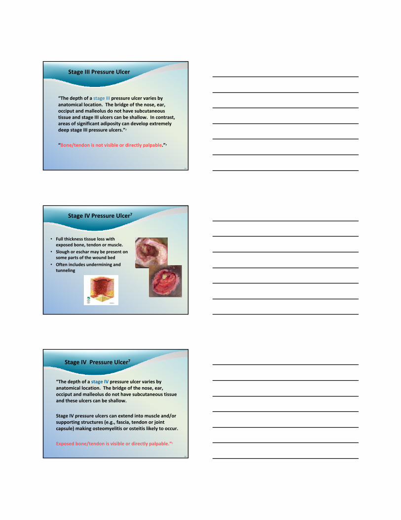
Stage III Pressure Ulcer7
• Full thickness tissue loss.
• Subcutaneous fat may be visible but bone, tendon or muscle are not exposed.
• Slough may be present but does not obscure the depth of tissue loss.
• May include undermining and tunneling.
33
Fascia
34
Stage III Pressure Ulcer
“The depth of a stage III pressure ulcer varies by anatomical location. The bridge of the nose, ear, occiput and malleolus do not have subcutaneous tissue and stage III ulcers can be shallow. In contrast, areas of significant adiposity can develop extremely deep stage III pressure ulcers.”7
“Bone/tendon is not visible or directly palpable.”7
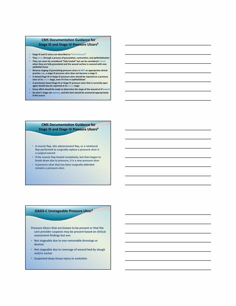
Stage IV Pressure Ulcer7
• Full thickness tissue loss with exposed bone, tendon or muscle.
• Slough or eschar may be present on some parts of the wound bed
• Often includes undermining and tunneling
36
Stage IV Pressure Ulcer7
“The depth of a stage IV pressure ulcer varies by anatomical location. The bridge of the nose, ear, occiput and malleolus do not have subcutaneous tissue and these ulcers can be shallow.
Stage IV pressure ulcers can extend into muscle and/or supporting structures (e.g., fascia, tendon or joint capsule) making osteomyelitis or osteitis likely to occur.
Exposed bone/tendon is visible or directly palpable.”7
37
• Stage III and IV ulcers are described as “full thickness”
• They close through a process of granulation, contraction, and epithelialization• They can never be considered “fully healed” but can be considered closed
when they are fully granulated and the wound surface is covered with new epithelial tissue
• Reverse staging of granulating pressure ulcers is NOT an appropriate clinical practice, i.e., a stage III pressure ulcer does not become a stage II.
• A closed Stage III or Stage IV pressure ulcer should be reported as a pressure ulcer at its worst stage, even if it has re‐epithelialized
• A previously closed Stage III or Stage IV pressure ulcer that is currently open again should also be reported at its worst stage
• Every effort should be made to determine the stage of the wound at it’s worst
• An ulcer’s stage can worsen, and this item should be answered appropriately if this occurs
CMS Documentation Guidance for Stage III and Stage IV Pressure Ulcers9
38
• A muscle flap, skin advancement flap, or a rotational flap performed to surgically replace a pressure ulcer is a surgical wound
• If the muscle flap healed completely, but then began to break down due to pressure, it is a new pressure ulcer
• A pressure ulcer that has been surgically debrided remains a pressure ulcer.
CMS Documentation Guidance for Stage III and Stage IV Pressure Ulcers9CMS Documentation Guidance for
Stage III and Stage IV Pressure Ulcers9
OASIS‐C Unstageable Pressure Ulcer9
Pressure Ulcers that are known to be present or that the care provider suspects may be present based on clinical assessment findings but are:
• Not stageable due to non‐removable dressings or devices
• Not stageable due to coverage of wound bed by slough and/or eschar
• Suspected deep tissue injury in evolution
40
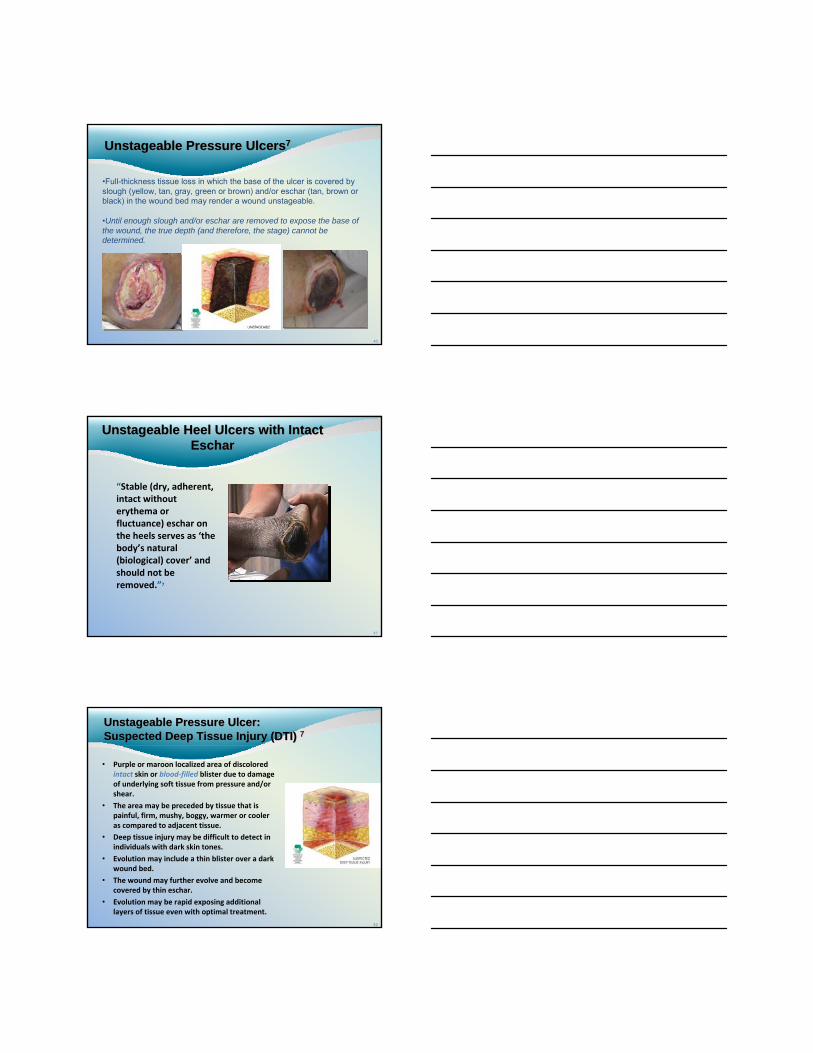
Unstageable Pressure Ulcers7Unstageable Pressure Ulcers7
•Full-thickness tissue loss in which the base of the ulcer is covered by slough (yellow, tan, gray, green or brown) and/or eschar (tan, brown or black) in the wound bed may render a wound unstageable.
•Until enough slough and/or eschar are removed to expose the base of the wound, the true depth (and therefore, the stage) cannot be determined.
41
“Stable (dry, adherent, intact without erythema or fluctuance) eschar on the heels serves as ‘the body’s natural (biological) cover’ and should not be removed.”7
Unstageable Heel Ulcers with Intact Eschar
Unstageable Heel Ulcers with Intact Eschar
42
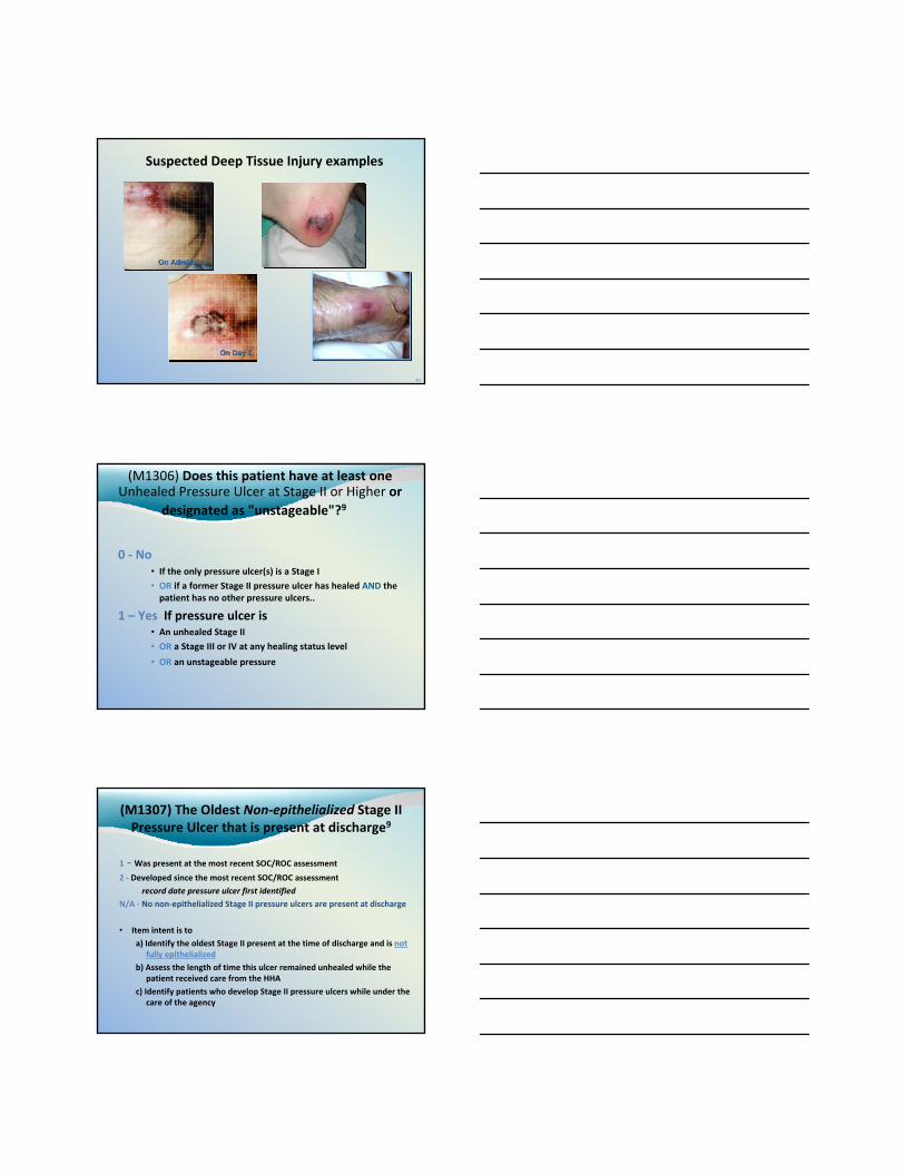
• Purple or maroon localized area of discolored intact skin or blood‐filled blister due to damage of underlying soft tissue from pressure and/or shear.
• The area may be preceded by tissue that is painful, firm, mushy, boggy, warmer or cooler as compared to adjacent tissue.
• Deep tissue injury may be difficult to detect in individuals with dark skin tones.
• Evolution may include a thin blister over a dark wound bed.
• The wound may further evolve and become covered by thin eschar.
• Evolution may be rapid exposing additional layers of tissue even with optimal treatment.
Unstageable Pressure Ulcer: Suspected Deep Tissue Injury (DTI) 7Unstageable Pressure Ulcer: Suspected Deep Tissue Injury (DTI) 7
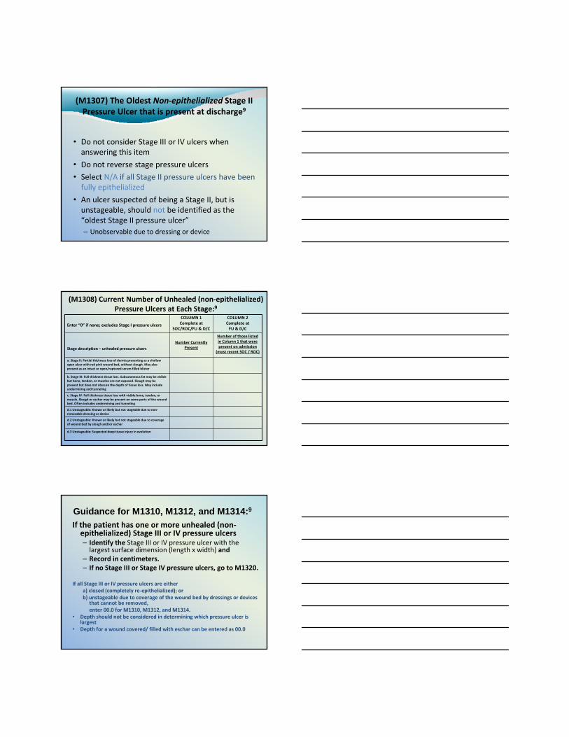
Suspected Deep Tissue Injury examples
On AdmissionOn Admission
OnOn Day 7Day 7
43
(M1306) Does this patient have at least one Unhealed Pressure Ulcer at Stage II or Higher or
designated as "unstageable"?9
0 ‐ No• If the only pressure ulcer(s) is a Stage I
• OR if a former Stage II pressure ulcer has healed AND the patient has no other pressure ulcers..
1 – Yes If pressure ulcer is• An unhealed Stage II
• OR a Stage III or IV at any healing status level
• OR an unstageable pressure
(M1307) The Oldest Non‐epithelialized Stage II Pressure Ulcer that is present at discharge9
1 ‐ Was present at the most recent SOC/ROC assessment
2 ‐ Developed since the most recent SOC/ROC assessment
record date pressure ulcer first identifiedN/A ‐ No non‐epithelialized Stage II pressure ulcers are present at discharge
• Item intent is toa) Identify the oldest Stage II present at the time of discharge and is not
fully epithelialized
b) Assess the length of time this ulcer remained unhealed while the patient received care from the HHA
c) Identify patients who develop Stage II pressure ulcers while under the care of the agency
(M1307) The Oldest Non‐epithelialized Stage II Pressure Ulcer that is present at discharge9
• Do not consider Stage III or IV ulcers when answering this item
• Do not reverse stage pressure ulcers
• Select N/A if all Stage II pressure ulcers have been fully epithelialized
• An ulcer suspected of being a Stage II, but is unstageable, should not be identified as the “oldest Stage II pressure ulcer”– Unobservable due to dressing or device
(M1308) Current Number of Unhealed (non‐epithelialized) Pressure Ulcers at Each Stage:9
d.3 Unstageable: Suspected deep tissue injury in evolution
d.2 Unstageable: Known or likely but not stageable due to coverage of wound bed by slough and/or eschar
d.1 Unstageable: Known or likely but not stageable due to non‐removable dressing or device
c. Stage IV: Full thickness tissue loss with visible bone, tendon, or muscle. Slough or eschar may be present on some parts of the wound bed. Often includes undermining and tunneling
b. Stage III: Full thickness tissue loss. Subcutaneous fat may be visible but bone, tendon, or muscles are not exposed. Slough may be present but does not obscure the depth of tissue loss. May include undermining and tunneling
a. Stage II: Partial thickness loss of dermis presenting as a shallow open ulcer with red pink wound bed, without slough. May also present as an intact or open/ruptured serum‐filled blister
Number of those listed in Column 1 that were present on admission
(most recent SOC / ROC)
Number Currently PresentStage description – unhealed pressure ulcers
COLUMN 2 Complete at FU & D/C
COLUMN 1 Complete at
SOC/ROC/FU & D/CEnter “0” if none; excludes Stage I pressure ulcers
If the patient has one or more unhealed (non‐epithelialized) Stage III or IV pressure ulcers – Identify the Stage III or IV pressure ulcer with the largest surface dimension (length x width) and
– Record in centimeters. – If no Stage III or Stage IV pressure ulcers, go to M1320.
If all Stage III or IV pressure ulcers are either a) closed (completely re‐epithelialized); orb) unstageable due to coverage of the wound bed by dressings or devices
that cannot be removed, enter 00.0 for M1310, M1312, and M1314.
• Depth should not be considered in determining which pressure ulcer is largest
• Depth for a wound covered/ filled with eschar can be entered as 00.0
Guidance for M1310, M1312, and M1314:9
(M1310) Pressure Ulcer Length: Longest length “head‐to‐toe” | ___ | ___ | . | ___ | (cm) 9
(M1312) Pressure Ulcer Width: Width of the same pressure ulcer; greatest width perpendicular to the length | ___ | ___ | . | ___ | (cm) 9
Head 12:00
L
W
(M1314) Pressure Ulcer Depth: Depth of the same pressure ulcer; from visible surface to the deepest area
| ___ | ___ | . | ___ | (cm) 9
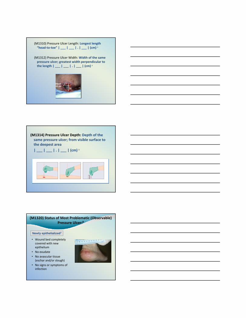
(M1320) Status of Most Problematic (Observable) Pressure Ulcer:9
• Wound bed completely covered with new epithelium
• No exudate
• No avascular tissue (eschar and/or slough)
• No signs or symptoms of infection
Newly epithelialized7
52
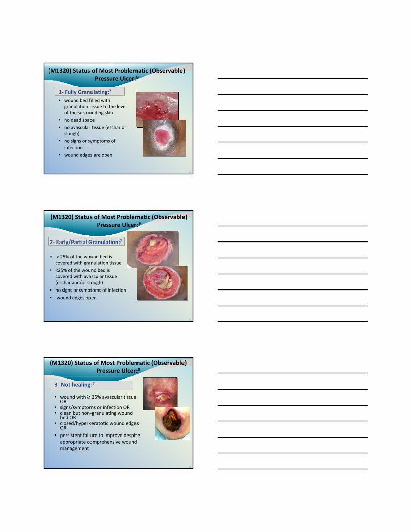
(M1320) Status of Most Problematic (Observable) Pressure Ulcer:9
1‐ Fully Granulating:7
• wound bed filled with granulation tissue to the level of the surrounding skin
• no dead space
• no avascular tissue (eschar or slough)
• no signs or symptoms of infection
• wound edges are open
53
(M1320) Status of Most Problematic (Observable) Pressure Ulcer:9
2‐ Early/Partial Granulation:7
• > 25% of the wound bed is covered with granulation tissue
• <25% of the wound bed is covered with avascular tissue (eschar and/or slough)
• no signs or symptoms of infection
• wound edges open
54
(M1320) Status of Most Problematic (Observable) Pressure Ulcer:9
3‐ Not healing:7
• wound with ≥ 25% avascular tissue OR
• signs/symptoms or infection OR• clean but non‐granulating wound
bed OR• closed/hyperkeratotic wound edges
OR• persistent failure to improve despite
appropriate comprehensive wound management
55
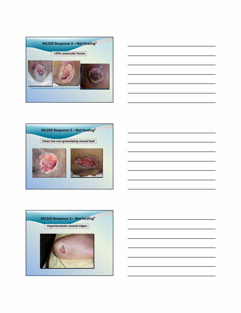
M1320 Response 3 – Not healing9
>25% avascular tissue
These wounds would be deemed unstageable due to the inability to view the base of the wound.9
56
M1320 Response 3 – Not healing9
Clean but non-granulating wound bed 1
57
M1320 Response 3 – Not healing9
Hyperkeratotic wound edges
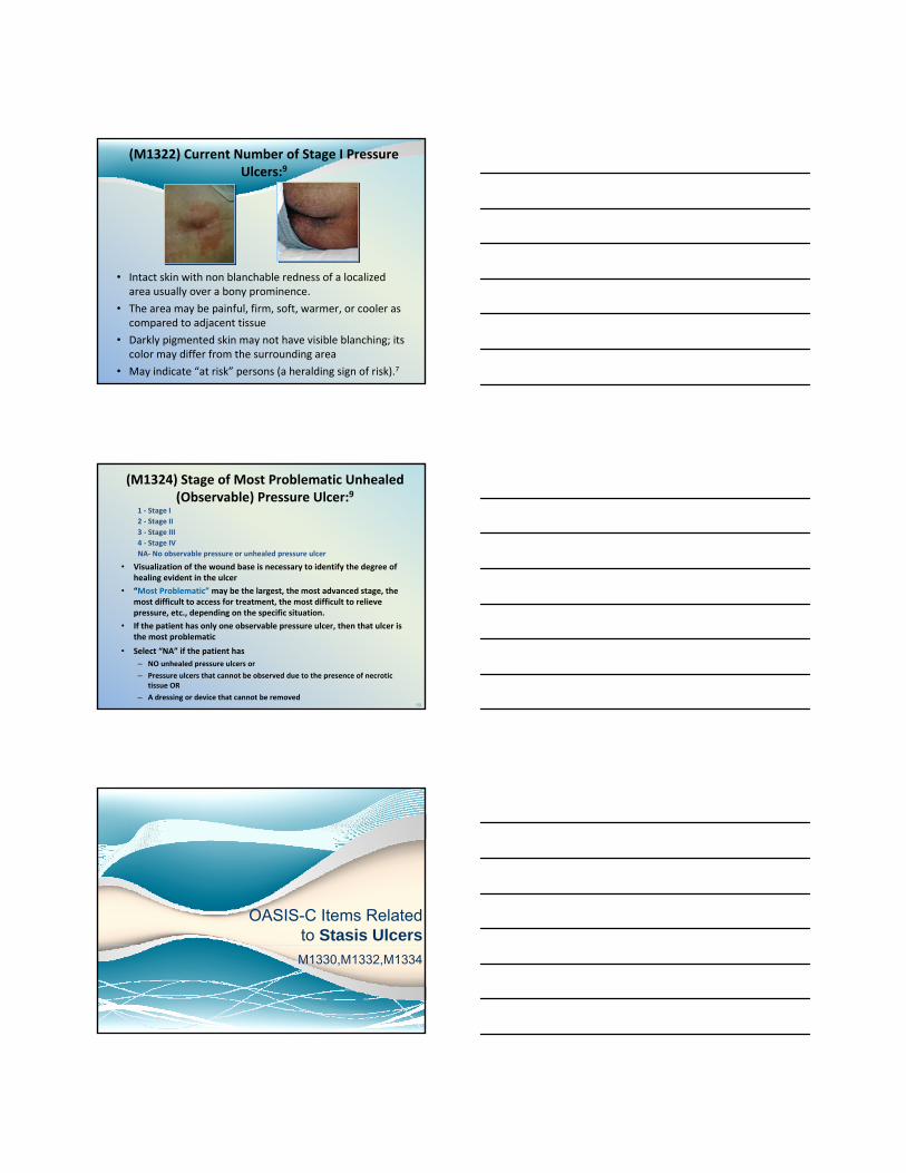
(M1322) Current Number of Stage I Pressure Ulcers:9
• Intact skin with non blanchable redness of a localized area usually over a bony prominence.
• The area may be painful, firm, soft, warmer, or cooler as compared to adjacent tissue
• Darkly pigmented skin may not have visible blanching; its color may differ from the surrounding area
• May indicate “at risk” persons (a heralding sign of risk).7
59
(M1324) Stage of Most Problematic Unhealed (Observable) Pressure Ulcer:9
1 ‐ Stage I 2 ‐ Stage II 3 ‐ Stage III 4 ‐ Stage IV NA‐ No observable pressure or unhealed pressure ulcer
• Visualization of the wound base is necessary to identify the degree of healing evident in the ulcer
• “Most Problematic” may be the largest, the most advanced stage, the most difficult to access for treatment, the most difficult to relieve pressure, etc., depending on the specific situation.
• If the patient has only one observable pressure ulcer, then that ulcer is the most problematic
• Select “NA” if the patient has– NO unhealed pressure ulcers or
– Pressure ulcers that cannot be observed due to the presence of necrotic tissue OR
– A dressing or device that cannot be removed
OASIS-C Items Related to Stasis UlcersM1330,M1332,M1334
61
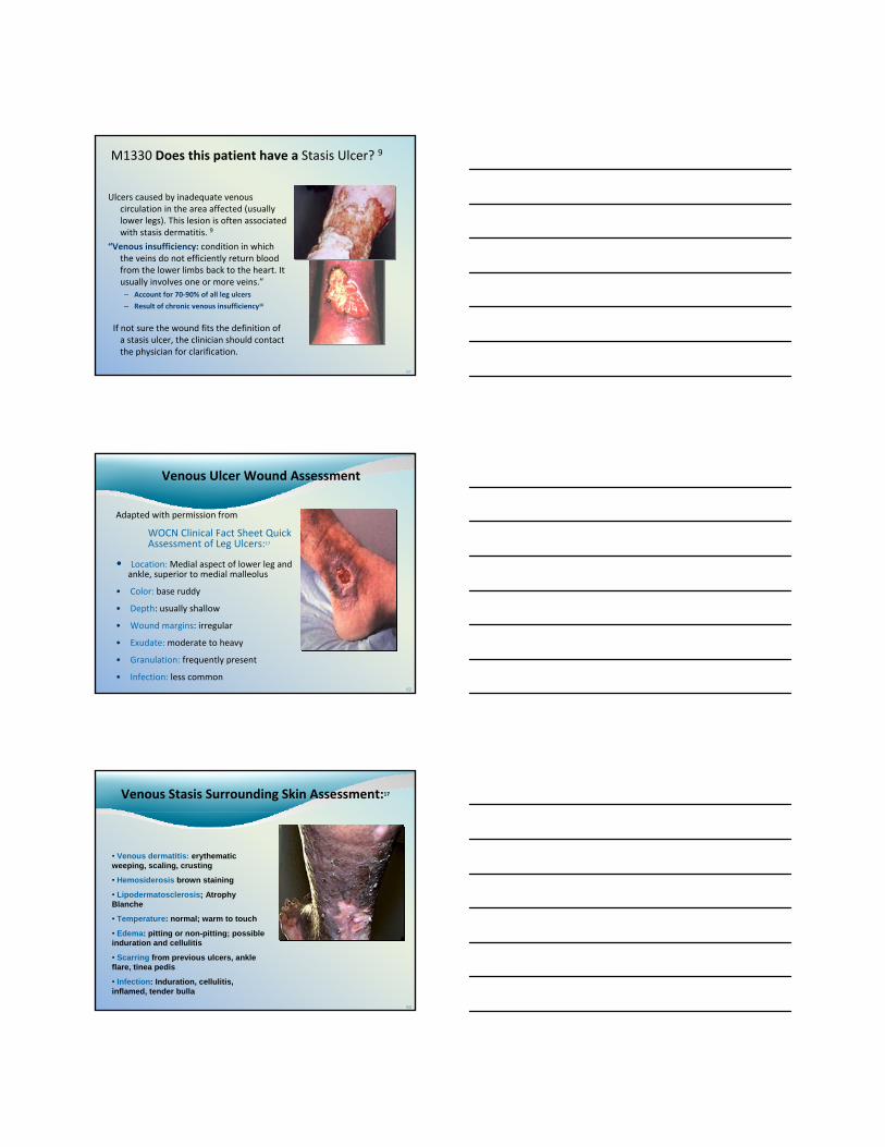
M1330 Does this patient have a Stasis Ulcer? 9
Ulcers caused by inadequate venous circulation in the area affected (usually lower legs). This lesion is often associated with stasis dermatitis. 9
“Venous insufficiency: condition in which the veins do not efficiently return blood from the lower limbs back to the heart. It usually involves one or more veins.”– Account for 70‐90% of all leg ulcers
– Result of chronic venous insufficiency16
If not sure the wound fits the definition of a stasis ulcer, the clinician should contact the physician for clarification.
62
Venous Ulcer Wound Assessment
Adapted with permission from
WOCN Clinical Fact Sheet QuickAssessment of Leg Ulcers:17
• Location: Medial aspect of lower leg and ankle, superior to medial malleolus
• Color: base ruddy
• Depth: usually shallow
• Wound margins: irregular
• Exudate: moderate to heavy
• Granulation: frequently present
• Infection: less common
63
Venous Stasis Surrounding Skin Assessment:17
• Venous dermatitis: erythematicweeping, scaling, crusting
• Hemosiderosis brown staining
• Lipodermatosclerosis; Atrophy Blanche
• Temperature: normal; warm to touch
• Edema: pitting or non-pitting; possibleinduration and cellulitis
• Scarring from previous ulcers, ankle flare, tinea pedis
• Infection: Induration, cellulitis, inflamed, tender bulla
64
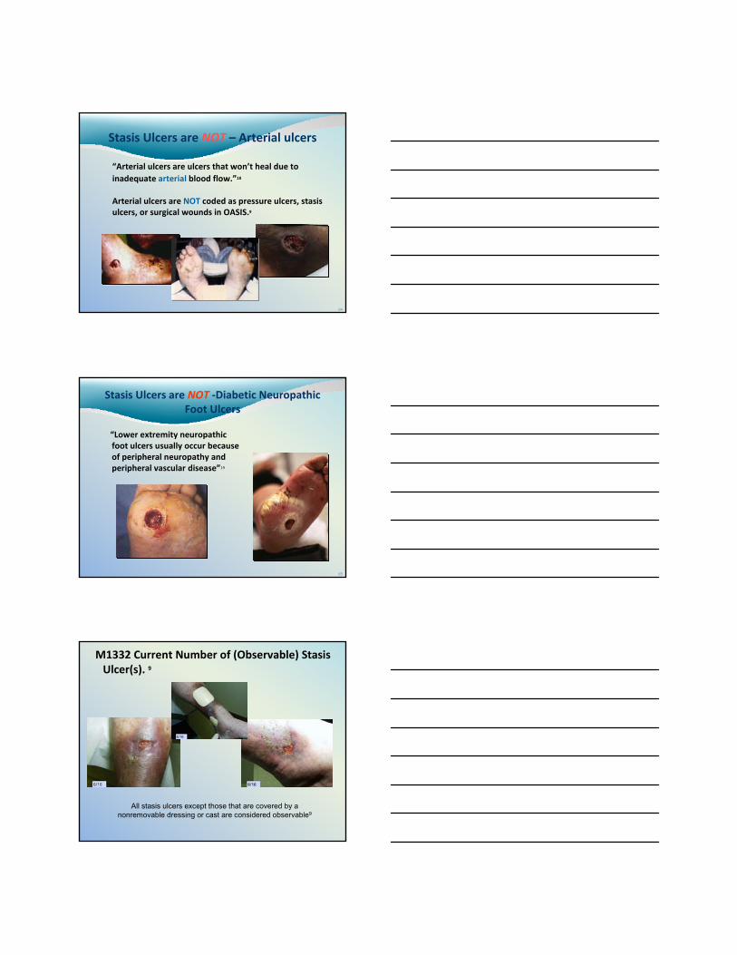
Stasis Ulcers are NOT – Arterial ulcers
“Arterial ulcers are ulcers that won’t heal due to inadequate arterial blood flow.”18
Arterial ulcers are NOT coded as pressure ulcers, stasis ulcers, or surgical wounds in OASIS.9
65
Stasis Ulcers are NOT ‐Diabetic Neuropathic Foot Ulcers
“Lower extremity neuropathic foot ulcers usually occur because of peripheral neuropathy and peripheral vascular disease”19
M1332 Current Number of (Observable) Stasis Ulcer(s). 9
All stasis ulcers except those that are covered by a nonremovable dressing or cast are considered observable9
67
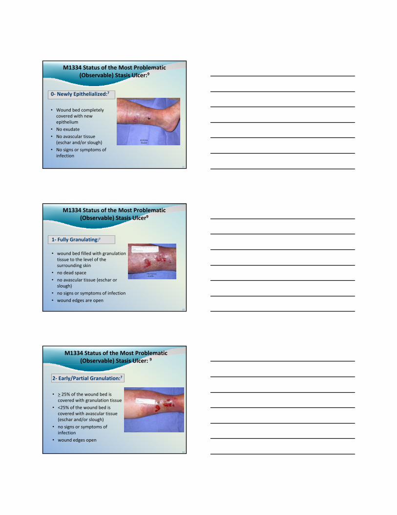
M1334 Status of the Most Problematic (Observable) Stasis Ulcer:9
0‐ Newly Epithelialized:7
• Wound bed completely covered with new epithelium
• No exudate
• No avascular tissue (eschar and/or slough)
• No signs or symptoms of infection
03/01/04Healed
68
M1334 Status of the Most Problematic (Observable) Stasis Ulcer9
1‐ Fully Granulating:7
• wound bed filled with granulation tissue to the level of the surrounding skin
• no dead space
• no avascular tissue (eschar or slough)
• no signs or symptoms of infection
• wound edges are open
After cleansing02/09/04
69
M1334 Status of the Most Problematic (Observable) Stasis Ulcer: 9
2‐ Early/Partial Granulation:7
• > 25% of the wound bed is covered with granulation tissue
• <25% of the wound bed is covered with avascular tissue (eschar and/or slough)
• no signs or symptoms of infection
• wound edges open
70
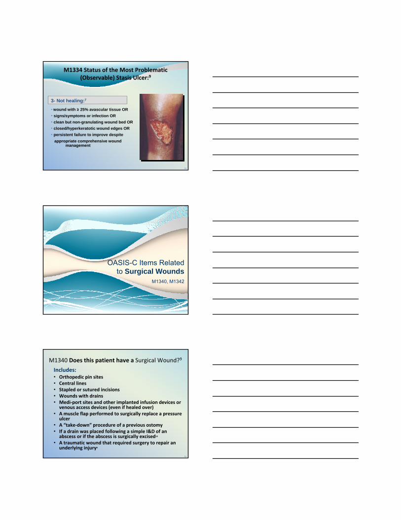
M1334 Status of the Most Problematic (Observable) Stasis Ulcer:9
3- Not healing:7
• wound with ≥ 25% avascular tissue OR• signs/symptoms or infection OR• clean but non-granulating wound bed OR• closed/hyperkeratotic wound edges OR• persistent failure to improve despite
appropriate comprehensive wound management
OASIS-C Items Related to Surgical Wounds
M1340, M1342
72
M1340 Does this patient have a Surgical Wound?9
Includes:• Orthopedic pin sites• Central lines• Stapled or sutured incisions• Wounds with drains• Medi‐port sites and other implanted infusion devices or
venous access devices (even if healed over)• A muscle flap performed to surgically replace a pressure
ulcer• A “take‐down” procedure of a previous ostomy• If a drain was placed following a simple I&D of an
abscess or if the abscess is surgically excised12
• A traumatic wound that required surgery to repair an underlying injury9
73
M1340 Does this patient have a Surgical Wound? 9
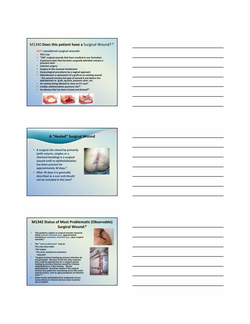
NOT considered surgical wounds:• PICC Line• “Old” surgical wounds that have resulted in scar formation• A pressure ulcer that has been surgically debrided remains a
pressure ulcer• Cataract surgery• Surgery to the mucosal membranes• Gynecological procedures by a vaginal approach• Debridement or placement of a graft on an existing wound.
‐ The wound remains the type of wound it was before the debridement or graft, eg burn, pressure ulcer, etc.
• An ostomy being allowed to close on it’s own9
• Cardiac catheterization puncture site20
• An abscess that has been incised and drained20
74
A “Healed” Surgical Wound
• A surgical site closed by primarily (with sutures, staples or a chemical bonding) is a surgical wound until re‐epithelializationhas been present for approximately 30 days.9
• After 30 days it is generally described as a scar and should not be included in this item9
75
M1342 Status of Most Problematic (Observable) Surgical Wound:9
• This guidance applies to surgical wounds closed by either primary intention (i.e., approximated incisions) or secondary intention (i.e., open surgical wounds).7
• The “most problematic” may be‐The only observable‐The largest‐The most resistant to treatment‐Infected9
• “Surgical incisions healing by primary intention do not granulate. Because of this the only response that could be appropriate for a surgical wound healing by primary intention would be 0‐Newly epithelialized or 3‐Not healing. “Newly epithelialized” should be chosen if the surgical incision has epidermal resurfacing across the entire wound surface, and no signs/symptoms of infection exist.”21
• Select newly epithelialized for implanted venous access device or infusion devices when insertion site is healed9
Day 28
76
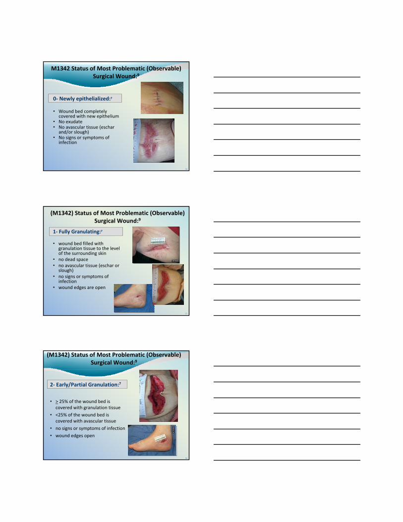
M1342 Status of Most Problematic (Observable) Surgical Wound:9
0‐ Newly epithelialized:7
• Wound bed completely covered with new epithelium
• No exudate• No avascular tissue (eschar
and/or slough)• No signs or symptoms of
infectionDay 28
77
(M1342) Status of Most Problematic (Observable) Surgical Wound:9
1‐ Fully Granulating:7
• wound bed filled with granulation tissue to the level of the surrounding skin
• no dead space• no avascular tissue (eschar or
slough)• no signs or symptoms of
infection• wound edges are open
78
(M1342) Status of Most Problematic (Observable) Surgical Wound:9
2‐ Early/Partial Granulation:7
• > 25% of the wound bed is covered with granulation tissue
• <25% of the wound bed is covered with avascular tissue
• no signs or symptoms of infection
• wound edges open
79
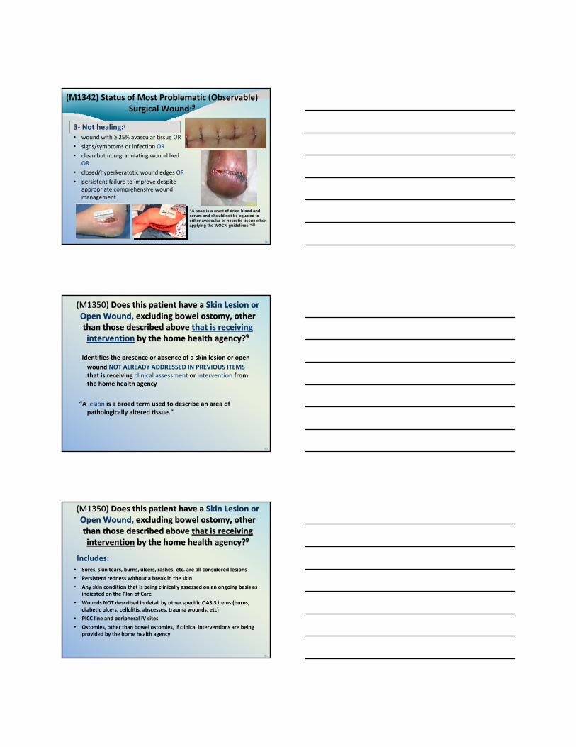
(M1342) Status of Most Problematic (Observable) Surgical Wound:9
3‐ Not healing:7
• wound with ≥ 25% avascular tissue OR
• signs/symptoms or infection OR
• clean but non‐granulating wound bed OR
• closed/hyperkeratotic wound edges OR
• persistent failure to improve despite appropriate comprehensive wound management
“A scab is a crust of dried blood and serum and should not be equated to either avascular or necrotic tissue when applying the WOCN guidelines.” 22
80
Identifies the presence or absence of a skin lesion or open wound NOT ALREADY ADDRESSED IN PREVIOUS ITEMSthat is receiving clinical assessment or intervention from the home health agency
“A lesion is a broad term used to describe an area of pathologically altered tissue.”
(M1350) Does this patient have a Skin Lesion or Open Wound, excluding bowel ostomy, other than those described above that is receiving intervention by the home health agency?9
(M1350) Does this patient have a Skin Lesion or Open Wound, excluding bowel ostomy, other than those described above that is receiving intervention by the home health agency?9
81
Includes:• Sores, skin tears, burns, ulcers, rashes, etc. are all considered lesions
• Persistent redness without a break in the skin
• Any skin condition that is being clinically assessed on an ongoing basis as indicated on the Plan of Care
• Wounds NOT described in detail by other specific OASIS items (burns, diabetic ulcers, cellulitis, abscesses, trauma wounds, etc)
• PICC line and peripheral IV sites
• Ostomies, other than bowel ostomies, if clinical interventions are being provided by the home health agency
(M1350) Does this patient have a Skin Lesion or Open Wound, excluding bowel ostomy, other than those described above that is receiving intervention by the home health agency?9
(M1350) Does this patient have a Skin Lesion or Open Wound, excluding bowel ostomy, other than those described above that is receiving intervention by the home health agency?9
82
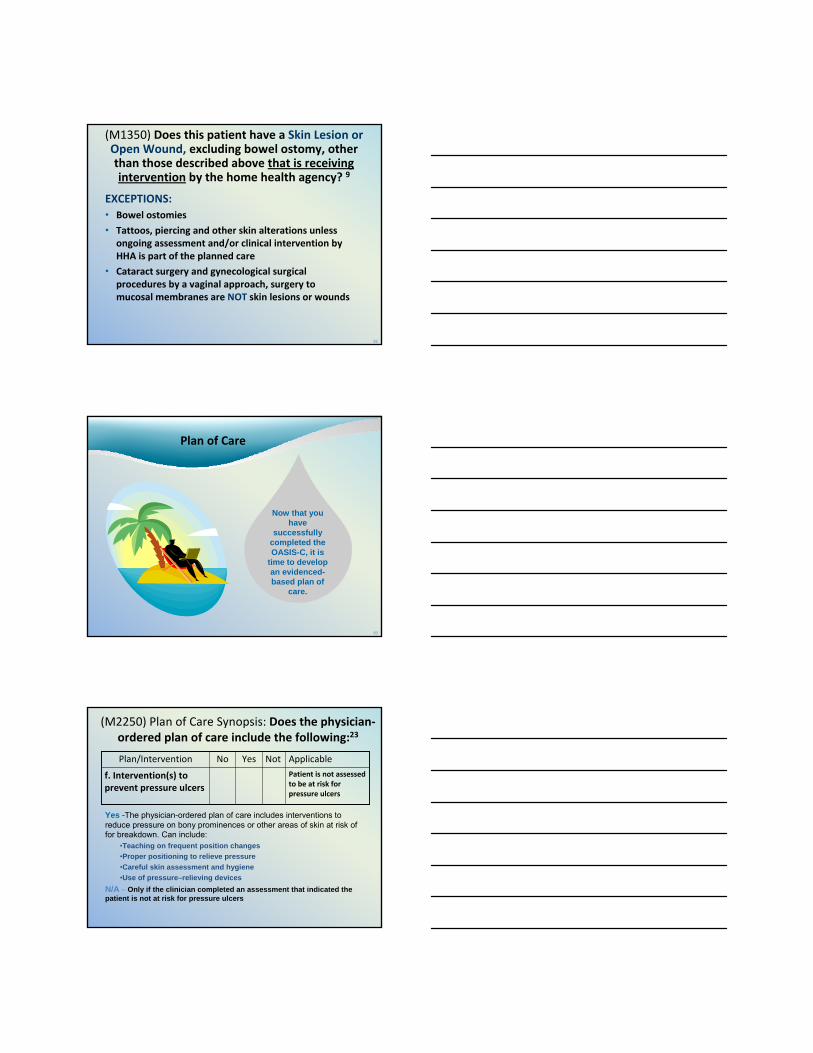
(M1350) Does this patient have a Skin Lesion or Open Wound, excluding bowel ostomy, other than those described above that is receiving intervention by the home health agency? 9
EXCEPTIONS:• Bowel ostomies
• Tattoos, piercing and other skin alterations unless ongoing assessment and/or clinical intervention by HHA is part of the planned care
• Cataract surgery and gynecological surgical procedures by a vaginal approach, surgery to mucosal membranes are NOT skin lesions or wounds
83
Plan of Care
Now that you have
successfully completed the OASIS-C, it is
time to develop an evidenced-based plan of
care.
(M2250) Plan of Care Synopsis: Does the physician‐ordered plan of care include the following:23
ApplicableNotYesNoPlan/InterventionPatient is not assessed to be at risk for pressure ulcers
f. Intervention(s) to prevent pressure ulcers
Yes -The physician-ordered plan of care includes interventions to reduce pressure on bony prominences or other areas of skin at risk of for breakdown. Can include:
•Teaching on frequent position changes•Proper positioning to relieve pressure•Careful skin assessment and hygiene•Use of pressure–relieving devices
N/A – Only if the clinician completed an assessment that indicated thepatient is not at risk for pressure ulcers
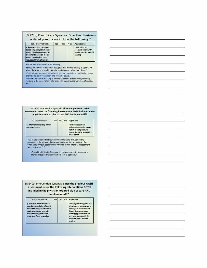
(M2250) Plan of Care Synopsis: Does the physician‐ordered plan of care include the following:23
ApplicableNotYesNoPlan/InterventionPatient has no pressure ulcers with need for moist wound healing
g. Pressure ulcer treatment based on principles of moist wound healing OR order for treatment based on moist wound healing has been requested from physician
Principles of moist wound healing•Since the 1960s, it has been accepted that wound healing is optimized when the wound is kept in a moist environment rather than dried.15
•Occlusive or semiocclusive dressings that maintain wound bed moisture promote re-epithelialization and wound closure.15
•Moisture-retentive dressing is one that is capable of consistently retaining moisture at the wound site by interfering with natural evaporative loss of moisture vapor24
(M2400) Intervention Synopsis. Since the previous OASIS assessment, were the following interventions BOTH included in the
physician‐ordered plan of care AND implemented?25
ApplicableNotYesNoPlan/Intervention
Formal assessment indicates the patient was not at risk of pressure ulcers since the last OASIS assessment
e. Intervention(s) to prevent pressure ulcers
Yes- if the specified clinical interventions were included in the physician ordered plan of care and implemented at the time of orsince the previous assessment whether or not a formal assessmentwas performed.12,25
•Recall for M1300 – Pressure Ulcer Assessment, the use of a standardized/formal assessment tool is optional.9
(M2400) Intervention Synopsis. Since the previous OASIS assessment, were the following interventions BOTH included in the physician‐ordered plan of care AND
implemented?25
ApplicableNotYesNoPlan/Intervention
Dressings that support the principles of moist wound healing not indicated for this patient’s pressure ulcers OR patient has no pressure ulcers with the need for moist wound healing
g. Pressure ulcer treatment based on principles of moist wound healing OR order for treatment based on moist wound healing has been requested from physician
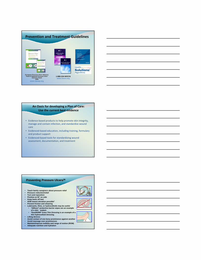
Prevention and Treatment Guidelines
European Pressure Ulcer Advisory European Pressure Ulcer Advisory Panel & National Pressure Ulcer Panel & National Pressure Ulcer
Advisory PanelAdvisory Panel20092009
www.npuap.org
11--888888--224224--WOCNWOCNwww.wocn.org
89
An Oasis for developing a Plan of Care:Use the current best evidence
• Evidence‐based products to help promote skin integrity, manage and contain infection, and standardize wound care
• Evidenced‐based education, including training, formulary and product support
• Evidenced‐based tools for standardizing wound assessment, documentation, and treatment
Teach family caregivers about pressure relief Pressure reduction/reliefTurn and repositionPosition at 30° on sideKeep heels off bedHOB lowest elevation possible7
Prevent friction and shearingLubricants, films, or hydrocolloids may be useful
*AllKare® protective barrier wipes are an example of a skin sealant. *DuoDERM® Extra Thin Dressing is an example of a thin hydrocolloid dressing.
Lifting devicesAvoid contact of one bony prominence against anotherAvoid massage over prominencesMaintain/improve mobility and range of motion (ROM)Adequate nutrition and hydration
Preventing Pressure Ulcers26Preventing Pressure Ulcers26
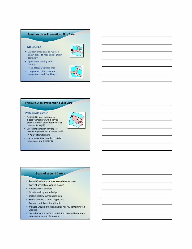
Moisturize
Use skin emollients to hydrate skin in order to reduce risk of skin damage15
Apply after bathing and as needed
Do not apply between toes
Use products that contain Humectants and Emollients
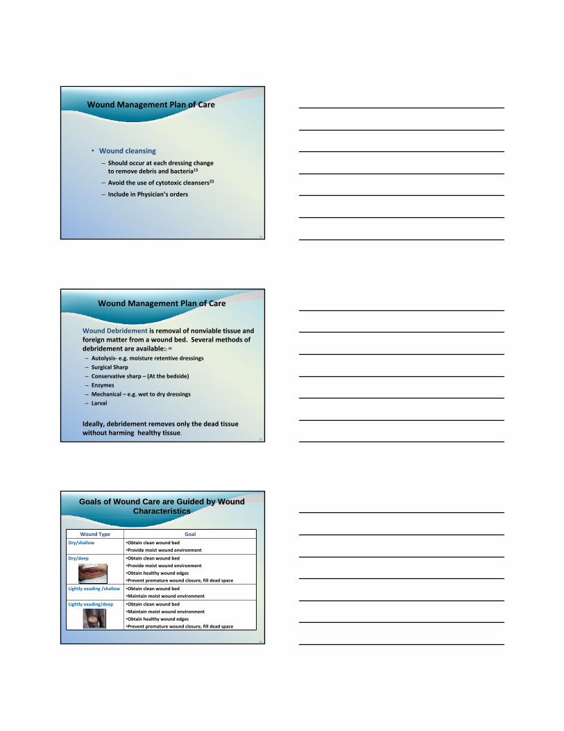
Pressure Ulcer Prevention: Skin Care
Protect with Barrier
Protect skin from exposure to excessive moisture with a barrier product in order to reduce the risk of pressure damage15
Use incontinent skin barriers…as needed to protect and maintain skin25
Apply after cleansing
Use protectant barriers that contain Humectants and Emollients
Pressure Ulcer Prevention : Skin Care
93
Goals of Wound Care:18
• Provide/maintain a moist wound environment
• Prevent premature wound closure
• Absorb excess exudate
• Obtain healthy wound edges
• Obtain healthy surrounding skin
• Eliminate dead space, if applicable
• Promote autolysis, if applicable
• Manage wound infection and/or heavily contaminated wounds
• Consider topical antimicrobials for bacterial bioburdenon wounds at risk of infection
94
Wound Management Plan of Care
• Wound cleansing
– Should occur at each dressing change to remove debris and bacteria15
– Avoid the use of cytotoxic cleansers25
– Include in Physician’s orders
95
Wound Management Plan of Care
Wound Debridement is removal of nonviable tissue and foreign matter from a wound bed. Several methods of debridement are available:: 26
– Autolysis‐ e.g. moisture retentive dressings
– Surgical Sharp
– Conservative sharp – (At the bedside)
– Enzymes
– Mechanical – e.g. wet to dry dressings
– Larval
Ideally, debridement removes only the dead tissue without harming healthy tissue.
96
Wound Type Goal
Dry/shallow •Obtain clean wound bed•Provide moist wound environment
Dry/deep •Obtain clean wound bed•Provide moist wound environment
•Obtain healthy wound edges•Prevent premature wound closure, fill dead space
Lightly exuding /shallow •Obtain clean wound bed•Maintain moist wound environment
Lightly exuding/deep •Obtain clean wound bed•Maintain moist wound environment
•Obtain healthy wound edges•Prevent premature wound closure, fill dead space
Goals of Wound Care are Guided by Wound Characteristics
Goals of Wound Care are Guided by Wound Characteristics
97
Wound Type Goal
Moderately exuding/shallow •Obtain clean wound bed•Maintain moist wound environment
•Absorb excess exudate
Moderately exuding/deep •Obtain clean wound bed•Maintain moist wound environment
•Obtain healthy wound edges•Prevent premature wound closure, fill dead space
•Absorb excess exudate
Goals of Wound Care are Guided by Wound Characteristics
Goals of Wound Care are Guided by Wound Characteristics
98
Wound Type Goal
Heavily exuding/shallow •Obtain clean wound bed•Maintain moist wound environment
•Absorb excess exudate
Heavily exuding/deep •Obtain clean wound bed•Maintain moist wound environment
•Obtain healthy wound edges•Prevent premature wound closure
•Absorb excess exudate
Goals of Wound Care are Guided by Wound Characteristics
Goals of Wound Care are Guided by Wound Characteristics
99
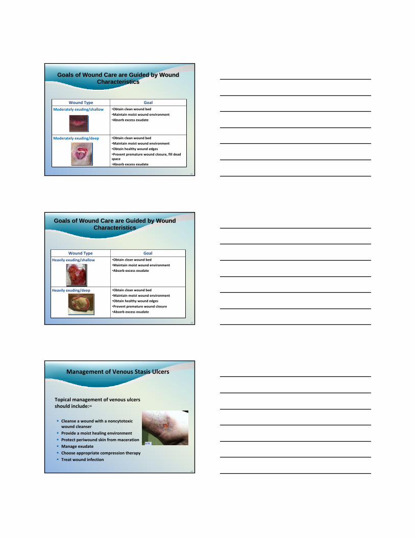
Management of Venous Stasis Ulcers
Topical management of venous ulcers should include:16
Cleanse a wound with a noncytotoxicwound cleanser
Provide a moist healing environment
Protect periwound skin from maceration
Manage exudate
Choose appropriate compression therapy
Treat wound infection
100
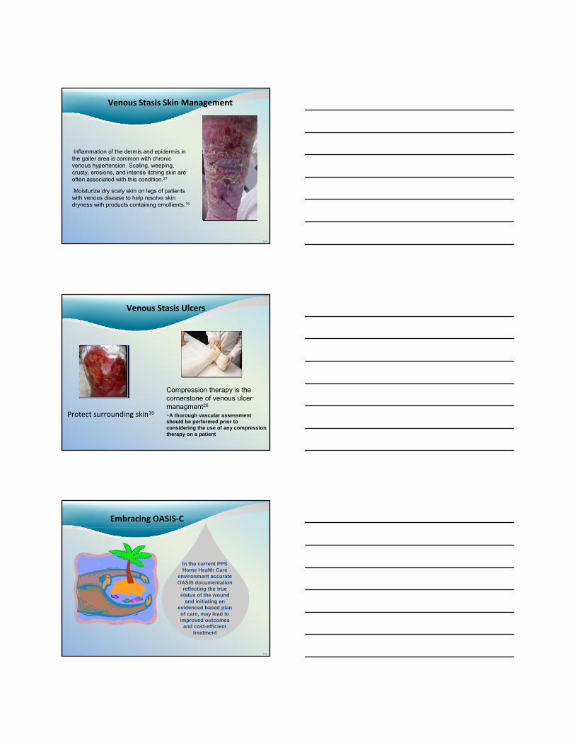
Venous Stasis Skin Management
Inflammation of the dermis and epidermis in the gaiter area is common with chronic venous hypertension. Scaling, weeping, crusty, erosions, and intense itching skin are often associated with this condition.27
Moisturize dry scaly skin on legs of patients with venous disease to help resolve skin dryness with products containing emollients.16
Venous Stasis Ulcers
Protect surrounding skin16
Compression therapy is the cornerstone of venous ulcer managment26
A thorough vascular assessment should be performed prior to considering the use of any compression therapy on a patient
102
Embracing OASIS‐C
In the current PPS Home Health Care
environment accurate OASIS documentation
reflecting the true status of the wound
and initiating an evidenced based plan of care, may lead to improved outcomes
and cost-efficient treatment


![OASIS-D Handouts [Read-Only]€¦ · • Describe the major changes from OASIS-C2 to OASIS-D • Understand OASIS M-item coding instructions to accurately code new and revised OASIS](https://static.fdocuments.us/doc/165x107/5ec3637ace40ce0748747c2e/oasis-d-handouts-read-only-a-describe-the-major-changes-from-oasis-c2-to-oasis-d.jpg)