Final Program and Abstracts - uni-due.de 2005.pdf · Final Program and Abstracts September 21st...
176
8 th Essen Symposium on Biomaterials and Biomechanics: Fundamental and Clinical Applications of the Working Group on „Biomaterials and Tissue Compatibility“ Final Program and Abstracts September 21 st – 23 rd , 2005 University Duisburg-Essen Essen, Germany Organizing Committee: M. Epple, D. Bingmann, A. Fischer, H. P. Jennissen Sponsored by: AG Biomaterialien NRW, Bochum LifeTecRuhr, Bochum Kulturstiftung Essen e.V., Essen Fahrzeugwerke Lueg AG, Essen
Transcript of Final Program and Abstracts - uni-due.de 2005.pdf · Final Program and Abstracts September 21st...

8th Essen Symposium onBiomaterials and Biomechanics:Fundamental and Clinical Applications
of the Working Group on „Biomaterials and Tissue Compatibility“
Final Program and Abstracts
September 21st – 23rd, 2005University Duisburg-EssenEssen, Germany
Organizing Committee:M. Epple, D. Bingmann, A. Fischer, H. P. Jennissen
Sponsored by:AG Biomaterialien NRW, BochumLifeTecRuhr, BochumKulturstiftung Essen e.V., EssenFahrzeugwerke Lueg AG, Essen

8th Essen Symposium on Biomaterials and Biomechanics: Fundamental and Clinical Applications
1
Scope of the Symposium Biomaterials constitute a still growing area of research in science, driven by the
demand of clinical medicine for suitable synthetic materials for the regeneration of
biological functions. A significant progress is only achievable by the joint efforts of
clinicians, biologists, biochemists, and materials scientists (e.g., chemists, engineers,
physicists). Biological, chemical, and mechanical requirements must be fulfilled by a
good implant. The interplay between biological function (including implant
performance) and the mechanical loads on the body is described as biomechanics,
another emerging field which tries to understand and subsequently to enhance the
mechanical performance of implants. Regenerative medicine is a further emerging
area of the biosciences where the biological aspect is even more important.
It is the aim of this symposium to bring together for the 8th time people from various
areas of research to accomplish an open and lively discussion on current problems in
the area of biomaterials. We invite you to join us at Essen for another productive and
enlightening meeting among colleagues from these areas.
For the organizing committee,
Matthias Epple

8th Essen Symposium on Biomaterials and Biomechanics: Fundamental and Clinical Applications
2

8th Essen Symposium on Biomaterials and Biomechanics: Fundamental and Clinical Applications
3
Program Wednesday, September 21st, 2005 9:00-12:30 Development of Biomaterials: Ceramics, Polymers, Metals,
Composites Chairman: Epple (Duisburg-Essen, Germany) 9:00 Welcome 9:30 Bone repair and regeneration: possibilities
Vallet-Regi (Madrid, Spain)
10:00 Silicon-substituted hydroxyapatite ceramics (Si-HAp): densification and grain growth through the prism of sintering theories Putlayev (Moscow, Russia)
10:30 Coffee Break and Posters 11:00 Impurity effects in the structure of nanocrystalline hydroxyapatite
during calcinations Zyman (Kharkiv, Ukraine)
11:30 Biomaterials of marine origin: perspectives for the development of bone implants and templates for tissue engineering Ehrlich (Dresden, Germany)
11:45 In-vitro bioactivity of nanoporous silica tested in simulated body fluid Krueger (Hannover, Germany)
12:00 Needs and trends for biomaterials and biodevices in oral and maxillofacial surgery Muster (Strasbourg, France)
12:15 A new in-situ hardening composite material for minimal invasive bone augmentation Pompe (Munich, Germany)
12:30 Lunch

8th Essen Symposium on Biomaterials and Biomechanics: Fundamental and Clinical Applications
4
14:00-15:00 Biomechanics of Hard Tissue, Orthopaedics Chairman: Fischer (Duisburg-Essen, Germany) 14:00 Palasept® - a bone substitute as a resorbable drug carrier
S. Vogt (Hanau, Germany) 14:15 Study of the three-dimensional model of the human knee joint
Tarnita (Bukarest, Romania)
14:30 Comparative study of patient individual implants from β-tricalcium phosphate made by different techniques based on CT data Peters (Kleinostheim, Germany)
14:45 Effect of TiO2 and SiC on hydration and mechanical properties of
mineral trioxide aggregate (MTA) Javadpour (Shiraz, Iran)
15:00 Coffee Break and Posters
8th Essen Symposium on Biomaterials and Biomechanics: Fundamental and Clinical Applications
5
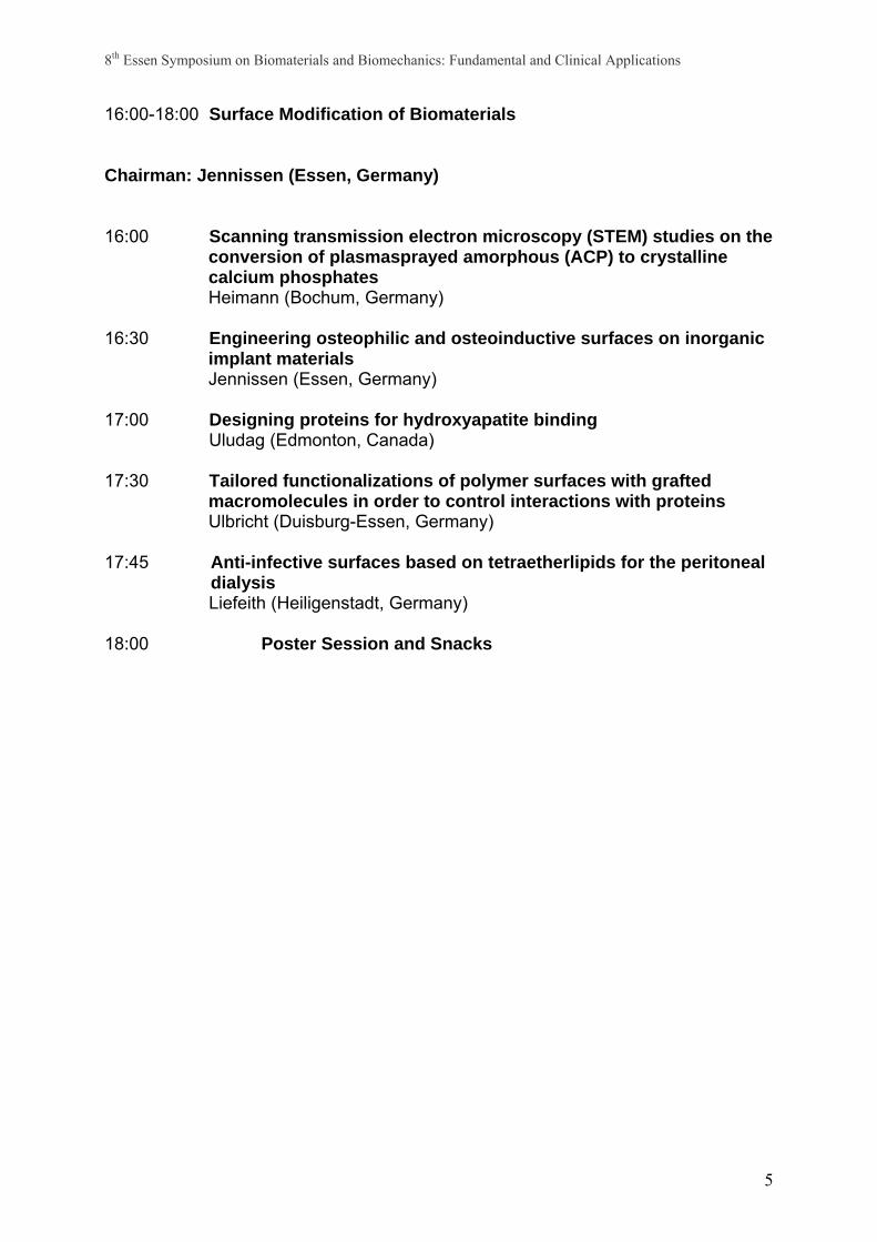
16:00-18:00 Surface Modification of Biomaterials Chairman: Jennissen (Essen, Germany) 16:00 Scanning transmission electron microscopy (STEM) studies on the
conversion of plasmasprayed amorphous (ACP) to crystalline calcium phosphates Heimann (Bochum, Germany)
16:30 Engineering osteophilic and osteoinductive surfaces on inorganic implant materials Jennissen (Essen, Germany)
17:00 Designing proteins for hydroxyapatite binding Uludag (Edmonton, Canada)
17:30 Tailored functionalizations of polymer surfaces with grafted macromolecules in order to control interactions with proteins Ulbricht (Duisburg-Essen, Germany)
17:45 Anti-infective surfaces based on tetraetherlipids for the peritoneal dialysis Liefeith (Heiligenstadt, Germany)
18:00 Poster Session and Snacks

8th Essen Symposium on Biomaterials and Biomechanics: Fundamental and Clinical Applications
6
Thursday, September 22nd, 2005 9:00-10:30 Biomechanics of Soft Tissue Chairman: Schröder (Duisburg-Essen, Germany) 9:00 On the load bearing mechanism of the human intervertebral disc
Ehlers (Stuttgart, Germany)
9:30 Computational bio-mechanics of bones with applications to artificial hip-joint replacement Nackenhorst (Hannover, Germany)
10:00 Modeling of atherosclerotic arteries – hyperelasticity and anisotropic damage Balzani (Darmstadt, Germany)
10:15 Modelling of the active and passive muscle behaviour by means of the finite element method Böl (Bochum, Germany)
10:30 Coffee Break and Posters 11:30-12:45 Simulation of the Mechanical Performance of Biomaterials and
Tissues Chairman: Klawonn (Duisburg-Essen, Germany) 11:30 Innovative numerical methods : their benefits for modeling cardio-
vascular systems Vidrascu (Chesnay, France)
12:00 Theory and implementation of mass changes in transversely isotropic materials Kuhl (Kaiserslautern, Germany)
12:15 A four-phasic theory for growth in biological tissue Ricken (Duisburg-Essen, Germany)
12:30 Mesoscopic simulations of biomaterials: the interaction of phospholipid membranes and inorganic nanoparticles Schulz (Essen, Germany)
12.45 Lunch

8th Essen Symposium on Biomaterials and Biomechanics: Fundamental and Clinical Applications
7
14:00-15:45 Implant Design, Properties and Prototyping Chairman: Bergers (Duisburg-Essen, Germany) 14:00 Material evaluation for the fabrication of patient-individual bone
grafts by 3D printing Irsen (Bonn, Germany)
14:30 A novel synthetic vascular prosthesis: effect of plasma protein adsorption on blood- and cyto-compatibility Knetsch (Maastricht, Netherlands)
15:00 Dense hydroxyapatite ceramics with Mg2+additives Naboka (Kharkiv, Ukraine)
15:15 Study on the influence of plasma spray processes and spray parameters on HA coating structure and crystallinity Zhao (Aachen, Germany)
15:30 A staple from nickel-titanium for foot surgery using the shape memory effect Mentz (Jülich, Germany)
15:45 Coffee Break and Posters
8th Essen Symposium on Biomaterials and Biomechanics: Fundamental and Clinical Applications
8
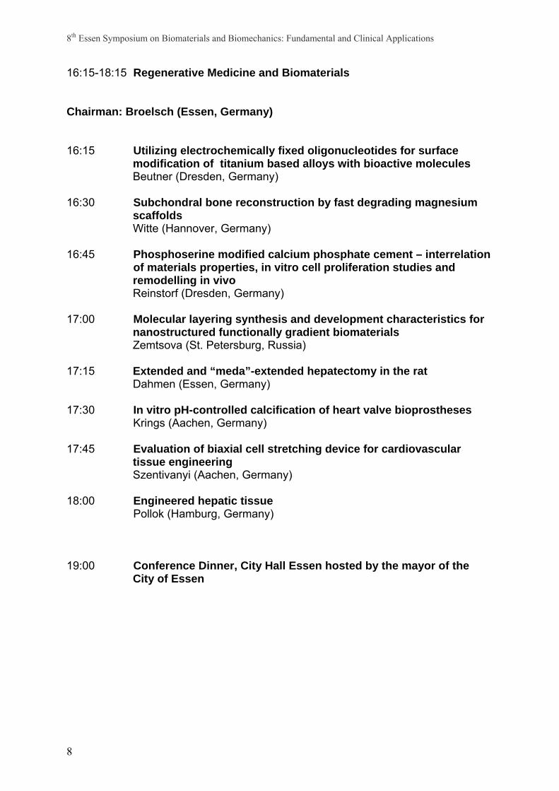
16:15-18:15 Regenerative Medicine and Biomaterials Chairman: Broelsch (Essen, Germany) 16:15 Utilizing electrochemically fixed oligonucleotides for surface
modification of titanium based alloys with bioactive molecules Beutner (Dresden, Germany)
16:30 Subchondral bone reconstruction by fast degrading magnesium scaffolds Witte (Hannover, Germany)
16:45 Phosphoserine modified calcium phosphate cement – interrelation
of materials properties, in vitro cell proliferation studies and remodelling in vivo Reinstorf (Dresden, Germany)
17:00 Molecular layering synthesis and development characteristics for nanostructured functionally gradient biomaterials Zemtsova (St. Petersburg, Russia)
17:15 Extended and “meda”-extended hepatectomy in the rat Dahmen (Essen, Germany)
17:30 In vitro pH-controlled calcification of heart valve bioprostheses
Krings (Aachen, Germany)
17:45 Evaluation of biaxial cell stretching device for cardiovascular tissue engineering Szentivanyi (Aachen, Germany)
18:00 Engineered hepatic tissue Pollok (Hamburg, Germany)
19:00 Conference Dinner, City Hall Essen hosted by the mayor of the
City of Essen

8th Essen Symposium on Biomaterials and Biomechanics: Fundamental and Clinical Applications
9
Friday, October 23rd, 2005 9:00-11:00 Interactions between Cells and Biomaterials Chairman: Bingmann (Essen, Germany) 9:00 Zinc-finger structures as potential targets for toxic metal ions and
essential trace elements Schwerdtle (Berlin, Germany)
9:20 Cartilage/bone progenitors, signalling, environmemtal and mechanical considerations Archer (Cardiff, UK)
9:40 Beta1- but not beta3-integrin adhesion structures are influenced by the surface roughness of titanium which correlates with the organization of fibronectin in human osteoblastic cells Nebe (Rostock, Germany)
10:00 Cell sensing of the surface aspects of mechanotransduction and cellular mechanics Jones (Marburg, Germany)
10:20 Integrin mediated chondrocyte-collagen type II interactions and their influence on chondrocyte function Shakibaei (Berlin, Germany)
10:40 The role of BMP-2-stimulated integrins for cell motility Wiemann (Essen, Germany)
11:00 Coffee Break and Posters

8th Essen Symposium on Biomaterials and Biomechanics: Fundamental and Clinical Applications
10
11:20- 12:45 Clinical Experience Chairman: Löer (Essen, Germany) 11:20 Severity of immunoinflammatory tissue response following plate
osteosynthesis of stainless steel and titanium in rabbits Taeger (Essen, Germany)
11:30 The density of nociceptive nerve fibres in the dura mater lumbalis of rats is enhanced after laminectomy, even after application of autologous fat grafts Saxler (Essen, Germany)
11:40 Generation of soluble mediators and periimplant cell activation after adherence of leukocytes to biomaterials Bogdanski (Bochum, Germany)
11:50 Linking metalloproteomics to new metalspecific immune responses in human nickel allergy Thierse (Freiburg, Germany)
12:00 Regulation of osteogenous potential of multicellular marrow systems in situ using orthopedic implants with the modified surface Karlov (Tomsk, Russia)
12:10 Push-out testing of cementless acetabular components with equatorial roughened surface von Knoch (Essen, Germany)
12:20 Generation of metallic nanocolloidal wear particles and their reaction on inflammatory cells Weuster (Essen, Germany)
12:30 The impact of nanocolloidal wear-particles on human mononuclear cells Podleska (Essen, Germany)
12:45 Lunch

8th Essen Symposium on Biomaterials and Biomechanics: Fundamental and Clinical Applications
11
Map of Essen
Map of the University Duisburg Essen
ParkplatzParking Lot
TagungsortConference
VenueEntrance S03Lecture Room
E33/E59
MensaCafeteria
ParkhausParking Garage
U-BahnhofSubway Station
ParkplatzParking Lot
TagungsortConference
VenueEntrance S03Lecture Room
E33/E59
MensaCafeteria
ParkhausParking Garage
U-BahnhofSubway Station

8th Essen Symposium on Biomaterials and Biomechanics: Fundamental and Clinical Applications
12

8th Essen Symposium on Biomaterials and Biomechanics: Fundamental and Clinical Applications
13
Session: Development of Biomaterials: Ceramics, Polymers,
Metals, Composites 9:30 Bone repair and regeneration: possibilities
M. Vallet-Regi (Madrid, Spain)
10:00 Silicon-substituted hydroxyapatite ceramics (Si-HAp): densification and grain growth through the prism of sintering theories V. Putlayev (Moscow, Russia)
11:00 Impurity effects in the structure of nanocrystalline hydroxyapatite during
calcinations Z. Zyman (Kharkiv, Ukraine)
11:30 Biomaterials of marine origin: perspectives for the development of bone implants and templates for tissue engineering H. Ehrlich (Dresden, Germany)
11:45 In-vitro bioactivity of nanoporous silica tested in simulated body fluid I. Krueger (Hannover, Germany)
12:00 Needs and trends for biomaterials and biodevices in oral and maxillofacial surgery D. Muster (Strasbourg, France)
12:15 A new in-situ hardening composite material for minimal invasive bone augmentation C. Pompe ( Munich, Germany)
8th Essen Symposium on Biomaterials and Biomechanics: Fundamental and Clinical Applications
14
Bone repair and regeneration: possibilities Prof. María Vallet Regí Departamento de Química Inorgánica y Bioinorgánica. Facultad de Farmacia. Universidad Complutense de Madrid, 28040 Madrid, Spain. The most significant demand for biomaterials has emerged as a consequence of the need to provide clinical treatment to a large number of patients. The key factors and driving forces are the increase in life expectancy and the aim to provide a minimum quality of life to an aging population. In particular, said increase in life expectancy has increased considerably the number of patients with osteoporosis. If we take also into account the continuous increase in the number of motor vehicles, with its associated social penalty in terms of traffic accidents, the incidence of bone pathologies is increasing at an alarming rate in recent years. The search for potential solutions in this field produces a large demand for materials suitable for bone repair or replacement. Calcium phosphates, bio-glasses, bio-glass ceramics and ordered silica mesoporous materials, among other types of materials, will be reviewed and studied from the point of view of their potential applications as replacement materials in bone repair and regeneration, as potential substrates in tissue engineering, and also as drug delivery systems. The ability of functionalising the material surfaces with various molecules of different nature and shape will allow to actuate selectively on the biological environment. An overview on the present achievements, but also on the ‘missing links’ will be presented. REFERENCES M. Vallet-Regí. CERAMICS FOR MEDICAL APPLICATIONS. Perspective Article. J. Chem. Soc. Dalton Trans. 02, 97-108. (2001) M. Vallet-Regi, A Ramila, R. P. del Real, J. Perez-Pariente. A NEW PROPERTY OF MCM-41: DRUG DELIVERY SYSTEM. Chem. Mater. 13, 308-311. (2001) M. Vallet-Regí and J. González-Calbet. CALCIUM PHOSPHATES IN THE SUBSTITUTION OF BONE TISSUE. Progress in Solid State Chemistry. 32, 1-31 (2004) I. Izquierdo-Barba, L. Ruiz-González, J.C. Doadrio, J.M. González-Calbet, and M.Vallet-Regí. TISSUE REGENERATION: A NEW PROPERTY OF MESOPOROUS MATERIALS. Solid. State Sci. 2005, In Press

8th Essen Symposium on Biomaterials and Biomechanics: Fundamental and Clinical Applications
15
Notes

8th Essen Symposium on Biomaterials and Biomechanics: Fundamental and Clinical Applications
16
Silicon-substituted hydroxyapatite ceramics (Si-HAp): densification and grain growth
through the prism of sintering theories
V. Putlayev1,2, A. Veresov2, M. Pulkin2, A. Soin2, V. Kuznetsov3 1Department of Chemistry, Moscow State University, Moscow 119899, Russia 2Department of Materials Science, Moscow State University, Moscow 119899, Russia 3Institute of Mechanics, Moscow State University, Moscow 119899, Russia
Ceramics based on calcium phosphates is known to be a prospective material for
biomedical applications. Noticeable attention is paid to hydroxyapatite Ca10(PO4)6(OH)2 (HAp) due to its affinity to a bone mineral. However, the rate of its resorption in vivo is considered to be quite low to induce a massive formation of new bone tissue. It was recognized recently that Si-doped HAp is a highly promising material in sense of bioactivity improvement. In the frame of distinct research activity structured around this subject, some special problems are to be solved: (i) solubility of Si in the bulk of HAp lattice (and its limit) vs. segregation of Si to free surfaces and grain boundaries, (ii) rationalization of Si effect on sintering of HAp ceramics, (iii) charge compensation over aliovalent doping of HAp with Si and its interplay with thermal stability of HAp phase. Our work was focused on a fabrication of Si-HAp with Са10-х(РО4)6-х(SiO4)х(ОН)2-х (x=0…1) nominal composition and evaluation the effect of Si on sintering of HAp ceramics. Fine powders of Si-HAp were fabricated via conventional wet-precipitation technique by dropwise addition of (NH4)2HPO4 (0.15-1.00 M) to the stock solution of Ca(NO3)2 (0.25 -1.67 M) with pre-adjusted pH at 60°C in the presence of TEOS. The powder samples were tested by XRD, FTIR, light-scattering , TEM and SEM/EDX to get particle sizes, morphology and chemical composition. It was found that Si-doped HAp is stable up to 1100°C. The limit of Si solubility in HAp lattice was estimated to be not higher than x=0.1 in the formula above (at 1000°C, according to XRD and SEM/EDX). Ceramics were sintered at 1000-1250°C in air and examined with SEM/EDX, density measurements and dilatometry. The essence of the silicon influence consists in significant suppression of grain growth during the initial stage of sintering. Silicon doping reduces grain boundary mobility (increasing activation energy of lattice diffusion) along with the increase of pore mobility (in fact, increasing grain boundary diffusion). Based on densification and grain growth data, sintering trajectories for Si-HAp ceramics were plotted. Their specific ‘plain’ habit suggests that for this material densification prevails over re-crystallization phenomena, and, thus, favours a fabrication of highly-dense ceramics (typical apparent density of 95% in this study, with 98% being the best). It should be noted that at temperatures higher than 1200°C the trajectories enter the region of pore/grain boundary separation leading to coarsening instead of densification. We believe that the effect of Si on sintering behaviour can be treated in terms of its segregation to grain boundaries, the phenomenon arising from a lattice instability of Si-HAp due to the charge compensation (through the loss of Ca-cations and OH-anions) in the course of aliovalent doping. The work was supported by RFBR (grant #05-03-32768), the program ‘Universities of Russia’ (grant # UR 06.02.556 ), interdisciplinary grant of MSU #26, and SEC ‘Synthesis’ (grant of Ministry of Education and Science).

8th Essen Symposium on Biomaterials and Biomechanics: Fundamental and Clinical Applications
17
Notes

8th Essen Symposium on Biomaterials and Biomechanics: Fundamental and Clinical Applications
18
Impurity effects in the structure of nanocrystalline hydroxyapatite during calcinations
Z. Zyman, D. Rokhmistrov, I. Ivanov, V. Glushko, M. Epple*
Physics of Solids Department, V.N. Karazin Kharkiv National University, Kharkiv, 61077
Ukraine *Institut fűr Anorganische Chemie, Universität Duisburg-Essen, Universitätsstraβe 5-7, D-
45117 Essen, Germany
1. Introduction Hydroxyapatite (HA) and materials based on HA have been widely used for medical purposes. Such materials are processed under definite conditions since there have been developed severe requirements for permissible extent of impurities in them. On the other hand, some impurities may be functional and give new useful properties for the materials. A simple way for inserting impurities lays through a conventional precipitation method. However, success in this way has been restricted since the role of complex ions of the starting reagents in the crystallization of HA has not been fully understood yet. Some aspects of this problem were studied in this work. 2. Materials and methods A HA powder was prepared by the known method of Jarcho et al (1976). An appropriate proportion in the initial analytical reagents was chosen to get stoichiometric molar ratio Ca/P=1,67 in the HA powder. The synthesis temperature was 96ºC in order to obtain crystallites large enough for giving sharp diffraction maxima. The precipitate was dried at RT. Equal batches of the powder were heated at 100-1250ºC in air with 100ºC step for 2h at each temperature. The batches after heating were subjected to XRD, IR, TG, DTA and SEM analysises by routine. The lattice constants, a and c, were measured with an error of less than ± 0, 004 Å using 300 and 002 reflections and aluminum as a standard. 3. Results and discussion The temperature dependence of the lattice constants (LC) may be divided into three intervals: 1) RT- 550ºC, 2) 550-1100ºC, 3)1100-1250ºC (Figure 1). HA reflections were fixed in diffractograms within all these intervals. However, past 1100ºC, the batches also contained traces of β-TCP (reflected by characteristic peaks for β-TCP of very low intensities in IR spectra). It meant that the Ca/P ratio in the powder was slightly less than 1,67. In the first interval, the LC curves decreased from values that were much higher than the corresponding stoichiometric ones (showed by continuous for a and dotted for c horizontal lines) to the mean values of a = 9,408 Å and c = 6,896 Å. A TGA curve (Figure 2) showed a wide strong maximum for the speed of mass loss (SML) with a top at 108ºC and a corresponding DTA curve – a prolonged endothermic effect. Besides, intensity of the band at 3430 cm-1 in IR spectra noticeably decreased with heating. These changes could be associated with the release of water adsorbed on the surface of powder particles and, what is worth to notice, localized in the lattice. Since, in addition, the curves of LC and SML showed irregularities at correlated temperatures, it was highly probable that the water released from the lattice had different origin. In the second interval, two major maxima in the SML curve and corresponding endothermic minima in the DTA curve at around 770 and 980ºC were observed. The main changes in the IR spectra were associated with decreasing intensities of −2
3CO bands and increasing
8th Essen Symposium on Biomaterials and Biomechanics: Fundamental and Clinical Applications
19
intensities of −OH bands (both at 3550 cm-1 and 630 cm-1) as the calcination temperature increased. According to the present bands, AB-type carbonated apatites were fixed. However, it should be noted that B-positions were mainly occupied at lower temperatures (~600ºC) and A- sites – at higher values (~1000ºC). Consequently, the mass loss, endothermic effects and changes in LC associated with these events were caused by release of −2
3CO groups from (probably, two sites in) the lattice. As a result, purification from the impurities incorporated in the lattice during precipitation led to the transformation of the powder to practically stoichiometric HA. It was proved by the LC and −2
3PO and −OH band characteristics which were equal (within experimental errors) to those for the stoichiometric HA in the powder calcined at 1100ºC. However, molecular water and carbonate bands were still fixed in the spectra. Probably, it was due to impurities mainly adsorbed on the particles. In the third interval, some traces of β-TCP appeared in the batches (as pointed above, they were not detected by XRD, just fixed in IR spectra), and this led to the slight change in LC of almost stoichiometric HA. The gas release (mass loss) also effected the crystallite sizes in the calcined powder (they were calculated from broadening of the 002 and 300 reflections). The effect started at 650ºC when the diffusion mobility of ions in HA began. There were two well-defined maxima in the temperature dependence of crystallite sizes within 650-1000ºC. They coincide well by temperature limits with the maxima in the SML curve. The gas release greatly promoted the growth of crystallites. Thus, while the crystallite size parallel with c – axis past calcinations at 950ºC (closely to that for a peak of gas release) was 5500 Å, the size at higher temperature of 1000ºC was only around 2000 Å. 4. Conclusions Structural changes in a nanocrystalline HA powder of nearly stoichiometric Ca/P ratio caused by evolution of impurities during calcination in a wide temperature range of RT-1250ºC (for 2h at each temperature) were studied. It was firstly revealed that the LC for as- received powder were much higher than the stoichiometric values for HA and diminished to the last values as a result of the calcinations by a complicated way. This reduction was mainly associated with the release of molecular water incorporated during precipitation and formed of the proper impurities in the crystal lattice within RT-550ºC range. Futher structural alterations were due to decreasing in quantity and redistribution in positions of −2
3CO groups substituted for −33PO and −OH ions in the 550-
1100ºC range. The as-received powder transformed to almost stoichiometric HA due to the purification by 1100ºC. A new effect of acceleration of crystallite growth owing to gas release was revealed. Within 1100-1250ºC, the lattice started decomposing as some traces of β-TCP was found in the powder.

8th Essen Symposium on Biomaterials and Biomechanics: Fundamental and Clinical Applications
20
Figure 1
Figure 2

8th Essen Symposium on Biomaterials and Biomechanics: Fundamental and Clinical Applications
21
Notes

8th Essen Symposium on Biomaterials and Biomechanics: Fundamental and Clinical Applications
22
Biomaterials of marine origin: perspectives for the development of bone implants and templates for tissue engineering H. Ehrlich*, P. Etnoyer**, Litvinov S.D.+, H. Domaschke++, H. Meissner*, T. Hanke*, R. Born* & H. Worch* *Max Bergmann Center of Biomaterials, Institute of Materials Science, Dresden University of Technology, Dresden, Germany ** Aquanautix Corp., Los Angeles, USA + Samara State Technical University, Samara, Russia ++ Max Planck Institute of Cell Biology and Genomics, Dresden, Germany Natural structural biomaterials of marine origin including mollusc shells, sponges and corals not only provide an abundant source of novel bone and cartilage replacements but also inspire investigations to develop nano-sized biomimetic composites. Natural coral has been used as a bone substitute for more than 10 years in orthopaedic, trauma, craniofacial, dental, and neurosurgery. At present, the tropical coral genera Porites, Alveopora, Acropora, and Goniopora are being used as bone substitutes; these are the only families known to have the correct pore diameter and the ability to connect property with bone. The deep-sea corals of Isididae family, also known as bamboo corals, are presently attracting increased scientific attention. These deep-sea jointed corals consist of bony calcareous structures alternated by proteinaceous nodes of gorgonin, giving the skeletal remains of the organism a unique fingerlike appearance. Gorgonin is known as a keratin-like biomaterial with elastic properties containing polyphenolic compounds. The mineral phase of these octocorals consists of calcite. The aim of the present study was to investigate for the first time the in situ behaviour of bamboo coral axial internodes as bone implants in an animal model (dog) and of the organic matrix of demineralised coral internodes as a template for tissue engineering. The demineralization procedure was carried out using OsteosoftTM solution at 37°C for 7 days. The nanostructure of the organic matrix obtained was analysed using SEM, TEM and LSM techniques. In addition, its nature was characterised by performing SDS-PAAG electrophoresis and amino acid analysis. The presence of calcite was determined using XRD, FTIR and Raman analysis. The implanted coral fragments were sterilized with formaldehyde vapour and subsequently gamma radiation (25 kGr). The bamboo coral implant was implanted in a trepanation hole in a dog’s tibia. The implantation zone was monitored using X-ray techniques. The duration of the experiment was 10 months. Afterwards a biopsy was performed under calypsol anaesthetic, the animal was not killed and continued to live without any handicap. The following results were obtained: Due to its high potential for colonisation with both human osteoblasts and osteoclasts, the organic matrix, composed of an acidic fibrillar protein framework, showed itself to be a very successful model for possible applications in tissue engineering. The resorption of the calcite-containing fragment of the coral implant was faster in relation to that of bioceramics. No infection or rejection of coral implant was observed. The material properties of the coral axial internode measured, namely Young’s modulus (160 ± 35 GPa, n=20), density (2.7 ± 0.02 g/cm3) and flexural strength (107 N/mm2) correlate well with the material property charts for materials used in orthopaedic surgery. On the basis of the high biomimetic potential of the results obtained, we propose that biotechnological processes for the aquacultural cultivation of Isididae corals as “living bone implants” should be developed in the near future.

8th Essen Symposium on Biomaterials and Biomechanics: Fundamental and Clinical Applications
23
Notes

8th Essen Symposium on Biomaterials and Biomechanics: Fundamental and Clinical Applications
24
In-vitro bioactivity of nanoporous silica tested in simulated body fluid
I. Krueger1, P. Behrens1, P.P. Müller2, M. Stieve3, H. Mojallal3, T. Lenarz3
1Institut für Anorganische Chemie, Universität Hannover; 2GBF/RDIF, Braunschweig; 3Hals-Nasen-Ohren-Klinik, Medizinische Hochschule Hannover
The surface structure of a biomaterial can affect cell attachment, growth and differentiation.
Especially, the investigation of the influence of nanostructured surfaces is currently an active
field. On the other hand, bone replacement materials still suffer from problems relating to
their interactions with the surrounding tissues, and it has been shown that the essential
condition for bone replacement materials to bond to living bone is the formation of an apatite
layer on their surface in the body [1]. Here we report on the in vitro bioactivity of nanoporous
silica in simulated body fluid [2], a solution with ion concentrations which are almost equal to
those of the human blood plasma.
To generate material samples for structural analysis and exposure in simulated body fluid,
thin films of nanostructured silica materials on standard glass slides were prepared by dip-
coating. Non-ionic surfactants (octaethyleneglycolmonodocecylether, C12EO8, or an
amphiphilic triblock copolymer, Pluronic P-123, EO20PO70EO20, EO: ethylene oxide, PO:
propylene oxide, Pluronic P103, EO17PO85EO17 or Pluronic F127, EO106PO70EO106) served as
structure-directing agents. Together with a silica precursor, these structure-directing agents
form organic-inorganic hybrid nanostructures; the organic part of the hybrid structure was
then removed by calcination to yield silica nanostructures with characteristic repeat distances
between 5 and 12 nm. A control sample of amorphous silica was obtained by applying a
similar synthesis procedure, but omitting the organic material. The silica films were immersed
in simulated body fluid for different lengths of times. The surface of the silica films became
covered with a calcium phosphate layer which crystallized from SBF and was identified as
apatite by X-ray diffraction and infrared spectroscopy. Scanning electron microscopy images
showed typical morphologies of apatite.
In conclusion, the nanoporous silica materials induces apatite formation on its surface in a
simulated body environment. However, a decisive influence of the dimension of the
nanostructure was not detected.
References [1] L.L. Hench, J.Am.Ceram. Soc., (1991), 74, 1487-510
[2] T. Kokubo et al, J. Biomed. Mater. Res. (1990), 24,721

8th Essen Symposium on Biomaterials and Biomechanics: Fundamental and Clinical Applications
25
Notes
8th Essen Symposium on Biomaterials and Biomechanics: Fundamental and Clinical Applications
26
NEEDS AND TRENDS FOR BIOMATERIALS AND BIODEVICES IN ORAL AND MAXILLOFACIAL SURGERY
D. Muster and C. Meyer
Service de Stomatologie, Chirurgie Maxillofaciale et Chirurgie Plastique Reconstructrice Hôpitaux Universitaires de Strasbourg BP 426 – 67091 STRASBOURG CEDEX (France)
The different categories of biomaterials actually available (metals, ceramics, synthetic polymers and materials of biological origin) have largely contributed to the improvement of the surgical repair or reconstruction. Three main fields of application will be considered.
Fracture fixation devices The evolution of the devices used for the fracture fixation is now oriented toward the bioresorbable materials (PLA, PGA, PDX) which should gradually replace the metallic materials (titanium, SS) in the next 10-15 years. The aim is the return to the initial state for the patient (children, adult or elderly) : that is no interference with growth and long term evolution (corrosion, impairment for further dental implants, electromagnetic perturbations,…). The osteosynthesis devices will have to fulfil more precise biomechanical criteria, in certain anatomical zones (i.e. trapezoidal plates for the mandibular condyle). For resorbable osteosynthesis, some difficulties have to be controlled : resorption of the material, overdimensioning of the device itself related to the weaker mechanical properties (excluding zones with high mechanical constraints like mandible), radiotransparency making clinical following difficult, costs (up to 3 times that of titanium). The fixation of bones with adhesives (synthetic polymers, invertebrates secretions, …) should also be reconsidered to overcome the barrier effect on bone repair, the toxicity of some constituants and eventual immunological problems. The modalities of application should also be optimized.
TMJ prostheses The most known TMJ prostheses are either definitive prostheses only available on the American market or temporary condylar prostheses. In this field the lag compared with the orthopedics is certainly over 20 years. Very few biomechanical studies have been performed and there is few interest from the industrials in Europe for this type of prostheses because of the small number of cases and the scarcity of the indications.
Materials for packing, augmentation and regeneration Numerous materials of all classes have been tried as bone substitutes but all are lacking of osteoinductivity and therefore combined materials are largely studied. But there is no global view and the networking is really needed for exchane of experience. Moreover, there are few multicentric clinical studies and the success of one material is often related to the importance of its use by its promoter. Actually, in maxillofacial surgery, the most used materials are HA, TCP, Coral eventually enriched with growth factors. Among many remaining problems are : the industrial feasability, the ease of handling by the surgeon, their appropriate training and the patient education. Concerning tissue engineering, nice models are elaborated in various laboratories in the world but huge difficulties remain regarding the optimal molecular environment and the biomechanical functionality. In conclusion, for the daily clinical use in

8th Essen Symposium on Biomaterials and Biomechanics: Fundamental and Clinical Applications
27
the next decade (and even the following), optimized traditional biomaterials will surely continue to keep a major place. Notes

8th Essen Symposium on Biomaterials and Biomechanics: Fundamental and Clinical Applications
28
A new in-situ hardening composite material for minimal invasive bone augmentation C. Pompe1,2, K. Hellerbrand2, S. Krüger2, M. Siedler2, A. Schütz2, W. Frieß1
1 Ludwig-Maximilians-University Munich Pharmaceutical Technology and Biopharmacy 2Scil Technology GmbH Fraunhoferstraße 15 82152-Martiensried The new composite material, comprising a bioresorbable polymer, a calcium phosphate cement, an organic solvent and an active pore forming agent is supposed to be applied as a ready-to-use device in the field of regenerative treatment of bone defects. Due to its pasty like consistency it is suitable to be administered within the scope of minimal invasive surgical techniques. Following the principle of an in-situ hardening scaffold the material solidifies immediately after application at the defect site when contacted with aqueous fluids. After one hour the material achieves approx. 80 % of the final mechanical strength which is achieved after approx. 48 hours. According to the interconnecting pore-structure and an average pore size higher than 150 µm exhibited by the material after complete setting, the scaffold allows cell migration and ingrowth. Moreover the new composite material is suitable to provide sustained release of incorporated growth factors like rhBMP-2 or rhGDF-5.

8th Essen Symposium on Biomaterials and Biomechanics: Fundamental and Clinical Applications
29
Notes

8th Essen Symposium on Biomaterials and Biomechanics: Fundamental and Clinical Applications
30

8th Essen Symposium on Biomaterials and Biomechanics: Fundamental and Clinical Applications
31
Session: Biomechanics of Hard Tissue, Orthopaedics 14:00 Palasept® - a bone substitute as a resorbable drug carrier S. Vogt (Hanau, Germany) 14:30 Study of the three-dimensional model of the human knee joint
D. Tarnita (Bukarest, Romania)
14:45 Comparative study of patient individual implants from β-tricalcium phosphate made by different techniques based on CT data F. Peters (Kleinostheim, Germany)
15:00 Effect of TiO2 and SiC on hydration and mechanical properties of
mineral trioxide aggregate (MTA) S. Javadpour (Shiraz, Iran)

8th Essen Symposium on Biomaterials and Biomechanics: Fundamental and Clinical Applications
32
PALASEPT® - A BONE SUBSTITUTE AS A RESORBABLE DRUG CARRIER S. Vogt, U. Gopp, P. Seide, K.-D. Kühn, Heraeus Kulzer, D-61273 Wehrheim; E-Mail: [email protected]
Introduction
Infections still represent a serious problem in bone graft procedures. Because of the poor accessibility of the infection site by systemically administered antibiotics, drug delivery systems with a retarding release of antibiotics are preferred to treat or to prevent bone infections. Antibiotic loaded PMMA beads are available for drug delivery, however, they are not resorbable and have to be removed after application. Therefore, resorbable drug carrier systems acting as bone substitute material with a retarding release profile are advantageous to avoid a second surgery for removal of the carrier.
Materials and Methods
PALASEPT®-implants with a hemispherical, cylindrical shape are made of calcium sulfate
and calcium carbonate. The implants with a total weight of about 250 mg contain a
resorbable, lipophilic additive to get high mechanical strength of the implants. Two different
types of PALASEPT®-implants were manufactured, first type with gentamicin sulfate and the
second type with a combination of gentamicin sulfate and clindamycin hydrochloride. In vitro
elution characteristics were studied in phosphate buffer solution using FPIA and HPLC for
antibiotic detection. Biocompatibility of the implants was verified according to ISO 10993.
Results and Discussion
PALASEPT®-implants showed a high initial antibiotic release during the first day
followed by a low but still efficient antibiotic release upto two weeks. The retarding
release profile is an effect of a complex and well-elaborated manufacturing process.
Toxicolgical studies according to ISO 10993 showed good biocompatibility of the
implants. Gentamicin sulfate is the standard antibiotic for medical devices in bone
surgery, therefore, gentamicin was incorporated in the implants. By adding
clindamycin hydrochloride and gentamicin sulfate to the implants the spectrum of
microbiological efficacy of PALASEPT®-implants is expanded to prevent infections
even in high-risk applications.

8th Essen Symposium on Biomaterials and Biomechanics: Fundamental and Clinical Applications
33
Notes

8th Essen Symposium on Biomaterials and Biomechanics: Fundamental and Clinical Applications
34
STUDY OF THE THREE-DIMENSIONAL MODEL
OF THE HUMAN KNEE JOINT
Daniela Tarnita, Dragos Popa, Dan Tarnita, Traian Preoteasa
The University of Craiova, Faculty of Mechanics, Dept. Of Applied Mechanics, 165 Bucuresti str., Craiova,
E –mail: [email protected]
The University of Craiova, Faculty of Mechanics, Dept. of Automotive, 165 Bucuresti str., Craiova,
E -mail: [email protected],
The University of Medicine and Pharmacy of Craiova, Faculty of Medicine, Dept. Of Human Anatomy, 2 Petru Rares str., Craiova,
E –mail: [email protected]
Ministry of Transportation
The paper is a part of the themes of high actuality which use the knowledge from various domains (anatomy, surgery techniques, orthopedic, mechanics, mechanisms, bio-mechanisms, computers science).
Human knee is the most important joint used for locomotion and bones, ligaments, tendons and cartilages compose it. Scientific studies are very difficult to realize because the knee is the most complex joint in the human body, almost they are made in a statically system. The paper presents a method of study and the steps to obtain a virtual knee joint. For that
purpose was used a CAD parametric software which permits to define models with a high degree of difficulty. First, were defined the bone components like femur, tibia, fibula and patella. The obtained model was prepared for kinematics and dynamic simulation in the classical cases of locomotion (walking, running, jumping). The input functions and parameters for the knee joint simulation were: the masses of the bone elements, the force applied on femur bone and the driver angle in the knee joint. The behavior of the virtual knee can give the important informations which can be used in the fields of
robotics, medicine sciences and medical robotics. Also, on the virtual knee joint can be attached virtual prosthetic elements for virtual post-surgery simulations.
The entire model was complete parametrised and can be adapted using dimension modifying to any anatomical values. Also, using the same medium densities, the assembly model was almost identically from inertial point of view and attaching the moving functions the model can permits to obtain the entire kinematics behaviour and the function for forces in every bone of the knee joint.
To complete the virtual mechanical model it was necessary to define the next input parameters:
-the dimensional and geometrical elements of the components of the mechanical assembly. -the masses of the components and the entire inertial behavior for the mechanical defined model. -the forces applied to the femur

8th Essen Symposium on Biomaterials and Biomechanics: Fundamental and Clinical Applications
35
Notes

8th Essen Symposium on Biomaterials and Biomechanics: Fundamental and Clinical Applications
36
Comparative Study of patient individual implants from β-tricalcium phosphate made by
different techniques based on CT data Fabian Peters1, Daniel Groisman2, Rolf Davids2, Thomas Hänel3, Holger Dürr3 and Martin Klein2 1 Curasan AG, Lindigstraße 4, D-63801 Kleinostheim, Bavaria, Germany 2 Campus Virchow Klinikum, Medizinische Fakultät der Universität zu Berlin, Augustenburger Platz 1, D-13353 Berlin 3 TU Chemnitz, Fakultät für Maschinenbau, Professur Fertigungslehre, D-09107 Chemnitz Bone substitution or bone augmentation with synthetic materials (e. g. β-tricalcium phosphate
materials) is usually done by standardized material shapes: block materials can be
individually shaped by the surgeon for the specific defect situation. Granular materials are
used for more complicated and hardly accessible defect situations. The granulate will be
mixed with blood. Induced by air and calcium from the biomaterial the material is moldable,
caused by blood coagulation and can be smoothed inside the defect. Also injectable putty
materials are available.
The disadvantage is that none of these material modifications can guarantee a complete and
strong press fit contact to the surrounding bone in complicated defect situations, what is
essential for a complete and successful bone regeneration. The treatment of complicated
defects were mechanical stability is necessary but not viable with a granular material (e. g.
large mandibular defects) is therefore difficult.
Especially defects were surgeries can have a longer planning horizon (e. g. bone tumours,
cysts) can be treated with custom made patient individual implants. CT data can be converted
to three dimensional CAD/CAM data for shaping or prototyping the implant.
For a manufacturer of bone augmentation materials also economical questions are necessary
to solve for the decision, whether this technology can be an interesting possibility for the
treatment of the above written defect situations.
Different techniques for making patient individual implants from β-tricalcium phosphate with
three-dimensional fabrication methods will be presented.
3D architectures of custom made porous scaffolds were produced by using conventional
shaping techniques as well as rapid prototyping methods.
The techniques, the fabrication times and the distances that had to be bridged from the
original CT data to the sterilized product, ready made for surgery will be compared in terms
of material properties and economical considerations.

8th Essen Symposium on Biomaterials and Biomechanics: Fundamental and Clinical Applications
37
Notes

8th Essen Symposium on Biomaterials and Biomechanics: Fundamental and Clinical Applications
38
Effect of TiO2 and SiC an Hydration and Mechanical Properties of Mineral Trioxide Aggregate (MTA)
S. Javadpour*, A. Khayyat**, M. Shakerzadeh*
*Department of Material Science and Engineering, School of Engineering, Shiraz University, Shiraz, Iran. [email protected]
** Department of Endodontic, School of Dentistry, Shiraz University of Medical Sciences, Shiraz, Iran.
___________________________________________________________
Abstract: Mineral Trioxide Aggregate (MTA) is an aggregation of three ceramic compounds, possesses many of the properties and applications in Endodontic practice. MTA has many of the properties of an ideal sealing material for perforation repair, retrograde filling, pulp capping, and apixification. Beside noble properties of MTA, it has some limitations concerning the setting time of the material, because the time to allow the MTA to set properly is too long. In this research, a new MTA with a formula similar wich the original white version is prepared. Different amounts of TiO2 and SiC were added and corresponding changes (e.g., hydration and mechanical properties) were determined. Based on the results, it is found that during hydration of MTA, TiO2 will react with components of MTA and forms some complex compounds that will affect the hydration of the material. Besides, addition of SiC has improved the mechanical properties of MTA. Other properties of this new version of MTA (e.g., biocompatibility) are under evaluation.

8th Essen Symposium on Biomaterials and Biomechanics: Fundamental and Clinical Applications
39
Notes

8th Essen Symposium on Biomaterials and Biomechanics: Fundamental and Clinical Applications
40

8th Essen Symposium on Biomaterials and Biomechanics: Fundamental and Clinical Applications
41
Session: Surface Modification of Biomaterials 16:00 Scanning transmission electron microscopy (STEM) studies on the
conversion of plasmasprayed amorphous (ACP) to crystalline calcium phosphates R. Heimann (Bochum, Germany)
16:30 Engineering osteophilic and osteoinductive surfaces on inorganic implant materials H. P. Jennissen (Essen, Germany)
17:00 Designing proteins for hydroxyapatite binding H. Uludag (Edmonton, Canada)
17:30 Tailored functionalizations of polymer surfaces with grafted macromolecules in order to control interactions with proteins M. Ulbricht (Duisburg-Essen, Germany)
17:45 Anti-infective surfaces based on tetraetherlipids for the peritoneal dialysis K. Liefeith (Heiligenstadt, Germany)
8th Essen Symposium on Biomaterials and Biomechanics: Fundamental and Clinical Applications
42
Scanning transmission microscopy (STEM) studies on the conversion of plasma-sprayed
amorphous (ACP) to crystalline calcium phosphates
Robert B. Heimann, Universitätszentrum Medizintechnik, Ruhr-Universität Bochum Richard Wirth, GeoForschungsZentrum Potsdam
Abstract Medical-grade hydroxyapatite (CAPTAL 90, Plasma Biotal Ltd., UK; -140+100µm) as well as duplex hydroxyapatite + titania (AMDRY™ 6500, Sulzer Metco GmbH; -22+5 µm) bond coat layers were deposited by atmospheric plasma spraying (APS) onto flat Ti6Al4V substrates at moderate plasma enthalpies. From as-sprayed coatings and coatings incubated for up to 24 weeks in simulated body fluid (r-SBF after Kokubo) electron-transparent samples were generated by focused ion beam (FIB) excavation and investigated by STEM in conjuction with energy-dispersive x-ray analysis (EDX), electron diffraction (ED), and electron energy loss spectroscopy (EELS).
Immediately adjacent to the titanium alloy substrate surface a thin layer (3-5 µm) of amorphous calcium phosphate (ACP) was deposited whose Ca/P ratio is determined by the presence or absence of the TiO2 bond coat. In the presence of a 10-15 µm thick titania bond coat, determined to consist of polycrystalline brookite (orthorhombic TiO2, S.G. Pbca (61)), the Ca/P ratio of the ACP increases with increasing distance from the interface from 1.1 to values that approach those of hydroxyapatite (1.67) far away from the interface in the bulk of the 150 µm thick calcium phosphate coating. EELS investigations of the ACP revealed the existence of substantial amounts of protons and OH- ions, respectively that are thought to act as driving force for the crystallisation of ACP to HAp during cool-down of the plasma-sprayed coatings. However, during in vitro incubation in r-SBF for 24 weeks secondary needle- or lath-shaped Ca-deficient defect apatite with Ca/P = 1.36 precipitated from ACP presumably by a dissolution-precipitation mechanism. The defect hydroxyapatite needles are being separated by porous regions consisting of nearly stoichiometric HAp. During prolonged irradiation with an electron beam in the TEM rapid and complete crystallisation of the ACP to well-crystallised tetracalcium phosphate (TetrCP) and poorly crystallised tricalcium phosphate (TCP) was observed.
In contrast to this, in coatings without a TiO2 bond coat no Ca/P gradient across the ACP layer was observed thus keeping its Ca/P ratio constant at approximately 1.38. Beyond the thin ACP layer there is a fully crystalline region consisting of more or less stoichiometric HAp. After incubation for 1 week in r-SBF well-crystallised secondary HAp was found near cracks in the transforming ACP, pointing to fluid flow of water through the coating. In ACP there are also diffusion bands visible of 1-2 µm width parallel to the interface metal/coating reminiscent of diffusion-controlled zoning in gels whose trailing edges show nucleation and nanocrystals of HAp. Such a mechanism is typical of a dissolution-precipitation sequence thought to govern the transformation of ACP to crystalline calcium phosphates.
In vitro and in vivo evidence exist that the addition of a bioinert TiO2 bond coat to plasma-sprayed hydroxyapatite coatings on titanium alloy implants increases the adhesion strength of the ceramic layers to the substrate metal thus suppressing the formation of a gap between metal and ceramic, and possibly subsequent invasion of connective tissue. However, no clear indication was found of a Ca-Ti-Oxide reaction layer at the interface brookite/ACP that could account for the enhanced adhesive strength of the coatings reported in earlier work.

8th Essen Symposium on Biomaterials and Biomechanics: Fundamental and Clinical Applications
43
Notes

8th Essen Symposium on Biomaterials and Biomechanics: Fundamental and Clinical Applications
44
Engineering Osteophilic and Osteoinductive Surfaces on Inorganic Implant Materials
Herbert P. Jennissen, Maria Chatzinikolaidou, Kristin Zurlinden and Markus Laub
Institut für Physiologische Chemie, Universitätsklinikum Essen, Hufelandstr. 55,
D-45122 Essen, Germany
Although the spreading of a sessile drop begins when the diameter of the drop contact exceeds the diameter of the spherical drop itself (i.e. contact angle < 90°) the begin of reasonable wetting (i.e. hydrophilicity) is generally defined as beginning at contact angles below 60° (for review see [1]). The surfaces of medicinal implant metals like cp titanium, 316L steel or cobalt chromium alloys (CoCr29Mo) however, generally possess contact angles of 60-80°, thus displaying effective hydrophobic surfaces. In 1972 Baier [2] suggested a model, in which a correlation exists between biocompatibiltiy, bioadhesion and the critical surface tension of solids. In this model he postulated a good bioadhesion on hydrophilic surfaces. Several years ago we discovered a novel wet chemical etching method with chromosulfuric acid (CSA) at 200-240 °C for the preparation of extremely hydrophilic surfaces on transition metals like titanium, steel (316L) [3], aluminum [4] and cobalt chromium (CoCrMo) alloys [5]. In subsequent work it was demonstrated for the first time that these metals with surface roughness values of Ra ~ 1 µm exhibited ultra-hydrophilic properties i.e. dynamic contact angles < 10° with absent contact angle hysteresis [6], a phenomenon which we have called "Inverse Lotus Effect" [7]. It will be shown that the development of ultra-hydrophilicity on titanium surfaces follows a typical time course. The advancing and receding contact angles respectively decrease from ca. 70°/60° at zero time to 1°/1° at 60 minutes and then increase again to 15°/3° or more at 120 minutes of heating in chromosulfuric acid. Thus the contact angles run through a minimum as a function of Chromosulfuric acid treatment. This typical minimum-function behavior was found for electropolished, anodically oxidized, SLA- (sand-blasted surface etched) and PVD- (plasma vapor deposited) titanium surfaces, inspite of the fact that the surface roughness (Ra value) varied between 1 µm (electropolished) and ~ 40 µm (PVD-surface). In pilot animal experiments such ultra-hydrophilic surfaces show an enhanced bone growth (osteophilicity) versus controls. For engineering osteoinductive surfaces, we have developed methods for immobilizing bone morphogenetic protein 2 (rhBMP-2) on metal surfaces [3-6]. In this way chemotactic-juxtacrine surfaces may be produced: chemotactic by way of a slow controlled release of rhBMP-2 and juxtacrine by simulating juxtacrine secretion in the form of a 2-dimensional layer of immobilized rhBMP-2 for solid phase interactions with the receptors of osteoprogenitor cells. 125I-rhBMP-2 can be immobilized in amounts between 0.1-5 µg/cm2 on different titanium surfaces in clinical use. The half-life of rhBMP-2 released from such surfaces is in the order of bone growth depending on the immobilization procedure and varies between 30 and 100 days [5]. In vivo and in vitro experimenters show that the immobilized BMP-2 is biologically active. Recently we have turned to the biocoating of hydroxyapatite ceramics and similar bone derived bone replacement materials [8] with model proteins and BMP-2. Here it is the aim to synthesize osteoinductive bone replacement materials. It will be shown that the chemical modification of such materials is possible allowing a covalent and non-covalent immobilization of BMP-2 in significant amounts. Future studies for demonstrating biological activity of these materials in vitro and in vivo are planned. [1] Jennissen, H.P. (2005) Macromol. Symp. , in press. [2] Baier, R.E. (1972) Bull N Y Acad Med, 48, 257-272. [3] Jennissen, H.P., Zumbrink, T., Chatzinikolaidou, M., & Steppuhn, J. (1999) Materialwiss. Werkstofftech.,
30, 838-845. [4] Jennissen, H.P. (1999) PCT Patent WO9926674A2 (Priority date Nov. 24, 1997), pp. 1-29 (+ 5 Figs),
Munich. [5] Chatzinikolaidou, M., Laub, M., Rumpf, H.M., & Jennissen, H.P. (2002) Materialwiss. Werkstofftech., 33,
720-727. [6] Jennissen, H.P., Chatzinikolaidou, M., Rumpf, H.M., Lichtinger, T., & Müller, R. (2000) DVM Bericht,
313, 127-140.

8th Essen Symposium on Biomaterials and Biomechanics: Fundamental and Clinical Applications
45
[7] Jennissen, H.P. (2001) Biomaterialien, 2, 45-53. [8] Zurlinden, K. & Jennissen, H.P. (2005) J. Artific. Org., 28, 424. Notes

8th Essen Symposium on Biomaterials and Biomechanics: Fundamental and Clinical Applications
46
DESIGNING PROTEINS FOR HYDROXYAPATITE AFFINITY: AN APPROACH TO CONTROL THE DELIVERY OF BONE THERAPEUTICS
Hasan Uludag, Geeti Bansal, Sebastien A. Gittens, Jennifer E.I. Wright, Cezary Kucharski
Department of Chemical and Materials Engineering, and Department of Biomedical Engineering,
University of Alberta, Edmonton, Alberta, T6G 2G6, Canada
Recombinant growth factors are being utilized in clinical bone repair since they are powerful stimulants of bone regeneration. The growth factors, such as Bone Morphogenetic Proteins, have been utilized in local bone repair by implanting the proteins with biomaterials. Biomaterials help to bind and retain the proteins at the application site so that sufficient concentrations of pharmacological agents are maintained at the site for local cells to respond to the proteins and accelerate new bone deposition. Systemic bone regeneration with growth factors has been more challenging since the proteins do not exhibit an affinity to bone tissue after systemic administration, and they are removed from the circulation by the clearance organs (kidneys and liver). This limitation can be alleviated if the proteins are engineered to exhibit a high bone affinity, so that the proteins ‘seek’ the bones after systemic administration.
Towards this goal, bisphosphonates (BPs), a class of compounds with exceptional affinity to bone-mineral hydroxyapatite (HA), were chemically conjugated to proteins using water-soluble crosslinkers. The resultant protein-BP conjugates held together with chemical tethers were shown to exhibit a high affinity to bone mineral, HA. Understanding the factors controlling the imparted HA affinity will be critical in controlling the in vivo fate of bone therapeutics. Towards this goal, we are conducting systemic studies on molecular determinant of BP-induced HA affinity. Our studies indicated that: (1) Proteins as large as 150 kDa (IgG) could be targeted to bone with the proposed BP conjugation approach (J. Pharm. Res. 2004, 93: 2788). (2) Both stable (thioether) and degradable (disulfide) tethers between the proteins and the BPs were suitable to impart the desired HA affinity (J. Biomed. Mat. Res. 2005, in press). Although significant differences were observed in the stability of the tethers in the presence of physiological thiols (Biomacromol. 2005, in press), no obvious differences in HA affinity was evident when the proteins were implanted in animal models (Biomat. 2005, in press). (3) Based on molecular dynamics simulations, correlations between the chemical tether length and the attached BP density were explored. Shorter chemical tethers were found to give a higher BP density in the vicinity of the proteins, which was experimentally shown to result in higher HA affinity both in vitro and in vivo (Pharm. Res. 2004, 21:608).
(4) Bone targeting of large proteins required a significant number of BP conjugations, for example ~10 BPs with 66 kDa albumin, and ~4 BPs for 14 kDa lysozyme (Biotech Prog. 2002, 18: 604). It is likely that such an extent of BP conjugation will alter bioactivity of recombinant growth factors. To overcome this limitation, novel dendritic BPs were synthesized where multiple copies of BPs can be attached to a protein at a single site (Angew. Chem. Int. Ed. 2005, 44:3710). These BPs allowed bone targeting with reduced numbers of BP attachment per protein. (5) Not all proteins were targeted to bone with the BPs. A notable exception was fetuin, a protein that controls biochemical sequence of mineralization at bones. Despite imparting a strong HA affinity, BP conjugation did not lead to significant bone targeting as seen with other proteins (Mol. Pharmaceutic. 2005, in press).
Collectively, our studies are establishing a foundation for rational design of protein-based bone therapeutics. Such proteins could be used in both implantable systems (where local retention of proteins is desired) and systemic injections (where bone targeting is desired). Our results will ultimately yield chemically-modified growth factors with intact pharmacopores. We believe that the proposed therapeutic agents will allow approaches to clinical bone repair that was not previously feasible.

8th Essen Symposium on Biomaterials and Biomechanics: Fundamental and Clinical Applications
47
Notes

8th Essen Symposium on Biomaterials and Biomechanics: Fundamental and Clinical Applications
48
Tailored functionalizations of polymer surfaces with grafted macromolecules in order to control interactions with proteins Mathias Ulbricht, Melvy G. Chuquimia-Beltran, Dimitrios Lazos, Heru Susanto Lehrstuhl für Technische Chemie II, Universität Duisburg-Essen, 45117 Essen, Germany. During the last decade we had explored various routes for surface functionalization of many different polymeric (or other) materials with grafted macromolecules [1-6]. One overall intention had been to establish easy and robust methods to modify technically interesting materials – such as membranes, microplates or biomaterials – without degradation of their useful intrinsic properties by attaching thin polymeric layers which then control the material’s function in particular applications. Photo-grafting had been especially efficient and flexible, and examples for functionalized materials include low-fouling filtration membranes [1,2], affinity adsorbers via ion-exchange [6], covalent immobilization of biomolecules [2,4] or molecular imprinting [5], or surfaces with an improved cell-compatibility [3].
Here we will focus on recent efforts to improve the functionality of such materials based on a more detailed analysis of the grafted layer structure and function by using thin-film or surface-selective analytical methods such as ellipsometry, surface plasmon resonance (SPR) and quartz crystal microbalance (QCM). Models for real material’s surfaces on silicon or gold substrates had been prepared by spin-coating of polymer (polystyrene, polysulfone and polypropylene) solutions or by preparing self-assembled monolayers (e.g. hexadecanthiol on gold – resembling the surface of polypropylene). Surface functionalizations had been done via end-on “grafting-to” of photo-reactive poly(ethylene glycol) (PEG) and “grafting-from” of functional acrylate monomers, including PEG acrylates. Further variations of functional layer architecture after “grafting-from” had been done by covalent coupling of dextrans. Additional surface characterizations had been based on contact angle and zeta potential measurements. The adsorption of proteins to the functionalized surfaces or/and antigen – antibody (protein – protein) interactions (“bioactivity”) had been studied by SPR and QCM as well as ELISA.
Based on all results for the end-on grafted PEGs, an ordered surface layer structure (thickness < 10 nm) had been proposed, with a PEG density controlled by the size of the PEG molecules and leaving significant fractions of the hydrophobic base polymer uncovered – these arrays of grafted PEGs effectively control the adsorption of proteins based on their size (“two-dimensional molecular sieving”). These data support strongly the earlier developed hypothesis that the biocompatibility of hydrophobic materials can be very much improved by PEG-modifications at incomplete surface coverage, but yielding an ordered layer structure controlled by the size and sterical interactions of surface-bound PEGs [3].
For the grafted polyacrylates at variied composition and the derived polyacrylate/dextran layers (dry thicknesses between 5 and ~50 nm), a broad range of protein binding capacities – via ion-exchange or covalent coupling – could be achieved. This binding capacity and, especially, the bioactivity of the bound protein depend very sensitively on the architecture of the layer, especially its density and thickness. The matrix properties introduced by the dextran seem to improve protein bioactivity along with a significantly reduced non-specific binding. [1] M. Ulbricht, K. Richau, H. Kamusewitz, Colloids and Surfaces A 1998, 138, 353. [2] M. Ulbricht, M. Riedel, Biomaterials 1998, 19, 1229. [3] G. Altankov, V. Thom, Th. Groth, K. Jankova, G. Jonsson, M. Ulbricht, J. Biomed. Mat.
Res. 2000, 52, 219. [4] H. Borcherding, H. G. Hicke, D. Jorcke, M. Ulbricht, Ann. NY Acad. Sci. 2003, 984, 470. [5] M. Ulbricht, J. Chromatogr. B 2004, 804, 113. [6] M. Ulbricht, H. Yang, Chem. Mater. 2005, 17, 2622.

8th Essen Symposium on Biomaterials and Biomechanics: Fundamental and Clinical Applications
49
Notes

8th Essen Symposium on Biomaterials and Biomechanics: Fundamental and Clinical Applications
50
Anti-infective surfaces based on tetraetherlipids for the peritoneal dialysis M. Frant1, K. Liefeith1, R. Schmid2, P. Stenstad2, H. Johnsen
1 Institute for Bioprocessing and Analytical Measurement Techniques, Rosenhof, DE-37308 Heiligenstadt, Germany, 2 SINTEF Materials and Chemistry, NO-7465 Trondheim, Norway
To prevent bacterial growth on silicone catheters and other medical devices a new coating strategy based on covalent immobilisation of a group of bipolar membrane spanning tetraether lipids from archaebacterium Thermoplasma acidophilum was developed. A lipophilic layer with optional further functionality was introduced to the surface of commercially available medical silicone elastomer slides and tubes. Physico-chemical characterisation of the different surface modified polymers and of potential pathogens represents the basis for the optimisation of the monomolecular coatings. The new tetraetherlipid coated catheter surfaces showed a successful reduction of bacterial adherence coupled with an excellent biobompatibility.
In general, medical device associated infections are thought to be strongly involved with the attachment of bacteria onto the device surface. During peritioneal dialysis (PD) often Peritonitis occurs, an infectious disease, which might result in loosing of the catheter. It has been estimated that in the year 2007 the number of PD catheters will grow up to more than 396,000 worldwide. Customary anti-fouling attempts aim at the direct inactivation of micro-organisms, e.g. leaching systems. As a serious disadvantage an exhaustion of the anti-microbial substances can appear. The specific modification of physicochemical material parameters is, optionally combined with the integration of specifically active molecules, the most promising concept from a today's point of view. In this study, tetraetherlipid coatings are used as an appropriate approach to achieve prevention of medical device associated infections by modification of surfaces.
Silicone sheets and discs (Raumedic SIK, a commercial catheter polymer) were kindly obtained from Humeca B.V. The tetraetherlipid was isolated from a raw lipid extract of the archaeabacterium Thermoplasma acidophilum by column chromatography followed by pre-activation procedures that allow the chemical coupling of the lipid to the silicone surface. Subsequently, the second, outward standing head groups of the lipid-layer has been further modified in order to obtain surfaces with different physical and chemical properties e.g. by introducing a flexible, hydrophilic polyethylene glycole or alternatively zwitterionic phosphorylcholine headgroups. The adhesion of a mixed culture from Staph. aureus/Staph. epidermidis was quantified in synthetic dialysis effluent. Static and dynamic test conditions were applied to simulate the real catheter situation. Moreover biocompatibility was tested corresponding to existing European regulations.
The present study has shown that biomimetic coatings consisting of monolayers of modified tetraetherlipid (Caldarchaeol) can be prepared on silicone surfaces. In the adhestion test procedure a significant reduced cell adhesion was observed for the tetraetherlipid coated polymer surfaces. Despite the observed inhibition of microbial adhesion the preservation of the cell vitality could be demonstrated. Biocompatibility tests have confirmed the biocompatibility of the developed coatings. Specific measuring techniques based on the evaluation of the free surface energy and microscopic findings by means of AFM and CLSM were employed to confirm the efficiency of the performed modification steps. Subsequent immobilisation of cyanur chloride activated lipids resulted in lipid-coated silicone surfaces with a distinct different behaviour in both physico-chemical as well as biological tests. To sum up, the used bacterial mixed culture led to a significant reduced adherence onto the coated silicone surfaces compared to uncoated silicone surface.

8th Essen Symposium on Biomaterials and Biomechanics: Fundamental and Clinical Applications
51
The innovative aspect of this study was the development of thin and stable biomimetic anti-infective surfaces based on polymerizable tetraetherlipid coatings for medical applications. This includes a value-added product and process technology as well as a strong increase of user-friendliness and lower costs e.g. for dialysis therapies associated with the expected reduction in morbidity and mortality, especially in high risk patient populations.
This work was supported by the EC under the Contract G5RD-CT-2001-00594.
Notes

8th Essen Symposium on Biomaterials and Biomechanics: Fundamental and Clinical Applications
52
8th Essen Symposium on Biomaterials and Biomechanics: Fundamental and Clinical Applications
53
Session: Biomechanics of Soft Tissue 9:00 On the load bearing mechanism of the human intervertebral disc
W. Ehlers (Stuttgart, Germany)
9:30 Computational bio-mechanics of bones with applications to artificial hip-joint replacement U. Nackenhorst (Hannover, Germany)
10:00 Modeling of atherosclerotic arteries – hyperelasticity and anisotropic damage D. Balzani (Darmstadt, Germany)
10:15 Modelling of the active and passive muscle behaviour by means of the finite element method M. Böl (Bochum, Germany)

8th Essen Symposium on Biomaterials and Biomechanics: Fundamental and Clinical Applications
54
ON THE LOAD BEARING MECHANISM OF THE HUMAN INTERVERTEBRAL DISC Wolfgang Ehlers, Bernd Markert, Nils Karajan Institute of Applied Mechanics (CE) University of Stuttgart Pfaffenwaldring 7 70569 Stuttgart, Germany e-mail: [email protected] http://www.mechbau.uni-stuttgart.de/ls2 The spinal discs are part of the load bearing system of our body. They serve as shock absorbers, transmit loads and enable the motion between the vertebrae. In particular, the intervertebral discs (IVD) of the lumbar spine (L1-L5) carry the complete weight of the upper body, which in combination with the convex curvature (lordosis) makes them suspectible to severe injuries, such as disc herniation (prolapse).
As a naturally grown material, the IVD belongs to the class of soft avascular cartilagenous tissues consisting of mostly ionized water (interstitial fluid) and collagen of type 1 and 2 embedded in an extracellular meshwork of charged protein compounds. More precisely, the type 1 collagen is organized in fiber bundles building a fiber-reinforced ring, the anulus fibrosus (AF), which encloses a highly charged gelatenous core, the nucleus pulposus (NP). Following this, it directly emerges the principle load bearing mechanism of the disc. Under compression, interstitial fluid pressure develops inside the NP forcing a lateral bulging, which is prevented by the AF with the collagen fibers carrying under tension.
However, from a medical perspective, this basic knowledge about the disc is too vague for a diagnosis. In order to get a more detailed insight into the functioning of the IVD in vivo, which is apparently impossible by standard examination methods, numerical simulations provide an elegant and cost-effective alternative. Accordingly, this requires an appropriate theoretical model, which is capable of describing the physiological behavior of charged hydrated tissues. This includes the electro-chemomechanical couplings as well as the viscoelastic and anisotropic properties of the extracellular matrix. In order to meet these requirements, the established Theory of Porous Media (TPM) can be applied, which allows the description of multicomponent continua with internal interactions and is proven to yield stable implementations within the mixed finite element method (FEM).
The overall possibilities offered by this approach are finally shown in a numerical analysis of a human IVD accounting for the essential inhomogeneities within its realistic anatomic structure. In general, the model can be used to simulate aging and degeneration of individual discs as well as their influence on the behavior of the spine. Hence, the simulation results might be used to improve preventive and therapeutical measures.
References:
R. Eberlein, G. A. Holzapfel, M. Fröhlich: Multi segment FEA of the human lumber spine including the heterogeneity of the annulus fibrosus. Computational Mechanics 34 (2004), 147-163.
W. Ehlers, B. Markert, A. Acartürk: Large strain viscoelastic swelling of charged hydrated porous media. In J.-L. Auriault, C. Geindreau, P. Royer, J.-L. Bloch, C. Boutin, J. Lewandowska (eds.): Poromechanics II, Proceedings of the second Biot conference on Poromechanice, Balkema at Swets & Zeitlinger, Lisse 2002, pp. 185-191.

8th Essen Symposium on Biomaterials and Biomechanics: Fundamental and Clinical Applications
55
Notes

8th Essen Symposium on Biomaterials and Biomechanics: Fundamental and Clinical Applications
56
Computational bio-mechanics of bones
with applications to artificial hip-joint replacement
Udo Nackenhorst Institut für Baumechanik und Numerische Mechanik
University of Hannover e-mail: [email protected]
Bones are living organs with the ability to adapt themselves to their mechanical demand. This phenomenon is of great importance in endoprosthetics or fracture healing processes. Due to a total hip joint replacement for example the bone is stressed in a non–physiological which causes bone remodelling. Therefore, the durability of the artificial device depends strongly on the prosthesis design.
In this presentation computational methods based on a continuum theory of mass variable systems will be presented. On the macroscopic length scale a pure phenomenological, but thermodynamically consistent constitutive approach of stress adaptive bone adaptation will be suggested. For approximate solution of the governed field equations a finite element discretization is used.
A special focus will be laid onto the engineering modelling approach. A central point will be the treatment of the uncertain boundary conditions describing muscle forces and joint load collectives. Here an optimization strategy will be suggested for the determination of statically equivalent load sets from CT-data. Furthermore, the meaning of mechanical anisotropy will be discussed.
These methods have been applied to various hip-joint endoprosthesis. The numerical results are in good agreement with clinical observations. It will be demonstrated that this approach allows for judging the bio-mechanical compatibility of different prosthesis designs.
By this hierarchical modelling approach will be shown that the mechanical demand is of first order importance. At this stage only qualitative statements can be concluded from numerical studies, which however, give clear hints for the design of more bio-mechanical endoprosthesis. Additionally ideas for surgery planning and rehabilitation treatment can be derived from this analysis. Actual work is done on model refinement taking second order effects into account. In this presentation the actual research related to
• numerical investigation on much smaller length scales for a better understanding of the mechano-sensation by the bone-cells and homogenized material properties for use on the macroscopic length scale, and
• the bio-active contact between implant and bone depending on the local mechanical demand, surface structure and coating of the implant
will be outlined.

8th Essen Symposium on Biomaterials and Biomechanics: Fundamental and Clinical Applications
57
Notes

8th Essen Symposium on Biomaterials and Biomechanics: Fundamental and Clinical Applications
58
Modeling of Atherosclerotic Arteries – Hyperelasticity and Anisotropic Damage
D. Balzani1, J. Schröder2, D. Gross1
1Institute of Mechanics (AG4),Department of Mechanics, Technical University of Darmstadt,
Germany; 2Institute of Mechanics, Department of Civil Engineering, University of Duisburg-Essen,
Campus Essen, Germany Soft biological tissues are characterized by an anisotropic and incompressible material behavior. Arterial walls are basically stiffened by collagen fibers arranged mainly in two directions, thus, the superposition of two transversely isotropic models reflects the anisotropy of the material. Herein, weak interactions between the particular fibers are assumed. In the present contribution an anisotropic damage model is proposed which is able to represent the basic properties of arterial walls observed in experiments. The model is embedded into the concept of internal variables and introduces a specific referential damage state, at which the damage evolution starts, cf. [2]. For the description of the effective hyperelastic response a polyconvex stored energy function is utilized in order to ensure the existence of minimizers of the underlying potential. Furthermore, polyconvex energy functions fulfill a priori the Legendre-Hadamard condition, which means that a stable material is obtained, see [1]. The stored energy is formulated in the concept of structural tensors and the representation theorems for isotropic tensor functions are applied. As a numerical example for simulation applications we consider the overexpansion of a diseased artery and investigate the damage distribution. In Fig. 1a) the mesh of the considered artery and the components are illustrated, cf. [3]. The artery is expanded by applying internal pressure up to the physiological deformation and here the referential damage state is reached. Then further internal pressure is applied and the damage evolves. Fig. 1b) shows the damage distribution for a specific internal pressure.
Fig. 1: a) mesh of the considered artery and b) damage distribution in deformed state. References: [1] Schröder, Neff, Balzani, “A Variational Approach for Materially Stable Anisotropic Hyperelasticity”, International Journal of Solids and Structures, Vol. 42/15, 4352-4371 (2004) [2] Schröder, Balzani, Gross, “Aspects of Modeling and Computer Simulation of Soft Tissues: Applications to Arterial Walls”, Materialwissenschaft und Werkstofftechnik, submitted (2005) [3] Holzapfel, Schulze-Bauer, Stadler, “Mechanics of Angioplasty: Wall, Balloon and Stent”, Mechanics in Biology, Vol. 46, 141-156 (2000)
D(1)
Adventitia
Media
Plaque
Calcification
Extracellular lipid

8th Essen Symposium on Biomaterials and Biomechanics: Fundamental and Clinical Applications
59
Notes

8th Essen Symposium on Biomaterials and Biomechanics: Fundamental and Clinical Applications
60
Modelling of the active and passive muscle behaviour by means of the finite element method
Markus Böl & Stefanie Reese
Department of Civil Engineering, Ruhr University of Bochum, D-44780, Germany
The structure of a skeletal muscle can be considered as a complex hierarchical organisation in which thousands of muscle fibres are arranged within a connective tissue network. The single muscle fibres consist of many force-producing cells, known as sarcomeres. However, the properties of the individual fibres have been studied over the last decades, see e.g. MCMAHON [1984], GORDON ET AL. [1996] and DENOTH ET AL. [2002]. The material behaviour of muscles is non-linear. Muscles can have large deformations in space, changing significantly their shape, so that the geometrical non-linearities must also be taken into account. The most authors use the finite element method to solve such materially and geometrically non-linear problems, see e.g. KOJIC ET AL. [1998] and BLEMKE ET AL. [2004]. In the present approach the finite element method is used too, where the material behaviour of the muscle is split into a so-called active and a passive part. To describe the passive part special unit cells consisting of one tetrahedral element and six truss elements have been derived. Additionally to these unit cells other truss elements are attached representing bundles of muscle fibres and therefore the active part of the material behaviour. To simulate a complex muscle structure, the muscle is discretised by the before described element ensembles. Depending on the discretisation it is possible to simulate the material behaviour of skeletal muscles (oriented fibres) as well as soft tissue behaviour (non-oriented fibres). References
BLEMKER, S. S., PINSKY, P. M., DELP, S. L., 2005, A 3D model of muscle reveals the causes of nonuniform strains in the biceps brachii, Journal of Biomechanics, 38, 657-665
DENOTH, J., STUSSI, E., CSUCS, G., DANUSER, G., 2002, Single muscle fiber contraction is dictated by inter-sarcomere dynamics, Journal of Theoretical Biology, 216, 101-121
GORDON, A. M., HUXLEY, A. F., JULIAN, F. J., 1966, The variation in isometric tension with sarcomere length in vertebrate muscle fibres, Journal of Physiology, 184, 170-192
KOJIC, M., MIJAILOVIC, S., ZDRAVKOVIC, N., 1998, Modelling of muscle behaviour by the finite element method using Hill’s three-element method, International Journal for Numerical Methods in Engineering, 43, 941-953
MC MAHON, T. A., 1984, Muscles, Reflexes and Locomotion, Princeton University Press, New York

8th Essen Symposium on Biomaterials and Biomechanics: Fundamental and Clinical Applications
61
Notes

8th Essen Symposium on Biomaterials and Biomechanics: Fundamental and Clinical Applications
62
8th Essen Symposium on Biomaterials and Biomechanics: Fundamental and Clinical Applications
63
Session: Simulation of the Mechanical Performance of
Biomaterials and Tissues 11:30 Innovative numerical methods: their benefits for modeling cardio-
vascular systems M. Vidrascu (Chesnay, France)
12:00 Theory and implementation of mass changes in transversely isotropic materials E. Kuhl (Kaiserslautern, Germany)
12:15 A four-phasic theory for growth in biological tissue T. Ricken (Duisburg-Essen, Germany)
12:30 Mesoscopic simulations of biomaterials: the interaction of phospholipid membranes and inorganic nanoparticles S. Schulz (Essen, Germany)
8th Essen Symposium on Biomaterials and Biomechanics: Fundamental and Clinical Applications
64
Innovative numerical methods: their benefits for
modeling cardio-vascular systems Marina Vidrascu
INRIA, Rocquencourt B.P. 105 F-78153 Le Chesnay Cedex, France
One of the long term objective for several teams at INRIA-Rocquencourt (MACS, REO, SOSSO) is to design a multi-scale hierarchical model for the cardio-vascular system. In this talk I will focus on two topics:
• a 3d fluid structure interaction model used at the level of the aortic valve. We designed an
efficient and robust algorithm for the simulation of blood flows in compliant vessels. This algorithm is using a Dirichlet-Neumann domain decomposition approach which leads to the solution of an interface problem. To insure the stability of the non-linear equation on the fluid-structure interface very time-consuming strongly-coupled algorithms are required. Based on a simplified approach we propose a Jacobian-free Newton-Krylov preconditioner that takes into account a fundamental mechanical feature of the original system, namely the added-mass effect.
• an electro-mechanical model of the heart The aim is to built a cardiac simulator. The current model (due to Bestel-Sorine) describes the contraction or relaxation of cardiac muscles including fibers under action potential control. This model is consistent with the behavior of myosin molecular motors. The active constitutive law used is a rheological model of Hill-Maxwell type. The blood inside each ventricle is modeled as a simplified 0D pression-volume relation. The dynamic system is discretized using the Newmark scheme. From a clinical point of view a simulator's advantage is the presentation of the numerical results in the form of classical indicators of the cardiac function such as volume variations, pressure-volume cycle and so on.
All the performed simulations aim to consider realistic geometrical models and thus require several software ingredients such as a procedure that starts with medical images and automatically generates a finite element mesh, tools to refine surface meshes, 3D mesh generation. Such tools were designed at INRIA by the GAMMA team. Several numerical results will illustrate the robustness end efficiency of this approach.

8th Essen Symposium on Biomaterials and Biomechanics: Fundamental and Clinical Applications
65
Notes
8th Essen Symposium on Biomaterials and Biomechanics: Fundamental and Clinical Applications
66
Theory and Implementation of Mass Changes in Transversely
Isotropic Materials
Grieta Himpel, Ellen Kuhl, Andreas Menzel, Paul Steinmann Chair of Applied Mechanics University of Kaiserslautern
PO Box 3049 D-67653 Kaiserslautern
For the modelling of biomaterials a change in mass can be introduced at two different levels. On the one hand the coupling of mass changes and deformations can be introduced at the constitutive level, see e.g. HARRIGAN & HAMILTON [1]. Herein a weighting of an elastic free energy function with respect to a relative density is assumed, which causes changes in mass by changes in density while the volume remains constant. We call this effect ‘remodelling’. On the other hand the coupling of mass changes and deformations can be introduced at the kinematic level. The kinematic coupling is characterised by a multiplicative decomposition of the deformation gradient into a growth part and an elastic part, see RODRIGUEZ ET AL. [2]. With such an ansatz changes in mass can be realised by changes in density and volume. With the denotation of ‘growth’ for changes in volume, the latter comprises that both remodelling and growth can be simulated. Based on the formulations for isotropy in [3], the present contribution aims at combining both approaches for a transversely isotropic material. Herein the transverse isotropy will be described by a characteristic direction, which is assumed to be a principal direction of the growth part of the deformation gradient. The discussed theory will be implemented into a finite element code and studied by numerical examples. [1] Harrigan, T. P.; Hamilton J. J.: Optimality conditions for finite element simulation of
adaptive bone remodelling, Int. J. Sol. Struct. 29, 2897-2906 (1992).
[2] Rodriguez, E. K.; Hoger, A. McCulloch, A. D.: Stress-dependent finite growth in soft elastic tissues, J. Biomech. 27, 455-467 (1994).
[3] Himpel, G.; Kuhl, E.; Menzel, A.; Steinmann, P.: Computational modelling of isotropic multiplicative growth, CMES, accepted for publication.

8th Essen Symposium on Biomaterials and Biomechanics: Fundamental and Clinical Applications
67
Notes

8th Essen Symposium on Biomaterials and Biomechanics: Fundamental and Clinical Applications
68
A Four-Phasic Theory for Growth in Biological Tissue Tim Ricken (a), Joachim Bluhm (b)
Institute of Mechanik, Universität Duisburg-Essen, Essen, Germany,
Universität Duisburg-Essen, D-45117 Essen, b) [email protected], b) [email protected]
Introduction: Biological soft tissues are highly complex materials capable of performing a wide range of functions. Two basic properties of biological soft tissues are optimized load transfer and the capacity of growth. The former is often realized by a combination of inner anisotropic structure components such as fibres situated in porous saturated tissue. Therefore, the optimized load transfer is solved for both the maximum stress and the hydrostatic pressure. The growth results from a phase transition inside the soft tissue where mass exchange between the biological skeleton and the nutrients carried by the saturating fluid is observed. This process strongly depends upon the state of stress, the availability of nutrients and the presence of chemical growth factors, i.e. growth hormone. Modeling: A formulation is developed for the phenomenological description of transverse isotropic and saturated soft tissues including the phenomena of growth. This is done within the framework of a macro-mechanical description based on the Theory of Porous Media (TPM), see e.g. DE BOER [1]. In order to characterize the transverse isotropic skeleton behavior an invariant formulation of the Helmholtz free energy function is used. Owing to the interior structure, an anisotropic tissue permeability can be observed. A transverse isotropic permeability tensor in an invariant formulation is introduced to account for this phenomena. The biological growth process in soft tissues is also investigated. In contrast to formulations wherein the mixture body is treated as a one component material, see e.g. [2] and [3], a four-phasic model is suggested using the TPM, where the continuum consists of, simultaneously, particles of the tissue skeleton as well as the saturating fluid and nutrient phases and the concentration of chemical growth factors. The nutrient phase is coupled with the fluid velocity whereas the chemical concentration is driven by the osmotic pressure. Therefore, the chemical potentials and the dependency between the stresses and the chemical concentration are considered. This has been done using an additional chemical rate in the Helmholtz free energies. In view of the growth description, this method allows a closed system approach wherein the mass source of one phase inside the body derives completely from the respective other phase. As a consequence, the validity of the mixture balance equation of mass can be guaranteed without the introduction of a surface mass flux. References: [1] DE BOER (2000), Springer-Verlag, Berlin Springer-Verlag Berlin [2] EPSTEIN & MAUGIN (2000) Int. J. Plasticity 16:951-978; [3] AMBROSI & MOLLICA (2002) Int. J. Engineering Science 40:1297-1316

8th Essen Symposium on Biomaterials and Biomechanics: Fundamental and Clinical Applications
69
Notes

8th Essen Symposium on Biomaterials and Biomechanics: Fundamental and Clinical Applications
70
Mesoscopic Simulations of Biomaterials:
The Interaction of Phospholipid Membranes and Inorganic
Nanoparticles.
Sarah G. Schulz1, Ulf Frieske1, Hubert Kuhn1, Christian Mayer2
1 CAM-D Technologies GmbH, Essen
2 Institut für Physikalische Chemie, Universität Duisburg-Essen, Duisburg
The simulation of the aggregation of complex systems represent a challenge for
conventional methods of simulation. The length and time scales of the dynamics of such
phenomena overcome the limits of most simulation methods. In this lecture we present a new
developed mesoscopic simulation method (MFD = Molecular Fragment Dynamics). The
introduction of nanoparticles in the mesoscopic simulation technique opens new insights into
the behavior of nanoparticles with biomaterials.
Experimental investigations show the embedding of nanoparticles in phospholipd
multilayers as a cell membrane model. The self-aggregation of the particles in regular two-
dimensional arrangement has been observed. These results could be reproduced with our new
simulation method, moreover new insight into the mechanism and dynamics of the
aggregation of the nanoparticles in the lipid layers were obtained. Thereby structural data e.g.
the nanoparticle distances and arrangement of the phospholipds was determined.
The presented simulation results of nanoparticle systems show a novel application of
mesocopic computer simulations and yield an important contribution to the understanding of
such systems. Experimental data could be reproduced as well as experimentally unavailable
information obtained.
Schulz, S. G., Frieske, U., Kuhn, H., Schmid, G., Müller, F., Mund, C. and Venzmer, J.: Tenside Surfactants
Detergents 41 (2004) 230.
Schulz, S. G., Kuhn, H., Schmid, G., Mund, C. and Venzmer, J.: J Colloid Polym Sci 283, (2004), 284.
Annegret Terheiden, Christian Mayer, Karsten Moh, Burkhard Stahlmecke, Sonja Stappert, Mehmet Acet, Bernd
Rellinghaus, Applied Physics Letters 84 (2004), 3891.

8th Essen Symposium on Biomaterials and Biomechanics: Fundamental and Clinical Applications
71
Notes

8th Essen Symposium on Biomaterials and Biomechanics: Fundamental and Clinical Applications
72

8th Essen Symposium on Biomaterials and Biomechanics: Fundamental and Clinical Applications
73
Session: Implant Design, Properties and Prototyping 14:00 Material evaluation for the fabrication of patient-individual bone grafts
by 3D printing S. H. Irsen (Bonn, Germany)
14:30 A novel synthetic vascular prosthesis: effect of plasma protein adsorption on blood- and cyto-compatibility M. Knetsch (Maastricht, Netherlands)
15:00 Dense hydroxyapatite ceramics with Mg2+additives M. Naboka (Kharkiv, Ukraine)
15:15 Study on the influence of plasma spray processes and spray parameters on HA coating structure and crystallinity L. Zhao (Aachen, Germany)
15:30 A staple from nickel-titanium for foot surgery using the shape memory effect J. Mentz (Jülich, Germany)
8th Essen Symposium on Biomaterials and Biomechanics: Fundamental and Clinical Applications
74
Material evaluation for the fabrication of patient-individual bone grafts by 3D printing
S. H. Irsen, B. Leukers, H. Seitz, C. Tille caesar research center, Ludwig-Erhard-Allee 2, 53175 Bonn Keywords: rapid prototyping, bone grafts, Hydroxyapatite, 3D printing During the last decade rapid prototyping (RP) technologies have become attractive for scaf-fold and implant generation especially in the field of bone tissue engineering. In a RP machine a three-dimensional object is fabricated directly from computer data. Especially the powder based 3D printing (3DP) process is well suited for fabricationof custom ceramic bone replacement implants from patients anatomical data, e.g. CT-scans in a short time. In 3DP a vat filled with a ceramic powder is imprinted with a special binder solution layer by layer. The powder is bonded in the wetted regions. After all, unglued powder can be removed and a ceramic body remains. The computer data sets can be optimized prior to the 3DP process. For example, an internal structure may easily be added to the data set and afterwards realized in the 3D printer. Matrices generated by 3DP can be used for bone tissue engineering using a patient’s cells seeded onto the scaffolds. The scaffolds serve as three-dimensional templates for initial cell attachment and subsequent tissue formation. The aim is to produce biocompatible 3D scaffolds for bone replacement with a fully interconnected pore network. The complete fabrication process, beginning from data preparation to 3D printing tests up to finally sintering of the scaffold was developed. First prototypes of scaffolds were successfully manufactured and characterized. The aim of the current study was the development and characterization of granulates with optimized properties for the use in a 3D printer. Powders for 3DP have to match special process driven demands like a high flowability or a suited particle size distribution. These material properties cannot be fulfilled with commercial available biomaterials. We use spray drying and fluidised bed agglomeration for granulation of the raw material Hydroxyapatite (HA). Various different granulates were produced and tested in an experimental 3D printer. A major task was finding a good balance between printing quality and green body strength. Several 3D test structures as well as a standardized test procedure were developed for an efficient evaluation of the granulates. Suitable granulates have been found showing a good compromise between the above mentioned demands. These materials were successfully used for the fabrication of more complex scaffolds for further investigations.

8th Essen Symposium on Biomaterials and Biomechanics: Fundamental and Clinical Applications
75
Notes

8th Essen Symposium on Biomaterials and Biomechanics: Fundamental and Clinical Applications
76
A NOVEL SYNTHETIC VASCULAR PROSTHESIS: EFFECT OF PLASMA PROTEIN ADSORPTION ON BLOOD- AND
CYTO-COMPATIBILITY
Menno L.W. Knetsch1,2, Yvette B.J. Aldenhoff1 and Leo H. Koole1,2
1 Centre for Biomaterials Research, University of Maastricht, The Netherlands 2 Dept. Biomedical Engineering, University of Technology Eindhoven, The Netherlands
Most vascular surgeons have a skeptical view on the use of polymeric prostheses to replace an occluded or diseased artery with a diameter below 5 mm. Synthetic vascular grafts in this regimen very often fail over time, due to thrombotic complications and/or the formation of intimal hyperplasia at the anastomoses. The underlying problem is that no synthetic surfaces with genuine long-term blood-compatibility are available. Most commercial vascular prostheses are composed of poly(tetrafluoroethylene) or Dacron. These passive, inert polymers are in striking contrast with the vascular endothelium that continuously secretes mediator molecules to avoid thrombotic complications. It has become clear that inert and “protein-repellent” surfaces (often PEO- or PEG-modified polymers) are not the solution for biomaterial induced thrombosis in vivo. Therefore, the luminal surface of a synthetic vascular graft should induce and/or support formation of an endothelial cell layer. In our search for an “off-the-shelf” vascular prosthesis, we chose a different approach: our device consists of thin metallic wires that are uniformly coated with a range of co-polymers, consisting of n-vinyl-pyrolidinone and butyl-methacrylate. Heparin, a common anticoagulant drug, can be incorporated in these coatings. Coiling the coated wires results in coils/tubes of any desired dimensions. We systematically studied blood- and cyto-compatiblity of the range of well-defined polymer coated coils/tubes. Inclusion of heparin in the coatings greatly improved blood-compatibility1. Furthermore, these coatings supported the formation of an endothelial cell layer in vitro2. In vivo however, endothelial cells do not interact with the polymeric surface directly because proteins from the blood rapidly adsorb and strongly influence the interaction of the cells with the material. To mimic this, we studied the effect of plasma protein adsorption on the performance of our polymer coated coils. We found that hydrophilic surfaces preferentially adsorbed high-density lipoprotein (HDL), while hydrophobic surfaces adsorbed albumin and fibrinogen. HDL is known for its protective effects in the cardiovascular system, e.g. by reducing arteriosclerosis formation and progression3. Adsorption of HDL to our coatings strongly improved the cyto-compatibility, and even reduced polymer induced thrombosis in vitro. We are currently performing in vivo experiments in goats to evaluate the performance of our novel vascular prosthesis. References 1. Aldenhoff et al., Biomaterials 25, 3125 (2004) 2. Knetsch et al., J. Biomed. Mater. Res. 71, 615 (2004) 3. Barter, Arterioscler. Thromb. Vasc. Biol. 25, 1305 (2005)

8th Essen Symposium on Biomaterials and Biomechanics: Fundamental and Clinical Applications
77
Notes
8th Essen Symposium on Biomaterials and Biomechanics: Fundamental and Clinical Applications
78
Dense hydroxyapatite ceramics with Mg2+ additives
Z.Zyman, N. Tkachenko, M. Epple*, M. Naboka
Physics of Solids Department, V.N. Karazin Kharkiv National University, Kharkiv, 61077 Ukraine
*Institut fűr Anorganische Chemie, Universität Duisburg-Essen, Universitätsstraβe 5-7, D-45117 Essen, Germany
In this paper, some new results on an effect of MgO additives on microstructure and mechanical properties including compressive strength, microhardness and fracture toughness (K1c) of hydroxyapatite (HA) ceramics are presented. A HA powder was synthesized by the reaction of CaCO3 and H3PO4. A processing route for adding into the reaction mixture of additives containing 0,25-3,0 wt.% of Mg2+ ions was developed. The ceramic samples were prepared by sintering of powder compacts at 1100-1250ºC for 2h. To preserve possible dehydration of HA, the sintering was conducted in humid atmosphere. XRD, IR, TEM and mechanical measurements were used for testing. It was revealed that incorporation of Mg2+ ions in HA lattice caused alterations in its microstructure which led to decreasing in density and values of some mechanical characteristics of the ceramics compared to those for non-doped samples. The results were in conformance with increase in microporosity found in the ceramics and known data on chemical bonds weakening in the HA structure caused by increasing content of Mg2+ ions. These changes though were considerable, however, the compressive strength and microhardenss in the ceramic samples still were much higher than those in living bone. Nevertheless, the fracture toughness (K1c) tended to increase with increasing content of Mg2+ ions up to 1 wt. %, and had an unchanged value around 2,8 MPa·m1/2 at further increasing concentration of the additive in the ceramics. Noticeable changes on the molecular scale as a result of doping were found in IR spectra. Some broadening the valence band of OH- and considerable broadening the bands of phosphate group were detected with increasing content of Mg2+. These changes in the spectra could be associated with substitution of Mg2+ ions both for peripheral +2
1Ca ions, bonded with the OH- group, and for internal +2
2Ca ions, bonded with −34PO orthophosphate group. Such
substitutions could led to weakening the bonds between OH- and Ca2+ ions and in the phosphate group. References 1. W. Suchanek, M. Yashima, M Kakhina, M. Yoshimura. J.Am.Ceram.Soc., 80 [11] 2805
(1997). 2. W. Suchanek, M. Yashima, M Kakhina, M. Yoshimura. Biomaterials, 18, 923 (1997).

8th Essen Symposium on Biomaterials and Biomechanics: Fundamental and Clinical Applications
79
Notes
8th Essen Symposium on Biomaterials and Biomechanics: Fundamental and Clinical Applications
80
Study on the influence of plasma spray processes and spray parameters on HA coating structure and crystallinity
Dr.-Ing. Lidong Zhao, Jochen Zwick, Felix Ernst, Prof. Dr.-Ing. K. Bobzin, Prof. Dr.-techn. E. Lugscheider Surface Engineering Institute of Aachen University, Aachen, Germany
Abstract
Hydroxylapatite (HA) is one of the most attractive materials for human hard tissue implants. Application of a HA coating can minimize the release of metallic ions from the underlying metal substrate and provide a stimulus for bone growth and surface for bone to establish a strong bond. Until now, HA coatings are most widely deposited on implants using the atmospheric plasma spraying (APS) process. However, due to the high temperatures of the plasma jet, overheating can occur, leading to decomposition of HA into tetracalcium phosphate, tricalcium phosphate and calcium oxide. In addition, the high cooling rates of spray particles result in the formation of amorphous phase. Therefore the crystallinity of APS coatings is usually lower than 60 %, although coarse spray powders like –150 +45 µm are used. In this study a finer HA powder –80 +50 µm was sprayed using both a conventional APS process and a new process “microplasma spraying” (MPS). The coatings were characterized in terms of microstructure and crystallinity. It was found that the spray parameters at APS influenced strongly the coating structure and phase composition. Despite the smaller particles than the usually used HA powders for APS, the coating crystallinity could be increased from 27 % to 72 % only by changing the spray parameters. The optimised APS coatings are very dense, having a porosity of less than 1 %. Compared with the APS spraying, even higher crytallinites over 85 % could be reached by microplasma spraying. The high crystallinity achieved by microplasma spraying is based on its much lower plasma energy. The MPS coatings are also very dense. It was very interesting to find that a texture structure can be formed by varying the spray parameters with both spraying processes.

8th Essen Symposium on Biomaterials and Biomechanics: Fundamental and Clinical Applications
81
Notes

8th Essen Symposium on Biomaterials and Biomechanics: Fundamental and Clinical Applications
82
A staple from nickel-titanium for foot surgery using the shape memory effect M. Bram, L. Krone, J. Mentz, D. Boganski, M. Köller, H.P. Buchkremer, D. Stöver
Staples for foot surgery were prepared by metal injection moulding (MIM) starting from
prealloyed nickel-titanium powders. This near net shape powder metallurgical process allows
the fabrication of large numbers of geometrically identical implants in a fast and inexpensive
way. The function of this temporary implant is based on the shape-memory-effect, which is a
specific property of nickel-titanium alloys. The staple is opened at cold temperatures and
remembers its shape during heating up to body temperature. Thus, a fracture can be clamed,
when the staple is inserted.
The staple was used to demonstrate the potential of metal injection moulding for the
manufacturing of nickel-titanium implants. The study was completed by chemical and
microscopical analyses, differential scanning calorimetry (DSC) measurements, and
mechanical tests. The implant showed a good biocompatibility in in-vitro osteoblast cell
cultures.

8th Essen Symposium on Biomaterials and Biomechanics: Fundamental and Clinical Applications
83
Notes

8th Essen Symposium on Biomaterials and Biomechanics: Fundamental and Clinical Applications
84

8th Essen Symposium on Biomaterials and Biomechanics: Fundamental and Clinical Applications
85
Session: Regenerative Medicine and Biomaterials 16:15 Utilizing electrochemically fixed oligonucleotides for surface
modification of titanium based alloys with bioactive molecules R. Beutner (Dresden, Germany)
16:30 Subchondral bone reconstruction by fast degrading magnesium scaffolds F. Witte (Hannover, Germany)
16:45 Phosphoserine modified calcium phosphate cement – interrelation of
materials properties, in vitro cell proliferation studies and remodelling in vivo A. Reinstorf (Dresden, Germany)
17:00 Molecular layering synthesis and development characteristics for nanostructured functionally gradient biomaterials E. G. Zemtsova (St. Petersburg, Russia)
17:15 Extended and “meda”-extended hepatectomy in the rat U. Dahmen (Essen, Germany)
17:30 In vitro pH-controlled calcification of heart valve bioprostheses
M. Krings (Aachen, Germany)
17:45 Evaluation of biaxial cell stretching device for cardiovascular tissue engineering A. Szentivanyi (Aachen, Germany)
18.00 Engineered hepatic tissue J.-M. Pollok (Hamburg, Germany)

8th Essen Symposium on Biomaterials and Biomechanics: Fundamental and Clinical Applications
86
Utilizing electrochemically fixed oligonucleotides for surface modification of
titanium based alloys with bioactive molecules R. Beutner1), J. Michael2), I. Israel2), U. Hempel3), D. Scharnweber1), B. Schwenzer2), H. Worch1) Technische Universität Dresden, 1) Max-Bergmann-Zentrum für Biomaterialien, 2) Institut für Biochemie, 3) Institut für Physiologische Chemie, Fetscherstr. 74,
Introduction In this contribution a method for surface mo-dification of titanium based implants is pre-sented, offering the possibility to adapt bio-chemical surface properties in a modular way specifically prior to implantation. The method is based on a staged process. In a first step oligonucleotides (ON) are fixed preferentially via one terminus (regiospecifically) by partial electrochemical entrapment into an anodic oxide layer. Such anchor strands are subse-quently hybridized with complementary ON conjugated to bioactive molecules. This ena-
bles flexibility and modularity of the system based on the hybridization ability of nucleic acids (fig. 1).
Materials and Methods Immobilization of a partially 32P-labelled, 60mer, and 5’-phosphorylated anchor strand (AS) at grinded and etched (HF/ HNO3) Discs (∅16 mm x 2 mm) of Ti6Al7Nb (Synthes Inc.) was performed in acetate buffer (pH = 4.0) up to potentials of 15 VSCE. Amounts of ON on the sur-face were determined using a PIPS-based spectrometer system (Canberra / Ortec). Stability of fixation was tested by immersion in 50 mM TRIS-HCl (pH = 7.5) for up to 24 h. Hybridiza-tion experiments with 31mer complementary (CS) and non-complementary (NS) strands to AS, respectively, were performed in the same electrolyte in two steps of 30 min each, with 10 mM MgCl2 added in the 2nd step. Bioactivity of a conjugate, synthesized from a cell adhesion peptide (GRGDSP, Bachem) and CS according to Ruth [1], was tested by blocking the inte-grine receptors of rat-calvaria osteoblasts with GRGDSP alone and with its conjugate, respec-tively, before seeding on polystyrene cell-culture plates.
Results and Discussion Stable fixation of oligonucleotides in anodic oxide layers on titanium based alloys is possible at and above potentials of 4 VSCE whereas merely adsorbed ON desorb to a large extent during storage at pH = 7.5 within 24 h. A maximum surface density of 4 pmol/cm2 after desorption could be achieved by immobilization from 400 nM AS solution at a potential of 8 VSCE. Hy-bridization efficiency depends on the surface density of immobilized anchor strand and rea-ches values of up to 1.0. Addition of MgCl2 increased hybridization rates. The same results were obtained for the conjugate GRGDSP-CS. It could be shown in solution that biological activity of the conjugate is preserved.
References [1] Ruth JL. Conjugation of enzymes to linker arm oligodeoxynucleotides. In: Eckstein F, editor. Oligonucleotides and Analogues. IRL Press, 1991. p. 270 et sqq.
Titanium (alloy) Titanium (alloy) Titanium (alloy)
Native oxide layer
anodic oxide layer
BA
M1
BA
M2
BA
M3
Figure 1: Principle of surface modification utilizing oligonucleotides (ON) and their conjugates with bioactive molecules (BAM)

8th Essen Symposium on Biomaterials and Biomechanics: Fundamental and Clinical Applications
87
Acknowledgments Financial support of the DFG (WO494/14 and SCHW638/3-1) is greatly acknowledged. The authors thank Synthes, Inc. for providing the Ti6Al7Nb alloy. Notes

8th Essen Symposium on Biomaterials and Biomechanics: Fundamental and Clinical Applications
88
SUBCHONDRAL BONE RECONSTRUCTION BY FAST DEGRADING MAGNESIUM SCAFFOLDS Witte F1, Reifenrath J1, Müller P2, Crostack H-A3, Nellesen J3, Bach F-W4, Bormann D4, Rudert M5
1 Laboratory of Biomechanics and Biomaterials, Hannover Medical School, Hannover, Germany 2 German Reseach Centre for Biotechnology, Braunschweig, Germany
3 Chair of Qualitymanagement, University of Dortmund, Dortmund, Germany 4 Institute of Material Science, University of Hannover, Hannover, Germany
5 Dept. of Orthopaedic Surgery, University of Tuebingen, Tuebingen, Germany INTRODUCTION: The replacement of the subchondral bone plate in the treatment of large osteochondral cartilage defects is reported to be a crucial step for optimal cartilage repair [1]. However, no artificial implant material can yet fully restore the mechanical properties of the subchondral plate [2]. To address this problem, we asked whether or not the implantation of an open porous degradable scaffold made of a magnesium alloy (AZ91) can serve as a sufficient temporary replacement of the subchondral bone plate. In this study, we evaluated the morphology of the cartilage tissue in a rabbit animal model after subchondral implantation of a magnesium scaffold in comparison to an autologous bone cylinder at the contralateral side. The mechanical properties of the AZ91 scaffolds were compared to cancellous bone. MATERIAL AND METHODS: An open porous metallic scaffolds were cast in a negative salt-pattern molding process using the magnesium alloy AZ91. Mechanical compression tests of the magnesium cylinders were carried out in material testing machine and compared to cylinders of cancellous bone. Thirteen adult New Zealand White Rabbits were used for osteochondral defects with a follow up of 12 weeks. The scaffold was inserted into the right knee while an autologous bone cylinder that was grafted from the left patellar groove was used as a control in the left knee. To determine the characteristics of the implant degradation, the magnesium scaffolds were scanned by micro computer-tomography (µCT) prior to implantation and after harvesting. After embedding and polymerisation in methyl-methacrylate standard staining and immunhistochemistry was performed on 5 µm thick sections. Safranin-O stained sections were scored by two blinded investigators using the O`Driscoll score. The regeneration of the subchondral bone plate was analyzed on van-Kossa stained sections by measuring the bone area per total area (B.Ar./T.Ar.) of the defect in a rectangle beneath the cartilage surface. Statistical analyses were performed using ANOVA, Tukey HSD and regression analysis (p < 0.05). RESULTS: The mechanical testing revealed that the magnesium scaffolds have similar compression properties compared to the cancellous bone. Pore sizes of 10-1000 µm were randomly distributed in the magnesium scaffolds. All magnesium scaffolds were fully degraded at twelve weeks postoperative as indicated by µCT. Most of the outer struts were replaced by a calcified tissue, but no sufficient replacement of the subchondral bone plate was present. No correlation was found between the subchondral bone area and the O`Driscoll score values. Staining for collagen type II was more intense in the patellar defect and the contralateral side than above the degraded magnesium scaffold. DISCUSSION: In this study, we found that an open porous scaffold made of the magnesium alloy AZ91 is a fast degrading biomaterial with similar mechanical properties to cancellous bone but its fast degradation impaired a sufficient mechanical support during the first 12 weeks of cartilage repair. Furthermore, the results confirmed that a mechanically stable replacement of the subchondral bone in osteochondral defect is vital for collagen type II expression in cartilage regeneration [3]. Fundamental osteoconductive properties in the implant rim were however observed during the degradation of the magnesium scaffold. In contrast to the bone inducing effect seen with the rare earth containing alloys in a guinea pig model [4], the open cell porous scaffolds made of an AZ91 magnesium alloy do not induce the formation of subchondral bone necessary for osteochondral defect repair. REFERENCES:[1] Muller-Gerbl, Adv Anat Embryol Cell Biol. 14:1-134, 1998. [2] Hunziker Osteoarthritis Cartilage. 10:432-463, 2002. [3] Ikenoue et al., J Orthop Res. 21:110-116, 2003 [4] Witte et al., Biomaterials 26:3557-3563, 2005.

8th Essen Symposium on Biomaterials and Biomechanics: Fundamental and Clinical Applications
89
Notes

8th Essen Symposium on Biomaterials and Biomechanics: Fundamental and Clinical Applications
90
Phosphoserine modified calcium phosphate cement – interrelation of materials properties, in vitro cell proliferation studies and remodeling in vivo A. Reinstorf1, U. Hempel2, R. Mai3, F. Olgemöller4, M. Gelinsky1, A. Rösen-Wolff4, U. Eckelt3, W. Pompe1
1 Max Bergmann Center of Biomaterials, Institute of Materials Science, University of Technology Dresden, 01069 Dresden, Budapesterstr. 27, Germany 2 Institute of Physiological Chemistry, University Clinic “Carl Gustav Carus”, University of Technology Dresden, 01307 Dresden, Fiedlerstr.42 3 Department of Craniomaxillofacial and Oral Surgery, University Clinic “Carl Gustav Carus”, University of Technology Dresden, 01309 Dresden, Fetscherstr. 74 4 Department of Pediatrics, University Clinic “Carl Gustav Carus”, University of Technology Dresden, 01309 Dresden, Fetscherstr. 74 One of the most important requirement for calcium phosphate bone cements (CPBC) is the fast remodeling in vivo. However, the destruction of the artificial material and the formation of native bone matrix have to interdigitate to avoid instability. CPBC on the basis of hydroxyapatite (HAP) have a very good biocompatibility, but the remodeling into native bone is slow. There are convenient bone cement systems (CPBC/HAP) available which could be used for designing materials with an elevated potential to be remodeld. One strategy can be the use of principles of biomineralization. Collagenous and non collagenous bone proteins are very effective in this field, because of specific peptide sequences and functional groups like phosphate- and carboxygroups. Often the phosphoserine residue is a part of such proteins, and seems to be important for specific interaction with the HAP-matrix. Therefore O-phospho-L-serin (PS), a small phosphorylated amino acid, may be a cheap and easy to handle possibility for calcium phosphate bone cement modification. In this study we investigated a CPBC modified with PS with regard to an improvement in microstructure and compressive strength as well as cell proliferation on this material and remodeling in vivo. A calcium phosphate bone cement (Calcibon, BIOMET) was modified with 2.5% mineralized collagen I and O-phospho-L-serine (25 mg/g). This material was characterized by SEM and XRD methods. The specific surface area, porosity and the setting times were determined. Cell culture studies were performed with primary rat calvaria osteoblasts and monocytes, the precursor cells of osteoclasts. For osteoblasts MTS oxidation, activity of alkaline phosphatase, and lactate dehydrogenase were determined over a period of 11 days. Monocytes from human blood were seeded on the material and the concentration of secreted TNF-alpha was determined by ELISA to estimate the efficiency for differentiation to osteoclasts. Animal experiments were performed with 12 adult mini pigs. Critical size defects were created in the lower jaw and filled with the modified cement paste. After 50 days the animals were sacrificed and the explanted materials were analysed histologically and histomorphometrically with regard to resorption and bone ingrowth. PS was found to be an accelerator of HAP-formation and an inhibitor of cement precursor dissolution. Due to this the modified cements showed a nano-crystalline microstructure with a high specific surface area and increased compressive strength (about 50%). The viability of osteoblasts was better on the modified material. Furthermore the rates of collagen synthesis and mineralization were higher. The TNF-alpha secretion of monocytes on PS-modified cements was found to be different from non modified cements. In particular within the first 30 hours it was very high on PS-cements, which indicated a high activation. The results of animal experiments showed an accelerated remodeling in vivo for PS-cements in comparison with cements without PS. Nearly no inflammatory reactions were observed. In this context we can conclude that PS is a very powerful chemical compound for modification of CPBC. The microstructural as well as the biological properties of the cement are enhanced which results in a faster remodeling.

8th Essen Symposium on Biomaterials and Biomechanics: Fundamental and Clinical Applications
91
Notes
8th Essen Symposium on Biomaterials and Biomechanics: Fundamental and Clinical Applications
92
MOLECULAR LAYERING SYNTHESIS AND DEVELOPMENT CHARACTERISTICS FOR NANOSTRUCTURED FUNCTIONALLY GRADIENT
BIOMATERIALS
E.G. Zemtsova, V.M. Smirnov
St. Petersburg State University, Dept. of Chemistry, 198504, St. Petersburg, Russia
e-mail: [email protected], [email protected]
This announcement reports the modern state and perspectives of researches in the
field of precise solid state synthesis of functionally gradient nanolayers with the different
level of microscopic organization and nanostructured functionally gradient materials on their
basis.
Actually, there is the necessity to synthesize new construction materials, which are
able to satisfy the strict use requirements. It forces us to search new ways to get materials
with the new properties.
The most perspective way to solve these problems is based on the idea of creation of
functionally gradient materials (FGMs). The main feature of such FGMs is the combination
of materials of the different types. It provides the possibility to use the advantages of each of
them. For the most applications, functionally gradient materials are developed as a body
(micron-size) covers (i.e. layers).
The new functional properties should appear for nanostructured functionally gradient
layers.
So, we consider here the development of the new route of high-precise synthesis. It is
a complex of the chemical and physical processes leading to the predicted order of atomic
arrangement in the synthesizing nanostructured substances on the surface of construction
material.
The problem of obtaining of biomaterials is discussing on the example of
development of pharmaceuticals on magnetic supports. The low toxicity and high
biocompatibility of nano-sized particles of the magnetic nanomaterial allow one to use them
as magnetic supports. We concern the synthesis of capsulated pharmaceuticals on the
surfaces of magnetic silica supports. Iron oxide nanolayers were obtained by the molecular
layering synthesis.
This work was financially supported by the Russian Scientific School Program (grant
NSch-2236.2003.3).

8th Essen Symposium on Biomaterials and Biomechanics: Fundamental and Clinical Applications
93
Notes
8th Essen Symposium on Biomaterials and Biomechanics: Fundamental and Clinical Applications
94
Extended and “meda”-extended hepatectomy in the rat Dahmen U, Madrahimov N, *Dirsch O, Madrahimova F, Broelsch CE AG Experimental Surgery, Department of General, Visceral and Transplantation Surgery, University Hospital Essen; *Institute of Pathology, University of Cologne Objective: Based on the three dimensional visualization of vascular supply and drainage, a
vessel oriented resection technique was optimized. The new surgical technique was used to
determine the maximal reduction in liver mass enabling a 50% one-week survival rate.
Background data: Determination of the minimal liver mass is necessary in clinical as well as in experimental liver surgery. In rats survival seems to depend on the surgical technique applied. Extended hepatectomy with removal of 90% of the liver mass was long regarded as a lethal model. Introduction of a vessel oriented approach enabled long term survival in this model. Methods: The lobar and vascular anatomy of rat livers was visualized by plastination of the
whole organ respectively corrosion casts of the portal vein, hepatic artery and liver veins.
The three-dimensional models were used to extract the underlying anatomical structure. In
90% partial hepatectomy the liver parenchyma was clamped close to the base of the
respective liver lobes (left lateral, median and right, liver lobe). Piercing sutures were placed
through the liver parenchyma, so that the stem of portal vein and the accompanying hepatic
artery but also the hepatic vein were included. Results: One week survival after 90% PH was 100%. Extending the procedure to 95%
“mega”-resection by additional removal of the upper caudate lobe led to a 1 week-survival
rate of 66%. Regeneration was initiated 24h after resection as in the 90% model. However, in
contrast to 90% resection, liver morphology was altered in terms liver architecture with
dilated sinusoids and the appearance of regenerative nodules. 97% partial hepatectomy,
achieved by additional resection of the lower caudate lobe only leaving the paracaval parts of
the liver behind, resulted in 100% lethality within 4 days.
Conclusions: Using a anatomically based, vessel oriented, parenchyma preserving surgical
technique in “mega”-extended” 95% liver resections led to longterm survival. This represents
the maximal reduction of liver mass compatible with survival and inititation of regeneration.

8th Essen Symposium on Biomaterials and Biomechanics: Fundamental and Clinical Applications
95
Notes
8th Essen Symposium on Biomaterials and Biomechanics: Fundamental and Clinical Applications
96
In vitro pH-controlled calcification of heart valve bioprostheses Author: Krings M, Kanellopoulou D*, Mavrilas D*, Glasmacher B Institution: Cryobiology & Biomaterials, Helmholtz-Institute for Biomedical Engineering and IZKF “BIOMAT.” RWTH Aachen University, Germany; * Departments of Mechanical and Chemical Engineering, University of Patras, Greece Background: Bioprosthetic heart valves still suffer from calcification. We have established an in vitro calcification test method at the Helmholtz-Institute Aachen (HIA) which is suitable for the investigation of calcification patterns of heart valves. Since new prostheses exhibit a lower risk of calcification, a highly prolonged calcification study period becomes necessary which additionally might increase a possible risk of microbiological contamination of the prostheses. In a modified test protocol we want to use the pH-dependency of the calcification process to control and possibly fasten the process by keeping the calcification ions more constant than in the established test protocol. Methods: Five glutaraldehyde fixed porcine heart valve bioprostheses individually underwent accelerated dynamic in vitro calcification. Three valves were tested according to the established test protocol by using a calcification solution with an initial solubility product of (Ca x P)=10.5 (mmol/l)2 with weekly fluid changes accounting for the drop in CaxP. Two valves were studied with the new pH-controlled loop system. pH drop was used to compensate for the consumption of the calcification ions to keep the initial values constant. Computer controlled syringe pumps were used for the titrants. The test period was four weeks at 300 cycles per min resulting in 12 million cycles. Degree of calcification was determined by means of µx-ray, conventional and micro computer tomography (CT, µ-CT). Results and conclusion: First results give reason to the assumption for a faster and stronger in vitro calcification process of the pH-value controlled method. Conclusion: It seems that a new more controllable in vitro calcification method is developed. Concrete data will be presented up to the time of congress. Keywords: Calcification, heart valve bioprostheses, pH-value control

8th Essen Symposium on Biomaterials and Biomechanics: Fundamental and Clinical Applications
97
Notes

8th Essen Symposium on Biomaterials and Biomechanics: Fundamental and Clinical Applications
98
Evaluation of Biaxial Cell Stretching Device for Cardiovascular Tissue Engineering Author: Szentivanyi A, Klein M*, Rau G, Glasmacher B. Institution: Helmholtz-Institute for Biomedical Engineering, RWTH Aachen University and * University Hospital Duesseldorf Background: Cardiovascular tissue engineering relies on physiological mechanical stimulation for correct phenotypic differentiation of cultured cells. In order to simulate the cardiovascular environment in vitro, a device for applying physiological cyclic tensile strains to cell cultures has been developed and built. The strain field generated by this device has been characterized using surface marker imaging techniques as well as in cell culture experiments. Methods: Microvasular endothelial cells (CDC:HMEC-1) were cultured to confluent monolayers on hydrophilized and collagen coated silicone substrates and strained for up to 144 hrs. at 11% average tensile strain. Endothelial cell response to strain and fluid shear stress were separated by selective disruption of the actin-/microtubule cytoskeleton. Cell orientation in the monolayer was visualized by PFA fixation and subsequent silver nitrate staining. For more detailed analysis of the tensile strain field, actin cytoskeleton morphology was visualized by rhodamine-phalloidin staining. Results: Analysis of the generated strain field delineated separate areas of uniaxial and biaxial strain on the silicone membrane. Fluid shear stresses were found to be turbulent over most of the membrane surface and elicited no significant reorientation of cells. Conclusion and Outlook: The cell straining device developed and built at the Helmholtz Institute is thus suitable for application in cardiovascular tissue engineering. In a second stage, the cell stretching device will be fitted with a module for applying an additional pulsatile pressure profile synchronized with the applied strain field. Keywords: Tissue engineering, cardiovascular, mechanical in vitro stimulation

8th Essen Symposium on Biomaterials and Biomechanics: Fundamental and Clinical Applications
99
Notes

8th Essen Symposium on Biomaterials and Biomechanics: Fundamental and Clinical Applications
100
ENGINEERED HEPATIC TISSUE Jörg-Matthias Pollok, M.D., Ph.D. Departmet of Hepatobiliary Surgery and Visceral Transplantation, Hamburg Medical Center, University of Hamburg, Germany Background and Objective: The aim of our studies was to optimize the culture conditions for rat and human hepatocytes on biodegradable polymer poly-L-lactic acid (PLLA) in a flow bioreactor dynamic culture system prior to implantation to improve cell survival and function after transplantation. This hepatocyte transplantation model may provide a treatment strategy for liver based metabolic disorders.
Methods/Material: Hepatocytes were isolated from male Lewis rats or healthy human liver tissue obtained from patients undergoing partial hepatectomy or from donor tissue remaining after reduced-size or split-liver transplantation using a two-step collagenase digestion. Highly porous PLLA discs were seeded with hepatocyte suspensions, exposed to a pulsatile medium flow and harvested after different time points up to 14 days.
Primary rat hepatocytes were isolated and transplanted onto the small bowel mesentery and explanted after 1, 4, 12 and 24 weeks. The samples were investigated for Pan-Cytokeratin, Actin, Laminin, CD26, β-Catenin using confocal laser-scanning microscopy:
Results: Hepatocytes were isolated with aviability higher than 80%. Spheroidal aggregation of hepatocytes could be observed already after one or two days respectively by phasecontrast microscopy. The average diameter and number of spheroids increased during the observation period, such as the albumin concentration in the culture medium. The Northern blot signal for albumin mRNA increased steadily. Electron microscopy revealed active cell-cell interaction and bile canaliculi formation of hepatocytes throughout the spheroids.
Via proliferation of the hepatocytes during the time course, stretched areas of liver neo-tissue were formed already after 4 weeks in vivo. Laminin-staining showed multiple new blood vessels within and around the heterotopic liver tissue. The heterotopic hepatocytes displayed a liver like cytoskeleton, as shown by Pan-Cytokeratin and Actin staining. The cell areas were surrounded by a newly formed thin Laminin membrane. The hepatocyte polarity markers CD26 und β-Catenin showed a liver typical distribution signal.
Conclusion: Hepatocytes seeded of PLLA polymer and cultured in a pulsatile flow bioreactor form spheroids and show hepatocyte specific morphology and function. These spheroids of hepatocytes may prove useful as a functional unit for in vitro studies of differentiated hepatocyte function and for tissue engineering of an in vivo liver substitute.

8th Essen Symposium on Biomaterials and Biomechanics: Fundamental and Clinical Applications
101
Notes

8th Essen Symposium on Biomaterials and Biomechanics: Fundamental and Clinical Applications
102

8th Essen Symposium on Biomaterials and Biomechanics: Fundamental and Clinical Applications
103
Session: Interactions between Cells and Biomaterials 9:00 Zinc-finger structures as potential targets for toxic metal ions and
essential trace elements T. Schwerdtle (Berlin, Germany)
9:20 Cartilage/bone progenitors, signalling, environmemtal and mechanical considerations Ch. W. Archer (Cardiff, UK)
9:40 Beta1- but not beta3-integrin adhesion structures are influenced by the surface roughness of titanium which correlates with the organization of fibronectin in human osteoblastic cells J. G. Nebe (Rostock, Germany)
10:00 Cell Sensing of the Surface Aspects of Mechanotransduction and Cellular Mechanics D. Jones (Marburg, Germany)
10:20 Integrin mediated chondrocyte-collagen type II interactions and their influence on chondrocyte function M. Shakibaei (Berlin, Germany)
10:40 The role of BMP-2-stimulated integrins for cell motility M. Wiemann (Essen, Germany)
8th Essen Symposium on Biomaterials and Biomechanics: Fundamental and Clinical Applications
104
Zinc finger structures as potential targets for toxic metal ions and essential trace elements. T. Schwerdtle, H. Blessing, C. Thuy, G. Jahnke, I. Walter, A. Hartwig Institute of Food Technology and Food Chemistry, Technical University of Berlin, Germany
Zinc finger structures are among the most abundant protein motifs with an estimated amount of about 3 % of genes encoding for these structures in the human genome. They represent a family of proteins where zinc is complexed through four invariant cysteine and/or histidine residues forming a finger like structure that is mostly involved in DNA-binding or in protein-protein-interactions. Zinc finger domains are present in transcription factors, DNA repair proteins and other proteins involved in maintaining genomic stability [1] and an intact zinc finger structure is essential for function of these proteins. Since some metal compounds show high reactivity towards thiol groups and/or are able to oxidize thiols, zinc finger proteins provide potential molecular targets for these metal compounds. In the present study trivalent inorganic and biomethylated arsenicals have been shown to release zinc from the zinc finger domain of the human repair protein XPA (XPAzf) [2]. NiCl2 and CdCl2 were able to deform the zinc finger structure of XPA, XPAzf showed a 1000-fold higher binding constant for cadmium than for zinc and NiCl2 additionally increased XPAzf sensitivity towards oxidation [3, 4]. Furthermore in cultured human cells nanomolar concentrations of trivalent arsenicals inhibited poly(ADP-ribosyl)ation, which is predominantly mediated by the zinc finger protein PARP-1. Poly(ADP-ribosyl)ation of proteins is one of the first nuclear events following DNA strand break induction, and these modifications are believed to promote changes in the nuclear structure and to direct repair proteins to sites of damage. A zinc binding motif is also present in the tumor suppressor protein p53, and water soluble as well as particulate cadmium compounds converted the correctly folded “wild type” conformation into a so-called “mutant” form with an unfolded zinc binding domain. Further experiments demonstrated changes in downstream events such as diminished transcription of the repair proteins p48 and XPC as well as altered cell cycle control in response to UVC irradiation. Finally the observed interaction of arsenic, nickel and cadmium compounds with the zinc binding proteins XPA, PARP-1 and/or p53 may at least in part explain the observed DNA repair inhibition by these compounds and/or their carcinogenic potential. Remarkably, an interaction with zinc finger structures may not only occur by toxic metal compounds but also by essential trace elements. Thus reducible selenium compounds inhibited XPA-DNA binding, released zinc from the zinc finger motif XPAzf [5] and in cultured human cells phenylseleninic acid caused an increase of p53 with structurally altered zinc binding motif. Regarding copper a strong inhibition of poly(ADP-ribosyl)ation was observed at non-cytotoxic concentrations. Altogether, these results indicate that for both trace elements, copper and selenium, a balance needs to be maintained to insure sufficient intake and to prevent overload that may compromise genetic stability via an interference with zinc binding proteins. [1] A. Hartwig (2001) Zinc finger proteins as potential targets for toxic metal ions: differential effects on
structure and function. Antioxid Redox Signal. 3(4), 625-34 [2] T. Schwerdtle, I. Walter, A. Hartwig (2003) Arsenite and its biomethylated metabolites interfere with the
formation and repair of stable BPDE-induced DNA adducts in human cells and impair XPAzf and Fpg. DNA Repair, 9, 2(12), 1449-63
[3] W. Bal, T. Schwerdtle, A. Hartwig (2003) Mechanism of nickel assault on the zinc finger of DNA repair protein XPA. Chem Res Toxicol. 16(2), 242-8
[4] E. Kopera, T. Schwerdtle, A. Hartwig, W. Bal (2004) Co(II) and Cd(II) substitute for Zn(II) in the zinc finger derived from the DNA repair protein XPA, demonstrating a variety of potential mechanisms of toxicity. Chem Res Toxicol. 17(11), 1452-8
[5] H. Blessing, S. Kraus, P. Heindl, A. Hartwig (2004) Interaction of selenium compounds with zinc finger proteins involved in DNA repair. Eur J Biochem. 271(15):3190-9

8th Essen Symposium on Biomaterials and Biomechanics: Fundamental and Clinical Applications
105
Notes

8th Essen Symposium on Biomaterials and Biomechanics: Fundamental and Clinical Applications
106
Cartilage/bone progenitors, signalling, environmental and mechanical considerations.
Professor Charles W. Archer School of Biosciences and
Cardiff Institute of Tissue Engineering and Repair, School of Biosciences, Cardiff University, Museum Avenue, Cardiff CF10 3US.
The development of synovial joints is characterised by its dependence upon epigenetic influences. One of the most important of these is secondary chondrogenesis, which occurs following ossification within certain membrane bone periostea, as a result of biomechanical articulation leading to the establishment of secondary cartilaginous joints. We have studied the genesis, character and function of the secondary chondrocytes of the quadratojugal of the chick between embryonic days 11 and 14. Analysis of gene expression revealed that secondary chondrocytes formed coincident with Sox9 upregulation from a precursor population expressing Cbfa1/Runx2: a reversal of the normal sequence. Such secondary chondrocytes rapidly acquired a phenotype that is a compound of prehypertrophic and hypertrophic chondrocytes, exited from the cell cycle and upregulated Ihh. Pulse and pulse/chase experiments with BrdU confirmed the germinal region as the highly proliferative source of the secondary chondrocytes, which formed by division of chondrocyte-committed precursors. By blocking Hh signalling in explant cultures we show that the enhanced proliferation of the germinal region surrounding the secondary chondrocytes derives from this Ihh source. Additionally, in vitro studies on membrane bone periosteal cells (non-germinal region) demonstrated that these cells can also respond to Ihh, and do so both by enhanced proliferation and precocious osteogenesis. Despite the pro-osteogenic effects of Ihh on periosteal cell differentiation, mechanical articulation of the quadratojugal/quadrate joint in explant culture revealed a negative role for articulation in the regulation of osteocalcin by germinal region descendants. Thus, the mechanical stimulus that is the spur to secondary chondrocyte formation appears able to override the osteogenic influence of Ihh on the periosteum, but does not interfere with the cell cycle-promoting component of Hh signalling.

8th Essen Symposium on Biomaterials and Biomechanics: Fundamental and Clinical Applications
107
Notes
8th Essen Symposium on Biomaterials and Biomechanics: Fundamental and Clinical Applications
108
β1- BUT NOT β3-INTEGRIN ADHESIONS ARE STRUCTURALLY INFLUENCED BY THE SURFACE ROUGHNESS OF TITANIUM WHICH CORRELATES WITH THE ORGANIZATION OF FIBRONECTIN IN HUMAN OSTEOBLASTIC CELLS Frank Lüthen1, Regina Lange2, Joachim Rychly1, Ulrich Beck2, J.G. Barbara Nebe1
1 Department of Internal Medicine, University of Rostock, Ernst-Heydemann-Str. 6, D-18057 Rostock, Germany [[email protected]] 2 Department of Electrical Engineering and Information Technology, University of Rostock, Albert-Einstein-Str. 2, D-18051 Rostock, Germany
Introduction: The functional activity of cells in contact with the biomaterial is determined by the characteristics of the surface, such as the topography. Today, investigations are increasingly focussed on cellular mechanisms which control cell physiology. Processes that play a role in the interaction of cells with the material surface involve initial attachment and cell adhesion followed by spreading and migration. The quality of this first phase of cell-material interaction will influence the cell’s capacity to proliferate and to differentiate [1]. Adhesion receptors such as integrins mediate these processes in the interplay with the actin cytoskeleton and the cytoskeletally associated proteins [2,3]. Materials and Methods: Titanium (Ti, grade 2) was modified by polishing (P), machining (NT) and blasting with corundum particles (CB) as described [4-7]. Human primary osteoblasts (pOb) derived from orthopaedic patients and human MG-63 cells (osteosarcoma cell line) were cultured in DMEM with 10% FCS [4]. Integrins (α5, β1, β3), vinculin, tensin, actin and fibronectin were stained as already described in [4,5] and analyzed by confocal microscopy (LSM410). Results: The cell adhesion components in osteoblasts were structurally influenced: the integrins β1 and α5 formed fibrillar adhesions in colocalization with tensin on P and NT, but on CB these subunits remained in focal adhesions. β3-integrin seems not to perceive the surface roughness. The fibronectin on the cell surface was formed in dependence on the structures of β1-integrins, actin and vinculin which was influenced by the titanium topography. Conclusions: Because the organization of adhesion components is related to the cell physiology the biomaterial characteristics, e.g. the topography, should maintain these cellular structures. Our vision is to strengthen our first results [6,7] concerning possible correlations between physico-chemical surface characteristics and sensitive cellular parameters. This knowledge could be of interest for the design of new material surfaces in the future. References: 1. Anselme K: Osteoblast adhesion on biomaterials. Biomaterials 21 (2000) 667-681.
2. Giancotti FG, Ruoslahti E: Integrin signalling. Science 285 (1999)1028-1032.
3. Wiesner S, Legate KR, Fässler R: Integrin-actin interactions. CMLS Cell Mol Life Sci 62 (2005) 001-019.
4. Lüthen F, Lange R, Becker P, Rychly J, Beck U, Nebe JGB: The influence of surface roughness of titanium on β1- and β3-integrin adhesion and the organization of fibronectin in human osteoblastic cells. Biomaterials 26 (2005) 2423-2440.
5. Nebe B, Lüthen F, Lange R, Becker P, Beck U, Rychly J: Topography-induced alterations in adhesion structures affect mineralization in human osteoblasts on titanium. Mater Science Engin: C 24 (2004) 619-624.

8th Essen Symposium on Biomaterials and Biomechanics: Fundamental and Clinical Applications
109
6. Lange R, Lüthen F, Kirbs A, Baumann A, Müller P, Rychly J, Nebe B, Beck U: A contribution to the correlation between physico-chemical properties of micro structured implants and cell biological parameters. BIOmaterialien 5 (2004) 74-75.
7. Nebe B, Lüthen F, Lange R, Bulnheim U, Müller P, Neumann HG, Rychly J, Beck U: Interface interaction of osteoblasts with structured titanium surfaces. BIOmaterialien 6/1 (2005) 35-41.
Notes

8th Essen Symposium on Biomaterials and Biomechanics: Fundamental and Clinical Applications
110
Cell Sensing of the Surface
Aspects of Mechanotransduction and Cellular Mechanics
David Jones and Erk Klopp EOBM University of Giessen-Marburg
There are many hypothesis about how cells sense the mechanical environment, surface stiffness, external mechanical forces, surface topography etc. We have used a combination of techniques- fluid shear, defined cell stretching, AFM stimulation, traction force microscopy, ratio ion imaging and molecular biology, sometimes all combined in the one experiment, to investigate many aspects of this problem. Using a new modification of traction force microscopy we have mapped the forces distributed through the cells when mechnaicall stimulated by AFM and by fluid shear. The first results indicate that the cells do not possess any so called ‘tensegrity’ This image shows the result of pressing on a cell with 300 dynes (indention of 500nm) results in only local load distribution onto the surface. The restoration oif the surface takes 4 minutes which fits with the estimated viscosity of the cell. Ie all force distributins and mechanical properties are consistent with viscous coupling with an insignificant contribution of rigid cytoskeletal elements. Signal transducition with AFM only shows poration and no downstream signalling which is not the case with stretch. Higher forces depper in the cell show increasing stiffness and the results indicate that all the significant forces and mechanical properties of the cell are associated in these cells with a layer of actin/myosin within 700nm of the surface acting inwards with significant forces. Focal adhesions can be stimulated at a distance to the applied force which indicates that there is no direct mechanical link but a chemical signalling mechanism acting at a distance. Our results are consistent with a mechanisensor being linked to the tension /attachment apparatus of the cell which is not the focal adhesion.

8th Essen Symposium on Biomaterials and Biomechanics: Fundamental and Clinical Applications
111
Notes
8th Essen Symposium on Biomaterials and Biomechanics: Fundamental and Clinical Applications
112
Integrin mediated chonodrocyte-collagen type II interactions and their
influence on chonodrocyte function.
Mehdi Shakibaei Musculoskeletal Research Group Institute of Anatomy, Ludwig-Maximilian-University Munich, Pettenkoferstrasse 11, 80336 Munich
Cultivation of chondrocytes in monolayer culture is unstable, i.e. after several days
they dedifferentiate to fibroblast-like cells, produce collagen type I, change the
pattern of their proteoglycan synthesis as well as that of their surface receptors or are
overgrown by fibroblast-like cells. The aim of this study was to optimize the cultivation
of chondrocytes in monolayer culture and to slow down their dedifferentiation to
fibroblast-like cells. For this reason freshly isolated human chondrocytes were grown
on plastic or collagen type II-coated substrates. Using this model, chondrocytes
grown on plastic had completely dedifferentiated to fibroblast-like cells after 5 days in
culture. When grown on collagen type II, they maintained their round phenotype more
than 2 weeks in culture. Immunomorphological investigations showed that
chondrocytes express collagen type II and surface specific adhesion and signaling
receptors (integrins of the ß1-group) on the membrane from day 1 until the end of the
culture period. Treatment with ß1-integrin antibodies clearly reduces the
chondrocytes adhesion on collagen type II by about 70%. Hence, these data indicate
a important role of ß1-integrins in chondrocyte-collagen type II interaction, and this
model represents a pure chondrocyte culture which offers the possibility to grow cells
for an extended period. In addition, chondrocyte mechanoreceptor, integrins,
incorporating key extracellular matrix macromolecules, the cytoskeleton and
subcellular signal transduction pathways that maintain the chondrocyte phenotype,
prevent chondrocyte apoptosis and regulate chondrocyte-specific gene expression.

8th Essen Symposium on Biomaterials and Biomechanics: Fundamental and Clinical Applications
113
Notes

8th Essen Symposium on Biomaterials and Biomechanics: Fundamental and Clinical Applications
114
The Role of BMP-2 Stimulated Integrins for Cell Motility
Martin Wiemann and Lars Winkler, Institut für Physiologie, Universität Duisburg-Essen In recent years evidence has been obtained that bone forming cells involved e.g. in bone
fracture healing are recruited from remote sites of the musculature. These cells may be
attracted by diffusible proteins one of which may be bone morphogenetic protein-2 (BMP-2).
The mechanisms involved in this response are widely unknown. However, other studies have
shown that a reversible attachment of cells to substrate surfaces underlies cell motility and
that this process is governed at least in part by integrin-binding to extracellular ligands. Also
cell cell attachment by cadherins may play a role. In this study we, therefore, asked how
expression of integrins and cadherins by bone cells depends on BMP-2.
All studies were carried out on murine MC3T3-E1 cells, which respond to BMP-2 by
differentiating into osteoblastic cells, characterized by increased expression of alkaline
phosphatase, osteocalcin and/or collagen I. BMP-2 was added either to the medium in defined
concentrations or was dot-immobilized onto small glass carriers, onto which cells were seeded
afterwards. As shown before, the latter procedure allows for an off-diffusion of BMP-2 from
the BMP-2 depot with a time constant of approximately 70 h.
In the first part of the study time lapse video data will be presented showing that immobilized
BMP-2 enhanced the motility of MC3T3-E1 cells on the BMP-2 depot. This increase in
motility became significant 8-24h after seeding the cells, possibly pointing to altered gene
expression. No change in motility was seen at that time when BMP-2 was inactivated by
excessive heat treatment, suggesting that increased motility was due to intact BMP-2
molecules but not to the layer of inactivated protein. In the second part of the study several
putative cell adhesion molecules used by osteoblasts were studied by conventional or real
time PCR. We found that neither N-, OB-, E-, or R-cadherin mRNA was changed by BMP-2
(6-600 ng/ml) after 24 or 72 h. Similarly, integrin α2, α5 and β1 mRNA remained unchanged.
However, there was a BMP-2 concentration dependent increase in integrin α9 mRNA and
protein. At its highest level a 30fold increase of integrin α9 mRNA was obtained by a 3d
lasting treatment with 600 ng BMP-2/ml. Dot-immobilized BMP-2 also strongly increased
integrin α9 mRNA but left unchanged the other proteins under study.
This is the first report of integrin α9 being up-regulated by BMP-2 in a dose dependent
fashion. As integrin α9 has recently been associated with enhanced cell migration or even
diapedesis, its role in BMP-2 mediated cell attraction or migration appears likely also for
osteoblasts and bone formation.

8th Essen Symposium on Biomaterials and Biomechanics: Fundamental and Clinical Applications
115
Notes

8th Essen Symposium on Biomaterials and Biomechanics: Fundamental and Clinical Applications
116

8th Essen Symposium on Biomaterials and Biomechanics: Fundamental and Clinical Applications
117
Session: Clinical Experience 11:20 Severity of immunoinflammatory tissue response following plate
osteosynthesis of stainless steel and titanium in rabbits G. Taeger (Essen, Germany)
11:30 The density of nociceptive nerve fibres in the dura mater lumbalis of rats is enhanced after laminectomy, even after application of autologous fat grafts G. Saxler (Essen, Germany)
11:40 Generation of soluble mediators and periimplant cell activation after adherence of leukocytes to biomaterials D. Bogdanski (Bochum, Germany)
11:50 Linking metalloproteomics to new metalspecific immune responses in human nickel allergy H.- J. Thierse (Freiburg, Germany)
12:00 Regulation of osteogenous potential of multicellular marrow systems in situ using orthopedic implants with the modified surface A. Karlov (Tomsk, Russia)
12:10 Push-out testing of cementless acetabular components with equatorial roughened surface M. von Knoch (Essen, Germany)
12:20 Generation of metallic nanocolloidal wear particles and their reaction on inflammatory cells M. Weuster (Essen, Germany)
12:30 The impact of nanocolloidal wear-particles on human mononuclear cells L. E. Podleska (Essen, Germany)

8th Essen Symposium on Biomaterials and Biomechanics: Fundamental and Clinical Applications
118
G. Taeger1, B. Hußmann1, F. Grabellus3, L. Podleska1, A. Fischer2, S. Ruchholtz1 1 Klinik für Unfallchirurgie, Universitätsklinikum Essen 2 Institut für Product Engineering, Universität Duisburg-Essen 3 Institut für Pathologie, Universitätsklinikum Essen Severity of immunoinflammatory tissue response following plate osteosynthesis of stainless steel and titanium in rabbits Introduction: Stainless steel (316L) and titanium (cpTi) represent the two alloys commonly used in manufacturing of implant devices for operative fracture treatment. Reflecting that Nickel has been highly debateable for many years as to whether it can exert adverse reactions, and reflecting inferior mechanical properties of more cost-intensive titanium, laboratory research is now focusing on the development and investigation of an austenitic steel which does not contain Nickel (P2000). While biologic responses on metals from steel and titanium alloy are sufficiently characterizable in the controlled environment of in vitro laboratory research, little is known about specific cell reactions in the clinical setting. Thus, this study aims to establish a standardized osteosynthesis model in animals which enables to investigate and compare the immunoinflammatory response on these implant materials. Materials&Methods: Implant devices from 316L, cpTi and P2000 were used for plate osteosynthesis with bicortical screw fixation (2.7mm) following horizontal tibia-osteotomy (gap 1mm) of the right hind leg in rabbits. Groups consisted of 15 rabbits each (New Zealand, 3.2kg). Osteosyntheses were performed under intravenous anesthesia, there were no restrictions for postoperative weight bearing under systemic analgesia (Temgesic®). Animals of all groups were kept following identical conditions. Animals were sacrificed after bone healing eight weeks postoperatively. Tissue samples from the implant site were taken without contamination (Kera-knife®), fixed, embedded in methacrylate and stained with hematoxylin as well as with macrophage antibody. Assessment of histopathologic findings and the severity of inflammatory reaction was performed using light microscopy in low and high power fields (HPF/LPF) according to Mirra´s modified classification. Results are shown as mean (SEM). Results: Tissue samples of cpTi-animals revealed the highest amount of macrophages with 18.8 cells/HPF (SEM 2.7) whereas 316L-samples and P2000-samples showed 8.3 (1.5) and 4.1 (0.5) cells/HPF respectively. These findings showed a strong correlation to the amount of particles within these tissue samples in cpTi animals (K=0.72) containing 14.1 (1.9) particles/HPF in contrast to 3.5 (1.1) and 1.8 (0.6) particles in 316L and P2000 animals. But also cells of chronic inflammation (lymphocytes etc) were found in higher numbers in cpTi animals (21.7 SEM 3.1) compared to 316L and P2000 (8.9 SEM 1.3; 3.6 SEM 0.47). All these results showed significant differences in all groups with cpTi-tissue samples mostly affected by wear debris and inflammatory response. Discussion: Plate osteosynthesis in a full weight bearing animal model, where the choice of implant material exclusively determines the only variable, shows wear and foreign body reaction in all groups. However, cpTi seem to release the highest amount of particles due to its hexagonal structure which cause a heavier inflammatory reaction in the implant surrounding tissue than 316L and P2000 what is contradictionary to previous reports about the bicompatibility of titanium implants. Thus, this study gives a glimpse on how biocompatibility of orthopedic implant materials could be tested more realistic and more effective as it was in the past.

8th Essen Symposium on Biomaterials and Biomechanics: Fundamental and Clinical Applications
119
Notes
8th Essen Symposium on Biomaterials and Biomechanics: Fundamental and Clinical Applications
120
The density of nociceptive nerve fibres in the dura mater lumbalis of rats is enhanced after laminectomy, even after application of autologous fat grafts
G. Saxler1, F. Löer1, M. von Knoch1, G. Hilken2 und U. Hanesch3
1Klinik und Poliklinik für Orthopädie, Universität Duisburg-Essen
2Zentrales Tierlabor, Universität Duisburg-Essen
3Institut für Physiologie, Universität Würzburg
Introduction Some patients complain about low back pain after lumbar surgery. The spinal dura mater has been debated as a possible source of this pain. In a rat model we studied the potentially nociceptive SP- and CGRP-immunopositive innervation of the lumbar dura after laminectomy.
Methods Altogether 18 adult Lewis rats divided in three groups (6 animals each) were used in this study. Group 1: controls, no surgical treatment. Group 2: laminectomy of lamina 4. Group 3: laminectomy, dura covered by an autologous fat graft. Six weeks after surgery the animals were anaesthetized and perfused intracardially using Zamboni`s fixative. The lumbar dura mater was removed and stained immunohistochemically for CGRP- and SP-containing nerve fibers. The neuronal density was determined by counting the immunopositive neurons. Results In group1, CGRP- and SP-positive nerve fibers were predominantly found in the ventral dura mater lumbalis, but rarely observed in dorsal areas. After laminectomy (group 2) the density of SP- and CGRP-immunopositive neurons increased in ventral as well as in dorsal parts of the dura. Application of autologous fat grafts (group 3) did not reduce the increase in the density of SP- and CGRP-positive afferents.
Conclusions Laminectomies, regardless of the use of fat grafts, induce an increase in the density of putative nociceptive SP- and CGRP-immunopositive neurons in the lumbar dura mater which is ascribable to an axonal sprouting of the fine nerve fibers. It is conceivable that the neuronal outgrowth of nociceptive afferents is a cause of low back pain observed after lumbar surgery.
Key words Laminectomy – pain – fat graft – dural innervation – SP-/CGRP-immunopositive nerve fibres Korrespondenzadresse Dr. G. Saxler Klinik und Poliklinik für Orthopädie Universität Duisburg-Essen 45122 Essen Tel: +49/201/4089/2139 Fax: +49/201/4089/2722 e-mail: [email protected]

8th Essen Symposium on Biomaterials and Biomechanics: Fundamental and Clinical Applications
121
Notes
8th Essen Symposium on Biomaterials and Biomechanics: Fundamental and Clinical Applications
122
Generation of soluble mediators and periimplant cell activation after adherence of leukocytes to biomaterials
D. Bogdanski, T.A. Schildhauer, M. Epple1, O. Prymak1, M. Bram2, G. Muhr, M. Köller Department of Surgery, Surgical Research, BG Kliniken Bergmannsheil,
Ruhr-University Bochum, 1Institute of Inorganic Chemistry, University of Duisburg-Essen
2Institute for Materials and Processes in Energy Systems, Forschungszentrum Jülich Germany
Introduction Blood leukocytes belong to the first cells which will immediately get into close contact with implant surfaces. Physiologically, these cells are necessary for a controlled tissue integration. Additionally, they provide growth factors necessary to initiate and accelerate wound healing, tissue repair and regeneration. Beside tissue remodeling soluble leukocyte mediaters are key signals for host defense and/or inflammatory reactions. Thus, we analyzed leukocyte functions in the presence of biomaterial condtioned media. Materials and Methods Conditioned media (CM) were obtained by interactions of leukocytes with implant biomaterial samples for 24 h using cell culture conditions. Following materials (10 mm diameter discs) were tested: polished solid metals (nickel titanium-NiTi, tantalum) or porous metal samples (tantalum, titanium) or calciumphosphate coated NiTi. Polymorphonuclear neutrophil leukocytes (PMN) and peripheral blood mononuclear cells (PBMC) were isolated from EDTA-anticoagulated peripheral blood. The influence of CM on leukocyte functions was analyzed (regulation of adhesion molecules, chemotaxis, phagocytosis, apoptosis, bacterial killing) using flow cytometric assay and microbiological assays. The cytokine pattern of respective CMs was analyzed by ELISA and protein array technique. Results and Discussion In contrast to CM obtained by polished solid materials, conditioned media obtained by coated or porous materials led to cell cluster formation and upregulation of adhesion molecules (CD11b, CD66, ICAM-1), to an increase in chemotaxis, to an increase in phagocytosis and to an increase in bacterial killing of freshly isolated leukocytes or whole blood. Cytokine analysis revealed significantly elevated concentrations of IL-1ra, IL-6, IL-8, TNF and GM-CSF of CM obtained from coated/porous material compared to CM from polished solid materials. In contrast, concentrations of IL-2 or IFN-γ were not significantly different in both CMs. Concluding remarks Our data demonstrate that the interaction of leukocytes with implant material led to the release of soluble mediators (such as cytokines) which may influence functional activities of cells in the peri-implant microenvironment. Among the analyzed cytokines those factors released from myeloid cells (PMN and monocytes within the PBMC) were elevated which indicates that these cells are predominantly activated during cell surface contact whereas lymphocytes are not activated under these experimental conditions. The material surface topology (e.g. roughness, porosity, total surface area) influences leukocyte mediater generation more than material composition. Whether a microenviroment enriched with cytokines from activated leukocytes will also provide an area of enhanced local host defense in vivo has to be proven by further studies.

8th Essen Symposium on Biomaterials and Biomechanics: Fundamental and Clinical Applications
123
Notes
8th Essen Symposium on Biomaterials and Biomechanics: Fundamental and Clinical Applications
124
Linking metalloproteomics to new metal-specific immune
responses in human nickel allergy The widely distributed metal nickel (Ni) represents the most common occupational as well as public contact allergen, causing metal-specific allergic reactions in about 10-15% of the human population. To elucidate molecular events potentially underlying this disease, we have previously demonstrated that such Ni-protein-interactions involving human serum albumin (HSA) lead to functional, Ni-specific human T cell clone activation. T cell receptor (TCR)-transfected cell lines were also activated in a Ni-dependent and HLA-restricted manner by such HSA-Ni metalloprotein-complexes (Thierse HJ et al., 2004, J.Immunol. 172.1926). With the aim of identifying unknown cellular Ni-protein interactions in human blood-derived professional antigen presenting cells (APC), we used in vitro generated human dendritic cells (DC), as a model system for a metal-specific subproteomic approach (metalloproteome). Results were compared to Ni-interacting proteins in human B cells, which have just recently been described by our laboratory (Heiss K et al., Proteomics, in press). Methods: Ni-protein interactions were detected via Ni-NTA-enrichment, 2-D electrophoresis, mass spectrometry and database analysis. If possible, data were con firmed by Western blotting, graphite furnace atomic absorption spectrometry and/or Biacore analysis. Conclusions: In DCs 32 out of 57 isolated Ni-interacting proteins were identified. Comparative analysis of both cell types revealed differential Ni-interacting molecules in B cells and in vitro generated human DCs. Among others, several chaperones/heat shock proteins were detected, which may be involved in Ni-epitope presentation and/or cellular stress responses towards heavy metal Ni. Functional understanding of these metal-protein interactions potentially helps to elucidate the development and pathophysiology of T cell-mediated human nickel allergy and/or processes like Ni-induced cell toxicity. Supported by Landesstiftung Baden-Wuerttemberg, Programm "Allergologie", Project P-LS-AL26 Heiss K, Junkes C, Guerreiro N, Swamy M, Camacho-Carvajal MM, Schamel WWA, Haidl ID, Wild D, Weltzien HU, Thierse H-J, Subproteomic Analysis of Metal-Interacting Proteins in Human B Cells, Proteomics, in press. Weltzien HU, Doetze A, Gamerdinger K, Hellwig S, and Thierse H-J, Molecular Recognition of Haptens by T Cells: More Than One Way to Tickle the Receptor, in Immune Mechanisms of Allergic Contact Dermatitis (ed. Cavani A) Landes Bioscience, Georgetown, TX, USA, (www.eurekah.com), 2004, 14-27.

8th Essen Symposium on Biomaterials and Biomechanics: Fundamental and Clinical Applications
125
Notes
8th Essen Symposium on Biomaterials and Biomechanics: Fundamental and Clinical Applications
126
REGULATION OF OSTEOGENOUS POTENTIAL OF MULTICELLULAR MARROW SYSTEMS in situ USING ORTHOPEDIC IMPLANTS WITH THE
MODIFIED SURFACE
A. Karlov
The Center for Orthopaedy and Medical Material Sciences of the Siberian Branch of the Russian Academy of Medical Sciences, Tomsk, Russian Federation.
[email protected] Osteogenious properties of calcium phosphate (CP) materials are convincingly proved by numerous experiments for ectopic bone formation. They have revealed following conditions, being necessary for this phenomenon: (i) presence of a source – of stromal cells-precursors – mesenchimal stem cells (MSC) ; (ii) presence of the specific growth factor ; (iii) presence of a specific bone microenvironment. Since in last years implants with CP surfaces are widely used in orthopaedy, influence of parameters of these surfaces on differentiation of stromal precursors has both theoretical, and practical interest. Influence of CP surfaces on osteogenious properties of MSC was studied in experiments for ectopic bone formation on 62 male mice of line BALB/c. 1 implant with a column of singenous marrow from a mice femur with average area 7.5 mm2 , which was applied in aseptic conditions, was introduces into 41 animals under ether narcosis. For adhesion of cells the organ marrow culture was cultivated within 45 minutes in the cultural medium containing 95 % of RPMI-1640 medium (ICN) and 5 % of calf embryonic serum. Marrow was source of MSC, MBC and microenvironment factors for stromal precursors. The implants was made from medical Ti and its alloy disks with diameter 12 mm and thickness 1 mm. – with following biocompatible surfaces: 1. Metal - ceramic one (МC); 2. Dense CP one (DCP); 3. Loose CP one (LCP); 4. Crystalline DCP one (CDCP); 5. Crystalline LCP one (CLCP); 6. Microarc CP one (MACP); 7. Glass-ceramic CP one (GCCP). The surfaces no. 1-6 have been applied by anode-spark method in solution of 10 % Н3РО4 with hydroxyapatite (HA) (2-5), with HA + Са2СО3 (6), and without additives (1). The surface no. 7 has been applied by shlicker technology with annealing at 600 ° C. Surface morphology, element and phase composition, coating thickness and roughness were determined. After 45 days the implants were harvested, photographed in reflected light with fixed parameters, quantitative morphometry of optical density, initial marrow areas and histological analysis were carried out. Statistical data processing was made by a standard method. Research of tissue reactions has shown, that in 45 day after subcutaneous implantation in no animals were signs of inflammatory reactions. Only slight encapsulation was noted. The probability of formation of tissue platelets from marrow was distributed as follows: MC <DCP <CDCP <GCCP <MACP <LCP <CLCP. CP surfaces promote migration of parent cells and their descendants. On the area of evolved tissue platelets the coatings were distributed in following order: CDCP <GCCP <MACP <CLCP <LCP. Macroporosity of GCCP substrates can promote movement of cells inside of the coating that underestimated results of a superficial conduction. Adhering and conduction of stromal cells on an implant surface is necessary, but insufficient condition for osteogenesis. Formation of a bone tissue has been determined by presence of osteoblasts. The maximal probability (100 %) of osteogenesis was observed for LCP and GCCP surfaces, this parameter for MACP and CLCP was 80 % and 67 %, accordingly. Thus, results of research have shown, that the surface structure can actively influence on functional properties of multicellular marrow systems.

8th Essen Symposium on Biomaterials and Biomechanics: Fundamental and Clinical Applications
127
Notes

8th Essen Symposium on Biomaterials and Biomechanics: Fundamental and Clinical Applications
128
Push-Out Testing of Cementless Acetabular Components with Equatorial Roughened Surface
Marius von Knoch, MD1; Robin Büscher, Dipl.-Ing.2, Andreas Piotrowski, PhD3;
Guido Saxler, MD1; Alfons Fischer, PhD2; Franz Löer, MD1
Investigation performed at the Institute of Product Engineering / Materials Science, University of Duisburg - Essen, Duisburg, Germany
1 Department of Orthopaedic Surgery, University of Duisburg-Essen, Essen, Germany 2 Institute of Product Engineering / Materials Science, University of Duisburg - Essen, Duisburg, Germany
3 gb Implantat-Technologie GmbH, Essen, Germany
Abstract
Aseptic loosening due to wear particles is the most important long-term complication after total hip arthroplasty. One concept to improve acetabular fixation is to focus on long-term preservation of bone at the equatorial area of uncemented acetabular components. This goal may be achieved by constructing acetabular components with an equatorial area which is rougher than the dome so that stress-shielding is less likely to occur within this area. This could potentially result in improved sealing of the implant-bone interface against wear particles and improved long-term fixation of acetabular implants. In this study we tested the primary press-fit quality of such uncemented acetabular components. With the chosen experimental setting we found that an uncemented acetabular component with a rough equatorial area and a smooth dome may have a press-fit quality comparable to that of a fully roughened acetabular component. This important finding supports the idea to construct acetabular components with equatorial coating from the perspective of primary press-fit.
Key words: Arthroplasty, Acetabular, Push-Out Testing, Equatorial Rough Surface
Corresponding Author: Priv.-Doz. Dr. med. Marius von Knoch Department of Orthopaedic Surgery University of Duisburg-Essen Pattbergstrasse 1-3 45239 Essen Germany Phone +49-201-4089-2161 Fax +49-201-4089-2722 Email [email protected]

8th Essen Symposium on Biomaterials and Biomechanics: Fundamental and Clinical Applications
129
Notes

8th Essen Symposium on Biomaterials and Biomechanics: Fundamental and Clinical Applications
130
Generation of metallic nanocolloidal wear particles and their reaction on inflammatory cells M. Weuster 1, L.E. Podleska 1,R. Büscher 2, B. Schmidt 1, A. Fischer 2, G. Taeger 1 1 Klinik für Unfallchirurgie, Universitätsklinikum Essen 2 Institut für Produkt Engineering und Werkstofftechnik (IPE II), Universität Duisburg-Essen The interaction of metallic wear derived from orthopaedic implants and the surrounding cells is still of major interest. In the ongoing discussion whether implants manufactured of titanium and its alloys or implants manufactured from stainless steel do perform better in respect of their biocompatibility, wear and particles of submicron size (nanocolloids) are of greatest interest. As several studies have shown so far, the focus of biocompatibility research was placed on solid body wear using mostly commercially available particles. Due to the fabrication process these particles are already oxidized when used for contamination of cell cultures, furthermore identical amounts of particles were used despite of the very different mechanical properties of titanium and steel. Thus it was the purpose of this study to create nanocolloidal particles within a physiological environment and to investigate their impact on mononuclear cells in vitro. With an oscillating pin-on-disc under sterile conditions and in a medium consisting of cell culture medium (RPMI-1640, supplemented with 2% FCS, Penicillin/Streptomycin and L-Glutamin) wear was produced from stainless steel (316L) and Titanium alloy (TiAl6V4). By centrifugation the wear medium was separated in a particle fraction with particles >200µm and in a nanocolloidal fraction with complexes <200µm according to Stoke´s law. The concentration of alloy components in both liquid fractions was measured by optical emission plasma spectrometry (ICP-OES). Murine macrophages (J-774) were incubated with increasing concentrations of both wear fractions (12,5%, 25% and 50% by volume). Supernatants from cell culture were taken for analysis of the proinflammatory cytokine TNF-α. Cells were then taken for an MTT-assay to determine the vitality of the cells. Particle fractions of 316L contained 9µg/ml of Iron whereas 2µg/ml Iron was measured in the nanocolloidal fractions. The concentrations of Ti were similar to those of 316L in all nanocolloidal fractions however, Ti was found with exceeding 650ug/ml in particle fractions. Looking at the vitality of J-774 cells after incubation with increasing amounts of nanocolloids with 12.5%, 25% and 50% there was a distinct and significant decrease in vitality with 71%, 50% and 8% vital cells respectively for 316L and with 67%, 50% and 22% vital cells for Ti-alloy. Contamination of macrophages with supernatants of the particle fractions did not show any significant impact on cell vitality without any dose dependent changes. The results of TNF-α synthesis indicated a close correlation to the decreasing vitality already seen in the MTT-assay. Values decrease from about 180pg/ml TNF-α at a concentration of 12, 5 % to about 100pg/ml at a concentration of 50 %, for both 316L and Titanium. No difference could be detected for 316L-particles. The sampled data fluctuate around 200pg/ml TNF-alpha, whereas Titanium particles induce a slight increase of TNF-α production from 180 pg/ml up to 250 pg/ml. This paper shows the feasibility of generating nanocolloidal complexes from orthopaedic implants in a physiologic environment. There are some indications that these nanocolloids have a strong effect on the vitality of mononuclear cells and thus impede TNF-secretion compared to particles, where only Titanium showed a clear increase of TNF production. As an outlook we expect that nanocolloidal particles bring a new aspect into biomaterial research.

8th Essen Symposium on Biomaterials and Biomechanics: Fundamental and Clinical Applications
131
Notes

8th Essen Symposium on Biomaterials and Biomechanics: Fundamental and Clinical Applications
132
The impact of nanocolloidal wear-particles on human mononuclear cells L.E. Podleska 1, E. Dose 1, R. Büscher 2, B. Schmidt 1, A. Fischer 2 G. Taeger 1 1 Exp. Unfallchirurgie, Universitätsklinikum Essen 2Institut für Produkt Engineering und Werkstofftechnik (IPE II), Universität Duisburg-Essen Many aspects of the mechanisms of adverse effects towards orthopedic implant materials still remain unclear. Furthermore the results from in vitro testing of different metallic biomaterials stay contradictory. In the discussion about possible explanation the argument could be raised that the settings used in most experiments still do not get close enough to the clinical situation in vivo. Equal amounts of wear products have been utilized though the wear rates from different materials differ strongly under identical load conditions. Additionally nanocolloidal particles (sized 200 nm and smaller) have not been considered either. To compensate for these phenomena we have contaminated human mononuclear cells with wear medium generated exclusively for in vitro testing, comparing 316L stainless steel and TiAl6V4 (sterile pin on disc suspended in cell-culture medium; following was the separation of particles and nanocolloidal particles; wear medium was alliquoted and stored at -20°C until use in the cell culture). Supernatants from cell culture were taken for analysis of the proinflammatory cytokine TNF-α. Cells were then taken for an MTT-assay to determine the vitality of the cells. In a second step we aged the wear medium for 14 days under physiological conditions (37°C; 5% CO2) to simulate prolonged exposure of wear medium to the implant tissue. We have found that an increasing amount of nanocolloidal wear medium lead to a strong decrease in vitality in the MTT assay while particles do not seem to affect the cells survival (see table, line 1: living cells [%]). Presumably resulting from reduced vitality, the TNF secretion in the nanocolloidal fraction decreases with increasing doses of nanocolloidal wear medium (see table line 2: TNF-α [pg/ml]). Most remarkable is the fact, that increasing doses of particles lead to a strong increase in TNF secretion with titanium showing significantly higher amounts of TNF-α compared to stainless steel. In contrast cells contaminated with aged wear medium behave very differently. Neither the nanocolloidal particles nor the particle fraction do interfere with cell survival (line 3: living cells [%] with aged wear medium). Summarizing we find that nanocolloidal wear particles and soluble wear products play an important role in material sciences since they have a strong impact on cell vitality. Aging phenomena in the wear medium seem to alter the reaction toward nanocolloidal wear since aged wear medium does not affect cell vitality any more. We can conclude that our newly developed method opens up new aspects in metallic wear science from orthopedic implants and that future analysis on biocompatibility should be connected closer to the wear situation in vivo.
wear medium / cell culture medium 12,5% 50% 12,5% 50% 12,5% 50% 12,5% 50%
living cells [%] 114 121 127 130 118 46 112 52
TNF-α [pg/ml] 125 631 420 1295 57 43 31 76
living cells [%] with aged wear medium 89 99 93 103 99 105 95 108
particles nanocolloids316L Titanium316LTitanium

8th Essen Symposium on Biomaterials and Biomechanics: Fundamental and Clinical Applications
133
Notes

8th Essen Symposium on Biomaterials and Biomechanics: Fundamental and Clinical Applications
134
8th Essen Symposium on Biomaterials and Biomechanics: Fundamental and Clinical Applications
135
Postersession S. Boukercha (Essen, Germany) Environmental Scanning Electron Microscopy (ESEM) of Biomaterials D. Büsselberg (Essen, Germany) Metal Containing Compounds: Differential Interaction with Calcium Homeostasis M. Chatzinikolaidou (Essen, Germany) Immobilization and Release of Bone Morphogenetic Protein 2 (BMP-2) on Osteophilic Titanium Plasma Sprayed Implants M. Chuquimia Beltran (Essen, Germany) Surface Functionalization of Synthetic Polymers with Tailor-Made 3D Grafted Layers of Polyacrylate and Polysaccharides for Protein Immobilization S. V. Dorozhkin (Moscow, Russia) A Hierarchical Structure of Apatite M. Ermrich (Reinheim, Germany) Structural characterization of biphasic calcium phosphate materials from Red Algae M. Ermrich (Reinheim, Germany) Refined X–ray powder diffraction data of pure synthetic β-Tricalcium Phosphate Ca3(PO4)2 A. Lagoa (Essen, Germany) An artificial elastic strut graft consisting of a metal core and a polymer surface O. N. Lazarenko (Kiev, Ukraine) Application of ZR Coatings for the Implants Surface Protection S. Magosch (Dortmund, Germany) Computerized identification of bone cell mobility parameters (in vitro) M. van der Meer (Hannover, Germany) Production and biomechanical testing of bioceramic implants F. Neues (Essen, Germany) Biomineralization in zebrafish (Danio Rerio) shown by scanning electron microscopy and synchrotron radiation computer microtomography D. Popa (Bukarest, Romania) The Three-Dimensional Model of the Femural Prosthesis Component O. Prymak (Essen, Germany) Fatigue resistance and nickel release of NiTi-based orthodontic wires

8th Essen Symposium on Biomaterials and Biomechanics: Fundamental and Clinical Applications
136
M. Pulkin (Moscow, Russia) The effect of silicon-substitution on densification and grain growth processes in hydroxylapatite ceramics U. Reich (Hannover, Germany) Cell growth on cochlear implant materials A. Roth (Dortmund, Germany) Enhanced stereoscopic 3D-Visualtisation of confocal images I. Schmitz (Recklinghausen, Germany) Morphological characterization of graded, biodegradable and buffered polytactide skull implants V. Schulz-von der Gathen (Duisburg, Germany) A 'cold' atmospheric pressure plasma jet for treatment of sensitive surfaces with biomedical relevance A. Shukla (Dortmund, Germany) Characteriazation of Core-Shell Dendritic Nanoparticles as Potential Drug Carrier: Laser Light Scattering V. Sokolova (Essen, Germany) Transfection of cells with multi-shell calcium phosphate-DNA nanoparticles A. A. Stepuk (Moscow, Russia) Influence of Cl-, NO3
- and СH3COO- anions on micromorphology of precipitated hydroxyapatite powders D. Tarnita (Bukarest, Romania) The Method of Finite Element Applied to the study of Stress Distribution of Tibia H. Urch ( Essen, Germany) Funktionalisierte Calciumphosphat-Nanopartikel auf Metall und Polymer-Oberflächen Functionalized calciumphospate nanoparticles on metal and polymer surfaces M. von Walter (Aachen, Germany) Porous TiO2/glass ceramic filled with heparinized collagen as a reservoir for the osteoinductive factor BMP-2 F. Watari (Sapporo, Japan) Tissue Reaction of Micro/Nanoparticles and Biomedical Application of Carbon Nanotubes M. Wehmöller (Bochum, Germany) Strength analysis of structured degradable implants for cranioplasty N. Wöhrl (Essen, Germany) Residual Stress Control in Nanocrystalline Diamond Films

8th Essen Symposium on Biomaterials and Biomechanics: Fundamental and Clinical Applications
137
D. Zander (Dortmund, Germany) Electrochemical interaction of H2O2 and hyaluronan at the surface of β-Ti45Nb O. Zinger (Aarau, Switzerland) Alumina contamination of grit-blasted surfaces and consequences on hard-soft bearing surfaces K. Zurlinden (Essen, Germany) Immobilization of Proteins on Bone Replacement Materials Z. Zyman (Kharkiv, Ukraine) Hydrothermal synthesis of hydroxyapatite whiskers of functional characteristics

8th Essen Symposium on Biomaterials and Biomechanics: Fundamental and Clinical Applications
138

8th Essen Symposium on Biomaterials and Biomechanics: Fundamental and Clinical Applications
139
Environmental Scanning Electron Microscopy (ESEM) of Biomaterials S. Boukercha, U. Giebel, M. Epple Institute of Inorganic Chemistry University Duisburg-Essen Universitätsstr. 5-7 45117 Essen, Germany
The Quanta 400 FEG is a versatile high-performance of a low-vacuum scanning electron microscope („ESEM“). It can be used in three modes: high vacuum, low vacuum and ESEM (Environmental Scanning Electron Microscopy). In this way the device covers the widest range of samples of any SEM system. The instrument operates also in a diluted water vapour atmosphere. In many cases it is not even necessary to coat the sample with an electrically conducting layer. With the ESEM microscope it is possible to show and analyse insulating or moist samples without prior treatment as it is needed by conventional devices. No further drying of the samples is necessary. Accordingly, investigations of untreated biological samples like cells or tissues are possible. Based on examples from biomaterials, we show pictures produced with the ESEM microscope, e.g. a biodegradable polymer, mouse bones and teeth.
8th Essen Symposium on Biomaterials and Biomechanics: Fundamental and Clinical Applications
140
Metal Containing Compounds: Differential Interaction With Calcium Homeostasis
A.-M. Florea, F. Splettstoesser, A. Tomaszewski, and D. Büsselberg
Institut für Physiologie, Universitätsklinikum Essen Humans get in contact with metal containing compounds every day, since they are found in air, water, food. Especially people who have (metalic) implants have an additional source of metal exposure. While some metal compounds are only known for their high toxicity (e.g. methylmercury, lead, tributyltin) others are used in anticancer therapy (arsenic trioxide, cisplatin). The mechanisms of metal interaction with body tissues and cells are very broad but it is supposed that the interference with calcium homeostasis of the cells is one major target site of metal interactions. Numerous studies have emphasised the importance of intracellular calcium ([Ca2+]i) signals. [Ca2+]i is involved in the modulation of signalling pathways. On the one hand physiological processes (gene expression, protein synthesis, differentiation, fertilization, proliferation, aging, cell adhesion and migration, development, neuronal transmission, learning and memory, muscle contraction, and secretion) are encoded by calcium signals. On the other hand these signals are also involved in pathological processes that lead to cell injury and end in death when [Ca2+]i is excessively high. Otherwise [Ca2+]i is strictly regulated by calcium channels and ATPases at the plasma membrane, mitochondria, and endoplasmic reticulum. [Ca2+]i rises by entry in the cytosol from extracellular sources, through membrane channels, and by release from the endoplasmic reticulum (ER) or from mitochondria through activation of inositol triphosphate (IP3) receptors and/or ryanodine (RyR) receptors. [Ca2+]i is reduced by uptake into mitochondria and ER or by pumping the Ca2+ into the extracellular space. In this study we have tested whether cispatin and arsenic trioxide have an effect on [Ca2+]i homeostasis with in vitro cell models (neuroblastoma and HeLa S3 cells), using laser scanning microscopy in combination with the calcium sensitive fluorecent dye fluo 4. Changes of calcium channel currents after the application of either of the substances were measured with the whole cell patch clamp technique. Cisplatin as well as arsenic trioxide increased [Ca2+]i in an concentration dependent manner. While there was always a slow increase to a higher [Ca2+]i concentration also fast calcium spikes were observed. The slow increase started a threshold concentrations in the low nanomolar range for both metal compounds. While concentrations between 0.1 and 1 µM, the calcium signal was elevated within 10 to 30 min, at concentrations between 0.1 and 1 nM it took up to 3 h before [Ca2+]i was significantly increased. Interestingly, the source of [Ca2+]i rise was different for the two metal compounds. While arsenic trioxide triggered a calcium release from the intracellular calcium stores (mithochondria/ER) cisplatin increased [Ca2+]i due to the entry of this ion from the extracellular calcium. Therefore this effect was totally depending on the extracellular calcium concentration. We were unable to define through which type of channels calcium enters the cells; surprisingly the conductance of voltage gated calcium channelss – through which calcium enters cells after a depolarisation - was concentration dependently reduced by cisplatin (as well as arsenic trioxide) with an IC50 above 10 µM. Overall cispatin and arsenic trioxide increase the [Ca2+]i concentration in different cell lines underlining functional importance of these metal compounds not only for calcium homeostasis but also for the intracellular signalling pathways.
8th Essen Symposium on Biomaterials and Biomechanics: Fundamental and Clinical Applications
141
Immobilization and Release of Bone Morphogenetic Protein 2 (BMP-2) on Osteophilic Titanium Plasma Sprayed Implants Recently we have shown that bone morphogenetic protein 2 (BMP-2) can be immobilized on model surfaces such as electrolytically polished titanium leading to bone growth and osseointegration. In expanding, this work, we now investigate real implant surfaces which are in medical use, such as titanium plasma spray coated (TPS) implants having very high surface areas and indicating osteophilic properties. Adsorption and release of immobilized rhBMP-2 in the native state were investigated in vitro and in vivo experiments. Two methods of chemical immobilization of rhBMP-2 were developed: covalently and non-covalently bound self prepared rhBMP-2 [1]. For non-covalent immobilization on chromosulfuric acid treated (CSA) titanium plasma sprayed surfaces native rhBMP-2 was hydrophobically adsorbed to an alkylsilane substituted (3-aminopropyltriethoxysilane, APS) titanium surface of high alkyl surface concentration. Covalent immobilization to these surface was enabled via silanization with APS and then coupling propylamino group with ε-amino group of lysine residue of protein by 1,1´-carbonyldiimidazole (CDI). By means of confocal laser scanning electron microscopy a ratio of real area/geometric area of 20 could be calculated. We could also show a nanostructure by means of scanning electron microscopy and a stable ultra-hydrophilic metal surface by dynamic contact angle measurement after surface treatment with chromosulfuric acid. The biological activity of immobilized rhBMP-2 was measured in the mouse osteoblast cell line MC3T3-E1 by in situ activation of alkaline phosphatase and illustrated by fluorescence microscopy [2]. The biological activity of rhBMP-2 coated implants in vivo was curried out in a gap healing model in a sheep condylus after 4 and 9 weeks [3] and later in a pilot study of determination of bone-implant contact in a dog tibia. Immobilization of 125I-rhBMP-2 on titanium plasma spray coated (TPS) implants leads to amounts of 5.2 µg/cm2 for non-covalently and 9 µg/cm2 for covalently bound protein respectively. In a desorption experiment we showed that bound BMP-2 released with half lives of 25 – 35 d making such surfaces applicable as long term drug delivery devices for enhancing bone growth and osseointegration of implant materials. In future work we plan to achieve a dose-response curve in vivo. [1]Chatzinikolaidou, M., Laub, M., Rumpf, H., Jennissen, H.P.: Biocoating of Elektropolished and Ultra-Hydrophilic Titanium and Cobalt Chromium Molybdenum Alloy Surfaces with Proteins. Materialwiss. Werkstofftech. 33: 720-727, 2002 [2]Chatzinikolaidou, M., Zumbrink, T., Jennissen, H.P.: Stability of surface-enhanced ultrahydrophilic metals as a basis for bioactive rhBMP-2 surfaces. Materialwiss. Werkstofftech. 34: 1106-1112, 2003 [3]Jennnissen H.P.: Accelerated and improved osteointegration of implants biocoated with bone morphogenetic protein 2 (BMP-2). Annals N. Y. Acad. Sci. 961: 139-142, 2002

8th Essen Symposium on Biomaterials and Biomechanics: Fundamental and Clinical Applications
142
SURFACE FUNCTIONALIZATION OF SYNTHETIC POLYMERS WITH TAILOR-MADE 3D GRAFTED LAYERS OF POLYACRYLATE AND
POLYSACCHARIDES FOR PROTEIN IMMOBILIZATION M. Chuquimia Beltrán, M. Ulbricht
Institute of Technical Chemistry II, University of Duisburg-Essen, Universitätsstr. 5, 45141 Essen, Germany, [email protected]
The development of effective methods for the immobilization of proteins, antibodies or enzymes on solid carriers while preserving their biological activity is of fundamental importance for Life Science applications, e.g. protein microarray technologies for the identification of protein-protein interaction. Here, an alternative strategy is proposed for the design of thin, three-dimensional and multi-functional layers on synthetic support materials. These layers possess reactive groups for a covalent immobilization as well as optimal loading capacity and maximal binding stability [1] as well as matrix properties for the minimization of denaturing. These layer characteristics are achieved through a combination of synthetic grafted copolymers and covalently coupled polysaccharides. Polystyrene (PSt) and Polypropylene (PP)- spin-coated films on quartz, glass or gold-sputtered sensor discs or commercial microtitre plates - was used as support material. Dextrane was selected as polysaccharide, since it exhibits a very low nonspecific adsorption of proteins, it is available within a broad range of molecular weights, and it can be modified with different functional groups [2]. A primary functionalization of the PSt or PP surfaces with amino, carboxyl and/or epoxy-functional layers was realized by a heterogeneous graft-copolymerization [1], photoinitiated with benzophenone, with polyethylene glycol-methacrylates and 2-aminoethyl-methacrylate, acrylic acid/ acrylamide and/or glycidyl methacrylate. Quantity of functional groups and morphology of the layers were systematically adjusted through the variation of the monomer concentrations. For the covalent coupling of dextrane and aminodextran the following methods were evaluated, on amino- functional layers: towards ”end-on” configuration, by direct addition to amino groups and reduction with borohydride [2] for the formation of a glycosylamide linkage, towards ”side-on” configuration, through dextrane activation by partial oxidation with periodate, and via further steps as outlined under a); on carboxyl or epoxid functional layers: towards ”side-on” configuration, via reductive amination of dextran and subsequent reaction with activated carboxyl groups or epoxide groups. The characterization of the grafted layers was done using atomic force microscopy, ellipsometry,contact angle measurements as well as dye derivatization reactions in combination with fluorescence and UV-Vis spectroscopy. The interactions with proteins were monitored and quantified through surface plasmon resonance as well as enzyme-linked immunoassays. The primary functionalization in combination with the different coupling methods for dextrane of varied molecular weight are compared with respect to minimal non-specific interactions. Furthermore, the quantity, the accessibility for affinity interactions as well as the stability of covalently immobilized proteins are discussed as a function of the layer structure. Such grafted polymer layers find also application for the functionalization of commercial filtration membranes, e.g. from polypropylene. [1] M.Ulbricht; M. Riedel, Biomaterials, 1998, 19(14), 1229-1237. [2] E. Östenberg, K. Bergström, J. Biomed. Mater.Res.,1995, 29,741-747.
8th Essen Symposium on Biomaterials and Biomechanics: Fundamental and Clinical Applications
143
A HIERARCHICAL STRUCTURE OF APATITE S.V. Dorozhkin
Currently unemployed, Kudrinskaja sq. 1 – 155, 123242 Moscow D-242, Russia. e-mail: [email protected]
INTRODUCTION: Calcium apatites are known to be the main inorganic part of all calcified tissues of mammals. Animal and human bones and teeth turn out to have a complicated hierarchical structure. Therefore, apatite by itself might have a hierarchical structure as well but, according to the author’s knowledge, this subject has never been studied before.
METHODS: Big (up to 5 mm3 in volume) single crystals of natural fluorpapatite (FA) and the coarsest fraction of a chemically pure hydroxyapatite (HA) were studied by electron microscopy and X-ray diffraction techniques. Both initial crystals and those after chemical etching for 20 s and 120 s in 0.1M aqueous solutions of chemically pure phosphoric acid were investigated.
RESULTS: Structural imperfection of FA was investigated by X-ray diffraction before and after chemical etching in acidic solutions. Two structural levels of irregularity were found: coherent scattering blocks approximately of 50 - 60 nm in size were discovered by X-ray diffraction and another blocks approximately of 0.2 - 3.0 µm in size were found by chemical etching. The larger blocks were described as dislocation ones. During chemical etching of FA, which had been previously plastically deformed by intending their surface, we revealed dislocation networks those formed blocks of 0.2 - 3.0 µm in size. The blocks had a shape of rhombi or parallelograms with the characteristic angles of the apatitic unit cell.
The crystal structure of HA was studied by high resolution transmission electron microscopy. Nanoparticles of apatite equal to 1 nm were discovered as a result [2]. According to a cluster growth model, HA contains two distinct, but same size (8.15 Å x 8.7 Å) Ca9(PO4)6 cluster units [3]. Both results are very close to the unit-cell dimensions of apatite.
Investigations by SEM of the chemically etched crystals of apatite always resulted in etch pit formation on their surface. Sometimes among the pits etch hillocks were also found. The hillocks appeared to have a smooth hexagonal shape and usually were of 0.3 - 2.0 µm in sizes. By tilting the samples inside SEM, their height was measured to be 0.5 - 2.0 µm above the average level of crystal faces.
In addition, big FA crystals were studied by SEM in the cathodoluminescent mode. A size distribution of the spots having equal colors and/or brightness was measured and plotted. The spots were found to be different in sizes, but their distribution had two maximums. One of them had an order of several square microns (or of 1 - 2 µm in length) and another had an order of 1000 - 2500 µm2 (or of 35 - 50 µm in length).
The first maximum in size distribution of the color spots coincided with the dimensions of the dissolution hillocks and dislocation blocks. No information about any irregularities and/or imperfections in the crystal structure of apatite equal to the second maximum (1000 - 2500 µm2) has been found in available literature. CONCLUSIONS: A hierarchical structure of apatite consists of four levels of hierarchy. The first one is equal to the apatite unit-cells (or cluster units) with the dimensions about 1 nm. The second level corresponds to the X-ray coherent scattering blocks of 50 - 60 nm in size. They should consist of 120,000 - 200,000 unit-cells. The third level corresponds to the dislocation blocks of 0.3 - 2.0 µm in size. Each of them should contain about 200 - 60,000 of the X-ray coherent scattering blocks. The forth level corresponds to macroblocks of 35 - 50 µm in size. Each of the latter should incorporate approximately 5,000 - 4,500,000 of the dislocation blocks.

8th Essen Symposium on Biomaterials and Biomechanics: Fundamental and Clinical Applications
144
REFERENCES 1. M.V. Chaikina, M.I. Tatarintseva, A.S. Kolosov, V.V. Boldyrev, Reports Siberian Brunch Sci, Acad. USSR, Chemical Series, 1983, 4, 15. 2. F.J.G. Cuisinier, J.C. Voegel, J. Yacaman, R.M. Frank, J. Crystal Growth, 1992, 116, 314. 3. K. Onuma, A. Ito, Chem. Mater. 1998, 10, 3346.
8th Essen Symposium on Biomaterials and Biomechanics: Fundamental and Clinical Applications
145
Structural characterization of biphasic calcium phosphate materials from Red Algae
Martin Ermrich, Röntgenlabor Dr. Emrich, Am Kandelborn 7, D-64354 Reinheim, Germany
Erich Halwax, Institute of Chemical Technologies and Analytics, Division of Structural Chemistry, Technical University, Getreidemarkt 9/164-SC, Vienna, A-1060 Wien, Austria
Else Spassova-Tzekova, AlgOss Biotechnologies, Schumanngasse 15, A-1180 Wien, Austria
Rolf Ewers, Institute of Oral and Maxillo-Facial Surgery, Vienna Medical University and Vienna General Hospital, Währinger Gürtel 18-20, A-1090 Wien, Austria
In the field of oral and maxillo-facial surgery, various kinds of materials have been used in the past few years for bone replacement or, properly speaking, bone reconstruction. Among those materials that are being produced from a natural source those based on marine algae appear particularly appropriate for several reasons. The material, which is essentially calcium phosphate in the form of hydroxyl apatite (HA) and/or ß-tricalcium phosphate (ß- TCP), can be synthesized in a way that it retains the same high porosity as is observed in the original algae. A summary of the structural and other properties, including osteo-conductivity, of pure HA material, which is commercially available as Algipore©, is given in [1]. As ß-TCP based material has been observed to be resorbed by human tissue much faster than hydroxyl apatite, biphasic material offers the potential of controlling the speed of resorption in dependence upon individual medical issues. The present work summarizes results obtained from X-ray diffraction for several samples with a HA/ß-TCP ratio varying from 70/30 to about 5/95 by wt%. As Mg2+ ion is known to stabilize the ß-TCP phase (actually Mg whitlockite, a phase with the approximate composition Ca18Mg2H2(PO4)14) [2], structural features of the material including the Mg distribution in the crystal structure deserve special interest. References: [1] Spassova E., Halwax E., Schopper Ch., Moser D., Ewers R.: Apatite ceramics from algae: Characterization of a porous bone-forming material. J. Mat. Sc. - Mat. Med. (in preparation) [2] Spassova E., Monophasic and composite bioactive calcium phosphate ceramic materials, synthsesized from marine algae. Doctoral Thesis (Prof. Y. Dimitriev, supervisor), University of Sofia 2004 (Bulgaria)

8th Essen Symposium on Biomaterials and Biomechanics: Fundamental and Clinical Applications
146
Refined X- ray powder diffraction data of pure synthetic ß- Tricalcium Phosphate Ca3(PO4)2
Martin Ermrich, Röntgenlabor Dr. Ermrich, Am Kandelborn 7, D-64354 Reinheim, Germany Fabian Peters, Curasan AG, In der Schildwacht 13, D-65933 Frankfurt/Main, Germany The ß- Tricalcium Phospate Ca3(PO4)2 has been established as an important bioceramic. The Whitlockite mineral and its PDF data (PDF #9-169) are generally in use to identify the ß- TCP phase in practice. But, the lattice parameters of ß- TCP change with different Mg- contents. That makes the assignment using Search/Match programs sometimes difficult. At the latest now, the synthesis of pure ß- TCP for the application as biomaterial succeeded. For that, only a very low amount of < 0,5 wt. % Mg is necessary to stabilize the structure as already given by the raw materials. This synthetic ß- TCP with the lowest Mg- content was investigated by X-ray powder diffraction. After refinement the rhombohedral cell parameters (R-3c, 167) were determined to a = 10.4183(5) Å c = 37.3464(23) Å V = 3510.52(47) Å3 Z = 21 Dx = 3.081 g/cm3. The new PDF- card #55-898 is created.
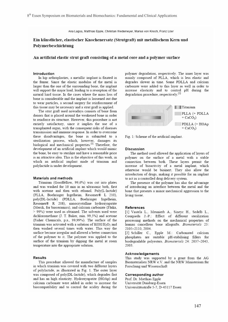
8th Essen Symposium on Biomaterials and Biomechanics: Fundamental and Clinical Applications
147
8th Essen Symposium on Biomaterials and Biomechanics: Fundamental and Clinical Applications
148
APPLICATION OF ZR COATINGS FOR THE IMPLANTS SURFACE PROTECTION (Experimental study)
Aleksyeyeva T.A.*, Lazarenko O.N., Kuznetsov M.V.**, Borisov Yu.S.**, Oshkaderov S.P*.
Kiev medical academy of postgraduate study, Kiev Ukraine *Institute of Physic of Metal NAS of Ukraine, Kiev Ukraine **Paton Welding Institute NAS of Ukraine, Kiev Ukraine
The use of different kinds of implants in medicine meets the problem of recipient organism reaction to the foreign body. Implants provoke to the lymphocytes activation and arises aseptic inflammation, which results in fibrous-connective capsule formation. Depending on the level of the recipient reaction the implant looses its functional properties. Due to this events it is actual the search the new materials which don’t arise the reaction – i.e. would be invisible for the recipients organism. Investigation of Zr properties, its behaviour in contact with biological mediums, low chemical activity leads very slow speed of the reaction with organic compounds. Thus there was a question to evaluate the influence of Zr coating and its alloys onto the implants surface on the organism reaction. Materials and methods The pure zirconium coatings were used in the study. The coatings were applied on to the stainless steel (316L) plate’s surface by the magnetron sputtering method. The thickness of the coating was 3-5 mkm. The experiment was carried out with the animals -8 rabbits, breed “Grey Giant”, weight 3,0 - 3,5 kg. The plates of stainless steel, coated with Zr and without coating were implanted subcutaneous along the back under the total anesthesia with intramuscular injection of ketamine 0,5 ml/kg and intravenous injection of thiopental. In order to receive the objective data the plates were implanted tow by tow per one animal: without Zirconium coating to the left and with one to the right. Such scheme of the implantation permits to escape the specific effect of the experimental animal’s organism on the results. The rabbits were fed with common diet for the 8 weeks. After that animals were killed by the lethal injection of thiopental and the plates with surrounding tissue were collected for the histological study and morphometry. The effect of Zirconium coating was evaluated by the thickness of the capsule that was formed over the plates. The plates were extracted from the capsules and their surface was studied by the SEM. Results The histological study shows that in the case with zirconium coating the wall of isolated fibrous-connective capsule was consist of single cell’s line. At the same time the wall over uncoated plates was significant thicker (50,2 ±8,3 mkm vs 122,03 ±.5,4 respectively) In this case at the histological slides artificial particles were found in the giant cells. The SEM data of the uncoated surface shows the manifest tracks of corrosion. There was no corrosion on the coated plates. Conclusion The coating Stainless steel with Zirconium by magnetron sputtering method significant decrease the reaction of the recipient’s tissue to the foreign body and prevent corrosion of base material.

8th Essen Symposium on Biomaterials and Biomechanics: Fundamental and Clinical Applications
149
Computerized identification of bone cell mobility parameters (in vitro)
S. Magosch+), H.-G. Lipinski+), L. Winkler*), M. Wiemann*), D. Bingmann*)
+) University of Applied Science (FH) Dortmund; Dept. of Medical Informatics, Emil-Figge-Str. 42, D-44227 Dortmund; email to [email protected]
*) University Duisburg-Essen, Physiological Institute, Hufelandstr. 55, 45147 Essen It is well knows that nursing or other substrata markedly influence the mobility of living bone cells within cell culture media. An efficient movement analysis of the moving cells is required to understand the physiology of the cellular movement pattern. During experiments a large number of cells (500 < N < 1200) has to be analysed for time sequences of about 400 images. Therefore, a set of computer programs was developed which allows for a successful (and rather fast) movement analysis. First we used certain digital image processing methods to prepare the microscopic raw data:
• digital band pass filter techniques (Fourier method), helpful to minimize image disturbances;
• cell segmentation (based on binary transformation techniques), useful to detect the whole cell body;
• edge detectors (Marr-Hildreth and Canny Operators), necessary for cell border identification;
• Skeleton algorithms were used to calculate the individual cell circumference. Next we developed algorithms to identify a certain cell and its mobility parameters from the complete cell stack. This technique allows for an automatic movement analysis. In detail the program analyzed (among others) the following mobility parameters:
• the actual position of the moving cell and the movement distance after a certain time;
• the actual speed of an individual cell and the total velocity vector field of all cells observed;
• the actual acceleration (and, hence, the impress force) of an individual cell and the total acceleration vector field;
• the geometrical form of the path of any cell (characterized by its fractal dimension);
• the optical flow of the moving cells. All cell objects are examined automatically with regard to their structure, size, form, moving paths and other information. Finally, the complete movement process (including alteration of the computed mobility parameters) could be presented as a digital video animation. The programs run fast on standard PCs (approximately 100 images can be completely analysed within one minute if a 2 GHz Intel® processor is used). All generated data can also be stored in a separate data base connectable to external data analysis programs like Excel® or MatLab®. Our program needs an interactive correction of the movement-detection only in very few cases and therefore it permits an efficient automatic analysis. Hence, the program contributes to the understanding of physiological processes in living bone cells under changing conditions of life or in cases of pathophysiological situations.

8th Essen Symposium on Biomaterials and Biomechanics: Fundamental and Clinical Applications
150
Production and biomechanical testing of bioceramic implants Subject: In the long term the application of conventional implants, consisting of metallic and plastic components, leads towards problems like implant loosening and immune reactions. Main reason for the failure of the interconnection between bone and prosthesis is the agglomeration of wear debris of the polyethylene part. Besides functional losses strong pain is the consequence and a revision operation becomes necessary. Inspired by the successfully employed ceramic hip endoprostheses the trend for the development of implant material pairings goes towards the manufacturing of hard surfaces. Within the scope of the Collaborative Research Centre “Sustainable Bioresorbable and Permanent Implants of Metallic and Ceramic Materials” the development of an automated machining of the free form surface and the biomechanical testing of low-wear bioceramic implants is promoted under medical and technical points of view with the aim of optimized lifetime in collaboration of the Institute of Production Engineering and Machine Tools and the Laboratory for Biomechanics and Biomaterials. The basic mechanisms of five-axis grinding and polishing are analyzed to design the processes and develop suitable manufacturing strategies. The developed technologies of five-axis grinding and polishing will enable the fabrication of variegated kinds of complex prostheses for medical technology. The biomechanical effects and tribological properties are investigated inter alia by kinemator-tests, to examine the functionality and durability of the developed bioceramic implants. The known superiority of hip joint replacements will be made accessible for patients, which are in need of complex shaped implants, for example knee joint prostheses. Authors: Prof. Dr.-Ing. B. Denkena1, Dipl.-Ing. M. Reichstein1, Dipl.-Ing. (FH) M. van der Meer1, Dr.-Ing. C. Hurschler2, PD. Dr. med. C. Stukenborg-Colsman2, Dr. med. S. Ostermeier2 1 Institute of Production Engineering and Machine Tools (IFW) University of Hannover Schönebecker Allee 2 30823 Garbsen, Germany 2 Laboratory for Biomechanics and Biomaterials (lbb)
Orthopaedic Clinic Medical University of Hannover Anna-von-Borries-Str. 1-7 30625 Hannover, Germany

8th Essen Symposium on Biomaterials and Biomechanics: Fundamental and Clinical Applications
151
Biomineralization in zebrafish (Danio Rerio) shown by scanning electron microscopy and synchrotron radiation computer
microtomography
Neues, F.1; Epple, M.1; Fischer, J.2; Beckmann, F.2 1University Duisburg-Essen; Institute of Inorganic Chemistry; Universitaetsstr. 5-7; 45117 Essen 2HASYLAB at DESY; Notkestr. 85; 22603 Hamburg The zebrafish is a model for studying vertebrate development and human diseases. It has become the most investigated fish during the past decade. The short generation time, embryonic transparency, ease of mutagenesis and ongoing genomic research have made zebrafish ideal for laboratory studies. Bones of zebrafish are assembled in a similar way to human bones. They are primary cellular (osteocyte containing) and cancellous. Transitional stages between bone and cartilage also exist. The bones were investigated by synchrotron radiation computer microtomography (SRµCT) and scanning electron microscopy (SEM). SRµCT can show the sample without a need for destruction or extraction out of the organism. Virtual cuts through the specimen can be performed. Hidden internal structures become visible and even quantitative conclusions can be drawn on the basis of virtual cuts which cannot be performed with "real" cutting, i.e. artifacts caused by preparation are avoided. It is also possible to determine the spatial variation of the thickness of a sample.

8th Essen Symposium on Biomaterials and Biomechanics: Fundamental and Clinical Applications
152
THE THREE-DIMENSIONAL MODEL OF THE FEMURAL PROSTHESIS COMPONENT
Dragos Popa, Daniela Tarnita, Dan Tarnita, Dan Grecu and Sorin Didu
The University of Craiova, Faculty of Mechanics, Dept. of Automotive, 165 Bucuresti str., Craiova,
E -mail: [email protected],
The University of Craiova, Faculty of Mechanics, Dept. Of Applied Mechanics, 165 Bucuresti str., Craiova,
E –mail: [email protected]
The University of Medicine and Pharmacy of Craiova, Faculty of Medicine, Dept. Of Human Anatomy, 2 Petru Rares str., Craiova,
E –mail: [email protected]
The University of Medicine and Pharmacy of Craiova, Faculty of Medicine, Dept. Of Human Anatomy, 2 Petru Rares str., Craiova,
The University of Medicine and Pharmacy of Craiova, Faculty of Medicine,
Dept. Of Human Anatomy, 2 Petru Rares str., Craiova,
The paper presents the steps of the obtaining of the three dimensional model of the femural prosthetic component for the human knee joint. The model was realized using a parametrical CAD software which permits the modeling of the most complex part of the knee prosthesis. The femural component was considered the most important part of the knee prosthesis and it was composed by many complicated elementary shapes.
The base shape presented in the first figure was a simple extruded shape, which was transformed, in the final stage, in a virtual complex part of the knee prosthesis (in the second figure).
That model can be used to include it in a virtual prosthesis and the entire assembly can be integrated in the virtual knee joint. With that complete assembly can be obtaining the complex analysis such kinematical and dynamical simulation or finite element analysis in the clasical cases of locomotion (walking, running and jumping). These complete studies has the very complex results like diagrams, tables of data or mapes of results, which can show the entire behaviour of the knee joint with the prosthesis.
The behavior of that model can give the important informations which can be used in the fields of robotics, medicine sciences and medical robotics.

8th Essen Symposium on Biomaterials and Biomechanics: Fundamental and Clinical Applications
153

8th Essen Symposium on Biomaterials and Biomechanics: Fundamental and Clinical Applications
154
8th Essen Symposium on Biomaterials and Biomechanics: Fundamental and Clinical Applications
155
The effect of silicon-substitution on densification and grain growth processes in hydroxylapatite ceramics.
M. Pulkin1,2, V . Putlayev2,3, A. Veresov2, A. Soin2, A. Garshev2 1Ceramic Materials and Components, University Bremen, Bremen 28359, Germany 2Department of Materials Science, Moscow State University, Moscow 119899, Russia 3Department of Chemistry, Moscow State University, Moscow 119899, Russia Hydroxylapatite Ca10(PO4)6(OH)2 (HAp) in form of dense or porous ceramics is an
attractive material for many biomedical applications due to its chemical composition and crystallographic structure similarities to those of inorganic component of the bones of vertebrates. To the disadvantages of HAp limiting its successfully application for bone defects repairing one can refer the low rate of resorption and the slow formation of bone-like HAp layer at the implant interface in vivo. One of the most promising way to improve the bioactivity of HAp via enhance of resoprtion rate and surface activity is the chemical modification of HAp. It has been recognised recently that Si-doping have a positive effect on both of those factors.
Thus, silicon-substituted HAp (Si-HAp) with Са10-х(РО4)6-x(SiO4)х(ОН)2-х (x = 0…1) nominal composition was the object of the study. The problems of silicon solubility in HAp lattice (and its distribution in the bulk of ceramics), charge compensation mechanism, si-doping effect on sintering of HAp ceramics were under the investigation.
The starting Si-HAp powders were prepared by precipitation technique starting from solutions: (NH4)2HPO4 (0.5 M) and Ca(NO3)2 (0.5 M) with pre-adjusted pH~9.5 at 60°C in the presence of (C2H5O)4Si. For the particle sizes distributions, morphology, chemical and phase composition analysis of the powders the following methods were used: light-scattering, TEM, SEM/EDX, XRD and FTIR. The pressed (at 30 MPa) compacts were sintered at 1100-1250°C (1-6 hours holding time) in air to produce Si-HAp ceramics, those was examined with SEM/EDX, density measurements and dilatometry.
HAp phase in the Si-HAp samples was found to be stable up to 1100°C. The charge compensation by aliovalent substitution of PO4
3- through SiO44- is provided via decreasing of
OH- groups content and additional aliovalent substitution of PO43- through CO3
2- at temperatures under ca. 1000°C. It was found that silicon-doping suppress the grain growth due to reduction of grain boundary mobility: activation energy of lattice diffusion increase from 160±30 kJ/mol for HAp to 430±90 kJ/mol for Si-HAp. Moreover, it enhances the densification rate due to increasing of pore mobility (in fact, activation energy of grain boundary diffusion decrease from 440±20 kJ/mol for HAp to 140±10 kJ/mol for Si-HAp) during the initial sintering stage. Sintering trajectories for Si-HAp ceramics based on densification and grain growth data revealed the prevalence of densification over re-crystallization phenomena at temperatures not higher than 1200°C. That allows to produce high-dense ceramics (over 98% of theoretical density) with small grain sizes (with beneficial mechanical properties). The effect of Si on sintering behaviour can be explained in terms of its segregation to grain boundaries.
The work was supported by RFBR (grant #05-03-32768), the program ‘Universities of Russia’ (grant # UR 06.02.556 ), interdisciplinary grant of MSU #26, and SEC ‘Synthesis’ (grant of Ministry of Education and Science).
8th Essen Symposium on Biomaterials and Biomechanics: Fundamental and Clinical Applications
156
Cell growth on cochlear implant materials
U. Reich1, G. Reuter1, P. Müller2, T. Stöver1,T. Fabian3, H. Lubatschowski4, T. Lenarz1. 1Department of Otolaryngology, Medical University of Hannover
²German Research Centre for Biotechnology, Braunschweig, ³University Hannover, Centre of Biomedical Engeneering
4 Laser Zentrum Hannover e.V. The optimization of the electrode-nerve-interface is a key aspects in cochlear implant (CI) research today is. A few days after implantation there is an increase of impedance at the individual electrode contacts. This may be affected by the growth of connective tissue around the electrode witch is visible on explanted CI-electrodes. In addition the electric stimulation signal become more unspecific. The aim of this study is a permanent reduction of connective tissue growth. The cochlear electrode contains platinium/iridium contacts in a silicone carrier. Silicon carrier is manufacured (Cochlear Ltd, Sydney) from two types of silicone (LSR 30, HCRP 50). For our studies this silicone was provided with a polished and unpolished surface. In the first step gradual patterns were inserted into the material. The gradual structures presented in this study have a width of 1-20 µm and a depth of approx. 1 µm in linear and radial configuration. To create structures in the material with high precision and sharp edges we use the femtosecond laser technology. GFP-marked fibroblasts makes it possible to observe cell growth even on non-transparent materials (e. g. electrode metals). Cell growth can be observed on one sample over a period of several days. This method particularly suitable for trials with only few samples. The fibroblasts grow on the electrode materials. While the cell growth rate on platinium/iridium is similar to that of cells on glass, the cells on silicones show reduced growth. Silicone HCRP 50 shows the lowest growth rate. Cell growth on silicone with a polished surface is smaller than on an unpolished surface. Laser structuring leads to further reduction of fibroblast growth. The cell growth rate correlates with the width of the microstructure. First experiments show that the cell growth rate is reduced on structures over 4 µm width. In further experiments structures of different size are to be tested. The aim is to reduce connective tissue growth while optimizing the electric contact to the neuronal target cells, the spiral ganglion cells. This project was supported by German Research Foundation, Collaborative Research Centre 599: “Suitable Bioresorbing and Permanent Implants of Metallic and Ceramic Materials”

8th Essen Symposium on Biomaterials and Biomechanics: Fundamental and Clinical Applications
157
Enhanced stereoscopic 3D-visualisation of confocal images
A. Roth+), H.-G. Lipinski+), M. Wiemann*), D. Bingmann*)
B. +) University of Applied Science (FH) Dortmund; Dept. of Medical Informatics,
Emil-Figge-Str. 42, D-44227 Dortmund; email to [email protected]
*) University of Duisburg-Essen, Physiological Institute, Hufelandstr. 55, 45147 Essen There are many ways to visualize 3-dimensional image data from a confocal microscope. In this paper a new technique for a clear and high quality visualisation will be demonstrated. The visualisation process takes thee steps:
• First, the image data have to be acquired by the aid of confocal laser scanning microscopes.
• Second, with some pre-filtering methods the wanted cellular object data have to be separated from the image raw data.
• Third, the visualisation-routine has to display the result of the filtered 3D-data on the computer screen.
For the second step a combination of high-pass filtering and an additional picture sharpening algorithm was created. The sharpening algorithm allows for a modification of the complex three-dimensional components of the Fourier spectrum of the image data. The data are weighted by a non-linear function which could be handled by only one parameter value. This value controls an exponential function which is used to generate a three-dimensional weighting matrix. The real and the imaginary parts of the Fourier spectrum are multiplied by the corresponding entries of this matrix. Thus, both an additional picture sharpening and a reduction of the high frequency noise in the picture occur. After some great results by using these pre-filtering methods, in the third step the possibilities of the displaying routines have been enhanced. A combined visualisation is possible, showing the depth-information by using special stereoscopic glasses and simultaneously showing the surface of the visualised cell. Key frame-animations can be rendered and presented for discussions or educational purposes. Both the picture pre-processing and the 3D-visualisation were programmed using the programming language C++ for WINDOWS® operating systems. All programs run without special hardware on a standard PC.

8th Essen Symposium on Biomaterials and Biomechanics: Fundamental and Clinical Applications
158
Morphological characterization of graded, biodegradable and buffered polylactide skull implants Schmitz*, I., Rasche**, Ch., Wehmöller****, M., Weihe*****, St., Eufinger**, H., Epple***, M., Schiller***, S., Müller, K.M*., Theegarten, D*. *BG- Clinic Bergmannsheil, University Hospital, Institute of Pathology, Bochum **Department of Oral and Maxillofacial Plastic Surgery, University Hospital, Ruhr University, Knappschaftskrankenhaus, Recklinghausen ***University Duisburg-Essen, Campus Essen, Institute of Anorganic Chemistry, Essen ****Institute for Productive Systems, Ruhr University, Bochum *****Department of Oral and Maxillofacial Surgery, Clinic Dortmund, Clinic Center North, Dortmund The aim of this study was to analyze the micromorphology of individual biodegradable skull implants, which should be substituted by bone according to their degradation. A new sheep model for silmultaneous craniectomy and cranioplasty was developed, and results obtained in 7 animals are reported. The inside of the implants degrades faster than the outside in order to allow the ingrowth of osteoblats. The skulls were investigated after 2, 9, 12 and 18 months using light microscopy and scanning electron microscopy. After 2 months the biomaterial showed a beginning degradation, with breaking and loss of integrity of the material. Foreign body reaction to the implant and formation of connective tissue was obvious. After 12 months the implants could be clearly distinguished from the surrounding organic structures. After 9 and clearly after 12 months bone cells invading the implant and beginning formation of new bone were seen. After 18 months the implants were very weak and degradation of the material was pronounced. Above the dura implanted material was focally replaced by bone. In conclusion the dura plays an important role in formation of new bone in the used animal model. Further studies have to prove if great defects could be sufficiently treated by this method.

8th Essen Symposium on Biomaterials and Biomechanics: Fundamental and Clinical Applications
159
A 'cold' atmospheric pressure plasma jet for treatment of sensitive surfaces with biomedical relevance V. Schulz-von der Gathen, St. Reuter, K. Niemi, H.F. Döbele Fachbereich Physik, Universität Duisburg-Essen Atmospheric pressure plasma jets (APPJ) emit a beam of relatively cold (T ~50°C) neutral gas containing large amounts of excited atoms or molecules and radicals into free air. It has been demonstrated that these discharges can be applied e.g. to etch or to clean surfaces, to deposit layers on and to sterilize them [1]. The compact, handy (10 cm length, 4 cm diameter) RF-excited plasma jet produces a homogeneous plasma of helium or argon with a minority admixture of molecular gases (~ 1 vol.-%) such as oxygen. The typical power of 10 to 150 W is supplied by a commercial radio-frequency transceiver at a frequency of 13.56 MHz. The jet presented here is is operated with a gap of about 1.5 mm heights and 4 cm width between powered and grounded electrodes. The plasma jet can be scaled up from dimensions of about one square millimeter. Within the gap the homogeneous discharge delivers the energy to dissociate the admixed molecules and to excite and to ionize the atoms. The effluent leaving the discharge region exhibits directly at the nozzle temperatures of about 70°C at a power of 150 W. This values fall off rapidly with the expansion of the beam into air as measured by a thermo-couple [2]. Within a project funded by the MWF-NRW some aspects of biomedical interest have been examined. First sterilizations experiments with AG-BioStrips containing bac. subtilis (niger) and bac. stearo-thermophilus yielded positive results at a distance of 1 cm from the jet and after an exposure time of 5 minutes. The analyses have been carried out at the 'Institut für Hygiene und Arbeitsmedizin des Universitäts-Klinikum Essen'. Up to now these experiments are neither representative nor systematic. The sterilization success is substantially determined by the concen-tration of atomic oxygen binding free radicals and reactive substances in an oxidation reaction. Absolute concentrations of atomic oxygen have been measured in the effluent of the plasma jet by two photon laser-induced fluorescence (TALIF). It could be demonstrated that about 1015cm-3 oxygen atoms are transported over several centimeters in a strongly collimated helium beam [3]. Another subject of the project is the investigation of the applicability of the jet for the treatment of biological or organic surfaces (e.g. skin or wounds). For these investigations the transmission of UV radiation from the discharge volume through the jet – representing its own helium or argon atmosphere – onto the surface is of great importance. VUV emission spectroscopic measurements in the spectral range down to 110 nm have been carried out with an evacuated spectrometer (Minuteman). The entrance slit has been closed vacuum-tight by a MgF2 window and is positioned directly in front of the plasma jet. First non-calibrated spectra demonstrate that strong emission lines of e.g. oxygen at 130 nm can be observed up to a few centimeters outside the discharge. This radiation - also important for the sterilization efficiency - may represent a load for the skin. As an initial experiment towards modification of sensitive surfaces in ambient air the cover of a Petri dish has been treated with the 'hand-held' plasma jet for 10 seconds. Within this time the cover changes from hydrophobic to hydrophilic behaviour without a significant heating up or melting of the dish. It has to be pointed out again that this change of surface energy as well as sterilization are achieved without the need to put the object into a vessel or to use any harmful chemicals. Acknowledgments This work was supported by the ‘Ministerium für Wissenschaft und Forschung des Landes Nordrhein-Westfalen’ in the frame of the ‘Verbund plasmagestützte Oberflachenmodifikation von

8th Essen Symposium on Biomaterials and Biomechanics: Fundamental and Clinical Applications
160
Medizin- und Biotechnischer Bedeutung’. [1] Selwyn G S, Herrmann H W, Park J, Henins I, Contrib. Plasma Phys. 41 (2001) 610-619 [2] S. Wang, V. Schulz-von der Gathen and H.F. Döbele, Appl. Phys. Letters 83 (2003) 3272-3274 [3] K. Niemi, S. Reuter, V. Schulz-von der Gathen, and H. Döbele, Plasma Sources Sci. Technol. 14 (2005) 375
– 386

8th Essen Symposium on Biomaterials and Biomechanics: Fundamental and Clinical Applications
161
Characterization of Core-Shell Dendritic Nanoparticles as
Potential Drug Carrier: Laser Light Scattering
Anuj Shuklaa, Holger Türkb, Rainer Haagb*, Heinz Rehagea
a Lehrstuhl für Physikalische Chemie II,University of Dortmund, Otto-Hahn-Str. 6, D-44227 Dortmund b Lehrstuhl für Organische Chemie,University of Dortmund, Otto-Hahn-Str. 6, D-44227 Dortmund
*Present Address: Institut für Chemie, Freie Universität Berlin, Takustrasse 3, D-14195
Berlin
During the development of better administration techniques and drug delivery systems in
pharmacy, water-soluble polymers have become potential candidates for this purpose.
Polymer matrixes are considered to have the ability to serve as drug carriers, where drug
release can be accomplished through either internal (self-regulated) or external triggers. Core
shell structured nanotransporters gain great attention in the last years in the field of drug
delivery, since they are stable under shear force and other environmental effects in contrast to
weak micellar assembly and additionally they can release encapsulated drug using very weak
external signal such as pH change.[1]
In this study, we characterized the dendritic core-shell architectures based on the selective and
reversible shell functionalization of dendritic polyglycerol (PG) polymer. Detailed synthetic
concept for the preparation of dendritic core-shell architectures are presented elsewhere.[2] We
performed a laser light scattering (LLS), and surface tension study of the association behavior
of these core-shell structured nanotransporters in water. By using a combination of static and
dynamic light scattering measurements, we have determined the molecular weight, size, and
size distribution. We also determined the critical aggregation concentration and the average
aggregation number of the aggregated particles. A model for the resulting aggregates is
presented. Results indicated that unfunctionalized polyglycerol dendtritic core-shell
nanotransporter behaves like so-called unimolecular micelles while functionalized
polyglycerol dendtritic core-shell nanotransporter can self-assemble to form spherical micellar
aggregates. Micellar aggregation number is equal to ~ 10. Dynamic light scattering
measurements suggest that structure of functionalized group has big impact on the size of
micellar aggregates. It is also observed that with increasing the degree of core
functionalization, micellar size also increases. Dynamic light scattering measurements
indicated that drug is preferentially located at the interfacial region of micelles and the size of
the nanotransporter slightly decreases in the presence of drug.

8th Essen Symposium on Biomaterials and Biomechanics: Fundamental and Clinical Applications
162
[1] R. Haag, Angew. Supramolecular Drug-Delivery Systems Based on Polymeric Core-Shell Architectures Chem. 2004, 116, 280-284
[2] H. Türk, et. al, in preparation

8th Essen Symposium on Biomaterials and Biomechanics: Fundamental and Clinical Applications
163
8th Essen Symposium on Biomaterials and Biomechanics: Fundamental and Clinical Applications
164
Influence of Cl-, NO3- and СH3COO- anions on micromorphology of precipitated
hydroxyapatite powders.
A.A. Stepuk*, A.G. Veresov, V.I. Putlayev
Material Science Department, Moscow State University Leninskie Gory, Moscow119992, Russia
* e-mail: [email protected]
Hydroxyapatite Ca10(PO4)6(OH)2 (HA) seems to be an ideal component of bone
implants due to its perfect biological compatibility. Micromorphology of artificial HA crystals
is a key parameter determining the practical application of the material. Precipitation in
aqueous media is the most common method of HA synthesis. Calcium compounds
Ca(NO3)2*4H2O, CaCl2, Ca(OH)2, CaSO4*2H2O can be mixed with H3PO4, (NH4)2HPO4,
Na2НPO4 (as phosphate source) to obtain HA powders. NaOH, KOH, NH4OHs are used to
control solution pH value:
The objective of the present research was synthesis of HA powders with different
micromorphology (needles, plates etc.) and average size less than 100 nm (to mimic the
natural bone nanocrystals). Different starting reagents were used to analyze the influence of
anion of calcium salt on the HA crystals shape:
10CaX2+6KH2PO4+14KOH→Ca10(PO4) 6(OH)2↓+20KX+12H2O, (1)
where −−−= COOCHClNOX 33 ,, .
The as-received HA specimens were annealed at 500-1000 oC to improve their
crystallinity (the heat treatment leads to particle coarsening). The materials were characterized
with XRD, SEM and IR spectroscopy methods.
It was revealed that powders annealed at T ≥ 700 oC possess different shapes depending
on the initial calcium salt. For instance, needle-like crystals were obtained with calcium
nitrate. Difference in crystals shapes and their sintering ability can be explained in terms of
adsorption (acetatate) or substitution (chloride for hydroxyl in HA lattice) of X- ions of
“impurities” (eq. 1).
The authors are grateful to the Russian foundation of basic research (grant # 05-03-32768-а) and to the program “University of Russia” (grant # 06.02.556) for financial support of present work.
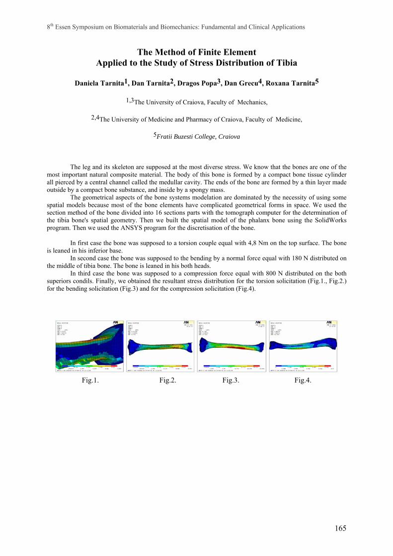
8th Essen Symposium on Biomaterials and Biomechanics: Fundamental and Clinical Applications
165
The Method of Finite Element Applied to the Study of Stress Distribution of Tibia
Daniela Tarnita1, Dan Tarnita2, Dragos Popa3, Dan Grecu4, Roxana Tarnita5
1,3The University of Craiova, Faculty of Mechanics,
2,4The University of Medicine and Pharmacy of Craiova, Faculty of Medicine,
5Fratii Buzesti College, Craiova
The leg and its skeleton are supposed at the most diverse stress. We know that the bones are one of the most important natural composite material. The body of this bone is formed by a compact bone tissue cylinder all pierced by a central channel called the medullar cavity. The ends of the bone are formed by a thin layer made outside by a compact bone substance, and inside by a spongy mass.
The geometrical aspects of the bone systems modelation are dominated by the necessity of using some spatial models because most of the bone elements have complicated geometrical forms in space. We used the section method of the bone divided into 16 sections parts with the tomograph computer for the determination of the tibia bone's spatial geometry. Then we built the spatial model of the phalanx bone using the SolidWorks program. Then we used the ANSYS program for the discretisation of the bone.
In first case the bone was supposed to a torsion couple equal with 4,8 Nm on the top surface. The bone is leaned in his inferior base.
In second case the bone was supposed to the bending by a normal force equal with 180 N distributed on the middle of tibia bone. The bone is leaned in his both heads.
In third case the bone was supposed to a compression force equal with 800 N distributed on the both superiors condils. Finally, we obtained the resultant stress distribution for the torsion solicitation (Fig.1., Fig.2.) for the bending solicitation (Fig.3) and for the compression solicitation (Fig.4).
Fig.1. Fig.2. Fig.3. Fig.4.

8th Essen Symposium on Biomaterials and Biomechanics: Fundamental and Clinical Applications
166
8th Essen Symposium on Biomaterials and Biomechanics: Fundamental and Clinical Applications
167
Porous TiO2/glass ceramic filled with heparinized collagen as a reservoir for the osteoinductive factor BMP-2
von Walter§ M., Erli§ H.J., Ragoss# C., Maier# H.R. and Steffens* G.C.M. §Department of Trauma Surgery and *Institute of Biochemistry,
University Hospital Aachen, Pauwelsstrasse 30, D-52074 Aachen, Germany #Institute of Ceramic Components in Mechanical Engineering, RWTH University Aachen,
Nizzaallee 32-34, 52072 Aachen, Germany The TiO2/glass ceramic Ecopore is a porous composite with basic biocompatibility. We are currently investigating whether this material can be used as a load-bearing bone substitute by improving osteo- and angiogenic capabilities. While covalent immobilization of fibronectin on the surface of Ecopore has been shown to enhance cellular adhesion and spreading of human osteoblasts (von Walter et al., Biomaterials 2005), the analogous modification with bone morphogenetic protein-2 (BMP-2) obviously lacked biological functionality, because it did not induce expression of alkaline phosphatase (AP) in embryonic fibroblasts (line MC3T3). In order to preserve biological functionality, we intended to avoid surface binding of the factor by realizing a second matrix inside the pore system of Ecopore, into which BMP-2 could be reversibly bound. For this purpose, the pore system of Ecopore was filled with collagen sponge: a suspension of bovine tendon collagen (type I) in 10% acetic acid was filled into the pores and the specimens were lyophilized. We included heparin as further modification, because many growth factors, among them BMP-2, vascular endothelial growth factor (VEGF) and basic fibroblast growth factor (bFGF), exhibit heparin binding domains, which allow them to bind to the extracellular matrix (ECM). Part of the ceramic/collagen hybrids were therefore treated with heparin in the presence of 1-ethyl-3(3-dimethylaminopropyl)carbodiimide (EDC) and N-hydroxysuccinimide (NHS). This procedure leads to the covalent immobilization of heparin and to an additional cross-linking of the collagen. Heparinized and non-heparinized hybrids were placed in the wells of 24-well plates and immersed in solutions containing variable amounts of BMP-2. After equilibration, the BMP-2 concentrations of the supernatants were determined by ELISA. Preliminary results show that the heparinized hybrids have a higher BMP-2 binding capacity than the non-heparinized hybrids. We also performed release kinetics of hybrids loaded with BMP-2: here the heparinized hybrids are characterized by a slower release, indicating that this hybrid might be functioning as a sustained release system. To prove biological activity of the released BMP-2, we co-incubated embryonic fibroblasts with BMP-2-loaded and blank hybrids and measured AP presence in the cultures applying an enzymatic photometric assay. The AP signal was clearly elevated in the cultures with BMP-2-loaded hybrids. Future research will focus on testing the Ecopore/collagen system with angiogenic growth factors, as well as evaluating its osteo- and angiogenic potential in in vivo experiments.

8th Essen Symposium on Biomaterials and Biomechanics: Fundamental and Clinical Applications
168
Tissue Reaction of Micro/Nanoparticles and Biomedical Application of Carbon Nanotubes
Fumio WATARI
Graduate School of Dental Medicine, Hokkaido University Sapporo 060-8586 (JAPAN)
The cell and tissue reaction changes from bulk to fine particles for the same material. Both in vitro biochemical cell functional test and in vivo animal test were in accordance that even the biocompatible materials in bulk such as Ti and TiO2 become stimulative as the size is decreased and this is pronounced below 10µm, about the cell size where the phagocytosis is induced. ICP elemental analysis showed that the dissolution from Ti particles was negligible. These phenomena were characterized as non-specific cytotoxicity arising from the physical size effect, and different from the chemical toxicity effect due to the ionic dissolution which is usually dominant in bulk to affect on material biocompatibility.
Carbon nanotubes (CNT) have attracted the attentions to the application in the electronic and chemical fields, but the application for the biomedical field has been very rare. There is an argument that CNT may have the serious toxicity due to its acicular or fibrous particle shape. So far as we investigated in vitro and in vivo, there is a weak stimulus due to the size of particles as general as in the biocompatible materials. We have rather found the properties favorable as biomaterials such as affinity for saccharides and proteins, cell adhesion, bacteria adsorption, and apatite precipitation. These properties could be the excellent basis of the development for numerous biomedical applications.

8th Essen Symposium on Biomaterials and Biomechanics: Fundamental and Clinical Applications
169

8th Essen Symposium on Biomaterials and Biomechanics: Fundamental and Clinical Applications
170
Residual stress control in nanocrystalline diamond films
N. Wöhrl, V. Buck
Thin Film Technology Group, Dept. of Physics, University of Duisburg-Essen, Universitätsstr.3-5, 45141 Essen, Germany, E-mail: [email protected]
In medical technology, artificial joints have been used successfully for many years now. Worldwide about 600.000 knee- and hip-endoprosthesis are implanted per year. Still there remain open questions in basic research. Especially the stability time of prosthesis, averaging from 10 to 15 years is unsatisfying. The main cause for failure is the loosening of the implant due to osteolysis, which is caused by wear particles. In recent years prosthesis have been coated with amorphous carbon (a-C; a-C:H) to avoid the development of wear particles, because a-C and a-C:H coatings are characterized by excellent biocompatibility, low friction and low wear. But this is not the central point, because the size of the wear particles is the crucial parameter for the biocompatibility. Nanostructured carbon films can be tailored in a very wide range to fulfil these conditions in a first approach. Residual stress is a critical parameter in thin film deposition and crucial for technical applications of nanocrystalline diamond films because the adhesion of the films on implants is affected by the intrinsic stress. High residual stress can lead to cracking or even to delamination of the film from the substrate. An ex-situ optical device (SSIOD “Surface Stress Induced Optical Deflection”) was used to measure the curvature of silicon substrates coated with nanocrystalline diamond films. With respect to Stoney’s equation one can calculate the residual stress from the curvature of the substrate. It is shown that the residual stress in the substrate can be varied in a wide range just by controlling the plasma parameters. A possible explanation for the changes in residual stress is given based on the data taken from Raman Spectroscopy and FTIR Spectroscopy.

8th Essen Symposium on Biomaterials and Biomechanics: Fundamental and Clinical Applications
171
Electrochemical interaction of H2O2 and hyaluronan at the surface of β-Ti-45Nb Beate Heisterkamp, Daniela Zander Department of Biochemical and Chemical Engineering, University of Dortmund, D-44221 Dortmund, Germany Titanium and titanium alloys, specially Ti-6Al-4V, are used for various medical applications, e.g. endoprosthesis. There is evidence that metallic dissolution into surrounding tissue can cause complications with the implant and the environment [1]. Conditions such as metallosis and inflammation have been associated to the migration of metallic ions into the tissue. An explanation for this behavior was attributed to the radical transfer reaction of free oxygen in the biological system [2] as well as the interaction of those radical reactions with the polysaccharide hyaluronic acid [3] during the osseointegration. So far the mechanism for the osseointegration of titanium based implants is not clear. Klinger et al. [3] assumed the mechanism of osseointegration as a radical transfer reaction by a radical peroxide layer on titanium to metabolize the hyaluronan in the chondroitinsulphate proteoglycan matrix. This assumtion is developed under exploitation of new monoclonal antibodies which directly allow the measurement of the glucosaminoglycans like hyaluronan. The aim of the present studies is on one hand to investigate the influence of H2O2 on the corrosion of Ti-45Nb. On the other hand the interaction of hyaluronan with the passive layer including the degradation process of hyaluronan will be investigated. The corrosion measurements of technical β-type Ti-45Nb were performed by potentiodynamic polarization and electrochemical impedance spectroscopy (EIS) in PBS without and with 0.1M H2O2 (pH 7.4, 37 °C). The effect of the hyaluronan was investigated by adding 1.5 g/l hyaluronan to the electrolyte. In addition viscosity measurements of the electrolytes were performed to investigate the degradation of hyaluronan after corrosion in more detail. The corrosion experiments revealed a lower open circuit potential as well as a decreased corrosion rate for PBS with hyaluronan in comparison to PBS with H2O2. EIS investigationsin PBS without and with hyaluronan for 168h showed a significant influence of hyaluronan on the layer formation of the system Ti-45Nb/electrolyte. Modeling of the EIS data indicate a second adsorption layer by hyaluronan besides an oxide layer and a first adsorption charge layer. The model for PBS only demonstrates an oxide layer with diffusion control and a strong adsorption layer. An increased degradation of hyaluronan was observed by viscosity measurements of PBS with H2O2 and hyaluronan indicating also a strong electrochemical interaction between H2O2, hyaluronan and the Ti-45Nb surface. 1. G. Meachim, D.F. Williams, Biomed. Mater. Res. 7, 1973, 555. 2. P. Tengvall, H. Elwing, L. Sjöqvist, I. Lundström, L.M. Bjursten, Biomater. 10, 1989,
118. 3. M.M. Klinger, F. Rahemtulla, C.W. Prince, L.C. Lucas, J.E. Lemons, Crit. Rev. Oral
Biol. Med. 9, 1998, 449

8th Essen Symposium on Biomaterials and Biomechanics: Fundamental and Clinical Applications
172
Alumina contamination of grit-blasted surfaces and consequences on hard-soft bearing surfaces.
O. Zinger, S. König, Y. Dirix, H. Schmotzer Plus Orthopedics AG, Schachenallee 29, 5001 Aarau, Switzerland,
Alumina (aluminium oxide) grit-blasted surfaces show excellent osseointegration in clinical studies [1]. However, it has been known for many years that such a roughening method leads to extensive embedding of grit particles. According to previous studies performed on commercial implants, contamination with aluminium oxide particles can cover up to 26% of the surface [2]. Those particles can apparently invade the joint space and then lead to third body wear in the metal-metal bearing surface [3]. Therefore, we measured the alumina contamination on new hip stems produced by different manufacturers and examined the bearing surfaces from retrieved metal-polyethylene pairings which were combined with an alumina-blasted implant.
Four hip stems produced by 4 different manufacturers (A-D) were analysed. Alumina contamination was determined by image analysis of Back-Scattered-Electrons (BSE) micrographs (measurement method validated by EDX mapping). The results can be seen in the chart below, where mean values and standard deviations are calculated from 5 different measurements performed at different locations.
0.0
5.0
10.0
15.0
20.0
25.0
30.0
35.0
40.0
45.0
manufacturers
Al 2O
3 sur
face
cov
erag
e [%
]
mean 24.1 26.3 34.1 40.2
A B C D
Those results confirmed that grit-blasted hip stems are covered by an extensive amount of alumina particles, which ranges from 24 to 40%. We then examined a total of 12 different bearing surfaces from retrieved hip joints and sorted them into 4 groups according to the amount of alumina particles observed by optical microscopy on the polyethylene (PE) surface. Most of the inlays were contaminated with aluminium oxide particles which were pressed into the PE (see picture above) whereas the number and size of the particles varied. One inlay (8%) was free of particles, 42% showed a few particles, 25% featured a medium level of contamination and 25% were heavily contaminated. Usually, both inlays and ball-heads showed scratches that may have been caused by the alumina particles embedded in the PE. Roughening of the ball head can firstly lead to an increase in PE wear, and secondly, those hard particles embedded into the PE surface could lead to an increase in metal wear and metal-ion release.
All cementless hip implants investigated showed aluminium oxide contamination originating from the grit-blasting process, with a surface coverage ranging from 24 to 40% and a clear difference between the manufacturers. Most of the retrieved inlays (92%) were contaminated with aluminium oxide particles which were pressed into the PE, and usually both inlays and ball-heads showed scratches which could increase PE and metal wear. 1. Zweymüller et al. CORR 235, 195-206 (1988) 2. Göske et al. Eur Mic Anal 18, 9-11 (2004) 3. Böhler et al. JBJS 84-B, 128-136 (2002)

8th Essen Symposium on Biomaterials and Biomechanics: Fundamental and Clinical Applications
173
Immobilization of Proteins on Bone Replacement Materials
Kristin Zurlinden and Herbert P. Jennissen
Institut für Physiologische Chemie, Universitätsklinikum Essen, Hufelandstr. 55, D-45122 Essen, Germany
Hydroxyapatite (HAP) has a great potential as a bone replacement material because of
its similarity to the crystal structure of inorganic matrix of bone. Several years ago we showed
that porous hydroxyapatite ceramics (Endobone, Merck) can be covalently modified for the
immobilization of proteins [1]. In this connection the immobilization of rhBMP-2 on
hydroxyapatite would be of high medical interest, because of the decisive role of bone
morphogenetic proteins (BMP) in bone development and osteogenesis. Therefore we have
performed model studies with the protein ubiquitin, which has been shown to possess very
similar immobilization properties as rhBMP-2 [2, 3].
The modification of the used porous HAP wafers (Orthoss, Geistlich) proceeds in two
steps. For non-covalently immobilization of the protein the HAP surface is modified with
APS (3-aminopropyl-triethoxysilane) [2]. For covalently immobilization the HAP-APS
wafers are activated with CDI (1,1'-carbonyldiimidazole). The wafers were incubated in 0.1 –
3.5 mg/ml ubiquitin solution to immobilize the protein. The amount of bound protein was
determined by using 125I-ubiquitin.
It could be shown that negligible protein is adsorbed to non-functionalized HAP
surfaces (control). In contrast ubiquitin can be very efficiently immobilized non-covalently
(up to 2.4 mg protein/g HAP) and covalently (up to 9.7 mg protein/g HAP) on porous HAP
wafers. In desorption experiments it is shown that the bound protein is slowly released
making such surfaces applicable as long term drug delivery devices for enhancing bone
growth and osteointegration of implant materials.
In future work we want to repeat these successful experiments by replacing ubiquitin
by BMP-2 to get a bioactive bone replacement material. First attemps have shown, that also
BMP-2 can be immobilized on modified HAP surfaces.
[1] Jennissen, H. P. (1999) PCT Patent WO9926674A2 (priority date Nov. 24 1997), 1-29, EP Munich
[2] Jennissen, H.P., Zumbrink, T., Chatzinikolaidou, M., Steppuhn, J., (1999) Materialwiss. Werkstofftech., 30, 838-845
[3] Chatzinikolaidou, M., Laub, M., Rumpf, H., Jennissen, H.P. (2002) Materialwiss. Werkstofftech., 33, 720-727
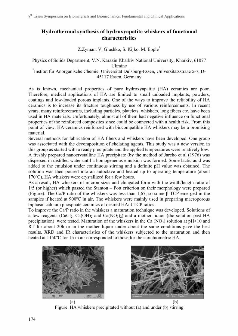
8th Essen Symposium on Biomaterials and Biomechanics: Fundamental and Clinical Applications
174
Hydrothermal synthesis of hydroxyapatite whiskers of functional characteristics
Z.Zyman, V. Glushko, S. Kijko, M. Epple*
Physics of Solids Department, V.N. Karazin Kharkiv National University, Kharkiv, 61077 Ukraine
*Institut fűr Anorganische Chemie, Universität Duisburg-Essen, Universitätsstraβe 5-7, D-45117 Essen, Germany
As is known, mechanical properties of pure hydroxyapatite (HA) ceramics are poor. Therefore, medical applications of HA are limited to small unloaded implants, powders, coatings and low-loaded porous implants. One of the ways to improve the reliability of HA ceramics is to increase its fracture toughness by use of various reinforcements. In recent years, many reinforcements, including particles, platelets, whiskers, long fibers etc. have been used in HA materials. Unfortunately, almost all of them had negative influence on functional properties of the reinforced composites since could be connected with a health risk. From this point of view, HA ceramics reinforced with biocompatible HA whiskers may be a promising material. Several methods for fabrication of HA fibers and whiskers have been developed. One group was associated with the decomposition of chelating agents. This study was a new version in this group as started with a ready precipitate and the applied temperatures were relatively low. A freshly prepared nanocrystalline HA precipitate (by the method of Jarcho et al (1976) was dispersed in distilled water until a homogeneous emulsion was formed. Some lactic acid was added to the emulsion under continuous stirring and a definite pH value was obtained. The solution was then poured into an autoclave and heated up to operating temperature (about 170˚C). HA whiskers were crystallized for a few hours. As a result, HA whiskers of micron sizes and elongated form with the width/length ratio of 1/5 (or higher) which passed the Stanton – Pott criterion on their morphology were prepared (Figure). The Ca/P ratio of the whiskers was less than 1,67, so some β-TCP emerged in the samples if heated at 900ºC in air. The whiskers were mainly used in preparing macroporous biphasic calcium phosphate ceramics of desired HA/β-TCP ratios. To improve the Ca/P ratio in the whiskers a maturation technique was developed. Solutions of a few reagents (CaCl2, Ca(OH)2 and Ca(NO3)2) and a mother liquor (the solution past HA precipitation) were tested. Maturation of the whiskers in the Ca (NO3) solution at pH>10 and RT for about 20h or in the mother liquor under about the same conditions gave the best results. XRD and IR characteristics of the whiskers subjected to the maturation and then heated at 1150ºC for 1h in air corresponded to those for the stoichiometric HA.
(a) (b)
Figure. HA whiskers precipitated without (a) and under (b) stirring

8th Essen Symposium on Biomaterials and Biomechanics: Fundamental and Clinical Applications
175