FIGO CG PAS Con Man - accepted version
29
1 FIGO GUIDELINES FIGO consensus guidelines on placenta accreta spectrum disorders: Conservative management ★ ,§ Loïc Sentilhes 1 , Gilles Kayem 2 , Edwin Chandraharan 3 , José Palacios- Jaraquemada 4 , Eric Jauniaux 5 ; for the FIGO Placenta Accreta Diagnosis and Management Expert Consensus Panel* 1 Department of Obstetrics and Gynecology, Bordeaux University Hospital, Bordeaux, France 2 Department of Obstetrics and Gynecology, Trousseau Hospital AP-HP, Paris, France 3 Department of Obstetrics and Gynecology, St George's University Hospitals NHS Foundation Trust, London, UK 4 Department of Obstetrics and Gynecology, CEMIC University Hospital, Buenos Aires, Argentina 5 EGA Institute for Women’s Health, Faculty of Population Health Sciences, University College London, London, UK ★ Developed by the FIGO Safe Motherhood and Newborn Health Committee; coordinated by Eric Jauniaux, lead developer and corresponding author: [email protected] § The views expressed in this document reflect the opinion of the individuals and not necessarily those of the institutions that they represent.
Transcript of FIGO CG PAS Con Man - accepted version

1
FIGO GUIDELINES
FIGO consensus guidelines on placenta accreta spectrum disorders:
Conservative management★,§
Loïc Sentilhes 1, Gilles Kayem 2, Edwin Chandraharan 3, José Palacios-
Jaraquemada 4, Eric Jauniaux 5; for the FIGO Placenta Accreta Diagnosis and
Management Expert Consensus Panel*
1 Department of Obstetrics and Gynecology, Bordeaux University Hospital, Bordeaux,
France
2 Department of Obstetrics and Gynecology, Trousseau Hospital AP-HP, Paris,
France
3 Department of Obstetrics and Gynecology, St George's University Hospitals NHS
Foundation Trust, London, UK
4 Department of Obstetrics and Gynecology, CEMIC University Hospital, Buenos
Aires, Argentina
5 EGA Institute for Women’s Health, Faculty of Population Health Sciences,
University College London, London, UK
★ Developed by the FIGO Safe Motherhood and Newborn Health Committee;
coordinated by Eric Jauniaux, lead developer and corresponding author:
§The views expressed in this document reflect the opinion of the individuals and not
necessarily those of the institutions that they represent.

2
* Consensus panel: Greg Duncombe (Australia and New Zealand), Philipp Klaritsch
(Germany), Frédéric Chantraine (Belgium), John Kingdom (Canada), Lene Grønbeck
(Denmark), Kristiina Rull (Estonia), Balkachew Nigatu (Ethiopia), Minna Tikkanen
(Finland), Loïc Sentilhes (France), Tengiz Asatiani (Georgia), Wing-Cheong Leung
(Hong Kong), Taghreed AIhaidari (Iraq), Donal Brennan (Ireland), Eiji Kondoh
(Japan), Jeong-In Yang (South Korea), Muhieddine Seoud (Lebanon), Ravindran
Jegasothy (Malaysia), Salvador Espino y Sosa (Mexico), Benoit Jacod (Netherlands),
Francesco D’Antonio (Norway), Nusrat Shah (Pakistan), Dorota Bomba-Opon
(Poland), Diogo Ayres-de-Campos (Portugal), Katarina Jeremic (Serbia), Tan Lay
Kok (Singapore), Priya Soma-Pillay (South Africa), Nataša Tul Mandić (Slovenia),
Pelle Lindqvist (Sweden), Thora Berglind Arnadottir (Sweden), Irene Hoesli
(Switzerland), Unnop Jaisamrarn (Thailand), Amal Al Mulla (United Arab Emirates),
Stephen Robson (UK), Rafael Cortez (Venezuela).

3
1. Introduction
Conservative management of both abnormally adherent (placenta creta) and invasive
placenta (placenta increta and percreta) defines all procedures that aim to avoid
peripartum hysterectomy and its related morbidity and consequences. Four different
primary methods of conservative management have been described in the
international literature: (1) the extirpative technique (manual removal of the placenta);
(2) leaving the placenta in situ or the expectant approach; (3) one-step conservative
surgery (removal of the accreta area); and (4) the Triple-P procedure (suturing
around the accreta area). These methods have been used alone or in combination
and in many cases with additional procedures such as those proposed by
interventional radiology.
The main aim of leaving the placenta in situ versus the extirpative method is
essentially to attempt to decrease the risks of severe maternal morbidity during
cesarean delivery [1–4]. Forcibly removing an invasive placenta—with placental villi
that have invaded the deep uterine vasculature [5]—increases the risks of massive
obstetric hemorrhage and the need for salvation hysterectomy. Uncontrolled bleeding
will lead to coagulopathy and will also complicate the surgical procedure, increasing
the risk of injuries mainly to the bladder and ureters and their possible long-term
complications such as vesicouterine fistula [1–4]. Successful conservative
management strategies will also preserve fertility and thus reduce the impact on a
woman’s societal status and self-esteem associated with the loss of her uterus.
The purpose of this chapter is to assist obstetrician-gynecologists in selecting the
most appropriate conservative treatment option to manage women with the different

4
type of PAS disorders according to their individual need and the local expertise of the
healthcare team. Since histopathological confirmation of adherent or invasive
placentation is rarely available in most cases of conservative management and few
authors provide detailed clinical information on the differential diagnosis between
retained placenta and abnormally adherent placenta or the depth and lateral
extension of accreta placentation, we use the term placenta accreta spectrum (PAS)
disorders to describe both adherent and invasive placentation. When available we
refer to the different depth of PAS disorders, i.e. creta, increta, and percreta.
2. The extirpative technique
This procedure consists of forcibly removing the placenta manually in an attempt to
empty the uterus at delivery. The aim of this approach is to avoid leaving retained
placental tissues in the uterine cavity and it is recommended by established
worldwide guidelines as one of the first steps to manage postpartum hemorrhage [6–
13]. However, in cases of PAS disorders, this procedure often results in massive
obstetric hemorrhage and, overall, not disturbing the accreta portion of the placenta
is associated with more than a 50% reduction in blood loss and need for transfusions
[13].
A retrospective study comparing two consecutive periods of PAS disorder
management in a single center found a reduction in the mean amount of red blood
cells transfused, disseminated intravascular coagulation, hysterectomy rates, and
secondary maternal infection during the second period when the placenta was left in
situ compared with the first period when the placenta was always removed manually

5
[14]. PAS disorders were diagnosed in the 51 cases included in this study using the
following clinical criteria:
(1) Manual removal of the placenta partially or totally impossible and with no
cleavage plane between all or part of the placenta and uterus.
(2) Prenatal diagnosis of accreta placentation, confirmed by the failure of gentle
attempts to remove it during the third stage of labor.
(3) Evidence of invasive placental tissue at the time of surgery.
(4) Histologic confirmation of PAS disorders on a hysterectomy specimen.
Overall, most experts in the management of PAS disorders consider that attempts at
manual removal of the placenta should be avoided in cases of planned cesarean
hysterectomy [16–21]. In women presenting with risk factors for abnormally invasive
placenta (placenta previa and multiple prior cesarean deliveries) but no suspicion of
PAS disorders on prenatal ultrasound (false-negative), surgeons performing the
cesarean delivery should not attempt to manually remove the placenta when the
clinical signs suggest PAS disorders and/or there are unusual or unexplained
difficulties at delivering the placenta. Within this context, new epidemiological data
are needed to better evaluate the numbers of false-negative and false-positive cases
of PAS disorders in the general obstetric population.
3. “Leaving the placenta in situ” approach
This approach consists of leaving the placenta in situ and waiting for its complete
spontaneous resorption. It was initially called the “conservative treatment of placenta
accreta” [19]. As other conservative approaches have since been described, it is
more accurate to use the terms “leaving the placenta in situ approach” or “expectant

6
management” [20]. This approach is based on the following evidence-based clinical
concepts [18–21]:
(1) Cesarean hysterectomy is considered the gold standard treatment for placenta
accreta but it remains associated with high rates (40%–50%) of severe
maternal morbidity and, in cases of placenta percreta, the mortality rates can
be as high as 7% owing to damage to pelvic organs and vasculature.
(2) The extirpative method is associated with severe maternal morbidity because
it leaves, within the myometrium, placental tissues connected to large feeding
vessels, which are responsible for uncontrolled massive obstetric hemorrhage.
By leaving the placenta accreta in situ after the delivery of the fetus, one can expect
a progressive decrease in blood circulation within the uterus, parametrium, and the
placenta. This will result in secondary necrosis of the villous tissue and theoretically
the placenta should progressively detach itself from the uterus and the percreta villi
from the adjacent pelvic organs.
Two separate surveys from the Society for Maternal–Fetal Medicine (SMFM) [22,23]
reported that 14.9% of practitioners would attempt to leave the placenta in situ in a
hemodynamically stable patient and 32% had attempted conservative expectant
management for PAS disorders. In an older survey on the preferences for surgical
versus conservative therapy in cases of placenta percreta, it was found that when
adjacent pelvic organs such as the bladder and bowel are involved, the majority of
members of the Society of Perinatal Obstetricians, with and without recent
experience in the management of placenta percreta, opt for conservative

7
management (69% and 70%, respectively) compared with 31% when the accreta
villous tissue is confined to the uterus [21].
3.1. Practical issues
In cases of invasive PAS disorders diagnosed prenatally, the exact position of the
placenta should be determined by preoperative ultrasound and the required surgical
equipment for an emergent hysterectomy should available in the operating theatre. A
low transverse skin incision allowing access to the lower half of the uterus can be
performed if the upper margin of the anterior aspect of the placenta does not rise into
the upper segment of the uterus. If the placenta is anterior and extending toward the
level of the umbilicus, a midline skin incision may be needed to allow for a high
upper-segment transverse uterine incision above the upper border of the placenta.
The opening of the uterus should be by a transverse incision at a distance from the
placental bed.
After delivery of the fetus, and only if there is no clinical evidence of invasive
placentation (i.e. no placental tissue seen invading through the surface of the uterus),
the surgeon may carefully attempt to remove the placenta by a controlled cord
traction and the use of uterotonics. Failure to do so suggests the diagnosis of a PAS
disorder and in these cases, the cord should be cut close to its placental insertion
and the uterine cavity should be closed. Postoperative antibiotic therapy is usually
administered prophylactically to minimize the risk of infection.
A literature review performed up to 2007, including 48 case reports describing the
outcome of 60 women presenting with PAS disorders and managed by leaving the

8
placenta in situ, found that of the 26 women managed without the use of additional
therapies, 22 (85%) had a favorable outcome [24]. Expectant management failed in 4
(15%) cases and secondary hysterectomy had to be performed owing to massive
obstetric hemorrhage or infection [24].
A French multicenter retrospective study of 167 cases of PAS disorders managed in
40 teaching hospitals evaluated the maternal outcome after conservative treatment
and found that 25 (63%) of the centers reported to have used conservative treatment
for PAS disorders at least once [2]. Conservative management in cases of PAS
disorders was defined by the decision of the obstetrician to leave the placenta
partially or totally in situ, with no attempt to remove it forcibly. In 59% of the cases the
placenta was left partially in situ and in 41% it was left completely in situ (Table 1).
The overall success rate of uterine preservation was 78% (95% CI, 71%–84%) and
severe maternal morbidity including sepsis, septic shock, peritonitis, uterine necrosis,
postpartum uterine rupture, fistula, injury to adjacent organs, acute pulmonary
edema, acute renal failure, deep vein thrombophlebitis or pulmonary embolism, or
maternal death was reported in 10 (6%) cases [2]. There was one maternal death
due to multiorgan failure and septic shock, following the additional injection of
methotrexate in the umbilical cord. Other rare morbidities including fistula and
arteriovenous fistula formation were also reported in this series and by other authors
[25–27]. An empty uterus was obtained spontaneously in 75% of cases after a
median of 13.5 weeks (range, 4–60 weeks) [2]. The results of this large study
suggest that it is possible for centers with limited experience in conservative
treatment of PAS disorders to attempt to preserve the uterus by leaving the placenta
in situ, but it is essential that these centers have emergency access to blood

9
products, obstetric anesthesia, interventional radiology, urology, and gynecological
oncology expertise.
There are limited data on the conservative management of placenta percreta. A small
series of three cases of placenta percreta and review of 57 cases from the literature
found that when managed conservatively with the placenta left in situ, hysterectomy
can be avoided in 60% of cases [28]. However, 42% of these cases had major
complications including sepsis, coagulopathy, hemorrhage, pulmonary embolism,
fistula, and arteriovenous malformation. In another review, in 36 cases of placenta
percreta managed by leaving the placenta in situ, delayed secondary hysterectomy
was required in 58% of cases [27]. In the French national study, there were 18 cases
of placenta percreta where the placenta was left in situ [2]. Conservative treatment
was successful in 10 (55.6%) cases but severe maternal morbidity was observed in 3
(16.7%) cases. Of the eight cases of placenta percreta with bladder involvement,
conservative treatment was successful in 6 (75%) cases but severe maternal
morbidity occurred in 2 (25%) cases [2].
Overall, these data suggest that leaving the placenta in situ may be an option for
women who desire to preserve their fertility and agree to close follow-up in centers
with adequate expertise [2,16–21].
3.2. Additional procedures
Additional procedures (i.e. embolization or vessel ligation, temporal internal iliac
balloon occlusion, methotrexate, hysteroscopic resection of retained tissues) have
been used in a conservative approach with the placenta left in situ to decrease

10
morbidity or to accelerate placental resorption [19]. There are no randomized
controlled trials comparing these different additional procedures and the quality of the
evidence varies according to the type of procedure used.
3.2.1. Gentle attempted removal of the placenta
In case of false-positive prenatal diagnosis with no clinical evidence of PAS disorders
at cesarean delivery, gentle attempted removal of the placenta can be tried. In cases
of PAS disorders visibly limited to a small portion of the uterine wall, it is sometimes
possible to remove the “non-accreta” portion of the placenta, thus reducing the
volume of villous tissue left in situ [19]. Overall, the main risk of this strategy is the
risk of massive obstetric hemorrhage and the need for emergent hysterectomy if the
placenta is accreta; thus, this can only be attempted if a multidisciplinary team is
available for an emergent hysterectomy.
3.2.2. Methotrexate adjuvant treatment
Some authors have proposed the use of methotrexate to hasten placental resolution
[29]. Only case reports and small case series with no control group have been
reported [24]. A recent observational case series including 24 women with PAS
disorders left in situ after birth and treated with methotrexate reported placental
delivery in 33.3% of the cases (spontaneously in 55% and 45% by means of
dilatation and curettage) [30]. The low rate of trophoblastic cell turnover compared
with that in early pregnancy indicates a much lower efficacy of methotrexate in late
compared with early pregnancy. In addition, methotrexate exposes the patient to the
risk of neutropenia or medullar aplasia and this has been reported even after a single

11
dose for treatment of ectopic pregnancy [31]. These adverse effects can precipitate
other possible complications, such as secondary infection of a placenta left in situ [2].
In women with a placenta in situ who are successfully treated with methotrexate, the
beta-human chorionic gonadotropin (b-hCG) levels and Doppler vascular resistance
indices of the uteroplacental arterial circulation decrease faster than in those with
treatment failure [30]. An earlier systematic review of different uterus preserving
treatment modalities in 16 women with PAS disorders found that methotrexate
therapy is associated with a low rate (6%) of secondary hysterectomy, although the
number of cases reviewed was low [32]. The authors also reported subsequent
menstruation in four out of five cases (80%) and a subsequent pregnancy in one out
of two cases (50%) [32]. One case of maternal death was reported in the French
national survey [2] and disseminated intravascular coagulation may develop requiring
a secondary hysterectomy [33]. Overall, the use of methotrexate is not recommended
until further evidence is available on its efficacy and safety.
3.2.3. Preventive surgical or radiological uterine devascularization
There are also very limited data on the use of these adjuvant techniques [34–44].
Preventive devascularization can be achieved by surgical or interventional radiology
procedures also used in the management of severe postpartum hemorrhage, such as
stepwise uterine surgical devascularization, bilateral uterine or hypogastric artery
surgical ligation, iliac artery embolization, or balloon occlusion. Embolization before
performing hysterectomy may reduce the risk of intraoperative blood loss [36] and
prophylactic devascularization may prevent the occurrence of secondary hemorrhage

12
[37] and could also accelerate placental resorption [38]. Overall, these uterine-
sparing procedures seem to be less effective in cases of PAS disorders [34,35].
A systematic review including 177 cases of PAS disorders reported success rates of
90% for arterial embolization, with secondary hysterectomy necessary in only 11.3%
[39]. In the remaining 85 women, subsequent menstruation occurred in 87% and
three women had a subsequent pregnancy. The indications for embolization and the
depth of placental invasion are not accurately reported by the authors, limiting the
interpretation of the data. This technique is associated with maternal morbidity [2,35].
The value of prophylactic placement of balloon catheters in the iliac arteries in cases
of PAS disorders is even more controversial, mainly owing to the higher risks of
complications than with embolization. In particular, there are two case reports, one of
a popliteal and one of an external iliac arterial thrombus [40,41], a case of iliac artery
rupture [42], and a case of ischemic nerve injury attributable to iliac artery thrombosis
complicating common iliac balloon catheterization at cesarean hysterectomy.
A recent single-institution observational cohort series of 45 cases of PAS disorders
reported the use of prophylactic lower abdominal aorta balloon occlusion and found a
reduced need for blood transfusion [43]. One of the cases was complicated by lower
extremity arterial thrombosis and another by ischemic injury to the femoral nerve. A
small randomized controlled trial of women presenting with a prenatal diagnosis of
PAS disorders was recently published [44]. Women were randomized to either
preoperative prophylactic balloon catheters (n=13) or to a control group (n=14). No
difference was observed for the number of women with blood loss greater than

13
2500 mL, number of plasma products transfused, duration of surgery, peripartum
complications, and hospitalization length. Reversible adverse effects related to
prophylactic balloon catheter insertion were observed in 2 of 13 (15.4%) cases (leg
pain and weakness without swelling in one case and buttock claudication and
abdominal pain in the other) [44]. Larger studies and randomized controlled trials are
essential to demonstrate the safety and efficacy of prophylactic bilateral iliac balloon
occlusion before this technique can be offered in the management of PAS disorders.
3.2.4. Systematic hysteroscopic resection of retained accreta tissue
In a small series of 23 women with PAS disorders with the placenta left in situ, 12
hysteroscopies were performed under ultrasound guidance owing to pain and/or
bleeding with retained tissues [45]. The use of bipolar energy was limited to avoid
any potential uterine perforation. The median size of the retained placenta was
54 mm (13–110 mm). No complication occurred. Complete removal (11/12) was
achieved after one, two, and three hysteroscopic procedures in 5 (41.7%), 2 (16.7%),
and 4 (33.3%) cases, respectively. These results suggest that hysteroscopic
resection could shorten the recovery time without major adverse effects. However, in
this series all women were symptomatic, thus the role of systematic hysteroscopic
resection in asymptomatic women remains to be determined.
High-intensity focused ultrasound (HIFU) is an ultrasound heat technique used in the
management of prostate cancer. HIFU has recently been used in the treatment of
PAS after vaginal delivery but the safety and efficiency remains to be demonstrated
in larger prospective trials [46]. The study included 12 women with PAS disorders.
The average period of residual placental involution was of 36.9 days. HIFU treatment
14
did not increase the risk of infection or hemorrhage and no patient required
hysterectomy.
3.3. Monitoring of leaving the placenta in situ approach
The pattern of follow-up after leaving the placenta in situ in cases of PAS disorders is
not supported by randomized controlled trials. The residual villous tissue in the
uterine wall may require up to 6 months to be completely absorbed [31]. In rare
cases, a coagulopathy or septicemia may develop, requiring an emergent secondary
hysterectomy [33]. Measuring serum b-hCG on a weekly basis to check it falls
continuously can reassure to some extent, but low levels do not guarantee complete
placental resorption and so this should be supplemented by expert ultrasound
imaging. There is insufficient evidence to recommend the use of MRI [38].
Subsequent management usually requires weekly follow-up visits during the first two
months and then in the absence of complications, monthly visits until complete
resorption of the placenta. The follow-up consultation should include a clinical
examination (bleeding, temperature, pelvic pain), pelvic ultrasound (size of retained
tissue), and laboratory tests for infection (hemoglobin and leukocytes count, vaginal
sample for bacteriological analysis) [2].
3.4. Long-term obstetric and fertility outcomes
Successful conservative treatment for PAS disorders does not appear to compromise
subsequent fertility or obstetric outcome, but data are limited. Pregnancies following
prior PAS disorders are at increased risk for adverse maternal outcomes including
recurrent PAS disorders, uterine rupture, postpartum hemorrhage, and peripartum
hysterectomy [47–49]. Overall, the risk of recurrence of PAS disorders ranges

15
between 22% [50] and 29% [49], whereas the risk of early postpartum hemorrhage
ranges between 8.6% [50] and 19% [49]. Long-term complications also include
intrauterine adhesions and secondary amenorrhea [49], which both have a direct
effect on fertility
All women included in the French national retrospective study who did not undergo a
hysterectomy were contacted to evaluate their fertility and pregnancy outcomes after
successful expectant management [49]. Follow-up data were available for 96 of the
131 women (73.3%) included in the study. Eight (8.3%) women had severe
intrauterine adhesions and were amenorrheic. Of the 27 women who wanted more
children, 24 (88.9%) women had 34 pregnancies with a mean time to conception of
17.3 months (range, 2–48 months). All 21 deliveries resulted in healthy babies born
after 34 weeks of gestation. PAS disorders recurred in 6 of 21 cases (28.6%) and
were associated with placenta previa in four cases. Postpartum hemorrhage occurred
in 4 (19%) cases, related to accreta placentation in three and to uterine atony in one.
These results indicate that pregnancy is possible in most cases of successful
conservative management, but is associated with an almost 30% risk of PAS
disorders in subsequent pregnancies [49].
4. Alternative conservative surgical procedures
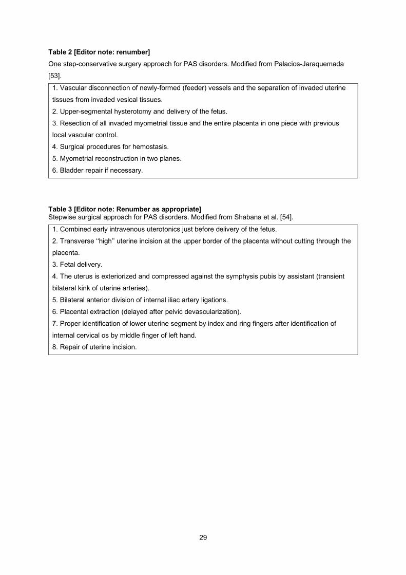
4.1. One-step conservative surgery
This surgical procedure has been described primarily by one author [51–53]. It
consists of resecting the invasive accreta area (partial myometrial resection) followed
by immediate uterine reconstruction and bladder reinforcement [52]. This strategy
aims to combine the advantages of both the “leaving in situ approach” of preserving

16
the uterus and cesarean hysterectomy with minimal risk of secondary bleeding or
infection. The main steps in this uterine-sparing technique can be performed via a
modified Pfannenstiel or midline incision [Table 2] (Box 1) [53]. It is advantageous for
low- and middle-income countries where expensive additional treatments such as
interventional radiology may not be available.
In a cohort study of 68 women presenting with placental invasion of the adjacent
organs including invasion of the posterior upper bladder section (n=46; group 1) or of
the posterior lower vesical section (n=22; group 2), uterine preservation was
achieved in 44 out of 46 (95.7%) and 6 out of 22 (27.3%) cases, respectively [51].
The indications for the 18 hysterectomies were segmental circumferential rupture
greater than 50% (n=13), coagulopathy (n=2), infection (n=1), and uncontrolled
hemodynamic instability (n=2). Among the 50 women with uterine preservation,
follow-up was available for 42 patients. A normal menstrual cycle returned between 3
and 16 months. Ten women had another uneventful pregnancy and delivery with no
recurrence of PAS disorders.
A recent prospective study of 71 patients presenting with placenta percreta evaluated
a variation of the stepwise approach [Table 3](Box 3) [Editor note: renumber] and
found that it was successful in controlling the bleeding and preserving the uterus in
65 (91.5%) of the cases [54]. Hemostasis was achieved firstly by retrovesical ligature
of vesicouterine vessels (upper pedicle) and secondly by stitch occlusion of the
colpo-uterine vessels in the cervical–vaginal junction (lower pedicle). Selective
devascularization was only applied to the vessels that provide irrigation to the
invaded area (pelvis subperitoneal pedicles) avoiding any procedure (ligature or

17
embolization) around the uterine arteries. Overall, this procedure may be less
reproducible than other approaches for conservative treatment, mainly because
efficient hemostasis is operator dependent. Removal of the area completely invaded
by placental tissue and uterine reconstruction using surrounding healthy myometrial
tissues results in a low rate of recurrence (2/108 cases) in future pregnancies [55].
4.2. The Triple-P procedure
A novel uterine-sparing procedure for PAS disorders called the “Triple-P procedure”
was recently proposed [4,56]. The aim of this procedure is to avoid incising through
the vascular placental venous sinuses, and to excise the myometrium with PAS
disorder tissue and to reconstitute the uterine defect. The main steps of this
procedure include: (1) perioperative placental ultrasound localization of the superior
edge of the placenta; (2) pelvic devascularization involving preoperative placement of
intra-arterial balloon catheters (anterior division of the internal iliac arteries); and (3)
no attempt to remove the entire placenta with large myometrial excision and uterine
repair. If the posterior wall of the bladder is involved, the placental tissue invading the
bladder is left in situ to avoid cystotomy.
A comparison of two periods (i.e. before implementation of the Triple-P procedure
[n=11] and after [n=19]) showed no difference in the estimated mean blood loss and
rate of transfusion; however, the rates of postpartum hemorrhage and hysterectomy
were lower in the Triple-P procedure group [4]. Larger studies are needed to
demonstrate the safety and efficacy of this technique.
4.3. Tamponade techniques

18
Small case series have also reported the successful use of compression sutures [57–
61], using the cervix as a natural tamponade by inverting it into the uterine cavity and
suturing the anterior and/or the posterior cervical lips into the anterior and/or posterior
walls of the lower uterine segment [62]. The latter technique of cervical inversion was
successful in stopping bleeding in 38 out of 40 patients. The mean time needed to
perform the technique was 5.4 ± 0.6 minutes. The complications observed included
bladder injury in the two patients who underwent hysterectomy and wound infection
in one patient.
Conflicts of Interest
The authors have no conflicts of interest to declare.

19
References
1. Kayem G, Davy C, Goffinet F, Thomas C, Clement D, Cabrol D. Conservative
versus extirpative management in cases of placenta accreta. Obstet Gynecol.
2004;104:531–6.
2. Sentilhes L, Ambroselli C, Kayem G, Provansal M, Fernandez H, Perrotin F, et
al. Maternal outcome after conservative treatment of placenta accreta. Obstet
Gynecol. 2010;115: 526–34.
3. Palacios-Jaraquemada JM, Pesaresi M, Nassif JC, Hermosid S. Anterior
placenta percreta: surgical approach, hemostasis and uterine repair. Acta
Obstet Gynecol Scand. 2004;83:738–44.
4. Teixidor Viñas M, Belli AM, Arulkumaran S, Chandraharan E. Prevention of
postpartum hemorrhage and hysterectomy in patients with morbidly adherent
placenta: a cohort study comparing outcomes before and after introduction of
the Triple-P procedure. Ultrasound Obstet Gynecol. 2015;46:350–5.
5. Jauniaux E, Collins SL, Burton GJ. The placenta accreta spectrum:
Pathophysiology and evidence-based anatomy for prenatal ultrasound imaging.
Am J Obstet Gynecol. 2017; doi: 10.1016/j.ajog.2017.05.067. [Epub ahead of
print].
6. American College of Obstetricians and Gynecologists. ACOG Practice Bulletin:
Clinical management guidelines for obstetricians-gynecologists. Number 76,
October 2006: postpartum hemorrhage. Obstet Gynecol 2006;108:1039–47.
7. Royal Australian and New Zealand College of Obstetricians and
Gynaecologists. Management of postpartum hemorrhage.
https://www.ranzcog.edu.au/RANZCOG_SITE/media/RANZCOG-
MEDIA/Women%27s%20Health/Statement%20and%20guidelines/Clinical-

20
Obstetrics/Management-of-Postpartum-Haemorrhage-(C-Obs-43)-Review-July-
2017.pdf?ext=.pdf.
8. Leduc D, Senikas V, Lalonde AB, Ballerman C, Biringer A, Delaney M, et al.
Active management of the third stage of labour: prevention and treatment of
postpartum hemorrhage. J Obstet Gynaecol Can. 2009;31:980–93.
9. World Health Organization. WHO recommendations for the prevention and
treatment of postpartum haemorrhage. Geneva: WHO; 2012.
10. Lalonde A, FIGO Safe Motherhood and Newborn Health Committee. Prevention
and treatment of postpartum hemorrhage in low-resource settings. Int J Gynecol
Obstet. 2012;117:108–118.
11. Sentilhes L, Vayssière C, Deneux-Taraux C, Aya AG, Bayoumeu F, Bonnet MP,
et al. Postpartum Hemorrhage: Guidelines for clinical practice from the French
College of Gynaecologists and Obstetricians (CNGOF) in collaboration with the
French Society of Anesthesiology and Intensive Care (SFAR). Eur J Obstet
Gynecol Biol Reprod .2016;198:12–21.
12. Sentilhes L, Goffinet F, Vayssière C, Deneux-Tharaux. Comparison of
postpartum haemorrhage guidelines: discrepancies underline our ignorance.
BJOG. 2017;124:718–22.
13. Fitzpatrick K, Sellers S, Spark P, Kurinczuk J, Brocklehurst P, Knight M. The
management and outcomes of placenta accreta, increta, and percreta in the
UK: a population-based descriptive study. BJOG. 2014;121:62–71.
14. Kayem G, Anselem O, Schmitz T, Goffinet F, Davy C, Mignon A, et al.
Conservative versus radical management in cases of placenta accreta: a
historical study. J Gynecol Obstet Biol Reprod. 2014;43:1142–60.

21
15. Belfort MA. Placenta accreta. Publications Committee, Society for Maternal-
Fetal Medicine Am J Obstet Gynecol. 2010;203:430–9.
16. Committee on Obstetric Practice. Committee opinion no. 529: placenta accreta.
Obstet Gynecol. 2012;120:207–11.
17. Royal College of Obstetricians and Gynecologists. Placenta praevia, placenta
praevia accreta and vasapraevia: diagnosis and management. Green-top
Guideline No. 27. London: RCOG; 2011.
18. Brennan DJ, Schulze B, Chetty N, Crandon A, Peterse, SG, Gardene, G, et al.
Surgical management of abnormally invasive placenta: a retrospective cohort
study demonstrating the benefits of a standardized operative approach. Acta
Obstet Gynecol Scand. 2015;94:1380–6.
19. Sentilhes L, Goffinet F, Kayem G. Management of placenta accreta. Acta
Obstet Gynecol Scand. 2013;92:1125–34.
20. Fox KA, Shamshirsaz AA, Carusi D, Secord AA, Lee P, Turan OM, et al.
Conservative management of morbidly adherent placenta: expert review. Am J
Obstet Gynecol. 2015;213:755–60.
21. O’Brien JM, Barton JR, Donaldson ES. The management of placenta percreta:
conservative and operative strategies. Am J Obstet Gynecol. 1996;175:1632–7.
22. Jolley JA, Nageotte MP, Wing DA, Shrivastava VK. Management of placenta
accreta: a survey of Maternal-Fetal Medicine practitioners. J Matern Fetal
Neonatal Med. 2012;25:756–60.
23. Esakoff TF, Handler SJ, Granados JM, Caughey AB. PAMUS: placenta accreta
management across the United States. J Matern Fetal Neonatal Med.
2012;25:761–5.

22
24. Timmermans S, van Hof AC, Duvekot JJ. Conservative management of
abnormally invasive placentation. Obstet Gynecol Surv. 2007;62:529–39.
25. Barber JT Jr, Tressler TB, Willis GS, Martinez FJ, Peisner DB, Goodman JD, et
al. Arteriovenous malformation identification after conservative management of
placenta percreta with uterine artery embolization and adjunctive therapy. Am J
Obstet Gynecol. 2011;204:e4–8.
26. Sentilhes L, Descamps P, Goffinet F. Arteriovenous malformation following
conservative treatment of placenta percreta with uterine artery embolization but
no adjunctive therapy. Am J Obstet Gynecol. 2011;205:e13.
27. Clausen C, Lönn L, Langhoff-Roos J. Management of placenta percreta: a
review of published cases. Acta Obstet Gynecol Scand .2014;93:138–43.
28. Pather S, Strockyj S, Richards A, Campbell N, de Vries B, Ogle R. Maternal
outcome after conservative management of placenta percreta at caesarean
section: a report of three cases and a review of the literature. Aust N Z J Obstet
Gynaecol. 2014;54:84–7.
29. Mussalli GM, Shah J, Berck DJ, Elimian A, Tejani N, Manning FA. Placenta
accreta and methotrexate therapy: three case reports. J Perinatol.
2000;20:331–4.
30. Lin K, Qin J, Xu K, Hu W, Lin J. Methotrexate management for placenta
accreta: a prospective study. Arch Gynecol Obstet. 2015;291:1259–64.
31. Isaacs JD Jr, McGehee RP, Cowan BD. Life-threatening neutropenia following
methotrexate treatment of ectopic pregnancy: a report of two cases. Obstet
Gynecol.1996;88:694–6.

23
32. Steins Bisschop CN, Schaap TP, Vogelvang TE, Scholten PC. Invasive
placentation and uterus preserving treatment modalities: a systematic review.
Arch Gynecol Obstet. 2011;284:491–502.
33. Judy AE, Lyell DJ, Druzin ML, Dorigo O. Disseminated Intravascular
Coagulation Complicating the Conservative Management of Placenta Percreta.
Obstet Gynecol. 2015;126:1016–8.
34. Sentilhes L, Trichot C, Resch B, Sergent F, Roman H, Marpeau L, et al. Fertility
and pregnancy outcomes following uterine devascularization for severe
postpartum haemorrhage. Hum Reprod .2008;23:1087–92.
35. Sentilhes L, Gromez A, Clavier E, Resch B, Verspyck E, Marpeau L. Predictors
of failed pelvic arterial embolization for severe postpartum hemorrhage. Obstet
Gynecol. 2009;113:992–9.
36. Angstmann T, Gard G, Harrington T, Ward E, Thomson A, Giles W. Surgical
management of placenta accreta: a cohort series and suggested approach. Am
J Obstet Gynecol. 2010;202:38.e1–9.
37. Bouvier A, Sentilhes L, Thouveny F, Bouet PE, Gillard P, Willoteaux S, et al.
Planned caesarean in the interventional radiology cath lab to enable immediate
uterine artery embolization for the conservative treatment of placenta accreta.
Clin Radiol. 2012;67:1089–94.
38. Soyer P, Sirol M, Fargeaudou Y, Bour L, Morel O, Dohan A, et al. Placental
vascularity and resorption delay after conservative management of invasive
placenta: MR imaging evaluation. Eur Radiol. 2013;23:262–71.
39. Mei J, Wang Y, Zou B, Hou Y, Ma T, Chen M, et al. Systematic review of
uterus-preserving treatment modalities for abnormally invasive placenta. J
Obstet Gynaecol. 2015;35:777–82.

24
40. Sewell MF, Rosenblum D, Ehrenberg H. Arterial embolus during common iliac
balloon catheterization at cesarean hysterectomy. Obstet Gynecol.
2006;108:746–8.
41. Matsueda S, Hidaka N, Kondo Y, Fujiwara A, Fukushima K, Kato K. External
iliac artery thrombosis after common iliac artery balloon occlusion during
cesarean hysterectomy for placenta accreta in cervico-isthmic pregnancy. J
Obstet Gynaecol Res. 2015;41:1826–30.
42. Gagnon J, Boucher L, Kaufman I, Brown R, Moore A. Iliac artery rupture related
to balloon insertion for placenta accreta causing maternal hemorrhage and
neonatal compromise. Can J Anaesth. 2013;60:1212–7.
43. Wei X, Zhang J, Chu Q, Du Y, Xing N, Xu X, et al. Prophylactic abdominal aorta
balloon occlusion during caesarean section: a retrospective case series. Int J
Obstet Anesth. 2016;27:3–8.
44. Salim R, Chulski A, Romano S, Garmi G, Rudin M, Shalev E. Precesarean
Prophylactic Balloon Catheters for Suspected Placenta Accreta: A Randomized
Controlled Trial. Obstet Gynecol. 2015;126:1022–8.
45. Legendre G, Zoulovits FJ, Kinn J, Sentilhes L, Fernandez H. Conservative
management of placenta accreta: hysteroscopic resection of retained tissues. J
Minim Invasive Gynecol. 2014;21:910–3.
46. Bai Y, Luo X, Li Q, Yin N, Fu X, Zhang H, et al. High-intensity focused
ultrasound treatment of placenta accreta after vaginal delivery: a preliminary
study. Ultrasound Obstet Gynecol. 2016;47:492–8.
47. Alanis M, Hurst BS, Marshburn PB, Matthews ML. Conservative management
of placenta increta with selective arterial embolization preserves future fertility

25
and results in a favorable outcome in subsequent pregnancies. Fertil Steril.
2006;86:1514.e3–7.
48. Kayem G, Pannier E, Goffinet F, Grange G, Cabrol D. Fertility after
conservative treatment of placenta accreta. Fertil Steril. 2002;78:637–8.
49. Sentilhes L, Kayem G, Ambroselli C, Provansal M, Fernandez H, Perrotin F, et
al. Fertility and pregnancy outcomes following conservative treatment for
placenta accreta. Hum Reprod. 2010;25:2803–10.
50. Kabiri D, Hants Y, Shanwetter N, Simons M, Weiniger CF, Gielchinsky Y, et al.
Outcomes of subsequent pregnancies after conservative treatment for placenta
accreta. Int J Gynaecol Obstet. 2014;127:206–10.
51. Palacios-Jaraquemada JM, Pesaresi M, Nassif JC, Hermosid S. Anterior
placenta percreta: surgical approach, hemostasis and uterine repair. Acta
Obstet Gynecol Scand. 2004;83:738–44.
52. Palacios-Jaraquemada JM. Diagnosis and management of placenta accreta.
Best Pract Res Clin Obstet Gynecol. 2008;22:1133–48.
53. Palacios-Jaraquemada JM. Placental adhesive disorders. Berlin/Boston: Walter
de Gruyter; 2012.
54. Shabana A, Fawzy M, Refaie W. Conservative management of placenta
percreta: a stepwise approach. Arch Gynecol Obstet. 2015;291:993–8.
55. Palacios-Jaraquemada JM. One-Step Conservative Surgery for Abnormal
Invasive Placenta (Placenta Accreta–Increta–Percreta). In: Arulkumaran S,
Karoshi M, Keith LG, Lalonde AB, B-Lynch C, eds. A Comprehensive Textbook
of Postpartum Hemorrhage. An essential clinical reference for Effective
Management 2nd Edition. London: Sapiens Publishing GLOWM; 2012: 263–71.

26
56. Chandraharan E, Rao S, Belli AM, Arulkumaran S. The Triple-P procedure as a
conservative surgical alternative to peripartum hysterectomy for placenta
percreta. Int J Gynaecol Obstet. 2012;117:191–194.
57. Shazly SA, Badee AY, Ali MK. The use of multiple 8 compression suturing as a
novel procedure to preserve fertility in patients with placenta accreta: case
series. Aust N Z J Obstet Gynaecol. 2012;52:395–9.
58. Huang G, Zhou R, Hu Y. A new suture technique for cesarean delivery
complicated by hemorrhage in cases of placenta previa accreta. Int J Gynaecol
Obstet. 2014;124:262–3.
59. Kaplanoğlu M, Kaplanoğlu DK, Koyuncu O. A different approach to placenta
previa accreta: intrauterine gauze compress combined B-Lynch uterine
compression suture. Clin Exp Obstet Gynecol. 2015;42:53–6.
60. Li GT, Li XF, Li J, Liu YJ, Xu HM. Reflexed Compression Suture for the
Management of Atonic Postpartum Hemorrhage with an Abnormally Adherent
Placenta. Gynecol Obstet Invest. 2015;80:228–33.
61. Li GT, Li XF, Wu B, Li G. Longitudinal parallel compression suture to control
postpartum hemorrhage due to placenta previa and accrete. Taiwan J Obstet
Gynecol. 2016;55:193–7.
62. El Gelany SA, Abdelraheim AR, Mohammed MM, Gad El-Rab MT, Yousef AM,
Ibrahim EM, et al. The cervix as a natural tamponade in postpartum
hemorrhage caused by placenta previa and placenta previa accreta: a
prospective study. BMC Pregnancy Childbirth. 2015;15:295.

27
Box 1 [Editor Note: Re-label as Table once placement determined] Recommendations for conservative management of placenta accreta spectrum (PAS) disorders.
Recommendations Resource
settings
Quality of evidence
and strength of
recommendation
Leaving the placenta in situ is an option for women who desire to preserve their fertility and agree to continuous long-
term monitoring in centers with adequate expertise.
High Moderate and Strong
The extirpative approach or forcible manual removal of the
placenta should be abandoned.
All High and Strong
When a conservative treatment is attempted in cases of PAS
disorders diagnosed prenatally, the exact position of the
placenta should be confirmed by a preoperative ultrasound
and the equipment and expert surgical team should be on
stand-by for an emergent hysterectomy.
High Moderate and Strong
After the delivery of the fetus, and only in cases with no clinical evidence of PAS disorders, the surgeon may carefully
attempt to remove the placenta by controlled cord traction
and the use of uterotonics.
All Low and Strong
Postoperative antibiotic therapy (amoxicillin and clavulanic
acid or clindamycin in case of penicillin allergy) should be
administered prophylactically to minimize the risk of infection
when the placenta is left in situ.
High Low and Weak
The use of methotrexate is not recommended until further
evidence is available on its efficacy and safety.
All Moderate and Strong
Preventive surgical or radiological uterine devascularization
is not recommended routinely.
High Low and Weak
There is insufficient evidence to recommend the use of
magnetic resonance imaging and/or measuring serum b-hCG
for the monitoring of conservative management.
High Low and Weak
Women who want another pregnancy should be advised that
the recurrence risk of PAS disorders is high.
All Low and Strong
The one-step conservative surgery is less reproducible than
other conservative management approaches, mainly
because the efficacy of hemostasis is operator dependent.
High Low and Weak

28
Table 1 Maternal morbidity after conservative treatment for placenta accreta spectrum. Modified from Sentilhes
et al., [2]
Characteristics PAS disorders including percreta
No. (%) Placenta left in situ 167 (100) Partially 99 (59.3) Entirely 68 (40.7) Primary postpartum hemorrhage 86 (51.5) No additional uterine devascularization procedure 58 (34.7) Additional uterine devascularization procedure 109 (65.3) Pelvic arterial embolization 62 (37.1) Vessel ligation 45 (26.9) Stepwise uterine devascularization 15 (9.0) Hypogastric artery ligation 23 (13.8) Stepwise uterine devascularization and hypogastric artery ligation
7 (4.2)
Uterine compression suture 16 (9.6) Balloon catheter occlusion 0 Methotrexate administration 21 (12.6) Primary hysterectomy 18 (10.8) Cause of primary hysterectomy Primary postpartum hemorrhage 18/18 (100) Postpartum prophylactic antibiotic therapy >5 days 54 (32.3) Transfusion patients 70 (41.9) Units of packed red blood cells transfused >5 25 (15.0) Transfer to intensive care unit 43 (25.7) Infection 47 (28.1) Septic shock 1 (0.6) Sepsis 7 (4.2) Vesicouterine fistula 1 (0.6) Uterine necrosis 2 (1.2) Deep vein thrombophlebitis or pulmonary embolism 4 (2.4) Secondary postpartum hemorrhage 18 (10.8) Delayed hysterectomy 18 (10.8) Median interval from delivery to delayed hysterectomy, d 22 (9–45) Cause of delayed hysterectomy Secondary postpartum hemorrhage 8/18 (44.4) Sepsis 2/18 (11.1) Secondary postpartum hemorrhage and sepsis 3/18 (16.7) Vesicouterine fistula 1/18 (5.6) Uterine necrosis and sepsis d 2/18 (11.1) Arteriovenous malformation 1/18 (5.6) Maternal request 1/18 (5.6) Death 1 (0.6) Success of conservative treatment 131 (78.4) Severe maternal morbidity 10 (6.0)
29
Table 2 [Editor note: renumber] One step-conservative surgery approach for PAS disorders. Modified from Palacios-Jaraquemada
[53].
1. Vascular disconnection of newly-formed (feeder) vessels and the separation of invaded uterine
tissues from invaded vesical tissues.
2. Upper-segmental hysterotomy and delivery of the fetus.
3. Resection of all invaded myometrial tissue and the entire placenta in one piece with previous local vascular control.
4. Surgical procedures for hemostasis.
5. Myometrial reconstruction in two planes.
6. Bladder repair if necessary.
Table 3 [Editor note: Renumber as appropriate] Stepwise surgical approach for PAS disorders. Modified from Shabana et al. [54].
1. Combined early intravenous uterotonics just before delivery of the fetus.
2. Transverse ‘‘high’’ uterine incision at the upper border of the placenta without cutting through the placenta.
3. Fetal delivery.
4. The uterus is exteriorized and compressed against the symphysis pubis by assistant (transient
bilateral kink of uterine arteries).
5. Bilateral anterior division of internal iliac artery ligations.
6. Placental extraction (delayed after pelvic devascularization).
7. Proper identification of lower uterine segment by index and ring fingers after identification of
internal cervical os by middle finger of left hand. 8. Repair of uterine incision.