Fibrillization DOI:10.1002/anie.200700861 Peptide ...lamosa/Hamley_AngewChem_2007_pp8128.pdf ·...
20
Fibrillization DOI: 10.1002/anie.200700861 Peptide Fibrillization Ian W. Hamley* Angewandte Chemie Keywords: amyloids · biomaterials · peptides · self-assembly · soft matter I. W. Hamley Reviews 8128 www.angewandte.org # 2007 Wiley-VCH Verlag GmbH & Co. KGaA, Weinheim Angew. Chem. Int. Ed. 2007, 46, 8128 – 8147
Transcript of Fibrillization DOI:10.1002/anie.200700861 Peptide ...lamosa/Hamley_AngewChem_2007_pp8128.pdf ·...

FibrillizationDOI: 10.1002/anie.200700861
Peptide FibrillizationIan W. Hamley*
AngewandteChemie
Keywords:amyloids · biomaterials · peptides ·self-assembly · soft matter
I. W. HamleyReviews
8128 www.angewandte.org � 2007 Wiley-VCH Verlag GmbH & Co. KGaA, Weinheim Angew. Chem. Int. Ed. 2007, 46, 8128 – 8147

1. Introduction
This Review is concerned with fibril formation bypeptides and peptide conjugates, with a focus primarily onamyloid-type fibrils that contain b sheets; other collagen-typefibrillar structures, for example, are not considered. This is asubject of great current interest because of the role of amyloidformation in numerous diseases and the possibilities to usefibrillar peptide structures as structural or structuring ele-ments in bionanotechnology.
The term amyloid refers to protein deposits that resemblethose first observed for starch (amyloid originally meantstarch-like). It is now specifically associated with proteins andpeptides that adopt fibrils based on the cross-b structure, inwhich the peptide backbone is orthogonal to the fibril axis.[1–5]
The b sheets self-assemble into protofilaments, which maycomprise a structure, such as a bundle of twisted b-sheets,which further packs into amyloid fibers[4,6] (Figure 1). The
structure of fibrils is discussed further in Section 2. Figure 2shows a typical TEM image of amyloid fibrils.
The formation of fibrils is symptomatic of many amyloiddiseases such as Alzheimer0s and Creutzfeldt-Jacob dis-ease[6,8, 9] (lists of other diseases that result from proteinaggregation can be found in Refs. [10,11]). In some species,amyloidosis may be exploited for useful purposes.[12–14] Indisease, fibrillization results from the aggregation of proteinsor peptides such as amyloid-b (Ab) or tau. As a consequenceof its relevance to a diverse number of conditions affecting
large numbers of people, fibril formation by the Ab peptidehas been widely studied. There are two variants of thisamyloid peptide in humans: Ab40 and Ab42 (this notationwill be used for the whole peptide with the number of residuesindicated), of which the latter forms fibrils more rapidly.[15] Itis now thought that protofilaments formed in the initial self-assembly process are the toxic agents.[8, 16–21] This is discussedin more detail in Section 5. The amyloid-b peptide (Figure 3)is produced by proteolytic cleavage of the amyloid precursorprotein (APP), a transmembrane protein of unknown func-tion.[8] The N terminus of the peptide is created by cleavageby b-secretase in the extracellular domain of APP, and theC terminus results from intramembrane cleavage by g-secre-tase (Figure 3). A third enzyme, a-secretase, cleaves betweenamino acids 16 and 17 in Ab, thus hindering fibrillization.Oligomerization of Ab occurs intracellularly, as revealed byin vivo experiments on human cerebrospinal fluid which
The fibrillization of peptides is relevant to many diseases basedon the deposition of amyloids. The formation of fibrils is beingintensively studied, especially in terms of nanotechnology appli-cations, where fibrillar peptide hydrogels are used for cell scaf-folds, as supports for functional and responsive biomaterials,biosensors, and nanowires. This Review is concerned withfundamental aspects of the self-assembly of peptides into fibrils,and discusses both natural amyloid-forming peptides andsynthetic materials, including peptide fragments, copolymers, andamphiphiles.
From the Contents
1. Introduction 8129
2. Structure of Fibrils 8130
3. Characterization of Fibrillization 8131
4. Kinetics (and Mechanism) ofFibrillization 8132
5. Prefibrillar Aggregates Are moreToxic than Final Fibrils 8134
6. Amyloid Formation Is not SequenceSpecific 8135
7. Fibrillization of Fragments 8136
8. Fibrillization of de novo DesignedPeptides 8137
9. Peptide Amphiphiles and PeptideCopolymers 8138
10. Peptide Nanotubes 8140
11. Peptide Fibrillar Gels 8140
12. Fibrils from a Helices 8142
13. Summary and Outlook 8143
Figure 1. Hierarchical structure of amyloid fibrils.
[*] Prof. I. W. HamleyDepartment of ChemistryUniversity of ReadingReading, Berkshire RG6 6AD (UK)Fax: (+44)118-378-8450E-mail: [email protected]
FibrillizationAngewandte
Chemie
8129Angew. Chem. Int. Ed. 2007, 46, 8128 – 8147 � 2007 Wiley-VCH Verlag GmbH & Co. KGaA, Weinheim

yielded dimers of Ab that were stable to sodium dodecylsul-fate (SDS).[22] Incubation did not lead to the production ofextracellular oligomers. However, oligomers were detected inneural and non-neural cell lines.
Several genes have been linked to Alzheimer0s disease,including the genes for APP,[23] presenilin 1 and 2 (PSEN1 andPSEN2),[24,25] and apolipoprotein E (APOE).[26–29] Recentwork suggests that in late-onset Alzheimer0s disease, Ab
accumulation occurs intracellularly in late endosomes (i.e. inthe deep end of the endosomal compartment) where theenzymes b-secretase and g-secretase cleave Ab, the latter in apresinilin-dependent fashion.[30] The gene involved in APP
recycling in endosomes has been identified, and is termedSORL1 or LR11. Normally the protein product of this genedirects APP into recycling endosomes; however, mutationslead to a decrease in the protein product which leads to thepathway where Ab production is increased by enzymes in thelate endosomes. There are several proposed therapies to treatAlzheimer0s disease, these are outside the scope of the currentreview and are discussed elsewhere.[9]
The tau protein is involved in microtubule assembly andstabilization within the cytoskeleton (in particular in F-actinfibrils). Mutations can lead to filamentous deposits whichhave been observed for several neurodegenerative diseasessuch as Pick0s disease and the Parkinsonism-dementia com-plex of Guam.[8] Filamentous tau deposits are invariablypresent even in the absence of Ab deposits, and it is not clearin the context of fibril deposition precisely how Ab and tauinteract, although there seems to be a synergistic effect whichenhances actin bundling and neurodegeneration.[8,31] It has,however, been suggested that, as in the case of Ab, oligomericspecies may be the toxic agents. Neuronal degenerationinduced by tau has been studied in vivo. Hyperphosphory-lated forms of this microtubule-associated protein induceaccumulation of F-actin. This has been confirmed by in vivostudies using drosophila and mouse models.[31]
The initial part of this Review focuses on fibrils formedfrom b sheets. Section 2 describes the structure of fibrils.Section 3 briefly covers methods used to characterize fibril-lization. Section 4 discusses aspects of the mechanism andkinetics of self-assembly. Sections 5 and 6 are concerned withthe toxicity and sequence specificity of Ab amyloid forma-tion. Sections 7–10 consider fibril formation with specifictypes of peptide and peptide conjugates. Section 11 concernsthe formation of fibrillar gels. Section 12 briefly summarizesrecent work on fibrils formed from coiled-coil peptides. Thereis finally a summary and outlook.
2. Structure of Fibrils
Amyloid fibrils contain bundles of b sheets with back-bones orthogonal to the fiber axis in the so-called “cross-b”structure.[1–5] Figure 4 shows a representation of the cross-bstructure within a helical fibril constructed from four b sheets.Helical ordering is often observed within the fibrils, forexample, for transthyretin,[32] the SH3 domain of phosphati-dylinositol-3’-kinase,[33] or bovine insulin[34] (other examplesare tabulated in Ref. [5]). The helical structure results fromthe preferred right-handed twist of b sheets.[35,36] The b sheetsin the fibrils are antiparallel, as revealed by infraredspectroscopy; an early example of such measurements wasmade in a study on human amyloid protein.[37]
Ian Hamley, born 1965, is Diamond Profes-sor of Physical Chemistry and head ofPhysical Chemistry at the University ofReading. He received his PhD from theUniversity of Southampton in 1991. He wasthen a Royal Society postdoctoral fellow atFOM-AMOLF, Amsterdam, and then in1992 he carried out postdocoral researchwith Frank Bates at the University of Minne-sota, Minneapolis. He returned to the UK in1993 as a lecturer in Physics at the Univer-sity of Durham, before moving to theChemistry department at the University of
Leeds in 1995, where he became Professor in 2004. He moved to theUniversity of Reading in 2005.
Figure 2. TEM image of amyloid fibrils, formed by peptide fragmentFFKLVFF.[7]
Figure 3. Amino acid sequence of Ab42. The enzyme cleavage sites areindicated and the polar residues are underlined.
Figure 4. Helical packing of a four protofilament bundle proposed forinsulin amyloid fibrils. Reprinted with permission from Ref. [34].
I. W. HamleyReviews
8130 www.angewandte.org � 2007 Wiley-VCH Verlag GmbH & Co. KGaA, Weinheim Angew. Chem. Int. Ed. 2007, 46, 8128 – 8147

Figure 5 shows the b strand-turn-b strand conformation ofAb(18–42) (residues 1–17 are disordered) within the cross-bfibril structure. Solid-state NMR experiments (with consider-ation of the X-ray diffraction and TEM measurements) hasindicated a similar structure in Ab40.[38]
The characteristic features in an X-ray scattering patternof the cross-b structure are a 4.7 C meridional reflectioncorresponding to the spacing between peptide backbones andan equatorial reflection at 8–12 C, which is broader and whichcorresponds to the stacking periodicity of the b sheets (therange of values reflects different side-chain dimensions).Most X-ray diffraction studies that showed a cross-b structurehave been performed on dried films. Since hydration canaffect the structure of b-sheet-containing fibrils, Squires et al.have examined whether the cross-b structure is retained inflow-aligned solution.[40] The wide-angle X-ray scattering(WAXS) results for a synthetic peptide (fragment of trans-thyretin) and lysozyme from hen egg white indeed showed thesame pattern as for a dried sample, thus indicating that thecross-b structure is present in solution and is not an artefactcaused by dehydration.
Perutz et al. proposed that amyloid fibers are water-fillednanotubes.[41] This proposal was based on the analysis of theX-ray diffraction data for the polyglutamine-rich peptideD2Q12K2 and for the polyglutamine-rich natural peptideshuntingtin and yeast prion Sup35, as well as on an electronmicroscope image of Sup35. However, this analysis has beenchallenged by two research groups who have proposed astacked b-sheet model for the X-ray diffraction data ofD2Q12K2
[42–44] (The second research group[43,44] also report astacked b-sheet structure for K2Q28K2 and K2Q45K2.) It hasalso been pointed out that distinctive features in thediffraction patterns of polyglutamine-rich peptides meanthat it is very unlikely that the water-filled nanotube modelcould be a general structure for amyloid fibers.[42]
3. Characterization of Fibrillization
3.1. Dye Staining
One method for identifying amyloid fibrils is by stainingwith Congo red. Under polarized light, amyloid samplesexhibit green birefringence when stained with Congo red.
Thioflavin T is a fluorescent dye widely used to studyamyloid formation. Excitation at 450 nm produces fluores-cence at 482 nm.[45,46]
3.2. X-ray Diffraction
As mentioned in Section 2, wide-angle X-ray scattering(WAXS) is usually performed on dried samples, in the form offilms or “stalks”, the latter being dried threads of solution.Other methods of alignment[47] include the use of stretchframes or cryoloops, with the latter producing a dried flat filmor “mat”. Stalks may also be dried in the presence of amagnetic field to improve alignment. Small-angle X-rayscattering (SAXS) can provide information on the secondarystructure over larger length scales (5–100 nm).
3.3. Dynamic and Static Light Scattering
Analysis of the intensity autocorrelation function ofscattered light from a particle undergoing translational orrotational diffusion enables the determination of a diffusioncoefficient, and hence, from the Stokes–Einstein equation,the effective hydrodynamic radius. There are several approx-imate methods to relate this quantity to the fibril length anddiameter.[48, 49]
Static light scattering can be used to obtain the molecularweight of peptide aggregates and also to provide an indicationof the particle shape, from measurements of the angulardependence of the scattered intensity.
Light scattering has been used quite widely to monitor theformation of amyloid protofilaments and fibrils.[50–53]
3.4. Circular Dichroism
Cicular dichrosim (CD) refers to the differential absorp-tion of right- and left-circularly polarized light. It is sensitiveto the presence of chiral groups and is a primary technique tocharacterize protein secondary structure. The usual method isbased on the “fingerprinting” of features in the 190–250 nm(far-UV) region.[54] Data in the near-UV region can provideinformation on the folding of peptides containing aromaticresidues. In the far-UV region, characteristic minima areobserved in the absorption spectra at approximately 208 and222 nm (a helix), 216–220 nm (b sheet), or 195 nm (randomcoil). CD spectra are usually analyzed by using algorithmsbased on databases compiled for peptides for which the X-raycrystal structure is known.[54] This permits an accuratedetermination of the secondary structure, which can then beused to “calibrate” CD spectra. Several algorithms are
Figure 5. Conformation of Ab(17–42) as revealed by H/D-exchangeNMR spectroscopy experiments. The hydrophobic, polar, negativelycharged, and positively charged amino acids are shown in yellow,green, red, and blue, respectively. Positively and negatively chargedsurface patches are shown in blue and red, others in white. Reprintedwith permission from Ref. [39].
FibrillizationAngewandte
Chemie
8131Angew. Chem. Int. Ed. 2007, 46, 8128 – 8147 � 2007 Wiley-VCH Verlag GmbH & Co. KGaA, Weinheim www.angewandte.org

available that are based on different databases. Most consideronly larger proteins, although there are limited reference datasets (and curve-fitting programs) for shorter peptides.[55]
3.5. FTIR Spectroscopy
The amide I band of the FTIR spectrum at 1620–1640 cm�1 is associated with b sheets.[56–60] In comparison tocoils or helices this band is shifted to lower frequency. Afurther side band at 1680–1690 cm�1 has been ascribed tohydrogen bonding, although ab initio calculations[61] suggestthat it may be due to the vibrational coupling of residues. Thenarrow intense band observed for some peptides at 1610–1620 cm�1 is ascribed to an antiparallel aggregated structure.
The amide II band around 1550 cm�1 mainly results fromthe N-H bending vibrations. The frequency of these vibrationsare responsive to deuteration (in D2O the hydrogen atom ofthe N�H bond exchanges with a deuterium atom) and, as aconsequence, the amide II band is shifted by approximately100 cm�1 to 1450 cm�1.
Since a full quantum chemical analysis of the vibrationalmodes of peptides or proteins is not usually possible, theanalysis of FTIR data relies on “fingerprinting” or peak-fitting methods.[56,57] These methods are prone to uncertaintybecause of the overlap of features in the spectra.[56,57,59]
FTIR measurements can be extended to study lineardichroism on aligned samples with isotope labeling (polarizedFTIR) and to vibrational circular dichroism.[58]
3.6. NMR Spectroscopy
Solid-state NMR spectroscopy has provided much detailon the structure of amyloid fibrils. Various high-resolutionexperiments employing magic-angle spinning can be per-formed using 13C- or 15N-labeled peptides.[62–65] Homonuclearand heteronuclear 2D and 3D spectroscopy may also beperformed to give information on interatomic distances andtorsion angles for isotopically labeled peptides in the driedstate.[66, 67]
Hydrogen/deuterium exchange techniques have also beenemployed to probe structural changes during amyloid for-mation.[66] Exchange rates provide information on the struc-tural rearrangements of subsegments of a protein or peptideduring folding, unfolding, or fibrillization. This method hasalso been used to provide a 3D structure for Ab42 (seeFigure 5).[39]
3.7. Atomic Force Microscopy and Electron Microscopy
These methods allow the direct imaging of peptide fibrils.Atomic force microscopy (AFM), which is a scanning probemicroscopy technique, provides images at the surface. Todate, the method has mainly been used to image fibrils fromsolutions dried onto planar solid substrates such as mica.Electron microscopy includes transmission electron micro-scopy (TEM) of dried films on holey grids (these are usually
stained with heavy-metal-containing compounds to enhancecontrast) or scanning electron microscopy (SEM) of surfaces.Cryotechniques are often preferred as a method to avoidartefacts caused by slow drying.
4. Kinetics (and Mechanism) of Fibrillization
Fibrillization appears to occur via a prefibrillarstage[11,15,50,68,69] consisting of small (spherical) protein multi-mers that are consumed as fibrillization proceeds.[50, 68] AFMstudies on Ab40 and Ab42 provided a compelling picture ofthe initial formation of protofilaments followed by theirreplacement with fibrils.[68] This technique was complementedwith light scattering studies, TEM, and size-exclusion chro-matography (SEC) analysis of the molecular weight offractions obtained at different stages of the polymerizationprocess.[50] The aggregation of Ab on hydrophilic mica andhydrophobic graphite has been investigated. On mica,pseudomicellar aggregates were noted at low concentration,and fibrils at higher concentration. In contrast, on graphite,sheets were observed with a thickness equal to the extendedpeptide length, oriented along the graphite lattice direc-tions.[68] A later AFM study examined Ab42 fibrillization andplaque formation, as well as the interaction between Ab40and Ab42.[70] Deposition was studied on a synthetic templatecomprising a solid surface activated with N-hydroxysuccin-imide ester. Ab42 oligomers were found to be more effectiveseeds for fibril growth than monomers or mature fibrils.
The question of whether amyloid fibrillization resultsfrom partially folded intermediates containing b-sheet struc-tures or from a fully denatured conformation has recentlybeen addressed. For most proteins, conditions that lead topartial unfolding favor fibrillization. Examples include trans-thyretin,[71,72] the prion protein PrPC,[73, 74] the immunoglobulinlight chain,[75–77] lysozyme,[78,79] the SH3 domain of bovinephosphatidylinositol 3-kinase,[80]and b2-microglobulin.[81–83]
The extent of unfolding and ultimately the fibril morphol-ogy seem to depend on the level of denaturation.[82, 84] Kadet al. correlated these two properties in a study of b2-microglobulin.[82] Dobson and co-workers studied the aggre-gation of acylphosphatase subjected to mild denaturing inaqueous solutions of 2,2,2-trifluoroethanol (TFE) of variousconcentrations.[85] Partial denaturing occurred and led tofibrillization. In contrast, in aggressive denaturing conditions,such as a high concentration of urea or guanidinium chloride,only a soluble highly unfolded state is usually observed. Acomplication in this study was the fact that the protein has asignificant b-sheet content in the native state, and was thusexpected to form b-sheet-rich intermediates. A study of fibrilformation by myoglobin by the same research group sug-gested, in contrast, that full denaturing occurs prior tofibrillization.[86] Myoglobin is predominately a helical in itsnative state and lacks b-sheet content. It is therefore a usefulmodel to study the mechanism of fibrillization. The CD,fluorescence, and FTIR experiments indicated that inter-mediate states did not contain b-sheet structures. The authorssuggested that this partial unfolding mechanism may begeneric. This hypothesis supports the evidence that many
I. W. HamleyReviews
8132 www.angewandte.org � 2007 Wiley-VCH Verlag GmbH & Co. KGaA, Weinheim Angew. Chem. Int. Ed. 2007, 46, 8128 – 8147

proteins lacking specific secondary structure can fibrillize (seeSection 6). The intermediate structure must be unfavorablewith respect to folding into the native structure, yet mustallow non-hydrogen-bonded extended transient structuresthat may be precursors to amyloid fibrillization.[85,86]
Dobson and co-workers as well as other researchers havenoted that the unfolded state may contain significant poly(l-proline) II (PPII) content.[87–89] Ramon spectroscopy studieswith polarized light (Raman optical activity) on differentforms of synuclein have shown this is not a sufficientcondition for b-sheet aggregation, since b- and g-synucleinhave a very limited tendency to form fibrils, despite being richin PPII.[90] By studying Ab40 and Ab42 in which themethionine (residue 35) side chain was in an oxidized orreduced state, Hou et al. found, from NMR experiments insolution, residue-specific interactions in the early stages ofaggregation.[89] These studies suggest that both hydrophobicand turnlike structures are required in the first self-assemblysteps. The specific region of Ab40 involved in contactsbetween fibrils has been identified by solution-state NMRexperiments as corresponding to Ab(15–24) (other studies onthis sequence are discussed in Section 7).[91] The NMR dataalso show that there is an exchange between a monomeric,soluble state and an oligomeric, aggregated state underappropriate (physiological) conditions of salt concentrationand that the equilibrium can be shifted by varying the anionicstrength. The molecular weight of the oligomer was found tobe greater than 100 kDa.[91]
Small-angle X-ray scattering (SAXS) has been used[92] tomonitor the self-assembly of mutant myogobin with anexpanded glutamine repeat sequence (this expansion isassociated with diseases such as Huntington0s[93]). The earlystage aggregation was studied upon thermally induced partialunfolding. A nonfibrillar aggregate without characteristic b-sheet features in the X-ray pattern was observed. The authorssuggested that this type of protofilament could be related tothe toxicity of polyglutamine-rich proteins.
It has been shown that fibrillization is a multistep processand that prefibrillar aggregates formed in the early stages candisassemble.[94] Fluorescence experiments were performed onhuman muscle acylphosphatase under mild denaturing con-ditions (25% TFE). Partial unfolding occurred to yieldglobular aggregates 60–200 nm in diameter that could furtheraggregate into clusters 400–800 nm in diameter. Above acritical concentration of the globular aggregates, larger (>5 mm) superstructures could form. The globular aggregatesand clusters disaggregated upon dilution (to 5% TFE).Protein monomers refold quickly whilst globular aggregatesand clusters (400–800 nm) disaggregate at a somewhat slowerrate. The larger superstructures are not affected by dilution.
Fibrillization of Ab40 occurs above a critical concentra-tion, which has been described in analogy with a criticalmicelle concentration.[48,95,96] Figure 6 is identical in form tothat for micellization.[97] Fibrillization can be described usingthe corresponding one-dimensional model of self-assembly.[97]
It is commonly observed that fibrillization occurs after alag phase, which suggests a nucleation and growth pro-cess.[48,82,95,96,98–101] The lag phase can be eliminated by additionof preformed aggregates, that is, by seeding (Figure 7).[95,96]
The initially formed protofibrillar species for severalproteins including Ab40, Ab42, and a-synuclein are spherical,porelike, annular species,[11,68,69] which may be linked intochains.[11,69] The protofibril formation process may occurbefore or during the lag phase.
Fibril formation from hen egg white lysozyme (HEWL)induced at low pH values and high temperature (57 8C) occursafter an extended lag time of 48 h. The lag time is independentof concentration, which indicates that fibril formation is asingle-molecule process.[84] The observation of an isodichroicpoint in the CD data points to a two-state cooperative processas the initial a-helix-rich structure unfolds. CD measurementson fractions separated by filtration from a sample heated for11 days showed that the monomer had a random coil structureand the fibrils were characterized by an additional small b-sheet signal. It was noted that the fibril morphology isdependent on the preparation conditions.
In contrast to the lag time observed with HEWL,aggregation of b-lactoglobulin occurs immediately uponheating.[102] This finding points to distinct mechanisms offibrillization. A possible factor is the b-sheet content in theprotein, since b-lactoglobulin is rich in this secondarystructure whereas HEWL is rich in a-helix structures (30–40%[103,104]). The suggested mechanism for heat-inducedfibrillization of b-lactoglobulin involves several steps: First,the incorporation of (partly) denatured protein moleculesinto fibrils or nonfibril-forming oligomers. A second stepinvolves the reversible formation of linear aggregates, fol-lowed by a third process of “consolidation”, which producesthermally stable fibrils.[102]
The structure of b-lactoglobulin aggregates produced byheat-induced denaturation at pH 2 has been probed by lightand neutron scattering studies.[105] The morphology was found
Figure 6. The amount of fibril and monomer as a function of addedprotein. Fibrils are formed above a critical concentration cR.
Figure 7. The addition of seed can eliminate the lag time in fibrilliza-tion.
FibrillizationAngewandte
Chemie
8133Angew. Chem. Int. Ed. 2007, 46, 8128 – 8147 � 2007 Wiley-VCH Verlag GmbH & Co. KGaA, Weinheim www.angewandte.org

to depend on the ionic strength, thereby pointing to the roleof electrostatic interactions in the self-assembly process.Rodlike aggregates were observed at low ionic strength,whereas a fractral structure was suggested for solutions athigher ionic strength. A two-step aggregation process was notobserved at pH 2, where fibrils form, but was seen at pH 7where globular aggregates form.[105]
The dependence of the morphology on the ionic strengthwas later probed by AFM and light scattering experiments.[106]
A critical concentration for fibril formation was reported andit was also noted that fibrils became shorter and more curvedon increasing the ionic strength. The formation of fibrillar gelsby b-lactoglobulin at high concentration is discussed inSection 11. As mentioned above, the lag time observed infibrillization can be eliminated or greatly reduced by seedingwith preformed fibrils.[6,107]
The role of shear flow on amyloid formation has beeninvestigated by fluorescence experiments on b-lactoglobulinand by AFM studies on extracted aliquots.[108] Shear-inducedformation of spheroidal aggregates was observed in a couettecell (concentric cylinder). These aggregates could be used toseed subsequent fibrillization by incubation; the fibrillizationwas significantly enhanced relative to the unseeded controlsolution. Preformed fibrils were degraded into shorter fibrilsduring prolonged steady shear at a high shear rate. Theseresults were placed in the context of physiological blood-flowconditions.[108] SAXS, TEM, and dynamic light scattering(DLS) studies also provide evidence for the break-up of b-lactoglobulin into short rodlike fragments after continuousshear (alignment is observed in the early stages of flow).[109]
Early work on the growth phase of fibrillization in Ab40suggested that the kinetics are first order, that is, the rate offibril elongation is proportional to the concentration of themonomers.[48,110,111] This was confirmed by light scatteringstudies on Ab40 in 0.1m HCl[48] (the aggregation kinetics arepH-dependent), in vitro studies of deposition onto plaques inbrain tissue of Alzheimer0s patients,[110] and thioflavin T(ThT) fluorescence studies.[111] The temperature-dependenceof aggregation for Ab40 (determined from size measurementsby dynamic light scattering) follows the Arrhenius equa-tion,[51] with an activation energy of 96 kJmol�1 that iscomparable to the value for the unfolding of several otherpeptides.[51] As mentioned above, the kinetics of dilution-induced disaggregation has been probed for monomers andearly stage protofilaments of human muscle acylphospha-tase.[94] More recent work indicates that the kinetics offibrillization are more complex, with a sigmoidal shape of thegrowth curve.
Two research groups have investigated the kinetics offibrillization of yeast prion protein Sup35.[112, 113] The growthkinetics following seeding could be interpreted using a three-step model involving nucleation, stepwise monomer addition,but also fragmentation of fibrils.[112] In other words, as well asthe assembly of fibrils by monomer addition there is acompeting disassembly process. A molecular chaperone wasshown to disaggregate Sup35 by subtraction of oligomers(hexamers to dodecamers).[113] These results point to theimportance of low-molecular-weight oligomers in the assem-bly and disassembly of fibrils.
The rate of fibrillization is strongly influenced by seed-ing.[95] The efficiency of seeding appears to be correlated tothe similarity in sequence between the added protein and theseeded protein, as shown by experiments where fibrils ofvarious proteins were added to a solution of hen lysozyme.[114]
Fibril morphology is also influenced by seeding, as revealedby TEM and solid-state NMR studies on Ab40.[115] It is alsoreported that preparation conditions, in particular, the use ofsonication, affect both fibril morphology and toxicity.[115]
AFM studies on b-lactoglobulin[102, 116] and HEWL[84] haveshown that morphology is strongly affected by the prepara-tion conditions, where unfolding is induced by the pH value,solvent, or heat.
It has been reported that the aggregation rate is correlatedto fibril length.[83] Fast aggregation is associated with shortfibrils, whereas conditions favoring slower growth lead tolonger fibrils. On the other hand, more recent ThT fluores-cence studies indicate that the seed-induced growth kineticsof b2-microglobulin varies from fibril to fibril, althoughgrowth is always unidirectional and first order.[117] Thisresult may reflect variations in the b-sheet configuration atthe growth front.
5. Prefibrillar Aggregates Are more Toxic than FinalFibrils
It is now thought that protofilaments formed in the initialself-assembly process of Ab are the toxic agents.[8, 15–21,118–124]
Evidence for this comes from several experiments on disease-related and nondisease-related proteins. In vivo and cellculture experiments showed that Ab42 oligomers, formedunder conditions that inhibited fibril formation, were neuro-toxic.[118–120] Oligomeric forms of Ab, specifically dimers andtrimers, were shown to disrupt learning behavior in rats.[124]
Anti-Ab antibodies isolated from immunoglobulin stronglydisrupt fibrillization.[125] Polyclonal antibody experimentsindicate that antibodies suppress the toxicity of solubleoligomers, whereas there is no antibody response to maturefibrils.[122] This behavior has been proposed as a possiblestarting point for the development of a vaccination usingAb42 oligomers.[126] Experiments on Ab, a- synucelin, andtransthyretin suggest that cytoxicity shares a common causenot related to the specific sequence (see the discussion inSection 6 on the common origin of amyloid aggregation).[16,21]
For example, two proteins (unrelated to disease)—the N-terminal HypF-N domain of E. coli and the SH31 domainfrom bovine phosphatidylinositol 3-kinase—have been shownto be toxic to cells only when added to the culture medium inprefibrillar form.[16] Recent in vivo studies on a mouse modelsuggest that specific soluble Ab multimeric species, specifi-cally dodacameric 56 kDa species, are associated withmemory loss in Alzheimer0s disease.[127]
The mechanism of cytotoxicity may be related to the factthat several amyloidogenic proteins and peptides have beenshown to form membrane pores or channels.[18, 128–130] Thiscould be due to the exposure of hydrophobic regions inmisfolded proteins, such as those that form amyloid fibrils.[129]
The presence of positive charge on a peptide which enables
I. W. HamleyReviews
8134 www.angewandte.org � 2007 Wiley-VCH Verlag GmbH & Co. KGaA, Weinheim Angew. Chem. Int. Ed. 2007, 46, 8128 – 8147

interaction with negatively charged lipidmembranes may also be important.[129]
According to the channel hypothesis,pore formation is responsible for theneurotoxicity of Ab. The original workby Arispe et al.[131–133] established thatAb is capable of forming membranechannels. Lambert et al. showed thatAb42 oligomers bind to cell membranesand cause cytotoxicity under conditionsin which mature fibrils do not form.[118]
Further support for this result is thefinding that pore formation is inhibitedby the binding of Congo red,[134] whichindicates that the Ab needs to beaggregated into (proto)filaments forthis mechanism to be effective. AFMstudies have revealed that Ab formspores in lipid membranes that containmultimers of the protein.[135] Uptake ofCa2+ across the ion channels leads toneuritic degeneration and ultimately celldeath.[135] This mechanism was alsoimplicated for the N-terminal Hyp-F domain and suggeststhat is it common to several amyloid-forming peptides,including those not related to disease and those involved inseveral rare degenerative diseases.[21] Protofibrillar variants ofa-synuclein (known to be linked to certain forms of Parkin-son0s disease) were shown to exhibit enhanced permeabilityacross phospholipid vesicle membranes relative to that of thewild-type protein.[136] Selective leakage of low-molecular-mass molecules suggested poration of the vesicles.[136] Theintracellular Ca2+ concentration and redox status of the Hyp-F N-terminal domain was studied by fluorescence experi-ments. The cytotoxicity behavior caused by an increase in freeCa2+ ions and reactive oxygen species is similar to that forproteins involved in specific amyloidoses[18] and consistentwith the conclusion, discussed above, that aggregation andresultant cytotoxicity is not sequence specific (Section 6).[21]
The importance of “gatekeeper residues” that cap aggre-gation-prone sequences in natural proteins and help to hinderaggregation into fibrils has been highlighted.[17] There isclearly scope for evolutionary pressure to ensure that proteinscontain residues that hinder aggregation and/or promotefolding into the native state.[17, 100] Specific residues thatoppose aggregation were analyzed using a computer algo-rithm that analyzes the aggregation propensity of sequen-ces.[17]
The toxicity of Ab42 is much greater than Ab40[119,123]
because of its greater tendency to fibrillize, as mentioned inSection 2. It is unclear whether extracellular or intracellularprocesses are responsible for the toxicity of Ab. IntracellularAb42 is neurotoxic, at least for human neurons.[123] Theobservation that protofilaments are more toxic than matureaggregates also seems to be the case for tau.[8]
Figure 8 shows the proposed pathways for in vivo aggre-gation of Ab42 that are relevant to age-related proteotox-icity.[19] The insulin/insulin growth factor-1-like signaling (IIS)pathway is regulated by the receptor DAF-2 (inhibition of
DAF-2 expression extends the lifespan of worm models). Thetwo transcription factors heat shock factor (HSF-1) and DAF-16 regulate the opposing disaggregation and aggregationprocesses. The preferred mechanism whereby toxic aggre-gates are rapidly degraded (5-II) is positively regulated byHSF-1 (stage 5-A) and negatively regulated by DAF-2(stage 5-C). When the HSF-1-regulated dissaggregationmechanism is overloaded, a second mechanism comes intoplay (5-III). This produces less toxic higher molecular weightaggregates. This is positively regulated by DAF-16 (stage 5-B)and negatively by DAF-2 (stage 5-D). The high-molecularweight aggregates can be eliminated by several methods, asindicated in Figure 8.
6. Amyloid Formation Is not Sequence Specific
The cross-b structure (Section 2) seems to be a commonfeature for amyloids formed by many different proteins andpeptides.[18, 20,137] Evidence that the formation of amyloidfibrils is a common state for many, if not all, proteins comesfrom several types of experiments: First, fibrils can be inducedto form by partial denaturing of proteins not involved withany disease[85] or using de novo designed peptide fragments(see Section 8). Second, amyloids can be induced to form byseeding with fibrils of the same, related, or unrelatedprotein.[82, 96,99,107,114,138] This process may be implicated in thetransmission of prion diseases,[96] although the transmission ofspongiform encephalopathies may involve cofactors in addi-tion to prions—the full mechanism is unclear at themoment.[139] Small peptide fragments can be designed toinhibit fibrillization; in particular, fragments containingsequences homologous to Ab40 can inhibit its fibrillization,as discussed in Section 7.
FNndrich and Dobson showed that cross-b structurescould be formed independently of the sequence or side-chain
Figure 8. Schematic representation of the regulation pathways of fibrillization in age-onset Ab
proteolysis. (Reprinted with permission from Ref. [19]. Copyright 2006, American Associationfor the Advancement of Science (AAAS).)
FibrillizationAngewandte
Chemie
8135Angew. Chem. Int. Ed. 2007, 46, 8128 – 8147 � 2007 Wiley-VCH Verlag GmbH & Co. KGaA, Weinheim www.angewandte.org

type for a series of polyamino acids, for example, poly(l-lysine), poly(l-glutamic acid), and poly(l-threonine).[140] Theypoint out that this is quite distinct to protein folding, whichdepends on the specificity of side-chain interactions. How-ever, under certain conditions (of temperature or pH),aggregation in amyloid fibrils can be overcome by specificside-chain interactions, which may lead to kinetically favor-able states or may destabilize fibrillar aggregates. It thusappears that fibril formation is due to the common main-chainpolypeptide backbone whereas folding is due to specificinteractions of the side chains.[100]
Amyloids also have the common property that they arestained by Congo red and thioflavin dyes. This may simplyreflect the common cross-b structure.[18] Glabe and co-work-ers studied an antibody that is specific to soluble oligomericintermediates of Ab and showed that it also recognizesoligomers from a range of other proteins and peptides.[122]
Recognition was not observed for low-molecular-weight orfibrillar Ab species. This finding indicates that the antibodyrecognizes a common epitope in soluble oligomers.
In recent work, mutants of wild-type Ab42 have beenprepared in which hydrophobic residues in the C terminal halfwere substituted with random nonpolar residues.[141] It wasshown that fibrillization was unaffected. This finding impliesthat generic hydrophobic sequences are sufficient to promoteAb42 fibrillization.
7. Fibrillization of Fragments
Recent work has focussed on determining the minimalpeptide sequence that can still exhibit amyloid-type fibrilliza-tion.[44] Much work has focused on Ab, as discussed in thefollowing. However, the minimal core domain sequence hasalso been determined for the PHF6 tau protein (VYK),[142]
medin (NFGSVQ),[143] human calcitonin (DFNK),[144] andyeast prion Sup35 (GNNQQNY).[138] It is interesting that coredomains can be as short as three amino acids.
The fibrillization of fragments of Ab40 and Ab42 has beeninvestigated extensively. Several early studies are summarizedin the reviews by Teplow[6] and Serpell.[4] In this section, thefocus is on fibrillization by the shortest fragment which iscritical for fibril formation. Hilbich et al. showed that a regionin the hydrophobic core around residues 17 to 20 (that is,LVFF) is crucial for b-sheet formation.[145] They preparedvariants of Ab42, by various substitutions of residues 17 to 20,and investigated fibrillization by CD, FTIR, and TEMexperiments. Substitution with hydrophilic amino acids ledto a significant reduction in amyloid formation. Tjernberget al. studied the binding of fragments and variant fragmentsof Ab40 to the full peptide.[146,147] The binding of 125I-labeledAb40 was studied by autoradiography. A series of fragmentsof Ab40 ranging from 3 to 10 residues was prepared. Onlypentapeptides or longer showed significant binding to Ab40,and fragment Ab(16–20) (that is, KLVFF) is contained in allstrongly binding sequences.[147] By preparing pentapeptidevariants of KLVFF with substituted amino acids, it was foundthat residues 2,3, and 5 (K,L,F) are most important for thebinding of this fragment to Ab40.[146, 147] A model for the
binding of KLVFF to Ab(13–23) confirmed the importance ofthese residues in forming an antiparallel b sheet. The bindingcapacity of pentapeptides containing d-amino acids instead ofl-amino acids was also studied, since the latter are resistant toproteolysis. Residues 2 and 3 were found to be most criticalfor binding, with d-Lys and d-Phe enhancing the binding.[147]
Findeis et al. reported the inhibition of Ab fibrillization by Ab
fragments.[148] This study revealed once again the importanceof the Ab(16–21) region. A derivative of Ab(17–21), namelycholyl-LVFFA-OH, was found to be a particularly potentinhibitor of fibrillization, although with limited biochmemicalstability. The d-amino acid version, however, was found to bestable in monkey cerebrospinal fluid.
The dependence of fibrillization on the fragment size wasinvestigated for Ab fragments containing the Ab(16–20)sequence.[149] Electron microscopy studies suggested that theshortest fibril-forming sequence was Ab(14–23) (that is, thedecapeptide HQKLVFFAED). The KLVFF sequence wasfound not to form fibrils itself. Meredith and co-workers laterstudied variants of KLVFF[150] and KLVFFAE[151] in which theamide protons in alternate residues were replaced by N-methyl groups.[150,151] Ac-K(Me)LV(Me)FF-NH2 was shownto form extended b strands.[150] It is also more water solublethan KLVFF, can permeate phospholipid vesicles and cellmembranes, and is resistant to denaturation by the addition ofsolvent or by an increase in temperature or pH value. It is alsoa potent inhibitor of Ab40 fibrillization, and can break uppreformed Ab40 fibrils; it is more effective than KLVFF inboth processes[150] (as is heptapeptide NH2-KLV(Me)F-(Me)F(Me)A(Me)E-CONH2
[151]).These fragments are believed to form b strands with
distinct faces: one with unmodified groups capable of forminghydrogen bonds and the other containing nonpolar methylgroups. This arrangement can disrupt the hydrogen-bonded b-sheet structure of the Ab peptide itself. Other fragments havebeen designed to inhibit Ab40 and Ab42 fibrillization.Rational design principles based on the knowledge of thepentapeptide binding sequence led to a study on LPFFD.[152]
This peptide incorporates proline, which is known to be a b-sheet blocker, and was found to reduce amyloid depositionin vivo and to disassemble preformed fibrils in vitro.[152] It hasbeen reported[153] that the retro-inverse peptide ffvlk (lowercase indicates d-amino acids) binds Ab40 fibrils withmoderate affinity, but that this binding can be significantlyenhanced by attaching multiple copies of this peptide to aneight-arm branched poly(ethylene glycol) (PEG). Tandemdimers of ffvlk linked by a k(bA) spacer or a difunctionalPEG chain also showed some enhancement of binding. All ofthese conjugates are effective in inhibiting fibrillization of thefull Ab40 peptide.[153]
TEM studies indicated that KLVFF itself forms fibrils inaqueous phosphate-buffered saline (PBS) solutions(pH 7.4),[150] which is contrary to the reports by Tjernberget al.[149] There is, therefore, some controversy as to whetherthis fragment itself fibrillizes. In a separate study, fibrilformation has been reported for the heptapeptide Ab(16–22)(that is, (CH3CO-)KLVFFAE(-NH2)).
[154] It has been sug-gested on the basis of electron microscopy, atomic forcemicroscopy, and small-angle scattering data that fibrils of this
I. W. HamleyReviews
8136 www.angewandte.org � 2007 Wiley-VCH Verlag GmbH & Co. KGaA, Weinheim Angew. Chem. Int. Ed. 2007, 46, 8128 – 8147

peptide actually comprise nanotubes.[155] Analysis of the 3Dstructure of Ab42 obtained from NMR measurementssuggests that residues Ab(18–26) form a b-sheet structure,as do residues 31–42, within the overall b-strand-turn-b-strand structure of residues 18–42 (Figure 5; residues 1–17are disordered).[39] The Ab(17–23) sequence, which seems tobe vital for amyloid self-assembly, has also been shown to beimportant in forming the correct b-pleated sheet structure ofthe Ab peptide.[145, 156]
Computer modeling studies based on the calculation ofthe partition functions of b-sheet peptide configurationspredicts that Ab(17–21) should be prone to b-sheet aggrega-tion.[17, 157] Algorithms based on the analysis of the aggregationproperties of the constituent amino acids also predictaggregation for this region of Ab(1–42).[158] On the basis ofresults obtained by using a similar methodology, Kallberget al.[159] suggest that Ab(16–23) is a so-called discordantsequence of amino acids, in the sense that this sequence ispredicted to adopt a b-strand conformation, whereas the fullprotein structure in the protein database (Ref. 1ba6) indicatesan a helix for this region of Ab40. The protein databasestructure 1ba6[160] is for Ab40 with oxidized methionine(residue 35) in aqueous SDS solution, a solvent which isknown to favor a helices.
NMR spectroscopic data on Ab40 in water/TFE solu-tion[161] and in SDS solution[162] also indicate an a helix forresidues 15–24 in aqueous solution (data from Sticht et al.[161]
corresponding to pdb structure 1 AML). As mentionedabove, NMR studies in aqueous solution[39] indicates a b-sheet structure in this region of Ab42. Other methods thatpredict secondary structure indeed lead to different predic-tions for the conformation of KLVFF.[163] The method ofGarnier predicts a helices for KLVFF, whereas the Chou–Fasman method predicts residues KLVare in b-strand and FFin a-helix structures.
Sequences in this central region of Ab42 are also of greatinterest because cleavage by the enzyme a-secretase occursbetween K and L.[8] The cleaved peptide fragments do notundergo fibrillization.
8. Fibrillization of de novo Designed Peptides
The self-assembly of two types of de novo designedpeptides has been studied extensively by Aggeli et al.Peptides K24 and K27 (the numbers indicate the numbersof the residues) are related to the transmembrane domain ofthe IsK protein and were designed to form b sheets in organicsolvents.[164,165] The second series of peptides prepared were11-residue peptides designed to form b sheets in water.[166]
The original peptide (DN1 or P11-II) contained six glutamineresidues that provided a hydrophobic face to the b sheet, andother residues that provided a hydrophilic surface.
The self-assembly of K24 into b sheets was studied and thegelation in sufficiently polar solvents was probed.[164, 166] Geldiagrams were compiled and the dependence on the param-eters solvent polarity (dielectric constant) and hydrogen-bonding ability determined. The rheological properties of thegels were investigated, and a strong increase in the modulus
was observed following pre-shear. The structure of the self-assembled tapes formed by K24 in 2-chloroethanol wasinvestigated in detail by dynamic light scattering studies.[167]
Networks above the critical gel concentration were describedin terms of entangled semirigid polymers.
The self-assembly of peptides such as DN1 was analyzedusing a model[168] that accounts for the energy of interactionbetween monomers (b strands), tapes (b sheets), and ribbons(stacks of b sheets) as well as the elastic penalty for twistingribbons into helices. Figure 9 provides a schematic represen-tation of the hierarchical self-assembly as well as a calculatedphase diagram.
The influence of amino acid substitutions on the self-assembly of peptides with 11 amino acids based on DN1 intofibrils has been examined. The substitutions affect the chargeand hydrophobicity of the peptides. Changes in the fibrillength, width, and aggregation state were noted.[170] Later,internal dynamics were studied by dynamic light scatter-ing.[170] The formation of nematic gels was also investi-
Figure 9. Self-assembly of chiral peptide tapes.[169] a) Successive stagesin the self-assembly process are illustrated, and the associatedinteraction energies e are indicated. b) Calculated phase diagram interms of the parameters eattr
fibril/e*fibril (relative side-by-side attractionenergy between ribbons) and hribbon/a (the relative helix pitch ofribbons). The thick lines divide regions where different aggregates arestable. The dotted lines are lines of stability for fibrils containing pribbons. The calculation[168] was performed for a ratio of the elasticconstants kbend/ktwist =0.1.
FibrillizationAngewandte
Chemie
8137Angew. Chem. Int. Ed. 2007, 46, 8128 – 8147 � 2007 Wiley-VCH Verlag GmbH & Co. KGaA, Weinheim www.angewandte.org

gated.[169–172] A fluid–fluid isotropic–nematic phase transitionoccurs as the concentration is increased; a transition betweenviscoelastic and gel nematic states is observed at higherconcentration.[169] Gelation can be switched very rapidly(within seconds) by variation of the pH value when DN1-type peptides are designed with appropriate sequences.[172]
A series of de novo designed hexapeptides were preparedon the basis of computer database searching of sequenceswith a propensity to form b sheets.[173] It was found thatfibrillization only occurred if the charge on the peptide was+1 or�1. The peptides assembled into a cross-b structure withfour antiparallel b sheets running parallel to the axis of theprotofilaments, similar to the model of Blake and Serpell[32]
(although there was no evidence for the twisting of b sheets).Chen has studied the self-assembly of a range of so-called
ionic complementary peptides into fibrils.[174] These containsequences of alternating positively and negatively chargedpeptides of several sequence types such as�+ (type I)��++
(type II), and ����++++ (type IV); one example isAEAEAKAKAEAEAKAK (type II). Other peptides stud-ied are listed in Ref. [174]. The self-assembly of thesepeptides depends on the sequence[175] and is also susceptibleto pH changes and added salt counterions.[174] The addition ofcopper salts was shown to change the conformation of atype II peptide with a metal-ion-binding GGH end groupfrom an a-helix/random coil to a b-sheet structure.[176] AFMstudies have shown that different anions change the fibrilmorphology: Divalent SO4
2� ions led to long fibrils, whereasmonovalent counterions such as Cl� and NO3
� led to shortfibrils.[176] It was suggested that the divalent anion could forman electrostatic bridge between the K units and lead to fibrils.In the case of the monovalent anions, there was evidence for amixed secondary structure, including a-helix/random coil thatcould disrupt fibrillization.[176] Other research groups havedesigned peptides that undergo conformational changes toform b sheets upon binding metal ions.[177,178]
The self-assembly of a series of amphiphilic b-sheetpeptides of the form PK-X1-K-X2-X2-E-X1-EP, where X1
and X2 are hydrophobic residues (F, I, V, or Y), has beeninvestigated.[179] Several variants with aromatic X1 and X2
residues were shown to form helical ribbons which aggregatedinto straight fibers, as in the model introduced by Aggeliet al.[169] However, variants with only aliphatic side groupsformed tapelike fibers, with no evidence of helical stackingwithin the fibrils. Considering the propensity of amino acidsto form b sheets, I and F were shown to strongly favorfibrillization, whereas peptides containing a VV sequence orone or two tyrosine units did not form fibrils. Tyrosine is notvery hydrophobic and these findings highlight the role ofhydrophobicity in the self-assembly process.[179]
9. Peptide Amphiphiles and Peptide Copolymers
Peptide amphiphiles (PAs) and copolymers are relatedstructures. PAs usually comprise a hydrophilic peptide towhich a hydrophobic tail is attached—this may be a hydro-carbon chain or a sequence of hydrophobic amino acids. Self-assembly drives these surfactants to form typical structures
such as micelles and vesicles, but nanotubes are also formed(discussed in Section 10). Block copolymers containing pep-tide units may have hydrophilic or hydrophobic syntheticpolymers conjugated to peptide sequences of oppositeamphiphilicity in architectures that include diblocks, tri-blocks, etc. Most work has been on conjugates of peptideswith hydrophilic poly(ethylene glycol) units.
Stupp and co-workers designed PAs with multiple func-tional building blocks which self-assembled into nanofibersand which could be used to fabricate biomaterials, such asscaffolds for biomineralization.[180,181] The PAs (Figure 10)comprised a hydrocarbon tail group (region 1) attached to aunit with four cysteine residues (to enable cross-linkingthrough formation of disulfide bonds, region 2); a flexibleregion (3) of three G residues linked the head group to thecross-linked region. The head group itself comprised a singlephosphorylated serine residue (4) attached to an RGD cell-binding ligand (5). The phosphorylated serine residue wasdesigned to interact with Ca2+ ions and to help directmineralization of hydroxyapatite during biomineralization.These PAs were shown to self-assemble reversibly at lowpH values.[180] The influences of amino acid sequence andmodification of the alkyl tail on fibril morphology were alsoinvestigated.[181] Gelation was observed upon addition ofdivalent cations, and fibril formation at pH 8 was observed ondrying, thus pointing to the role of concentration in the self-
Figure 10. a) Chemical structure and b) molecular model of the pep-tide amphiphile designed by Stupp and co-workers (see text) andc) molecular model of a self-assembled cylindrical micelle. (Reprintedwith permission from Ref. [180]. Copyright 2006, American Associationfor the Advancement of Science (AAAS).)
I. W. HamleyReviews
8138 www.angewandte.org � 2007 Wiley-VCH Verlag GmbH & Co. KGaA, Weinheim Angew. Chem. Int. Ed. 2007, 46, 8128 – 8147

assembly process.[181] Related peptide amphiphiles weredesigned with distinct head groups based on other recognitionsequences such as IKVAVand YIGSR, these being known tointeract with mammalian neurons.[182] Pairs of oppositelycharged peptide amphiphiles were shown to self-assembleinto fibrils at neutral pH value. Fibril formation was alsoobserved for the negatively charged molecules in acid and forpositively charged molecules in base. FTIR measurementsindicated significant b-sheet content in the fibrils. The sameresearch group also reported the fibrillization in aqueoussolution of a peptide bolamphiphile comprising two hydro-philic head groups linked by a hydrophobic spacer.[183]
Fibrillar networks based on nanotube structural elementshave been observed for peptide amphiphiles.[184, 185] Thesepeptides comprised 7 or 8 residues with a charged head groupof 1 or 2 amino acids and a repeated sequence of hydrophobicamino acids in the tail, for example, A6D, G6D2 etc. Thenanotubes were proposed to comprise a peptide bilayersimilar to a lipid bilayer (the peptides were of similar length tophospholipids). TEM images showed helicity and branchingof the nanotubes.[184] The same research group also reviewedother aspects of the self-assembly of amphiphilic peptides.[186]
Peptide nanofibers were also observed for PAs containing 13amino acids and an enzyme-cleavable unit, a glutamic acid toassist in calcium binding, and an RGDS cell-adhesionsequence.[187] Networks of these fibers could be enzymaticallydegraded. The PAs could also function as cell-growth media.
The formation of amyloid-like structures at the air/waterinterface was reported for “peptidolipids” with a peptidesequence based on Ab(31–35) (that is, IIGLM) attached to aC18 chain.[188] Epifluorescence microscopy showed the forma-tion of threadlike and needlelike aggregates.
Peptide amphiphiles have been prepared with DNA-binding head groups (GCN4 sequence) and C12 chainsterminated with polymerizable methyl methacrylategroups.[189] Fibril formation was observed in aqueous solution.A change in morphology to lamellae was observed uponbinding to DNA (caused by a change in the area of the headgroup). Giant PAs have been prepared in which a largeprotein (a lipase) was conjugated to a synthetic polymer(polystyrene).[190] The end-functionalized polystyrene waslinked to the protein through a disulfide bridge exposed atthe surface. TEM images showed rodlike fibrillar structures.
Several types of PAs have been prepared by van Hest andco-workers. Fibril formation was observed for the peptideamphiphile C18-GANPNAAG-OH.[191] The fibrils comprisedtwisted b-sheet ribbons. Shorter N-terminally acylated pep-tides (C8, C10, or C12 chains) showed random coil behaviorindependent of the temperature. Some temperature-depen-dent self-assembly was observed with the hydrophobic C14
and C16 groups, with a b-sheet structure observed at lowtemperature. These results point to the possibility to stabilizethe peptide secondary structure by conjugation to a chain ofappropriate length (this will be discussed further for peptideblock copolymers below). The same research group haverecently investigated the influence of terminal hydrophobicalkyl chains on the self-assembly of Ac-KTVIIE-NH2.
[192] Thishexapeptide forms b-sheet fibrils. The effect of alkyl chains(CH3 to C16H33) at either or both termini on the stability of
fibrils was examined mainly using CD measurements. It wasfound that alkylation enhances the thermal stability of thepeptide, as does cross-linking with PEG (see below). Fur-thermore, the increase in hydrophobicity leads to fibrillizationat lower concentration.
The conjugation of peptides to synthetic polymers such asPEG may lead to improved solubility, enhanced stabilityagainst dilution, reduced toxicity, and immunogenicity.[193–195]
A review on the self-assembly of peptide-containing blockcopolymers in solution can be found in Ref. [196].
In a pioneering series of reports, Meredith and co-workershave confirmed the formation of fibrils in aqueous solutionsof PEG-peptide block copolymers, where the peptide blockwas based on the central hydrophobic domain Ab(10–35) ofthe b-amyloid peptide and the PEG block had a molar mass of3000 gmol�1.[197–199] They found from small-angle neutronscattering (SANS) and TEM measurements that the PEGforms a coating around the fibril, and thus acts as a “stericstabilization” layer. Collier and Messersmith have investi-gated the effect of conjugation to PEG on the width of thepeptide fibril by studying PEG-b-peptide and peptide-b-PEG-b-peptide copolymers containing b-sheet-formingsequences.[200] They investigated the secondary structure byFTIR and imaged fibrils by TEM for a peptide with 11 aminoacids (and a core domain of 7 amino acids), which wasdesigned as a transglutaminase substrate, and copolymerswith terminal PEG chains or with a central PEG domainconnecting two chains each of 7 amino acids.
The effect of the conjugation of PEG on the thermal andpH stability of the secondary structures formed by two classesof short peptides has been investigated by Klok and co-workers.[193, 201] The first class consisted of de novo designedcoiled-coil peptides[202] and the second of “switch” peptides.Switch peptides are patterned with hydrophobic and hydro-philic substituents in such a way that they can form eitheramphiphilic a helices or amphiphilic b strands, depending onthe pH value; the b-sheet structure is stable near pH 7.[203] ThePEG does not disrupt the secondary structure but providesenhanced stability against variations in the concentration andpH value relative to the unconjugated peptide.[201] The self-assembly of a number of hybrid block copolymers containingamphiphilic b-strand sequences flanked by one or two PEGterminal chains was investigated in aqueous solution bycircular dichroism spectroscopy, small-angle X-ray scattering,and transmission electron microscopy.[204] Circular dichroismmeasurements revealed primarily b-strand secondary struc-tures. In comparison with the native peptide sequence, it wasfound that the secondary structure in the di- and triblockcopolymers with PEG was stabilized against pH changes andtemperature variation. SAXS experiments indicated thepresence of fibrillar structures, and the dimensions of thesewere comparable to the estimated width of a b strand (withterminal PEG chains in the case of the copolymers). TEMstudies on selectively stained and dried specimens showeddirectly the presence of fibrils. It was proposed that thesefibrils result from the hierarchical aggregation of b strandsinto helical tapes which then stack into fibrils (see Figure 9).
Helical filaments as well as other morphologies have beenobserved for poly(styrene)-b-poly(isocyanodipeptide)
FibrillizationAngewandte
Chemie
8139Angew. Chem. Int. Ed. 2007, 46, 8128 – 8147 � 2007 Wiley-VCH Verlag GmbH & Co. KGaA, Weinheim www.angewandte.org

diblock copolymers.[205] The poly(isocyanide) backbones inthe peptide adopt helical conformations. Self-assembly waspromoted by hydrogen bonding and electrostatic interactionsbetween the peptides.
Fibril formation has been observed for samples dried frommethanol for ABA triblocks with a silklike b-sheet polypep-tide flanked by PEG end blocks.[206] The polypeptide com-prising [(AG)3EG]10 is inspired by the repeated AGAGsequence found in crystalline domains of silkworm silk. Thefibril morphology was influenced by the PEG chain length,with shorter fibrils being observed for the PEG with thehighest molecular weight studied (5000 gmol�1).
10. Peptide Nanotubes
Remarkably, it has been observed that the simpledipeptide diphenylalanine forms nanotubes in aqueous solu-tion.[207] Their formation has been ascribed to the effect ofaromatic p–p stacking interactions.[208,209] With the aim oftesting this hypothesis, the end groups of the FF dipeptidewere changed to probe whether charge has any effect on theself-assembly.[210] Variants of the original peptide NH2-FF-COOH (which has oppositely charged end groups) withuncharged end groups or with net charge also formednanotubes or amyloid-like structures. Other aromatic dipep-tides have also been shown to form nanotubes.[211] Thesefindings underlined the important role of aromatic interac-tions. However, it has been pointed out that the formation ofnanotubes may have alternative or additional causes. X-raydiffraction experiments suggest that the nanotube shell hasthe same structure as that of the single crystal,[212] thussuggesting that it is not necessary to invoke p–p stacking toaccount for the self-assembly process in solution. Theformation of hydrophobic pores in the crystal structure ofseveral hydrophobic dipeptides[213,214] was noted before theformation of nanotubes by self-assembly was observed insolution.
The studies discussed elsewhere in this Review highlightthe importance of electrostatic, hydrogen-bonding, andhydrophobic interactions in driving the self-assembly ofpeptides with four or more amino acids into amyloid fibrils.Tracz et al. studied variants and fragments of amylin andshowed that substitution of the single aromatic residue (F) byL does not prevent amyloid formation, although it is hinderedwhen F is substituted by A. They suggest that these resultshighlight the importance of hydrophobicity and b-sheet-forming tendency in forming fibrils.[215] Bemporad et al.investigated the effects of aromatic residues on the kineticsof aggregation of mutants of human muscle acylphosphataseby monitoring thioflavin T fluorescence.[216] The substitutionof aromatic residues reduced aggregation, but this wascorrelated to a reduced hydrophobicity and intrinsic b-sheet-forming propensity rather than any specific interactionsbetween the aromatic groups. The important role of aromaticinteractions in stabilizing amyloid fibrils has also beenhighlighted for a synthetic peptide containing FF units[217]
(this report also tabulates other studies in which aromaticinteractions were implicated in amyloid formation).
X-ray diffraction studies have also provided evidence forthe formation of nanotubes from peptides related to thepaired helical filament morphology observed for tau andcontaining the sequences VQIINK and VQIVYK.[218] Thehydrogen bond lies along the fibril direction with a radialpeptide bilayer tube structure. Aromatic interactions betweentyrosine groups of different layers stabilize the structure.
The formation of hydrogels with a fibrillar networkstructure has been observed for various dipeptides.[219,220]
The dipeptide nanotubes have been used as templates tocast metal nanowires,[207] and other applications in bionano-technology have been discussed.[221, 222]
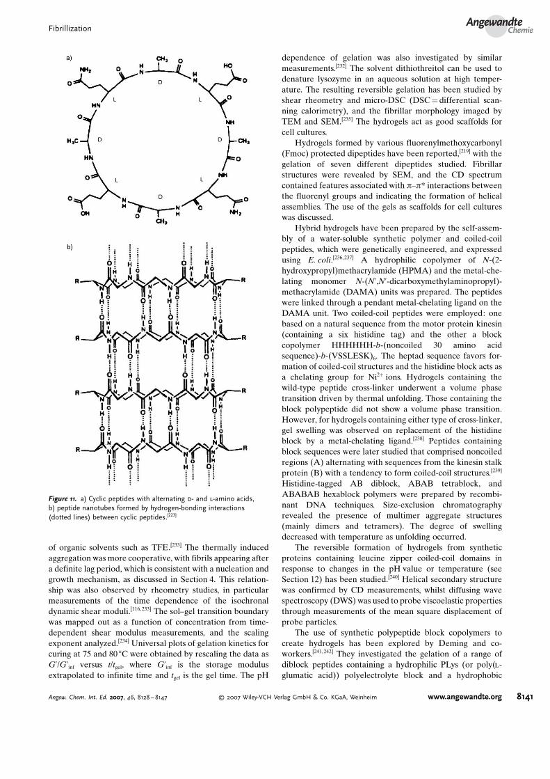
Ghadiri et al. pioneered the design of peptide nanotubesusing stacked cyclic peptides comprising alternating d- and l-amino acids.[223] These peptides were shown to form hollownanotubes based on a b helix—a structure that had beenanticipated prior to their fabrication based on conformationalmodeling studies.[224] Figure 11 shows these structures, whichhave been used as artificial membrane channels[225] and mayfind other applications in nanotechnology.[226] A recentdevelopment has been the preparation of hybrid peptidenanotubes with a thermoresponsive poly(N-isopropylacryl-amide) (PNIPAM) polymer shell around a cyclic peptidecore.[227]
Other peptide motifs that can form nanotubes[218] includehydrophobic dipeptides in the crystal state,[214] b-helix-form-ing peptides,[228] and peptide amphiphiles/copolymers (seeSection 9). The controversy over the Perutz model for theformation of nanotubes from amyloid peptides is discussed inSection 2.
11. Peptide Fibrillar Gels
At sufficiently high concentration, the fibrillization ofpeptides is accompanied by gelation. In this section weconsider the structure of fibrillar hydrogels and organogelsformed by several natural and synthetic peptides.
The formation of fibrils by the thermally induced dena-turing of b-lactoglobulin and lysozyme has been discussed inSection 4. b-lactoglobulin forms fibrillar gels on heating atlow pH values. Particulate gels are formed at higher pH val-ues, close to the dielectric point where the protein has a lownet charge.[229,230] This is outside the scope of the currentReview. In regard to gel formation, it should be noted that thegels can comprise two-phase systems with nematic dropletsdispersed in an isotropic phase.[231] The phase diagram hasbeen mapped out as a function of b-lactoglobulin concen-tration and ionic strength. The persistence length of the fibrilshas been obtained from rheometry measurements of thecritical percolation concentration for network formation. Thepersistence length for b-lactoglobulin is much longer than thatfor bovine serum albumin (BSA) and ovalbumin at pH 2.[231]
In a series of papers, Ross-Murphy and co-workers havestudied the formation of fibrillar gels from b-lactoglobu-lin.[116, 232–234] The structure of the fibrils was probed by AFM,FTIR, and Raman spectroscopy as well as by X-ray scatteringstudies.[116,233,234] The mechanism of fibrillization induced byheating at pH 2 was compared to that induced by the addition
I. W. HamleyReviews
8140 www.angewandte.org � 2007 Wiley-VCH Verlag GmbH & Co. KGaA, Weinheim Angew. Chem. Int. Ed. 2007, 46, 8128 – 8147
of organic solvents such as TFE.[233] The thermally inducedaggregation was more cooperative, with fibrils appearing aftera definite lag period, which is consistent with a nucleation andgrowth mechanism, as discussed in Section 4. This relation-ship was also observed by rheometry studies, in particularmeasurements of the time dependence of the isochronaldynamic shear moduli.[116,233] The sol–gel transition boundarywas mapped out as a function of concentration from time-dependent shear modulus measurements, and the scalingexponent analyzed.[234] Universal plots of gelation kinetics forcuring at 75 and 80 8C were obtained by rescaling the data asG’/G’inf versus t/tgel, where G’inf is the storage modulusextrapolated to infinite time and tgel is the gel time. The pH
dependence of gelation was also investigated by similarmeasurements.[232] The solvent dithiothreitol can be used todenature lysozyme in an aqueous solution at high temper-ature. The resulting reversible gelation has been studied byshear rheometry and micro-DSC (DSC =differential scan-ning calorimetry), and the fibrillar morphology imaged byTEM and SEM.[235] The hydrogels act as good scaffolds forcell cultures.
Hydrogels formed by various fluorenylmethoxycarbonyl(Fmoc) protected dipeptides have been reported,[219] with thegelation of seven different dipeptides studied. Fibrillarstructures were revealed by SEM, and the CD spectrumcontained features associated with p–p* interactions betweenthe fluorenyl groups and indicating the formation of helicalassemblies. The use of the gels as scaffolds for cell cultureswas discussed.
Hybrid hydrogels have been prepared by the self-assem-bly of a water-soluble synthetic polymer and coiled-coilpeptides, which were genetically engineered, and expressedusing E. coli.[236, 237] A hydrophilic copolymer of N-(2-hydroxypropyl)methacrylamide (HPMA) and the metal-che-lating monomer N-(N’,N’-dicarboxymethylaminopropyl)-methacrylamide (DAMA) units was prepared. The peptideswere linked through a pendant metal-chelating ligand on theDAMA unit. Two coiled-coil peptides were employed: onebased on a natural sequence from the motor protein kinesin(containing a six histidine tag) and the other a blockcopolymer HHHHHH-b-(noncoiled 30 amino acidsequence)-b-(VSSLESK)6. The heptad sequence favors for-mation of coiled-coil structures and the histidine block acts asa chelating group for Ni2+ ions. Hydrogels containing thewild-type peptide cross-linker underwent a volume phasetransition driven by thermal unfolding. Those containing theblock polypeptide did not show a volume phase transition.However, for hydrogels containing either type of cross-linker,gel swelling was observed on replacement of the histidineblock by a metal-chelating ligand.[238] Peptides containingblock sequences were later studied that comprised noncoiledregions (A) alternating with sequences from the kinesin stalkprotein (B) with a tendency to form coiled-coil structures.[239]
Histidine-tagged AB diblock, ABAB tetrablock, andABABAB hexablock polymers were prepared by recombi-nant DNA techniques. Size-exclusion chromatographyrevealed the presence of multimer aggregate structures(mainly dimers and tetramers). The degree of swellingdecreased with temperature as unfolding occurred.
The reversible formation of hydrogels from syntheticproteins containing leucine zipper coiled-coil domains inresponse to changes in the pH value or temperature (seeSection 12) has been studied.[240] Helical secondary structurewas confirmed by CD measurements, whilst diffusing wavespectroscopy (DWS) was used to probe viscoelastic propertiesthrough measurements of the mean square displacement ofprobe particles.
The use of synthetic polypeptide block copolymers tocreate hydrogels has been explored by Deming and co-workers.[241, 242] They investigated the gelation of a range ofdiblock peptides containing a hydrophilic PLys (or poly(l-glumatic acid)) polyelectrolyte block and a hydrophobic
Figure 11. a) Cyclic peptides with alternating d- and l-amino acids,b) peptide nanotubes formed by hydrogen-bonding interactions(dotted lines) between cyclic peptides.[223]
FibrillizationAngewandte
Chemie
8141Angew. Chem. Int. Ed. 2007, 46, 8128 – 8147 � 2007 Wiley-VCH Verlag GmbH & Co. KGaA, Weinheim www.angewandte.org

block based on an a helix for poly(l-leucine) or a b sheet inthe case of poly(l-valine).[241] It was found that a high degreeof conformational order of the hydrophobic chain wasrequired to observe gelation, and that the gelation wasslightly better with a-helical domains. The importance ofchain packing was highlighted by the fact that gelation wasnot observed in a mixture containing racemic leucine at aconcentration at which gelation occurred for a copolymercontaining the enantiomeric form. Hierarchical ordering wasobserved with a nanoscale interpenetrating network structureand a porous bicontinuous morphology comprising a gelmatrix and water channels on the microscale.[242] Laserscanning confocal microscopy and cryo-TEM measurementswere used to probe the cellular network structure for a PLys-b-poly(l-leucine-stat-l-valine) diblock (Figure 12). SANSmeasurements confirmed a nanoscale-ordered structure.
Pochan and co-workers have investigated the pH-depen-dent self-assembly of a peptide containing a DPLP (d-proline,l-proline) b-hairpin link between repeating VK units.[243–245]
The peptide folds under basic conditions, and forms hydro-gels. Folding can also be induced by increasing the ionicstrength, which screens electrostatic interactions betweenpositively charged lysine residues.[244] A variant of the originalpeptide was later prepared with threonine in place of severalvaline residues (T is less hydrophobic than V).[245] The newpeptide was shown to exhibit thermally reversible hydro-gelation, with gelation occuring at high temperature. Light-activated hydrogelation was later demonstrated for a homol-ogous b-hairpin peptide containing an a-carboxy-2-nitroben-zyl group attached through a cysteine unit.[246] Intramolecularfolding was induced by exposure to UV light, and wasaccompanied by hydrogelation.
12. Fibrils from a Helices
There have been far fewer reports on the formation offibrils by a-helical peptides. Woolfson and co-workers have
designed so-called “SAF” (“self-assembling fibril”) peptideswith coiled-coil structures based on the heptad of hydro-phobic and polar residues HPPHPPP.[247–249] Complementaryinteractions in the core and complementary flanking ion pairswere used to create staggered heterodimers (Fig-ure 13a).[247, 250] These also had “sticky ends” to promote theformation of long fibers (Figure 13b), again as a result ofinteractions between charged residues.
The design of the peptide was later improved to enhancethe protofibril–protofibril interactions. Nonlinear “fiber-shaping” (FiSh) peptides were also introduced into mixedpeptide systems to engineer branched fibrils (Figure13c,d).[250,251] Hyperbranched networks as well as regularlysegmented and terminated fibrils could also be produced byengineering peptides around three-arm dendritic spacers.[252]
Modification of the original linear SAF peptides by substitu-tion at the e-amino group of lysine enabled fibrils to befunctionalized with ligands such as biotin or the FLAGoctapeptide.[253] Both of these peptides were used to bind withstreptavidin-conjugated gold nanoparticles (in the latter casebiotinylated anti-FLAG antibody was required to effectrecognition).
Several research groups have reported switch peptideswith a design based on a dimeric parallel coiled-coil structure(known as a leucine-zipper sequence).[201, 203,254–256] The use ofappropriate residues in the heptad coiled-coil sequence canfavor b-sheet formation under appropriate conditions.[203] Thede novo switch peptide synthesized by Mutter et al. adopted ab-sheet structure at pH 4, but formed a partly a-helical
Figure 12. Cryo-TEM image showing the cellular structure in a gelformed by a poly(l-lysine)-b-poly(l-leucine-stat-l-valine) diblock copoly-mer.[242]
Figure 13. a) Self-assembly of fiber peptides through complementaryelectrostatic interactions within heterodimers (A with D, B with C).Interactions between the asparagine residues indicated by * furtherstabilize the structures.[250] The peptides are staggered leading to theformation of fibrils (b). Kinked peptides containing flexible b-alanineresidues (c,d) can introduce branches into the fibrils.
I. W. HamleyReviews
8142 www.angewandte.org � 2007 Wiley-VCH Verlag GmbH & Co. KGaA, Weinheim Angew. Chem. Int. Ed. 2007, 46, 8128 – 8147

structure at higher pH values. The design of Woolfson et al.contained a b hairpin (Figure 14).[254] Heat induced a tran-sition from an a-helix to a b-sheet structure that was driven byformation of intramolecular disulfide bonds between the Cys
residues flanking the hairpin. The formation of the b sheetswas accompanied by gelation. This was later extended to thedesign of related peptides which responded to chemicaltriggers as well as thermal processing.[257] Pagel et al. designeda 26-residue peptide that can form random coil, b-sheet, orcoiled-coil structures at appropriate pH values or concentra-tions.[256] A 17-residue peptide has been designed that formscoiled-coil structures at ambient temperatures, but transformsinto amyloid fibrils at high temperature.[255]
Fibril formation from a peptide containing three heptadsequences and designed to form water-soluble helix bundleswas observed under appropriate pH conditions.[258] Fibershave also been observed for five-stranded fibrils of a peptidedesigned to associate in a staggered fashion.[259] Designedthree-helix bundles have been shown to form fibrils providedthe helices have an appropriate orientation.[260] The intermo-lecular association of the monomers is an example of a 3Ddomain-swapped protein structure in which one structuraldomain of a protein monomer is exchanged with the samedomain from another protein, resulting in an intertwinedoligomer.
13. Summary and Outlook
A clear picture is slowly emerging on the mechanism ofamyloid formation by Ab and related proteins. Use of thisknowledge to design inhibitors of fibrillization is underwayand may lead to breakthroughs in therapies. Importantquestions remain on the thermodynamic basis for b-sheetfibrillization and its relationship to protein folding. Mean-while, the principles of self-assembly based on noncovalentinteractions are being used to design novel peptides andpeptide conjugates with desired properties such as controlledfibril morphology, enhanced stability, responsive gelation, orfunctionalization for bio-inorganic hybrids. This is still quite anew field, and exciting new findings can be anticipated,although precise details cannot always be predicted inadvance.
I thank Dr Adam Squires (University of Reading) for usefuldiscussions and Marta Krysmann for the TEM images used in
the frontispiece and Figure 2. The referees are thanked forpointing out several additional interesting references.
Received: February 26, 2007Published online: October 12, 2007
[1] E. D. Eanes, G. G. Glenner, J. Histochem. Cytochem. 1968, 16,673.
[2] D. A. Kirschner, C. Abraham, D. J. Selkoe, Proc. Natl. Acad.Sci. USA 1986, 83, 503.
[3] M. Sunde, C. C. F. Blake, Adv. Protein Chem. 1997, 50, 123.[4] L. C. Serpell, Biochim. Biophys. Acta Mol. Basis Dis. 2000,
1502, 16.[5] O. S. Makin, L. C. Serpell, FEBS J. 2005, 272, 5950.[6] D. B. Teplow, Amyloid 1998, 5, 121.[7] M. J. Krysmann, I. W. Hamley, P. J. F. Harris, unpublished
results.[8] M. Goedert, M. G. Spillantini, Science 2006, 314, 777.[9] E. D. Roberson, L. Mucke, Science 2006, 314, 781.
[10] J. D. Sipe, Annu. Rev. Biochem. 1992, 61, 947.[11] B. Caughey, P. T. Lansbury, Annu. Rev. Neurosci. 2003, 26, 267.[12] M. R. Chapman, L. S. Robinson, J. S. Pinkner, R. Roth, J.
Heuser, M. Hammar, S. Normark, S. J. Hultgren, Science 2002,295, 851.
[13] J. W. Kelly, W. E. Balch, J. Cell Biol. 2003, 161, 461.[14] J. F. Berson, A. C. Theos, D. C. Harper, D. Tenza, G. Raposo,
M. S. Marks, J. Cell Biol. 2003, 161, 521.[15] P. T. Lansbury, H. A. Lashuel, Nature 2006, 443, 774.[16] M. Bucciantini, E. Giannoni, F. Chiti, F. Baroni, L. Formigli, J.
Zurdo, N. Taddei, G. Ramponi, C. M. Dobson, M. Stefani,Nature 2002, 416, 507.
[17] F. Rousseau, J. Schmykovitz, L. Serrano, Curr. Opin. Struct.Biol. 2006, 16, 118.
[18] C. G. Glabe, Neurobiol. Aging 2006, 27, 570.[19] E. Cohen, J. Bieschke, R. M. Perciavalle, J. W. Kelly, A. Dillin,
Science 2006, 313, 1604.[20] C. M. Dobson, Trends Biochem. Sci. 1999, 24, 329.[21] M. Bucciantini, G. Calloni, F. Chiti, L. Formigli, D. Nosi, C. M.
Dobson, M. Stefani, J. Biol. Chem. 2004, 279, 31374.[22] D. M. Walsh, B. P. Tseng, R. E. Rydel, M. B. Podlisny, D. J.
Selkoe, Biochemistry 2000, 39, 10831.[23] A. Goate, M. C. Chartierharlin, M. Mullan, J. Brown, F.
Crawford, L. Fidani, L. Giuffra, A. Haynes, N. Irving, L.James, R. Mant, P. Newton, K. Rooke, P. Roques, C. Talbot, M.Pericakvance, A. Roses, R. Williamson, M. Rossor, M. Owen, J.Hardy, Nature 1991, 349, 704.
[24] R. Sherrington, E. I. Rogaev, Y. Liang, E. A. Rogaeva, G.Levesque, M. Ikeda, H. Chi, C. Lin, G. Li, K. Holman, T. Tsuda,L. Mar, J. F. Foncin, A. C. Bruni, M. P. Montesi, S. Sorbi, I.Rainero, L. Pinessi, L. Nee, I. Chumakov, D. Pollen, A.Brookes, P. Sanseau, R. J. Polinsky, W. Wasco, H. A. R. Dasilva,J. L. Haines, M. A. Pericakvance, R. E. Tanzi, A. D. Roses, P. E.Fraser, J. M. Rommens, P. H. St George-Hyslop, Nature 1995,375, 754.
[25] E. I. Rogaev, R. Sherrington, E. A. Rogaeva, G. Levesque, M.Ikeda, Y. Liang, H. Chi, C. Lin, K. Holman, T. Tsuda, L. Mar, S.Sorbi, B. Nacmias, S. Piacentini, L. Amaducci, I. Chumakov, D.Cohen, L. Lannfelt, P. E. Fraser, J. M. Rommens, P. H.St George-Hyslop, Nature 1995, 376, 775.
[26] D. E. Schmechel, A. M. Saunders, W. J. Strittmatter, B. J. Crain,C. M. Hulette, S. H. Joo, M. A. Pericakvance, D. Goldgaber,A. D. Roses, Proc. Natl. Acad. Sci. USA 1993, 90, 9649.
[27] A. M. Saunders, W. J. Strittmatter, D. Schmechel, P. H.St George-Hyslop, M. A. Pericakvance, S. H. Joo, B. L. Rosi,J. F. Gusella, D. R. Crappermaclachlan, M. J. Alberts, C.
Figure 14. a) Helical wheel representation of a coiled-coil dimer.b) Structure of the b-hairpin; the interstrand hydrogen bonds areindicated by dashed lines.[254]
FibrillizationAngewandte
Chemie
8143Angew. Chem. Int. Ed. 2007, 46, 8128 – 8147 � 2007 Wiley-VCH Verlag GmbH & Co. KGaA, Weinheim www.angewandte.org

Hulette, B. Crain, D. Goldgaber, A. D. Roses, Neurology 1993,43, 1467.
[28] E. H. Corder, A. M. Saunders, W. J. Strittmatter, D. E. Schme-chel, P. C. Gaskell, G. W. Small, A. D. Roses, J. L. Haines, M. A.Pericakvance, Science 1993, 261, 921.
[29] K. R. Bales, T. Verina, R. C. Dodel, Y. S. Du, L. Altstiel, M.Bender, P. Hyslop, E. M. Johnstone, S. P. Little, D. J. Cummins,P. Piccardo, B. Ghetti, S. M. Paul, Nat. Genet. 1997, 17, 263.
[30] E. Rogaeva, Y. Meng, J. H. Lee, Y. Gu, T. Kawarai, F. Zou, T.Katayama, C. T. Baldwin, R. Cheng, H. Hasegawa, F. Chen, N.Shibata, K. L. Lunetta, R. Pardossi-Piquard, C. Bohm, Y.Wakutani, L. A. Cupples, K. T. Cuenco, R. C. Green, L. Pinessi,I. Rainero, S. Sorbi, A. Bruni, R. Duara, R. P. Friedland, R.Inzelberg, W. Hampe, H. Bujo, Y. Q. Song, O. M. Andersen,T. E. Willnow, N. Graff-Radford, R. C. Petersen, D. Dickson,S. D. Der, P. E. Fraser, G. Schmitt-Ulms, S. Younkin, R.Mayeux, L. A. Farrer, P. St George-Hyslop, Nat. Genet. 2007,39, 168.
[31] T. A. Fulga, I. Elson-Schwab, V. Khurana, M. L. Steinhilb, T. L.Spires, B. T. Hyman, M. B. Feany, Nat. Cell Biol. 2006, 9, 139.
[32] C. Blake, L. C. Serpell, Structure 1996, 4, 989.[33] J. L. JimRnez, J. L. Guijarro, E. Orlova, J. Zurdo, C. M. Dobson,
M. Sunde, H. R. Saibil, EMBO J. 1999, 18, 815.[34] J. L. Jimenez, E. J. Nettleton, M. Bouchard, C. V. Robinson,
C. M. Dobson, H. R. Saibil, Proc. Natl. Acad. Sci. USA 2002,99, 9196.
[35] D. Voet, J. G. Voet, Biochemistry, John Wiley & Sons, NewYork, 1995.
[36] M. Sunde, L. C. Serpell, M. Bartlam, P. E. Fraser, M. B. Pepys,C. C. F. Blake, J. Mol. Biol. 1997, 273, 729.
[37] J. D. Termine, E. D. Eanes, D. Ein, G. G. Glenner, Biopolymers1972, 11, 1103.
[38] A. T. Petkova, Y. Ishii, J. J. Balbach, O. N. Antzutkin, R. D.Leapman, F. Delaglio, R. Tycko, Proc. Natl. Acad. Sci. USA2002, 99, 16742.
[39] T. LThrs, C. Ritter, M. Adrian, D. Riek-Loher, B. Bohrmann, H.DUbeli, D. Schubert, R. Riek, Proc. Natl. Acad. Sci. USA 2005,102, 17342.
[40] A. M. Squires, G. L. Devlin, S. L. Gras, A. K. Tickler, C. E.MacPhee, C. M. Dobson, J. Am. Chem. Soc. 2006, 128, 11738.
[41] M. F. Perutz, J. T. Finch, J. Berriman, A. Lesk, Proc. Natl. Acad.Sci. USA 2002, 99, 5591.
[42] P. Sikorski, E. Atkins, Biomacromolecules 2005, 6, 425.[43] D. Sharma, L. M. Shinchuk, H. Inouye, R. Wetzel, D. A.
Kirschner, Proteins Struct. Funct. Bioinf. 2005, 61, 398.[44] H. Inouye, D. A. Kirschner in Adv. Protein Chem. , Vol. 73
(Eds.: A. Kakava, J. M. Squire, D. A. D. Parry), ElsevierAcademic, San Diego, 2006.
[45] H. LeVine, Protein Sci. 1993, 2, 404.[46] S. J. Wood, B. Maleef, T. Hart, R. Wetzel, J. Mol. Biol. 1996, 256,
870.[47] O. S. Makin, L. C. Serpell, Fibre Diffr. Rev. 2004, 12, 29.[48] A. Lomakin, D. S. Chung, G. B. Benedek, D. A. Kirschner,
D. B. Teplow, Proc. Natl. Acad. Sci. USA 1996, 93, 1125.[49] J. Riseman, J. G. Kirkwood, J. Chem. Phys. 1950, 18, 512.[50] D. M. Walsh, A. Lomakin, G. B. Benedek, M. M. Condron,
D. B. Teplow, J. Biol. Chem. 1997, 272, 22364.[51] Y. Kusumoto, A. Lomakin, D. B. Teplow, G. B. Benedek, Proc.
Natl. Acad. Sci. USA 1998, 95, 12277.[52] F. Sokolowski, A. J. Modler, R. Masuch, D. Zirwer, M. Baier, G.
Lutsch, D. A. Moss, K. Gast, D. Naumann, J. Biol. Chem. 2003,278, 40481.
[53] A. J. Modler, K. Gast, G. Lutsch, G. Damaschun, J. Mol. Biol.2003, 325, 135.
[54] S. M. Kelly, T. J. Jess, N. C. Price, Biochim. Biophys. ActaProteins Proteomics 2005, 1751, 119.
[55] J. Reed, A. Reed, Anal. Biochem. 1997, 254, 36.
[56] W. K. Surewicz, H. H. Mantsch, D. Chapman, Biochemistry1993, 32, 389.
[57] B. Stuart, Biological Applications of Infrared Spectroscopy,Wiley, Chichester, 1997.
[58] H. Hiramatsu, T. Kitagawa, Biochim. Biophys. Acta ProteinsProteomics 2005, 1753, 100.
[59] M. Jackson, H. H. Mantsch, Crit. Rev. Biochem. Mol. Biol.1995, 30, 95.
[60] P. Haris, D. Chapman, Biopolymers 1995, 37, 251.[61] J. Kubelka, T. A. Keiderling, J. Am. Chem. Soc. 2001, 123, 6142.[62] P. R. Costa, D. A. Kocisko, B. Q. Sun, P. T. Lansbury, R. G.
Griffin, J. Am. Chem. Soc. 1997, 119, 10487.[63] T. L. S. Benzinger, D. M. Gregory, T. S. Burkoth, H. Miller-
Auer, D. G. Lynn, R. E. Botto, S. C. Meredith, Biochemistry2000, 39, 3491.
[64] A. B. Siemer, C. Ritter, M. Ernst, R. Riek, B. H. Meier,Angew.Chem. 2005, 117, 2494; Angew. Chem. Int. Ed. 2005, 44, 2441.
[65] H. Heise, W. Hoyer, S. Becker, O. C. Andronesi, D. Riedel, M.Baldus, Proc. Natl. Acad. Sci. USA 2005, 102, 15871.
[66] R. Tycko, Curr. Opin. Struct. Biol. 2004, 14, 96.[67] R. Tycko, Q. Rev. Biophys. 2006, 39, 1.[68] J. D. Harper, S. S. Wong, C. M. Lieber, P. T. Lansbury, Chem.
Biol. 1997, 4, 119.[69] G. Bitan, M. D. Kirkitadze, A. Lomakin, S. S. Vollers, G. B.
Benedek, D. B. Teplow, Proc. Natl. Acad. Sci. USA 2003, 100,330.
[70] C. Ha, C. B. Park, Langmuir 2006, 22, 6977.[71] S. L. McCutchen, Z. H. Lai, G. J. Miroy, J. W. Kelly, W. Colon,
Biochemistry 1995, 34, 13527.[72] Z. Lai, W. ColVn, J. W. Kelly, Biochemistry 1996, 35, 6470.[73] G. S. Jackson, L. L. P. Hosszu, A. Power, A. F. Hill, J. Kenney,
H. Saibil, C. J. Craven, J. P. Waltho, A. R. Clarke, J. Collinge,Science 1999, 283, 1935.
[74] L. L. P. Hosszu, N. J. Baxter, G. S. Jackson, A. Power, A. R.Clarke, J. P. Waltho, C. J. Craven, J. Collinge, Nat. Struct. Biol.1999, 6, 740.
[75] M. R. Hurle, L. R. Helms, L. Li, W. Chan, R. Wetzel,Proc. Natl.Acad. Sci. USA 1994, 91, 5446.
[76] L. R. Helms, R. Wetzel, J. Mol. Biol. 1996, 257, 77.[77] R. Raffen, L. J. Dieckman, M. Szpunar, C. Wunschl, P. R.
Pokkuluri, P. Dave, P. W. Stevens, X. Y. Cai, M. Schiffer, F. J.Stevens, Protein Sci. 1999, 8, 509.
[78] S. E. Radford, C. M. Dobson, P. A. Evans, Nature 1992, 358,302.
[79] D. R. Booth, M. Sunde, V. Bellotti, C. V. Robinson, W. L.Hutchinson, P. E. Fraser, P. N. Hawkins, C. M. Dobson, S. E.Radford, C. C. F. Blake, M. B. Pepys, Nature 1997, 385, 787.
[80] J. I. Guijarro, M. Sunde, J. A. Jones, I. D. Campbell, C. M.Dobson, Proc. Natl. Acad. Sci. USA 1998, 95, 4224.
[81] V. J. McParland, N. M. Kad, A. P. Kalverda, A. Brown, P.Kirwin-Jones, M. G. Hunter, M. Sunde, S. E. Radford, Bio-chemistry 2000, 39, 8735.
[82] N. M. Kad, N. H. Thomson, D. P. Smith, D. A. Smith, S. E.Radford, J. Mol. Biol. 2001, 313, 559.
[83] D. P. Smith, S. E. Radford, Protein Sci. 2001, 10, 1775.[84] L. N. Arnaudov, R. de Vries, Biophys. J. 2004, 88, 515.[85] F. Chiti, P. Webster, N. Taddei, A. Clark, M. Stefani, G.
Ramponi, C. M. Dobson, Proc. Natl. Acad. Sci. USA 1999, 96,3590.
[86] M. FNndrich, V. Forge, K. Buder, M. Kittler, C. M. Dobson, S.Diekmann, Proc. Natl. Acad. Sci. USA 2003, 100, 15463.
[87] R. Schweitzer-Stenner, T. Measey, A. Hagarman, F. Eker, K.Griebenow, Biochemistry 2006, 45, 2810.
[88] F. Eker, K. Griebenow, R. Schweitzer-Stenner, Biochemistry2004, 43, 6893.
[89] L. M. Hou, H. Y. Shao, Y. B. Zhang, H. Li, N. K. Menon, E. B.Neuhaus, J. M. Brewer, I. J. L. Byeon, D. G. Ray, M. P. Vitek, T.
I. W. HamleyReviews
8144 www.angewandte.org � 2007 Wiley-VCH Verlag GmbH & Co. KGaA, Weinheim Angew. Chem. Int. Ed. 2007, 46, 8128 – 8147

Iwashita, R. A. Makula, A. B. Przybyla, M. G. Zagorski, J. Am.Chem. Soc. 2004, 126, 1992.
[90] C. D. Syme, E. W. Blanch, C. Holt, R. Jakes, M. Goedert, L.Hecht, L. D. Barron, Eur. J. Biochem. 2002, 269, 148.
[91] S. Narayanan, B. Reif, Biochemistry 2005, 44, 1444.[92] M. Tanaka, Y. Machida, Y. Nishikawa, T. Akagi, T. Hashikawa,
T. Fujisawa, N. Nukina, J. Biol. Chem. 2003, 278, 34717.[93] G. P. Bates, C. Benn in Huntington@s Disease (Eds.: G. P. Bates,
P. S. Harper, L. Jones), Oxford University Press, Oxford, 2002.[94] M. Calamai, C. Canale, A. Relini, M. Stefani, F. Chiti, C. M.
Dobson, J. Mol. Biol. 2005, 346, 603.[95] J. T. Jarrett, P. T. Lansbury, Cell 1993, 73, 1055.[96] J. D. Harper, P. T. Lansbury,Annu. Rev. Biochem. 1997, 66, 385.[97] J. N. Israelachvili, Intermolecular and Surface Forces, Academic
Press, San Diego, 1991.[98] J. M. Andreu, S. N. Timasheff,Methods Enzymol. 1986, 130, 47.[99] K. Johan, G. Westermark, U. EngstrUm, A. Gustavsson, P.
Hultman, P. Westermark, Proc. Natl. Acad. Sci. USA 1998, 95,2558.
[100] C. M. Dobson, Nature 2003, 426, 884.[101] I. V. Baskakov, G. Legname, Z. Gryczynski, S. B. Prusiner,
Protein Sci. 2004, 13, 586.[102] L. N. Arnaudov, R. de Vries, Biomacromolecules 2003, 4, 1614.[103] Y.-H. Chen, J. T. Yang, H. M. Martinez, Biochemistry 1972, 11,
4120.[104] Y.-H. Chen, J. T. Yang, K. H. Chau, Biochemistry 1974, 13,
3350.[105] P. Aymard, T. Nicolai, D. Durand, A. Clark, Macromolecules
1999, 32, 2542.[106] L. N. Arnaudov, R. de Vries, Biomacromolecules 2006, 7, 3490.[107] M. R. H. Krebs, D. K. Wilkins, E. W. Chung, M. C. Pitkeathly,
A. K. Chamberlain, J. Zurdo, C. V. Robinson, C. M. Dobson, J.Mol. Biol. 2000, 300, 541.
[108] E. K. Hill, B. Krebs, D. G. Goodall, G. J. Howlett, D. E.Dunstan, Biomacromolecules 2006, 7, 10.
[109] V. Castelletto, I. W. Hamley, Biomacromolecules 2007, 8, 77.[110] W. P. Esler, E. R. Stimson, J. R. Ghilardi, H. V. Vinters, J. P.
Lee, P. W. Mantyh, J. E. Maggio, Biochemistry 1996, 35, 749.[111] H. Naiki, K. Nakakuki, Lab. Invest. 1996, 74, 374.[112] S. R. Collins, A. Douglass, R. D. Vale, J. S. Weissman, PLoS
Biol. 2004, 2, 1582.[113] S. Narayanan, S. Walter, B. Reif, ChemBioChem 2006, 7, 757.[114] M. R. H. Krebs, L. A. Morozova-Roche, K. Daniel, C. V.
Robinson, C. M. Dobson, Protein Sci. 2004, 13, 1933.[115] A. T. Petkova, R. D. Leapman, Z. H. Guo, W. M. Yau, M. P.
Mattson, R. Tycko, Science 2005, 307, 262.[116] W. S. Gosal, A. H. Clark, P. D. A. Pudney, S. B. Ross-Murphy,
Langmuir 2002, 18, 7174.[117] T. Ban, D. Hamada, K. Hasegawa, H. Naiki, Y. Goto, J. Biol.
Chem. 2003, 278, 16462.[118] M. P. Lambert, A. K. Barlow, B. A. Chromy, C. Edwards, R.
Freed, M. Liosatos, T. E. Morgan, I. Rozovsky, B. Trommer,K. L. Viola, P. Wals, C. Zhang, C. E. Finch, G. A. Krafft, W. L.Klein, Proc. Natl. Acad. Sci. USA 1998, 95, 6448.
[119] K. N. Dahlgren, A. M. Manelli, W. B. Stine, L. K. Baker, G. A.Krafft, M. J. LaDu, J. Biol. Chem. 2002, 277, 32046.
[120] D. M. Hartley, D. M. Walsh, C. P. P. Ye, T. Diehl, S. Vasquez,P. M. Vassilev, D. B. Teplow, D. J. Selkoe, J. Neurosci. 1999, 19,8876.
[121] K. Andersson, A. Olofsson, E. H. Nielsen, S. E. Svehag, E.Lundgren, Biochem. Biophys. Res. Commun. 2002, 294, 309.
[122] R. Kayed, E. Head, J. L. Thompson, T. M. McIntire, S. C.Milton, C. W. Cotman, C. G. Glabe, Science 2003, 300, 486.
[123] Y. Zhang, R. McLaughlin, C. Goodyer, A. LeBlanc, J. Cell Biol.2002, 156, 519.
[124] J. P. Cleary, D. M. Walsh, J. J. Hofmeister, G. M. Shankar, M. A.Kuskowski, D. J. Selkoe, K. H. Ashe, Nat. Neurosci. 2005, 8, 79.
[125] Y. S. Du, X. Wei, R. Dodel, N. Sommer, H. Hampel, F. Gao,Z. Z. Ma, L. M. Zhao, W. H. Oertel, M. Farlow,Brain 2003, 126,1935.
[126] M. P. Lambert, K. L. Viola, B. A. Chromy, L. Chang, T. E.Morgan, J. X. Yu, D. L. Venton, G. A. Krafft, C. E. Finch, W. L.Klein, J. Neurochem. 2001, 79, 595.
[127] S. LesnR, M. T. Koh, L. Kotilinek, R. Kayed, C. G. Glabe, A.Yang, M. Gallagher, K. H. Ashe, Nature 2006, 440, 352.
[128] H. B. Pollard, N. Arispe, E. Rojas, Cell. Mol. Neurobiol. 1995,15, 513.
[129] J. I. Kourie, C. L. Henry, Clin. Exp. Pharmacol. Physiol. 2002,29, 741.
[130] H. A. Lashuel, D. Hartley, B. M. Petre, T. Walz, P. T. Lansbury,Nature 2002, 418, 291.
[131] N. Arispe, E. Rojas, H. B. Pollard, Proc. Natl. Acad. Sci. USA1993, 90, 567.
[132] N. Arispe, H. B. Pollard, E. Rojas, Proc. Natl. Acad. Sci. USA1993, 90, 10573.
[133] N. Arispe, H. B. Pollard, E. Rojas, Proc. Natl. Acad. Sci. USA1996, 93, 1710.
[134] Y. Hirakura, M. C. Lin, B. L. Kagan, J. Neurosci. Res. 1999, 57,458.
[135] H. Lin, R. Bhatia, R. Lal, FASEB J. 2001, 15, 2433.[136] M. J. Volles, P. T. Lansbury, Biochemistry 2002, 41, 4595.[137] T. R. Jahn, S. E. Radford, FEBS J. 2005, 272, 5962.[138] M. Balbirnie, R. Grothe, D. S. Eisenberg, Proc. Natl. Acad. Sci.
USA 2001, 98, 2375.[139] B. Gaughey, G. S. Baron, Nature 2006, 443, 803.[140] M. FNndrich, C. M. Dobson, EMBO J. 2002, 21, 5682.[141] W. K. Kim, M. H. Hecht, Proc. Natl. Acad. Sci. USA 2006, 103,
15824.[142] W. J. Goux, L. Kopplin, A. D. Nguyen, K. Leak, M. Rutkofsky,
V. D. Shanmuganandam, D. Sharma, H. Inouye, D. A. Kirsch-ner, J. Biol. Chem. 2004, 279, 26868.
[143] M. Reches, E. Gazit, Amyloid 2004, 11, 81.[144] M. Reches, Y. Porat, E. Gazit, J. Biol. Chem. 2002, 277, 35475.[145] C. Hilbich, B. Kisterswoike, J. Reed, C. L. Masters, K.
Beyreuther, J. Mol. Biol. 1992, 228, 460.[146] L. O. Tjernberg, J. Naslund, F. Lindqvist, J. Johansson, A. R.
Karlstrom, J. Thyberg, L. Terenius, C. Nordstedt, J. Biol. Chem.1996, 271, 8545.
[147] L. O. Tjernberg, C. Lilliehook, D. J. E. Callaway, J. Naslund, S.Hahne, J. Thyberg, L. Terenius, C. Nordstedt, J. Biol. Chem.1997, 272, 12601.
[148] M. A. Findeis, G. M. Musso, C. C. Arico-Muendel, H. W.Benjamin, A. M. Hundal, J. J. Lee, J. Chin, M. Kelley, J.Wakefield, N. J. Hayward, S. M. Molineaux, Biochemistry 1999,38, 6791.
[149] L. O. Tjernberg, D. J. E. Callaway, A. Tjernberg, S. Hahne, C.LilliehUUk, L. Terenius, J. Thyberg, C. Nordstedt, J. Biol. Chem.1999, 274, 12619.
[150] D. J. Gordon, R. Tappe, S. C. Meredith, J. Pept. Res. 2002, 60,37.
[151] D. J. Gordon, K. L. Sciarretta, S. C. Meredith, Biochemistry2001, 40, 8237.
[152] C. Soto, E. M. Sigurdsson, L. Morelli, R. A. Kumar, E. M.Castano, B. Frangione, Nat. Med. 1998, 4, 822.
[153] G. Zhang, M. J. Leibowitz, P. J. Sinko, S. Stein, BioconjugateChem. 2003, 14, 86.
[154] J. J. Balbach, Y. Ishii, O. N. Antzutkin, R. D. Leapman, N. W.Rizzo, F. Dyda, J. Reed, R. Tycko,Biochemistry 2000, 39, 13748.
[155] K. Lu, J. Jacob, P. Thiyagarajan, V. P. Conticello, D. G. Lynn, J.Am. Chem. Soc. 2003, 125, 6391.
[156] S. J. Wood, R. Wetzel, J. D. Martin, M. R. Hurle, Biochemistry1995, 34, 724.
[157] A.-M. Fernandez-Escamilla, F. Rousseau, J. Schymkowitz, L.Serrano, Nat. Biotechnol. 2004, 22, 1302.
FibrillizationAngewandte
Chemie
8145Angew. Chem. Int. Ed. 2007, 46, 8128 – 8147 � 2007 Wiley-VCH Verlag GmbH & Co. KGaA, Weinheim www.angewandte.org

[158] A. P. Pawar, K. F. DuBay, J. Zurdo, F. Chiti, M. Vendruscolo,C. M. Dobson, J. Mol. Biol. 2005, 350, 379.
[159] Y. Kallberg, M. Gustafsson, B. Persson, J. Thyberg, J. Johans-son, J. Biol. Chem. 2001, 276, 12945.
[160] A. A. Watson, D. P. Fairlie, D. J. Craik, Biochemistry 1998, 37,12700.
[161] H. Sticht, P. Bayer, D. Willbold, S. Dames, C. Hilbich, K.Beyreuther, R. W. Frank, P. Rosch, Eur. J. Biochem. 1995, 233,293.
[162] M. Coles, W. Bicknell, A. A. Watson, D. P. Fairlie, D. J. Craik,Biochemistry 1998, 37, 11064.
[163] S. B. Malinchik, H. Inouye, K. E. Szumowski, D. A. Kirschner,Biophys. J. 1998, 74, 537.
[164] A. Aggeli, M. Bell, N. Boden, J. N. Keen, P. F. Knowles, T. C. B.McLeish, M. Pitkeathly, S. E. Radford, Nature 1997, 386, 259.
[165] A. Aggeli, M. L. Bannister, M. Bell, N. Boden, J. B. C. Findlay,M. Hunter, P. F. Knowles, J.-C. Yang, Biochemistry 1998, 37,8121.
[166] A. Aggeli, M. Bell, N. Boden, J. N. Keen, T. C. B. McLeish, I.Nyrkova, S. E. Radford, A. Semenov, J. Mater. Chem. 1997, 7,1135.
[167] A. Aggeli, G. Fytas, D. Vlassopoulos, T. C. B. McLeish, P. J.Mawer, N. Boden, Biomacromolecules 2001, 2, 378.
[168] I. A. Nyrkova, A. N. Semenov, A. Aggeli, M. Bell, N. Boden,T. C. B. McLeish, Eur. Phys. J. B 2000, 17, 499.
[169] A. Aggeli, I. A. Nyrkova, M. Bell, R. Harding, L. Carrick,T. C. B. McLeish, A. N. Semenov, N. Boden, Proc. Natl. Acad.Sci. USA 2001, 98, 11857.
[170] L. Carrick, M. Tassieri, T. A. Waigh, A. Aggeli, N. Boden, C.Bell, J. Fisher, E. Ingham, R. M. L. Evans, Langmuir 2005, 21,3733.
[171] A. Aggeli, M. Bell, N. Boden, L. M. Carrick, A. E. Strong,Angew. Chem. 2003, 115, 5761; Angew. Chem. Int. Ed. 2003, 42,5603.
[172] A. Aggeli, M. Bell, N. Boden, L. Carrick, C. W. G. Fishwick,P. J. Mawer, S. E. Radford, A. E. Strong, J. Am. Chem. Soc.2003, 125, 9619.
[173] M. L. de la Paz, K. Goldie, J. Zurdo, E. Lacroix, C. M. Dobson,A. Hoenger, L. Serrano, Proc. Natl. Acad. Sci. USA 2002, 99,16052.
[174] P. Chen, Colloids Surf. A 2005, 261, 3.[175] S. Jun, Y. Hong, H. Imamura, B.-Y. Ha, J. Bechhoefer, P. Chen,
Biophys. J. 2004, 87, 1249.[176] H. Yang, M. Pritzker, S. Y. Fung, Y. Sheng, W. Wang, P. Chen,
Langmuir 2006, 22, 8553.[177] K. Pagel, T. Vagt, T. Kohajda, B. Koksch, Org. Biomol. Chem.
2005, 3, 2500.[178] K. Pagel, T. Vagt, B. Koksch,Org. Biomol. Chem. 2005, 3, 3843.[179] S. Matsumura, S. Uemura, H. Mihara, Chem. Eur. J. 2004, 10,
2789.[180] J. D. Hartgerink, E. Beniash, S. I. Stupp, Science 2001, 294,
1684.[181] J. D. Hartgerink, E. Beniash, S. I. Stupp, Proc. Natl. Acad. Sci.
USA 2002, 99, 5133.[182] K. L. Niece, J. D. Hartgerink, J. J. J. M. Donners, S. I. Stupp, J.
Am. Chem. Soc. 2003, 125, 7146.[183] R. C. Claussen, B. M. Rabatic, S. I. Stupp, J. Am. Chem. Soc.
2003, 125, 12680.[184] S. Vauthey, S. Santoso, H. Gong, N. Watson, S. Zhang, Proc.
Natl. Acad. Sci. USA 2002, 99, 5355.[185] S. Santoso, W. Hwang, H. Hartman, S. Zhang, Nano Lett. 2002,
2, 687.[186] S. S. Santoso, S. Vauthey, S. Zhang, Curr. Opin. Colloid
Interface Sci. 2002, 7, 262.[187] H.-W. Jun, V. Yuwono, S. E. Paramonov, J. D. Hartgerink, Adv.
Mater. 2005, 17, 2612.
[188] C. Li, J. Orbulescu, G. Sui, R. M. Leblanc, Langmuir 2004, 20,8641.
[189] R. Bitton, J. Schmidt, M. Biesalski, R. Tu, M. Tirrell, H. Bianco-Peled, Langmuir 2005, 21, 11888.
[190] K. Velonia, A. E. Rowan, R. J. M. Nolte, J. Am. Chem. Soc.2002, 124, 4224.
[191] D. W. P. M. LUwik, J. Garcia-Hartjes, J. T. Meijer, J. C. M.van Hest, Langmuir 2005, 21, 524.
[192] J. T. Meijer, M. Roeters, V. Viola, D. W. P. M. LUwik, G. Vriend,J. C. M. van Hest, Langmuir 2007, 23, 2058.
[193] G. W. M. Vandermeulen, C. Tziatzios, H.-A. Klok, Macro-molecules 2003, 36, 4107.
[194] G. W. M. Vandermeulen, H.-A. Klok, Macromol. Biosci. 2004,4, 383.
[195] I. W. Hamley,Block Copolymers in Solution, Wiley, Chichester,2005.
[196] H. Schlaad, Adv. Polym. Sci. 2006, 202, 53.[197] T. S. Burkoth, T. L. S. Benzinger, D. N. M. Jones, K. Hallenga,
S. C. Meredith, D. G. Lynn, J. Am. Chem. Soc. 1998, 120, 7655.[198] T. S. Burkoth, T. L. S. Benzinger, V. Urban, D. G. Lynn, S. C.
Meredith, P. Thiyagarajan, J. Am. Chem. Soc. 1999, 121, 7429.[199] P. Thiyagarajan, T. S. Burkoth, V. Urban, S. Seifert, T. L. S.
Benzinger, D. M. Morgan, D. Gordon, S. C. Meredith, D. G.Lynn, J. Appl. Crystallogr. 2000, 33, 535.
[200] J. H. Collier, P. B. Messersmith, Adv. Mater. 2004, 16, 907.[201] H.-A. Klok, G. W. M. Vandermeulen, H. Nuhn, A. RUsler, I. W.
Hamley , V. Castelletto, H. Xu, S. Sheiko, Faraday Discuss.2005, 128, 29.
[202] J. Y. Su, R. S. Hodges, C. M. Kay, Biochemistry 1994, 33, 15501.[203] M. Mutter, R. Gassmann, U. Buttkus, K.-H. Altmann, Angew.
Chem. 1991, 103, 1504; Angew. Chem. Int. Ed. Engl. 1991, 30,1514.
[204] I. W. Hamley , I. A. Ansari, V. Castelletto, H. Nuhn, A. RUsler,H.-A. Klok, Biomacromolecules 2005, 6, 1310.
[205] J. J. L. M. Cornelissen, M. Fischer, N. A. J. M. Sommerdijk,R. J. M. Nolte, Science 1998, 280, 1427.
[206] J. M. Smeenk, P. SchUn, M. B. J. Otten, S. Speller, H. G.Stunnenberg, J. C. M. van Hest,Macromolecules 2006, 39, 2989.
[207] M. Reches, E. Gazit, Science 2003, 300, 625.[208] E. Gazit, FEBS J. 2005, 272, 5971.[209] S. Gilead, E. Gazit, Supramol. Chem. 2005, 17, 87.[210] M. Reches, E. Gazit, Isr. J. Chem. 2005, 45, 363.[211] M. Reches, E. Gazit, Nano Lett. 2004, 4, 581.[212] C. H. GUrbitz, Chem. Commun. 2006, 2332.[213] C. H. GUrbitz, Chem. Eur. J. 2001, 7, 5153.[214] C. H. GUrbitz, New J. Chem. 2003, 27, 1789.[215] S. M. Tracz, A. Abedini, M. Driscoll, D. P. Raleigh, Biochem-
istry 2004, 43, 15901.[216] F. Bemporad, N. Taddei, M. Stefani, F. Chiti, Protein Sci. 2006,
15, 862.[217] O. S. Makin, E. Atkins, P. Sikorski, J. Johansson, L. C. Serpell,
Proc. Natl. Acad. Sci. USA 2005, 102, 315.[218] H. Inouye, D. Sharma, W. J. Goux, D. A. Kirschner, Biophys. J.
2005, 90, 1774.[219] V. Jayawarna, M. Ali, T. A. Jowitt, A. F. Miller, A. Saiani, J. E.
Gough, R. V. Ulijn, Adv. Mater. 2006, 18, 611.[220] A. Mahler, M. Reches, M. Rechter, S. Cohen, E. Gazit, Adv.
Mater. 2006, 18, 1365.[221] M. Yemini, M. Reches, J. Rishpon, E. Gazit,Nano Lett. 2004, 4,
183.[222] X. Gao, H. Matsui, Adv. Mater. 2005, 17, 2037.[223] M. R. Ghadiri, J. R. Granja, R. A. Milligan, D. E. McRee, N.
Khazanovich, Nature 1993, 366, 324.[224] P. De Santis, S. Morosetti, R. Rizzo, Macromolecules 1974, 7,
52.[225] M. R. Ghadiri, J. R. Granja, L. K. Buehler, Nature 1994, 369,
301.
I. W. HamleyReviews
8146 www.angewandte.org � 2007 Wiley-VCH Verlag GmbH & Co. KGaA, Weinheim Angew. Chem. Int. Ed. 2007, 46, 8128 – 8147

[226] D. T. Bong, T. D. Clark, J. R. Granja, M. R. Ghadiri, Angew.Chem. 2001, 113, 1016; Angew. Chem. Int. Ed. 2001, 40, 988.
[227] J. Couet, J. D. J. S. Samuel, A. Kopyshev, S. Santer, M.Biesalski, Angew. Chem. 2005, 117, 3361; Angew. Chem. Int.Ed. 2005, 44, 3297.
[228] C. Valery, M. Paternostre, B. Robert, T. Gulik-Krzywicki, T.Narayanan, J. C. Dedieu, G. Keller, M. L. Torres, R. Cherif-Cheikh, P. Calvo, F. Artzner, Proc. Natl. Acad. Sci. USA 2003,100, 10258.
[229] R. Parker, T. E. Noel, G. J. Brownsey, K. Laos, S. G. Ring,Biophys. J. 2005, 89, 1227.
[230] E. H. C. Bromley, M. R. H. Krebs, A. M. Donald, Eur. Phys. J.E 2006, 21, 145.
[231] L. M. C. Sagis, C. Veerman, E. van der Linden, Langmuir 2004,20, 924.
[232] G. M. Kavanagh, A. H. Clark, S. B. Ross-Murphy, Langmuir2000, 16, 9584.
[233] W. S. Gosal, A. H. Clark, S. B. Ross-Murphy, Biomacromole-cules 2004, 5, 2408.
[234] W. S. Gosal, A. H. Clark, S. B. Ross-Murphy, Biomacromole-cules 2004, 5, 2420.
[235] H. Yan, A. Saiani, J. E. Gough, A. F. Miller, Biomacromole-cules 2006, 7, 2776.
[236] C. Wang, R. J. Stewart, J. Kopecek, Nature 1999, 397, 417.[237] J. Kopecek, A. Tang, C. Wang, R. J. Stewart, Macromol. Symp.
2001, 174, 31.[238] A. Tang, C. Wang, R. J. Stewart, J. Kopecek, J. Controlled
Release 2001, 72, 57.[239] C. Wang, J. Kopecek, R. J. Stewart, Biomacromolecules 2001, 2,
912.[240] W. A. Petka, J. L. Harden, K. P. McGrath, D. Wirtz, D. A.
Tirrell, Science 1998, 281, 389.[241] A. P. Nowak, V. Breedveld, L. Pakstis, B. Ozbas, D. J. Pine, D.
Pochan, T. J. Deming, Nature 2002, 417, 424.[242] D. J. Pochan, L. Pakstis, B. Ozbas, A. P. Nowak, T. J. Deming,
Macromolecules 2002, 35, 5358.[243] J. P. Schneider, D. J. Pochan, B. Ozbas, K. Rajogopal, L. Pakstis,
J. Kretsinger, J. Am. Chem. Soc. 2002, 124, 15030.
[244] B. Ozbas, J. Kretsinger, K. Rajogopal, J. P. Schneider, D. J.Pochan, Macromolecules 2004, 37, 7331.
[245] D. J. Pochan, J. P. Schneider, J. Kretsinger, B. Ozbas, K.Rajogopal, L. Haines, J. Am. Chem. Soc. 2003, 125, 11802.
[246] L. A. Haines, K. Rajogopal, B. Ozbas, D. A. Salick, D. J.Pochan, J. P. Schneider, J. Am. Chem. Soc. 2005, 127, 17025.
[247] M. J. Pandya, G. M. Spooner, M. Sunde, J. R. Thorpe, A.Rodger, D. N. Woolfson, Biochemistry 2000, 39, 8728.
[248] C. E. MacPhee, D. N. Woolfson, Curr. Opin. Solid State Mater.Sci. 2004, 8, 141.
[249] D. N. Woolfson, Adv. Protein Chem. 2005, 70, 79.[250] M. G. Ryadnov, D. N. Woolfson, Nat. Mater. 2003, 2, 329.[251] M. G. Ryadnov, D. N. Woolfson,Angew. Chem. 2003, 115, 3129;
Angew. Chem. Int. Ed. 2003, 42, 3021.[252] M. G. Ryadnov, D. N. Woolfson, J. Am. Chem. Soc. 2005, 127,
12407.[253] M. G. Ryadnov, D. N. Woolfson, J. Am. Chem. Soc. 2004, 126,
7454.[254] B. Ciani, E. G. Hutchinson, R. B. Sessions, D. N. Woolfson, J.
Biol. Chem. 2002, 277, 10150.[255] R. A. Kammerer, D. Kostrewa, J. Zurdo, A. Detken, C. Garcia-
Echeverria, J. D. Green, S. A. Muller, B. H. Meier, F. K.Winkler, C. M. Dobson, M. O. Steinmetz, Proc. Natl. Acad.Sci. USA 2004, 101, 4435.
[256] K. Pagel, S. C. Wagner, K. Samedov, H. von Berlepsch, C.Bottcher, B. Koksch, J. Am. Chem. Soc. 2006, 128, 2196.
[257] M. J. Pandya, E. Cerasoli, A. Joseph, R. G. Stoneman, E. Waite,D. N. Woolfson, J. Am. Chem. Soc. 2004, 126, 17016.
[258] S. Kojima, Y. Kuriki, T. Yoshida, K. Yazaki, K. Miura, Proc.Jpn. Acad. Ser. B 1997, 73, 7.
[259] S. A. Potekhin, T. N. Melnik, V. Popov, N. F. Lanina, A. A.Vazina, P. Rigler, A. S. Verdini, G. Corradin, A. V. Kajava,Chem. Biol. 2001, 8, 1025.
[260] N. L. Ogihara, G. Ghirlanda, J. W. Bryson, M. Gingery, W. F.DeGrado, D. Eisenberg, Proc. Natl. Acad. Sci. USA 2001, 98,1404.
FibrillizationAngewandte
Chemie
8147Angew. Chem. Int. Ed. 2007, 46, 8128 – 8147 � 2007 Wiley-VCH Verlag GmbH & Co. KGaA, Weinheim www.angewandte.org