
Fetal Heart Rate Interpretation in the Second Stage of ... · McDonnell Care recommends...
14
_____________________________________________________________________________________________________ *Corresponding author: Email: [email protected], [email protected]; British Journal of Medicine & Medical Research 7(12): 957-970, 2015, Article no.BJMMR.2015.411 ISSN: 2231-0614 SCIENCEDOMAIN international www.sciencedomain.org Fetal Heart Rate Interpretation in the Second Stage of Labour: Pearls and Pitfalls Sian McDonnell 1 and Edwin Chandraharan 1* 1 St. George’s University Hospitals NHS Foundation Trust, London SW17 0RE, UK. Authors’ contributions This work was carried out in collaboration between both authors. Author SMD wrote the Manuscript. Author EC was the supervising author who edited the manuscript and provided CTG Traces and Algorithms. Both authors read and approved the final manuscript. Article Information DOI: 10.9734/BJMMR/2015/17022 Editor(s): (1) Gaetano Santulli, College of Physicians and Surgeons, Columbia University Medical Center, New York, USA. Reviewers: (1) Erbil Karaman, Yuzuncu Yil University, Department of Obstetrics and Gynecology, Van, Turkey. (2) Allegaert karel, Neonatal Intensive Care Unit, University Hospitals Leuven, Belgium. (3) Damian Hutter, Pediatric Critical Care Medicine and Pediatric Cardiology, University Children’s Hospital, 3010 Berne, Switzerland. Complete Peer review History: http://www.sciencedomain.org/review-history.php?iid=952&id=12&aid=8740 Received 23 rd February 2015 Accepted 24 th March 2015 Published 10 th April 2015 ABSTRACT It is vital to determine whether a fetus is showing a normal physiological response to the stress of labour or if the fetus is exposed to intrapartum hypoxia to ensure timely and appropriate management. Failure to interpret fetal heart rate correctly during second stage of labour may lead to increased maternal and neonatal morbidity due to an unnecessary caesarean section or an instrumental vaginal delivery. Conversely, delay in timely and appropriate intervention can also result in increased perinatal morbidity and mortality. This review addresses the pathophysiology behind features observed on the CTG trace as well as the types of intrapartum hypoxia during second stage of labour and aims to identify common pitfalls including inadvertent monitoring of maternal heart rate as well as monitoring and interpretation of cardiotocograph of twin pregnancies in the second stage of labour. Keywords: Cardiotocograph; pathophysiology; decelerations; maternal heart rate; types of hypoxia. Review Article
Transcript of Fetal Heart Rate Interpretation in the Second Stage of ... · McDonnell Care recommends...

_____________________________________________________________________________________________________ *Corresponding author: Email: [email protected], [email protected];
British Journal of Medicine & Medical Research 7(12): 957-970, 2015, Article no.BJMMR.2015.411
ISSN: 2231-0614
SCIENCEDOMAIN international www.sciencedomain.org
Fetal Heart Rate Interpretation in the Second Stage of Labour: Pearls and Pitfalls
Sian McDonnell1 and Edwin Chandraharan1*
1St. George’s University Hospitals NHS Foundation Trust, London SW17 0RE, UK.
Authors’ contributions
This work was carried out in collaboration between both authors. Author SMD wrote the Manuscript.
Author EC was the supervising author who edited the manuscript and provided CTG Traces and Algorithms. Both authors read and approved the final manuscript.
Article Information
DOI: 10.9734/BJMMR/2015/17022
Editor(s): (1) Gaetano Santulli, College of Physicians and Surgeons, Columbia University Medical Center, New York, USA.
Reviewers: (1) Erbil Karaman, Yuzuncu Yil University, Department of Obstetrics and Gynecology, Van, Turkey.
(2) Allegaert karel, Neonatal Intensive Care Unit, University Hospitals Leuven, Belgium. (3) Damian Hutter, Pediatric Critical Care Medicine and Pediatric Cardiology, University Children’s Hospital, 3010 Berne,
Switzerland. Complete Peer review History: http://www.sciencedomain.org/review-history.php?iid=952&id=12&aid=8740
Received 23rd
February 2015 Accepted 24th March 2015 Published 10th April 2015
ABSTRACT
It is vital to determine whether a fetus is showing a normal physiological response to the stress of labour or if the fetus is exposed to intrapartum hypoxia to ensure timely and appropriate management. Failure to interpret fetal heart rate correctly during second stage of labour may lead to increased maternal and neonatal morbidity due to an unnecessary caesarean section or an instrumental vaginal delivery. Conversely, delay in timely and appropriate intervention can also result in increased perinatal morbidity and mortality. This review addresses the pathophysiology behind features observed on the CTG trace as well as the types of intrapartum hypoxia during second stage of labour and aims to identify common pitfalls including inadvertent monitoring of maternal heart rate as well as monitoring and interpretation of cardiotocograph of twin pregnancies in the second stage of labour.
Keywords: Cardiotocograph; pathophysiology; decelerations; maternal heart rate; types of hypoxia.
Review Article

McDonnell and Chandraharan; BJMMR, 7(12): 957-970, 2015; Article no.BJMMR.2015.411
958
1. INTRODUCTION
The second stage of labour reflects the period of maximal stress for both the fetus and the mother. Pathophysiological changes that occur during the second stage of labour could result in a rapidly evolving fetal compromise. During active second stage of labour, the repeated Valsalva manoeuvres secondary to maternal pushing could result in decreased maternal oxygenation leading to reduced fresh oxygenation of the placental venous sinuses. In addition, reduced venous return secondary to increased intra-thoracic pressure and a subsequent reduction in cardiac output also threatens utero-placental circulation. Moreover, increased uterine vascular resistance occurs during the contractions. Aldrich et al. [1] showed that coordinated and sustained pushing during the second stage of labour is associated with a significant decrease in fetal cerebral oxygenation which occurs within 10 minutes of the onset of pushing compared to the passive phase of labour when no significant changes were observed in the first 30 minutes.
As the fetal head descends into the maternal pelvis during the second stage of labour, compression of the fetal head causes an increase in intracranial pressure. This leads to compression of the dura mater of the fetal brain, which is richly innervated by the parasympathetic nervous system, leading to early decelerations in the fetal heart rate trace.
In addition, umbilical cord compression and resultant fetal systemic hypertension secondary to constriction of umbilical arteries results in stimulation of the baroreceptors in the fetal carotid sinus and aortic arch. This results in parasympathetic stimulation (via the vagus nerve)leading to a rapid fall in fetal heart rate recorded as variable decelerations on a CTG trace. The presence of decelerations on a CTG Trace warrants continued monitoring of the fetal heart rate parameters but does not indicate a need for intervention if there is adequate fetal compensation to ongoing hypoxic and mechanical stress. Fetal head descent may also cause compression of the maternal uterine arteriesleading to a reduction in utero-placental perfusion in an adverse manner.
Fetal monitoring in the second stage of labour, both intermittent and continuous, is also fraught with errors. These include poor signal quality and erroneous monitoring of maternal heart rate as fetal heart rate. In addition, practical difficulties such as maternal choice with regard to birth
positions during second stage of labour and increased body mass index (BMI) may also affect effective monitoring of fetal heart rate. Even if there is accurate monitoring, clinicians need to interpret the CTG trace correctly to avoid unnecessary interventions. This includes understanding the pathophysiology behind the features observed on the CTG trace to differentiate between fetal compensatory response to ongoing hypoxic or mechanical stress and the onset of fetal decompensation during second stage of labour, which could lead to hypoxic ischaemic encephalopathy (HIE) or perinatal death, if timely action is not instituted.
Fetal hypoxia in the first stage of labour usuallyevolves over a longer period of time and the fetus often has sufficient time to mount a compensatory response to avoid hypoxic injury. In contrast, during the second stage of labour, because of the additional pathophysiological changes discussed above, the rate of development of fetal hypoxia could be very rapid. Therefore, a fetus may not have adequate time to redistribute its blood from the non-essential peripheral tissues to central organs, which may lead to an earlier fetal decompensation and resultant fetal hypoxic injury.
This review aims to discuss the tools of fetal monitoring available during the second stage of labour, the pathophysiology behind the features of fetal heart rate that may be observed on the CTG Trace as well as common pitfalls of second stage fetal heart rate monitoring. It is hoped that by better understanding of fetal physiological response to mechanical and hypoxic stresses during labour, clinicians will be able institute timely and appropriate interventions. Such a timely and appropriate interventions may help avoid unnecessary maternal and fetal trauma related to operative vaginal delivery and caesarean section during second stage of labour, whilst at the same time may help reduce hypoxic injury due to delay in intervention.
2. HOW TO MONITOR A FETUS DURING SECOND STAGE OF LABOUR?
Clinicians should determine whether continuous electronic fetal heart rate monitoring using a cardiotocograph (CTG) is required based on antenatal and intrapartum risk factors.
2.1 Intermittent Auscultation (IA)
National Institute for Health and Clinical Excellence (NICE) guidelines for Intrapartum

McDonnell
Care recommends auscultation of the fetal heart rate after a uterine contraction for 1 minute at least every 5 minutes [2] in the second stage of labour. There is however no robust evidence to support this recommendation. From a practical point of view, listening for one minute at least every five minutes may be difficult if one considers that the midwife also needs time for contemporaneous record keeping as well as providing other care and support for the woman and her birth partner. In 2007, Westgate et al. [3] warned of the inadvertent interpretation of ‘accelerations’ after each uterinecontraction which are infact ‘overshoots’ which occur after a variable deceleration. They hypothesised that these overshoots can be a potential marker for fetal compromise and may be associated with profound fetalypoxia and acidosis. The exact mechanisms for these overshoots of the fetal heart rate are as yet unclear. However, they appear to represent the fetal compensatory ‘tachycardic’ response to ongoing profound fetal hypotension. Therefore, if accelerations are heard following every uterine contraction during second stage of labour, clinicians need to auscultate throughout the next uterine contraction to identify a deceleration followed by an acceleration (i.e. an ‘overshootbecause it is not physiological for a fetus to show accelerations after each uterine contraction when the utero-placental blood flow has not been restored fully immediately after a contraction.
McDonnell and Chandraharan; BJMMR, 7(12): 957-970, 2015; Article no.
959
Care recommends auscultation of the fetal heart rate after a uterine contraction for 1 minute at
in the second stage of our. There is however no robust evidence to
From a practical point of view, listening for one minute at least every five minutes may be difficult if one considers that the midwife also needs time for
ing as well as and support for the woman
warned of the inadvertent interpretation of ‘accelerations’ after each uterinecontraction which are infact ‘overshoots’ which occur after a variable deceleration. They hypothesised that these overshoots can be a potential marker for fetal
ay be associated with profound fetalypoxia and acidosis. The exact mechanisms for these overshoots of the fetal heart rate are as yet unclear. However, they appear to represent the fetal compensatory ‘tachycardic’ response to ongoing profound fetal
accelerations are heard following every uterine contraction during second stage of labour, clinicians need to auscultate throughout the next uterine contraction to identify a deceleration followed by an acceleration (i.e. an ‘overshoot’). This is because it is not physiological for a fetus to show accelerations after each uterine contraction when
placental blood flow has not been restored fully immediately after a contraction.
CTG may confirm such fetal heart rate ‘overshoots’ (Fig. 1).
2.2 Continuous Electronic Fetal Monitoring (CEFM)
According to current scientific evidence, CEFM has not resulted in the anticipated reduction in perinatal mortality and morbidity.updated 2013 Cochrane Systematic Reviewelectronic fetal heart rate monitoring showed no reduction in perinatal death or cerebral palsy when compared CTG to IA (relative risk [RR] 0.85; 95% confidence interval [95% CI] 0.591.23 and RR 1.74; 95% CI, 0.97respectively). It did however demonstratCTG versus IA leads to higher operative delivery rates, be that by caesarean section or assisted vaginal delivery (RR 1.66; 95% CI, 1.30and RR 1.16; 95% CI, 1.01–1.32, respectively). The only apparent beneficial effect was a 50% reduction in neonatal seizures. One of the reasons for lack of benefit is because most existing guidelines on CTG interpretation are purely based on ‘pattern recognition’ and do not consider ‘fetal physiological response to hypoxic stress’. Therefore, it is not surprisipattern recognition alone leads to inappropriate interventions [5] in up to 60% of cases. addition, it is associated with a significant interobserver error of 25% [6,7]. The Indications for CEFM are shown in Table 1.
Fig. 1. Overshoots
; Article no.BJMMR.2015.411
CTG may confirm such fetal heart rate
Continuous Electronic Fetal
According to current scientific evidence, CEFM has not resulted in the anticipated reduction in perinatal mortality and morbidity. The recently updated 2013 Cochrane Systematic Review[4]on
tronic fetal heart rate monitoring showed no reduction in perinatal death or cerebral palsy when compared CTG to IA (relative risk [RR] 0.85; 95% confidence interval [95% CI] 0.59–1.23 and RR 1.74; 95% CI, 0.97–3.11 respectively). It did however demonstrate that CTG versus IA leads to higher operative delivery rates, be that by caesarean section or assisted
RR 1.66; 95% CI, 1.30–2.13 1.32, respectively).
The only apparent beneficial effect was a 50% eonatal seizures. One of the
reasons for lack of benefit is because most existing guidelines on CTG interpretation are purely based on ‘pattern recognition’ and do not consider ‘fetal physiological response to hypoxic stress’. Therefore, it is not surprising that use of pattern recognition alone leads to inappropriate
in up to 60% of cases. In addition, it is associated with a significant inter-
The Indications for CEFM are shown in Table 1.
McDonnell and Chandraharan; BJMMR, 7(12): 957-970, 2015; Article no.BJMMR.2015.411
960
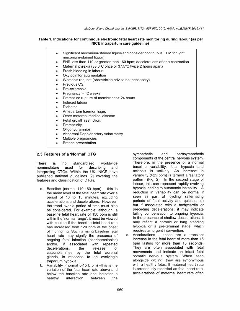
Table 1. Indications for continuous electronic fetal heart rate monitoring during labour (as per NICE intrapartum care guideline)
2.3 Features of a ‘Normal’ CTG There is no standardised worldwide nomenclature used for describing and interpreting CTGs. Within the UK, NICE have published national guidelines [2] covering the features and classification of CTGs.
a. Baseline (normal 110-160 bpm) – this is the mean level of the fetal heart rate over a period of 10 to 15 minutes, excluding accelerations and decelerations. However, the trend over a period of time must also be considered. For example, although, a baseline fetal heart rate of 150 bpm is still within the ‘normal range’, it must be viewed with caution if the baseline fetal heart rate has increased from 120 bpm at the onset of monitoring. Such a rising baseline fetal heart rate may signify the presence of ongoing fetal infection (chorioamnionitis) and/or, if associated with repeated decelerations, the release of catecholamines by the fetal adrenal glands, in response to an evolvingin trapartum hypoxia.
b. Variability (normal 5-15 b pm) –this is the variation of the fetal heart rate above and below the baseline rate and indicates a healthy interaction between the
sympathetic and parasympathetic components of the central nervous system. Therefore, in the presence of a normal baseline variability, fetal hypoxia and acidosis is unlikely. An increase in variability (>25 bpm) is termed a ‘saltatory pattern’ (Fig. 2). In the second stage of labour, this can represent rapidly evolving hypoxia leading to autonomic instability. A reduction in variability can be normal if seen as part of ‘cycling’ (alternating periods of fetal activity and quiescence) but if associated with a tachycardia or preceding decelerations, it may indicate failing compensation to ongoing hypoxia. In the presence of shallow decelerations, it may reflect a chronic or long standing hypoxia or a pre-terminal stage, which requires an urgent intervention.
c. Accelerations – these are a transient increase in the fetal heart of more than 15 bpm lasting for more than 15 seconds. They are often associated with fetal movements and indicate an intact fetal somatic nervous system. When seen alongside cycling, they are synonymous with a healthy fetus. If maternal heart rate is erroneously recorded as fetal heart rate, accelerations of maternal heart rate often
Significant meconium-stained liquor(and consider continuous EFM for light meconium-stained liquor)
FHR less than 110 or greater than 160 bpm; decelerations after a contraction Maternal pyrexia (38.0ºC once or 37.5ºC twice 2 hours apart) Fresh bleeding in labour Oxytocin for augmentation Woman's request (obstetrician advice not necessary). Previous CS. Pre-eclampsia. Pregnancy > 42 weeks. Premature rupture of membranes> 24 hours. Induced labour Diabetes Antepartum haemorrhage. Other maternal medical disease. Fetal growth restriction. Prematurity. Oligohydramnios. Abnormal Doppler artery velocimetry. Multiple pregnancies Breech presentation.
McDonnell and Chandraharan; BJMMR, 7(12): 957-970, 2015; Article no.BJMMR.2015.411
961
last throughout the uterine contraction and are of much higher amplitude.
d. Decelerations – these are defined as a decrease in the fetal heart rate of more than 15 bpm for more than 15 seconds. Each type of deceleration has a different pathophysiological basis and hence their shape, timing and duration vary significantly.
2.4 Types of Decelerations and Their Significance (Table 2)
Variable decelerations occur because of umbilical cord compression. At the onset of a uterine contraction, the soft, thin walled umbilical vein with its low intraluminal pressure is compressed. This causes a slight reduction in fetal blood volume with resultant stimulation of the sympathetic nervous system and an increase in fetal heart rate. This is the first ‘shoulder’ seen on the CTG trace, prior to the drop in the fetal heart rate. The continued uterine contraction causes compression of the umbilical artery leading to an increased systemic vascular resistance and systemic hypertension resulting in a swift drop in the fetal heart rate. This reflex response to systemic hypertension is mediated by stimulation of the baroreceptors. As the contraction eases, this stimulation of the baroreceptors is removed as the umbilical arteries are no longer compressed leading to a quick recovery in the fetal heart rate to its normal baseline. The second shoulder results from the umbilical vein, which is thin-walled and collapsible being compressed slightly longer than the umbilical artery leading to a relative fetal hypotension and resultant sympathetic stimulation. These uncomplicated or ‘typical’ variable decelerations (Fig. 4) can continue for a surprising length of time before fetal hypoxia develops, provided that the fetus continues to show a stable baseline fetal heart rate and reassuring baseline fetal heart rate variability in between these decelerations. However, complicated or ‘atypical’ variable decelerations (Fig. 5) are more suggestive of worsening fetal hypoxic stress and especially when seen with changes in the baseline fetal heart rate. In the presence of changes in baseline variability, it is likely that fetus may be losing its ability to effectively compensate for hypoxic stress. This process of fetal decompensation could occurmuch more rapidly during the second stage of labour compared to the first stage of labour.
This is because with the onset of active maternal pushing there is arapid reduction in maternal oxygenation. In addition, there is a reduction in maternal cardiac output due to reduced venous return secondary to Valsalva Manoeuvre as well as an intense umbilical cord and fetal head compression and reduction in utero-placental circulation secondary to compression of uterine arteries by the fetal head, all of which occur during active maternal pushing. Features of Complicated or ‘Atypical’ decelerations [8]: Deceleration lasting for more than 60
seconds Exaggerated shouldering or the presence
of an ‘overshoot’ Loss of shouldering Loss of variability within the deceleration
(often with associated loss of variability on the baseline)
Slow recovery (a variable deceleration combined with a late deceleration)
Biphasic decelerations However, the use of CTG solely based on pattern recognition without understanding fetal physiology and wider clinical picture may lead to poor neonatal outcomes and may also increase operative interventions. Therefore, clinicians need to always consider fetal compensatory response to ongoing hypoxic or mechanical stresses during labour and determine whether fetal central organs (i.e. brain and the heart) are well oxygenated (i.e. a stable baseline fetal heart rate and a reassuring variability) irrespective of the type of deceleration on the CTG Trace. Features that may be associated with a poor outcome are [2]: Fetal tachycardia >160 bpm Reduced baseline variability <5 bpm for
>90 min Complicated variable decelerations Late decelerations Prolonged decelerations for > 3 min,
especially if reduced variability prior to the deceleration.
There is a greater chance of fetal compromise if the above adverse features are observed. However, despite these ‘adverse’ features, if the fetus continues to have a stable baseline fetal heart rate and reassuring baseline variability,

McDonnell
fetal hypoxia and acidosis is very unlikely. Presence of other risk factors such as reduced fetal physiological reserve, meconium, intrapartum fever and chorioamnionitis may significantly impair fetal compensatory response or cause fetal damage via non-hypoxic pathways (e.g. neurological damage secondary to inflammatory mediators in chorioamnionitis). The following features, when seen in isolation, are unlikely to represent significant fetal compromise: Baseline rate 100-109 bpm Absence of accelerations Early decelerations
Fig. 2. Saltatory pattern with active pushing
McDonnell and Chandraharan; BJMMR, 7(12): 957-970, 2015; Article no.
962
fetal hypoxia and acidosis is very unlikely. Presence of other risk factors such as reduced fetal physiological reserve, meconium, intrapartum fever and chorioamnionitis may significantly impair fetal compensatory response
hypoxic pathways (e.g. neurological damage secondary to inflammatory mediators in chorioamnionitis).
The following features, when seen in isolation, are unlikely to represent significant fetal
Uncomplicated variable decelerations 2.5 Types of Hypoxia The type of intrapartum hypoxia that develops depends very much on its speed of onset. 2.5.1 Acute hypoxia This is evidenced by a single and sudden drop in the baseline rate termed ‘a single prolonged deceleration’. Three major intrapartum ‘accidents’ of labour must be excluded umbilical cord prolapse, placental abruption anduterine rupture and delivery needs to be expedited if this is the case. Iatrogenic causes of
Saltatory pattern with active pushing- Arrow
Fig. 3. Early decelerations
; Article no.BJMMR.2015.411
Uncomplicated variable decelerations
The type of intrapartum hypoxia that develops depends very much on its speed of onset.
and sudden drop in the baseline rate termed ‘a single prolonged deceleration’. Three major intrapartum ‘accidents’ of labour must be excluded – umbilical cord prolapse, placental abruption and
and delivery needs to be expedited if this is the case. Iatrogenic causes of

McDonnell
Fig. 4. Uncom
Fig. 5. Complicated or ‘atypical’
prolonged decelerations include prolonged or frequent uterine contractions secondary to syntocinon or maternal hypotension secondary to epidural analgesia. If such iatrogenic causes are identified, then, depending on the cause, immediate action should be taken to improve utero-placental oxygenation by stopping oxytocin, changes in maternal position or by increasing intravenous fluids instead of performing an unnecessary operative intervention.
In addition, the condition of the fetus at the onset of an acute event must be considered as a fetus with a normal pH (i.e. normal CTG) prior to the onset of deceleration will cope far better than a fetus already demonstrating signs of ongoing intrapartum hypoxia on the CTG prior to the ofthe deceleration.
McDonnell and Chandraharan; BJMMR, 7(12): 957-970, 2015; Article no.
963
Uncomplicated variable decelerations
Complicated or ‘atypical’ variable decelerations
prolonged decelerations include prolonged or frequent uterine contractions secondary to syntocinon or maternal hypotension secondary to epidural analgesia. If such iatrogenic causes are identified, then, depending on the cause, mmediate action should be taken to improve
oxygenation by stopping oxytocin, changes in maternal position or by increasing intravenous fluids instead of performing an unnecessary operative
fetus at the onset of an acute event must be considered as a fetus with a normal pH (i.e. normal CTG) prior to the onset of deceleration will cope far better than a fetus already demonstrating signs of ongoing intrapartum hypoxia on the CTG prior to the of
Once these acute intrapartum events have been excluded, the 3-6-9-12-15 [2,9] followed, provided the CTG trace prior to the onset of prolonged deceleration was normal with reassuring baseline variability and the variability within the first 3 minutes of deceleration is also normal. If these criteria are satisfied, then:
At 6 minutes – 90% of decelerations will recover to its original baseline.
At 9 minutes, 95% of decelerations will have recovered to the original baseline anin the absence of any attempts of recovery, woman should be moved to the theatre at this stage, unless an instrumental delivery is possible in the room. At 12 minutes, delivery should be underway by the quickest and the safest method and in
; Article no.BJMMR.2015.411
Once these acute intrapartum events have been rule can be
followed, provided the CTG trace prior to the onset of prolonged deceleration was normal with reassuring baseline variability and the variability
ithin the first 3 minutes of deceleration is also normal. If these criteria are satisfied, then:
90% of decelerations will recover
At 9 minutes, 95% of decelerations will have recovered to the original baseline and therefore, in the absence of any attempts of recovery, woman should be moved to the theatre at this stage, unless an instrumental delivery is possible
At 12 minutes, delivery should be underway by the quickest and the safest method and in

McDonnell and Chandraharan; BJMMR, 7(12): 957-970, 2015; Article no.BJMMR.2015.411
964
second stage of labour, this would be an operative vaginal delivery or an emergency caesarean section, if such an operative vaginal delivery is not possible or not deemed safe in the operator’s hands. Fetus should be delivered by 15 minutes of the onset of a prolonged deceleration. 2.5.2 Sub-acute hypoxia In this scenario, a fetus spends more time decelerating than at the normal baseline, typically with more than 90seconds of deceleration and only 30 seconds at the baseline. The fetal pH is expected to fall at a rate 0.01 every 2-3 minutes (Fig. 6) [10], which may be even more marked in the second stage of labour because of maternal Valsalva Manoeuvres reducing the pool of fresh oxygenation to the placenta. 2.5.3 Gradually evolving hypoxia A stepwise progression is seen with decelerations followed by loss of accelerations (conservation of energy by the fetus) and an increase in baseline rate (catecholamine
mediated attempt to increase tissue perfusion). This ‘compensated’ state may be followed onset of decompensation characterized by loss of baseline variability (reduced brain perfusion). If no intervention is instituted, the baseline heart rate becomes unstable and eventually would result in a terminal bradycardia and fetal death due to myocardial hypoxia and acidosis. Gradually evolving hypoxia occurs over a period of hours and highlights why any CTG in the second stage of labour must be viewed in context of the entire labour. Contrary to the misconception amongst some clinicians, there is no ‘normal second stage CTG with decelerations’ and one needs to determine whether the fetus is compensating to ongoing hypoxic stress to decide on the need for intervention. Onset of subacute hypoxia during second stage of labour on the background of a normal CTG will be tolerated for longer period than a fetus who has already been exposed to a gradually evolving hypoxia over the preceding hours. In this case, decompensation may occur much earlier due to progressive depletion of fetal reserves prior to the onset of subacute or acute hypoxia during second stage of labour.
Table 2. Types of decelerations and their significance
Incidence Stage of
labour Nadir of deceleration
Physiological basis Action
Early (Fig. 3)
Uncommon Late 1st/ 2
nd stage
Peak of contraction
Head compression Observe once confirmed true early decelerations. Exclude variable or late decelerations, especially if seen in early labour.
Late
8-10% Any More than 20 seconds after peak of contraction
Often associated with uteroplacental insufficiency. Stimulation of chemoreceptors by fetal hypoxia and hypercarbia. As contraction eases, placenta fills with oxygenated blood with slow return of fetal heart to base rate.
If no intervention or secondary test of fetal wellbeing, hypoxia will ensue. In the second stage, this may be of rapid onset.
Variable Common (85-90%)
Any Variable Cord compression See text

McDonnell
2.6 Pitfalls in CEFM
2.6.1 Inadvertent maternal heart rate monitoring
As previously discussed, maternal heart rate (via the maternal iliac vessels) can be mistaken for the fetal heart rate, both if monitoring is performed abdominally or, more rarely, via a fetal scalp electrode. Poor perinatal outcomes associated with erroneous monitoring of the maternal heart rate as fetal heart rate resulted in the release of a ‘Medical Device Alert’ by the Medicines and Healthcare Products Regulatory Agency (MHRA) in 2010 [11]. Due to the issuewe have highlighted in our article, second stage of labour increases the chances of such erroneous monitoring. The expulsive effort of women during the second stage of labour is associated with a maternal tachycardia and hence ‘accelerations’ of the fetheart rate observed with contractions or maternal pushing during second stage must be viewed with caution. One method of trying to avoid this is to place the transducer in the midline rather than laterally during the second stage to avoid picking up audible signals from maternal iliac vessels. Fig. 7 demonstrates the monitoring of maternal and fetal heart rates. In this case, the fetus was monitored using a fetal scalp electrode using fetal ECG (ST-Analyser or STAN).indicates the time at which the transducer was very likely to switch monitoring maternal heart rate erroneously as fetal if there was a prolonged
McDonnell and Chandraharan; BJMMR, 7(12): 957-970, 2015; Article no.
965
Fig. 6. Subacute hypoxia
maternal heart rate
As previously discussed, maternal heart rate (via the maternal iliac vessels) can be mistaken for the fetal heart rate, both if monitoring is performed abdominally or, more rarely, via a fetal
Poor perinatal outcomes associated with erroneous monitoring of the maternal heart rate as fetal heart rate resulted in the release of a ‘Medical Device Alert’ by the Medicines and Healthcare Products Regulatory
. Due to the issues we have highlighted in our article, second stage of labour increases the chances of such
The expulsive effort of women during the second stage of labour is associated with a maternal tachycardia and hence ‘accelerations’ of the fetal heart rate observed with contractions or maternal pushing during second stage must be viewed with caution. One method of trying to avoid this is to place the transducer in the midline rather than laterally during the second stage to avoid picking
ible signals from maternal iliac vessels.
demonstrates the monitoring of maternal and fetal heart rates. In this case, the fetus was monitored using a fetal scalp electrode using
Analyser or STAN). The arrow h the transducer was
very likely to switch monitoring maternal heart rate erroneously as fetal if there was a prolonged
deceleration. This is because signals from a more powerful maternal iliac vessel would be easily picked up by the transducer. In this cathe ST Analyser highlighted ‘ST Events’ secondary to ongoing myocardial hypoxia from to prolonged deceleration that enabled the clinicians to institute timely and appropriate action. In 2012, Nurani et al. [12] accelerations coinciding with uterine contractions were seen in 11% when external monitoring was used compared to 4% when an internal monitoring with a fetal scalp electrode was employed. In addition, the presence of ‘p’ waves on the ECG waveform helped to differentiate between maternal and fetal heart rates as the ‘p’ wave will not be seen if maternal heart rate was erroneously monitored as fetal. Table 3 describes features of Maternal Heart Rate [13]. 2.6.2 Monitoring of twins during second
stage of labour The NICE Multiple Pregnancy guidelineaddresses only antenatal as opposed to intrapartum care. NICE does however advocate continuous monitoring in labour for all multiple pregnancies because of the well documented increased risk of adverse events.feature and overall classification are given in Tables 4 and 5, respectively. As with a singleton pregnancy, the maternal heart rate can be inadvertently recorded but with the added
; Article no.BJMMR.2015.411
deceleration. This is because signals from a more powerful maternal iliac vessel would be easily picked up by the transducer. In this case, the ST Analyser highlighted ‘ST Events’ secondary to ongoing myocardial hypoxia from to prolonged deceleration that enabled the clinicians to institute timely and appropriate
reported that ith uterine contractions
were seen in 11% when external monitoring was used compared to 4% when an internal monitoring with a fetal scalp electrode was employed. In addition, the presence of ‘p’ waves on the ECG waveform helped to differentiate
ernal and fetal heart rates as the ‘p’ wave will not be seen if maternal heart rate was
Table 3 describes features of Maternal Heart
Monitoring of twins during second
Pregnancy guideline [14] as opposed to
NICE does however advocate continuous monitoring in labour for all multiple pregnancies because of the well documented
creased risk of adverse events. Individual d overall classification are given in
As with a singleton pregnancy, the maternal heart rate can be inadvertently recorded but with the added

McDonnell
complication of the same twin being monitored twice. In order to reduce this risk, madvocate internal monitoring using a fetal scalp electrode (FSE), if there are no contraindications (i.e. risk of vertical transmission of infections or fetal bleeding disorders), for the presenting twin with external monitoring for the seconalso good practice to use ultrasound to locate the two separate fetal heart beats at the start of monitoring and at any stage in labour if there are any difficulties in determining the fetal heart rate or a suspicion that the same twin is bemonitored twice.
Monitoring the fetal heart of the second twin following delivery of the first twin can be particularly difficult and inadvertent maternal heart rate monitoring at this stage is not uncommon. Real-time ultrasound is often helpful in this situation to locate the fetal heart rate prior to positioning the transducer.
2.6.3 Monitoring during operative vaginal delivery
It is vital to continue to monitor the fetal heart rate when preparations are made for an operative vaginal delivery as well as during operative vaginal delivery. Clinicians should be aware that application of a ventouse cup on the fetal head may lead to a sudden and sustained parasympathetic stimulation leading to a prolonged deceleration. This deceleration is secondary to a mechanical stimulation and not due to fetal hypoxia. Therefore, if the fetal heart rate variability is maintained during this
Fig. 7. Maternal heart rate shows accelerations whereas the fetal HR shows decelerations
Table 3.
• Lower baseline (usually 60• Repeated • Increased baseline variability
McDonnell and Chandraharan; BJMMR, 7(12): 957-970, 2015; Article no.
966
ame twin being monitored In order to reduce this risk, most units now
advocate internal monitoring using a fetal scalp electrode (FSE), if there are no contraindications (i.e. risk of vertical transmission of infections or fetal bleeding disorders), for the presenting twin with external monitoring for the second twin. It is also good practice to use ultrasound to locate the two separate fetal heart beats at the start of monitoring and at any stage in labour if there are any difficulties in determining the fetal heart rate or a suspicion that the same twin is being
Monitoring the fetal heart of the second twin following delivery of the first twin can be particularly difficult and inadvertent maternal heart rate monitoring at this stage is not
time ultrasound is often helpful is situation to locate the fetal heart rate prior
Monitoring during operative vaginal
It is vital to continue to monitor the fetal heart rate when preparations are made for an operative vaginal delivery as well as during operative vaginal delivery. Clinicians should be aware that application of a ventouse cup on the
sudden and sustained parasympathetic stimulation leading to a prolonged deceleration. This deceleration is secondary to a mechanical stimulation and not due to fetal hypoxia. Therefore, if the fetal heart rate variability is maintained during this
prolonged deceleration and if there was no evidence of ongoing hypoxia on the CTG trace prior to the commencement of the prolonged deceleration, then increasing traction to expedite delivery should be avoided. Such injudicious increase in traction may result in cup detachment which may lead to fetal trauma or the need for a ‘double instrumentation’, which may lead to a further delay in accomplishing delivery. Conversely, if there is absent or reduced baseline variability during a prolonged deceleration, then delivery should be expedited by the safest and fastest route to avoid hypoxic injury.
2.7 Prediction of Fetal Reserve In their intrapartum guidelines, NICE advocate CTG classification into Normal, Suspicious or Pathological [2], dependent upon four individualfeatures. Subsequent management, be it observation, fetal blood sampling (or another secondary test of fetal wellbeing such as ST Analysis) or delivery, is based upon this classification purely based on ‘pattern recognition’. However, as previously discusthe rate and intensity of development of hypoxia in the first stage of labour cannot be compared to the second stage of labour and therefore, clinicians should exercise caution whilst applying arbitrary time limits recommended by NICE Guidelines whilst managing a fetus in second stage of labour.
Maternal heart rate shows accelerations whereas the fetal HR shows decelerations
Table 3. Features of maternal heart rate
Lower baseline (usually 60-100 bpm) Repeated accelerations with contractions Increased baseline variability
; Article no.BJMMR.2015.411
ed deceleration and if there was no evidence of ongoing hypoxia on the CTG trace prior to the commencement of the prolonged deceleration, then increasing traction to expedite delivery should be avoided. Such injudicious
up detachment which may lead to fetal trauma or the need for a ‘double instrumentation’, which may lead to a further delay in accomplishing delivery. Conversely, if there is absent or reduced baseline variability during a prolonged
ery should be expedited by the safest and fastest route to avoid hypoxic
2.7 Prediction of Fetal Reserve
In their intrapartum guidelines, NICE advocate CTG classification into Normal, Suspicious or Pathological [2], dependent upon four individual features. Subsequent management, be it observation, fetal blood sampling (or another secondary test of fetal wellbeing such as ST Analysis) or delivery, is based upon this classification purely based on ‘pattern recognition’. However, as previously discussed, the rate and intensity of development of hypoxia in the first stage of labour cannot be compared to the second stage of labour and therefore, clinicians should exercise caution whilst applying arbitrary time limits recommended by NICE
t managing a fetus in second
Maternal heart rate shows accelerations whereas the fetal HR shows decelerations

McDonnell and Chandraharan; BJMMR, 7(12): 957-970, 2015; Article no.BJMMR.2015.411
967
Table 4. NICE categorisation of individual CTG features
Feature Baseline (bpm) Variability (bpm) Decelerations Accelerations
Reassuring 110-160 ≥5 None Present
Non-reassuring 100-109
161-180
<5 for 40-90 minutes
Typical variable decelerations with ≥50% of contractions, occurring for ≥90 minutes.
Single prolonged deceleration up to 3 min
The absence of accelerations with otherwise normal CTG is of uncertain significance.
Abnormal <100 >180 Sinusoidal pattern ≥ 10minutes
< 5 for 90 minutes Either atypical variable decelerations with over 50% of contractions or late decelerations, both for ≥30 min. Single prolonged deceleration for more than 3 mins
Table 5. NICE: ‘Overall’ CTG classification
Normal All for features are in the ‘reassuring’ category Suspicious One feature is ‘non-reassuring’ and the remainder are reassuring Pathological Two or more features are ‘non-reassuring’ or one or more is ‘abnormal’
Every fetus goes into the second stage of labour with its own individual physiological reserve to withstand hypoxic stress. Factors both in the antenatal and intrapartum period can affect this physiological reserve of the given fetus but it is often impossible to determine the individual physiological reserve and the duration of hypoxia that a fetus is able to withstand with any accuracy. Other factors that affect this ability include the rapidity of onset of hypoxia and other intrapartum risk factors such as use of oxytocin to augment labour, nature of uterine activity and presence of intrauterine infection.
For example, sensitivity for the detection of a severely small for gestational age (SGA fetus) (<2.3rd centile) is only 28% [15] on abdominal palpation and serial symphysio-fundal height measurements are only slightly better with a sensitivity of 48% [16]. Ultrasound for the detection of an SGA fetus has been found to be most beneficial as a screening tool in those women whom already have risk factors for SGA but in a low-risk population, has not shown to be
of clinical benefit. In the high risk population, the sensitivity for detection of an abdominal circumference < 10th centile is between 72.9-94.5% [17].
As our ability to detect fetuses with SGA is poor, having arbitrary time limits for instituting interventions during second stage of labour would lead to delay in interventions for SGA fetuses leading to hypoxic injury. This is because the ability for each individual fetus to mount a successful compensatory response to hypoxic stresses during labour varies with inherent fetal reserve. Other influencing factors may include the presence of intra-amniotic infection, meconium or prematurity.
Therefore, in order to avoid hypoxic-ischaemic fetal injury, clinicians should determine the type of intrapartum hypoxia as well as ongoing fetal compensation (baseline fetal heart rate and variability) on the CTG trace to ensure timely and appropriate interventions.

McDonnell and Chandraharan; BJMMR, 7(12): 957-970, 2015; Article no.BJMMR.2015.411
968
2.8 Role of Additional Tests of Fetal Wellbeing - Fetal ECG (ST-Analyser or STAN) & Fetal Scalp Blood Sampling (FBS) During Second Stage of Labour
Fetal ECG (STAN) is used as an additional test of fetal wellbeing to reduce the false positive rate of CTG. ST-Analyser (STAN) could be commenced when a woman is in ‘passive’ second stage of labour. However, once active pushing commences, ST-Analyser should not be commenced, especially if the CTG trace is not normal as the rate of development of hypoxia could be very rapid leading to insufficient time for the STAN technology to calculate the stable T/QRS ratio. Some obstetricians perform fetal scalp blood sampling (FBS) during second stage of labour prior to operative deliveries, if the CTG is abnormal or ‘pathological’. However, it must be
recognized that FBS is a ‘snapshot’ test and is not useful if hypoxia evolves rapidly during second stage of labour because the values may not represent the actual fetal condition. Recent Cochrane Review has concluded that fetal scalp blood sampling (FBS) does not reduce caesarean section rate as previously believed [18]. In addition, this review also concluded that FBS does not improve longer term neurological outcomes. The use of FBS in clinically practice has been recently questioned due to lack of anatomical and physiological justification as well as scientific evidence of benefit [19]. Therefore, use of FBS during second stage of labour is not recommended and clinicians need to exclude evidence of fetal decompensation by scrutinizing the baseline fetal heart rate and variability. Similarly, a recent systematic review has concluded that fetal scalp lactate was ineffective in improving perinatal outcomes [20].
Table 6. CTG interpretation in active second stage of labour: ‘St George’s management
algorithm’
CTG changes Wider clinical picture Intervention Normal None Continue observation Decelerations / increase in baseline fetal heart rate present
None Continue Observation: Intervene if changes in variability or saltatory pattern
Decelerations / increase in baseline fetal heart rate present
Meconium or evidence of clinical chorioamnionitis
Stop oxytocin infusion to avoid meconium aspiration syndrome. Expedite Delivery if CTG changes persist after stopping oxytocin
Subacute hypoxia or saltatory pattern
With or without additional risk factors
Stop oxytocin and maternal active pushing to improve fetal oxygenation Expedite Delivery if CTG changes persist after above interventions
Acute hypoxia With or without additional risk factors
Stop oxytocin to improve fetal oxygenation and stop active maternal pushing. In the absence of intrapartum accidents, if the prolonged deceleration persists for more than 6 minutes despite above measures, perform an urgent operative vaginal delivery or a ‘Crash Caesarean section’ if indicated.
Remember: A growth restricted fetus would have a lower physiological reserve, fetal infection lowers the threshold of fetal neurological injury secondary to hypoxia, deep prolonged decelerations and baseline
tachycardia are associated with increased incidence of fetal meconium aspiration syndrome

McDonnell and Chandraharan; BJMMR, 7(12): 957-970, 2015; Article no.BJMMR.2015.411
969
3. CONCLUSION In the UK, approximately 500 intrapartum stillbirths occur every year and unfortunately many of these occur as a result of errors in CTG interpretation. Obstetricians and midwives owe a duty of care to women to ensure that they understand the physiology behind the changes seen on a CTG trace both during first and second stages of labour. Better understanding of fetal heart rate changes during second stage of labour may help avoid unnecessary interventions secondary to misinterpretation of CTG traces with resultant fetal and maternal morbidity that can be associated with operative vaginal delivery and emergency caesarean section. Equally, clinicians need to avoid delay in action. Failure to recognise rapidly evolving hypoxic stress during second stage of labour with resultant fetal decompensation as well as failure to recognize erroneous recording of maternal heart rate as fetal during second stage of labour may lead to poor perinatal outcomes.
CTG interpretation during second stage of labour should focus on fetal response to rapidly evolving hypoxic stress. A stable baseline fetal heart rate and a reassuring variability would indicate the oxygenation of central organs (brain and the myocardium) and the type of hypoxia (acute or subacute) would help guide urgency of interventions required to improve outcomes (Table 6 above).
CONSENT It is not applicable.
ETHICAL APPROVAL It is not applicable.
COMPETING INTERESTS Authors have declared that no competing interests exist.
REFERENCES 1. Aldrich CJ, D’Antonia D, Spencer JAD,
Wyatt JS, Peebles DM, Delpy DT, Reynolds EOR. The effect of maternal pushing on fetal cerebral oxygenation and blood volume during the second stage of labour. BJOG. 1995;102:448-453.
2. NICE Intrapartum Care: Care of healthy women and their babies during childbirth. Guideline Number. 2007;55.
3. Westgate JA, Wibbens B, BennetL, Wassink G, Parer J, Gunn AJ. The intrapartum deceleration in center stage: A physiologic approach to the interpretation of fetal heart rate changes in labor. Am J Obstet Gynecol. 2007;197:236.e1-236.e11.
4. Alfirevic Z, Devane D, Gyte GM, et al. Continuous tocography (CTG) as a form of electronic fetal monitoring (EFM) for fetal assessment during labor. Cochrane Database Syst Rev. 2006;3. Updated May 2013.
5. Beard RW, Filshie GM, Knight CA, Roberts GM. The significance of the changes in the continuous fetal heart rate in the first stage of labour. J Obstet Gynecol Br C'wlth. 1971;78:865-81.
6. Chauhan SP, Klauser CK, Woodring TC, Sanderson M, Magann EF, Morrison JC. Intrapartum nonreassuring fetal heart rate tracing and prediction of adverse outcomes: Interobserver variability. Am J Obstet Gynecol. 2008;199:623.e1-623.e5.
7. Donker D, Van Geijn HP, Hasman A. Interobserver variation in the assessment of fetal heart rate recordings. Eur. J. Obstet. Gynecol. Reprod. Biol. 1993;52: 21-28.
8. Clinical Effectiveness Support Unit. The use of electronic fetal monitoring. The use and interpretation of cardiotocography in intrapartum fetal surveillance. London, Royal College of Obstetricians and Gynaecologists; 2001.
9. Chandraharan E, Arulkumaran S. Prevention of birth asphyxia: Responding appropriately to cardiotocograph (CTG) traces. Best Practice & Research Clinical Obstetrics and Gynaecology. 2007;21(4): 609-624.
10. Chandraharan E. Fetal compromise: Diagnosis & management. In: Obstetric & Intrapartum Emergencies: A Practical Guide to Management. Cambridge University Press; 2012.
11. Medical Device Alert Ref MDA/2010/054. Device–Fetal monitor/ cardiotocograph. Medicines and Healthcare Products Regulatory Agency; 2010.
12. Nurani R, Chandraharan E, Lowe V, Ugwumadu A, Arulkumaran S. Misidentification of maternal heart rate as fetal on cardiotocography during the

McDonnell and Chandraharan; BJMMR, 7(12): 957-970, 2015; Article no.BJMMR.2015.411
970
second stage of labour: The role of the fetal electrocardiograph. Acta Obstet Gynecol Scand. 2012;91.
13. Chandraharan E, Arulkumaran S. Electronic fetal heart rate monitoring in current and future practice. J Obstet Gynecol India. 2008;58(2):121-130.
14. NICE/RCOG. Multiple Pregnancy: The management of twins and triplet pregnancies in the antenatal period; 2011.
15. Bais JM, Eskes M, Pel M, Bonsel GJ, Bleker OP. Effectiveness of detection of intrauterine growth retardation by abdominal palpation as screening test in a low risk population: An observational study. Eur J Obstet Gynecol Reprod Biol. 2004;116:164–9.
16. Gardosi J, Francis A. Controlled trial of fundal height measurement plotted on customised antenatal growth charts. BJOG. 1999;106:309–17.
17. RCOG Greentop Guideline No 31. February 2013. The Investigation and Management of the Small-for-Gestational Age fetus. RCOG Press; 2013.
18. Alfirevic Z, Devane D, Gyte GML. Continuous cardiotocography (CTG) as a form of electronic fetal monitoring (EFM) for fetal assessment during labour. Cochrane Database Syst Rev; 2013.
19. Chandraharan E. Fetal scalp blood sampling during labour: Is it a useful diagnostic test or a historical test that no longer has a place in modern clinical obstetrics? BJOG. 2014;121(9):1056-60.
20. East CE, Leader LR, Sheehan P, Henshall NE, Colditz PB. Intrapartum fetal scalp lactate sampling for fetal assessment in the presence of a non-reassuring fetal heart rate trace. Cochrane Database Syst Rev. 2010:CD006174.
© 2015 McDonnell and Chandraharan; This is an Open Access article distributed under the terms of the Creative Commons Attribution License (http://creativecommons.org/licenses/by/4.0), which permits unrestricted use, distribution, and reproduction in any medium, provided the original work is properly cited.
Peer-review history: The peer review history for this paper can be accessed here:
http://www.sciencedomain.org/review-history.php?iid=952&id=12&aid=8740