Fetal cardiac arrhythmias: Diagnosis and … CARDIAC ARRHYTHMIAS: DIAGNOSIS AND MANAGEMENT Lindsey...
38
FETAL CARDIAC ARRHYTHMIAS: DIAGNOSIS AND MANAGEMENT Lindsey Malloy-Walton DO MPH Assistant Professor, Pediatric Electrophysiology Ward Family Heart Center Children’s Mercy Hospital
-
Upload
nguyendung -
Category
Documents
-
view
223 -
download
2
Transcript of Fetal cardiac arrhythmias: Diagnosis and … CARDIAC ARRHYTHMIAS: DIAGNOSIS AND MANAGEMENT Lindsey...

FETAL CARDIAC ARRHYTHMIAS:DIAGNOSIS AND MANAGEMENT
Lindsey Malloy-Walton DO MPH
Assistant Professor, Pediatric Electrophysiology
Ward Family Heart Center
Children’s Mercy Hospital

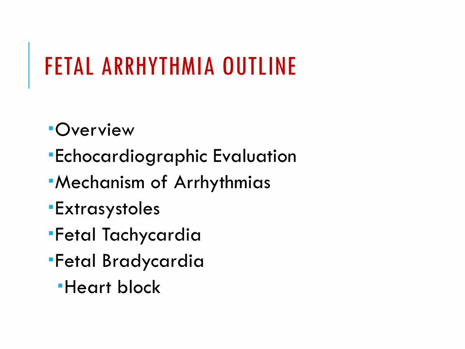
FETAL ARRHYTHMIA OUTLINE
Overview
Echocardiographic Evaluation
Mechanism of Arrhythmias
Extrasystoles
Fetal Tachycardia
Fetal Bradycardia
Heart block

FETAL ARRHYTHMIAS OVERVIEW
• 1-3% of pregnancies
• 10-20% of cardiac referrals
• Challenges with fetal EKG
•Low p wave amplitudes
•Signal acquisition from 27-34 weeks
•Early recognition is important
•M-mode and Doppler Techniques

FETAL ARRHYTHMIA OUTLINE
Overview
Echocardiographic Evaluation
Mechanism of Arrhythmias
Extrasystoles
Fetal Tachycardia
Fetal Bradycardia
Heart block

ECHOCARDIOGRAPHIC ANALYSIS OF FETAL CARDIAC RHYTHM : M-MODE ECHO

ECHOCARDIOGRAPHIC ANALYSIS OF FETAL CARDIAC RHYTHM: PULSED DOPPLER
FETAL ARRHYTHMIA OUTLINE
Overview
Echocardiographic Evaluation
Mechanism of Arrhythmias
Extrasystoles
Fetal Tachycardia
Fetal Bradycardia
Heart block

FETAL ARRHYTHMIA MECHANISMS
Extrasystoles
Atrial*, junctional, ventricular
Tachycardia
Sinus Tachycardia
Supraventricular tachycardia
Reentrant
Atrioventricular (AV) reentrant tachycardia *
Atrial flutter *
AV nodal reentrant tachycardia
Paroxysmal junctional reciprocating tachycardia (PJRT)
Ectopic
Ectopic Atrial tachycardia
Junctional ectopic tachycardia
Ventricular
Ventricular Tachycardia
Bradycardia
Sinus Bradycardia
Sinoatrial block
Atrioventricular * block

FETAL ARRHYTHMIA OUTLINE
Overview
Echocardiographic Evaluation
Mechanism of Arrhythmias
Extrasystoles
Fetal Tachycardia
Fetal Bradycardia
Heart block

EXTRASYSTOLES
•Atrial extrasystoles
•Ventricular extrasystoles

PACS: CONDUCTED AND NON-CONDUCTED

VENTRICULAR ECTOPY: BIGEMINY

FETAL ARRHYTHMIA OUTLINE
Overview
Echocardiographic Evaluation
Mechanism of Arrhythmias
Extrasystoles
Fetal Tachycardia
Fetal Bradycardia
Heart block

FETAL TACHYCARDIA
HR> 160 bpm
Sinus (160-200 bpm)
Pathologic mechanisms (180-280 bpm)
70-90% AV reentry tachycardia
Evaluate for congenital heart disease


SINUS TACHYCARDIA
Fetal distress
Hypoxemia, anemia, infection
Maternal use of medications
Fetal or maternal thyrotoxicosis
Maternal electrolyte abnormalities
Maternal fever

PATHOLOGIC FETAL TACHYCARDIA:AV 1:1
Short VA tachycardias
AV reentrant tachycardia
Preexcitation about 30% of the time
AVN reentrant tachycardia, typical
Junctional ectopic tachycardia (JET)
Long VA tachycardias
Ectopic atrial tachycardia (EAT)
Paroxysmal junctional reciprocating tachycardia (PJRT)

SHORT VA TACHYCARDIA: ACCESSORY PATHWAY
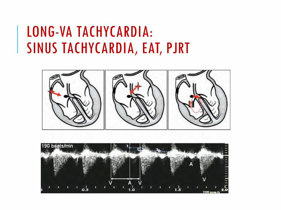
LONG-VA TACHYCARDIA: SINUS TACHYCARDIA, EAT, PJRT

PATHOLOGIC FETAL TACHYCARDIA:AV>1:1
Atrial Flutter
30% of cases of fetal tachycardia
More As than Vs
Atrial rate 300-500 bpm
Regular
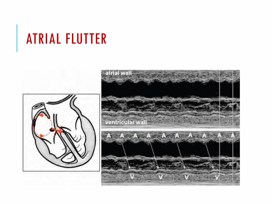
ATRIAL FLUTTER

FETAL TACHYCARDIA: VENTRICULAR AV<1:1
Relatively rare
HR 180-300 bpm
AV dissociation, can be 1:1
Usually automatic, non-sustained
Rare underlying etiology: Long QT, LV non-compaction, rhabdomyoma, fibroma, LV aneurysm

FETAL TACHYCARDIA: VENTRICULAR
Long QT syndrome
Possible cause of intrauterine death
High suspicion if:Constant bradycardia(110-120 bpm), low HR variability
2nd degree AV block (functional)
Torsade des pointes
Diagnosis: Fetal magnetocardiography

Fetal Arrhythmia Monitoring Guideline: ectopy & tachycardia
ASL, DRAFT, 6.2.2014
Ectopy or tachycardia noted on OB scan Refer for fetal echocardiogram If ectopy noted on fetal echocardiogram:
Follow- up with OB weekly for FHR checks and
possible fetal echo in 4 weeks
If not ectopy noted, to see
OB in 1 week for FHR check
If fetal tachycardia noted on fetal echo
- If FHR > 200 bpm or < 100 bpm, call OB
-OB to admit for observation for 4-6 hrs
-Obtain maternal baseline labs of a renal panel,
calcium, phosphorus, magnesium & ECG*
-May need US to check for hydrops and/or fetal
echo to assess function/AVVR
*If admitted recently for previous observation,
no need to repeat lab work/ECG again
Please see fetal tachycardia treatment guideline
- will include involvement of pediatric cardiology,
pediatric EP, MFM and possibly adult EP
If fetal tachycardia not frequent/<50%
of monitored time, then discharge w/o
treatment with close follow-up.
FHR with OB twice weekly
Fetal echo every other week for AVVR &
hydrops check
If fetal tachycardia noted > 50% of the monitoring time,
maternal admission for treatment

FETAL TACHYCARDIA: TREATMENT
In the final stages of pregnancy: Brief therapeutic attempt, if unsuccessful, delivery (preferably with CS), direct neonatal therapy
Fetuses <34 weeks with sustained tachycardia need medical therapy to avoid complications of prematurity

ASL, DRAFT, 6.2.2014
Fetal tachycardia treatment guideline:
***AVOID PRETERM DELIVERY UNLESS IN
DIRE SITUATION***
***All treatment requires***:
-Daily maternal ECGs
-Inpatient monitoring until fetal rhythm is <50% SVT
Outpatient monitoring with:
-Fetal echo once weekly at first, then every
other week until delivery
-Discontinue anti-arrhythmic therapy at
37 weeks gestation if heart rate control achieved
Without hydrops:SVT:
-First line:
-Digoxin IV load (1200-1500 micrograms/day divided every 8hours) for goal maternal Digoxin level 1.5 – 2
-Then switch to oral Digoxin (375- 750 micrograms/day divided every 12 hours); level s 1.5-2
- Flecainide can be considered first line (100-300 mg/day divided every 8 hours orally); levels 0.2- 1
-Second line (in addition to the above):
-Amiodarone oral load (1800-2400 mg/d divided every 6 hrs for 48 hrs)
-Then switch to maintenance dose of 200-600 mg/day orally
Atrial Flutter:
- Sotalol (160-480 mg/day divided every 8 hours orally)
With hydrops:SVT:
-First line consider direct intramuscular fetal tx (Digoxin IM dose
88 micrograms/kg every 12hrs, repeat 2 times) + transplacental tx
Ventricular tachycardia:
-Maternal IV Magnesium (loading dose 2-6 g IV over 20 min, followed by
1-2 g/hr for < 48 hours; can treat again if maternal Magnesium level
< 6 mEq/L)
After Magnesium, consider one of the following with unclear QTc interval:
-Lidocaine load 1-1.5 mg/kg IV followed by 1-4 mg/min infusion (esp if hydrops_
-Propranolol 60-320 mg/day divided every 6 hrs oral
-Mexiletine 600-900 mg/day divided every 8 hrs oral
After Magnesium, if normal QTc interval (DO NOT give if LQTS suspected or
confirmed):
-Sotalol
-Flecainide
-Amiodarone (for short term)

POSTNATAL OUTCOME OF FETAL TACHYCARDIA
2/3 of newborns with fetal tachycardia present with recurrence postnatally
Predicting factors: Presence of hydrops, lack of response to intrauterine therapy and female sex
Most patients free of tachycardia after first 6 months

FETAL ARRHYTHMIA OUTLINE
Overview
Echocardiographic Evaluation
Mechanism of Arrhythmias
Extrasystoles
Fetal Tachycardia
Fetal Bradycardia
Heart block

FETAL BRADYCARDIA
Heart rate <100 bpm
•Sinus bradycardia
•Atrial or junctional bigeminy with non-conducted extrasystoles
•AV block (high grade or complete)

SINUS BRADYCARDIA
Transient (vagal, e.g. from transducer pressure)
Persistent
Fetal distress
Maternal hypothermia
Sinus node disease
Primary (genetic etiology)
In the context of heterotaxy syndrome (left atrial isomerism / polysplenia syndrome)
Long QT syndrome

PERSISTENT BRADYCARDIA: DIFFERENTIAL DIAGNOSIS
Sinus bradycardia
Atrial bigeminy

ISOLATED CONGENITAL AV BLOCK
1:20,000 births
Age of diagnosis: 18-24 weeks
Usually autoimmune mediated
Maternal collagen vascular disease (SLE, Sjögren’s)
Transplacental passage of Anti-Ro (SSA), anti-La (SSB) antibodies
Possibility of AV block in the presence of maternal antibodies: 2-5%
Possibility of appearance in subsequent pregnancies: 15-20%
Occasionally progressive
Survival: 75% in isolated Complete Congenital AV block (CCAVB)
Indices of poor prognosis: Ventricular rate<55 bpm, endocardial fibroelastosis, myocardial dysfunction, hydrops fetalis

THIRD DEGREE AV BLOCK

CONGENITAL AV BLOCK IN THE CONTEXT OF CONGENITAL HEART DISEASE (CHD)
AV discordance (L-TGA, isolated ventricular discordance)
Heterotaxy syndrome (Left atrial isomerism)
LV non-compaction

CONGENITAL AV BLOCK IN THE CONTEXT OF CHD: PROGNOSIS
Much worse than isolated CCAVB
29-40% mortality
Poor prognosis
Hydropic fetus with CCAVB and CHD
Heart rate <55 bpm
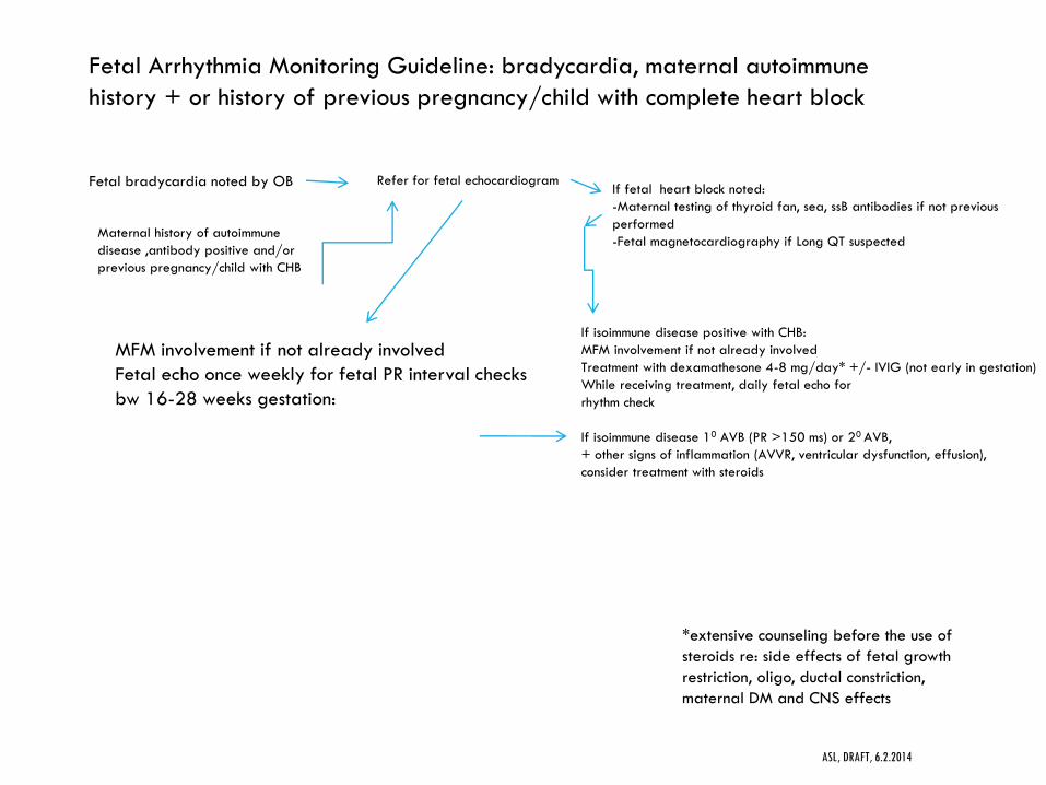
ASL, DRAFT, 6.2.2014
Fetal Arrhythmia Monitoring Guideline: bradycardia, maternal autoimmune
history + or history of previous pregnancy/child with complete heart block
Fetal bradycardia noted by OB Refer for fetal echocardiogram
Maternal history of autoimmune
disease ,antibody positive and/or
previous pregnancy/child with CHB
If fetal heart block noted:
-Maternal testing of thyroid fan, sea, ssB antibodies if not previous
performed
-Fetal magnetocardiography if Long QT suspected
MFM involvement if not already involved
Fetal echo once weekly for fetal PR interval checks
bw 16-28 weeks gestation:
If isoimmune disease positive with CHB:
MFM involvement if not already involved
Treatment with dexamathesone 4-8 mg/day* +/- IVIG (not early in gestation)
While receiving treatment, daily fetal echo for
rhythm check
If isoimmune disease 10 AVB (PR >150 ms) or 20 AVB,
+ other signs of inflammation (AVVR, ventricular dysfunction, effusion),
consider treatment with steroids
*extensive counseling before the use of
steroids re: side effects of fetal growth
restriction, oligo, ductal constriction,
maternal DM and CNS effects

CONCLUSIONS
Fetal arrhythmia can be diagnosed with high degree of accuracy with fetal echocardiographic methods
Treatment, depending on etiology, severity, can be delivered either transplacentally or intraumbilically, or directly after emergent delivery

REFERENCES
Hornberger, L. Echocardiographic assessment of fetal arrhythmias. HEART. BMJ. 2007;93 (11): 1331-1333.
Strasburger J et al. Overview of fetal arrhythmias. Curr Opin Pediatric. 2008 522-531.
ASL, draft, 6.2.2014
Moss and Adams Heart Disease in Infants, Children, and Adolescents Including the Fetus and Young Adult. Volume I. 7th Edition.