FERNANDEZ-MORAN - pnas.org · 446 PHYSICS: H. FERNANDEZ-MORAN PROC. N. A. S....
7
VOL. 53, 1965 PHYSICS: H. FERNANDEZ-MORAN 445 16Shechter, E., J. P. Carver, and E. R. Blout, these PROCEEDINGS, 51, 1029 (1964). 17 Urnes, P., private communication. An alternate graphical solution of equation (10) is to plot [m'] (X2/X1"2 - 1) against (X2/X1932 - 1)/(X2/X222 - 1), which directly yields Am5 as the slope and A193 as the intercept. 18 Rosenfeld, L., Z. Physik, 52, 161 (1928). 19 Iizuka, E., and J. T. Yang, Biochemistry, 3, 1519 (1964). 20 Katzin, L. I., and E. Gulyas, J. Am. Chem. Soc., 86, 1655 (1964). 21 Yang, J. T., in Polyamino Acids, Polypeptides, and Proteins, ed. M. A. Stahmann (Madison, Wisconsin: University of Wisconsin Press, 1962), p. 225. 22 Cassim, J., and E. W. Taylor, Abstracts, 5th Annual Meeting, Biophysical Society, Chicago, 1964, p. TC5. 23 Doty, P., A. M. Holtzer, J. H. Bradbury, and E. R. Blout, J. Am. Chem. Soc., 76, 4493 (195-1). 24 Yang, J. T., and T. Samejima, J. Biol. Chem., 238, 3262 (1963). 25 Yang, J. T., and W. J. McCabe, to be published. 26Yang, J. T., Federation Proc., 21, 406 (1962). 27 Doty, P., and R. D. Lundberg, these PROCEEDINGS, 43, 213 (1957). 28 Yang, J. T., Tetrahedron, 13, 143 (1961). 29Blout, E. R., presented orally at the 6th International Congress of Biochemistry, New York, 1964. 30 Sogami, M., W. J. Leonard, Jr., and J. F. Foster, Arch. Biochem. Biophys., 100, 260 (1963). ELECTRON MICROSCOPY WITH HIGH-FIELD SUPERCONDUCTING SOLENOID LENSES* BY HUMBERTO FEIIANDNDEZ-MORAN DEPARTMENT OF BIOPHYSICS, UNIVERSITY OF CHICAGO Communicated by W. Bloom, December 21, 1964 The resolving power of the electron microscope has extended the range of direct visualization to structural details of the order of a few angstroms. This corresponds to the size of small molecules and to the atomic spacing in crystalline lattices.'3' 9 However, although the wavelength of electrons in standard microscopes is 100,000 times shorter than the wavelength of light, the best electromagnetic and electro- static lenses available have usable apertures limited, by aberrations, to semiangles of the order of l/loo radian as compared with the numerical apertures of 1.5 of the best light microscope lenses. Considering the numerous complex instrumental and preparative factors involved,1-3' 9 the major steps which have to be taken for attainment of the ultimate theoretical resolution are correction of lens aberrations (mainly spherical and chromatic aberrations), stabilization of the lens excitation current, and accelerating voltage. Thus, the degree of stability required for very high resolving powers (in the range of 4 A) is of the order of 1 to 2 parts per million, since the focal length of a magnetic lens is dependent on the electron energy as well as on the lens excitation current. In addition, if the present limitations of the strength and configuration of the axially symmetrical field formed by iron pole pieces could be overcome, "stronger lenses" of shorter focal length could be designed with correspondingly reduced aberrations. With the introduction and availability of new high-field superconducting sole- noi.ds of alloys of nipobjqum-irconium and niobium-tin,4-7 it is now possible to obtain
Transcript of FERNANDEZ-MORAN - pnas.org · 446 PHYSICS: H. FERNANDEZ-MORAN PROC. N. A. S....
VOL. 53, 1965 PHYSICS: H. FERNANDEZ-MORAN 445
16Shechter, E., J. P. Carver, and E. R. Blout, these PROCEEDINGS, 51, 1029 (1964).17 Urnes, P., private communication. An alternate graphical solution of equation (10) is to plot
[m'] (X2/X1"2 - 1) against (X2/X1932 - 1)/(X2/X222- 1), which directly yields Am5 as the slopeand A193 as the intercept.
18 Rosenfeld, L., Z. Physik, 52, 161 (1928).19 Iizuka, E., and J. T. Yang, Biochemistry, 3, 1519 (1964).20 Katzin, L. I., and E. Gulyas, J. Am. Chem. Soc., 86, 1655 (1964).21 Yang, J. T., in Polyamino Acids, Polypeptides, and Proteins, ed. M. A. Stahmann (Madison,
Wisconsin: University of Wisconsin Press, 1962), p. 225.22 Cassim, J., and E. W. Taylor, Abstracts, 5th Annual Meeting, Biophysical Society, Chicago,
1964, p. TC5.23 Doty, P., A. M. Holtzer, J. H. Bradbury, and E. R. Blout, J. Am. Chem. Soc., 76, 4493 (195-1).24 Yang, J. T., and T. Samejima, J. Biol. Chem., 238, 3262 (1963).25 Yang, J. T., and W. J. McCabe, to be published.26Yang, J. T., Federation Proc., 21, 406 (1962).27 Doty, P., and R. D. Lundberg, these PROCEEDINGS, 43, 213 (1957).28 Yang, J. T., Tetrahedron, 13, 143 (1961).29Blout, E. R., presented orally at the 6th International Congress of Biochemistry, New York,
1964.30 Sogami, M., W. J. Leonard, Jr., and J. F. Foster, Arch. Biochem. Biophys., 100, 260 (1963).
ELECTRON MICROSCOPY WITH HIGH-FIELDSUPERCONDUCTING SOLENOID LENSES*
BY HUMBERTO FEIIANDNDEZ-MORAN
DEPARTMENT OF BIOPHYSICS, UNIVERSITY OF CHICAGO
Communicated by W. Bloom, December 21, 1964
The resolving power of the electron microscope has extended the range of directvisualization to structural details of the order of a few angstroms. This correspondsto the size of small molecules and to the atomic spacing in crystalline lattices.'3' 9However, although the wavelength of electrons in standard microscopes is 100,000times shorter than the wavelength of light, the best electromagnetic and electro-static lenses available have usable apertures limited, by aberrations, to semianglesof the order of l/loo radian as compared with the numerical apertures of 1.5 of thebest light microscope lenses. Considering the numerous complex instrumental andpreparative factors involved,1-3' 9 the major steps which have to be taken forattainment of the ultimate theoretical resolution are correction of lens aberrations(mainly spherical and chromatic aberrations), stabilization of the lens excitationcurrent, and accelerating voltage. Thus, the degree of stability required forvery high resolving powers (in the range of 4 A) is of the order of 1 to 2 parts permillion, since the focal length of a magnetic lens is dependent on the electron energyas well as on the lens excitation current. In addition, if the present limitations ofthe strength and configuration of the axially symmetrical field formed by iron polepieces could be overcome, "stronger lenses" of shorter focal length could be designedwith correspondingly reduced aberrations.With the introduction and availability of new high-field superconducting sole-
noi.ds of alloys of nipobjqum-irconium and niobium-tin,4-7 it is now possible to obtain

446 PHYSICS: H. FERNANDEZ-MORAN PROC. N. A. S.
magnetic fields in excess of 60 kilogauss over relatively large volumes when operatingat liquid helium temperatures, without the continual expenditure of vast amounts ofelectrical power. It has been demonstrated that operation of a superconductingsolenoid, short-circuited, or in "the persistent current mode," yields large uniformmagnetic fields which are highly homogeneous to better than one part in 106 to 107,and are highly stable and noise-free under appropriately controlled conditions.Based on previous work in low-temperature electron microscopy,8-'0 the author
has pointed out the unique advantages to be derived from the use of superconductingelectromagnetic lenses operating at liquid helium temperatures, with regard to bothinstrumentation and specimen preservation. As part of a comprehensive programunder way in the Department of Biophysics at the University of Chicago, prelimi-nary experiments have been successfully carried out with a simple electron micro-scope which can be used for transmission electron microscopy and electron diffrac-tion, using high-field superconducting solenoid lenses in an open-air-core, liquid-helium Dewar, preferably operating in the persistent current mode. In a series ofcontrolled, reproducible experiments, electron microscopic images have been re-corded of test specimens with accelerating potentials of 4-8 kY, using a niobium-zirconium solenoid without pole pieces, operating at 32.2 kilogauss in the persistentcurrent mode. These preliminary experiments have demonstrated over a periodof 4-8 hr of continuous operation the exceptional stability of the images, and alsotheir relatively high quality at magnifications of 50 to 100 X. The results ob-tained with this experimental approach, as well as some interesting observationsbearing on imaging phenomena with superconducting solenoids, are described inthis report.
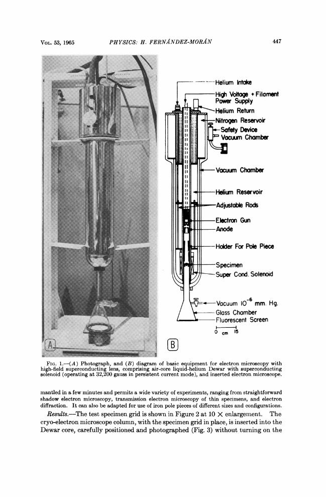
Experimental.-As shown in Figure 1A and B, the system consists of a vertical-air-core Dewar,provided by Westinghouse Cryogenics Systems Division. This Dewar is a nitrogen-shielded,stainless steel vessel consisting of six concentric cylinders providing the required cryogenic en-vironment, and with an inner vessel containing 6-7 liters of liquid helium in which a superconduct-ing solenoid is immersed. An important feature of this Dewar is that it has an open air core of1.5 inches in diameter for ready access of specimens at room temperature. The superconductingmagnet coil used for these experiments is a niobium-zirconium solenoid, which can be operatedin the persistent current mode at a field of 32,200 gauss; rated field uniformity is 6% in a 1/2 inchdiameter sphere; rated current is 15.3 amp; working volume: 2.000 inch diameter X 2.427 inchlong cylinder. Auxiliary equipment provided by Westinghouse Cryogenics Systems Division in-cludes a transistorized power supply, model 503, a gaussmeter, liquid helium level gauge, andaccessories.The electron microscope (Fig. 1B) is a simple, miniaturized instrument consisting of a stainless
steel, nonmagnetic tube with suitably scaled electron gun (of the re-entry cathode shield type)which can be used with standard or pointed filaments.9 The filament was operated at 2.3 volts,3-amp heating current with a variable bias of 180-200 volts. The high voltage in these initialexperiments was 4-8 kV, which can be highly stabilized by using a special motor-generator setwith Transistat type TRA-11, Westinghouse solid-state regulator. This system is sealed andevacuated to a vacuum of 10-6 to 10-6 mm Hg. The test specimen, a 200-mesh copper grid (holediameter: 136 A) can be placed at a convenient position within the field of the superconductingsolenoid and can be moved during the experiment. The image is focused on the fluorescent screenand photographed from the outside of the glass chamber by using Polaroid film (types 42 and 47),35-mm Plus-X or Royal-X Pan Kodak film, and also movie film (16-mm Royal-X Pan). As shownin Figures 3 and 4, photography from the outside results in a characteristic distorted perspectiveof the original image. Adequate provisions were taken for ensuring a high degree of mechanicalstability and for shielding from magnetic and electrical perturbations.The system is actually a very flexible electron optical bench which can be taken out and dis-
VOL. 53, 1965 PHYSICS: H. FERNANDEZ-MORAN 447
~.
----Helium Intake
High Voltage + FilamentPower SupplyHelium Return
-iNitroe Reservoir
Safety Device!tt1ffi ' 5 | J l Vwm Chamber
- Vacuum Chamber
Vacuum Chamervi
Adjustable Rods
Electron GunAroode
Holder For Pole Piece
Specimen-Super Cond. Solenoid
-6-Vacuum 10 mm. Hg.
Glass ChamberFluorescent Screen0 CM 15
A'-_:__ .C-_~~~~~~~~~~~~~~~........| \..../
FIG. l.-(A) Photograph, and (B) diagram of basic equipment for electron microscopy withhigh-field superconducting lens, comprising air-core liquid-helium Dewar with superconductingsolenoid (operating at 32,200 gauss in persistent current mode), and inserted electron microscope.
mantled in a few minutes and permits a wide variety of experiments, ranging from straightforwardshadow electron microscopy, transmission electron microscopy of thin specimens, and electrondiffraction. It can also be adapted for use of iron pole pieces of different sizes and configurations.
Results.--The test specimen grid is shown in Figure 2 at 10 X enlargement. Thecryo-electron microscope column, with the specimen grid in place, is inserted into theDewar core, carefully positioned and photographed (Fig. 3) without turning on the

448 PHYSICS: H. FERNANDEZ-MORAN PROC. N. A. S.
FIG. 2.-Micrograph of copper specimen grid (200-mesh) at 10 X magnification.FIG. 3.-Same grid photographed externally from fluorescent screen in oblique position within the
cryo-electron microscope.FIG. 4.-Electron micrograph of specimen grid recorded from fluorescent screen with high-
field superconducting lens (32,200 gauss in persistent current mode); 4-kv accelerating potential.Original electron optical magnification: 100 X
field. The solenoid is gradually energized manually according to standard proce-dure, and a remarkable series of changes in the image occurs. The image starts tospiral and is successively enlarged (about 5 10 X initially), and comes into focusfleetingly. As the field continues to build up, the image of the specimen grid is fur-ther enlarged, with progressive alternation of focusing and spiraling. One gains theimpression that the alternating succession of enlargement and spiraling seems totake place by "steps"~or "jumps." NIovie film frames were recorded of this inter-esting phenomenon, which differs from the usual spiraling that occurs during focus-ing of images in the standard electromagnetic lenses. The nature of these phenom-ena is being further investigated. They are subject, of course, to the rate of ener-gizing of the coil current and to the field strength parameters At any time duringthe process, the coil can be switched into the persistent current mode by short-circuiting the solenoid, and the desired magnification at the lower magnetic fields isobtained. There is an actual focusing control of the superconducting solenoid, es-sentially similar to that of standard lenses. Moreover, there is a possibility of ad-justing the focus by moving the column and the specimen within the air core of theDewar.
In two of our experiments, the field was driven to the maximum of 32,200 gaussand then held in the persistent current mode for 4-8 hr. Under these conditions,
VOL. 53, 1965 PHYSICS: H. FERNANDEZ-MORAN 449
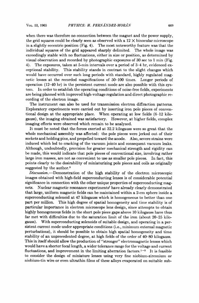
when there was therefore no connection between the magnet and the power supply,the grid squares could be clearly seen as observed with a 12 X binocular microscopein a slightly eccentric position (Fig. 4). The most noteworthy feature was that theindividual squares of the grid appeared sharply delimited. The whole image wasexceedingly stable with no fluctuations, either in size or position, as determined byvisual observation and recorded by photographic exposures of 30 sec to 1 min (Fig.4). The exposures, taken at 5-min intervals over a period of 3-4 hr, evidenced ex-ceptional stability. This stability stands in contrast to the slight changes whichwould have occurred over such long periods with standard, highly regulated mag-netic lenses at the recorded magnifications of 50-100 times. Longer periods ofoperation (12-40 hr) in the persistent current mode are also possible with this sys-tem. In order to establish the operating conditions of noise-free fields, experimentsare being planned with improved high-voltage regulation and direct photographic re-cording of the electron image.The instrument can also be used for transmission electron diffraction patterns.
Exploratory experiments were carried out by inserting iron pole pieces of conven-tional design at the appropriate place. When operating at low fields (6-12 kilo-gauss), the imaging obtained was satisfactory. However, at higher fields, compleximaging effects were observed which remain to be analyzed.
It must be noted that the forces exerted at 32.2 kilogauss were so great that thewhole mechanical assembly was affected: the pole pieces were jerked out of theirsockets and holding pins, and propelled toward the anode. - Also, severe strains wereinduced which led to cracking of the vacuum joints and consequent vacuum leaks.Although, undoubtedly, provision for greater mechanical strength and rigidity canbe made, this would indicate that pole pieces of conventional size, involving ratherlarge iron masses, are not as convenient to use as smaller pole pieces. In fact, thispoints clearly to the desirability of miniaturizing pole pieces and coils as originallysuggested by the author.9Discussion.-Demonstration of the high stability of the electron microscopic
images obtained with high-field superconducting lenses is of considerable potentialsignificance in connection with the other unique properties of superconducting mag-nets. Nuclear magnetic resonance experiments' have already clearly demonstratedthat large, uniform magnetic fields can be maintained within a 2-cm sphere inside asuperconducting solenoid at 47 kilogauss which is homogeneous to better than onepart per million. This high degree of spatial homogeneity and time stability is ofparticular importance in electron microscope lens design, since attempts to obtainhighly homogeneous fields in the short pole piece gaps above 10 kilogauss have thusfar met with difficulties due to the saturation limit of the iron (about 20-25 kilo-gauss). With superconducting solenoids of suitable design, and operating in a per-sistent current mode under appropriate conditions (i.e., minimum external magneticperturbations), it should be possible to obtain high spatial homogeneity and timestability of an unprecedented degree, at high fields of the order of 40-80 kilogauss.This in itself should allow the production of "stronger" electromagnetic lenses whichwould have a shorter focal length, a wider tolerance range for the voltage and currentfluctuations, and improvement in the limiting aberration factors.'-' It is feasibleto consider the design of miniature lenses using very fine niobium-zirconium orniobium-tin wire or even ultrathin films of these alloys evaporated on suitable sub-

450 PHYSICS: H. FERNANDEZ-MORAN PROC. N. A. S.
strates. Moreover, by using appropriately designed objective lens pole pieces of therare earth metals dysprosium, holmium, or erbium" instead of the standard ironpole pieces, it should be possible to obtain stronger objective lenses of much shorterfocal length and generally improved characteristics, if the miniature coil approachdoes not prove feasible. Dysprosium and the above-mentioned rare earth metalshave a higher saturation value (about 40 kilogauss) at liquid helium temperaturesthan iron. Finally, there are numerous additional possibilities of forming and com-pensating asymmetries in the superconducting magnetic field by the use of shim-ming coils and related devices.As described previously,9 the desirability of examining specimens at liquid helium
temperatures would represent in itself a major advantage in pursuing this experi-mental approach. Thus, by development of the concepts embodied in our low-temperature electron microscopy work,8' 10 it is possible to design a new type ofminiaturized high-resolution electron microscope totally immersed in liquid heliumwhich makes use of these completely stable superconducting lenses, including single-crystal pointed filaments and other distinctive features. These "cryo-electronmicroscopes" operating at temperatures of 1-40K would embody the following sig-nificant features: (a) highly stable superconducting electromagnetic lenses (20-60kilogauss) with noise-free magnetic fields of a persistent current in the optimumcase; (b) operation in the ultrahigh cryogenic vacuum at liquid helium temperaturesresulting in decisive advantages of minimized specimen contamination, specimendamage, and thermal noise; (c) optimum conditions for both low-voltage (i.e., 1-10kV) and high-voltage electron microscopy. In addition, the use of high-efficiencyimage viewing (single-crystal fluorescent screens), electronic image intensifiers, andrecording devices which operate at optimum low temperatures would make it possi-ble to use high-speed cinematography and stroboscopic recording (e.g., obtainedthrough pulsed T-F emission from pointed filaments) for attainment of high tempo-ral resolution combined with high spatial resolution. In principle, such a "cryo-electron microscope" would also be an ideal device for controlled application ofelectron microbeams (ca. 50-500 A diameter) of precisely defined intensity and dura-tion for ultraminiaturization, storage of information, and in general, for controlledobservation, irradiation, and manipulation of hydrated biological systems at themolecular level under conditions of minimum perturbation. An instrument of thistype is being developed at our laboratories in the University of Chicago, as part of acomprehensive research program in the field of low-temperature electron micros-copy. The described simple microscope design is well-suited as an electron opticalbench which would enable us to make the necessary preliminary tests using super-conducting magnetic fields with a minimum of manipulations at room temperature.It is also a simple tool for basic research of the electron optical phenomena encoun-tered in the new domain of superconducting high magnetic fields, providing strikingvisual demonstration of their properties. This work will be reported in greater de-tail in subsequent papers.Summary.-In a series of experiments with a simple electron microscope without
pole pieces, using high-field superconducting niobium-zirconium solenoid lenses in anopen-air-core, liquid-helium Dewar, electron microscopic images of test specimenshave been recorded while operating at 32,200 kilogauss in a persistent current mode,with accelerating potentials of 4-8 kV. These preliminary experiments have dem-

VOL. 53, 1965 GENETICS: CLARK AND MARGULIES 451
onstrated the exceptional stability of the images (both short-term and long-term)over a period of 4-8 hr, and the relatively high quality of the images at magnifica-tions of 50-100 X. The results obtained with this experimental approach and someinteresting observations bearing on imaging phenomena with superconducting sole-noids have been described here.
I gratefully acknowledge the expert technical assistance of Mr. Louis P. Ouwerkerk, Mr. R. J.Szara, and Dr. K. Yada in the course of these experiments. I also wish to thank Prof. L. Meyerand his colleagues of the Low-Temperature Laboratory, and Prof. W. Bloom of the Universityof Chicago for valuable suggestions. I am greatly indebted to Prof. W. H. Sweet, MassachusettsGeneral Hospital, Dr. S. Autler, Prof. S. C. Collins, and Prof. F. 0. Schmitt of MassachusettsInstitute of Technology for their early interest and encouragement in this project since 1961.Sincere thanks are also due to Mr. C. L. Hough, Mr. R. Luftig, Miss L. LaDeur, Miss J. Richard-son, Miss A. Hollinger, and other associates from our laboratory. I am also indebted to Mr. J.Handkek, Mr. J. Costa, and other members of the workshop for their valuable assistance. Sincereappreciation is due Mr. F. Lins, Mr. C. L. Berrington, and other members of the WestinghouseCryogenics Systems Department for their kind help.
* This work was in part supported by U.S. Atomic Energy Commission contract AT (30-1)-2278, by grants B-2460, C-3174, and NB-04267 from the National Institutes of Health, and byNASA grant NsG 441-63 from the National Aeronautics and Space Administration, and by theL. Block Fund of the University of Chicago.
I Ruska, E., in Proceedings Fifth International Congress for Electron Microscopy, ed. S. Breese(New York: Academic Press 1962), vol. 1, A-1.
2 Leisegang, S., in Encyclopedia of Physics (Berlin: Springer, 1955), vol. 33, p. 396.3 Haine, M. E., and V. E. Cosslett, The Electron Microscope (New York: Interscience Pub-
lishers, Inc., 1961).4 Autler, S. H., in High Magnetic Fields, ed. H. Kolm et al. (M.i.T. Press and J. Wiley & Sons,
Inc., 1962), p. 324.5 Kunzler, J. E., Rev. Mod. Phys., 33, 1 (1961).6 Hulm, J. K., M. J. Fraser, H. Riemersma, A. J. Venturiano, and R. E. Wien, in High Magnetic
Fields, ed. H. Kolm et al. (M.I.T. Press and J. Wiley & Sons, Inc., 1962), p. 332.7 Marshall, H. L., and H. E. Weaver, J. Appl. Phys., 34, 3175 (1963).8 FernAndez-MorAn, H., Ann. N. Y. Acad. Sci., 85, 689 (1960).9 Fernaindez-MorAn, H., J. Roy. Microscop. Soc., 83, 183 (1964).°0Fernfidez-MorAi, H., Circulatwn, 26, 1039 (1962).11 Henry, W. E., in High Magnetic Fields, ed. H. Kolm et al. (M.I.T. Press and J. Wiley &
Sons, Inc., 1962), p. 324.
ISOLATION AND CHARACTERIZATION OFRECOMBINATION-DEFICIENT MUTANTS OF ESCHERICHIA COLI K12*
BY ALvIN J. CLARK AND ANN DEE MARGULIESt
DEPARTMENT OF BACTERIOLOGY, UNIVERSITY OF CALIFORNIA, BERKELEY
Communicated by Michael Doudoroff, December 30, 1964
Certain features of the process of genetic recombination at the molecular levelhave recently become evident: (1) Recombination in bacteria and virusesinvolves the physical interaction of and subsequent inheritance by recombinantprogeny of double-stranded elements of DNA derived from two parents.1-5 (2)The unreplicated recombinant DNA may contain a double-stranded region in which