FEMALE REPRODUCTIVE SYSTEM. TO REVIEW THE COMPONENTS OF THE FEMALE REPRODUCTIVE SYSTEM TO...
93
FEMALE REPRODUCTIVE SYSTEM
-
date post
21-Dec-2015 -
Category
Documents
-
view
226 -
download
1
Transcript of FEMALE REPRODUCTIVE SYSTEM. TO REVIEW THE COMPONENTS OF THE FEMALE REPRODUCTIVE SYSTEM TO...

FEMALE REPRODUCTIVE SYSTEMFEMALE REPRODUCTIVE SYSTEM
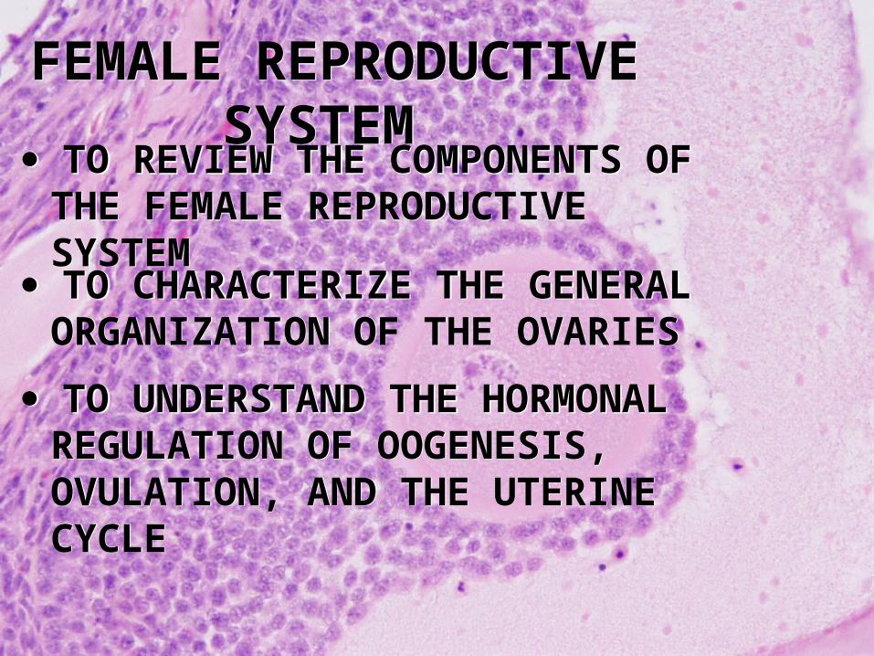
TO REVIEW THE COMPONENTS OF THE FEMALE REPRODUCTIVE SYSTEM
TO REVIEW THE COMPONENTS OF THE FEMALE REPRODUCTIVE SYSTEM
TO UNDERSTAND THE HORMONAL REGULATION OF OOGENESIS, OVULATION, AND THE UTERINE CYCLE
TO UNDERSTAND THE HORMONAL REGULATION OF OOGENESIS, OVULATION, AND THE UTERINE CYCLE
TO CHARACTERIZE THE GENERAL ORGANIZATION OF THE OVARIES
TO CHARACTERIZE THE GENERAL ORGANIZATION OF THE OVARIES
FEMALE REPRODUCTIVE SYSTEMFEMALE REPRODUCTIVE SYSTEM

Hormones of the Female Reproductive Cycle
• Control the reproductive cycle
• Coordinate the ovarian and uterine cycles

Hormones of the Female Reproductive Cycle
• Key hormones include:– FSH
• Stimulates follicular development
– LH • Maintains structure and secretory function of corpus
luteum
– Estrogens • Have multiple functions
– Progesterones • Stimulate endometrial growth and secretion
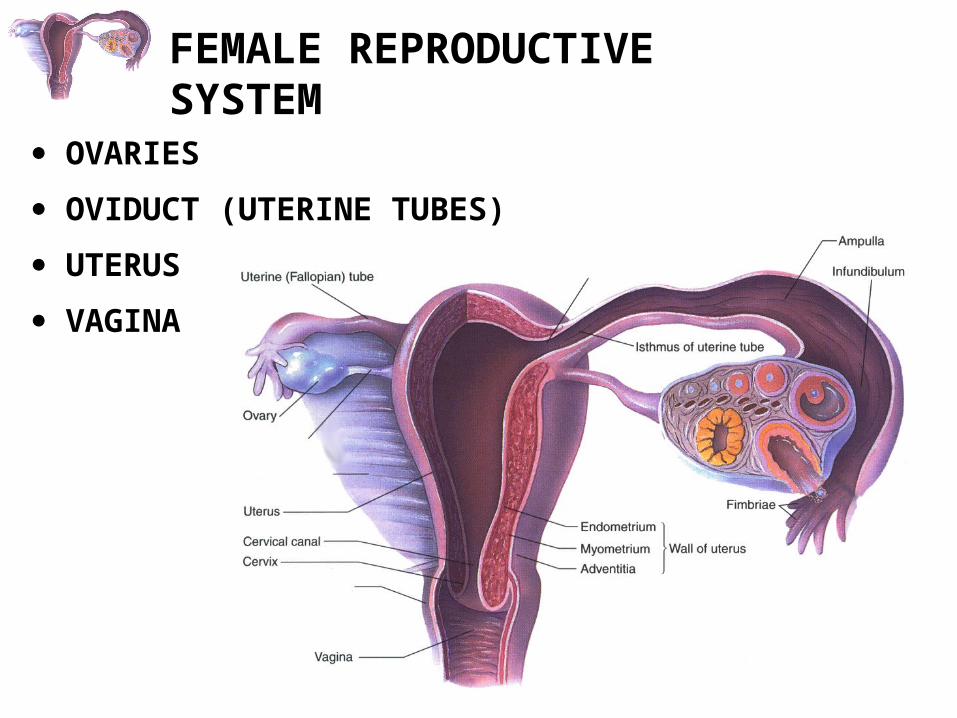
OVARIES
OVIDUCT (UTERINE TUBES)
UTERUS
VAGINA
FEMALE REPRODUCTIVE SYSTEM
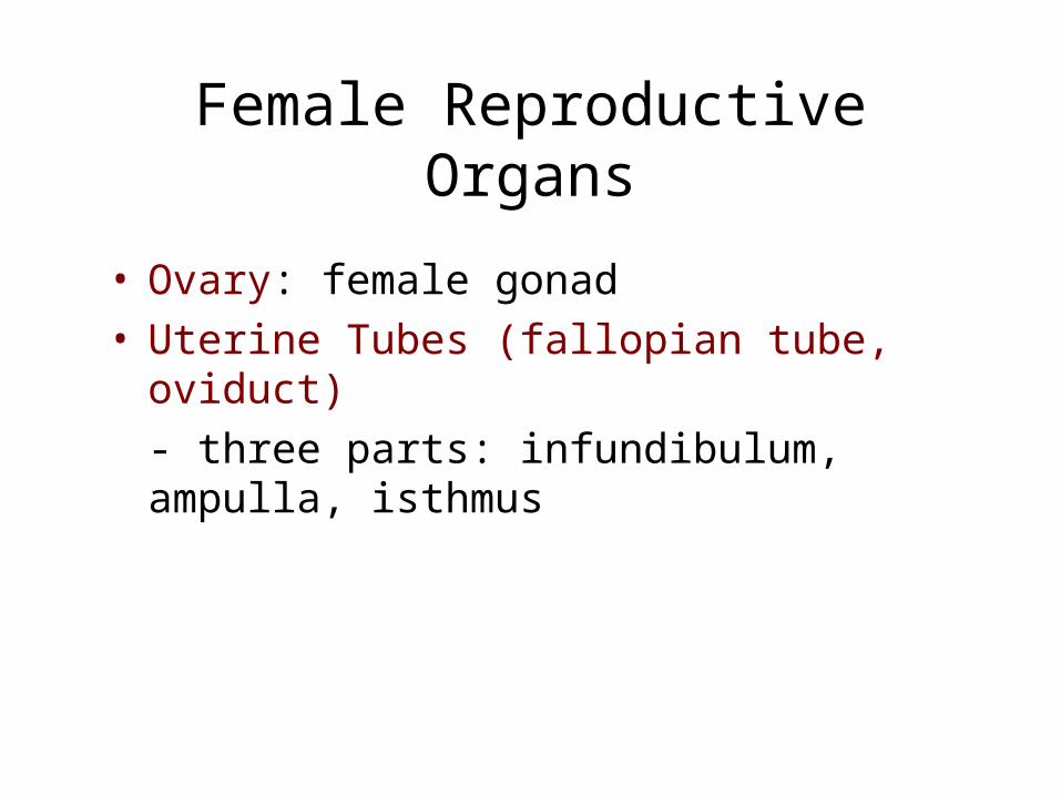
Female Reproductive Organs
• Ovary: female gonad• Uterine Tubes (fallopian tube, oviduct)
- three parts: infundibulum, ampulla, isthmus

Figure 28.13
The Female Reproductive System in Midsagital View

Figure 28.14a, b
The Ovaries and Their Relationships to the Uterine Tube and Uterus
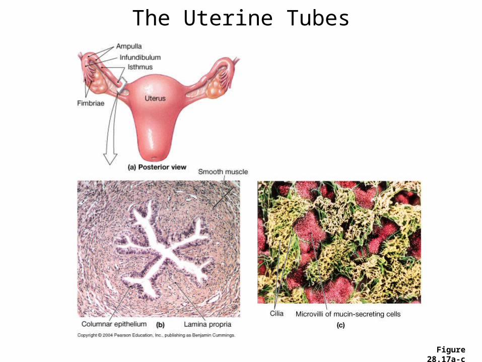
Figure 28.17a-c
The Uterine Tubes

The Uterus• Muscular organ
– Mechanical protection– Nutritional support– Waste removal for the developing embryo and fetus
• Supported by the broad ligament and 3 pairs of suspensory ligaments

Uterine Wall Consists of 3 Layers:
• Myometrium – outer muscular layer
• Endometrium – a thin, inner, glandular mucosa
• Perimetrium – an incomplete serosa continuous with the peritoneum
• The site of implantation of developing embryo• And 3 parts: fundus, body, and cervix

Female Accessory Sex Organs: Uterus
• Uterine endometrium has two layers:
- basal layer
- functional layer: built up and shed each cycle
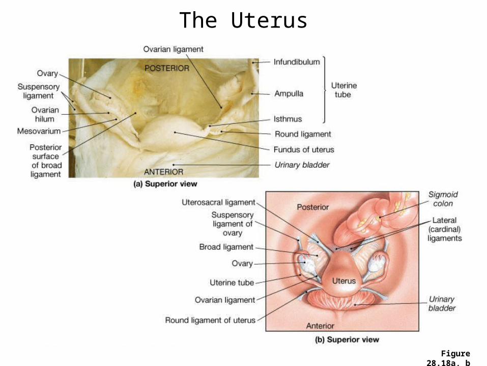
Figure 28.18a, b
The Uterus
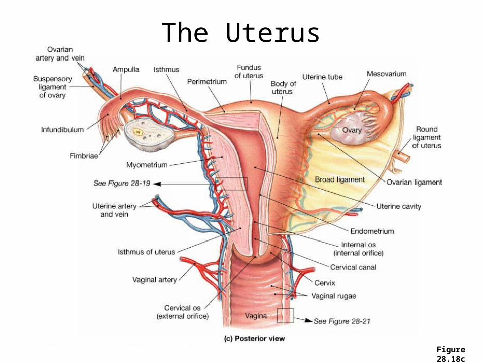
Figure 28.18c
The Uterus
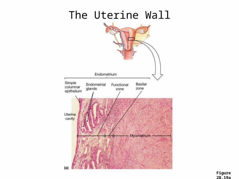
Figure 28.19a
The Uterine Wall

Figure 28.19b
The Uterine Wall

Figure 28.20
The Uterine CycleTo be discussed below
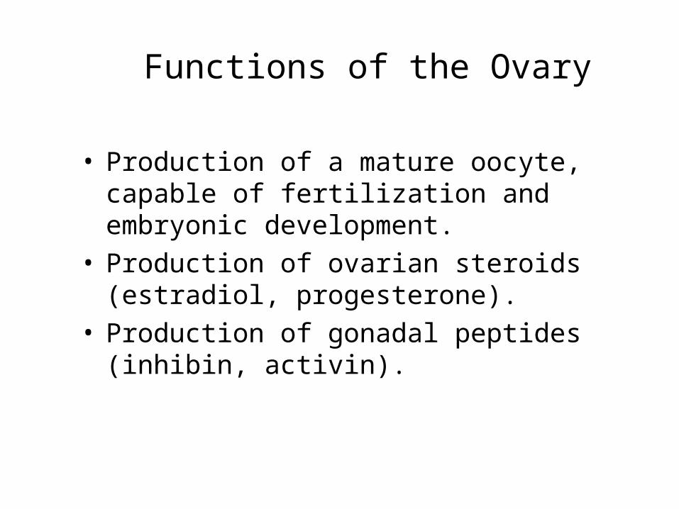
Functions of the Ovary
• Production of a mature oocyte, capable of fertilization and embryonic development.
• Production of ovarian steroids (estradiol, progesterone).
• Production of gonadal peptides (inhibin, activin).

Structural Organization of the Ovary
• The main functional unit of the ovary is the follicle.
• Follicles are composed of the oocyte, granulosa cells, and theca cells.

Stages of Follicular Growth
• Follicles are present in a number of different stages of growth:- primordial follicles (resting)- primary, secondary, and antral follicles- preovulatory (Graafian) follicles

The Corpus Luteum
• After the preovulatory follicle ovulates (releases its egg), it forms the corpus luteum.
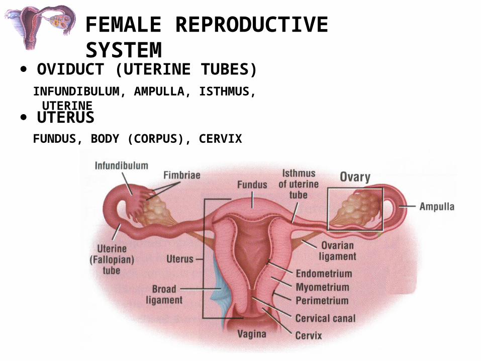
FEMALE REPRODUCTIVE SYSTEM
OVIDUCT (UTERINE TUBES)
UTERUS
INFUNDIBULUM, AMPULLA, ISTHMUS, UTERINE
FUNDUS, BODY (CORPUS), CERVIX

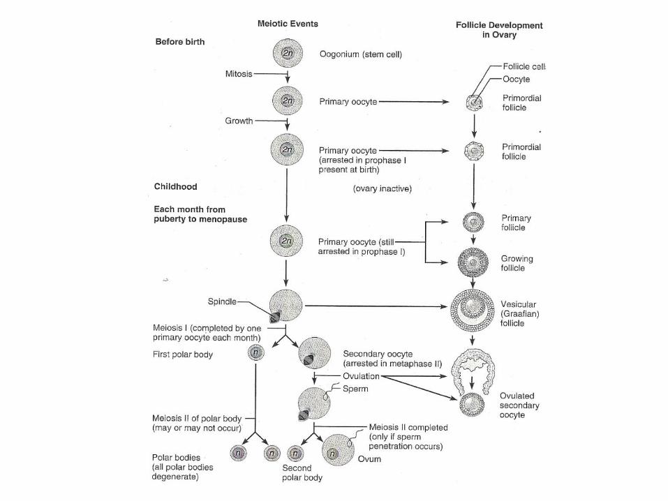
Oogenesis and Oocyte Maturation
• Recall that germ cells must go through meiosis in order to produce unique haploid cells.
• From one spermatogonia, end up with four spermatozoa.
• Oocytes must also go through meiosis, but they do it during the course of follicular development.
• Primordial follicles contain primary oocytes that are arrested in prophase I, prior to the first meiotic division (diploid).
• How do they go through the rest of meiosis?
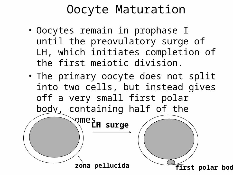
Oocyte Maturation
• Oocytes remain in prophase I until the preovulatory surge of LH, which initiates completion of the first meiotic division.
• The primary oocyte does not split into two cells, but instead gives off a very small first polar body, containing half of the chromosomes.
zona pellucida
LH surge
first polar body

Oocyte Maturation• Thus, the ovulated “egg” is actually not completely
mature (hasn’t completed meiosis II).
• Maturation goes to completion only if the oocyte is fertilized.
• Fertilization causes completion of meiosis II, and expulsion of a second polar body.
• Meiosis of the oocyte results in only one gamete.
first polar body first polar body
second polar body
fertilization
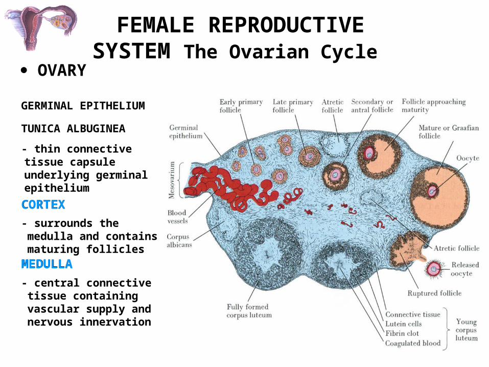
FEMALE REPRODUCTIVE SYSTEM The Ovarian Cycle
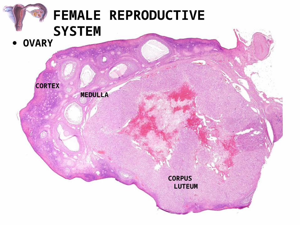
OVARY
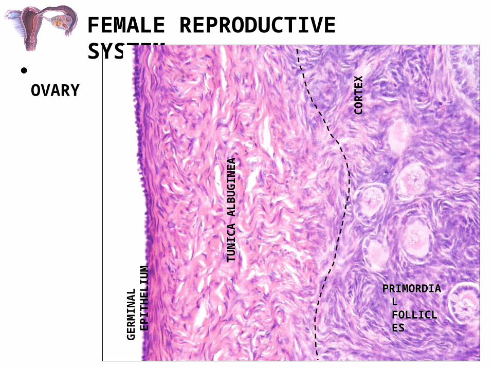
- thin connective tissue capsule underlying germinal epithelium
TUNICA ALBUGINEA
GERMINAL EPITHELIUM
CORTEXCORTEX
- surrounds the medulla and contains maturing follicles
MEDULLAMEDULLA
- central connective tissue containing vascular supply and nervous innervation
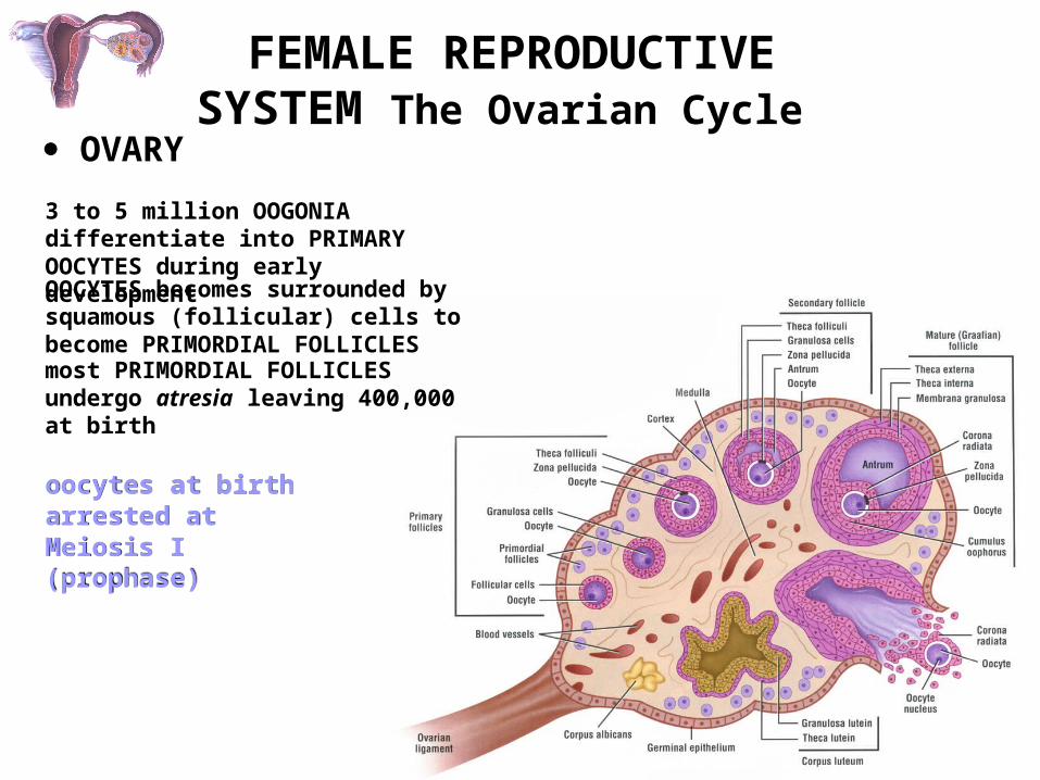
FEMALE REPRODUCTIVE SYSTEM The Ovarian Cycle
OVARY
3 to 5 million OOGONIA differentiate into PRIMARY OOCYTES during early development
OOCYTES becomes surrounded by squamous (follicular) cells to become PRIMORDIAL FOLLICLES
most PRIMORDIAL FOLLICLES undergo atresia leaving 400,000 at birth
oocytes at birth arrested at Meiosis I (prophase)oocytes at birth arrested at Meiosis I (prophase)

FEMALE REPRODUCTIVE SYSTEM
OVARY
THREE STAGES OF OVARIAN FOLLICLES CAN BE IDENTIFIED FOLLOWING PUBERTY:(each follicle contains one oocyte)
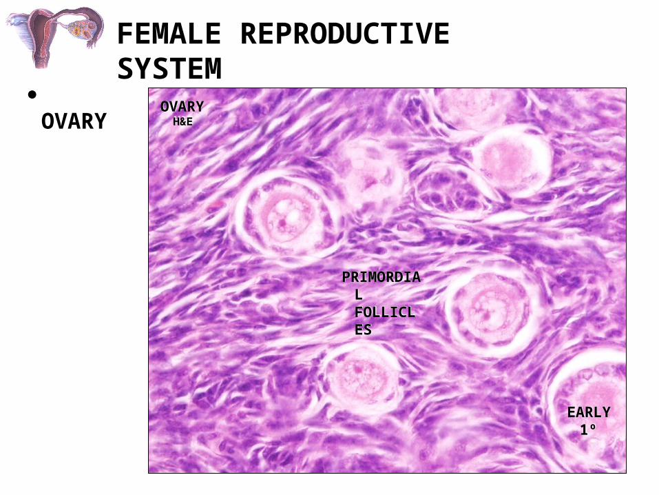
(1) PRIMORDIAL FOLLICLES
- very prevalent; located in the periphery of the cortex
- a single layer of squamous follicular cells surround the oocyte
(2) GROWING FOLLICLES
- three recognizable stages:
(a) early primary follicle
(b) late primary follicle(c) secondary (antral) follicle
(3) MATURE (GRAAFIAN) FOLLICLES- follicle reaches maximum size
OO
GEN
ESIS
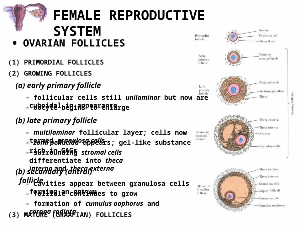
FEMALE REPRODUCTIVE SYSTEM
OVARIAN FOLLICLES
(1) PRIMORDIAL FOLLICLES
(2) GROWING FOLLICLES
(a) early primary follicle
- follicular cells still unilaminar but now are cuboidal in appearance
- oocyte begins to enlarge
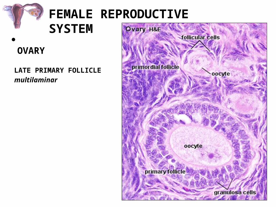
(b) late primary follicle
- multilaminar follicular layer; cells now termed granulosa cells
- zona pellucida appears; gel-like substance rich in GAGs
- surrounding stromal cells differentiate into theca interna and theca externa
(b) secondary (antral) follicle
- cavities appear between granulosa cells forming an antrum
- follicle continues to grow
- formation of cumulus oophorus and corona radiata
(3) MATURE (GRAAFIAN) FOLLICLES
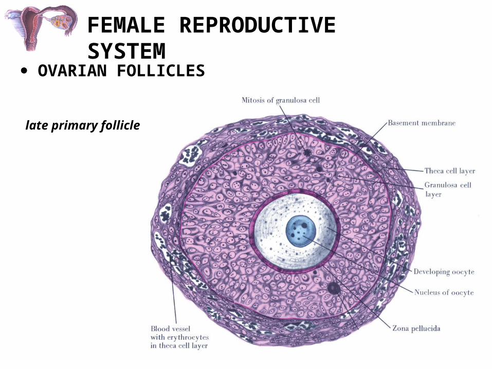
FEMALE REPRODUCTIVE SYSTEM
OVARIAN FOLLICLES
late primary follicle

FEMALE REPRODUCTIVE SYSTEM
OVARIAN FOLLICLES
GRANULOSA (FOLLICULAR) CELLS
ZONA PELLUCIDA
OOCYTE
FEMALE REPRODUCTIVE SYSTEM
OVARY
CORPUS LUTEUM
CORTEXMEDULLA
FEMALE REPRODUCTIVE SYSTEM
OVARY
GER
MIN
AL
EPIT
HEL
IUM
TUN
ICA
ALB
UG
INEA
CO
RTE
X
PRIMORDIAL FOLLICLES
PRIMORDIAL FOLLICLES

FEMALE REPRODUCTIVE SYSTEM
OVARY
GER
MIN
AL
EPIT
HEL
IUM
TUN
ICA
ALB
UG
INEA
FEMALE REPRODUCTIVE SYSTEM
OVARYOVARY H&EOVARY H&E
PRIMORDIAL FOLLICLES
PRIMORDIAL FOLLICLES
EARLY 1ºEARLY 1º

FEMALE REPRODUCTIVE SYSTEM
OVARY OVARY H&EOVARY H&E
CORPUS ALBICANSCORPUS
ALBICANS
EARLY PRIMARY FOLLICLES
EARLY PRIMARY FOLLICLES
OVARY
PRIMORDIAL FOLLICLE
PRIMORDIAL FOLLICLE
FEMALE REPRODUCTIVE SYSTEM
OVARY
LATE PRIMARY FOLLICLEmultilaminar

FEMALE REPRODUCTIVE SYSTEM
OVARY
MATURE (GRAAFIAN) FOLLICLE
cumulus oophoruscorona radiatatheca interna and externa
zona pellucida
theca interna cells begin to produce androgens that are converted to estrogens
FEMALE REPRODUCTIVE SYSTEM
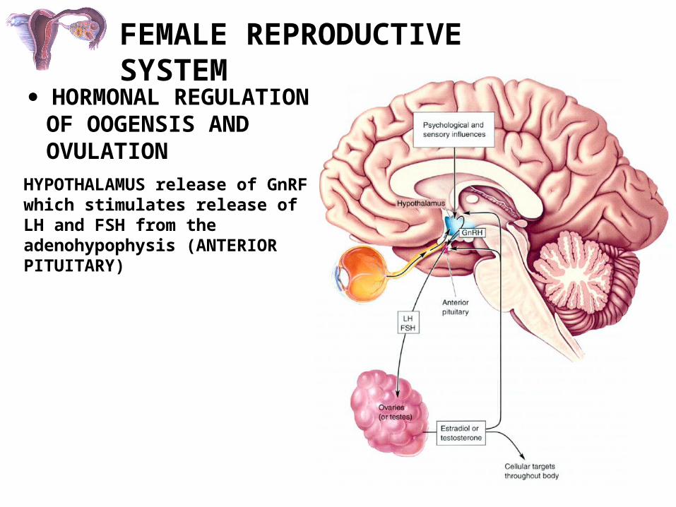
HORMONAL REGULATION OF OOGENSIS AND OVULATION
HYPOTHALAMUS release of GnRF which stimulates release of LH and FSH from the adenohypophysis (ANTERIOR PITUITARY)
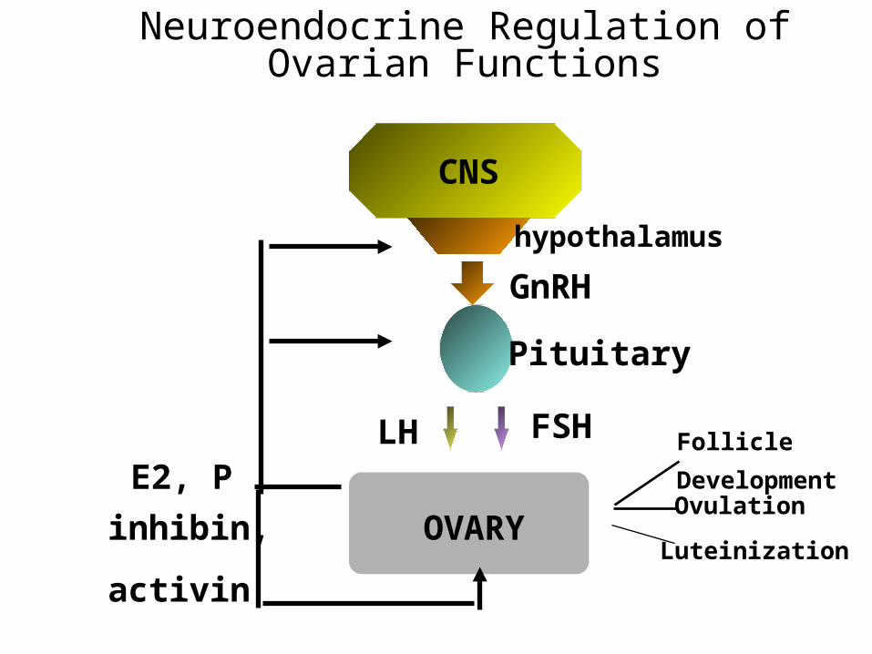
Neuroendocrine Regulation of Ovarian Functions
OVARY
E2, P
inhibin,
activin
Ovulation
Pituitary
GnRH
FSHLH
CNS
hypothalamus
Follicle
Development
Luteinization
Effects of GnRH on Gonadotropins
• GnRH is released in a pulsatile manner, stimulating the synthesis and release of LH and FSH.
• GnRH acts through its receptor on the pituitary gonadotroph cells, stimulating production of phospholipase C.
• Recall that IP3 pathway causes gonadotropin release, while the DAG/PKC pathway causes gonadotropin synthesis.

Actions of FSH on Granulosa Cells
FSH
Gs
AC
ATP
cAMP
PKA
CREB CRE
Steroidogenic
enzymes
LH Receptor
Inhibin Subunits
Plasminogen
activator
Gene Expression

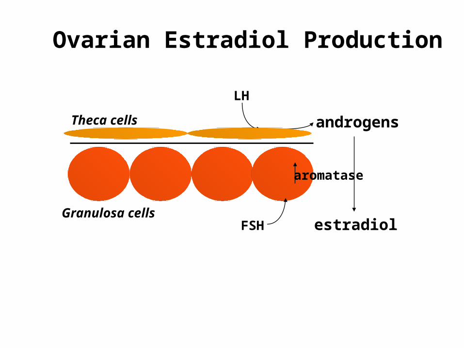
Regulation of Estradiol Production
• Recall the two-cell theory of estradiol production (lecture 4):
- LH acts on theca cells to produce androgens
- FSH acts on granulosa cells to increase aromatase activity, resulting in conversion of androgens to estrogens
(granulosa cells lack 17 hydroxylase activity)
LH
androgens
estradiol
Theca cells
Granulosa cells
aromatase
FSH
Ovarian Estradiol Production

Regulation of Progesterone Production
• Progesterone is produced from theca cells, mature granulosa cells, and from the corpus luteum.
• In this case, gonadotropins induce expression of
- steroidogenic acute regulatory protein
- P450 side chain cleavage

Actions of Estradiol
• Estradiol plays an important role in feedback regulation of gonadotropin release.
• Low estradiol levels exert negative feedback (via inhibition of GnRH release)
Question: what happens to LH and FSH levels if you remove the ovary (increase, decrease, no change)?
Answer:

• High estradiol levels exert positive feedback (via increase in GnRH receptors, stimulation of GnRH release, increased pituitary response to GnRH, and effects on LH)- increase in stimulatory neurotransmitters regulating GnRH neurons (ie, norepinephrine)- decrease in inhibitory neurotransmitters (ie, beta endorphin)- increased activity of GnRH neurons- increased expression of GnRH receptors- increased expression of LH gene
Actions of Estradiol

• Estradiol also has important actions in a number of other tissues:- causes proliferation of uterine endometrium- increases contractility of uterine myometrium- stimulates development of mammary glands- stimulates follicle growth (granulosa cell proliferation)- effects on bone metabolism, hepatic lipoprotein production, genitourinary tract, mood, and cognition
• Effects are mediated through the intracellular estrogen receptors (alpha and beta), and possible membrane effects.
Actions of Estradiol

Actions of Progesterone
• Progesterone exerts positive and negative feedback effects on gonadotropin synthesis and release.
• Progesterone also acts on many tissues:- stimulates secretory activity of the uterine endometrium- inhibits contractility of the uterine myometrium- stimulates mammary growth
• The actions of progesterone are mediated through an intracellular P receptor, which acts as a transcription factor.
Regulation of Follicle Growth
• Primordial follicles can “rest” for many decades before being recruited for growth by an unknown mechanism.
• The recruitment of primordial follicles and subsequent growth to the small antral follicle stage can occur without gonadotropin stimulation (gonadotropin-independent).
• Small antral follicles (and larger) must be stimulated by FSH and LH in order to continue growth to the preovulatory stage (gonadotropin dependent).

Regulation of Follicle Growth
• Each day many follicles begin to grow (recruitment).
• Each cycle, only one follicle is “selected” for continued development to the preovulatory stage, by an unknown mechanism.
• This follicle is called the dominant follicle• The remaining recruited follicles become atretic
and degenerate.

Regulation of Follicle Growth
recruitment selection
atresia
preovulatory
follicle

Follicular Ovulation
• As the preovulatory follicle grows, it produces increasing amounts of estradiol.
• When the preovulatory follicle is mature, plasma estradiol levels are very high.
• High estradiol levels exert positive feedback on the hypothalamus and pituitary, resulting in LH and FSH surges.
• These preovulatory gonadotropin surges cause ovulation of the preovulatory follicle (follicular rupture and release of the egg for fertilization).

Mechanism of Gonadotropin-induced Ovulation
• The preovulatory LH and FSH surges induce expression of enzymes in the preovulatory follicle which break down the follicle wall.
• Tissue-type plasminogen activator: results in breakdown of fibrin.
• Metalloproteinases: result in breakdown of collagen.
• Result: follicle wall is ruptured, resulting in release of the oocyte into the peritoneal cavity.
Formation of the Corpus Luteum
• After the follicle ovulates, the remaining granulosa cells and theca cells luteinize.
• Luteinization: accumulation of cholesterol and lipid, cells swell.
• Luteinized cells switch the ovary from predominant production of estradiol to production of progesterone and estradiol.
• This prepares the uterine endometrium for pregnancy.
FEMALE REPRODUCTIVE SYSTEM The Menstrual Cycle
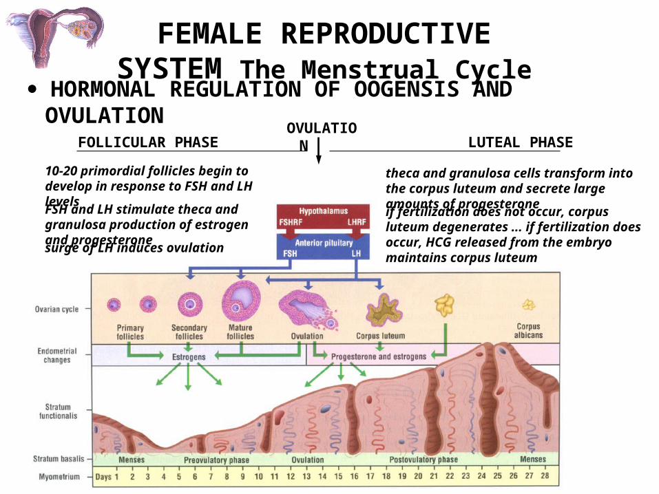
HORMONAL REGULATION OF OOGENSIS AND OVULATION
FOLLICULAR PHASE LUTEAL PHASEOVULATION
10-20 primordial follicles begin to develop in response to FSH and LH levels
FSH and LH stimulate theca and granulosa production of estrogen and progesterone
surge of LH induces ovulation
theca and granulosa cells transform into the corpus luteum and secrete large amounts of progesterone
if fertilization does not occur, corpus luteum degenerates ... if fertilization does occur, HCG released from the embryo maintains corpus luteum

FEMALE REPRODUCTIVE SYSTEM The Menstrual Cycle
HORMONAL REGULATION OF OOGENSIS AND OVULATION
OVULATION:
sharp surge in LH with simulataneous increase in FSH
Meiosis I resumes; oocyte and surrounding cumulus break away and are extruded
oocyte passes into oviduct
ECTOPIC IMPLANTATIONS

The Menstrual Cycle
• Women have ovulatory cycles of about 28 days in length.
• Day 1 of the cycle is defined as the first day of menstruation.
• There are two phases of the cycle, named after ovarian and uterine function during that phase:- first two weeks: follicular or proliferative stage- second two weeks: luteal or secretory stage
• The preovulatory gonadotropin surges occur in the middle of the cycle (around day 14).
The Menstrual Cycle: The Ovary
• Follicular phase: small antral follicles develop, a dominant follicle is selected and grows to the preovulatory stage.
• Midcycle: the gonadotropin surges cause ovulation of the dominant follicle.
• Luteal phase: the corpus luteum forms and becomes functional, secreting large amounts of progesterone, followed by estradiol (results in negative feedback, not positive feedback, because P increases before E2).
• If pregnancy does not take place, the corpus luteum regresses, and P and E2 levels decrease.

• Proliferative stage: increasing estradiol levels stimulate proliferation of the functional layer of the uterine endometrium.- Results in increased thickness of the endometrium.- Increased growth of uterine glands (secrete mucus) and uterine arteries.
• Secretory stage: progesterone acts on the endometrium.- uterine glands become coiled and secrete more mucus- uterine arteries become coiled (spiral arteries)
The Menstrual Cycle: The Uterus

The Menstrual Cycle: The Uterus
• If pregnancy doesn’t occur, P and E2 levels decrease at the end of the secretory stage.
- vasospasm of arteries causes necrosis of tissue
- loss of functional layer with bleeding of uterine arteries (menstruation)

FEMALE REPRODUCTIVE SYSTEM
CORPUS LUTEUM
FORMED FROM FOLLICLE WALL WHICH REMAINS FOLLOWING OVULATION
(1) GRANULOSA LUTEIN CELLS
- large, light cells derived from granulosa cells
TRANSFORMED CELLS SECRETE ESTROGENS AND PROGESTERONE:
(2) THECA LUTEIN CELLS
- strands of small cells derived from theca interna

FEMALE REPRODUCTIVE SYSTEM
CORPUS LUTEUM
(1) GRANULOSA LUTEIN CELLS
(2) THECA LUTEIN CELLS
CORPUS LUTEUM H&ECORPUS LUTEUM H&E
(1)(1)
(2)(2)
FEMALE REPRODUCTIVE SYSTEM
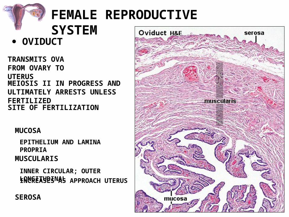
OVIDUCT
SITE OF FERTILIZATION
TRANSMITS OVA FROM OVARY TO UTERUS
MUCOSA
EPITHELIUM AND LAMINA PROPRIA
MUSCULARIS
INNER CIRCULAR; OUTER LONGITUDINAL
INCREASES AS APPROACH UTERUS
SEROSA
MEIOSIS II IN PROGRESS AND ULTIMATELY ARRESTS UNLESS FERTILIZED
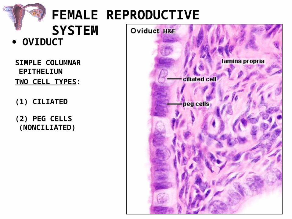
FEMALE REPRODUCTIVE SYSTEM
OVIDUCT
SIMPLE COLUMNAR EPITHELIUM
TWO CELL TYPES:
(1) CILIATED
(2) PEG CELLS (NONCILIATED)

FEMALE REPRODUCTIVE SYSTEM
UTERUSPERIMETRIUM, MYOMETRIUM, ENDOMETRIUM

FEMALE REPRODUCTIVE SYSTEM
UTERUS
ENDOMETRIUM
TWO LAYERS:
undergoes cyclic changes which prepare it for implantation of a fertilized ovum
(1) FUNCTIONAL LAYER (stratum functionalis)
(2) BASAL LAYER (stratum basale)
- BORDERS UTERINE LUMEN- SLOUGHED OFF AT MENSTRATION
- RETAINED AT MENSTRATION- SOURCE OF CELLS FOR REGENERATION OF
FUNCTIONAL LAYER
- CONTAINS UTERINE GLANDS
STRAIGHT AND SPIRAL ARTERIESSTRAIGHT AND SPIRAL ARTERIES
FEMALE REPRODUCTIVE SYSTEM
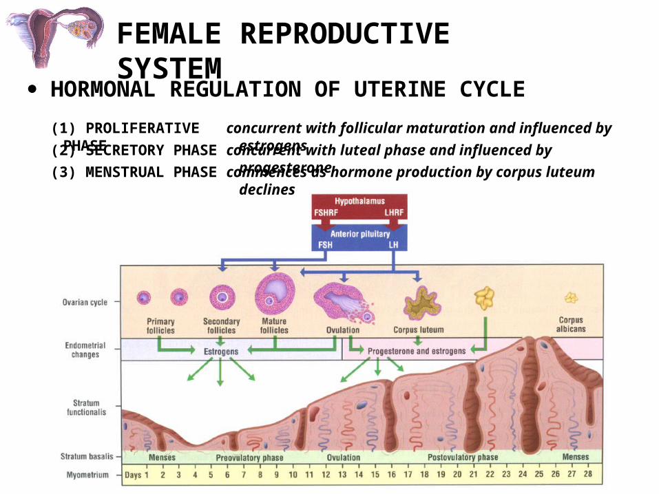
HORMONAL REGULATION OF UTERINE CYCLE
(1) PROLIFERATIVE PHASE concurrent with follicular maturation and influenced by estrogens
(2) SECRETORY PHASE concurrent with luteal phase and influenced by progesterone
(3) MENSTRUAL PHASE commences as hormone production by corpus luteum declines
FEMALE REPRODUCTIVE SYSTEM
UTERUS
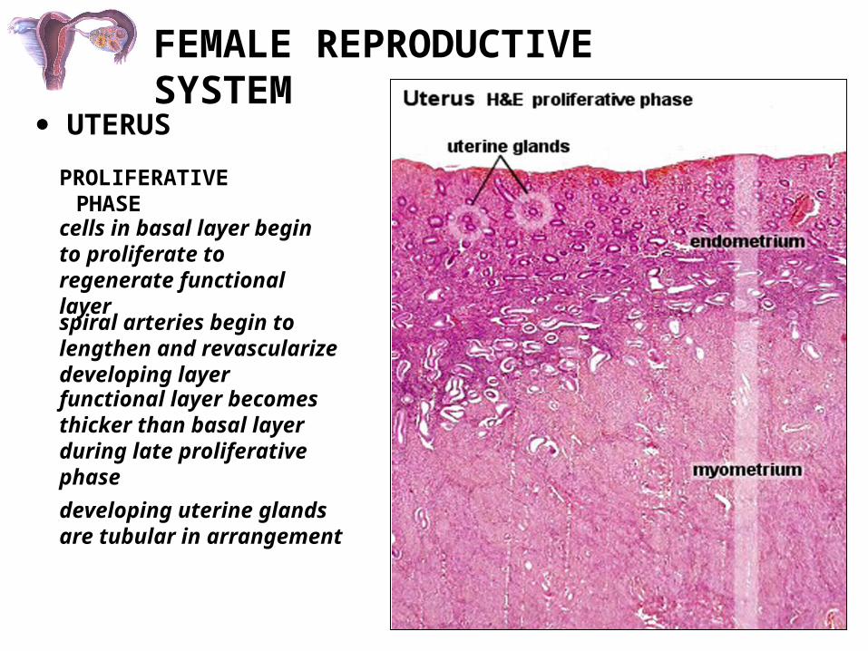
PROLIFERATIVE PHASE
cells in basal layer begin to proliferate to regenerate functional layer
spiral arteries begin to lengthen and revascularize developing layer
functional layer becomes thicker than basal layer during late proliferative phase
developing uterine glands are tubular in arrangement

FEMALE REPRODUCTIVE SYSTEM
UTERUS
PROLIFERATIVE PHASE
tubular uterine glands
simple columnar lining

FEMALE REPRODUCTIVE SYSTEM
UTERUS
PROLIFERATIVE PHASE
tubular uterine glands
simple columnar lining
UTERUS H&EUTERUS H&E
UTERINE GLANDSUTERINE GLANDS
PROLIFERATIVE PHASEPROLIFERATIVE PHASE

FEMALE REPRODUCTIVE SYSTEM
UTERUS
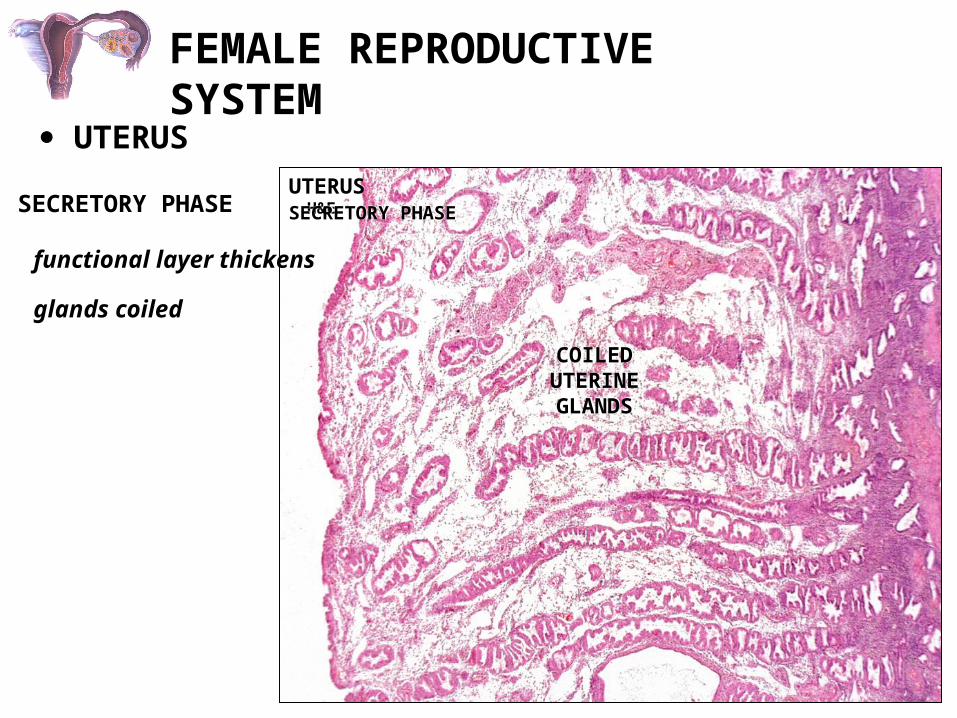
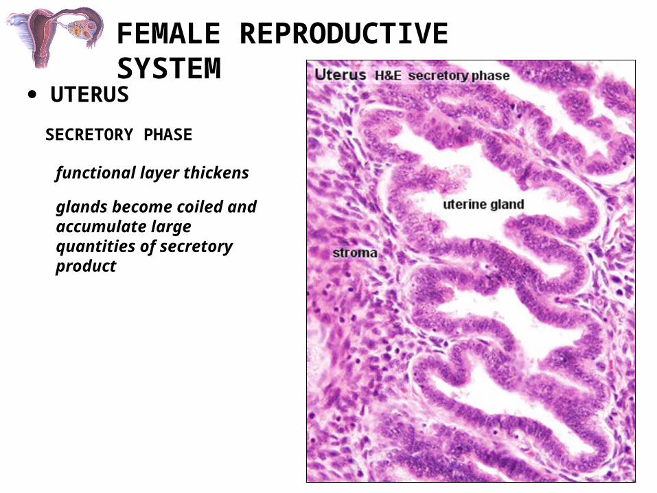
SECRETORY PHASE
functional layer thickens
glands become coiled and accumulate large quantities of secretory product
FEMALE REPRODUCTIVE SYSTEM
UTERUS
SECRETORY PHASE
functional layer thickens
glands coiled
UTERUS H&EUTERUS H&E
COILED UTERINE GLANDS
COILED UTERINE GLANDS
SECRETORY PHASESECRETORY PHASE
FEMALE REPRODUCTIVE SYSTEM
UTERUS
SECRETORY PHASE
functional layer thickens
glands become coiled and accumulate large quantities of secretory product

FEMALE REPRODUCTIVE SYSTEM
VAGINA
STRATIFIED SQUAMOUS EPITHELIUM
LAMINA PROPRIA
---------------- no glands ----------------
MUSCULARIS
INNER CIRCULAR
OUTER LONGITUDINAL
ADVENTITIA
MUCOSA

FEMALE REPRODUCTIVE SYSTEM
LABIA MINORUM
LABIA MINORUM H&ELABIA MINORUM H&E
Endocrinology of Pregnancy
• To maintain the uterine endometrium and inhibit contraction of myometrium, must maintain plasma progesterone levels during pregnancy.
• Early in pregnancy, this is accomplished by the action of human chorionic gonadotropin (hCG) on the corpus luteum (first 8 weeks of pregnancy).
• Later in pregnancy, progesterone is produced by the placenta.
Early Embryonic Development
• After fertilization, the embryo spends the first four days in the oviduct (fallopian tube).
• The developing embryo then goes to the uterus, and implants in the uterine endometrium on Day 6 (blastocyst stage of development).
• By day 6, the trophoblast cells of the embryo begin to produce hCG.
• In a normally developing embryo, hCG levels (in maternal circulation) will double every 3 days, reaching peak at about 2 months of pregnancy.

Actions of Human Chorionnic Gonadotropin (hCG)
• hCG binds to the LH receptor in the corpus luteum, maintaining luteal steroidogenesis during the first 8 weeks of pregnancy.
• In addition, hCG may act to stimulate testosterone production from the developing testes in male embryos.

Role of the Fetal-Placental-Maternal Unit in Steroid Production
• Later in pregnancy, the placenta becomes the major steroidogenic organ of pregnancy.
• However, the placenta requires maternal LDL as a source of cholesterol for progesterone production.
• The placenta also produces estrogens during pregnancy (primarily estriol). However, the placenta is dependent upon the maternal and fetal adrenal as a source of androgens for aromatization to estrogen.
• Estrogens may be important in increasing uterine blood flow to the fetus, and in the maturation of fetal organ systems.

Parturition: The Process of Childbirth
• The mechanisms signaling the onset of labor are not clearly understood, although several theories exist.
• Potential role of progesterone?:- decreasing progesterone prior to labor would allow uterine contractions to occur- however, there is no decline in progesterone before labor in humans- some studies suggest there is a decline in uterine progesterone receptors, resulting in decreased progesterone action, leading to labor
Potential Role of Oxytocin in Parturition?
• Oxytocin causes uterine contraction.• However, oxytocin levels do not increase until
after labor starts, according to more recent studies.• Oxytocin may play a role in uterine contraction
following labor, resulting in decreased blood loss.
Potential Role of Relaxin in Parturition?
• Relaxin acts on the cervix, causing dilatation and softening.
• In some animals relaxin increases before labor starts.• In humans, relaxin is high beginning early in
pregnancy and stays elevated until labor.• Relaxin does act to soften connective tissues, such as
the ligaments connecting the pelvic bones, to allow increase in size of the birth canal.
• Relaxin also decreases uterine contractility during pregnancy.
Potential Role of Prostaglandins in Parturition
• Prostaglandins cause dilation and softening of the cervix.
• Prostaglandins also cause uterine contractions.• The levels of prostaglandins increase in fetal
membranes before the onset of labor.• It is believed that some (unknown) signal from the
fetus causes increased prostaglandin production from fetal membranes, which then act on the uterus and cervix to initiate labor.

Lactation• Lactation is the delivery of milk from the mammary
gland.• There are four main stages of lactation, controlled by
different hormones:- milk synthesis in alveolar cells- secretion of milk from alveolar cells to alveolar lumen- maintainance of established milk production and release into alveolar lumen- milk ejection: movement of milk from alveoli into the duct system and out of the breast
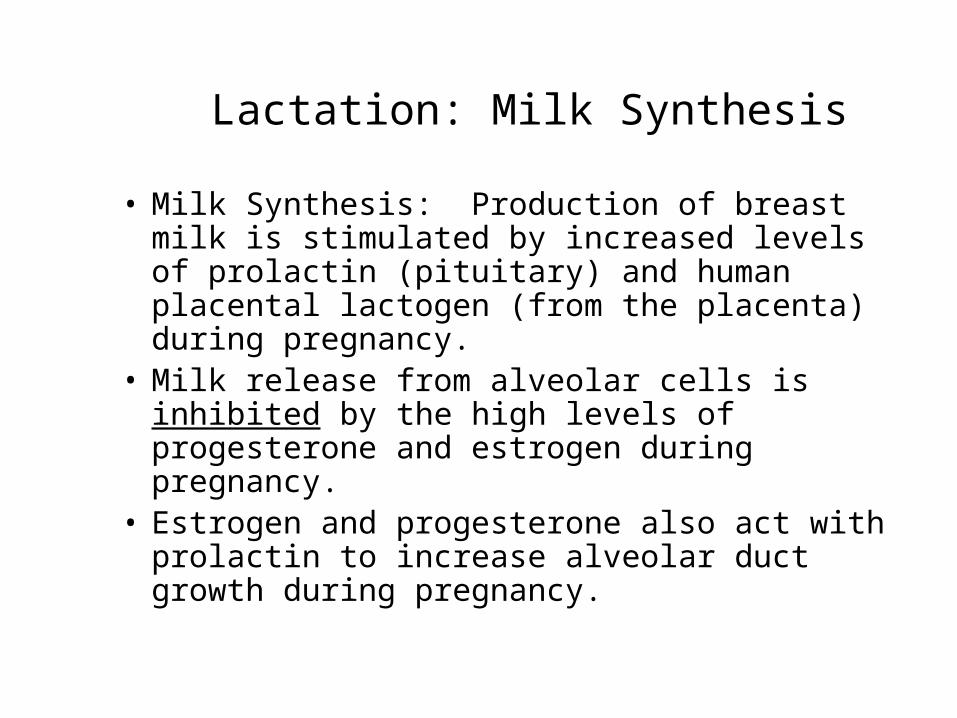
Lactation: Milk Synthesis
• Milk Synthesis: Production of breast milk is stimulated by increased levels of prolactin (pituitary) and human placental lactogen (from the placenta) during pregnancy.
• Milk release from alveolar cells is inhibited by the high levels of progesterone and estrogen during pregnancy.
• Estrogen and progesterone also act with prolactin to increase alveolar duct growth during pregnancy.

Lactation: Lactogenesis
• Lactogenesis: secretion of milk from alveolar cells into the alveoli of the breast.
• Stimulated by prolactin, and occurs after parturition when estradiol and progesterone levels are decreased.
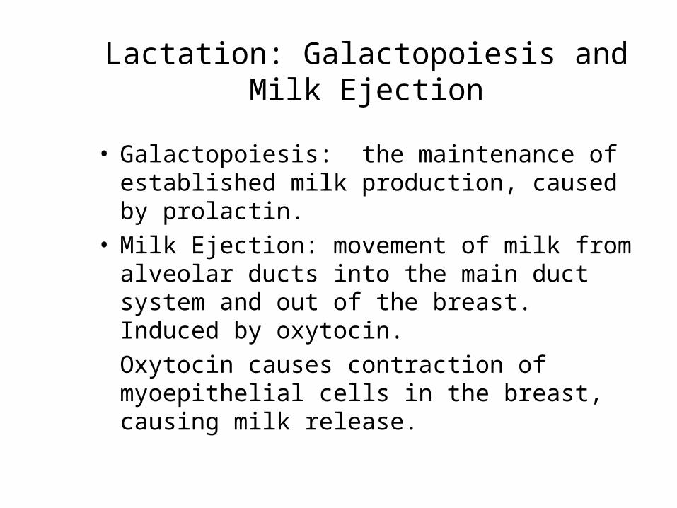
Lactation: Galactopoiesis and Milk Ejection
• Galactopoiesis: the maintenance of established milk production, caused by prolactin.
• Milk Ejection: movement of milk from alveolar ducts into the main duct system and out of the breast. Induced by oxytocin.
Oxytocin causes contraction of myoepithelial cells in the breast, causing milk release.

Regulation of Oxytocin and Prolactin During Lactation
• There is a positive feedback effect of breastfeeding on the production of oxytocin and prolactin.
• Oxytocin levels increase due to suckling of the breast by the infant. In addition, sight, sound, or thought of the infant can also increase oxytocin levels, causing milk ejection.
• Prolactin release is also increased by suckling of the breast by the infant (but not by audiovisual stimuli). Associated with suppression of dopamine release.

Hormonal Induction of Labor
Estrogen
From ovaries
Induces oxytocin receptors on uterus
Baby in utero
Oxytocin
Stim uterus to contract
Stim placenta to make PGs
Stim more vigorous contractions of uterus
Physical/emotional stress
(+)
(+)
Positive feedback
(+)
From fetus and mother’s post pit
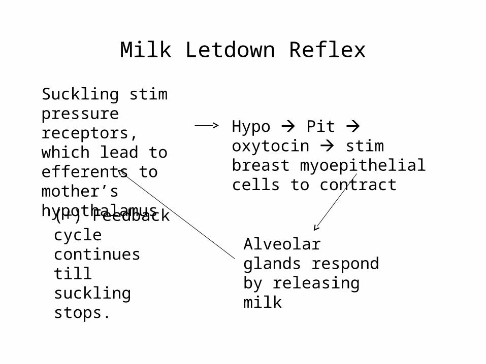
Milk Letdown Reflex
Suckling stim pressure receptors, which lead to efferents to mother’s hypothalamus
Hypo Pit oxytocin stim breast myoepithelial cells to contract
Alveolar glands respond by releasing milk
(+) Feedback cycle continues till suckling stops.