Unilateral Biportal Endoscopic Posterior Cervical Laminectomy and Discectomy
FDA Executive Summary: Page 1 of 101
FDA Executive Summary
Intrinsic Therapeutics
Barricaid® Anular Closure Device
Prepared for the December 12, 2017 Meeting of the
Orthopaedic and Rehabilitation Devices Panel

FDA Executive Summary: Page 2 of 101
Table of Contents
1 INTRODUCTION..................................................................................................... 6
1.1 RATIONALE FOR PRESENTATION TO PANEL ...................................................................................... 6
2 HERNIATED DISCS – CURRENT TREATMENT OPTIONS .......................... 7
2.1 CLINICAL CONDITION ....................................................................................................................... 7 2.2 CLINICAL ASSESSMENT ..................................................................................................................... 7 2.3 IMAGING ........................................................................................................................................... 7 2.4 NONSURGICAL TREATMENT .............................................................................................................. 8 2.5 SURGICAL TREATMENT ..................................................................................................................... 9
2.5.1 General .............................................................................................................................. 9 2.5.2 Surgery and Adverse Events ............................................................................................ 10 2.5.3 Subsequent Surgical Interventions - Overview ................................................................ 10
2.6 RECURRENT DISC HERNIATION ....................................................................................................... 11 2.6.1 Definitions and Diagnostic Challenges ........................................................................... 11 2.6.2 Surgical Factors –Extent of Anular Resection ................................................................. 12
3 BACKGROUND FOR FDA QUESTIONS FOR THE PANEL ......................... 13
3.1 STUDY POPULATION ....................................................................................................................... 13 3.2 PRESENCE AND SAFETY RISKS OF ENDPLATE LESIONS ................................................................... 14 3.3 APPROPRIATE ENDPOINTS FOR REHERNIATIONS ............................................................................. 14
4 DEVICE DESCRIPTION ...................................................................................... 15
4.1 IMPLANT AND INSTRUMENTS .......................................................................................................... 15 4.2 DEVICE MODIFICATIONS ................................................................................................................. 16
5 PROPOSED INDICATIONS FOR USE .............................................................. 17
6 DEVICE HISTORY................................................................................................ 17
6.1 REGULATORY HISTORY .................................................................................................................. 17 6.2 MARKETING HISTORY ..................................................................................................................... 18
7 NON-CLINICAL STUDIES .................................................................................. 18
7.1 CHARACTERIZATION ....................................................................................................................... 18 7.2 MECHANICAL TESTING TO REPLICATE CLINICAL FAILURES ........................................................... 18 7.3 CADAVERIC IMPLANTATION ........................................................................................................... 20 7.4 BIOCOMPATIBILITY ......................................................................................................................... 20 7.5 ANIMAL STUDIES ............................................................................................................................ 21
7.5.1 Rabbit Particulate Study .................................................................................................. 21 7.5.2 Baboon Implantation Study .............................................................................................. 22
7.6 STERILIZATION ............................................................................................................................... 24 7.7 MAGNETIC RESONANCE (MR) SAFETY ........................................................................................... 24
8 CLINCIAL STUDY AND DESIGN ...................................................................... 25
8.1 STUDY DESIGN ................................................................................................................................ 25 8.2 ENROLLMENT CRITERIA .................................................................................................................. 25
8.2.1 Inclusion Criteria: ........................................................................................................... 25 8.2.2 Exclusion Criteria ............................................................................................................ 27
8.3 IMPLANTATION, RANDOMIZATION, AND TREATMENT PROTOCOL ................................................... 28 8.3.1 Limited Discectomy and Randomization .......................................................................... 28 8.3.2 Barricaid Group Treatment ............................................................................................. 29
8.4 STUDY ASSESSMENTS ..................................................................................................................... 30 8.5 RADIOGRAPHIC ANALYSIS: ............................................................................................................. 31 8.6 PRIMARY ENDPOINTS AND STUDY SUCCESS .................................................................................... 34
FDA Executive Summary: Page 3 of 101
8.7 SECONDARY ENDPOINTS ................................................................................................................. 35 8.8 STATISTICAL DESIGN ...................................................................................................................... 35
8.8.1 Primary data set ............................................................................................................... 35 8.8.2 Other Subanalyses ........................................................................................................... 36
8.9 DATA ANALYSIS, ADJUDICATION, AND STUDY OVERSIGHT ........................................................... 36
9 STUDY RESULTS .................................................................................................. 37
9.1 SUBJECT ACCOUNTING ................................................................................................................... 38 9.2 PROTOCOL DEVIATIONS .................................................................................................................. 40 9.3 BASELINE DEMOGRAPHICS AND BASELINE STATUS ........................................................................ 42
10 SAFETY RESULTS................................................................................................ 44
10.1 ADVERSE EVENTS (AES) ............................................................................................................ 45 10.2 DEVICE AND PROCEDURE RELATED, AND SERIOUS AES ............................................................ 47 10.3 NEUROLOGICAL STATUS ............................................................................................................ 47 10.4 SECONDARY SURGERY ............................................................................................................... 48 10.5 ENDPLATE LESIONS (EPLS) ....................................................................................................... 52
10.5.1 Qualitative Assessment .................................................................................................... 53 10.5.2 Quantitative Assessment .................................................................................................. 59
10.6 RETRIEVAL ANALYSIS AND PRODUCT COMPLAINTS .................................................................. 64 10.6.1 Device Failures ................................................................................................................ 64 10.6.2 Retrieval Analysis ............................................................................................................ 65 10.6.3 Product Complaints ......................................................................................................... 67
11 EFFECTIVENESS RESULTS .............................................................................. 68
11.1 OVERALL SUCCESS .................................................................................................................... 68 11.1.1 Co-Primary Endpoint – Reherniation .............................................................................. 68 11.1.2 Co-Primary Endpoint – Composite .................................................................................. 69 11.1.3 Exploratory Analysis: Alternative Primary Endpoint ...................................................... 71 11.1.4 Longer-Term Clinical Composite Success (CCS) ............................................................ 75
11.2 INDIVIDUAL ENDPOINTS ............................................................................................................. 76 11.2.1 All Reherniations ............................................................................................................. 76 11.2.2 Clinical Endpoints – Oswestry Disability Index (ODI), VAS – Leg Pain, Neurologic
Outcomes ......................................................................................................................... 79 11.2.3 Radiographic Endpoints – Maintenance of Disc Height, Device Integrity and
Spontaneous Fusion ......................................................................................................... 79 11.2.4 Spontaneous Fusion ......................................................................................................... 81 11.2.5 Index Level Secondary Surgical Interventions (SSI) ........................................................ 81
11.3 SECONDARY ENDPOINTS ............................................................................................................ 82 11.4 EXPLORATORY/SUBGROUP ANALYSES ....................................................................................... 82
12 STATISTICAL ANALYSIS .................................................................................. 83
12.1 MISSING DATA IMPUTATION ...................................................................................................... 83 12.2 POOLABILITY OF SITES ............................................................................................................... 83 12.3 FINANCIAL INTERESTS ............................................................................................................... 83 12.4 PATIENT ENROLLMENT .............................................................................................................. 84 12.5 PATIENT POPULATION COMPATIBILITY ...................................................................................... 84 12.6 BLINDING ................................................................................................................................... 84
13 BENEFIT RISK ASSESSMENT ........................................................................... 86
13.1 SUMMARY OF BENEFITS ............................................................................................................. 86 13.2 SUMMARY OF RISKS ................................................................................................................... 89 13.3 ADDITIONAL CONSIDERATION FOR BENEFIT RISK ASSESSMENT ................................................ 92 13.4 BENEFIT-RISK SUMMARY .......................................................................................................... 95
14 POST APPROVAL STUDY .................................................................................. 96

FDA Executive Summary: Page 4 of 101
14.1 OVERVIEW OF THE PROPOSED PAS ............................................................................................ 96 14.2 FDA COMMENTS ON THE PROPOSED PAS .................................................................................. 97
15 BIBLIOGRAPHY ................................................................................................... 98
APPENDICES Appendix A – Surgical Technique Manual (STM) Appendix B – Contraindications, Warnings and Precautions Appendix C – Baboon Pathology Report Appendix D – Clinical Protocol Appendix E – Imaging Charter Appendix F – Patient Accounting Appendix G – Adverse Events Appendix H – Secondary Surgeries Appendix I – EPL Analyses Appendix J – Endpoints Appendix K – OUS Literature
LIST OF FIGURES
Figure 4.1 Barricaid® Anular Closure Device ............................................................................................. 15 Figure 7.1 Migration Test Samples .............................................................................................................. 19 Figure 7.2 12 Month Baboon Study Results ................................................................................................. 24 Figure 8.1 Correct placement of Barricaid ................................................................................................... 30 Figure 8.2 Estimation of EPL Volume ......................................................................................................... 33 Figure 10.1 Example Images of Radiographic Findings in Control Subjects ............................................... 55 Figure 10.2 Example Images of Radiographic Findings in Barricaid Subjects ............................................ 56 Figure 10.3 Progression of EPL in Barricaid Subject ................................................................................... 57 Figure 10.4 Barricaid EPLs in Lateral and Anterior View ........................................................................... 58 Figure 10.5 Subject with Barricaid and no lesion at 5 year point. ................................................. 59 Figure 10.6 Mesh proximate lesion .............................................................................................................. 60 Figure 10.7 Example of Mesh Detachment in a Retrieved Barricaid Implant .............................................. 65 Figure 10.8 Example of Mesh Migration ..................................................................................................... 65 Figure 11.1 Symptomatic Reherniation Algorithm ...................................................................................... 73 LIST OF TABLES Table 2.1 – Carragee classification system *Adapted from Carragee 2003 ................................................. 12 Table 4.1 Materials in the Barricaid® Anular Closure Device ...................................................................... 16 Table 4.2 Generations of the Barricaid Implants .......................................................................................... 16 Table 7.1 Summary of cycles to migration for the in vitro migration study ................................................. 19 Table 7.2 Status of Recommended Biocompatibility Testing per ISO 10993-1 ........................................... 21 Table 8.1 Study Assessments ....................................................................................................................... 31 Table 8.2 Composite Clinical Success – Clinical Protocol Definition (CCS-CPD) ..................................... 34 Table 9.1 Long-Term (>Month 24) Patient Accounting and Follow-up Compliance Table ........................ 38 Table 9.2 Clinical follow-up information for five control subjects who received Barricaid after failure .... 39
(b) (6)
FDA Executive Summary: Page 5 of 101
Table 9.3 Major Protocol Deviations............................................................................................................ 41 Table 9.4 Summary of Baseline Intra-Operative Data ................................................................................. 43 Table 10.1 AE Relationship Definitions ....................................................................................................... 45 Table 10.2 Comparisons of Summary Adverse Event Rates between Barricaid and Control Discectomy at
24 months – AT Analysis Sets ....................................................................................................... 46 Table 10.3 Time-course cumulative distribution of proportion of subjects with an SSI at Index level ........ 48 Table 10.4 Survival Analysis – Index Level SSI (mITT) ............................................................................. 48 Table 10.5 Secondary Surgical Interventions by Type ................................................................................. 50 Table 10.6 Time course of SSI Event Type .................................................................................................. 51 Table 10.7 Number of Subjects with EPLs .................................................................................................. 59 Table 10.8 Presentation of EPL Total Areas Corresponding per Subject (mm2) ......................................... 60 Table 10.9 EPL Area of Mesh Opposed vs Anchor Side Vertebral Body (Axial Plane) .............................. 61 Table 10.10 Per Subject Growth of Mesh Proximate EPLs (mm2) .............................................................. 62 Table 10.11 Per Subject Growth of Mesh Proximate EPLs with Subsidence (mm2) ................................... 62 Table 10.12 No Symptomatic Reherniation and EPL Sum Area at Month 24 ............................................. 63 Table 10.13: Overall Rate of Device Integrity Failures ................................................................................ 64 Table 11.1 Composite Endpoint out to 60 months (CCS-CPD) ................................................................... 70 Table 11.2 Composite Endpoint out to 60 months (CCS-mCPD) ................................................................ 74 Table 11.3 Comparisons of the Percentages of Subjects with Reherniations – mITT Analysis Set ............. 76 Table 11.4 Comparisons of the Percentages of Subjects with Symptomatic Reherniations – mITT Analysis
Set (Restricted to Expected Due) (Nominal Significance) ............................................................. 77 Table 11.5 Number of Symptomatic Recurrent Disc Herniations at Index Level (Nominal Significance) .. 77 Table 11.6 Comparisons of the Percentages of Subjects with Asymptomatic Reherniations – mITT Analysis
Set (Restricted to Theoretically Due) (Nominal Signficance) ........................................................ 78 Table 11.7 Survival Analysis – All Reherniations (mITT) ........................................................................... 78 Table 11.8 Survival Analysis – Symptomatic Reherniations (mITT) .......................................................... 79 Table 11.9 Overall Rates of Device Integrity Failure in Barricaid Subjects ................................................ 80 Table 12.1 Blinding CCS analysis ................................................................................................................ 86 Table 13.1 Twenty-four month Outcomes-Clinical Protocol Definition (CPD) ........................................... 87 Table 13.2 Twenty-four month Outcomes-Modified Clinical Protocol Definition (mCPD) ........................ 87 Table 13.3 Agency Determined Relevant Device or Procedure Related AE ................................................ 91
FDA Executive Summary: Page 6 of 101
1 Introduction This is Food and Drug Administration’s Executive Summary of the pre-market approval (PMA) application, P160050/M140021, for the Barricaid® Anular Closure Device (Barricaid) from Intrinsic Therapeutics (sponsor). The implant is a polymeric mesh that sits in the posterior intervertebral disc space, which is connected to a metallic anchor that is attached to the vertebral body. The implant is accompanied by a delivery tool and manual surgical instruments, which are used in a posterior/posterolateral approach. The Barricaid® Anular Closure Device is a permanent implant used after a limited lumbar discectomy performed for treatment of lumbar radiculopathy. The device is designed to mechanically block an opening in the anulus, thereby maintaining the relative position of nucleus within the disc space to prevent reherniation following limited discectomy in patients with large anular defects at an increased risk of reherniation.
This summary contains an overview of current treatment options, a device description, a summary of the non-clinical and clinical studies conducted by the sponsor, and additional analyses performed by Food and Drug Administration (FDA, also referred to as the Agency). The clinical data presented in this PMA application was collected outside of the United States (OUS). FDA did not reach consensus with the sponsor regarding the investigational protocols, including the protocol for the OUS randomized, controlled, clinical trial (RCT) presented in this summary, although the sponsor did partially incorporate FDA feedback in their study. The purpose of this panel meeting is to obtain the Panel’s feedback regarding the data provided in this PMA application. The Advisory Committee (Panel) will be asked to comment on several topics of interest to FDA as highlighted throughout this summary. In addition, Panel members will be asked to provide recommendations and vote on whether the data provided demonstrate a reasonable assurance of safety and effectiveness, as well as a favorable benefit-risk profile, for the Barricaid. Your time and effort in the review of this PMA application are greatly appreciated.
Rationale for Presentation to Panel
In addition to the fact that the Barricaid is a first-of-a-kind anular closure device, the Agency is presenting this PMA application to the Panel based on the reasons listed below. The Agency has questions regarding the original study design, study results and interpretation of the clinical findings. Specifically, the Agency has identified to following issues related to this clinical study:
The representativeness of the study population to the general primary lumbar disc herniation population is unclear.
The notable incidence of asymptomatic and symptomatic lumbar disc herniations identified in both the treatment and control groups.
There is uncertainty regarding elements of the surgical technique described during the study when compared with the surgical technique described in the Study Protocol.
The clinical relevance of the pre-specified Co-Primary Endpoint definition for assessment of recurrent lumbar disc reherniation confirmed surgically or radiographically is unclear. The alternative primary endpoint was developed post
FDA Executive Summary: Page 7 of 101
hoc to identify symptomatic recurrent disc herniations and does not appear to require correlation of physical examination findings, imaging findings, and outcome measures with the side and level of the recurrent disc herniation and the patient’s radicular symptoms.
The relevance and long term impact of the endplate lesions (EPLs) identified in Barricaid subjects are unclear with respect to the determination of safety risks.
It is unclear how to interpret study outcomes in view of the notable rate of device integrity issues (i.e. migration, dissociation).
2 Herniated Discs – Current Treatment Options Clinical Condition
Lumbar disc herniation is one of the most common clinical diagnoses in the US population. The incidence of symptomatic herniated lumbar discs has been estimated between 1-2%[1]. More than 480,000 lumbar discectomies are performed annually in the United States[2, 3]. Herniated lumbar discs are a common cause of lumbar radiculopathy (pain with possible motor and sensory disturbances in a nerve-root distribution) but also are observed on imaging studies (MRI or CT) in asymptomatic patients. Approximately 90% of lumbar herniated lumbar discs occur at the L4-5 and L5-S1 spinal levels. The herniated disc material includes not only nucleus pulposus, but potentially also, cartilage, fragmented apophyseal bone, or anulus fibrosus tissue, which may exert mechanical pressure upon the adjacent neural structures and lead to nerve root inflammation. In the absence of major neurologic deficit (cauda equina syndrome, progressive neurologic deficit), both non-operative management and operative management are viable treatment options as the natural history of herniated lumbar discs is favorable[4].
Clinical Assessment Clinical assessment of a patient with a suspected lumbar disc herniation begins with a detailed history and physical exam to identify patients with cauda equina symptoms who require urgent surgery, and exclude intraspinal and extraspinal conditions which may mimic radiculopathy secondary to lumbar disc herniation. Symptoms most frequently include leg pain radiating below the knee when originating from the L5 or S1 nerves (sciatic nerve) and may be associated with numbness or weakness. Less commonly, pain may radiate into the anterior thigh or groin due to compression of the L2, L3, or L4 nerve roots (femoral nerve). Physical examination includes evaluation of lower extremity sensory, motor and reflex function as well as testing of nerve root tension signs (straight leg raise, contralateral straight leg raise, femoral nerve stretch). The straight leg raise test is commonly used to evaluate the lower lumbar nerve roots and is positive if radicular pain is reproduced when the patient’s leg is elevated between 30 and 70 degrees. Although the sensitivity of this test approaches 90%, it has been shown to have low specificity[5].
Imaging MRI is currently the imaging modality of choice for evaluating the relationship of disc material to soft tissue and neural structures. The Combined Task Force (CTF) and van Rijn classification systems are the most reliable methods for describing lumbar disc herniation and nerve root compression[6].
FDA Executive Summary: Page 8 of 101
Herniation is broadly defined by the CTF classification[7] as a localized or focal displacement of disc material beyond the limits of the intervertebral disc space. The term ‘‘localized’’ or ‘‘focal’’ refers to the extension of the disc material less than 25% (90°) of the periphery of the disc as viewed in the axial plane. Herniated discs may be classified as protrusion or extrusion, based on the shape of the displaced material. Protrusion is present if the greatest distance between the edges of the disc material presenting outside the disc space is less than the distance between the edges of the base of that disc material extending outside the disc space. The base is defined as the width of disc material at the outer margin of the disc space of origin, where disc material displaced beyond the disc space is continuous with the disc material within the disc space. Extrusion is present when, in at least one plane, any one distance between the edges of the disc material beyond the disc space is greater than the distance between the edges of the base of the disc material beyond the disc space or when no continuity exists between the disc material beyond the disc space and that within the disc space. The latter form of extrusion is best further specified or sub-classified as sequestration if the displaced disc material has lost continuity completely with the parent disc. Disc herniations may be further specifically categorized as contained, if the displaced portion is covered by outer anulus fibers and/or the posterior longitudinal ligament, or uncontained when absent of any such covering. Herniated discs in the craniocaudad (vertical) direction through a gap in the vertebral body end plate are referred to as intravertebral herniations or Schmorl’s nodes. The presence of disc tissue extending beyond the edges of the ring apophyses, throughout the circumference of the disc, is called ‘‘bulging’’ and is not considered a form of herniation. Symmetric bulging of disc tissue greater than 25% of the disc circumference often seen as an adaptation to adjacent deformity, and is not considered a form of herniation. Nerve root compression grading by the van Rijn classification system[8] uses a five-point scale: definitely no root compression, possibly no root compression, indeterminate, possibly root compression, and definitely root compression. These ratings may be dichotomized into no root compression (first three categories) and root compression (last two categories).
Nonsurgical Treatment As many patients with a symptomatic herniated lumbar disc experience improvement of leg pain symptoms within 6 weeks, initial nonsurgical treatment is generally recommended. Nonsurgical treatment options include a short period of rest, medication, physical therapy, and return to activity as symptoms permit. Epidural glucocorticoid injections may be considered, but their effectiveness and safety have not been established by FDA and have not been approved for this use. For patients with persistent symptoms despite nonsurgical measures, surgical treatment with lumbar laminotomy and discectomy is an appropriate treatment option for patients with radicular pain and evidence of nerve root compression and positive nerve root tension signs. Surgery is also considered for patients with reflex, sensory, or motor deficit with associated radicular symptoms and positive nerve root tension signs. Appropriate surgical candidates have a confirmatory imaging study (MRI or CT) which shows a lumbar disc herniation at a location (level and side) corresponding to the patient’s radicular signs or symptoms[9] .

FDA Executive Summary: Page 9 of 101
Multiple randomized controlled trials (RCTs) have been performed to compare nonsurgical and surgical treatment for lumbar disc herniation. Surgical intervention may result in more rapid relief of symptoms and earlier return to function, although long-term results appear similar regardless of the type of management. However, limitations regarding the methodology of these RCTs, especially their high cross over rates, impact their ability to inform clinical practice[10, 11] . As both nonsurgical and surgical treatment are valid options, a shared decision making approach which involves a patient and physician who are both well informed regarding the benefits and risks of each treatment and considers patient preference is recommended[4].
Surgical Treatment General
The goal of surgical treatment of a symptomatic lumbar disc herniation is to provide adequate nerve root decompression and minimize soft-tissue damage and iatrogenic destabilization[12]. Disc space access and nerve root decompression may be achieved through a variety of posterior surgical approaches. Central and posterolateral disc herniations are most commonly accessed through an interlaminar approach. Disc herniations located in the extraforaminal zone (lateral to the pedicle) are most commonly accessed through an intertransverse approach, although an interlaminar approach has been advocated at the L5-S1 level. The surgical approach for a disc herniation located in the foraminal zone is influenced by a combination of factors, including the level and size of the disc herniation. Microsurgical lumbar discectomy is the most commonly performed technique for lumbar discectomy. A trend in discectomy surgery toward minimization of soft tissue dissection has led to the evolution of alternative techniques involving minimally invasive approaches which utilize tubular retractors and/or endoscopes. The extent of intraoperative discectomy required remains controversial. Historically, a “complete” or “radical” lumbar discectomy, including curettage of the disc space and removal of adjacent endplates, was performed based on the rationale that extensive removal these structures would decrease the risk of reherniation, but is no longer advocated[13]. Subtotal discectomy is one currently utilized technique which includes removal of the free disc fragment followed by opening of the anulus to allow for removal of additional disc material and may/may not include endplate curettage[14, 15].The technique of “limited discectomy” was introduced based on the rationale that decreased manipulation of neural elements during surgery would limit resultant peridural fibrosis. This technique avoided the use of curettes and was specific to the type of disc herniation encountered during surgery. As initially described[16], “if the disc was sequestered, the free fragment was removed but the annulus was not entered. In cases of extruded disc herniations, the extruded fragment was excised, and loose fragments near the annular defect were removed with a small pituitary rongeur. When a protruded disc was identified, the annulus was incised, and removal of all loose disc tissue was performed. After disc removal, the neural foramen was assessed… Incision into the annulus fibrosis was necessary only when a protruded disc herniation was identified… the surgeon must be prepared to perform a foraminotomy in addition to the lumbar discectomy if the nerve root remains tight after disc excision”. An alternate technique, sequestrectomy, was advocated and restricted disc removal to the free disc fragments located posterior to the vertebral body without probing of anulus or removal of nucleus pulposus. The rationale
FDA Executive Summary: Page 10 of 101
for limiting disc excision to removal of only the material responsible for compression of the neural elements was directed toward minimization of postoperative pain and maintenance of stability at the surgical level. Level 1 evidence is not available to guide surgeons in the selection of conservative versus aggressive discectomy for the treatment of primary lumbar disc herniation. However, systematic review of the literature suggests that conservative discectomy may result in shorter operative time, quicker return to work, and a decreased incidence of long-term recurrent low back pain but with an increased incidence of recurrent disc herniation[17].
Surgery and Adverse Events Surgical outcomes following lumbar discectomy are generally reported as favorable in appropriately selected patients, but outcomes may be adversely affected by a range of factors including the preoperative duration of symptoms[18], surgical technique, level of herniation, psychosocial factors, and pending litigation. Complications associated with lumbar discectomy may be classified as intraoperative, immediately postoperative, or late postoperative[19]. Intraoperative complications include wrong level surgery, durotomy, missed pathology, excessive bleeding, epidural hematoma, nerve root injury, injuries due to surgical positioning, and anterior blood vessel or visceral injury. Early postoperative complications include general medical complications, increased leg pain, increased back pain, neurologic deficit including cauda equina syndrome, epidural hematoma, wound infection, and wound dehiscence. Late postoperative complications include general medical complications as well as specific complications such as recurrent disc herniation, recurrent back and/or leg pain, disc space infection, peridural fibrosis, and spinal instability.
Subsequent Surgical Interventions - Overview Unplanned subsequent surgical interventions following lumbar discectomy may be required for treatment of adverse events attributed to the index procedure (ex. infection), for treatment of persistent or recurrent symptoms, as well as for treatment of symptoms attributed to progression of the underlying degenerative spinal process. Leven[20] performed a subgroup analysis of patients from the intervertebral disc herniation arm of the Spine Outcomes Research Trial (SPORT) to evaluate reoperation rates. The overall reoperation rate was 6% of the cohort by one year; 8% within two years; 10% within four years; 13% within six years; and 15% by 8 years. Sixty-two percent of patients underwent reoperation because of a recurrent disc herniation; 25%, because of a complication or other factor; and 11%, because of a new condition. Of the patients who underwent reoperation, greater than half (55%) underwent reoperation within the first two years. Across all surgically treated patients, the risk of a recurrent disc herniation at the same level as the index operation was 9%. The proportion of reoperations attributed to recurrent disc herniation over the eight-year study period was generally similar and accounted for 58% to 62% of reoperations each year. Martin[21] used a US statewide inpatient discharge registry to examine variation in reoperation rates among hospitals and surgeons after lumbar decompressions for herniated disc, and compared these rates to rates published for SPORT. The most common diagnoses at the time of reoperation remained herniated disc (70.6%), followed by spondylosis (13.9%), stenosis (11.1%), listhesis (3.8%), and scoliosis or other diagnosis (11%). The most common procedures at the time of reoperation were decompressions without fusion (73%) and fusion with or
FDA Executive Summary: Page 11 of 101
without decompression (25.7%). Reoperation rates varied substantially despite adjustments for patient characteristics, and the rates for many hospitals and surgeons exceeded the long-term SPORT reoperation rates. Reasons for these variations in reoperation rates were unclear and were potentially attributed to professional uncertainty regarding criteria or indications for revision surgery, surgical complications, potential quality problems, postoperative care differences, variable surgical training and practice philosophy, local practice patterns, and patient expectations. An additional consideration is that individual surgeons may have different thresholds for progressing patients with poor outcomes following an initial lumbar discectomy to procedures that are perceived to be more ‘‘definitive’’ such as spinal fusion. A recent survey of US spinal surgeons designed to assess the surgical treatment patterns among neurologic and orthopedic spine surgeons in the US for the treatment of one- and two-time recurrent lumbar disc herniation reported that the vast majority of surgeons selected revision microdiscectomy for treatment of a first time recurrence, but there was wide variability in the treatment choices for second surgeries to treat recurrent disc herniation[22].
Recurrent Disc Herniation Definitions and Diagnostic Challenges
Assessment of the rates of recurrent disc herniation is challenging due to lack of a standardized definition for recurrent disc herniation, the inability for imaging studies to distinguish between symptomatic and asymptomatic reherniation[23], and because prior studies report patients with prevalent pain and fail to separate out patients with persistent symptoms from those who experienced initial resolution of symptoms[24]. A strict definition of recurrent disc herniation considers disc reherniation as a reherniation occurring at the same level and the same side as a previously operated lumbar disc, with a pain-free interval after the primary discectomy of greater than 3 weeks to 6 months[25]. Other definitions require that the return of symptoms be consistent with the previous presentation in the same patient[26]. Many definitions include both ipsilateral and contralateral herniations at the previously operated level as recurrent herniations, but exclude adjacent level herniations[27, 28] . Contralateral disc herniations at the level of a previous discectomy occur less frequently than ipsilateral recurrent herniations. Choi[25] reported that contralateral herniations presented at a significantly longer mean time-interval to reherniation compared with patients with ipsilateral reherniation (33 vs. 18.6 months). While postoperative MRI can identify the presence of lumbar disc protrusions or extrusions, these imaging findings may or may not be related to clinical symptoms. For example, Barzouhi[29] reported a series of patients who were evaluated with repeat MRI one year after treatment for symptomatic lumbar disc herniation which showed that anatomical abnormalities visible on MRI were unable to distinguish patients with persistent or recurrent symptoms of sciatica from asymptomatic patients as a disc herniation was visible in 35% with a favorable outcome and in 33% with an unfavorable outcome. Barth[30, 31] observed an extremely high incidence (approximately 67%) of disc extrusions and protrusions two years postoperatively after lumbar microdiscectomy or sequestrectomy and noted these findings did not correlate with clinical outcome. However, repeat MRI studies (often performed with gadolinium) are frequently obtained for patients with persistent or recurrent symptoms following lumbar discectomy and abnormalities detected on MRI may lead to additional interventional procedures including epidural injections or surgical treatment. It can be difficult to determine
FDA Executive Summary: Page 12 of 101
whether the patients undergoing repeat surgery truly had a recurrent disc herniation, or ongoing/recurrent sciatic symptoms after discectomy surgery with subsequent imaging studies showing disc pathology that triggered further surgery[32].
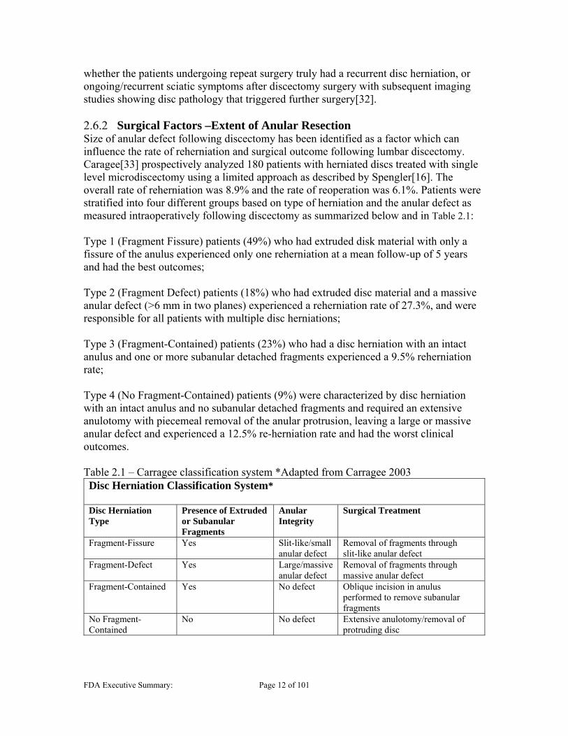
Surgical Factors –Extent of Anular Resection Size of anular defect following discectomy has been identified as a factor which can influence the rate of reherniation and surgical outcome following lumbar discectomy. Caragee[33] prospectively analyzed 180 patients with herniated discs treated with single level microdiscectomy using a limited approach as described by Spengler[16]. The overall rate of reherniation was 8.9% and the rate of reoperation was 6.1%. Patients were stratified into four different groups based on type of herniation and the anular defect as measured intraoperatively following discectomy as summarized below and in Table 2.1: Type 1 (Fragment Fissure) patients (49%) who had extruded disk material with only a fissure of the anulus experienced only one reherniation at a mean follow-up of 5 years and had the best outcomes; Type 2 (Fragment Defect) patients (18%) who had extruded disc material and a massive anular defect (>6 mm in two planes) experienced a reherniation rate of 27.3%, and were responsible for all patients with multiple disc herniations; Type 3 (Fragment-Contained) patients (23%) who had a disc herniation with an intact anulus and one or more subanular detached fragments experienced a 9.5% reherniation rate; Type 4 (No Fragment-Contained) patients (9%) were characterized by disc herniation with an intact anulus and no subanular detached fragments and required an extensive anulotomy with piecemeal removal of the anular protrusion, leaving a large or massive anular defect and experienced a 12.5% re-herniation rate and had the worst clinical outcomes. Table 2.1 – Carragee classification system *Adapted from Carragee 2003 Disc Herniation Classification System* Disc Herniation Type
Presence of Extruded or Subanular Fragments
Anular Integrity
Surgical Treatment
Fragment-Fissure Yes Slit-like/small anular defect
Removal of fragments through slit-like anular defect
Fragment-Defect Yes Large/massive anular defect
Removal of fragments through massive anular defect
Fragment-Contained Yes No defect Oblique incision in anulus performed to remove subanular fragments
No Fragment-Contained
No No defect Extensive anulotomy/removal of protruding disc
FDA Executive Summary: Page 13 of 101
3 Background for FDA Questions for the Panel The Agency would like the Panel to provide responses to a series of questions regarding the safety and effectiveness data presented in the PMA application. These questions are located in the “FDA Panel Questions” section of the Panel package, and Panel input will be solicited at the December 12, 2017 Panel meeting. To help outline background for the questions, the following subsections are provided as a guide to the information presented in greater detail later in the summary.
Study Population The overall target population for this device was patients who were of higher risk for reherniation following a limited discectomy. The sponsor presented literature identifying a greater incidence of reherniations in patients with “large” anular defects (>6mm) following discectomy[27, 34] in the reherniation population compared to patients in the non-recurrent group. This was the basis for the inclusion criteria for the treatment population and design for the sizing of the device. However, other studies[33] presented that the patient population from a consecutive cohort included 49% (89/180) of patients that were categorized as having a “Fragment-Fissure” type herniation that resulted in slit-like or small anular defect (<6mm). Given that a notable portion of the general lumbar disc herniation population is expected to have small size anular defects associated with low reherniation risk (i.e. Caragee type 1 and type 3 herniations), in the sponsor's study it is unclear why the enrolled population included only 26 subjects out of a total of 647 subjects (4%) that failed intraoperative screening and did not proceed to randomization due to anular defects that were considered too small for study inclusion. The enrollment of consecutive subjects with minimal exclusions also raises concern regarding the “at risk” nature of the herniation population. The sponsor asserts that some of these smaller size anular defect subjects may have been accounted for in the 3,332 subjects that were screened but never enrolled for various exclusion criteria. However, the impact of the identified screening process on such subjects is unclear. The screening process was applied to all patients who “presented to the clinic with complaints concordant with a herniated lumbar disc” and would include a large number who did not require surgery or did not have a herniated disc. The study protocol identified that subjects would be treated with a limited discectomy per the technique described in Spengler et al [16]. This technique differs from other more aggressive techniques (i.e. subtotal discectomy) that may include the use of curettes, as only pituitaries are used for removal of fragments. It appears that the extensive anular resections were performed in a number of study subjects as evidenced by frequent use (62% of all subjects in the study) of box anulotomies during discectomy. A “box” type anulotomy has been described in conjunction with a subtotal discectomy[15], and is inconsistent with published technique for limited discectomy [16, 35] and is also used during the preparation the disc space for insertion of a PLIF or TLIF interbody fusion device, in which case the anular incision may be larger than for a subtotal discectomy. The Panel will be asked to discuss the extent of anular resection and the potential impact on the study.
FDA Executive Summary: Page 14 of 101
Presence and Safety Risks of Endplate Lesions The presence of lesions in the vertebral body was seen in both the control and experimental groups, and has been a key concern regarding the safety evaluation of the subject device. These are collectively referred to in this Executive Summary as Endplate Lesions (EPLs), and are also referred to as “Endplate Changes” or EPCs in the sponsor’s Executive Summary. CT scans were used post procedure for subsequent follow-up of subjects to track the progression of these lesions due to the presence of lesions in the vertebral endplate seen in animal studies and other OUS clinical experience. The Barricaid study group presented with a large number of subjects with these identified lesions (483 EPLs in 235/267 [88%] subjects). The control group also presented with a subset of subjects with these lesions (190 EPLs in 113/283 [40%] subjects). However, the qualitative radiographic analysis noted the EPLs in the Barricaid group had notably different radiographic features than those generally seen in the control group as discussed in Section 10.5.1 below. The qualitative analysis also demonstrated that the Barricaid lesions were generally larger and faster progressing than those in the control group. They were located in both vertebral bodies in the area surrounding the implant, most often proximal to where the mesh interacts with and compromises the integrity of the endplate. Given the unknown impact of these lesions and the safety risk they may represent, the sponsor conducted extensive analyses investigating possible correlation between the presence of EPLs and clinical outcomes. Subgroup analyses included investigation of subgroups that had EPLs, large EPLs, EPLs proximal to the mesh, and EPLs with mesh subsidence among others. The endpoints investigated included reherniation, secondary surgeries and clinical performance. These analyses do not consistently show correlation between EPLs and the outcomes analyzed. While analyses showed a slowing or stabilization of the Barricaid EPLs at around 5 years, in addition to being smaller in size, the control group showed reduction in size beginning after year 4 based on the sponsor’s quantitative area analyses. It is notable that these conclusions are based on the sponsor’s qualitative analyses of growth that may be limited due to limited data available at later time points. It is unclear at this time if the Barricaid EPLs will eventually reduce in size as well, or if the size will still slowly increase; however, the concern regarding the long term presence of these induced lesions that alter the endplate needs to be weighed. It is not clear what impact the continued presence of these lesions should have on the evaluation of the safety of the Barricaid device.
Appropriate Endpoints for Reherniations The original proposed study design for the OUS dataset presented in this PMA consists of a 24 month co-primary endpoint that includes a measure of reherniation success and a composite success which includes safety and efficacy endpoints. The primary reherniation endpoint relies on radiographic and/or surgical assessment and does not include a clinical assessment. This endpoint is intended to capture all herniations, both symptomatic or asymptomatic, in order to assess the effectiveness of the device. The sponsor has since included a post-hoc analysis attempting to identify a more clinically relevant symptomatic reherniation assessment through use of a sorting algorithm. This alternative primary endpoint which was developed post hoc to identify symptomatic
FDA Executive Summary: Page 15 of 101
recurrent disc herniations does not appear to require that physical examination findings, imaging findings, and outcome measures correlate with the side and level of the recurrent disc herniation and the subject’s radicular symptoms. Additionally, given the dataset included data on a number of subjects with follow-up beyond the 24 month time point (out to 60 months), it was noted that there were also a large number of reherniations that occurred after the proposed 24 month endpoint. Despite findings that show many of the endpoints as statistically superior for the Barricaid group, analyses indicate that some results may change with longer term follow up, but this assessment is limited by the large number of subjects that are not yet due for assessment. Another consideration is the potential effect of the EPLs that do not approach a stable size until 60 months. It is unclear whether the 24 month endpoint is appropriate or whether a different time point would provide a more appropriate benefit risk profile.
4 Device Description Implant and Instruments
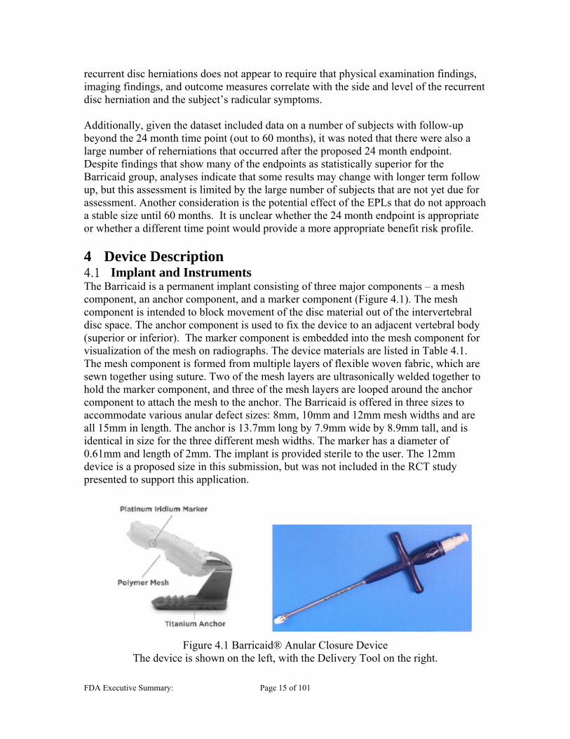
The Barricaid is a permanent implant consisting of three major components – a mesh component, an anchor component, and a marker component (Figure 4.1). The mesh component is intended to block movement of the disc material out of the intervertebral disc space. The anchor component is used to fix the device to an adjacent vertebral body (superior or inferior). The marker component is embedded into the mesh component for visualization of the mesh on radiographs. The device materials are listed in Table 4.1. The mesh component is formed from multiple layers of flexible woven fabric, which are sewn together using suture. Two of the mesh layers are ultrasonically welded together to hold the marker component, and three of the mesh layers are looped around the anchor component to attach the mesh to the anchor. The Barricaid is offered in three sizes to accommodate various anular defect sizes: 8mm, 10mm and 12mm mesh widths and are all 15mm in length. The anchor is 13.7mm long by 7.9mm wide by 8.9mm tall, and is identical in size for the three different mesh widths. The marker has a diameter of 0.61mm and length of 2mm. The implant is provided sterile to the user. The 12mm device is a proposed size in this submission, but was not included in the RCT study presented to support this application.
Figure 4.1 Barricaid® Anular Closure Device
The device is shown on the left, with the Delivery Tool on the right.

FDA Executive Summary: Page 16 of 101
Table 4.1 Materials in the Barricaid® Anular Closure Device Component Material
Mesh Mesh Layers: Polyethylene-terephthalate (PET) Mesh Sutures: Polytetrafluoroethylene (PTFE)-coated PET
Anchor Titanium Alloy (Ti6Al4V ELI) Marker 90% Platinum, 10% Iridium
The Barricaid implant is delivered using the Barricaid Delivery Tool, which is a single-use, sterile disposable instrument (Figure 4.1). This tool is designed to facilitate delivery and implantation during the surgical procedure, and the implant is pre-loaded on the delivery tool. The delivery tool is comprised of a delivery sheath, a pusher, and a strike cap, which are made from stainless steel, polyphenylsulfone (Radel), and/or nickel titanium (Nitinol). The system also includes the following manual surgical instruments provided non-sterile and are reusable: alignment trials, extractor, impactor, hammer, retraction wedge, and defect sizing tools. Please refer to the sponsor’s surgical technique manual in Appendix A for additional information.
Device Modifications Intrinsic Therapeutics described three generations of the Barricaid implant and associated instruments. The PMA included non-clinical and clinical data on all generations; however, the majority of the data and was on the Generation 3 device and its associated instruments, for which the sponsor is seeking to market. The differences between the generations are provided in Table 4.2. The sponsor described that the implant modifications were made to increase the strength between the mesh and anchor components after incidences of mesh detachment and mesh rotation were reported. The sponsor explained that the Surgical Technique Manual was modified and three manual surgical instruments were added based on surgeon feedback and other specific reasons (e.g., to improve mechanical leverage). Differences between generations are noted below. Table 4.2 Generations of the Barricaid Implants (*No Generation 1 implants were part of the RCT, both Generation 1 and 2 were included in other feasibility studies that are outside the scope of this panel meeting)
Generation 1 Generation 2 Generation 3
Mesh Component
mesh layers mesh layers mesh layers
(new attachment location/stitching)
Anchor Component
Unchanged
Marker Component
2 markers 2 markers 1 marker
Data Collection
30 subjects implanted in
OUS feasibility*
44 subjects implanted in
OUS Post Market Study*
44 subjects implanted in
OUS RCT for PMA
222 subjects implanted in OUS
RCT for PMA
(b)(4)
(b)
(b)

FDA Executive Summary: Page 17 of 101
5 Proposed Indications for Use Intrinsic Therapeutics proposed the following Indications for Use:
The Barricaid is intended to be implanted following a limited discectomy, to prevent reherniation and the recurrence of pain or dysfunction. The Barricaid is indicated for subjects with radiculopathy (with or without back pain), a posterior or posterolateral herniation, characterized by radiographic confirmation of neural compression using MRI, and a large anular defect (e.g., between 4-6 mm tall and between 6-12 mm wide) determined intraoperatively post discectomy, at one level between L4 and S1. The sponsor’s proposed Contraindications, Warnings and Precautions are provided in Appendix B.
6 Device History Regulatory History
Intrinsic Therapeutics has an extensive regulatory history with FDA as it pertains to the Barricaid device. In summary, the sponsor initially sought to perform a clinical study in the United States by submitting an Investigational Device Exemption (IDE) application for this device in 2009. However, the IDE was never approved by the Agency due to safety concerns. Specifically, FDA identified safety concerns regarding progressive bone and tissue resorption (described as “endplate lesions” by the Agency and “endplate changes” or “EPCs” by the sponsor) reported in both animal and early OUS feasibility clinical studies. The Agency requested a root cause analysis to address these safety concerns. Subsequently, Intrinsic Therapeutics initiated a prospective, randomized, controlled clinical trial in Europe and communicated their plan to use the OUS RCT data to support initiation of a U.S. clinical study. However, a U.S. clinical study was never initiated and in November 2016, the sponsor submitted OUS RCT data along with non-clinical studies in the subject PMA submission. It is important to note that an IDE is not required for studies conducted entirely outside the US.
FDA and the sponsor never reached consensus regarding the OUS study design, associated protocols (clinical, radiographic, and statistical), or documents (e.g., informed consent, clinical events charter, imaging charter) prior to or throughout the OUS clinical trial. FDA and Intrinsic Therapeutics discussed numerous items during the review of the IDE submission such as informed consent, patient selection, statistical analysis plan, definitions of study success, clinically meaningful differences, patient selection criteria, radiographic protocol and study duration. While FDA acknowledges that the sponsor incorporated many of the elements discussed during the review of their IDE submissions and pre-submissions, consensus was not reached regarding many details of the study protocol, including study endpoints. Intrinsic Therapeutics revised their study protocols multiple times throughout the OUS study. FDA was not aware of the protocol changes that occurred until the study had been initiated. These protocol modifications ranged from administrative corrections to adverse event reporting modifications to revisions to the statistical analysis plan. In addition, the modifications to the Barricaid implant and instruments were not fully identified until the PMA was received by the Agency.
FDA Executive Summary: Page 18 of 101
Marketing History The Barricaid device has been marketed outside the United States since 2009 in the following countries: Germany, Austria, Greece, Netherlands, Italy, Switzerland, Belgium, Turkey, Israel, South Korea, Russia, South Africa, Chile, Costa Rica, Poland, Hungary, Bulgaria, Saudi Arabia, and Australia.
The Barricaid has not been withdrawn from marketing for any reason.
7 NON-CLINICAL STUDIES Intrinsic Therapeutics performed a range of non-clinical studies to evaluate the integrity of the Barricaid and its effectiveness in closing anular defects. Bench testing was provided on the Generation 3 design with the addition of some Generation 2 testing used as comparison. Animal testing was conducted on Generation 1 implants. The following testing was submitted as part of the PMA.
Characterization The sponsor provided a number of bench studies to characterize the Barricaid implant the Agency determined that the testing yielded limited utility other than characterization. The following tests were performed:
Monotonic and dynamic anchor push-out tests Monotonic and dynamic compression shear Mesh resistance to rotation Monotonic and dynamic silicon bead herniation model
Mechanical Testing to Replicate Clinical Failures
Mesh detachment from the anchor and mesh migration are the two most common device-related clinical failures of this device as seen in the OUS RCT as reported by the sponsor (also see Section 10.4 below for retrieval analysis). These non-standard tests were developed and conducted after the RCT study was complete and were not used to demonstrate safety or efficacy prior to initiation of the study. The sponsor provided bench testing designed to evaluate both of these failure mechanisms as well as compare Generation 2 and 3 implants. Mesh detachment Study Description: The device anchor was held in place. A wire was placed through a hole in the mesh. The rod was moved in either an anterior/posterior or medial/lateral direction. and increased incrementally over the course of the test. Test Results: Three-fourths of the test articles replicated the mesh detachment failures that have occurred in vivo. Due to the high loads applied, the remaining devices failed by anchor fracture rather than mesh detachment. The sponsor provided a comparison of Generation 2 and 3 devices to establish that the two generations are similarly prone to mesh detachment in the benchtop testing, and similar to what was observed clinically.
In Vitro Migration Study
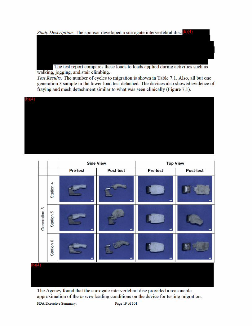
(b)(4)
FDA Executive Summary: Page 20 of 101
Although the Generation 3 devices did not detach as frequently as the Generation 2 devices, the Generation 3 devices consistently migrated at lower cycle numbers than the Generation 2 devices in the more physiologically relevant bench test. The sponsor states that this test was designed to recreate mesh migrations in 100% of the samples, which is substantially higher than the observed clinical migration rate. Because the applied loads were physiologically relevant, and the cycles to migration were relatively low, the discrepancy between the migration rate of this test and of the clinical migration rate is likely explained by other elements of the test set-up. While the sponsor stated that the intent of the testing was to replicate clinical failures and demonstrate that the two generations were comparable, the Agency had requested testing to demonstrate that the modifications of Generation 3 resolved migrations seen clinically. The testing was able to demonstrate the sponsor’s intent, but did not address the Agency’s request to demonstrate that the clinical concern was mitigated with the device modification.
Cadaveric Implantation
The sponsor performed cadaveric implantation studies to validate the surgical technique used to implant the Barricaid. The Agency previously expressed a number of concerns regarding the cadaver study. Many of these concerns related to verification of key instructions provided in the surgical technique manual (STM), such as assembling components and correct angulation of the device. There have been design changes and additions to the instrument set to address instrument failures. Design changes to the delivery instrument were made prior to initiation of the RCT, while modifications to the STM and additional instruments were introduced during the study. While the modified delivery instrument was used on all subjects in the RCT, the modified instruments, new instruments and modified STM were not re-evaluated in a new implantation study. Rather than provide a new implantation study to address these issues, the sponsor responded that the OUS clinical trial and commercial use have provided sufficient validation of the surgical technique. As a result, our concerns regarding the delivery instrument (e.g. guide buckling or premature deployment) and implantation technique remain unresolved, but will not be a specific question for the Panel.
Biocompatibility The Barricaid device, based on its intended use, is classified as a permanent implant (>30 days) in contact with tissue/bone. To thoroughly evaluate the safety of the device, the FDA Blue Book Memo G95-1 and the most recent FDA-recognized ISO 10993-1 standard recommend testing as shown below in Table 7.2. The sponsor has provided testing or rationales to support all the required biocompatibility endpoints, with the exception of implantation. With regard to implantation, the sponsor provided a two-week subcutaneous implantation study in rabbits to evaluate the local tissue response to the Barricaid device. However, the study was of limited utility due to the insufficient duration and location of implantation. The sponsor did provide two additional animal studies, a 6-month particulate study in rabbits, and a 12-month functional animal study in baboons (discussed below in Section 7.5) whose adequacy remains unresolved and could potentially serve to address the implantation and chronic toxicity endpoints.
FDA Executive Summary: Page 21 of 101
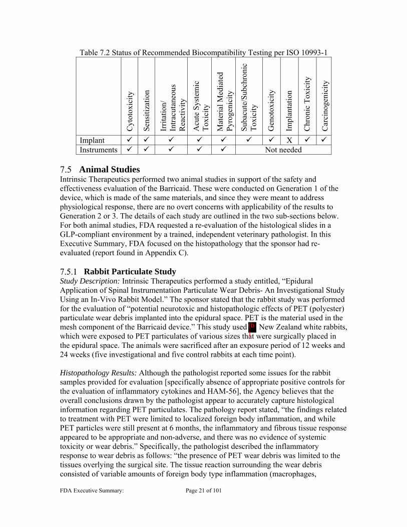
Table 7.2 Status of Recommended Biocompatibility Testing per ISO 10993-1
Cyt
otox
icit
y
Sen
siti
zati
on
Irri
tati
on/
Intr
acut
aneo
us
Rea
ctiv
ity
Acu
te S
yste
mic
T
oxic
ity
Mat
eria
l Med
iate
d P
yrog
enic
ity
Sub
acut
e/S
ubch
roni
c T
oxic
ity
Gen
otox
icit
y
Impl
anta
tion
Chr
onic
Tox
icit
y
Car
cino
geni
city
Implant X Instruments Not needed
Animal Studies
Intrinsic Therapeutics performed two animal studies in support of the safety and effectiveness evaluation of the Barricaid. These were conducted on Generation 1 of the device, which is made of the same materials, and since they were meant to address physiological response, there are no overt concerns with applicability of the results to Generation 2 or 3. The details of each study are outlined in the two sub-sections below. For both animal studies, FDA requested a re-evaluation of the histological slides in a GLP-compliant environment by a trained, independent veterinary pathologist. In this Executive Summary, FDA focused on the histopathology that the sponsor had re-evaluated (report found in Appendix C).
Rabbit Particulate Study Study Description: Intrinsic Therapeutics performed a study entitled, “Epidural Application of Spinal Instrumentation Particulate Wear Debris- An Investigational Study Using an In-Vivo Rabbit Model.” The sponsor stated that the rabbit study was performed for the evaluation of “potential neurotoxic and histopathologic effects of PET (polyester) particulate wear debris implanted into the epidural space. PET is the material used in the mesh component of the Barricaid device.” This study used New Zealand white rabbits, which were exposed to PET particulates of various sizes that were surgically placed in the epidural space. The animals were sacrificed after an exposure period of 12 weeks and 24 weeks (five investigational and five control rabbits at each time point).
Histopathology Results: Although the pathologist reported some issues for the rabbit samples provided for evaluation [specifically absence of appropriate positive controls for the evaluation of inflammatory cytokines and HAM-56], the Agency believes that the overall conclusions drawn by the pathologist appear to accurately capture histological information regarding PET particulates. The pathology report stated, “the findings related to treatment with PET were limited to localized foreign body inflammation, and while PET particles were still present at 6 months, the inflammatory and fibrous tissue response appeared to be appropriate and non-adverse, and there was no evidence of systemic toxicity or wear debris.” Specifically, the pathologist described the inflammatory response to wear debris as follows: “the presence of PET wear debris was limited to the tissues overlying the surgical site. The tissue reaction surrounding the wear debris consisted of variable amounts of foreign body type inflammation (macrophages,
(b)


FDA Executive Summary: Page 25 of 101
8 CLINCIAL STUDY AND DESIGN Study design
The clinical trial was a multi-center, prospective, randomized (1:1), largely unmasked, concurrently controlled superiority OUS clinical trial. The study enrolled 554 subjects at 21 clinical sites, all located in Northwestern Europe implanting 44 Generation 2 devices and 222 Generation 3 devices. The trial duration was from December 2010 to May 2017. The last subject was enrolled on October 14, 2014. The purpose of the trial was to evaluate the safety and effectiveness of the Barricaid following a limited discectomy, compared to a limited discectomy alone in subjects with radiculopathy, a posterior or posterolateral herniation and a large anular defect (identified intraoperatively). The sponsor chose the study control, to be a limited discectomy, a common technique. However as outlined in Section 2.5, it is important to note that there are many appropriate surgical options for treatment of symptomatic lumbar disc herniations. The sponsor notes that the inclusion/exclusion criteria for the study ensure that only a high-risk patient population (i.e., patients with large anular defects) was included in the pivotal clinical trial. The sponsor maintains that this patient population is easily identified intra-operatively with a simple defect measurement technique. According to the study protocol, “The surgeon will perform a conservative or limited (Spengler technique) discectomy. This technique will remove any nucleus that has migrated within the anular defect or beyond the anular wall (including sequestered fragments). Surgeons will be specifically trained to remove loose fragments of nucleus from within the disc in patients with extrusions or protrusions, per Spengler’s published technique. Upon completion of the discectomy and measurement of the defect, the patient will be randomized if not excluded due to defect size.”
Enrollment Criteria Patients that presented to clinic with symptoms concordant with herniated lumbar discs were screened. Subjects were enrolled in the study based on inclusion and exclusion criteria prior to randomization. Randomization (1:1) was performed intra-operatively, following measurement of the anular defect, which, according to the sponsor, minimizes any potential patient selection or treatment bias. Enrolled subjects who failed to meet the study protocol requirement during intraoperative screening (i.e. anular defect too large, anular defect too small, or “other reasons”) did not proceed to randomization. All subjects randomized to the treatment group and who subsequently did not have the Barricaid implanted were considered as treatment failures and were required to be followed per the protocol. Study investigators enrolled subjects with radiculopathy (with or without back pain), who were unresponsive to at least 6 weeks of non-operative treatment. Candidates for enrollment had a posterior or posterolateral herniation, characterized by radiographic confirmation of neural compression using MRI. The following specific eligibility criteria were specified in the protocol:
Inclusion Criteria: Any subject meeting all of the following criteria was eligible for inclusion in this trial:
FDA Executive Summary: Page 26 of 101
1. Age 21 to 75 years old and skeletally mature (male or female). 2. Subjects with posterior or posterolateral disc herniations at one level between L1 and S1 with radiographic confirmation of neural compression using MRI. [Note: Intraoperatively, only patients with an anular defect (post discectomy) between 4mm - 6mm tall and 6mm - 10mm wide shall qualify.] 3. At least six (6) weeks of failed, conservative treatment prior to surgery, including physical therapy, use of anti-inflammatory medications at maximum specified dosage and/or administration of epidural/facet injections.; 4. Minimum posterior disc height of 5mm at the index level 5. Radiculopathy (with or without back pain) with a positive Straight Leg Raise (0 – 60 degrees) (L45, L5S1) or Femoral Stretch Test (L12, L23, L34) 6. Oswestry Questionnaire score of at least 40/100 at baseline. 7. VAS leg pain (one or both legs) of at least 40/100 at baseline 8. Psychosocially, mentally and physically able to fully comply with the clinical protocol and willing to adhere to follow-up schedule and requirements
The Agency noted that Inclusion Criterion #2 identified posterior or posterolateral herniations for study inclusion. However, “posterior” herniatons could include a range of locations along the circumference of the disc anulus (e.g. central, posterolateral, foraminal, extraforaminal, etc.). Surgical access required for placement of an anular device is associated with different levels of complexity and risk depending on location, especially for the treatment of central and extraforaminal disc herniations. Notably, the sponsor also included subjects with grade 1 spondylolisthesis and subjects who have undergone prior lumbar surgery not at the index level. Such factors potentially increased the risk of enrollment of subjects with leg pain due to etiologies other than lumbar disc herniation and increased the likelihood of enrollment of a non- homogeneous patient population. Inclusion Criterion #5 states that subjects with “Radiculopathy (with or without back pain) with a positive Straight Leg Raise (0 – 60 degrees) (L45, L5S1) or Femoral Stretch Test (L12, L23, L34) are eligible for inclusion”. However, neither a specific definition for radiculopathy nor specific criteria for identification of subjects with radiculopathy were identified in the Study Protocol. Notably, the study protocol included patients with either unilateral or bilateral leg pain. In contrast, a recent publication by Genevay et al.[36] states that the item with the highest weight in diagnosis of radicular pain is monoradicular leg pain distribution. It was also noted that pathology other than a disc herniation may be responsible for a positive straight leg raising test less than 30˚ [37]. Additionally, Straight Leg Raise test used has a degree of imprecision due to the varied angle criteria and its high sensitivity, but low specificity as outlined in Section 2.2. [37]. It is common for lumbar discectomy studies to require that eligible subjects have clinical signs and symptoms of lumbar radiculopathy which correlate with level and side of a lumbar disc herniation observed on imaging studies. For example, SPORT used the following criteria for study eligibility[9]:
“For intervertebral disc herniation, patients are eligible for SPORT if they have radicular pain and evidence of nerve root compression with a positive nerve root

FDA Executive Summary: Page 27 of 101
tension sign (positive straight leg raise test or femoral tension sign). Alternatively, they may have a reflex (asymmetric depressed reflex), sensory (asymmetric decreased sensation in a dermatomal distribution), or motor (asymmetric weakness in a myotomal distribution) deficit with associated radicular symptoms and positive nerve root tension signs. In addition, a confirmatory imaging study (MRI or CT) must indicate an IDH (a protrusion, extrusion, or sequestered fragment) at a location (level and side) corresponding to the patient’s radicular signs or symptoms. Patients with only a bulging disc (circumferential symmetric extension beyond the interspace) are not eligible.”
Exclusion Criteria
Any subject meeting any one of the following criteria was not eligible for enrollment into the trial. 1. Spondylolisthesis Grade II or higher (25% slip or greater) 2. Subject requires spinal surgery other than a discectomy (with or without laminotomy) to treat leg/back pain (scar tissue and osteophyte removal is allowed) 3. Subject has back or non-radicular leg pain of unknown etiology 4. Prior surgery at the index lumbar vertebral level 5. Subject requiring a spine DEXA (i.e., patients with SCORE of ≥ 6) with a T Score less than -2.0 at the index level. For patients with a herniation at L5/S1, the average T score of L1-L4 shall be used 6. Subject has clinically compromised vertebral bodies in the lumbosacral region due to any traumatic, neoplastic, metabolic, or infectious pathology 7. Subject has sustained pathologic fractures of the vertebra or multiple fractures of the vertebra or hip 8. Subject has scoliosis of greater than ten (10) degrees (both angular and rotational) 9. Any metabolic bone disease 10. Subject has an active infection either systemic or local 11. Subject has cauda equina syndrome or neurogenic bowel/bladder dysfunction 12. Subject has severe arterial insufficiency of the legs or other peripheral vascular disease. (Screening on physical examination for patients with diminution or absence of dorsalis pedis or posterior tibialis pulses. If diminished or absent by palpation, then an arterial ultrasound is required with vascular plethysmography. If the absolute arterial pressure is below 50mm of Hg at the calf or ankle level, then the patient is to be excluded.) 13. Subject has significant peripheral neuropathy, patient defined as a patient with Type I or Type II diabetes or similar systemic metabolic condition causing decreased sensation in a stocking-like or non-radicular and non-dermatomal distribution in the lower extremities 14. Subject has insulin-dependent diabetes mellitus 15. Subject is morbidly obese (defined as a body mass index >40, or weighs more than 100 lbs. over ideal body weight) 16. Subject has been diagnosed with active hepatitis, AIDS, or HIV 17. Subject has been diagnosed with rheumatoid arthritis or other autoimmune disease 18. Subject has a known allergy to titanium, polyethylene or polyester materials 19. Any subject that cannot have a baseline MRI taken

FDA Executive Summary: Page 29 of 101
In the current study, the protocol called for the surgeon to measure and record the defect height and width by inserting different sized proprietary Defect Measurement Tools, until the tool fits snugly into the anulotomy. Upon completion of the discectomy and measurement of the defect, the subject was randomized if not excluded due to defect size. Please note that only subjects with an anular defect (post-discectomy) between 4mm - 6mm tall and 6mm - 10mm wide were eligible for randomization (the sponsor is seeking approval for the 12mm device, but this device size was not studied in the RCT).
Barricaid Group Treatment The Barricaid was designed for implantation into either the inferior or the superior vertebral body. Successful implantation requires proper alignment in the sagittal and transverse axes, as well as proper placement above the endplate of the vertebral body. Depending on the location of the anular defect, removal of bone from the lamina may be required to allow adequate access. The surgeon inserts the distal end of the Sizing Trial Tool through the anulotomy, keeping the endplate guide along the surface of the endplate of the target vertebral body and ensuring the distal end of the Tool is against the posterior wall of the vertebra. After selection of the appropriate Barricaid size (8 mm, 10 mm or 12 mm), the device is inserted using a pre-loaded, disposable delivery tool. The disposable delivery tool is comprised of a delivery sheath, a pusher, and a strike-cap as shown in the Surgical Technique Manual (Appendix A). Proper positioning of the Barricaid requires centering of the device within the anular defect. If the delivery tool and Barricaid are not centered within the anular defect, the mesh may be directed into healthy anulus and/or buckling of the mesh may occur. If a surgeon notes mesh buckling, he or she is instructed to stop implantation and remove and discard the implant and delivery tool. If the anchor penetrated the bone in the first attempt, the surgeon should target the opposing vertebral body for implantation, repeating measurement of the defect and use the sizing trial to confirm access and angle. Before pulling back on the strike cap of the delivery tool, the surgeon should confirm proper placement of the device. The baseplate (i.e., bottom of the anchor) of the Barricaid should be parallel to the surface of the endplate. The head of the anchor of the Barricaid should rest on the surface of the endplate. If the head of the anchor is too high, it can contact and damage the opposing endplate. If the head is too low and penetrates the vertebral body below the endplate, the head can damage the Barricaid mesh and the endplate. Figure 8.1 demonstrates correct alignment of the Barricaid and insertion tool:


FDA Executive Summary: Page 31 of 101
Table 8.1 Study Assessments
Study Time Period
Procedure
Pre
-op
as
sess
men
t/S
cree
nin
g
Day
of
Su
rger
y
6 w
ks±
2 k
s
3 m
os ±
2 w
ks
6 m
os ±
2 w
ks
12 m
os ±
2 w
ks
24 m
os ±
2 w
ks
(an
d
annu
al lo
ng-t
erm
fol
low
-u
p)
Pre-treatment information
Informed Consent X
Entry Criteria/Pre-operative screening X
Intra-operative screening X
Prior Treatments X
Physical Examination X
Case Report Forms (CRF) X X X X X X X
Medications X X X X X X X
Clinical Assessment
Pregnancy Screening X
Oswestry (Pain/Function) X X X X X X X
Back and Leg VAS Score X X X X X X X
SF-36v2 Health Survey X X X X X X X
Neurological Assessment X X X X X X X
Adverse event (AE)(if applicable) X X X X X X
Outstanding AE (if applicable) X X X X X
Study deviation-patient (if applicable) X X X X X X
Study deviation-site (if applicable) X X X X X X
Radiographic Assessment
MRI X X X
CT X X X
Lateral Flexion/Extension X X X
Lateral X X X X X X X
Anterior/Posterior X X X X X X X
Radiographic Analysis: The sponsor provided an Imaging Charter that provided instruction on how they quantitatively and qualitatively measured the radiographic findings. Please note that while this was discussed in pre-submissions and interactions with FDA, no agreement on the assessment methods of the imaging charter was reached. The imaging analysis was done by an independent imaging core lab under an Imaging Charter as provided in Appendix E.

FDA Executive Summary: Page 32 of 101
Imaging Charter: The Imaging Charter documented the evaluation definitions for the independent core lab to make assessments on the radiographic images taken as part of the study assessments. The main analysis performed includes the assessment of reherniation, disc height, device integrity, and spontaneous fusion, which are all part of the primary endpoint. These are described in greater detail below in Section 8.6.2. In addition, due to safety concerns related to the EPLs, there are a number of evaluations conducted to assess, characterize and quantify the lesions in terms of size, growth and behavior. A number of other measures are also described in the imaging charter, which include anular tears/fissures on MRI, septations, mineralization, endplate Modic changes, Pfirrmann Grade, ossification, spontaneous fusion, and disc degeneration (Kellgren and Lawrence method). Quantitative measurements include: angular motion, translational motion, disc height, and spondylolisthesis. The charter also describes a number of assessments related to characterization of the EPLs. The number of lesions in each vertebral body (superior and inferior) was recorded and location within the vertebral body was noted in each plane (grid method). Lesions were qualitatively evaluated to proximity of device (e.g. not proximate, proximate to mesh, proximate to anchor head, proximate to anchor plate, proximal to multiple components including mesh, proximal to multiple components not including mesh, no lesion, or control group), and lesion side (ipsilateral or contralateral). Lesion Size Measurement: Radiologists used sagittal, coronal and axial plane images to identify the lesion(s) separately for all superior and inferior vertebrae. The slice with the largest lesion area in each plane was subsequently chosen to be measured. The lesion was approximated independently for each plane (sagittal, coronal and axial), and for each vertebral body (superior and inferior) as an ellipse. Volume of the lesions was approximated via linear regression with the three-plane average area of the lesion. Sponsor conducted a pilot study to develop an approximation method to measure EPC volume and reduce radiation. This pilot study used a “true volume” of lesions plotted against a “3-plane average area” of each lesion to determine a linear regression best fit line to predict volume seen the table below:

FDA Executive Summary: Page 33 of 101
Figure 8.2 Estimation of EPL Volume The regression line used to estimate volume based on correlation of volume to 3-plane average of EPL area. Note that there is significant correlation between volume and area; however, additional calculations and conclusion based on specific volumetric numbers should be considered with caution. This linear approximation showed that there was a highly significant correlation between volume and area. The r2 = 0.7129, which the sponsor states is a high level of correlation and conclusions drawn from the volume estimations of size are expected to be no different from conclusions drawn from area analyses. Based on FDA’s assessment, this method appears to have a large degree of variability with the linear approximation of volume. While area and volume have a degree of correlation, additional calculations based on a volume approximation may not be accurate, particularly when attempting to track the change in volume of specific lesion, so these volume estimations should be considered with caution. This volume approximation may further confound the measurements and calculations of growth and stability over time due to the imprecise measurements. The area approximations for lesion size in each plane may be most accurate when tracking the size of each lesion. However, it was noted that the largest lesion sizes, based on 3-plane average, and their associated vertebral bodies were imaged and reconstructed for a more accurate true measurement. Vertebral Body Size Measurement: Vertebral body area of the superior vertebral body was measured using sagittal, coronal and axial slices using MRI, with CT used if MRI was unavailable or has missing anatomy. The inferior vertebral body area was also measured in three planes; however, initial measurements were collected using CT, with MRI used when CT is unavailable or has missing anatomy. The area of each vertebral body in each plane was estimated by the measurement in the x and y axis. This includes area estimate in the axial plane as well, which is more ellipse in shape. Vertebral body volume measurements were taken in a pilot study using CT measurements and averaged. This average vertebral body size was for all quantitative summary analyses of volume percentage calculations.


FDA Executive Summary: Page 36 of 101
received (i.e., subjects randomized to the treatment group who do not have the Barricaid device implanted will be classified as control subjects for this analysis). The sponsor evaluated safety in the AT population. Per-protocol patient population (PP): The PP patient population included all mITT subjects who received the assigned treatment and had no major inclusion/exclusion as protocol deviations identified by the Independent Physician Adjudicator (IPA) likely to impact primary and key secondary outcome assessments. Screen failure population (SF): The SF patient population included all subjects consented in whom the discectomy procedure is initiated but randomization did not occur. These subjects were not followed after surgery. Generation 3 Population (G3): The Generation 3 population included all mITT subjects randomized to control and all mITT subjects randomized to the treatment in which an attempt at treatment with the Generation 3 device was made. Subjects will be included in the treatment group to which they are randomized, regardless of the treatment received. The primary randomized ITT population was 554 subjects, 276 to Barricaid and 278 to Control. However, in four Barricaid subjects, implantation was not attempted, leaving 272 Barricaid and 278 Control subjects in the primary mITT population. Note that there were also 93 subjects which underwent surgery but were intra-operative screen failures and were not randomized. The As-Treated (AT) population consists of the 550 mITT subjects grouped as 267 Barricaid and 283 Control since there were 5 of the 272 attempted Barricaid subjects which did not receive the Barricaid treatment. Note that the sponsor also states that there were 5 control subjects who were treated with a commercially-available Barricaid device after becoming terminal study failures. These subjects were considered control composite clinical success failures due to the secondary surgical intervention. All follow-up among these subjects were included as part of the control group.
Other Subanalyses The sponsor has done many analyses of subjects with endplate lesions. Note that the sponsor calls the endplate lesions EPCs (endplate changes). FDA will refer to these findings as EPL’s (endplate lesions). The population with lesions consisted of 223 subjects (83.5%) in the Barricaid group and 103 subjects (36.4%) in the Control. Further, in the Barricaid group, the sponsor focused on subjects with EPL’s that were proximate to the occlusion component of the device (mesh-proximate), and there were 125 (46.8%) Barricaid subjects in this group. Note that initially, FDA had asked for a “mesh-proximate” determination with respect to lesions in Control subjects, defined as lesions in areas in a similar location to where the occlusion component was located in Barricaid subjects. However, this was determined later not to be helpful and subsequent comparisons were made to all Control subjects.
Data Analysis, Adjudication, and Study Oversight Primary and secondary effectiveness endpoint measurements and adjudications were performed by a combination of an independent adjudication committee, a radiographic
FDA Executive Summary: Page 37 of 101
core laboratory, a Contract Research Organization (CRO), or a Contract Research associate (CRA) using pre-specified charters and processes, as follows: Data and Safety Monitoring Board (DSMB) was responsible for monitoring the overall conduct of the study. The DSMB reviewed accumulating data from the ongoing clinical trial on an at least a quarterly basis. This board consisted of independent experts based in the United States in the field of neurological and orthopedic spine surgery and radiology, with access to an independent statistician as needed. The purpose of the DSMB was to advise Intrinsic Therapeutics regarding the continued safety of then-current and future participants. Radiographic Core Laboratory (Intrinsic Imaging, Inc.-unrelated to the sponsor) evaluated all subject imaging (x-rays, MR and CT) in accordance with the sponsor’s proprietary Radiographic Evaluation Protocol. Recall that the radiologic protocol was formulated independent of FDA. The radiographic core lab provided data to the sponsor approximately 2-3 times annually. A Contract Research Organization (CRO) or sponsor Clinical Research Associate (CRA) monitored each study site regularly, according to the Clinical Monitoring Plan. Through the CRO or CRA, the sponsor audited each site regularly and handled any required corrective or preventive actions through the sponsor’s quality management system.
9 STUDY RESULTS

FDA Executive Summary: Page 38 of 101
Subject Accounting The sponsor provided patient accounting and compliance data in Table 9.1 below, showing over 94% visit compliance at Month 24 for expected due Barricaid subjects, and 91% compliance for control subjects. Table 9.1 Long-Term (>Month 24) Patient Accounting and Follow-up Compliance Table Efficacy Evaluable (mITT) Barricaid (I) and Control Subjects (C)

FDA Executive Summary: Page 40 of 101
Other (18); The sponsor notes that eight listed as “other” by site should have failed screening with inclusion/exclusion criteria (i.e., defect too large/small, two-level, or no herniation).
Surgeons did not enroll 26 (approximately 4%) subjects because of anular defects that were too small. The distribution of anular defect types in the Barricaid study differed from the Carragee series[33], in which approximately half of the subjects were identified as having a small slit-like/small anular defect, and were called the “Fragment-Fissure” group (49%). Carragee described treatment of this group with removal of fragments through the slit-like anular defect, and noted a 1% rate of reherniation in this group. The sponsor attributed differences in the distribution of anular defect types in the Barricaid study compared to the Carragee series, including the small number of subjects with “fragment-fissure” anular defects, to the specific inclusion/exclusion criteria in their study. The population that was screened in the Barricaid study was broad and simply presented with “complaints concordant with a herniated lumbar disc.” This would not appear to limit enrollment to subjects with specific types of anular defects associated with lumbar disc herniations, and would seemingly also include subjects with conditions other than disc herniations, subjects whose symptoms did not necessitate surgery as well as disc herniation subjects who preferred nonsurgical treatment. No conclusions could be made based on the description of patient screening provided by the sponsor, as this information was not presented as part of the PMA. It was also noted that some of the “other” failures were due to inaccessibility (e.g. location “too medial” or the “angle was too difficult”). The Panel will be asked whether additional revisions or clarifications to the Indications for Use should be considered, including modification of the terms “posterior” or “posterolateral”, as use as these terms are vague and may include approaches that are not feasible for this procedure.
Protocol deviations An Independent Physician Adjudicator was responsible for categorizing protocol deviations as device-related deviations, inclusion/exclusion related deviations, deviations related to informed consent process, or as visit/assessment related deviations (visit not done or out of window, testing not completed or not per protocol, etc.). These deviations occurred in 2267 total visits for the Barricaid arm and 1822 visits in the control arm. Major Protocol Deviations: The Independent Physician Adjudicator classified five deviations in 5 subjects as major deviations that could impact the analysis of study endpoints; this resulted in exclusion of these subjects from the per protocol analysis. The sponsor provided a list of these major deviations below in Table 9.3:

FDA Executive Summary: Page 41 of 101
Table 9.3 Major Protocol Deviations
Device-Related Deviations: Device-related deviations were associated with sizing, the use of a commercial implant, or second attempts to place the device. By definition, device-related deviations occurred in the treatment group only, and included six devices that were undersized for the defect width at three different investigational sites. In all cases, defects were wider than 8mm, which per the Surgical Technique Manual would dictate the use of a 10mm wide device; however, an 8mm device was implanted. Reasons for undersizing included “there was not enough space to place the larger device without undue damage to the dura or nerve root” and “It was the decision of the surgeon” Three of the six subjects with undersized implants had secondary surgeries:
: 73 months after index surgery the subject underwent a removal of the device after the subject reported new or increased pain due to reherniation at the index level.
: 39 months after index surgery the subject underwent an osteophyte and scar tissue removal, as well as removal of bulging nucleus at the index level for nerve root compression.
: 34 months after index surgery the subject underwent a revision surgery during which the surgeon removed the detached occlusion component and an extruded reherniation.
Inclusion/Exclusion Related Deviations: There were 10 subjects with 11 inclusion/exclusion criteria deviations in the Treatment group and 11 subjects with 12 inclusion/exclusion criteria deviations in the control group. Five of the inclusion/exclusion related deviations are included above in Table 9.3. The remainder of the inclusion/exclusion related deviations involved pre-op pregnancy, DEXA scan, negative femoral stretch, and negative straight leg raise tests.
Informed Consent Related Deviations: Informed Consent Form (ICF) Related Deviations primarily related to use of the wrong consent version (77 subjects). The unapproved and expired ICF versions differed from approved ICFs primarily by
(b) (6)
(b) (6)
(b) (6)

FDA Executive Summary: Page 42 of 101
formatting differences and did not did not differ substantially in the ICF language. These deviations occurred equally in the treatment and control groups. Visit/Assessment Related Deviations: Missed visits occurred in 26 Barricaid and 32 Control subject visits through the 24-month follow-up. Visit and assessment deviations were most commonly associated with visits or assessments done out of window and were similar between groups. There were 133 out-of-window deviations for 34.1% (94/276) of subjects in the Barricaid group. For the control group, there were 129 window deviations in 37.1% (103/278) of subjects. Assessment-related deviations occurred for Radiology assessments, neurological exam and questionnaires. Fifty-two (32 control and 20 Barricaid) visits had incomplete imaging sets. One hundred fifty-three (76 control and 77 Barricaid) visits had partial missing imaging. There were 18 of 276 Barricaid subjects (6.5%) with a complete imaging set missing, and 29 of 278 control subjects with a complete imaging set missing. There were similar proportions of subjects in each group (23% Barricaid, 24% control) with partially missing imaging sets.
Baseline Demographics and Baseline Status Among the subject population for the Barricaid clinical trial, there were no apparent statistical differences in any of the baseline functional outcomes scores, demographics (sex, age, height, weight, BMI, smoker or race) or comorbidity variables. The baseline data between the two groups was similar for work status, symptom duration and medication use. Seventeen control subjects had non index-level prior lumbar decompression surgery as compared to six Barricaid subjects. The sponsor presented intraoperative data in Table 9.4:

FDA Executive Summary: Page 43 of 101
Table 9.4 Summary of Baseline Intra-Operative Data
The patient population included 96 Barricaid subjects (35.3%) and 108 control subjects (38.8%) with Fragment-Contained disc herniation morphology, as described by Carragee[33]. Carragee treated such subjects with an oblique incision in the anulus, and removal of subligamentous fragments without creation of a large anular defect. Forty-two

FDA Executive Summary: Page 45 of 101
Adverse Events (AEs) To demonstrate that Barricaid is safe, Intrinsic Therapeutics collected all adverse event data and had safety data adjudicated by an independent Data Safety Monitoring Board (DSMB). All safety data in this section represents the As Treated (AT) dataset so the safety information directly reflects the actual treatment received. Adverse Events: AEs were assessed and recorded in the subject’s medical record and then transcribed onto the appropriate case report form at each visit. AE definitions “Any untoward medical occurrence, unintended disease or injury or untoward clinical signs (including an abnormal laboratory finding) in subjects, users, or other persons whether or not related to the investigational medical device [ISO 14155].” Definitions used to categorize AEs are included in Appendix G. Relationship definitions “Due to the temporal proximity of the AE to investigational product administration, there is a reasonable possibility that the product may have caused the AE or may have contributed to the severity or duration of an event caused by other means.” Table 10.1 below from the protocol shows AE relationship assessment: Table 10.1 AE Relationship Definitions
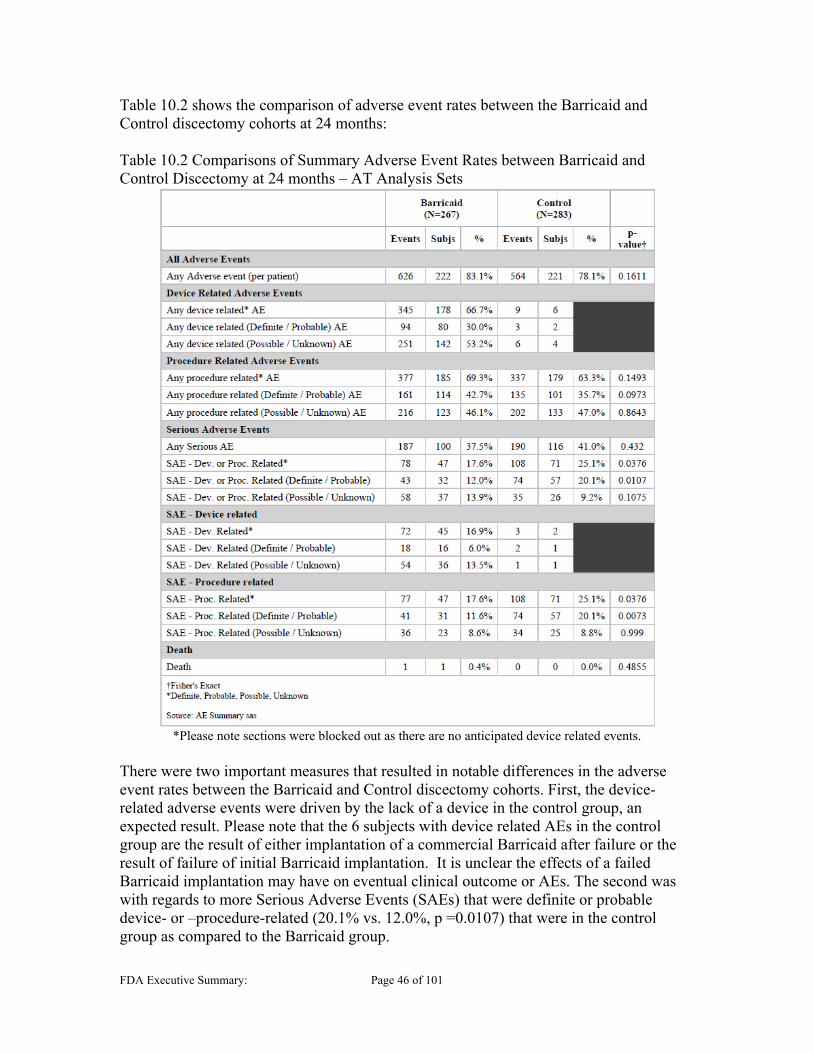
FDA Executive Summary: Page 46 of 101
Table 10.2 shows the comparison of adverse event rates between the Barricaid and Control discectomy cohorts at 24 months: Table 10.2 Comparisons of Summary Adverse Event Rates between Barricaid and Control Discectomy at 24 months – AT Analysis Sets
*Please note sections were blocked out as there are no anticipated device related events.
There were two important measures that resulted in notable differences in the adverse event rates between the Barricaid and Control discectomy cohorts. First, the device-related adverse events were driven by the lack of a device in the control group, an expected result. Please note that the 6 subjects with device related AEs in the control group are the result of either implantation of a commercial Barricaid after failure or the result of failure of initial Barricaid implantation. It is unclear the effects of a failed Barricaid implantation may have on eventual clinical outcome or AEs. The second was with regards to more Serious Adverse Events (SAEs) that were definite or probable device- or –procedure-related (20.1% vs. 12.0%, p =0.0107) that were in the control group as compared to the Barricaid group.
FDA Executive Summary: Page 47 of 101
Device and Procedure Related, and Serious AEs When comparing the procedure related AEs, the Barricaid and control group have comparable numbers; however, when this is further parsed into device or procedure related SAE, there is a nominal difference. It is unclear why there are more procedure related SAEs for the control group if both groups have the same procedure conducted. Despite the addition of a device, the combined device- or procedure-related SAE rate was still higher in the control group, suggesting discectomy with Barricaid has a more favorable safety profile compared to discectomy alone. Additional AE tables outlining All AEs, SAEs, procedure-related AEs, device related AEs and device- and procedure-related SAEs are included in Appendix G. There were two notably different outcomes seen. When considering all AEs, Device Deficiencies AE (as categorized by the DSMB defined in Appendix G) were observed in greater number in the Barricaid group as compared to the control (13.1% vs. 0.4%, p <.001) which was expected since there is no device in the Control group. Secondly, the disc herniation events were lower in the Barricaid group (24.7% vs. 38.5%, p=.002). When considering SAEs, again the Device Deficiency SAEs (4.5% vs. 0.0%, p<.001) were driven by the lack of a device in the control group and expected and disc herniation SAEs were again more prevalent in the control group (23.3% vs. 13.9%, p=0.010). This trend continued in other breakdowns, including device or procedure related AEs and SAEs. One other notable difference was Necrosis of Bone or Resorption (as categorized by DSMB) when considering all AEs, which represents the end plate lesions in Barricaid subjects observed by the clinical sites. With the Barricaid device, a long-standing focus of concern has been continued growth of end plate lesions, especially those in proximity to the mesh component of the device. These AE observations are further supported by observations in the baboon study as described in Section 7.5.2 and histology conducted on peri-prosthetic tissue as part of the retrieval analysis described in Section 10.6.2. Increasing volume of lesions could theoretically be of clinical consequence. However, structural failure of vertebral bodies or compromised SSI due to loss of bone stock as yet remain theoretical concerns. End plate lesions are discussed in detail in Section 10.2 of this document. Table G8 in Appendix G shows counts of procedure or device related SAE. This is notable as it shows that approximately one third of procedure- or device-related SAEs occurred after 24 months, calling into question the adequacy of a 24 month endpoint assessment. The accrual of device-related reoperations after 24 months may make assumption of greater safety with the Barricaid device premature at this time.
Neurological Status Subjects who have either maintained or improved in their neurological status as it relates to the subject’s index level are a success. Neurological status success is based upon Straight Leg Raising (SLR) (L4/5 and L5/S1) or Femoral Stretch Test (FST)(L1/2, L2/3, L3/4 only), motor examination, sensory examination, and reflex examination. The overall success as well as individual components for motor, sensor, reflex as well as SLR or FST

FDA Executive Summary: Page 48 of 101
show comparable outcomes at all time points. The tables showing neurological success are included in Appendix J.
Secondary Surgery Index Level Secondary Surgical Interventions (SSIs) The Barricaid study included success criteria that required no SSI (reoperations, revisions, removals or supplemental fixations). Through 24 months, there were 23 subjects with SSIs in the Barricaid group (23/267, 8.6%) compared with 45 subjects with SSIs in the control group (45/278, 16.2%, p=0.007). In the Barricaid group, there were 16 SSI events occurring in 15 subjects who did not undergo secondary surgery until after 24 months. In the control group, there were 14 SSI events occurring in 12 subjects who did not undergo secondary surgery until after 24 months. Secondary surgeries in subjects up to day 790 of treatment (24 months + 60 days) were failures in the primary endpoint. SSIs after 24 months were not counted as primary endpoint failures. Table 10.3 shows the index-level SSIs cumulatively as the proportion of subjects who had an SSI in at each time point: Table 10.3 Time-course cumulative distribution of proportion of subjects with an SSI at Index level
*Please note that the risk set calculations are based on “days” and not assessment windows and therefore may have numbers that are slightly different than reported in text at each timepoint.
Survival analysis present an estimated approach that attempts to eliminate the bias of failures carried forward. Again, conclusions from the theoretically due analyses are limited, since this approach does not include all events observed during the study. The following shows the Kaplan-Meier (product-limit) estimate for SSI in Table 10.4. It is also important to note that the nominal p-value presented on the difference on the entire dataset and should not be applied to significance at any one specific time point. Table 10.4 Survival Analysis – Index Level SSI (mITT) The “F” columns show failures, while “C” are censored subjects. The cumulative % is the survival estimate at each time point, with the predicted upper (UB) and lower bounds (LB). The p-value is reflective the conclusion that the entire curve is significantly different, not at each individual time points.
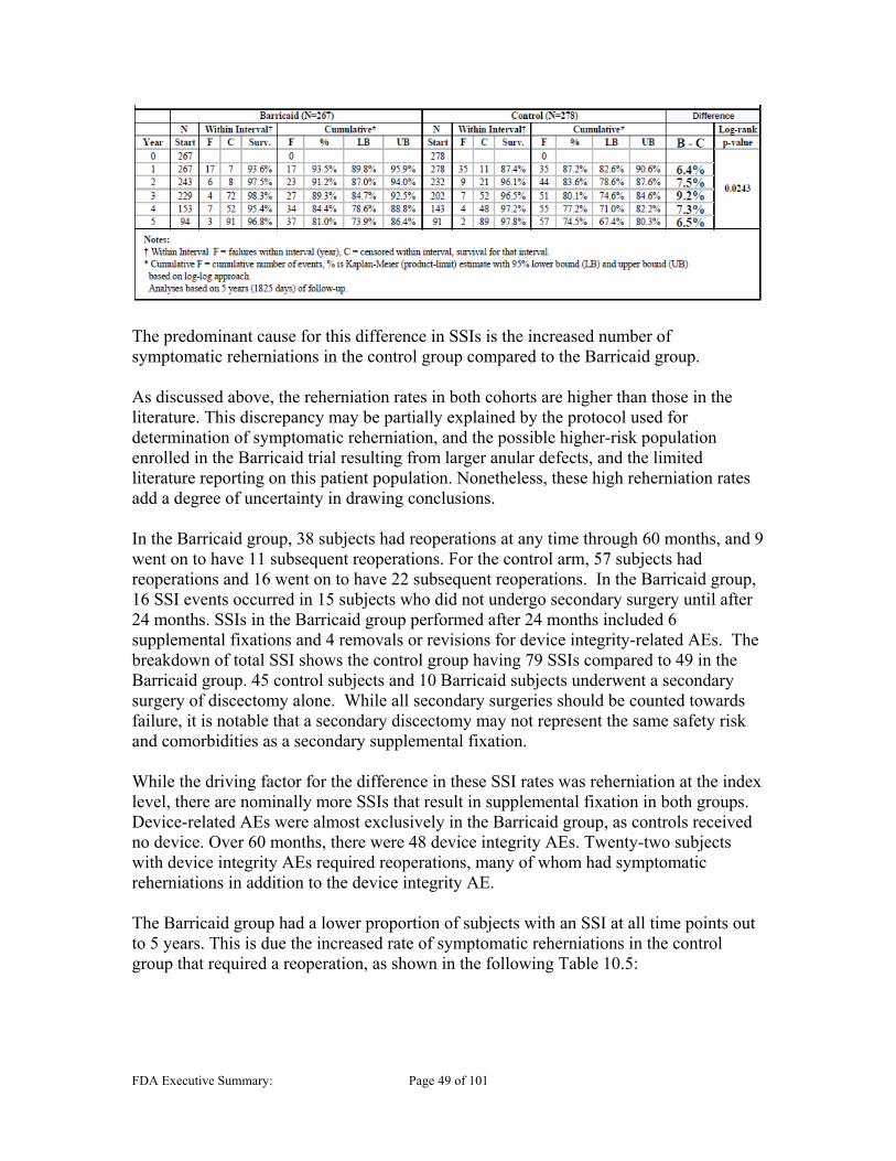
FDA Executive Summary: Page 49 of 101
The predominant cause for this difference in SSIs is the increased number of symptomatic reherniations in the control group compared to the Barricaid group. As discussed above, the reherniation rates in both cohorts are higher than those in the literature. This discrepancy may be partially explained by the protocol used for determination of symptomatic reherniation, and the possible higher-risk population enrolled in the Barricaid trial resulting from larger anular defects, and the limited literature reporting on this patient population. Nonetheless, these high reherniation rates add a degree of uncertainty in drawing conclusions. In the Barricaid group, 38 subjects had reoperations at any time through 60 months, and 9 went on to have 11 subsequent reoperations. For the control arm, 57 subjects had reoperations and 16 went on to have 22 subsequent reoperations. In the Barricaid group, 16 SSI events occurred in 15 subjects who did not undergo secondary surgery until after 24 months. SSIs in the Barricaid group performed after 24 months included 6 supplemental fixations and 4 removals or revisions for device integrity-related AEs. The breakdown of total SSI shows the control group having 79 SSIs compared to 49 in the Barricaid group. 45 control subjects and 10 Barricaid subjects underwent a secondary surgery of discectomy alone. While all secondary surgeries should be counted towards failure, it is notable that a secondary discectomy may not represent the same safety risk and comorbidities as a secondary supplemental fixation. While the driving factor for the difference in these SSI rates was reherniation at the index level, there are nominally more SSIs that result in supplemental fixation in both groups. Device-related AEs were almost exclusively in the Barricaid group, as controls received no device. Over 60 months, there were 48 device integrity AEs. Twenty-two subjects with device integrity AEs required reoperations, many of whom had symptomatic reherniations in addition to the device integrity AE. The Barricaid group had a lower proportion of subjects with an SSI at all time points out to 5 years. This is due the increased rate of symptomatic reherniations in the control group that required a reoperation, as shown in the following Table 10.5:

FDA Executive Summary: Page 50 of 101
Table 10.5 Secondary Surgical Interventions by Type Procedure Type Barricaid Control Discectomy 10 45 Discectomy + Barricaid Device* 0 5 Device Removal ± Discectomy 13 0 Supplemental Fixation ± Discectomy and/or Device Removal
21 16
Other 5 13 Total Procedures 49 79
*Please note that these 5 subjects were the subjects that received the device commercially after failure. Discounting excision of recurrent disc herniations alone, 34 Barricaid subjects required device removals or supplemental fixations with or without discectomy, with or without device removals. Five control subjects had subsequent excision of recurrent herniation and Barricaid implantation. Sixteen control subjects required excision of recurrent disc herniation and supplemental fixation. The “Other” procedures were primarily performed for wound issues.
Multiple subjects underwent more than one secondary surgical interventions. Reoperation trees for Barricaid and control treatment subjects can be found in Appendix H that visualizes first reoperations and all subsequent reoperations. The reoperation tree for the Barricaid group shows that 38 subjects had reoperations at any time through Day 1885 (60 months + 60 days) and nine went on to have subsequent reoperations. Reoperations included additional discectomies with and without fusion, fusions, pedicle fixations, wound revisions, decompressions and Barricaid removals. Two subjects had a third reoperation, and none had a fourth. The reoperation tree for the control arm shows that 57 subjects had reoperations and 16 went on to have 22 subsequent reoperations. These reoperations included additional discectomies with and without fusion, fusion with pedicle fixations, wound revisions and hematoma drainage. Four subjects went on to have a third reoperation, and two had a fourth reoperation. A time course for the types of SSI events that occurred is presented in Table 10.6. It is important to consider that some subjects in Table 10.6 had multiple events that occurred during the same time period. It is also notable that while the 60 month endpoint is presented, there are still subjects not yet due, so a rate calculation would not be representative.

FDA Executive Summary: Page 51 of 101
Table 10.6 Time course of SSI Event Type
In the Barricaid cohort, the 23 subjects who qualified as SSI failures by 24 months underwent 33 secondary surgeries. A summary of SSIs was provided in tabular form; however, theinformation provided was nonspecific when describing the “Reason for SSI.” These tables are included in Appendix H. The review team analyzed the operative reports and narratives of a symmetrical sample of Barricaid and control subjects who underwent SSI. Reasons for SSI in these tables include “new/increased pain”, “suspected or confirmed recurrent herniation” and “unknown.” In addition, the described surgical findings did not reliably permit an understanding of the reasons for the SSI. By 24 months there were also 7 supplemental fixation SSI’s in the Barricaid cohort and 17 in the control group mostly due to “instability.” In the control group, 14 SSI events occurred in 12 subjects who did not undergo secondary surgery until after 24 months. Summary of these events as well as for Barricaid subjects are included in Appendix H. Assessment of Difficulty of Barricaid SSI Relative to Control In assessing the benefit of the lower rate of SSI in the Barricaid group, the sponsor considered the possibility of increased difficulty in performing such procedures with an implant in place. To evaluate the difficulty of performing SSI in a subject with an implanted Barricaid, the sponsor compared the operative times of SSIs between Barricaid and Control subjects. The sponsor divided SSIs in each treatment arm into two groups: those involving a fusion or pedicle fixation and those that did not. The sponsor’s analysis found no statistical difference in operative time between Barricaid and Control for either category of intervention. The sponsor also compared adverse event rates rate and types of

FDA Executive Summary: Page 53 of 101
Qualitative Assessment
FDA solicited the help of a special government employee (SGE) with expertise in musculoskeletal radiology. The sponsor provided access to the raw images from the OUS clinical studies, and this expert musculoskeletal radiologist performed an independent, qualitative assessment of the raw images. The information in this sub-section is based on the qualitative review performed by the independent musculoskeletal radiologist. Please note that the conclusions reached by the SGE differ from the analysis performed by the core lab that compared the presence of sclerotic margins, septations, high attenuation within EPCs and reactive edema surrounding EPCs. Please defer to the sponsor’s summary for their qualitative assessments. The SGE musculoskeletal radiologist performed a qualitative review of a selection of images from study subjects to independently assess the findings reported by the sponsor. This review included assessment of images from each group Barricaid subjects with lesions, Barricaid subjects without lesions and control subjects with and without lesions. The sampling strategy for subject selection also attempted to sample from a variety of study sites for both Barricaid and control groups. X-rays, MRI and CT scans were reviewed, with CT being the primary focus and other imaging modalities serving as supplemental information for incomplete scans. It was noted that the MRI and X-rays were of inconsistent quality, while CT scans were more uniform in quality. Quantitative measurements were not evaluated as part of this independent radiographic review, and clinical significance or outcomes were not linked to the radiologic findings. The qualitative review by the SGE revealed that lesions in subjects from the Barricaid group, especially those proximal to the mesh component, were in her evaluation, visually distinguishable from the lesions in the control group. Endplate lesions/changes could be visually divided into three categories: endplate changes, endplate lesions, and lytic lesions. End plate changes This category could be seen in the control or Barricaid subjects but most often in the control subjects and not generally present on baseline imaging. Endplate changes are represented by more diffuse, less discrete endplate irregularity with some surrounding sclerosis and little propensity to increase over time; in some cases, these areas would become less prominent. The sclerotic margin tended to be thicker than those of the lytic lesions. There were no discrete, radiolucent lesions (lytic lesions) in association with these changes. This type of finding can be seen in general with disc degeneration or after discectomy. Early on there might be some reactive changes (edema on MRI or amorphous increased density on CT) but these were not intense and did not persist. End plate lesions The term endplate lesion is an accepted term for Schmorl’s nodes, which represent herniation or invagination of disc material into the vertebral endplate, and in the SGE’s analysis refers to the typical appearance of Schmorl’s nodes. These could be seen in both the control and Barricaid groups and were almost always present on the baseline exams for both groups. This finding is not unexpected as these endplate lesions are common in
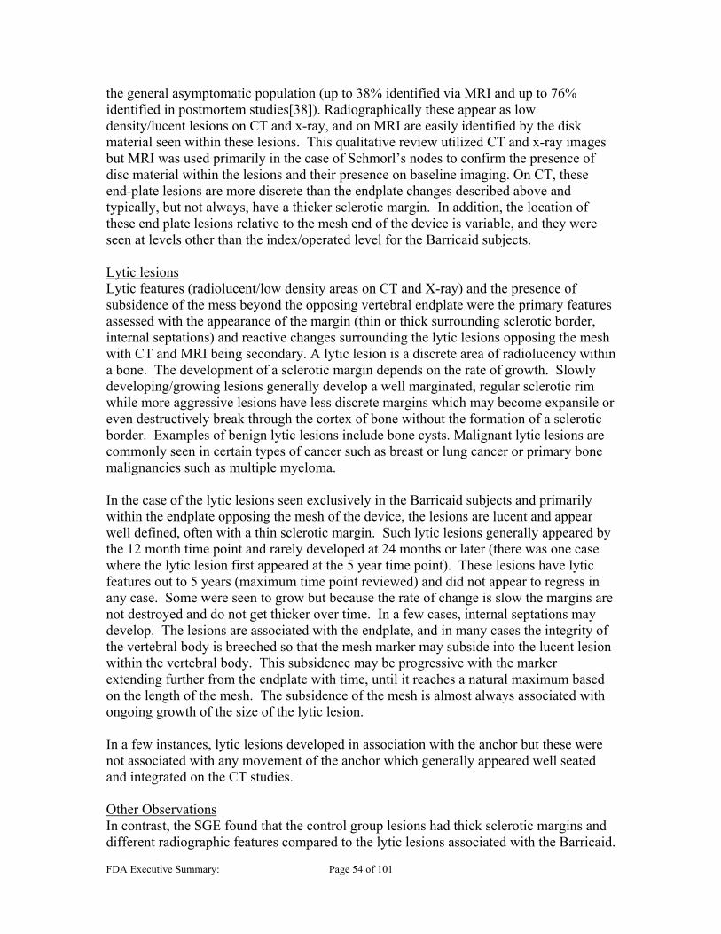
FDA Executive Summary: Page 54 of 101
the general asymptomatic population (up to 38% identified via MRI and up to 76% identified in postmortem studies[38]). Radiographically these appear as low density/lucent lesions on CT and x-ray, and on MRI are easily identified by the disk material seen within these lesions. This qualitative review utilized CT and x-ray images but MRI was used primarily in the case of Schmorl’s nodes to confirm the presence of disc material within the lesions and their presence on baseline imaging. On CT, these end-plate lesions are more discrete than the endplate changes described above and typically, but not always, have a thicker sclerotic margin. In addition, the location of these end plate lesions relative to the mesh end of the device is variable, and they were seen at levels other than the index/operated level for the Barricaid subjects. Lytic lesions Lytic features (radiolucent/low density areas on CT and X-ray) and the presence of subsidence of the mess beyond the opposing vertebral endplate were the primary features assessed with the appearance of the margin (thin or thick surrounding sclerotic border, internal septations) and reactive changes surrounding the lytic lesions opposing the mesh with CT and MRI being secondary. A lytic lesion is a discrete area of radiolucency within a bone. The development of a sclerotic margin depends on the rate of growth. Slowly developing/growing lesions generally develop a well marginated, regular sclerotic rim while more aggressive lesions have less discrete margins which may become expansile or even destructively break through the cortex of bone without the formation of a sclerotic border. Examples of benign lytic lesions include bone cysts. Malignant lytic lesions are commonly seen in certain types of cancer such as breast or lung cancer or primary bone malignancies such as multiple myeloma. In the case of the lytic lesions seen exclusively in the Barricaid subjects and primarily within the endplate opposing the mesh of the device, the lesions are lucent and appear well defined, often with a thin sclerotic margin. Such lytic lesions generally appeared by the 12 month time point and rarely developed at 24 months or later (there was one case where the lytic lesion first appeared at the 5 year time point). These lesions have lytic features out to 5 years (maximum time point reviewed) and did not appear to regress in any case. Some were seen to grow but because the rate of change is slow the margins are not destroyed and do not get thicker over time. In a few cases, internal septations may develop. The lesions are associated with the endplate, and in many cases the integrity of the vertebral body is breeched so that the mesh marker may subside into the lucent lesion within the vertebral body. This subsidence may be progressive with the marker extending further from the endplate with time, until it reaches a natural maximum based on the length of the mesh. The subsidence of the mesh is almost always associated with ongoing growth of the size of the lytic lesion. In a few instances, lytic lesions developed in association with the anchor but these were not associated with any movement of the anchor which generally appeared well seated and integrated on the CT studies. Other Observations In contrast, the SGE found that the control group lesions had thick sclerotic margins and different radiographic features compared to the lytic lesions associated with the Barricaid.

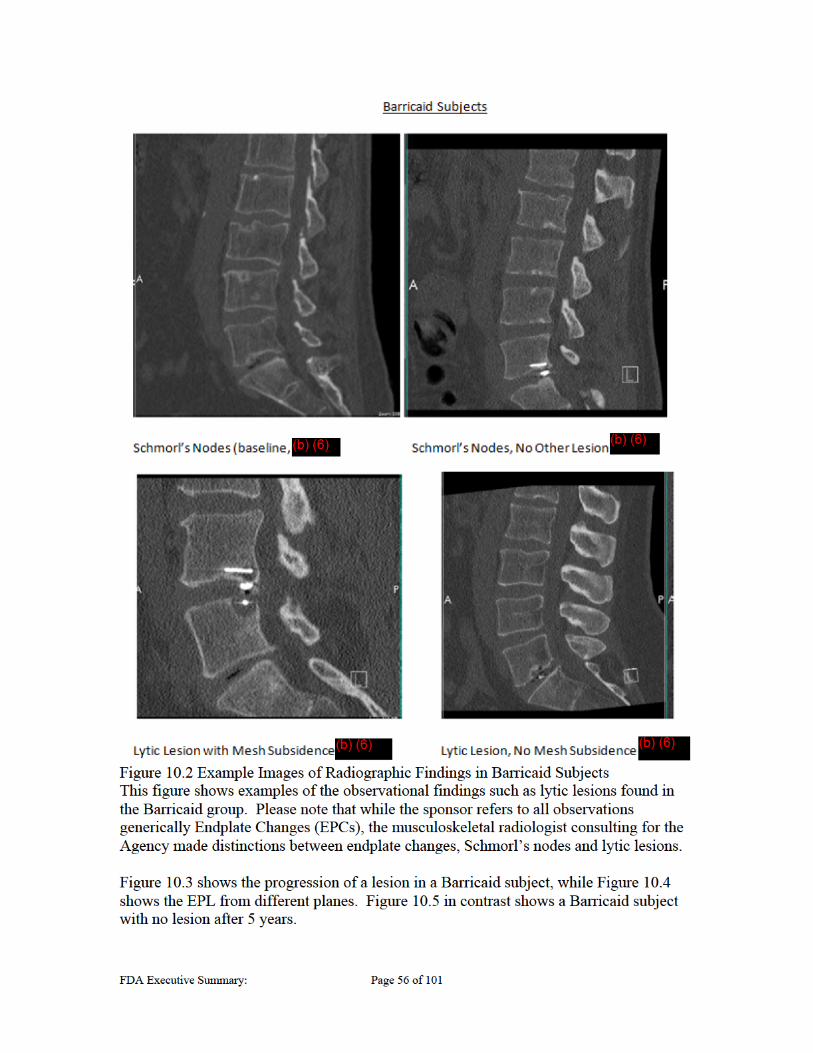

FDA Executive Summary: Page 57 of 101
Figure 10.3 Progression of EPL in Barricaid Subject Patient with Barricaid and a lytic endplate lesion and device subsidence across five years. Panel A shows preoperative films, B shows 1 year, C shows 2 year and D shows 5 year images. The lytic lesions in this example are associated with both the mesh and anchor. The lytic lesions associated with the mesh also enlarges over time. Note reactive sclerosis reaching maximum at 2 years.
(b) (6)
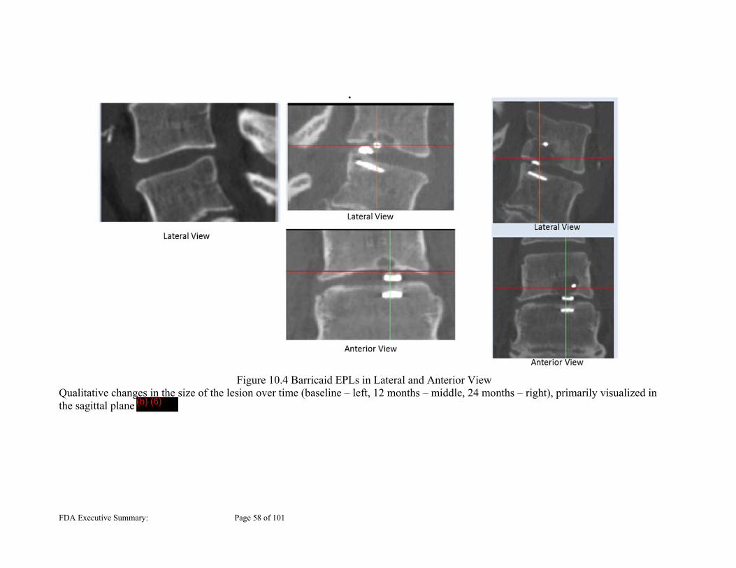
FDA Executive Summary: Page 58 of 101
Figure 10.4 Barricaid EPLs in Lateral and Anterior View
Qualitative changes in the size of the lesion over time (baseline – left, 12 months – middle, 24 months – right), primarily visualized in the sagittal plane (b) (6)

FDA Executive Summary: Page 59 of 101
Figure 10.5 Subject with Barricaid and no lesion at 5 year point.
Quantitative Assessment
The sponsor’s core radiology lab performed the quantitative assessments and, therefore, FDA’s summary of this information is based on data generated from their assessments. There were substantially more EPL’s, and subjects with EPL’s, in the Barricaid group compared to the Control group. Please note that all statistical analyses conducted on the EPL measurements were designed post-hoc and should be interpreted as nominal differences.
Table 10.7 Number of Subjects with EPLs
The sponsor notes that EPL’s (Table 10.7 Sponsor refers to EPLs as EPCs) were present at baseline in both groups. However, the Barricaid group had a larger proportion of post-baseline EPL’s, that is, 85.8% of EPL’s were post-baseline in the Barricaid compared to 68.4% in the Control group. The sponsor also noted that EPL’s in both groups tended to develop earlier, with a peak incidence at 12 months. For analysis purposes, the sponsor has identified a subset of EPL’s proximate to the mesh component of the device, while it was determined that the entire control group be used for comparison. Mesh proximate lesions are defined as a lesion within the vertebra opposing the mesh component of the Barricaid device on the vertebral body opposite the anchor. Note also that the 162 mesh-proximate EPL’s in the Barricaid group were observed in 125 Barricaid subjects.
(b) (6)

FDA Executive Summary: Page 61 of 101
Results of a per-subject analysis show diverging results between the Barricaid and control groups in total EPL area per subject over time. For example, the average EPL area per subject in the sagittal plate was 108.75 ± 75.12 mm2 in the Barricaid group at 60 months compared to 43.46 ± 34.71 mm2 in the control group. Appendix I includes additional area and volume approximations over time broken down and including measurements from each plane and proximate to the mesh component. The per subject calculations are particularly notable as we believe it is important to consider per subject results in addition to analyses at the individual EPC level. Additional analysis also showed that the vertebral body that opposed the mesh and had larger EPLs. This finding is consistent with the findings of Barth et al. who reported that the majority of endplate lesions associated with the Barricaid device were found in the vertebral body opposite the anchor[39]. While at baseline, the sizes are comparable, the area measurements show larger lesions in the mesh opposed vertebral body at 24 months and even larger difference between groups over time. This is particularly noted in the axial plane, as shown in Table 10.9 below. Tables of areas in the other planes are provided in Appendix I. Table 10.9 EPL Area of Mesh Opposed vs Anchor Side Vertebral Body (Axial Plane) This table is an example of the mesh opposed side (No group) and anchor side (Yes group) EPL area measurements in the axial plane over time.
Progression of Lesions A focus of FDA concern has been the continued growth of lesions and whether or not they may become of clinical consequence. To address this concern, the sponsor has plotted the sizes of the largest lesions over time and has shown that the growth rate of the largest lesions decelerates and they appear to stabilize over longer follow-up. This conclusion, at the level of individual EPL, is supported by analyses of individual lesion growth rate and change in the growth rate. This conclusion seems to also be supported when evaluating EPL area and growth rate per subject, rather than per individual EPL. Table 10.10 and Table 10.11 shows the growth rate at each between each time point for EPLs that were proximate to the mesh and proximate to a subsided mesh. The tables present the growth of the control group slowing down after the first year, in both median and mean calculations, while the Barricaid group appears to take longer. The growth of the Barricaid lesions that are mesh proximate or mesh proximate with subsidence clearly
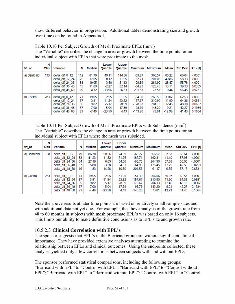
FDA Executive Summary: Page 62 of 101
show different behavior in progression. Additional tables demonstrating size and growth over time can be found in Appendix I. Table 10.10 Per Subject Growth of Mesh Proximate EPLs (mm2) The “Variable” describes the change in area or growth between the time points for an individual subject with EPLs that were proximate to the mesh.
Table 10.11 Per Subject Growth of Mesh Proximate EPLs with Subsidence (mm2) The “Variable” describes the change in area or growth between the time points for an individual subject with EPLs where the mesh was subsided.
Note the above results at later time points are based on relatively small sample sizes and with additional data not yet due. For example, the above analysis of the growth rate from 48 to 60 months in subjects with mesh proximate EPL’s was based on only 16 subjects. This limits our ability to make definitive conclusions as to EPL size and growth rate.
Clinical Correlation with EPL’s The sponsor suggests that EPL’s in the Barricaid group are without significant clinical importance. They have provided extensive analyses attempting to examine the relationship between EPLs and clinical outcomes. Using the endpoints collected, these analyses yielded only a few correlations between subjects with and without EPLs. The sponsor performed statistical comparisons, including the following groups: “Barricaid with EPL” to “Control with EPL”; “Barricaid with EPL” to “Control without EPL”; “Barricaid with EPL” to “Barricaid without EPL”; “Control with EPL” to “Control


FDA Executive Summary: Page 65 of 101
Figure 10.7: Example of Mesh Detachment in a Retrieved Barricaid Implant
Figure 10.8: Example of Mesh Migration
Immediate Post-Op (left), 6 Month (middle), and 12 Month (right) Time Points. Note that the Marker Component (circled) has rotated posteriorly.
Retrieval Analysis
Intrinsic Therapeutics collected 63 implant and instrument retrievals during the OUS clinical studies and commercial use; however, the sponsor stated that a number of retrievals were not properly prepared for a detailed retrieval analysis per the protocol (e.g., collected before protocol was activated) or were not analyzed for various reasons (e.g., Barricaid was not implanted). During the OUS RCT, the sponsor reported 25 subjects with implant removals in 26 procedures. This included one subject that had a staged removal of the mesh followed by the anchor. They also reported an additional six instrument failures during the OUS RCT. However, FDA’s review was limited to 21 implants with full retrieval reports available from the OUS RCT. FDA identified the following main points during the review of the retrieval analysis:
FDA Executive Summary: Page 66 of 101
Retrieved implants were removed primarily due to mesh detachment, device migration (mesh and/or anchor), reherniation, new or worsening pain, and/or segmental instability. Seven retrieved implants involved the mesh component of the Barricaid migrating into the spinal canal/epidural space or impinging on the nerve roots.
The average implantation time for the retrieved implants was 2.4 ± 1.8 years
(range: 0.1 – 5.8 years).
Mesh fraying at the attachment point to the anchor was reported in 19 out of 21 (90.5%) retrieved implants. This was similar to the results of the preclinical testing. Oxidative degradation of the mesh was not significant in the retrieved implants in the middle of the mesh. The sponsor was asked to further evaluate oxidation of all retrievals at the section of the mesh where it attaches to the anchor; however, the PMA did not include a reanalysis of all retrievals. Instead, the sponsor stated that six retrievals were analyzed at the attachment point and the conclusions were no different than those at the middle of the mesh. FDA could not verify these conclusions with the information provided.
Two surgeons commented in the operative reports that the removal surgery was “complicated” or “difficult” but successful for the Barricaid. Fifteen subjects had the entire device removed and 10 subjects had only the mesh component removed. The anchor component was left implanted at the surgeon’s discretion. The sponsor stated that a common explanation for leaving the anchor implanted “was that the remaining portion was well fixed and did not pose any increased risk of pain or subsequent complication.” They also stated that a dural tear was reported in three subjects when the entire implant was removed and one dural tear when the mesh only was removed. Scar tissue in the disc area was reported as well as scar tissue or adhesions around the Barricaid.
Failed instruments were retrieved but no retrieval analyses were provided. The sponsor reported that implantation was not successful in these six cases (not included in the analysis of the 21 retrieved implants), but no further analysis on the instruments were provided.
The sponsor performed additional retrieval analyses, including optical
microscopy, scanning electron microscopy, and micro CT, on five of these retrieved implants to further investigate device failure and to compare Generation 2 (n=1) and Generation 3 (n=4) implants. The results of the analyses showed similar failures across both Generation 2 and Generation 3 implants. There did not appear to be any difference between either generation, despite modifications attempting to strengthen the mesh attachment component, as the same failures occurred regardless of generation.
Histology of Peri-Prosthetic Tissue Eleven out of 21 retrieved implants were accompanied by peri-prosthetic tissue samples that surrounded the Barricaid implant. Intrinsic Therapeutics had these tissues evaluated


FDA Executive Summary: Page 69 of 101
confirmed that the suspected herniation is not a herniation, e.g. scar tissue or residual nucleus material).”
The purpose of this primary endpoint was to directly measure the Barricaid intended use-prevention of reherniation at the index level on either side (ipsilateral or contralateral to the index surgery). The sponsor presented the outcome for the first co-primary endpoint which can be found in Appendix J. This showed that at 24 months, 50.8% of the Barricaid subjects as compared to 30.1% of the control group had reherniation. The reherniation rate in the Barricaid group was 20.8 percentage points lower with statistically significant superiority (p<.001). Please note it has been documented that lumbar disc abnormalities are present in a substantial number of asymptomatic subjects evaluated with magnetic resonance imaging and computed tomography, and correlation of imaging findings with clinical signs and symptoms is important for interpretation of the clinical relevance of positive imaging findings. In this study, approximately half of Barricaid subjects were diagnosed with recurrent disc herniations on imaging studies. The reported reherniation rates vary in the literature. McGirt et al. conducted a 108 subject prospective study to study symptomatic and asymptomatic recurrent disc herniation following lumbar discectomy with serial imaging every 3 months with follow-up out to 24 months[41]. This study reported that 23.1% of subjects demonstrated radiographic evidence of disc herniation during this time, with 13% being asymptomatic and 10% being symptomatic. In contrast Barth reported asymptomatic reherniaton up to 67%[30, 31]. The results of the Barricaid clinical trial show a notable herniation rate in both groups, despite the presence of an anular closure device in the investigational group. These findings further contribute to the uncertainty in the interpretability of results. Clinically silent recurrent disc herniation is common after lumbar discectomy. The clinical relevance of recurrence of disc material noted on MRI after discectomy is unclear, particularly in asymptomatic subjects or subjects with poorly specified pain after surgery. However, assessment of freedom from all reherniations, whether symptomatic or not, contributes to understanding of device efficacy, and permits assessment of the effect of an anular device on the contralateral anulus.
Co-Primary Endpoint – Composite The second pre-defined co-primary endpoint was a responder analysis featuring eight components related to pain, function, safety and radiographic observation designed to be a composite of safety and effectiveness. These include categories intended to capture outcomes with use of a device in conjunction with standard surgical treatment (e.g., device integrity, removals). The sponsor determined success of each individual subject and the study at the 24-month evaluation time point and was defined as follows:
“Composite Success: To be considered a success, a patient will have achieved success in each of the following components:
15 point (out of 100 points) improvement in Oswestry compared to pre-op

FDA Executive Summary: Page 70 of 101
20 point (on a 100 point scale) improvement in VAS Leg (based on the primary leg complaint; if both legs have a minimum of 40/100 pre-operatively, the average leg score will be used)
Maintenance of average disc height (75% or greater of preoperative disc height) compared to pre-op
No deterioration of neurological status at the index level Device integrity and lack of implant migration (radiographic, implanted
patients only) No spontaneous fusion No reherniation at the index level (on either side) No secondary surgical interventions at the index level”
The outcome for the second co-primary endpoint, the Barricaid Composite Clinical Success – Clinical Protocol Definition (CCS-CPD) is provided in Appendix I and shown with long-term endpoints in Table 11.1 below: Table 11.1 Composite Endpoint out to 60 months (CCS-CPD)
The data in Table 11.1 demonstrate the Barricaid CCS-CPD was higher by a statistically significant amount compared to Control discectomy (27.8% vs. 18.1%, p=0.010), a 9.6%
FDA Executive Summary: Page 71 of 101
difference and a 53.6% relative improvement. When considering the components, the superiority margin was driven by the higher reherniation rate and SSIs in the control group, as the other endpoints were comparable. However as noted above, this difference in reherniation rate may not be clinically meaningful, as the definition of reherniation used in this determination was based primarily on imaging findings and may not have been correlated with relevant clinical findings. CCS – CPD Data Inclusion and Exclusion: For the CCS-CPD, the availability of an endpoint assessment at 24 months varies based on the individual endpoint. An assessment for presence or absence of secondary surgical intervention (SSI) is included for all subjects except those Barricaid subjects who had an unsuccessful device implantation, who were already considered study failures. For the endpoints of reherniation, spontaneous fusion, and device integrity, subjects included in the assessment are those who had data from a 24 month visit as well as those subjects who were a failure in that category at a prior time point. For disc height measurement and neurological status, only subjects who had that assessment at 24 months are included in the endpoint. For functional outcomes (ODI and VAS), only subjects who had that assessment at 24 months are included, with subjects having an SSI excluded from the endpoint. The composite endpoint includes subjects with success/failure measurements for every component of the endpoint as well as those subjects who were failures at any one component of the endpoint. However, subjects who are not failures by all components of the endpoint but are missing data from one component of the composite endpoint are considered missing but are imputed at 24 months using multiple Bayesian imputation. Missing data beyond 24 months was not imputed.
Exploratory Analysis: Alternative Primary Endpoint
The sponsor proposed a post-hoc primary endpoint that is intended to be more clinically meaningful than the original endpoint. In addition, the sponsor provided a rationale that ODI and VAS provide somewhat redundant assessments of pain, in addition to the function component provided by ODI. The sponsor’s position is that these factors in the original composite co-primary endpoint only add “noise” to the safety and effectiveness determination, such that it is a mathematical calculation, rather than reflective of actual clinical performance. Accordingly, the sponsor developed a post-hoc alternative primary endpoint (CCS-mCPD, “Composite Clinical Success-modified Clinical Protocol Definition”) to assess clinical performance, using a more clinically relevant definition. To be a success with regard to the CCS-mCPD, a subject will have achieved success in each of the following components at 24 months:
No Symptomatic Reherniation: To be considered a success, a patient will have no evidence of recurrent symptomatic herniation at the index level at any time up to and including the 24-month follow-up. Recurrent symptomatic herniation may be confirmed surgically, or radiographically as determined by an independent review (unless surgically confirmed that the suspected herniation is not a herniation, e.g. scar tissue or residual nucleus material);
15 point improvement in Oswestry Disability Index (ODI) compared to baseline;
FDA Executive Summary: Page 72 of 101
No deterioration of neurological status at the index level; No secondary surgical interventions at the index level; and No implant- (i.e., device migration or device condition issue) or procedure-related
serious adverse events; As compared to the original CPD, the mCPD uses the symptomatic reherniation rate instead of all reherniations. This is notable because, while the symptomatic reherniations are the only reherniations with associated clinical or meaningful health outcomes, the device is intended to block all reherniations, not just symptomatic ones. Both endpoints should be carefully considered under appropriate context. The mCPD also eliminates the use of VAS-Leg and considers only ODI. While there is some overlap in these assessments, VAS-Leg is an outcome more directly related to an intervention for treatment of sciatica due to a lumbar disc herniation in contrast to ODI, which provides a method for measure of condition-specific disability[42, 43]. Neurological status and SSI measures remain the same for both the CPD and mCPD endpoints. The original CPD considers a number of radiographic assessments (disc height, device integrity and spontaneous fusion) which the sponsor subsequently considered not clinically meaningful. These were originally included to aid in the evaluation of device performance. The lack of device or procedure-related serious adverse events is used instead of a radiographic device integrity assessment. Again, this is a more clinically meaningful measure; however, the radiographic assessments were originally included to assist in monitoring the function of the device. If the device is not functioning as intended (e.g. migrated out of the disc space or dissociated completely), it is challenging to attribute clinical success to the device. Similar to the asymptomatic vs all reherniation analysis, the original CPD was intended to measure the success of the device, while the mCPD is intended to measure clinical performance only. Since this was a post-hoc analysis, the Barricaid study Data Safety Monitoring Board (DSMB) oversaw the development of the algorithm presented in Figure 11.1 to determine if a reherniation was symptomatic:

FDA Executive Summary: Page 73 of 101
Figure 11.1 Symptomatic Reherniation Algorithm
The potential for bias exists as this adjudication algorithm uses lumbar related pain as a criterion. Such pain is common in a subset of post-discectomy subjects and its interpretation by its nature is subjective. A neurologic deficit or combined VAS and ODI scores and decreased neurological status may or may not accurately determine if a reherniation is symptomatic. The adjudication criteria for identification of symptomatic reherniations are unclear. For example, details regarding the criteria used for neurologic assessment are not provided, and the algorithm includes clinical outcomes that may not be related to the observed radiographic herniation. In addition, the algorithm does not consider the possible presence of other painful spinal or extra-spinal conditions which may be present. While a physical examination is part of the inclusion criteria and original assessment for enrollment in this study for a primary herniated disc, the algorithm above does not necessitate this diagnostic element and may lack specificity. These are limitations regarding the definition used to define “symptomatic reherniation” in this study and should be considered when reviewing the data. For example, a recurrent disc herniation has been defined in literature as the return of symptoms consistent with a previous presentation in the same subject after a minimum of 6 weeks of improvement following a discectomy procedure[26]. Many imaging-based degenerative features may be part of normal aging and unassociated with symptoms[44]. Additional factors such as the presence of a pain free interval
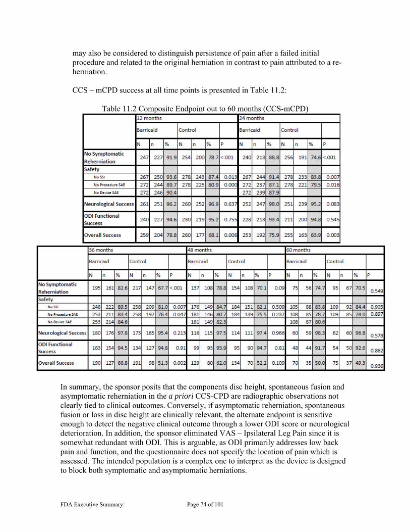
FDA Executive Summary: Page 74 of 101
may also be considered to distinguish persistence of pain after a failed initial procedure and related to the original herniation in contrast to pain attributed to a re-herniation. CCS – mCPD success at all time points is presented in Table 11.2:
Table 11.2 Composite Endpoint out to 60 months (CCS-mCPD)
In summary, the sponsor posits that the components disc height, spontaneous fusion and asymptomatic reherniation in the a priori CCS-CPD are radiographic observations not clearly tied to clinical outcomes. Conversely, if asymptomatic reherniation, spontaneous fusion or loss in disc height are clinically relevant, the alternate endpoint is sensitive enough to detect the negative clinical outcome through a lower ODI score or neurological deterioration. In addition, the sponsor eliminated VAS – Ipsilateral Leg Pain since it is somewhat redundant with ODI. This is arguable, as ODI primarily addresses low back pain and function, and the questionnaire does not specify the location of pain which is assessed. The intended population is a complex one to interpret as the device is designed to block both symptomatic and asymptomatic herniations.


FDA Executive Summary: Page 77 of 101
was not a predefined analysis and all statistical conclusions should be considered nominal. Table 11.4 Comparisons of the Percentages of Subjects with Symptomatic Reherniations – mITT Analysis Set (Restricted to Expected Due) (Nominal Significance)
Table 11.5 Number of Symptomatic Recurrent Disc Herniations at Index Level (Nominal Significance)
These results show a lower rate of symptomatic reherniations in the Barricaid group by a lower margin beginning at 12 months through 36 months. At 24 months, the difference is 11.3% vs. 25.4% (p<.001). This is a 14.1 percentage-point reduction in symptomatic reherniations for the Barricaid subjects compared to control. However, at Month 60, the Barricaid group was only 4.1% better than controls with respect to symptomatic reherniations and only marginally different. Retrospective studies report the incidence of same-level symptomatic recurrent disc herniation after lumbar discectomy varies approximately between 3% and 18%[45, 46]. This wide range is secondary to variations in the follow-up time, management paradigms, anular disruption, and surgical technique[47]. For comparison, Table 11.6 below shows the counts and percentages of subjects in each group with asymptomatic reherniations out to Month 60:

FDA Executive Summary: Page 78 of 101
Table 11.6 Comparisons of the Percentages of Subjects with Asymptomatic Reherniations – mITT Analysis Set (Restricted to Theoretically Due) (Nominal Signficance)
The results do show a lower rate of asymptomatic reherniations in the Barricaid group at all time points; however, there were no nominal differences in asymptomatic reherniations after the initial 12 month assessment. 37.9% of Barricaid subjects compared to 44.5% of control subjects had asymptomatic reherniations at 24 months, resulting in a 6.6% smaller difference. This rate further converged over time, resulting in only 2.1% difference at 60 months. When considering symptomatic or asymptomatic herniations, the Barricaid and control groups show a trend towards convergence at later time points despite initially starting out significantly different at the 24 month time point. Table 11.7 and Table 11.8 show the Kaplan-Meier (product-limit) estimate for All Reherniations and Symptomatic Reherniations. It is again important to note that the nominal p-value presented on the difference on the entire dataset and should not be applied to significance at any one specific time point. As previously stated, conclusions are limited due to a large number of subjects that are not yet due at later time points. The “F” columns show failures, while “C” are censored subjects. The cumulative % is the survival estimate at each time point, with the predicted upper (UB) and lower bounds (LB). The p-value is reflective the conclusion that the entire curve is significantly different, not at each individual time points. Table 11.7 Survival Analysis – All Reherniations (mITT)

FDA Executive Summary: Page 79 of 101
Table 11.8 Survival Analysis – Symptomatic Reherniations (mITT)
Clinical Endpoints – Oswestry Disability Index (ODI), VAS – Leg Pain, Neurologic Outcomes
Both cohorts showed similar outcome success in ODI, VAS-Leg and Neurological Status through 24 months and beyond, with no statistical or clinically meaningful difference between the two groups. ODI scores through 24 months were similarly successful between experimental and control groups (93.4% and 94.8% Success for Barricaid and control). Both cohorts had similar trends of improving VAS – Ipsilateral Leg Pain scores through 24 months, with a greater improvement in the Barricaid group in the immediate post-operative period. However, the success at 24 months is comparable (94.7% vs 96.2% Success, Barricaid vs control), with similar success rates out to 60 months. The overall assessment of neurologic function indicates that there are similar rates of maintenance or improvement of neurologic function in both groups at all time points. Tables are provided in Appendix I. One additional note of clarification was that the measure of VAS-Leg scores used VAS from the ipsilateral leg, except in cases when the subject has pain in both legs, where the average of the two scores was used. There is concern that this measure is confounded when the leg pain is averaged, especially since disc herniation is attributed to unilateral symptoms.
Radiographic Endpoints – Maintenance of Disc Height, Device Integrity and Spontaneous Fusion
The sponsor questions the utility of the following radiographic outcome measures as they are indirectly related to clinical outcomes. Maintenance of Disc Height The composite co-primary endpoint includes a component that requires subjects to maintain disc height (≥ 75% compared to Pre-Op value) in order to be a success. The disc height calculation is based on data provided by the independent core radiographic laboratory. Successful maintenance of disc height trends does not appear different between the Investigational (65.4%) and control (67.3%) groups at 24 months. The disc height maintenance of 75% of baseline drops off considerably in both groups over time, falling to 35.0% and 26.8% at 60 months. The prospectively measured success rate was only 50.0% of subjects with ≥ 75% disc height maintenance at Month 24, out of 108 consecutive subjects. Device Integrity

FDA Executive Summary: Page 80 of 101
Device integrity was assessed using radiographic findings alone, and evaluates the device’s ability to maintain device condition (no anchor fracture or occlusion component detachment) and avoid migration in order to be a success. The sponsor refers to these two assessments collectively as “device integrity”. The sponsor subdivided device integrity failures into device condition (fracture and/or disassembly) and migration failures (includes mesh rotation). These observations were reported and assessed by the radiographic core lab using the definitions of Device Condition and Migration established in the Imaging Charter. The following table shows the before and after two-year device integrity component data and reflects the overlap of “device condition” and “device migration.” Specifically Table 11.9 reports the device integrity failures and the resultant reoperations for before and after 24 months. This demonstrates that while the device may have failed, it may not require a subsequent reoperation.
Table 11.9 Overall Rates of Device Integrity Failure in Barricaid Subjects By 24 Months After 24 Months N n % Reoperations** N† n % Reoperations**Device Integrity Issue
242
32
13.2
13 164 16 9.8
4 Condition 238 11* 4.6 174 3 1.7 Migration 243 29 11.9 168 14 8.3
*There were 8 cases with both device condition and device migration issues, resulting in 32 cases by Month 24.
** At the time of or subsequent to device integrity observation
†Number of subjects who did not exhibit a device integrity failure through 24 months and who had at least one post-24 month follow-
up visit in which the x-rays could be assessed for device condition or migration
Device Condition events occurred in the form of fractured or disassembled device both before and after the 24 month time point. The same was observed in regards to Device Migration. Given the outcomes measured, the sponsor presented information to assert that subjects with Device Integrity issues had mostly positive clinical outcomes, and many of the secondary surgeries that occurred in these subjects were the result of other factors. It is also important to note that while there is an 86.8% success in the Device Integrity assessment at 24 months, by 60 months, that percentage drops to 69.5% as calculated using product-limit approximation. In addition to our concern for the proper clinical measures to assess the device, other findings create challenges in the ability to interpret the data. For example, the sponsor reports successful clinical outcomes in in 63.6% of subjects with device condition issues through Month 24, and in one of three subjects (33%) with device disassembly after Month 24. However, there were five Barricaid subjects with device migration AEs (including subject who also had a device condition AE) and successful outcomes at 24 months went on to have SSI subsequently. This again questions the appropriateness of a 24 month outcome assessment, as both device integrity, as well as potential secondary surgeries and SAE may occur after a good 24 month clinical assessment.
(b) (6)
FDA Executive Summary: Page 81 of 101
The sponsor states “Only 4 of the Barricaid SSIs (1.5% of treated subjects) reported were due to device-specific events such as loosening or migration without a concurrent suspected or confirmed reherniation. An additional 3 Barricaid SSIs (1.1% of treated subjects) were due to device-specific events and other causes including suspected or confirmed reherniation.” While the sponsor presents a case that only the clinical outcome matters, the utility of the device is unclear if it has an integrity or migration failure. It is unclear how to interpret outcomes when the device is not performing as intended. The design of the Barricaid device is for the mesh component to be positioned within the disc space to block herniation of disc material. The composite endpoint for the study includes an element that requires that a successful subject not have any radiographic findings that show a device integrity issue. This includes migration or dissociation of any portion of the device, notably, the mesh, which is intended to block herniation. However, of the subjects that failed the study due to device integrity issues, many subjects exhibited otherwise positive clinical outcomes. The clinical success of subjects regardless of radiographic device integrity concerns such as migration raises questions about the function of the device even among subjects with positive clinical outcomes. Please note that the relief of symptoms following the index surgical procedure in both groups is due to decompression of the nerve root due to removal of disc material and occurs in both groups and is not due to a direct device mediated effect. The Panel will be asked to consider how to interpret the clinical outcomes in light of these radiographic findings of device integrity and what appropriate endpoints should be considered to evaluate this device.
Spontaneous Fusion The primary endpoint includes a component that requires subjects to have no spontaneous fusions, as radiologically documented, in order to be a success. The independent core radiographic lab reported only one spontaneous fusion at 24 months, which occurred in a Control subject. There were two and four spontaneous fusions in the Barricaid and Control groups, respectively, at 36 months through 60 months.
Index Level Secondary Surgical Interventions (SSI) Please refer to Section 10.2 above under the safety results section in this document for SSI results. Briefly, the Barricaid group had fewer SSIs at the 24-month time point (8.84% vs 16.36%). This is maintained through 60 months. By 60 months, the 37 Barricaid subjects had reoperations at any time through Month 60 and nine went on to have subsequent reoperations. Reoperations included additional discectomies with and without fusion, fusions with or without pedicle fixations, wound revisions, decompressions and Barricaid removals. Two Barricaid subjects had a third reoperation, and none had a fourth. In the control arm, 57 subjects had reoperations at any time through Month 60 and 16 went on to have 22 subsequent reoperations. These reoperations included additional discectomies with and without fusion, fusions, pedicle fixations, wound revisions and hematoma drainage. Four subjects went on to have a third reoperation, and two had a fourth reoperation.

FDA Executive Summary: Page 83 of 101
type), baseline outcomes (ODI, VAS, disc height), completer vs non-completer, previous lumbar spine surgery, and medication usage. Many of these groups have low numbers in the subanalysis group or are otherwise not notable. Notable analyses conducted include exploratory analyses on the effects of endplate lesions and subsidence of the mesh and anchor. These analyses demonstrated no evidence of correlations between these factors and clinical outcomes.
12 Statistical Analysis The study had two co-primary endpoints, absence of recurrent herniation and a composite of clinical success (CCS). The sponsor claims to have met both endpoints with a predictive probability of 0.996 in a Bayesian early claim analysis. The final posterior probabilities for superiority, using the final data set, were determined using Bayesian multiple imputation with no borrowing from informative priors. Frequentist analyses were generally used in other analyses including analyses of EPL’s, blinding, poolability by site and various exploratory analyses. Significance was primarily demonstrated through tests for proportions such as Chi-square and Fisher’s Exact tests.
Missing Data Imputation The sponsor has provided a Bayesian multiple imputation analysis. This analysis follows the approach of using results from completers to impute missing subjects in a way that parallels the Bayesian early claim analyses. Results of this analysis show posterior probabilities of 0.9999 and 0.9977 for the freedom from reherniation and CCS analyses respectively. In addition, complete case, all-missing-as-failure, all-missing-as-success, and best case analyses are consistent with meeting the co-primary endpoint thresholds. The sponsor has also provided tipping point analyses. These show that the freedom from reherniation analysis is highly robust to the missing data with only 0.7% of the analyses showing a reversal of the superiority conclusion. The CCS endpoint was somewhat less robust with 25.3% of missing data combinations resulting in a non-significant conclusion. Thus, the data at 24 months are quite robust to missing data. However, results at later time points are less conclusive as a substantial fraction of subjects has not reached the later follow-up time points. Missing data at the later time points also appears slightly greater than at 24 months.
Poolability of Sites The sponsor’s statistical consultant used a random effects meta-analysis approach to test the significance of site-to-site and surgeon-to-surgeon variability in the two co-primary endpoints of freedom from reherniation and Month 24 CCS. No evidence of significant heterogeneity was found.
Financial Interests The sponsor has provided analyses stratified by financial interest greater than or less than $25k. The results demonstrate that the Barricaid and Control groups experienced a similar difference in CCS outcomes, 9.0% and 9.6%, respectively, in the comparison of subjects with and without financial interest. Of interest, the difference in reherniation rates was negligible within Barricaid (0.9%). However, the reherniation rates within the
FDA Executive Summary: Page 84 of 101
Control group were noticeably different (14.1%) with the >$25k group having a lower reherniation rate. This is important since it demonstrates there was no negative bias towards the Control group as a result of having significant financial compensation. In other words, if financial compensation biased the investigators, one would expect worse Control outcomes at sites with greater financial interest. In addition, the sponsor has provided analyses of the correlation between financial interest category and various clinical variables. No significant correlations were found.
Patient Enrollment Surgeons did not randomize almost 20% of subjects after enrollment. A total of ninety-three (93) subjects passed pre-operative screening, but during further intraoperative screening were found to have failed to meet the study protocol requirement, and did not proceed to randomization. Anular defects were out-of-range by sizing criteria for inclusion in 75 subjects and 18 others should have failed screening by the eligibility criteria for enrollment. Twenty-six subjects had anular defects that were too small for enrollment. Other observations on the patient population enrolled were detailed in Section 9.1. The uncertainty regarding these pathoanatomic characteristics raises the question of applicability of the studied patient population. As previously discussed, there is also concern that extensive anular resections may have impacted the results of the study.
Patient Population Compatibility The Barricaid study investigational sites were located in Western Europe (Belgium, Switzerland, the Netherlands, Germany, Austria and France) which may have influenced the validity of the study data as applicable to the US population. The sponsor reports reviewing the study design, the subjects, the standard of diagnosis and treatment prior to surgery, and follow-up following treatment for the OUS study subjects to show that the study provides outcomes that are generalizable to the United States subject population. In addition, the sponsor performed a relevant literature review in support of this generalizability. Many of the Agency’s concerns are centered around the population of subjects studied and the types of procedures conducted as part of standard of care for a herniated disc (i.e. limited discectomy). The potential for a patient population, whose control group outcomes differs significantly from literature, could be due to differences due to an OUS study.
Blinding One potential source of bias in the Barricaid clinical trial was the lack of blinding in all subjects receiving treatment outside of the Netherlands. In this study only 85 subjects at three sites in the Netherlands were blinded. The sponsor states that, “It was not possible in the remainder of the clinical sites due to limitations from the local EC authorities.” To assess possible bias from lack of blinding, the sponsor has provided a comparison of blinded subjects to unblinded subjects separately for the Barricaid and Control groups. Specifically, tables are presented comparing extensive Demographic, Baseline, Intra-Operative and Clinical Outcome variables. Note that due to the nature of the blinding, the results for blinding are confounded with the effects of site and geographic region.

FDA Executive Summary: Page 85 of 101
The sponsor first presents a comparison of 38 Blinded and 234 Unblinded subjects in the Barricaid group. There were a number of nominally significant findings in the baseline and intraoperative variables, but these can be attributed to the effect of geographic region (Height), study site (Operative Time, Anesthesia Time, and Bone Removed) or to other perhaps chance characteristics of the site population (Geometry, Defect Width and Height). Note that there is not a logical connection to subject blinding for any of the nominally significant variables mentioned previously. In terms of clinical outcomes for the Barricaid group, there was a nominally significant difference (p = 0.0002) in the proportion of subjects having device related AE’s that were categorized as “Definite/Probable” (D/P) as compared to “Possible/Unknown”(P/U), i.e. 2.6% (D/P) vs. 60.5%(P/U) in the Blinded and 28.4%(D/P) vs. 47.2%(P/U) in the Unblinded group. However, note that the overall proportion of subjects with “Any device related AE” was not statistically different between the two groups, being 60.5% in the Blinded and 63.3% in the Unblinded group. As device relatedness is not determined by the subject, the above can probably be dismissed as a chance finding. There was one other nominally significant result, which also cannot logically be attributed to blinding. This was a larger decrease over time in Posterior Disc Height for Blinded subjects. Such a finding could easily be due to chance, being noticed as a result of the very large number of comparisons made. In terms of the Control group, the sponsor presents a comparison of 47 Blinded and 231 Unblinded subjects. These comparisons yielded almost the same nominally significant findings in the baseline and intraoperative variables as for the Barricaid group. This is reassuring as it indicates the absence of an interaction between blinding (or other confounding factors) and treatment group. In terms of clinical outcomes, there was a nominally significant difference in the proportion of subjects having an asymptomatic reherniation with more unblinded subjects having this event, but this was statistically borderline and could easily have been due to chance. Also, the rates of symptomatic reherniation were very similar between the two groups and blinding should not have logically affected an endpoint without clinical symptoms. However, in addition to the above finding, for most of the clinical measures there was a significant difference at Month 24, with notably greater improvement in the blinded group, but these differences were not consistent with the other time points. Therefore, these latter findings are likely the result of an error, unusual chance variation or possibly operational bias combined with the effect of blinding, as the primary endpoint would have been known to be at 24 months. Note, however, that a similar finding was not observed in the responder versions of the clinical endpoints.

FDA Executive Summary: Page 86 of 101
Table 12.1 Blinding CCS analysis
13 Benefit Risk Assessment Benefit Risk Overview When making a determination of the benefit-risk profile of a device, the Agency considers the following:
Benefits: type of benefits, magnitude of benefits, probability of the subject experiencing one or more benefits, duration of effect and degree of uncertainty
Risks: types, number, and rates of harmful events associated with the use of the device (device-related serious, device-related non-serious, and procedure-related adverse events), probability of a harmful event, and duration of harmful events.
Additional factors (if applicable): uncertainty, characterization of the disease, subject tolerance for risk and perspective on benefit, availability of alternate treatments, risk mitigation, post market data, and novel technology addressing unmet needs.
Summary of Benefits Over the course of the study, the following benefits were associated with use of the Barricaid Anular Closure Device when compared to the control group treated with limited discectomy alone. These benefits should be interpreted in the context of the 24-month endpoint selected by the sponsor and assuming definitions of the endpoints are appropriate and acceptable for the proposed patient populations that are used in each of the analyses. Types of benefit: The primary probable benefits of the Barricaid Anular Closure Device are:
1. Limitation of lumbar disc reherniation, and 2. Limitation of secondary surgical interventions
The OUS protocol pre-defined co-primary endpoints were 1) all reherniations and 2) a composite responder analysis including eight different independent components measuring the effect of the Barricaid device on pain, function, and safety to provide a
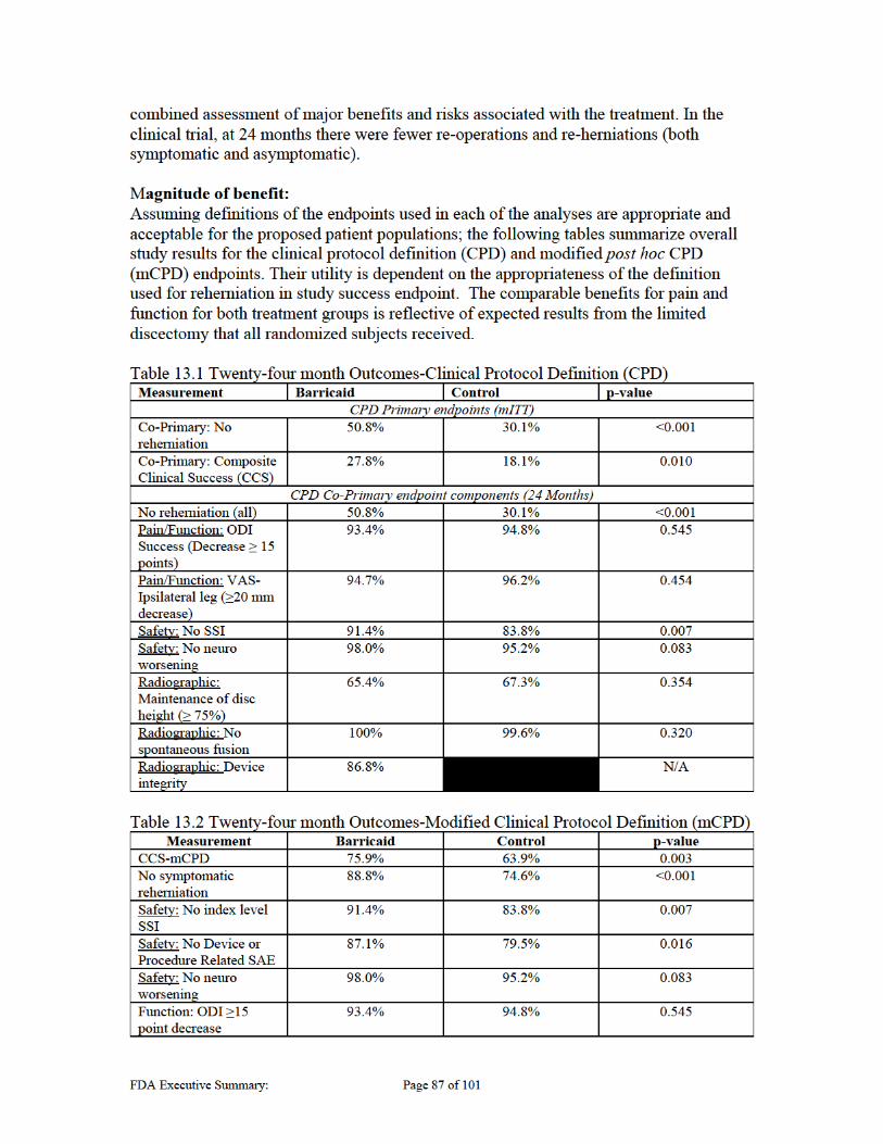

FDA Executive Summary: Page 88 of 101
For the CPD-CCS, the Barricaid device in combination with limited discecetomy demonstrated superiority over limited discectomy alone on both safety and efficacy co-primary endpoints at 24 months (p=0.010). The Barricaid had a responder success rate of 27.8% (68/245) compared to 18.1% (47/259) for control. This superiority margin is driven by superior reductions in reherniations (p<0.001) and freedom from SSI (p=0.007). The sponsor also presented results of an alternative composite endpoint (modified Clinical Protocol Definition – mCPD) that, in their judgement, is potentially more clinically meaningful compared to the CPD endpoint. The primary distinction of this post hoc composite endpoint was the inclusion of symptomatic rather than all post-operative reherniations, as determined by the sponsor. As discussed previously, there is potential for bias regarding the determination of symptomatic recurrent lumbar disc herniation. The mCPD also does not consider radiographic device integrity concerns as only device or procedure related SAEs were considered in this endpoint. At 24 months, this alternative composite endpoint resulted in a Barricaid success rate of 75.9% (192/253) compared to 63.9% (163/255) for the control (p=0.003). Probability of the Patient Experiencing a Benefit (at 24 months):
All reherniations: In the Barricaid group, 50.8% (122/240) were free from reherniation at 24 months. In the control group 30.1% (77/256) were reherniation-free, a difference of 20.8% in favor of Barricaid (p˂.001).
Symptomatic reherniations: In the Barricaid group, 27 subjects (11.6%) had a symptomatic reherniation at any time through month 24, compared to 61 subjects (24.8%) in the Control group. This is a 55.7% reduction in favor of Barricaid (nominal p<0.001).
SSIs: By 24 months, 23 subjects (8.6%) in the Barricaid group received an index-level SSI, compared to 44 subjects (15.8%) in the Control group. This is a 47.7% reduction of SSIs in favor of Barricaid (p=0.010).
SSIs for reherniation: Within the first 2 years, 24 subjects (9.0%) in the Barricaid group received a secondary operation to treat reherniation, compared to 47 subjects (16.9%) in the control group, a 48.9% reduction (nominal p= 0.007).
The 24-month data shows that subjects with large anular defects prone to reherniation could benefit from the Barricaid device. A subject who could avoid reherniation and SSI would highly value these benefits. The benefits did not appear to vary across subpopulations. The sponsor performed a demographic exploratory analysis to evaluate trends within the clinical data outcomes that may have been influenced by different demographic variables, including age, gender, BMI, and race. The study was unable to identify a subpopulation that might disproportionately benefit from the subject device. Duration of Effect: As noted above, the sponsor designed the clinical trial to power the co- primary endpoints based on 24-month outcomes and the data at most reflect the durability of outcomes through this interval. However, partial data accrual continued to
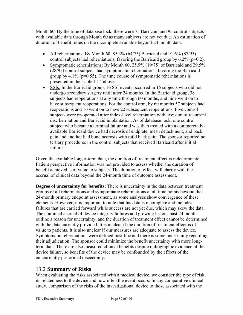
FDA Executive Summary: Page 89 of 101
Month 60. By the time of database lock, there were 75 Barricaid and 95 control subjects with available data through Month 60 as many subjects are not yet due. An estimation of duration of benefit relies on the incomplete available beyond-24 month data:
All reherniations: By Month 60, 85.3% (64/75) Barricaid and 91.6% (87/95) control subjects had reherniations, favoring the Barricaid group by 6.2% (p=0.2).
Symptomatic reherniations: By Month 60, 25.9% (19/75) of Barricaid and 29.5% (28/95) control subjects had symptomatic reherniations, favoring the Barricaid group by 4.1% (p=0.55). The time course of symptomatic reherniations is presented in the Table 11.4 above.
SSIs: In the Barricaid group, 16 SSI events occurred in 15 subjects who did not undergo secondary surgery until after 24 months. In the Barricaid group, 38 subjects had reoperations at any time through 60 months, and nine went on to have subsequent reoperations. For the control arm, by 60 months 57 subjects had reoperations and 16 went on to have 22 subsequent reoperations. Five control subjects were re-operated after index-level reherniation with excision of recurrent disc herniation and Barricaid implantation. As of database lock, one control subject who became a terminal failure and was then treated with a commercially-available Barricaid device had necrosis of endplate, mesh detachment, and back pain and another had bone necrosis with mild back pain. The sponsor reported no tertiary procedures in the control subjects that received Barricaid after initial failure.
Given the available longer-term data, the duration of treatment effect is indeterminate. Patient perspective information was not provided to assess whether the duration of benefit achieved is of value to subjects. The duration of effect will clarify with the accrual of clinical data beyond the 24-month time of outcome assessment. Degree of uncertainty for benefits: There is uncertainty in the data between treatment groups of all reherniations and symptomatic reherniations at all time points beyond the 24-month primary endpoint assessment, as some analyses show convergence of these elements. However, it is important to note that his data is incomplete and includes failures that are carried forward while success are not yet due, which may skew the data. The continual accrual of device integrity failures and growing lesions past 24 month outline a reason for uncertainty, and the duration of treatment effect cannot be determined with the data currently provided. It is unclear if the duration of treatment effect is of value to patients. It is also unclear if our measures are adequate to assess the device. Symptomatic reherniations were defined post-hoc and there is some uncertainty regarding their adjudication. The sponsor could minimize the benefit uncertainty with more long-term data. There are also measured clinical benefits despite radiographic evidence of the device failure, so benefits of the device may be confounded by the effects of the concurrently performed discectomy.
Summary of Risks When evaluating the risks associated with a medical device, we consider the type of risk, its relatedness to the device and how often the event occurs. In any comparative clinical study, comparison of the risks of the investigational device to those associated with the
FDA Executive Summary: Page 90 of 101
standard of care and/or control is appropriate. With the exception of the device-related complications and end plate lesions (EPLs), most of the risks of Barricaid overlap with those of limited discectomy. Severity, types, number and rates of harmful events (events and consequences): Types of risks: Over the course of the study, the results documented the following risks: Secondary Surgical Interventions: At 24 months, the Barricaid group had 23 subjects identified as failures due to SSIs as compared to 45 subjects identified as failures in the control group. The Barricaid group experienced failure in 38 subjects due to SSI, while the control group had 57 subjects experience failures due to SSI by 60 months. The Barricaid group had 17 subjects with SSIs that resulted in supplemental fixation, while the control group had only 12 subjects treated with supplemental fixation. Further, the initial SSI in the Barricaid groups resulted in 12 subjects out of 38 the resulted in a supplemental fixation procedure, while only 6 subjects out of 57 in the control group had initial supplemental fixation. There was a greater percentage of control group subjects whose initial SSIs were discectomy as compared to the Barricaid group. Device-related serious adverse events: The sponsor used an independent Data Safety Monitoring Board (DSMB) to adjudicate the study adverse events in terms of their relationship to the device and/or procedure. The notable device-related adverse events classified as serious for the Barricaid are device deficiency adverse events (primarily migration of the whole device (anchor) or migration of the occlusion component (mesh)), disc herniation at the index level (classified as either reherniation or residual herniation), and adverse events related to ongoing lumbar and/or lower extremity pain.
1. Device Integrity failures: At 24 months, 13.2% (34/267) Barricaid subjects had device integrity failures, with 11 instances of device condition (fracture and/or disassembly) and 29 instances of migration. By Month 24, there were 13 total reoperations of the index level performed at the time of or subsequent to the identification of the device integrity observation. Two subjects with device integrity failures and successful outcomes at 24 months went on to have SSI later. After 24 months, the core lab noted 14 instances of migration and 3 instances of condition in 16 subjects. One subject exhibited both modes. This group required four reoperations for apparent device integrity-related AEs only. These events are summarized in Table 11.9 above. 2. Reherniation at the index level As discussed above under benefits, at 24 months the rate of symptomatic reherniation was 11.3% in the Barricaid group compared to 25.4% for control. There is a convergence of reherniation rates between treatment groups at longer time points.
3. Ongoing lumbar and/or lower extremity pain

FDA Executive Summary: Page 91 of 101
At 24 months, 5.3%% of Barricaid subjects and 3.8% of control subjects had no improvement or worsening in VAS-leg pain scores. At 60 months, 4.2% of Barricaid subjects and 7.4% of control subjects had unimproved or worse leg pain.
Forty-seven of 267 as-treated subjects experienced Serious Device- or Procedure-Related Adverse Events in the Barricaid group (47/267, 17.6%) compared with 71 subjects in the control group (71/283, 25.1%). Forty-seven subjects in the Barricaid group and 71 subjects in the control experienced more than one device- or procedure-related SAE for a total count of 78 events in the Barricaid group and 108 events in the Control group. In total, 20.5% (55/267) of Barricaid subjects failed the modified primary endpoint due to a device-related SAE and/or a radiographically observed device integrity failure. Probability of a harmful event The following table presents the 24-month counts of specific procedure and device-related AEs germane to benefit-risk determination: Table 13.3 Agency Determined Relevant Device or Procedure Related AE
AE Barricaid (N=267)
Control (N=283)
Nominal Significance
Events n % Events n % ∆ p-value Device
Deficiency 34 33 12.7 1 1 0.4 12.4% ˂ 0.001
Reherniation 40 34 15.0 85 74 30 -15.1% ˂ 0.001 Wound issue: SSI at index
level
25
22
9.4
20
18
7.1
2.3%
0.416
Necrosis of Bone or Resorption
49 47 18.4 1 1 0.4 18% ˂ 0.001
*AE categorization determined by DSMB and definitions included in Appendix G. Incomplete data beyond 24 months prevents assessment of the probabilities of longer-term specific procedure and device-related AEs. Duration of events Most device deficiencies had successful 24-month outcomes, although two of such subjects went on to later SSI for symptomatic device integrity failures. The duration of device integrity and migration AEs is therefore indeterminate at this time. Symptomatic disc reherniations and SSIs in both groups continued to accrue after 24 months. The duration of EPL and osteonecrosis AEs cannot be determined at this time, and it is unclear what, if any, intervention may be required for these AEs. Procedure-related complications Because both the treatment and control required limited lumbar discectomy, surgical risks were common to both treatment groups. Procedure-related complications unique to Barricaid implantation were reported to relate to the device selection and insertion processes. Surgeons implanted six undersized devices for the defect width. In all cases 8 mm implants were inserted in defects that required 10 mm implants according to the

FDA Executive Summary: Page 92 of 101
surgical technique manual. Three of the six subjects with undersized implants had secondary surgeries, two of which included device removal. There were five subjects randomized to Barricaid in whom the device could not be successfully implanted. There were 6 devices implanted that were too small for the defect and 3 of these subjects required SSI. One Barricaid subject sustained transection of the S1 nerve root. Radiographic risks
1. End plate lesions: While endplate lesions were seen at baseline and de novo in both treatment groups, they were larger, more prevalent and appeared earlier in the Barricaid group. As of database closure, the Barricaid and control as-treated (AT) per EPL analysis set included 673 EPLs identified by the Core Lab as existing at one or more time points. These included 483 EPLs in 235 Barricaid AT subjects and 190 EPLs observed in 113 control as-treated subjects. EPLs in Barricaid subjects differ qualitatively from controls in radiographic characteristics, with reactive sclerosis, bone marrow edema and osteolysis associated with the mesh portion of the device. The radiology consultant for this application believes that these characteristics reported and seen in qualitative review speak to an ongoing and progressive inflammatory/reactive process.
2. Necrosis of Bone or Resorption: At 24 months, 19.5% (49/267) of Barricaid
subjects and 1.4% (4/283) of control subjects had “necrosis of bone” as adjudicated by the core lab. The osteonecrosis was principally associated with EPLs. Peri-prosthetic tissue was also analyzed from device retrievals that yielded signs of inflammation and necrosis. This is further supported by results from the preclinical baboon study.
Degree of risk uncertainty Depending on the interpretation of devices not performing as intended, impact of EPLs, and osteonecrosis, the degree of risk uncertainty is high without longer-term data.
Additional consideration for Benefit Risk Assessment As discussed in the overview above, additional considerations factor into the benefit-risk determination, and subsequently, the determination of the safety and effectiveness of a device. These considerations include uncertainty, disease characterization, patient tolerance for risk and perspective on benefit, availability of alternate treatments, risk mitigation, post-market data, and novel technology addressing unmet needs. In the Barricaid clinical trial, uncertainty resulted from issues related to the study design, the method (and type) of data collected and analyzed, observations/results reported, and the absence of patient and investigator blinding. Uncertainty related to these factors was considered in the benefit-risk determination. The adequacy regarding the characterization of the medical condition under study (i.e. herniated lumbar discs with large anular defects prone to reherniation) was also considered in the benefit-risk determination.

FDA Executive Summary: Page 93 of 101
The sections below present details of these additional considerations on the benefit-risk of the Barricaid device. Study Population Considerations The sponsor planned to enroll a study population that includes subjects with an increased risk of disc reherniation due to large anular defects after primary discectomy. The sponsor enrolled a consecutive series of patients with disc herniations; however, there were less patient exclusions based on size of defect than expected, which may have been due to inadequate patient selection/training, differing OUS standard of practice for discectomies, or chance. The distribution of the anular defect types found intraoperatively also does not appear to match the disc herniation population described in US literature. Another factor to consider is that subjects who are candidates for decompression of a herniated lumbar disc are relatively young; the mean age of all subjects enrolled in the Barricaid study was approximately 43 years. The Barricaid device is intended as a permanent implant in a young population for whom the current standard is decompression with no device implantation. Risk tolerance All subjects undergoing lumbar discectomy accept the risks of recurrent herniation and subsequent surgery, including fusion. Subjects receiving the Barricaid device must be willing to accept the risks of the device in exchange for the probable benefits of reduced rates of reherniation and SSI and the development of EPLs, the clinical significance of which are as yet unknown. Availability of alternate/novel technology addressing unmet needs There are no available alternative technologies for the prevention of reherniation after lumbar discectomy. However, it is unclear if there is an unmet need for the proposed indication whose appropriate target population is not clearly understood at this time. Overall reherniation rates after index discectomy have been reported to range from approximately 3% to 18% for a wide range of discectomy techniques. The sponsor has identified a subset of patients with post-discectomy anular defects ≥ 6mm as having a greater risk of reherniation. However, literature linking anular defect size with reherniation risk also reported a wide range of symptomatic reherniation rates. In the Carragee study (N=180)[33], there was a 21.2% rate of reherniations that required reoperation in the subset of “fragment-defect” patients (n=33) who had removal of fragments through a large anular defect. While it was also reported by Wera (N=259), that subjects with a large anular defect following discectomy (n=60) had only a 3.3% rate of reherniation. Due to the wide variability even in literature, it is unclear if there is an unmet need given such low rates reported in some cases. Quality of the study design The sponsor conducted a multi-center, prospective, randomized (1:1), largely unmasked, concurrently controlled, superiority clinical trial entirely outside of the United States (therefore not under an Investigational Device Exemption) to evaluate the safety and
FDA Executive Summary: Page 94 of 101
effectiveness of the Barricaid device following a limited discectomy as compared to a limited discectomy alone. Limitations of the study design include:
the lack of masking in the majority of subjects, uncertainty and potential bias in selection of the patient population, the surgeon’s choice of anulotomy and extent of discectomy performed, subjectivity and relativity to device performance and patient outcome for some of
the study endpoints (e.g., pain and function), and challenges related to the determination of whether a reherniation was
symptomatic using a post-hoc analysis and no additional patient interaction. The study used an independent core laboratory for radiographic assessments as well as an independent DSMB to adjudicate adverse events in terms of their severity and relationship to the device and/or procedure. Medical condition under study The sponsor characterized the patients at-risk for reherniation by the presence of large anular defects after decompression. However, there is concern with the lack of representation of lower risk defect types in the screening process as well as the number of resultant aggressive anulotomies. In addition, the reherniation rates in both treatment groups in the Barricaid study are much higher than those reported in the literature and appear unique to this clinical trial. Given the variety of accepted techniques and good clinical outcomes associated outlined in Section 2.4 and 2.5, it is unclear if the control treatment group is representative of the alternative treatment. Postmarket Data There are no devices with similar indications on the market, and the current typical treatment for the subjects in this study is discectomy alone with no permanently implanted medical device. Although this device is commercially available in Europe, there is no definitive postmarket data available that changes the risk/benefit evaluation. There is some registry data noted; however, this was incomplete and did not provide additional useful data. There are also commercial failures and complaints that were discussed. Notably, there are articles in the literature discussing the Barricaid device including Barth et al.[39] that comment on the lack of “clinical meaningful advantage” with use of this device. Their study similarly noted the presence of “endplate changes such as cysts or erosions” prior to surgery in both groups. It is then noted that “while these findings remained stable” in the sequestrectomy group, the group with the anular closure device had a significantly higher prevalence of new endplate erosions. It is noted that most of these new erosions occur adjacent to the mesh which the authors conclude are potentially caused by the mesh and are not Schmorl’s nodes. Their analysis also included results that depended on the definition of recurrent reherniation. The Barricaid subjects had 2.2% recurrent symptomatic reherniations compared to 12.5% in sequestrectomy group (p=0.095) when considering reherniations only on the ipsilateral side; however, the Barricaid group had 8.9% compared to 12.5% (p=0.729) when including both sides. Their conclusions stated that “the addition of an [anular closure device] does not seem to result

FDA Executive Summary: Page 95 of 101
in a clinically meaningful advantage” and that clinical relevance of the EPLs should be “closely followed in the present population for a longer period of time (>18 months). Additional literature was found regarding use of the Barricaid OUS and was summarized in Appendix K[39, 47, 50-52]. Risk Mitigation The sponsor has attempted to mitigate risks of device integrity failure through appropriate training, clear Instructions for Use and a detailed Surgical Technique manual as well as modifications of the device, now in its third reiteration. However, device integrity failures persist. The sponsor has used a hazard ratio approach to determine specific risk factors such as defect geometry and implant orientation and implant sizing. The sponsor has proposed additional changes to labeling and additional training to reduce the clinically relevant device integrity failure however, as noted in Section 7.3, no non-clinical or clinical studies have evaluated these changes. It is unclear whether these modifications actually influence clinical outcome.
Benefit-Risk Summary Assuming definitions of the endpoints are appropriate and acceptable for the proposed patient populations that are used in each of the analyses; the advantages of the Barricaid device appear meaningful. Barricaid is superior on both safety and efficacy co-primary endpoints at 24 months; however, this is dependent on the appropriateness of the endpoints (e.g. definition of recurrent symptomatic herniation) and whether the study was conducted on a patient population that was representative of what was intended. In the clinical trial, at 24 months there were fewer re-operations and symptomatic and asymptomatic re-herniations in the Barricaid cohort. The Barricaid therefore may have a particular role in subjects who have failed conservative therapy and may be at higher risk of re-herniation due to the size of their anular defect. However, there is uncertainty regarding the appropriate target population, and the duration of these benefits beyond 24 months is unclear. Reherniations and SSIs continued to accrue in both treatment groups after 24 months, with a tendency towards convergence of outcomes at longer time points. For symptomatic disc herniations at 60 months, the rates were 25.3% vs. 29.5% (Barricaid vs. Control) with a difference of 4.1% favoring Barricaid (nominal p-value 0.606). With the exception of device related complications and EPLs, most of the risks of Barricaid overlap with those of partial discectomy. The Barricaid group had fewer device or procedure related SAEs. Many of the 13.2% of Barricaid subjects with device-related complications at 24 months were clinically successful. Two Barricaid subjects with successful 24-month outcomes did go on to SSI for symptomatic device integrity failures. Barricaid devices were also revised or removed in conjunction with SSI for symptomatic reherniations, before and after 24 months. The duration of device deficiency AEs is therefore indeterminate at this time. The duration of EPL and osteonecrosis AEs and their clinical significance cannot be determined at this time, and it is unclear what, if any, intervention may be required for these AEs. With an implanted Barricaid device, the door is left open for future interventions, such as fusions with bone grafting, intervertebral body fusion devices and supplemental

FDA Executive Summary: Page 96 of 101
instrumentation. The Barricaid may have a particular role in young patients who have failed conservative therapy and are at higher risk of re-herniation due to the size of the anular defect. However, the development of EPLs might affect the feasibility of later reconstructive efforts if bone loss is significant or in critical locations (e.g. increased subsidence risk for interbody device). Uncertainty still exists in the following areas: studied patient population as compared to literature, standard discectomy treatment of control group, appropriate and well defined endpoints, length of study, and link between study effect and mechanism of action.
14 Post Approval Study Note: The inclusion of a Post-Approval Study section in this summary should not be interpreted to mean that FDA has made a decision, or is making a recommendation, on the approvability of this PMA device. The presence of a post-approval study plan or commitment does not in any way alter the requirements for pre-market approval and a recommendation from the Panel on whether the risks outweigh the benefits. The premarket data must reach the threshold for providing reasonable assurance of safety and effectiveness before the device can be found approvable and any post-approval study could be considered. The issues noted below are FDA’s comments regarding potential post-approval studies, for the Panel to include in the deliberations, should FDA find the device approvable based upon the clinical premarket data. The FDA review team has made the recommendation that if the Barricaid device is approved, a post-approval study (PAS) should be required as a condition of approval. Through premarket review of the PMA, the FDA team has identified the following reasons for conducting a PAS:
The occurrence and persistence of Endplate Lesions (EPLs) in both groups, but more prominent in the Barricaid group than in the Control group. In the Barricaid group, current analysis shows that lesion growth begins to slow and possibly reach a maximum size at 4-5 years. In the Control group, EPLs have different morphology and are generally smaller, slower growing, and demonstrate stability and, in some cases, reduction in lesion size at 4-5 years. Due to limited clinical evidence from the clinical trials regarding long-term performance, it is not clear whether the EPLs will reduce in size after 5-year follow-up point among subjects with Barricaid device.
Limited clinical evidence on specific adverse event in a longer term (> 2 years) notably SSI and re-herniation.
Need for imaging data and retrieval analysis on explants under longer-term of follow-up.
Overview of the Proposed PAS In the original submission, the sponsor proposed a continued access study and included a preliminary study plan. This study is designed to evaluate the long-term safety and

FDA Executive Summary: Page 97 of 101
effectiveness of the Barricaid device at 5 years when used in conjunction with discectomy, when compared to discectomy alone. All subjects still enrolled in the original OUS study will be invited to participate in this study and will be followed up to 5 years. The study hypotheses are that the safety and effectiveness the Barricaid device is maintained through 5 years when compared to treatment of discectomy alone. The following endpoints will be collected:
Adverse Events Reoperations Symptomatic reherniations Device integrity failures Device migration failures VAS Leg Pain Oswestry Disability Index (ODI)
VAS – Back Pain SF-36 PCS SF-36 MCS Radiographic assessments
including: endplate lesions and disc height
Additionally, the sponsor proposed a single-arm new enrollment PAS collecting 2-year clinical outcomes in the treatment of lumbar disc herniation with the Barricaid device. The primary objective of this study is to confirm that Barricaid performance is not clinically inferior in the PAS population compared to the pivotal Barricaid OUS trial population in respect to 15 point (out of 100 points) improvement in Oswestry compared to pre-op, secondary surgical interventions at the index level, symptomatic reherniations at the index level (either side), deterioration of neurological status at the index level, and implant- or procedure-related serious adverse events. However, the review team determined that a new enrollment study may not be needed because it is currently unclear if there is a specific gap in the premarket evidence that could only be addressed by conducting this new enrollment study. Without a postmarket question, the requirement of an additional PAS may pose unreasonable burden to the sponsor, the FDA and, ultimately, the subjects. As a result, we only recommend the inclusion of the first proposed PAS.
FDA Comments on the Proposed PAS
The review team agrees with the sponsor’s original proposal of a continued access study with the following recommendations:
PAS study endpoint extend out to 10 years post-implantation due to uncertainty of
clinical significance of the EPLs.
The sponsor should define a mechanism to ensure sufficient number of subjects from the original OUS study will participate in this PAS.
Size of EPLs should also be collected as one of the primary endpoints;
Adverse events, serious adverse events, and device-related adverse events should be clearly defined.

FDA Executive Summary: Page 98 of 101
Retrieval analysis should also be included.
15 Bibliography 1. Deyo, R.A. and Y.J. Tsui-Wu, Descriptive epidemiology of low-back pain and its
related medical care in the United States. Spine (Phila Pa 1976), 1987. 12(3): p. 264-8.
2. Gray, D.T., et al., Population-based trends in volumes and rates of ambulatory lumbar spine surgery. Spine (Phila Pa 1976), 2006. 31(17): p. 1957-63; discussion 1964.
3. Sherman, J., et al., Economic impact of improving outcomes of lumbar discectomy. Spine J, 2010. 10(2): p. 108-16.
4. Deyo, R.A. and S.K. Mirza, CLINICAL PRACTICE. Herniated Lumbar Intervertebral Disk. N Engl J Med, 2016. 374(18): p. 1763-72.
5. Ropper, A.H. and R.D. Zafonte, Sciatica. N Engl J Med, 2015. 372(13): p. 1240-8.
6. Li, Y., V. Fredrickson, and D.K. Resnick, How should we grade lumbar disc herniation and nerve root compression? A systematic review. Clin Orthop Relat Res, 2015. 473(6): p. 1896-902.
7. Fardon, D.F., et al., Lumbar disc nomenclature: version 2.0: Recommendations of the combined task forces of the North American Spine Society, the American Society of Spine Radiology and the American Society of Neuroradiology. Spine J, 2014. 14(11): p. 2525-45.
8. van Rijn, J.C., et al., Observer variation in MRI evaluation of patients suspected of lumbar disk herniation. AJR Am J Roentgenol, 2005. 184(1): p. 299-303.
9. Birkmeyer, N.J., et al., Design of the Spine Patient outcomes Research Trial (SPORT). Spine (Phila Pa 1976), 2002. 27(12): p. 1361-72.
10. Anderson, P.A., P.C. McCormick, and P.D. Angevine, Randomized controlled trials of the treatment of lumbar disk herniation: 1983-2007. J Am Acad Orthop Surg, 2008. 16(10): p. 566-73.
11. Schoenfeld, A.J. and B.K. Weiner, Treatment of lumbar disc herniation: Evidence-based practice. Int J Gen Med, 2010. 3: p. 209-14.
12. Aleem, I., R. Patel, and A. Nassr, Lumbar Disk Herniation, in Orthopaedic Knowledge Update: Spine 5, E. Truumees and H. Prather, Editors. 2017. p. 243-252.
13. Cauchoix, J., C. Ficat, and B. Girard, Repeat surgery after disc excision. Spine (Phila Pa 1976), 1978. 3(3): p. 256-9.
14. Kosztowski, T. and Z.L. Gokaslan, Determining the Extent of Lumbar Discectomy in Patients with Herniated Lumbar Discs. Insights in Neurosurgery, 2016. 1(1:2): p. 1-3.
15. Wera, G.D., et al., Failure within one year following subtotal lumbar discectomy. J Bone Joint Surg Am, 2008. 90(1): p. 10-5.
16. Spengler, D.M., Lumbar discectomy. Results with limited disc excision and selective foraminotomy. Spine (Phila Pa 1976), 1982. 7(6): p. 604-7.

FDA Executive Summary: Page 99 of 101
17. Watters, W.C., 3rd and M.J. McGirt, An evidence-based review of the literature on the consequences of conservative versus aggressive discectomy for the treatment of primary disc herniation with radiculopathy. Spine J, 2009. 9(3): p. 240-57.
18. Schoenfeld, A.J. and C.M. Bono, Does surgical timing influence functional recovery after lumbar discectomy? A systematic review. Clin Orthop Relat Res, 2015. 473(6): p. 1963-70.
19. Kraemer, R., et al., Microscopic Lumbar Discectomy. , in The Lumbar Spine, H.N. Herkowitz, et al., Editors. 2004, Lippincott Williams & Wilkins: Philadelphia. p. 453-463.
20. Leven, D., et al., Risk Factors for Reoperation in Patients Treated Surgically for Intervertebral Disc Herniation: A Subanalysis of Eight-Year SPORT Data. J Bone Joint Surg Am, 2015. 97(16): p. 1316-25.
21. Martin, B.I., et al., Repeat surgery after lumbar decompression for herniated disc: the quality implications of hospital and surgeon variation. Spine J, 2012. 12(2): p. 89-97.
22. Mroz, T.E., et al., Differences in the surgical treatment of recurrent lumbar disc herniation among spine surgeons in the United States. Spine J, 2014. 14(10): p. 2334-43.
23. Drazin, D., et al., Treatment of Recurrent Disc Herniation: A Systematic Review. Cureus, 2016. 8(5): p. e622.
24. Suri, P., et al., Pain Recurrence After Discectomy for Symptomatic Lumbar Disc Herniation. Spine (Phila Pa 1976), 2017. 42(10): p. 755-763.
25. Choi, K.B., D.Y. Lee, and S.H. Lee, Contralateral reherniation after open lumbar microdiscectomy : a comparison with ipsilateral reherniation. J Korean Neurosurg Soc, 2008. 44(5): p. 320-6.
26. Lumbar and Thoracic Disc Herniations, in Orthopaedic Knowedge Update 12, J.N. Grauer, Editor. 2017. p. 591-600.
27. Kim, K.T., et al., Preoperative Risk Factors for Recurrent Lumbar Disk Herniation in L5-S1. J Spinal Disord Tech, 2015. 28(10): p. E571-7.
28. Swartz, K.R. and G.R. Trost, Recurrent lumbar disc herniation. Neurosurg Focus, 2003. 15(3): p. E10.
29. el Barzouhi, A., et al., Magnetic resonance imaging in follow-up assessment of sciatica. N Engl J Med, 2013. 368(11): p. 999-1007.
30. Barth, M., C. Weiss, and C. Thome, Two-Year Outcome After Lumbar Microdiscectomy Versus Microscopic Sequestrectomy: Part 1 Evaluation of Clinical Outcome. SPINE, 2008. 33(3): p. 265-272.
31. Barth, M., et al., Two-year Outcome After Lumbar Microdiscectomy Versus Microscopic Sequestrectomy: Part 2 Radiographic Evaluation and Correlation with Clinical Outcome. SPINE, 2008. 33(3): p. 273-279.
32. Phillips, F.M., CORR Insights((R)): Recurrent Versus Primary Lumbar Disc Herniation Surgery: Patient-reported Outcomes in the Swedish Spine Register Swespine. Clin Orthop Relat Res, 2015. 473(6): p. 1985-7.
33. Carragee, E.J., et al., Clinical outcomes after lumbar discectomy for sciatica: the effects of fragment type and anular competence. J Bone Joint Surg Am, 2003. 85-a(1): p. 102-8.
FDA Executive Summary: Page 100 of 101
34. Zhou, B., et al., Adjacent level disc degeneration: a prognstic factor for recurrent lumbar disc herniation after transforaminal endoscopic lumbar discectomy in 409 cases. International Journal of Clinical and Exerpimental Medicine, 2016. 9(11): p. 21854-21859.
35. Carragee, E.J., et al., A prospective controlled study of limited versus subtotal posterior discectomy: short-term outcomes in patients with herniated lumbar intervertebral discs and large posterior anular defect. Spine (Phila Pa 1976), 2006. 31(6): p. 653-7.
36. Genevay, S., et al., Clinical classification criteria for radicular pain caused by lumbar disc herniation: the radicular pain caused by disc herniation (RAPIDH) criteria. Spine J, 2017. 17(10): p. 1464-1471.
37. Speed, C., Low Back Pain. British Medical Journal, 2004. 328: p. 1119-1121. 38. Mok, F.P., et al., ISSLS Prize Winnder: Prevalence, Determinants, and
Association of Schmorl Nodes of the Lumbar Spine with Disc Degereration. SPINE, 2010. 35(21): p. 1944-1952.
39. Barth, M., et al., Occurrence of discal and non-discal changes after sequestrectomy alone versus sequestrectomy and implantation of an anulus closure device. J Clin Neurosci, 2016. 34: p. 288-293.
40. Boden, S., et al., Abnormal Magnetic-Resonance Scans of the Lumbar Spine in Asymptomatic Subjects. The Jounral of Bone and Joint Surgery, 1990. 72-A(3): p. 403-408.
41. McGirt, M.J., et al., A prospective cohort study of close interval computed tomography and magnetic resonance imaging after primary lumbar discectomy: factors associated with recurrent disc herniation and disc height loss. Spine (Phila Pa 1976), 2009. 34(19): p. 2044-51.
42. Patrick, D., et al., Assessing Health-Related Quality of Life in Patients with Sciatica. SPINE, 1995. 20(17): p. 1899-1909.
43. Fairbank, J.C. and P.B. Pynsent, The Oswestry Disability Index. Spine (Phila Pa 1976), 2000. 25(22): p. 2940-52; discussion 2952.
44. Brinjikji, W., et al., Systematic literature review of imaging features of spinal degeneration in asymptomatic populations. AJNR Am J Neuroradiol, 2015. 36(4): p. 811-6.
45. Henriksen, L., et al., A controlled study of microsurgical versus standard lumbar discectomy. Br J Neurosurg, 1996. 10(3): p. 289-93.
46. Daneyemez, M., et al., Outcome analyses in 1072 surgically treated lumbar disc herniations. Minim Invasive Neurosurg, 1999. 42(2): p. 63-8.
47. Parker, S.L., et al., Effect of an Annular Closure Device (Barricaid) on Same-Level Recurrent Disk Herniation and Disk Height Loss After Primary Lumbar Discectomy: Two-year Results of a Multicenter Prospective Cohort Study. Clin Spine Surg, 2016. 29(10): p. 454-460.
48. McGirt, M.J., et al., Recurrent disc herniation and long-term back pain after primary lumbar discectomy: review of outcomes reported for limited versus aggressive disc removal. Neurosurgery, 2009. 64(2): p. 338-44; discussion 344-5.
49. Wera, G., et al., Reherniation and Failure After Lumbar Discectomy: A Comparison of Fragment Excision Alone Versus Subtotal Discectomy. Journal of Spinal Disorder and Technology, 2008. 21(5): p. 316-319.
FDA Executive Summary: Page 101 of 101
50. Lequin, M., et al., Primary LImited Lumbar Discectomy with an Annulus Closure Device: One-Year Clinical and Radiographic Results from a Prospective Multi-Center Study. Korean Journal of Spine, 2012. 9(4): p. 340-347.
51. Bouma, G., et al., The high-risk discectomy patient: prevention of reherniation in patients with large anular defects using an anular closure device. European Spine Journal, 2013. 22(5): p. 1030-1036.
52. Trummer, M., et al., Protecting facet joints post-lumbar discectomy: Barricaid annular closure device reduce risk of facet degeneration. Clinical Neurology and Neurosurgery, 2013. 115(8): p. 1440-1445.


![Incidence of Low Back Pain After Lumbar Discectomy for ...discectomy [5, 10]. In 2003, 2.1 per 1000 Medicare en-rollees received a lumbar discectomy/laminectomy [92]. Although the](https://static.fdocuments.us/doc/165x107/600b85aa673f433b006b3d0b/incidence-of-low-back-pain-after-lumbar-discectomy-for-discectomy-5-10-in.jpg)