DNA-bindingprotein RAPistimulates meiotic recombination at ...

FbsC, a Novel Fibrinogen-binding Protein, PromotesStreptococcus agalactiae-Host Cell Interactions*
Received for publication, January 26, 2014, and in revised form, June 3, 2014 Published, JBC Papers in Press, June 5, 2014, DOI 10.1074/jbc.M114.553073
Marco Buscetta‡1, Salvatore Papasergi‡1, Arnaud Firon§2, Giampiero Pietrocola¶, Carmelo Biondo‡,Giuseppe Mancuso‡, Angelina Midiri‡, Letizia Romeo‡, Giuseppe Teti‡3, Pietro Speziale¶, Patrick Trieu-Cuot§2,and Concetta Beninati‡
From the ‡Metchnikoff Laboratory, Dipartimento di Scienze Pediatriche, Ginecologiche, Microbiologiche e Biomediche (SPGMB),University of Messina, 98125 Messina, Italy, the §Institut Pasteur, Unité de Biologie des Bactéries Pathogènes à Gram Positif, CNRSERL3526, 75015 Paris, France, and the ¶Department of Molecular Medicine, Unit of Biochemistry, University of Pavia,27100 Pavia, Italy
Background: Streptococcus agalactiae (GBS) must bind to fibrinogen to cross host barriers and cause disease.Results: A novel fibrinogen-binding protein of GBS, named FbsC, was shown to be required for efficient invasion of human cells.Conclusion: GBS utilizes FbsC to adhere to fibrinogen on human cells and invade them.Significance: Blocking the function of FbsC may be useful to prevent or treat infections by GBS.
Streptococcus agalactiae (group B Streptococcus or GBS) is acommon cause of invasive infections in newborn infants andadults. The ability of GBS to bind human fibrinogen is of crucialimportance in promoting colonization and invasion of host bar-riers. We characterized here a novel fibrinogen-binding proteinof GBS, designated FbsC (Gbs0791), which is encoded by theprototype GBS strain NEM316. FbsC, which bears two bacterialimmunoglobulin-like tandem repeat domains and a C-terminalcell wall-anchoring motif (LPXTG), was found to be covalentlylinked to the cell wall by the housekeeping sortase A. Studiesusing recombinant FbsC indicated that it binds fibrinogen in adose-dependent and saturable manner, and with moderateaffinity. Expression of FbsC was detected in all clinical GBS iso-lates, except those belonging to the hypervirulent lineageST17. Deletion of fbsC decreases NEM316 abilities to adhereto and invade human epithelial and endothelial cells, and toform biofilm in vitro. Notably, bacterial adhesion to fibrino-gen and fibrinogen binding to bacterial cells were abolishedfollowing fbsC deletion in NEM316. Moreover, the virulenceof the fbsC deletion mutant and its ability to colonize thebrain were impaired in murine models of infection. Finally,immunization with recombinant FbsC significantly pro-tected mice from lethal GBS challenge. In conclusion, FbsC isa novel fibrinogen-binding protein expressed by most GBSisolates that functions as a virulence factor by promotinginvasion of epithelial and endothelial barriers. In addition,the protein has significant immunoprotective activity andmay be a useful component of an anti-GBS vaccine.
Infections by Streptococcus agalactiae (group B Streptococ-cus, GBS)4 are a major health problem worldwide (1, 2). ThisGram-positive bacterium persists as the main cause of life-threatening conditions in the neonate, including pneumonia,sepsis, and meningitis. In addition, the incidence of infectionsin adults with underlying chronic disease and in elderly peoplehas been steadily increasing in recent years (3). GBS is part ofthe normal flora of the intestine, which represents the mainreservoir of this organism, and is also found in the vagina of15–30% of healthy women (4). The ability of these bacteria toadhere to mucosal epithelial cells, particularly to those of therespiratory and intestinal tracts, is a crucial determinant of col-onization and infection. Moreover, translocation across epithe-lial and endothelial barriers is a necessary step for these bacteriato reach the bloodstream and eventually spread to targetorgans, including the meninges and the central nervous system(5, 6). Both colonization and invasion of host barriers by GBSare related to their ability to bind to extracellular matrix pro-teins, particularly to human fibrinogen (7–9). The interactionof GBS with human fibrinogen has been reported by severalauthors (10, 11). Because it is present in plasma, tissues, and onthe surface of host cells (12), fibrinogen acts as a molecularbridge between GBS and human tissues and can participate in anumber of pathogenic processes, including colonization ofmucosal surfaces, biofilm formation, invasion of epithelial andendothelial cells, interference with phagocytosis and thrombusformation (9). It has been well established that strains causingsevere invasive infections display stronger binding to fibrino-gen than colonizing strains (13).
GBS interactions with fibrinogen were initially associatedwith the expression of the cell wall-anchored LPXTG proteinFbsA and of the secreted protein FbsB, two structurally unre-lated proteins that are both capable of binding fibrinogen invitro (14 –16). FbsA might be involved in adhesion to epithelialcells (7), but not in cell invasion, a process for which FbsB is
* This work was supported in part by CLUSTER MEDINTECH ProjectCTN01_00177_962865 and Ministero dell’Istruzione Grant PON01_00117,the dell’Università e della Ricerca of Italy.
1 Both authors contributed equally to this work.2 Supported by the Institut Pasteur, CNRS, French Government’s Investisse-
ment d’Avenir program, Laboratoire d’Excellence “Integrative Biology ofEmerging Infectious Diseases” Grant ANR-10-LABX-62-IBEID, and Fonda-tion pour la Recherche Médicale Grant DEQ20130326538.
3 To whom correspondence should be addressed: Torre Biologica II p, Poli-clinico Universitario, Via Consolare Valeria, 1, 98125 Messina, Italy. Tel.:39-090-221-3310; Fax: 39-090-221-3312; E-mail: [email protected].
4 The abbreviations used are: GBS, group B Streptococcus; SrtA, sortase A; CC,clonal complex.
THE JOURNAL OF BIOLOGICAL CHEMISTRY VOL. 289, NO. 30, pp. 21003–21015, July 25, 2014© 2014 by The American Society for Biochemistry and Molecular Biology, Inc. Published in the U.S.A.
JULY 25, 2014 • VOLUME 289 • NUMBER 30 JOURNAL OF BIOLOGICAL CHEMISTRY 21003
by guest on August 1, 2019
http://ww
w.jbc.org/
Dow
nloaded from

required instead (15). Moreover, FbsA expression promotesgrowth in human blood (14) and mediates platelet aggregation,suggesting a role of this protein in GBS-induced endocarditis(17). Recently, it was reported that LPXTG glycoproteins Srr1and Srr2 also contribute to fibrinogen binding (18) and thatSrr1 mediated invasion of brain vascular endothelial cells andtranslocation through the blood-brain barrier (19).
Very recently, a novel fibronectin-binding protein, namedBsaB, was identified in GBS strains (20). However, its interac-tion with fibrinogen was not studied (20) and we describe herethat BsaB is in fact a specific fibrinogen-binding protein that werenamed FbsC, which is encoded by the gbs0791 locus in strainNEM316. FbsC, which bears two immunoglobulin-like tandemrepeat domains and a C-terminal cell wall-anchoring motif, wasfound here to mediate fibrinogen binding, biofilm formation,and invasion of epithelial and brain endothelial cells by GBS.Collectively, our data indicate that FbsC is an important viru-lence factor and a potential target for strategies aimed at con-trolling GBS infections.
EXPERIMENTAL PROCEDURES
Bacterial Strains and Reagents—The following referenceGBS strains (21) were used: NEM316 (serotype III, CC23),6313 (serotype III, CC 23), BM110 (serotype III, CC17),COH1 (serotype III, CC17), A909 (serotype Ia, CC1), and
2603V/R (serotype V, CC19). The relevant characteristics ofthe other bacterial strains and plasmids used in this study aresummarized in Table 1. GBS were grown at 37 °C in Todd-Hewitt broth (Difco Laboratories) or in Carey’s chemicallydefined medium (22). Antibiotics were used at the followingconcentrations for Escherichia coli: ticarcillin, 100 �g/ml;erythromycin, 150 �g/ml; kanamycin, 25 �g/ml; and forGBS: erythromycin, 10 �g/ml; kanamycin, 500 �g/ml. Anhy-drotetracycline (Sigma or Clontech) for gene induction inGBS was used at 500 ng/ml. Human fibrinogen was preparedas previously described (17). Human fibronectin and plas-minogen were purchased from Calbiochem and bovineserum albumin was purchased from Sigma.
DNA Manipulation and Mutant Construction—Purificationof GBS genomic DNA and E. coli plasmid DNA was performedon Qiagen columns following the manufacturer’s instructions(DNeasy Blood and Tissue kit and Qiaprep Spin Minipreps kit,respectively). The oligonucleotides used in this study were pro-vided by Eurofins MWG Operon or Sigma and are listed in Table2. Analytical PCR was used standard Taq polymerase (Invitrogen).Preparative PCR for cloning and PCR for sequencing werecarried out with a high fidelity polymerase (MyFi or PhusionDNA polymerase, Bioline and Thermo Scientific, respectively).Sanger sequencing was carried out at GATC Biotech.
TABLE 1GBS strainsThe abbreviations used include: aTc, anhydrotetracycline; pTCV_TetO, “empty” vector with an aTc-inducible promoter; pTCV_TetO_ fbsC, vector with the fbsC geneunder the control of the aTc-inducible promoter.
ID strains Strains Features
NEM316 GBS WT Clinical isolate, serotype IIIa
NEM2511 GBS SrtA* NEM316 with an inactive sortase A enzymeb
NEM3296 GBS �fbsC fbsC deletion in NEM316NEM3788 GBS WT � pTCV_TetO NEM316 with the empty vectorNEM3790 GBS WT � pTCV_TetO_ fbsC NEM316 with aTc-inducible fbsC vectorNEM3792 GBS �fbsC � pTCV_TetO NEM3296 with the empty vectorNEM3794 GBS �fbsC � pTCV_TetO_ fbsC NEM3296 with aTc-inducible fbsC vector
a From Ref 53.b From Ref. 54.
TABLE 2Oligonucleotides and plasmids
Oligos and plasmid Descriptiona,b Source
PlasmidspGEX-SN E. coli expression vector, Ampr Ref. 10pGEX-SN_ fbsC pGEX-SN expression vec to r carrying FbsC (residues 37 to 386) Ref. 36pTCV_TetO GBS (anhydro)tetracycline inducible expression vector, Pxyl/tetO promoter, ErmR, Km
R This studypG1ts Thermosensible shuttle vector, ErmR Ref. 55pG1_�fbcC pG1ts with fbsC in frame deletion cassette This studypTCV_TetO_ fbcC pTCV_TetO with the full lenght fbsC ORF This study
Oligosgbs0791_BamH1 TTTGGATCCTAATGGCAGCAAGTGCACAACAAgbs0791_NotI TTTTTTTGCGGCCGCCACTACCAACAAGGGCAGTTTTA383_EcoRI AGATGAATTCCCAGACTTTTACCCTTACCAG384_�0791 GTGTCTAAAGACCCAAGCTTCTAACCGGTTAAGTTTTTATTACG385_�0791 CGTAATAAAAACTTAACCGGTTAGAAGCTTGGGTCTTTAGACAC386_BamHI AGTAGGATCCAAACCGGAATATTACGATGCTTA562 TCCCCTTTACCATTGTCGAATAG563 ATTATTGGCAAACAGCTGATCAC389 TCAATTGATGGAAAATCAAAGG390 TTTAATTGGTGCTGTTGGTTTCpRPF185_Eco TTATGAATTCTTAAGACCCACTTTCACpRPF185_Bam CTGCAGGATCCCAGATCTGTTAACGC537_BamHI TGATGGATCCTTCTGGAGGAAAATAGTAATGAATAAATC538_PstI TGATCTGCAGTGTGTCTAAAGACCCAAGCTTC
a ErmR, erythromycin resistance; KmR, kanamycin resistance; Ampr, ampicillin resistance.
b Underlined nucleotides represent restriction sites.
FbsC, a Fibrinogen-binding Protein from S. agalactiae
21004 JOURNAL OF BIOLOGICAL CHEMISTRY VOLUME 289 • NUMBER 30 • JULY 25, 2014
by guest on August 1, 2019
http://ww
w.jbc.org/
Dow
nloaded from
The pG1_�FbsC deletion vector was constructed as described(23), using a splicing by overlap-extension method (24) with prim-ers 383_EcoRI � 384_�0791 and 385_�0791 � 386_BamHII.After GBS transformation with pG1_�fbsC and selection ofpG1_�fbsC integration and de-recombination events, marker-lessdeletion of fbsC was confirmed on genomic DNA with primers562 � 563 (positive PCR product in case of fbsC deletion) and389 � 390 (positive PCR product in case of a WT fbsC gene). ThefbsC deletion was further confirmed by Sanger sequencing ofthe 562 � 563 PCR product. The multicopy shuttle vectorpTCV_TetO was constructed to allow anydrotetracycline-induc-ible expression in GBS. This vector is based on the TetR-controlledPxyl/tetO promoter developed in Staphylococcus aureus (25) andClostridium difficile (26). We amplified the TetR activator and thePxyl/tetO promoter from the pRPF185 vector (26) with primerspRPF185_Eco and pRPF185_Bam. The purified PCR product wasdigested by EcoRI and BamHI and cloned into the GBS shuttlevector pTCV-erm (27) to give pTCV_TetO. A PCR product con-taining the full-length fbsC ORF (1539 bp), the 18-bp sequencedownstream of the fbsC start codon (to include the fbsC nativeribosome binding site), and 31 bp upstream of the fbsC stop codonwas obtained with primers 537_BamHI and 538_PstI. The purifiedPCR product was digested by BamHI and PstI and cloned intopTCV_TetO to give pTCV_TetO_ fbsC. The full-length insertwas sequenced to confirm the absence of mutations. ThepTCV_TetO_ fbsC was introduced in GBS by electroporation andtransformants were selected on TH agar supplemented withkanamycin.
Production of Recombinant FbsC—Recombinant FbsC wasproduced as described (28, 29). Briefly, the fbsC gene was amplifiedusing primers gbs0791_BamHI and gbs0791_NotI (Table 2) andcloned into the pGEX-SN bacterial expression vector (30). Thecorresponding pGEX-SN_FbsC allows the expression of therecombinant FbsC fused to a glutathione S-transferase (GST) tagat its amino-terminal end. After induction, the recombinant pro-tein, designated as GST-FbsC, was purified from the cytoplasm ofbacterial cells using affinity chromatography (29). RecombinantGST was produced and purified using the same method and usedas a negative control.
Production of Anti-FbsC Antisera—CD1 mice (5 weeks old,Charles River Labs) were injected intraperitoneally with 20 �gof GST-FbsC or GST in complete (first injection) or incomplete(second and third injections) Freund’s adjuvant emulsions (in atotal volume of 0.2 ml) on days 0, 14, and 28. The use of com-plete Freund’s adjuvant in the first immunization was justifiedby our previous observations that high-titer sera were moreconsistently obtained with this adjuvant, as compared withother less “inflammatory” adjuvants such as alum. However,care was taken to minimize discomfort to the animals by inject-ing a low volume (0.1 ml, containing 0.05 mg of mycobacteria)of the oily component of the emulsion and by using sterile solu-tions and techniques to prepare it. Under these conditions, nosignificant abdominal distension or other complications at theinjection site were observed throughout the experimentalperiod. The mice were bled at 2 weeks after the last immuniza-tion, and the sera were tested for reactivity to the purified anti-gen using ELISA and Western blot assays.
Bacterial Extracts and Immunoblots—To analyze secretedproteins, supernatants from 40 ml of Carey’s chemicallydefined medium cultures were collected at mid-exponentialphase (A600 nm � 0.5), filter sterilized, and concentrated50-fold using centrifugation in Amicon Ultra-15 tubes (Mil-lipore). Cell wall extracts were obtained as described (28),after digestion of purified cell walls with mutanolysin(Sigma) in osmo-protective buffer. Hot SDS extraction ofwhole bacterial cells was performed as previously described(31). After SDS-PAGE, proteins were transferred to nitrocel-lulose membranes and FbsC was detected using mouse anti-GST-FbsC serum followed by alkaline phosphatase-conju-gated goat anti-mouse IgG (Sigma), as described (31). Theamounts of proteins loaded on gels were calculated fromprotein concentrations, as determined by the Bradford assayusing BSA as a standard. Loading controls consisted of par-allel Coomassie-stained gels. For Far Western blots, fibrino-gen (10 �g) was run on 12% acrylamide gels, transferred onnitrocellulose, and overlaid with 0.5 �M GST-FbsC or GST in1% of nonfat dry milk supplemented with 2% Tween 20.Complex formation was detected using goat anti-GST IgG(1:4,000) followed by horseradish peroxidase-conjugatedanti-goat IgG (1:5,000). Five �g of GST-FbsC or GST werealso run on 12% acrylamide gels, transferred on nitrocellu-lose, and overlaid with fibrinogen (1 �g/ml). Complex for-mation was detected using anti-fibrinogen mouse monoclo-nal antibody 1F3 and alkaline phosphatase-conjugated goatanti-mouse IgG, as described (31).
Analysis of FbsC Binding to Extracellular Matrix Compo-nents by ELISA—Extracellular matrix components, includingfibrinogen, fibronectin, plasminogen, and BSA, used as a con-trol, were coated onto microtiter wells overnight at 4 °C in 0.1 M
carbonate buffer (pH 9.0). The wells were washed with phos-phate-buffered saline (PBS) supplemented with 0.05% Tween20, blocked with PBS supplemented with 0.01% Tween 20 and1% nonfat dry milk for 2 h at 20 °C, and incubated with 5 �g/mlof GST-FbsC or GST for 1 h. Complex formation was detectedwith goat anti-GST (1:4,000; GE Healthcare), followed by theaddition of alkaline phosphatase-conjugated rabbit anti-goatIgG (1:5,000; Sigma). For the competitive ELISA, GST-FbsCwas co-incubated with the indicated amounts of soluble inhib-itors for 15 min at 20 °C before the addition to fibrinogen-coated plates.
Immunofluorescence Microscopy and Flow CytometryAnalysis—Binding of fibrinogen or anti-FbsC antibodies to thebacterial cell surface was visualized using immunofluorescencemicroscopy on an Axio Observer microscope equipped with astructured illumination apparatus (Apotome), using previouslydescribed methods (29). Briefly, GBS strains grown to the sta-tionary phase in Todd-Hewitt broth were washed in PBS, driedon glass coverslips, fixed with 3.7% formaldehyde, and thenblocked using PBS supplemented with 5% dry milk. For fibrin-ogen-binding studies, slides were sequentially incubated withfibrinogen (50 �g/ml) in PBS supplemented with 1% milk(mPBS) and with an anti-fibrinogen mouse monoclonal anti-body (1F3, Abcam, diluted 1:5,000 in 1% mPBS). The slideswere then treated with FITC-conjugated goat anti-mouse IgG(Sigma) diluted 1:1,000 in 1% mPBS in the presence of DAPI
FbsC, a Fibrinogen-binding Protein from S. agalactiae
JULY 25, 2014 • VOLUME 289 • NUMBER 30 JOURNAL OF BIOLOGICAL CHEMISTRY 21005
by guest on August 1, 2019
http://ww
w.jbc.org/
Dow
nloaded from

(0.5 �g/ml, Sigma). To visualize surface-expressed FbsC, slideswere incubated with anti-GST-FbsC or anti-GST serum diluted1:100 followed by FITC-conjugated goat anti-mouse IgG(diluted 1:1,000, Sigma). Flow cytometry immunofluorescenceanalysis was also used to visualize FbsC expression on the bac-terial surface, using previously described methods (28 –29).Briefly, bacteria grown to the early-log phase were sequentiallyincubated with mouse anti-GST-FbsC antiserum (diluted1:100) and FITC-conjugated goat anti-mouse IgG. Fluorescentbacteria were analyzed with FACSCantoII flow cytometer usingthe FlowJo software (both from BD Biosciences).
Surface Plasmon Resonance—Surface plasmon resonancestudies were performed using the BIAcore X system (GEHealthcare). To measure KD values of fibrinogen binding torecombinant GST-FbsC, goat anti-GST antibody (30 �g/ml)dissolved in 10 mM sodium acetate buffer (pH 5.0) wasimmobilized onto a carboxy-derivatized sensor chip. GST-FbsC (500 nM) was passed over a flow cell, whereas GSTalone was passed in a reference cell. Human fibrinogen wasthen flowed over the surface of both flow cells at concentra-tions ranging from 2.92 to 750 nM at a rate of 20 �l/min.Assay channel data were subtracted from reference flow celldata to eliminate the effects of nonspecific interactions. Thedata were analyzed using the BIA evaluation software ver-sion 3.0. A plot of the level of binding (response units) atequilibrium against analyte concentration was used to deter-mine KD values.
Adhesion and Invasion—Human epithelial (Caco-2 andA549) cell lines were obtained from the American Type CultureCollection. The human brain endothelial cell line hCMEC/D3(32) was provided by P.O. Couraud (INSERM, Paris, France).Cell lines were cultured as previously described (29, 32). Theadherence and invasion assays were performed as described(29). Briefly, bacteria were grown to the mid-log phase andadded to confluent monolayers at a multiplicity of infection of25. After a 2-h incubation, monolayers were extensively washedwith PBS to remove the non-adherent bacteria, lysed, andplated to enumerate cell-associated bacteria. For the invasionassay, after washing, the monolayers were further incubated for1 h with medium supplemented with penicillin and streptomy-cin (200 units/ml and 200 �g/ml, respectively) to kill extracel-
lular bacteria. Bacterial adherence and invasion were calculatedas follows: recovered cfu/initial inoculum cfu � 100. Whereindicated, bacteria were pre-treated with exogenous fibrinogen(50 �g/ml) for 30 min at 20 °C before addition to the cellmonolayers.
Bacterial Attachment to Immobilized Fibrinogen—Microti-ter plates were coated overnight at 4 °C with fibrinogen at theindicated concentrations in PBS. The wells were washed threetimes with PBS before the addition of 105 cfu of GBS to eachwell, and the plates were then incubated for 1 h at 37 °C. Afterextensive washing, the wells were treated with trypsin (2.5mg/ml, Sigma) for 10 min at 37 °C to release the attached bac-teria, which were then enumerated by agar plate counts. Forinhibition experiments, fibrinogen-coated plates or bacteriawere pretreated for 15 min at 20 °C with the indicated inhibi-tors before the assay.
Biofilm Formation Assays—GBS strains grown overnight inTodd-Hewitt broth were diluted in Luria broth supplementedwith 1% glucose (Difco Laboratories) to reach a final A600 nm of0.1. Next, 100 �l were added to each well of 96-well polystyreneflat-bottom microtiter plates, whereas wells filled with non-inoculated growth medium were included as negative controls.Plates were incubated without shaking at 37 °C for 24 h in 5%CO2. Before biofilm quantification, bacterial growth was assessedby measuring A600 nm values and medium, including any unat-tached bacteria, was decanted from the wells. These were thenrinsed with PBS and air dried, and adherent bacteria werestained for 15 min with a 0.1% (w/v) solution of crystal violet(Sigma). After rinsing with PBS, bound dye was released fromstained cells using ethanol/acetone (80:20) and quantified bymeasuring A590 nm values. For SE observations, GBS strainswere diluted and incubated as above, and seeded on coverslipsin 24-well plates. After incubation, the slides were fixed in glu-teraldehyde/formaldeyde (both at a 2.5% concentration), dehy-drated, and imaged by SE according to standard procedures(33).
Immunoprotective Activities of FbsC Immunization and Vir-ulence Studies—To study the protective activity of FbsC immu-nization, CD1 mice (5 weeks old, Charles River Labs) wereinjected intraperitoneally with 20 �g of GST-FbsC or GST incomplete (first injection) or incomplete (second and third
FIGURE 1. Schematic representation of FbsC. A, the relevant characteristics of FbsC are a signal peptide (SP), two tandemly repeated domains (D1 and D2), anda carboxylic cell wall anchoring (LPXTG) motif; B, sequence comparison of domains 1 and 2, displaying 45.5% identity.
FbsC, a Fibrinogen-binding Protein from S. agalactiae
21006 JOURNAL OF BIOLOGICAL CHEMISTRY VOLUME 289 • NUMBER 30 • JULY 25, 2014
by guest on August 1, 2019
http://ww
w.jbc.org/
Dow
nloaded from

injections) Freund’s adjuvant emulsions (in a total volume of0.2 ml) on days 0, 14, and 28. Three weeks after the last immu-nization, mice were challenged intraperitoneally with GBSstrain NEM316 (2 � 108 cfu). Mice were monitored at leastonce a day for lethality and signs of disease for a total of 14 daysafter challenge. Animals with signs of irreversible sepsis werehumanely euthanized and their organs were cultured to con-firm GBS as the cause of disease. In further experiments, GBS-infected mice were sacrificed at 24 or 48 h after infection tocollect blood, brains, and kidneys. The number of cfu was mea-
sured in organ homogenates using standard methods (30). Tomeasure the virulence of GBS mutants, 8-week-old CD1 micewere infected intraperitoneally or intravenously with the indi-cated bacterial doses. Survival and organ cfu were determinedas described above.
RESULTS
FbsC Is Anchored to the Cell Wall by Sortase A—Gbs0791(thereafter referred to as FbsC, standing for fibrinogen bindingsurface protein C) is one of the five genes encoding LPXTG
FIGURE 2. Western blot and immunofluorescence analysis of parental wild-type NEM316 (WT) and its fbsC deletion (�fbsC) or sortase A-defective(srtA*) mutants using a mouse antiserum raised against the GST-FbsC fusion protein (�-GST-FbsC). A, Western blot analysis: 10 �g (protein weight) of cellwall extracts or concentrated supernatants from WT or �fbsC strains were run on PAGE gels, stained with Coomassie or transferred to nitrocellulose, andprobed with mouse anti-GST-FbsC serum followed by alkaline phosphatase-conjugated anti-mouse IgG. The numbers between the panels indicate the Mr �1000 of Mr standards. The arrows indicate the positions corresponding to the FbsC protein bands. B, immunofluorescence flow cytometry analysis of WT or�fbsC strains incubated with mouse anti-GST-FbsC (red line), or control anti-GST serum (blue line), followed by FITC-conjugated anti-mouse IgG (�-mouse IgGFITC). C, immunofluorescence microscopy analysis of WT or �fbsC strains incubated with mouse anti-GST-FbsC, followed by FITC-conjugated anti-mouse IgG.D and E, analysis of FbsC expression in the srtA* mutant strain using anti-GST-FbsC mouse sera. D, Western blot analysis; 10 �g (protein weight) of cell walldigests or whole cell SDS extracts from WT or srtA* strains were run on PAGE gels, stained with Coomassie, or transferred to nitrocellulose and probed withmouse anti-GST-FbsC serum followed by alkaline phosphatase-conjugated anti-mouse IgG. The numbers between the panels indicate the molecular mass ofprotein standards in kDa. The arrows indicate the positions corresponding to the FbsC protein bands. E, immunofluorescence flow cytometry analysis of WT orsrtA* strains incubated with mouse anti-GST-FbsC serum (red line) or control anti-GST serum (blue line) followed by FITC-conjugated anti-mouse IgG.
FbsC, a Fibrinogen-binding Protein from S. agalactiae
JULY 25, 2014 • VOLUME 289 • NUMBER 30 JOURNAL OF BIOLOGICAL CHEMISTRY 21007
by guest on August 1, 2019
http://ww
w.jbc.org/
Dow
nloaded from

surface proteins whose transcription is strongly repressed bythe pleiotropic two-component regulatory system CovRS (alsocalled CsrRS) in GBS strain NEM316 (34), a finding recentlyvalidated at the protein level (35, 36). FbsC possesses two bac-terial immunoglobulin-like tandem repeat domains and aC-terminal cell wall-anchoring motif (LPXTG, Fig. 1). To char-acterize its biological functions, we constructed an isogenicmutant strain bearing an in-frame deletion in the fbsC gene(�fbsC, Table 1). Viability, morphology, and growth in Todd-Hewitt broth of the mutant were similar to those of the wild-type strain (not shown). Western blot analysis performed withspecific polyclonal mouse serum (pAb) directed against arecombinant form of FbsC in fusion with GST (GST-FbsC)revealed that the antigen was detected in the cell wall extractsbut not in the concentrated supernatants of the NEM316 GBSstrain (Fig. 2A), whereas no bands were observed using controlanti-GST serum (not shown). The specificity of anti-GST-FbsCpAb was confirmed by the absence of immunoreactive bands inthe cell wall extracts of a �fbsC strain (Fig. 2A). Flow cytometryand fluorescent microscope analysis of NEM316 WT and �fbsCmutant stained with anti-GST-FbsC pAb consistently revealedthat this protein was present only at the surface of the WTstrain (Fig. 2, B and C). The antigen was mainly localized to the
cell poles (Fig. 2C), in a pattern reminiscent of that observedwith other cell wall proteins of GBS (37).
To demonstrate that FbsC is anchored to the cell wall bythe housekeeping sortase A (SrtA), cell wall digests and SDSextracts from NEM316 WT and srtA* mutant strains were ana-lyzed by Western blot analysis with the specific mouse pAb.FbsC was detected in the cell wall digests of the WT strain butnot in those of the srtA* mutant (Fig. 2A). In contrast, weobserved that this protein was only present in SDS extractsfrom the srtA* mutant (Fig. 2D), which is unable to covalentlyanchor LPXTG surface adhesins to the peptidoglycan andretains the unprocessed adhesins into the bacterial membranes(31). Presence of FbsC in cell wall digests of the WT strain, butnot in the SDS extracts, indicated that it is covalently linked tothe cell wall. Flow cytometry analysis confirmed the display ofFbsC at the surface of the WT strain only (Fig. 2E). Collectively,these results demonstrate that FbsC is a surface-exposed pro-tein covalently linked to the cell wall by a sortase A-dependentmechanism.
Last, FbsC expression was tested by flow cytometry analysisin additional GBS reference strains representative of the mainclonal complex (CC) lineages. FbsC was found on the surface ofA909 (serotype Ia, CC1), 2603V/R (serotype V, CC19), and
FIGURE 3. Binding of recombinant FbsC to human fibrinogen. A, ELISA in which plates were sensitized with increasing doses (1, 5, and 10 �g/ml) of bovineserum albumin (BSA), human fibronectin (Fnt), fibrinogen (Fng), or plasminogen (Plg) followed by incubation with recombinant GST-FbsC or GST (both at aconcentration of 10 �g/ml). Bound recombinant proteins were detected using alkaline phosphatase-conjugated anti-GST IgG. Columns and bars indicatemean � S.D. from three independent experiments. *, p � 0.05 by analysis of variance and Student’s Neuman Keuls test. B, competitive ELISA using platessensitized with fibrinogen: a fixed GST-FbsC concentration (10 �g/ml) was mixed with soluble fibrinogen, or with fibronectin or plasminogen as inhibitor, at theconcentrations indicated in the horizontal axis, before the addition to plates sensitized with immobilized fibrinogen (5 �g/ml). Bound GST-FbsC was detectedusing alkaline phosphatase-conjugated anti-GST IgG. Points and bars indicate mean � S.D. from three independent experiments. C, Far Western blot analysisin which fibrinogen (10 �g) was run on PAGE gels under non-reducing conditions, transferred to nitrocellulose membranes, and probed using GST-FbsC or GST.Bound recombinant proteins were detected using alkaline phosphatase-conjugated anti-GST IgG. D, Far Western blot analysis in which 5 �g of GST-FbsC or GSTwere run on PAGE gels, transferred to nitrocellulose membranes, and probed using fibrinogen. Bound fibrinogen was detected using mouse anti-fibrinogenIgG followed by alkaline phosphatase-conjugated anti-mouse IgG. Numbers indicate the molecular mass of protein standards in kDa. E, Coomassie staining andFar Western blot analysis in which fibrinogen (10 �g) was run on PAGE gels under reducing conditions, transferred to nitrocellulose membranes, and probedusing GST-FbsC or GST followed by alkaline phosphatase-conjugated anti-GST IgG.
FbsC, a Fibrinogen-binding Protein from S. agalactiae
21008 JOURNAL OF BIOLOGICAL CHEMISTRY VOLUME 289 • NUMBER 30 • JULY 25, 2014
by guest on August 1, 2019
http://ww
w.jbc.org/
Dow
nloaded from

6313 (serotype III, CC23) strains, but not on strains BM110 andCOH1 that are representative of the CC17 lineage (not shown).Accordingly, analysis of genomic sequences of 20 CC17 iso-lates, including BM110 and COH1, revealed deletion of an ade-nine at position 600 of the fbsC gene in all strains (data notshown).
FbsC Binds Fibrinogen in Vitro—As mentioned above, FbsCcontains two repeated motifs forming a bacterial Ig-like domain, afeature found in many bacterial surface-exposed proteins that bindto host components, in particular those present in the extracellularmatrix. We thus tested the ability of FbsC to bind to plasminogen,fibrinogen, and fibronectin, which are ubiquitous components ofhuman tissues. In an ELISA in which these extracellular matrixcomponents were immobilized on plastic plates, recombinantGST-FbsC was able to specifically bind fibrinogen, but not to plas-minogen, fibronectin, or bovine serum albumin (Fig. 3A). More-over, when GST-FbsC was preincubated with different concentra-tions of soluble fibrinogen, but not plasminogen or fibronectin, adose-dependent inhibition of binding to immobilized fibrinogenwas observed (Fig. 3B). The binding of FbsC to fibrinogen wasfurther confirmed in Far Western blotting experiments wherethese proteins were used alternatively as bait or pray (Fig. 3, C andD). FbsC binding was predominantly localized to the fibrinogenB� chain and only weakly to the A� and � chains (Fig. 3E). Theaffinity of interaction between fibrinogen and FbsC was investi-gated using BIAcore (Fig. 4). To this end, FbsC-GST was immobi-
lized on a sensor chip, over which different concentrations offibrinogen were subsequently flowed. The obtained sensorgramsyielded a KD � 1.425 � 0.171 � 10�7 M for fibrinogen-FbsCinteraction.
FbsC Expression Is Required for Bacterial Binding toFibrinogen—Fibrinogen is present in the host both as a surface-bound molecule (e.g. on the surface of epithelial cells or in theextracellular matrix) and in soluble form (e.g. in the blood). Totest the contribution of FbsC on the binding of GBS to fibrino-gen, we first compared the ability of NEM316 WT and �fbsCmutant strains to adhere to plastic plates coated with fibrino-gen, fibronectin, or plasminogen. Under these conditions,adherence of the �fbsC mutant to fibrinogen, but not fibronec-tin or plasminogen, was almost completely abrogated in com-parison to the WT strain (Fig. 5A). Residual adherence of the�fbsC mutant to fibrinogen was similar to that of the srtA*mutant, which is unable to covalently anchor LPXTG surfaceadhesins to the peptidoglycan. Genetic complementation of the�fbsC mutant restored FbsC expression (Fig. 5B) and the WTability to bind immobilized fibrinogen (Fig. 5C), thus confirm-ing that the inability of the mutant strain to bind fibrinogen isdue to deletion of the fbsC gene. Moreover, binding of WTbacteria to immobilized fibrinogen was almost completelyabrogated in the presence of soluble fibrinogen, GST-FbsC, oranti-GST-FbsC pAb (Fig. 5D). Collectively, these results dem-onstrate that, in the tested conditions, FbsC is a major fibrino-gen-binding protein coded by NEM316.
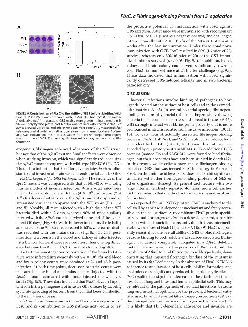
FbsC Expression Increases Biofilm Formation—Bacterialgrowth on epithelial surfaces or on implantable devices can beassociated with the elaboration of a self-formed matrix or abiofilm. We investigated whether the ability of GBS to formbiofilms depends on the expression of FbsC. To this end, GBSstrains were grown in polystyrene plates and bacterial biomassformation was measured by crystal violet staining. As expected,the srtA* mutant produced minimal biofilm, whereas robustbiomass formation was observed using wild type NEM316 (Fig.6A). Under these conditions, biofilm production by the �fbsCdeletion mutant was intermediate between that of NEM316WT and the srtA* mutant (Fig. 6A). Scanning electron micros-copy of the �fbsC biofilm reveals the formation of mainly iso-lated bacterial chains in contrast to WT biofilm forming tightlycompact clusters of bacterial chains surrounded by a denseextracellular matrix (Fig. 6B). This data indicated that FbsCplays a significant role in biofilm formation, probably by pro-moting bacterial aggregation on solid surfaces.
FbsC Expression Is Necessary for Adhesion to and Invasion ofEpithelial Cells—Attachment to and invasion of epithelial cellsby GBS play a crucial role in the initial stages of the infectionprocess. Alveolar and intestinal epithelia are considered aslikely entry sites in early- and late-onset GBS disease (38, 39).To test whether FbsC contributes to interactions between GBSand epithelial cells, we compared the binding of NEM316 WT,�fbsC, and srtA* mutant strains to Caco-2 (intestinal) and A549(pulmonary) human epithelial cell lines. The adherence of the�fbsC strain to CaCo-2 or A549 cells was significantly reducedcompared with NEM316, indicating that FbsC is required foroptimal bacterial adherence (Fig. 7A). The �fbsC mutant, how-ever, was more adherent to both cell types than the srtA*
FIGURE 4. Analysis of fibrinogen binding to recombinant GST-FbsC usingsurface plasmon resonance. GST-FbsC or GST were immobilized on anti-GST-coated sensor chips. Increasing concentrations of fibrinogen (Fng) werethen flowed through the cells. Assay channel data were subtracted from ref-erence flow cell data to eliminate the effects of nonspecific interactions. Thedata shown are from one experiment, representative of three and performedin triplicate.
FbsC, a Fibrinogen-binding Protein from S. agalactiae
JULY 25, 2014 • VOLUME 289 • NUMBER 30 JOURNAL OF BIOLOGICAL CHEMISTRY 21009
by guest on August 1, 2019
http://ww
w.jbc.org/
Dow
nloaded from

mutant, suggesting that besides FbsC other LPXTG-anchoredadhesins play a role in adherence to epithelial cells. Comple-mentation of the fbsC mutation with the wild-type allelerestored bacterial adherence to A549 cells (Fig. 7A). As shownfor adherence, invasion of both epithelial cell lines by the �fbsCmutant was significantly reduced, but was still higher than thatobserved with the srtA* mutant (Fig. 7B). Moreover, adhesionto and invasion of epithelial cells by WT bacteria was almostcompletely abrogated in the presence of GST-FbsC (Fig. 7C).Collectively these results indicate that GBS interactions withepithelial cells are largely dependent on surface-expressedFbsC. Because fibrinogen levels can increase at sites of injuryand during inflammation of mucosal tissues (12), we also stud-ied the interactions of GBS with A549 and Caco-2 cells after
treatment of bacteria with fibrinogen. Fibrinogen pre-treat-ment of NEM316 WT, but not of the �fbsC mutant, was asso-ciated with a significant increase in both adherence and inva-sion (data not shown), suggesting that both processes largelydepend on FbsC-mediated fibrinogen binding.
Role of FbsC in Invasion of Brain Endothelial Cells by GBS—Interaction with human brain vascular endothelial cells is con-sidered a crucial step in the invasion of the blood-brain barrierby GBS (40 – 42). To investigate whether FbsC is involved ininteraction with the endothelial cell line hCMEC/D3, we com-pared the adhesion properties of NEM316 WT with those of itsisogenic mutant �fbsC. After 1 h, NEM316 WT efficientlyadhered to these cells, whereas binding of the �fbsC mutant wassignificantly reduced (Fig. 7D). Preincubation of bacteria with
FIGURE 5. Contribution of FbsC to the overall ability of GBS to bind fibrinogen. Wild-type NEM316 (WT) was compared with its fbsC deletion (�fbsC) orsortase A-defective (srtA*) mutants. * � p � 0.05 by analysis of variance and Student’s Neuman Keuls test. A, binding of GBS strains to fibrinogen (Fng),fibronectin (Fnt), or plasminogen (Plg) immobilized on plastic plates at the concentrations indicated in the horizontal axis. Shown are mean � S.D. of cfu fromthree independent experiments. B, effects of complementation of the fbsc deletion on GBS binding to soluble fibrinogen (1 �g/ml). Bound fibrinogen wasvisualized with mouse anti-fibrinogen IgG followed by FITC-conjugated anti-mouse IgG. NEM3788, WT strain containing the pTCV_TetO (empty) vector;NEM3790, WT strain containing the pTCV_TetO_ fbsC vector and overexpressing the protein; NEM3792, �fbsC containing the pTCV_TetO (empty) vector;NEM3794, �fbsC complemented with the pTCV_TetO_ fbsC vector. C, effects of complementation of the fbsC deletion on adherence to immobilized fibrinogen(5 �g/ml). The results shown are mean � S.D. of cfu counts from three independent experiments. D, inhibition of GBS binding to immobilized fibrinogen (5�g/ml) by soluble fibrinogen, recombinant FbsC, or anti-FbsC antibodies used at the indicated concentrations. The results shown are mean � S.D. of cfu countsfrom three independent experiments.
FbsC, a Fibrinogen-binding Protein from S. agalactiae
21010 JOURNAL OF BIOLOGICAL CHEMISTRY VOLUME 289 • NUMBER 30 • JULY 25, 2014
by guest on August 1, 2019
http://ww
w.jbc.org/
Dow
nloaded from
exogenous fibrinogen enhanced adherence of the WT strain,but not that of the �fbsC mutant. Similar effects were observedwhen studying invasion, which was significantly reduced usingthe �fbsC mutant compared with wild-type NEM316 (Fig. 7D).These data indicated that FbsC largely mediates in vitro adhe-sion to and invasion of brain vascular endothelial cells by GBS.
FbsC Is Required for GBS Pathogenicity—The virulence of the�fbsC mutant was compared with that of NEM316 WT usingmurine models of invasive infection. When adult mice wereinfected intraperitoneally with high (4 � 108 cfu) or low (2 �108 cfu) doses of either strain, the �fbsC mutant displayed anattenuated virulence compared with the WT strain (Fig. 8, Aand B). Notably, all mice infected with a high dose of the WTbacteria died within 2 days, whereas 90% of mice similarlyinfected with the �fbsC mutant survived at the end of the exper-iment (10 days) (Fig. 8A). With the lower inoculum, the lethalityassociated to the WT strain decreased to 63%, whereas no deathwas recorded with the mutant strain (Fig. 8B). By 24 h post-infection, cfu counts in the blood and kidney of mice infectedwith the low bacterial dose revealed more than one log differ-ence between the WT and �fbsC mutant strains (Fig. 8C).
To test the hematogenous colonization of the brain by GBS,mice were infected intravenously with 4 � 108 cfu and bloodand brain colony counts were obtained at 24 and 48 h post-infection. At both time points, decreased bacterial counts weremeasured in the blood and brains of mice injected with the�fbsC mutant compared with those injected the wild-typestrain (Fig. 8D). These data indicated that FbsC plays an impor-tant role in the pathogenesis of invasive GBS disease by favoringsystemic spreading of bacteria from the initial focus of infectionto the invasion of organs.
FbsC-induced Immunoprotection—The surface exposition ofFbsC and its contribution to GBS pathogenicity led us to test
the protective potential of immunization with FbsC againstGBS infection. Adult mice were immunized with recombinantGST-FbsC or GST (used as a negative control) and challengedintraperitoneally with 2 � 108 cfu of the NEM316 strain at 3weeks after the last immunization. Under these conditions,immunization with GST-FbsC resulted in 80% (16 mice of 20)survival, whereas only 30% (6 mice of 20) of the GST-immu-nized animals survived (p � 0.05; Fig. 9A). In addition, blood,kidney, and brain colony counts were significantly lower inGST-FbsC-immunized mice at 24 h after challenge (Fig. 9B).These data indicated that immunization with FbsC signifi-cantly decreased GBS-induced lethality and in vivo bacterialpathogenicity.
DISCUSSION
Bacterial infections involve binding of pathogens to hostligands located on the surface of host cells and in the extracel-lular matrix (43– 45). In several bacterial species, fibrinogen-binding proteins play crucial roles in pathogenesis by allowingbacteria to penetrate host barriers and spread in tissues (9, 46).GBS strongly interact with fibrinogen, a property that is morepronounced in strains isolated from invasive infections (10, 11,13). To date, four structurally unrelated fibrinogen-bindingproteins (FbsA, FbsB, Srr1, and Srr2) involved in virulence havebeen identified in GBS (14 –16, 18, 19) and three of these areencoded by our prototype strain NEM316. Two additional GBSproteins (named Fib and SAG0242) were found to bind fibrin-ogen, but their properties have not been studied in depth (47).In this report, we describe a novel major fibrinogen-bindingprotein of GBS that was termed FbsC in analogy to FbsA andFbsB. On the amino acid level, FbsC does not exhibit significantsimilarity with other fibrinogen-binding proteins of GBS orother organisms, although its general architecture with twolarge internal tandemly repeated domains and a cell anchormotif is similar to that of other bacterial adhesins and virulencefactors (48).
As expected for an LPXTG protein, FbsC is anchored to thecell wall by a sortase A-dependent mechanism and freely acces-sible on the cell surface. A recombinant FbsC protein specifi-cally bound fibrinogen in vitro in a dose-dependent, saturablefashion with a dissociation constant value that was intermedi-ate between those of FbsB (15) and FbsA (15, 49). FbsC is appar-ently essential for the overall ability of GBS to bind fibrinogen,because binding to both soluble and surface-associated fibrin-ogen was almost completely abrogated in a �fbsC deletionmutant. Plasmid-mediated expression of fbsC restored thecapability of �fbsC to bind fibrinogen to wild-type levels, dem-onstrating that impaired fibrinogen binding of the mutant iscaused by its fbsC deficiency. In the absence of FbsC, NEM316adherence to and invasion of host cells, biofilm formation, andits virulence are significantly reduced. In particular, deletion offbsC resulted in a significant decrease in the attachment to andinvasion of lung and intestinal human epithelial cells. This maybe relevant to the pathogenesis of neonatal infections, becauselung and intestinal epithelia are the presumed bacterial entrysites in early- and late-onset GBS diseases, respectively (38, 39).Because epithelial cells express fibrinogen on their surface (50)it is likely that FbsC-dependent adherence and invasion are
FIGURE 6. Contribution of FbsC to the ability of GBS to form biofilm. Wild-type NEM316 (WT) was compared with its fbsC deletion (�fbsC) or sortaseA-defective (srtA*) mutants. A, GBS strains were grown in liquid medium in96-well polystyrene plates and biofilm was stained with crystal violet. Leftpanel, a crystal violet-stained microtiter plate; right panel, A595 measured afterreleasing crystal violet with ethanol/acetone from stained biofilms. Columnand bars indicate the mean � S.D. values from three independent experi-ments * � p � 0.05. B, scanning electron microscopy analysis of biofilmformation.
FbsC, a Fibrinogen-binding Protein from S. agalactiae
JULY 25, 2014 • VOLUME 289 • NUMBER 30 JOURNAL OF BIOLOGICAL CHEMISTRY 21011
by guest on August 1, 2019
http://ww
w.jbc.org/
Dow
nloaded from

mediated by GBS-fibrinogen interactions. This hypothesis isstrengthened by our observations that pretreatment withfibrinogen of wild-type bacteria, but not of those lacking FbsC,increased their ability to bind to epithelial and endothelial cells.
During the preparation of this manuscript, a novel GBSadhesin named BsaB (bacterial surface adhesin of GBS) was
described in GBS strain 515 (20). In vitro functional analysis ofBsaB revealed that it participates in GBS binding to humanfibronectin and laminin, in the adhesion of GBS to human epi-thelial cells, and in biofilm formation. Genome and sequenceanalysis revealed that FbsC and BsaB were identical and encodedby the same gene. However, whereas our results confirmed that
FIGURE 7. Association to human epithelial and endothelial cell lines of wild-type NEM316 (WT) and its fbsC deletion (�fbsC) or sortase A (srtA*)-defectivemutants. Each panel shows the mean � S.D. of cfu counts from three independent experiments; *, p � 0.05 by analysis of variance and Student’s Neuman Keuls test.A, adhesion to intestinal (left panel) and respiratory (center panel) epithelial cell lines; right panel, effects of complementation of the fbsc deletion on adherence tointestinal epithelial cells. NEM3788, WT containing the pTCV_TetO (empty) vector; NEM3790, WT containing the pTCV_TetO_ fbsC vector and overexpressing theprotein; NEM3792, �fbsC containing the pTCV_TetO (empty) vector; and NEM3794, �fbsC complemented with the pTCV_TetO_ fbsC vector. B, invasion of intestinal(left) and respiratory (right) cell lines; C, inhibition of GBS adhesion to and invasion of intestinal epithelial cells by soluble recombinant GST-FbsC or GST, used at theindicated concentrations. D, interactions of GBS with the brain endothelial cell line hCMEC/D3 in the presence and absence of pretreatment of bacteria with fibrinogen.
FbsC, a Fibrinogen-binding Protein from S. agalactiae
21012 JOURNAL OF BIOLOGICAL CHEMISTRY VOLUME 289 • NUMBER 30 • JULY 25, 2014
by guest on August 1, 2019
http://ww
w.jbc.org/
Dow
nloaded from

FbsC/BsaB is involved in adhesion to epithelial cells and in bio-film formation, we demonstrate that this adhesin specificallybinds fibrinogen, but not fibronectin. In fact, in their study (20),FbsC/BsaB binding to fibrinogen was not tested and the adher-ence rates to fibronectin were very low (less than 4%). We there-fore maintained that FbsC/BsaB is a GBS surface protein thatinteracts with fibrinogen and conserved its designation as FbsC.
The respective contribution of cognate GBS fibrinogen-binding proteins (FbsA, FbsB, Srr11, Srr2, and now FbsC) foroptimal binding to fibrinogen remains to be elucidated. How-ever, it is likely that optimal adhesion of GBS to fibrinogenrequires the simultaneous expression of different types offibrinogen-binding proteins. It is interesting to note that, in thisrespect, FbsC preferentially interacts with the B� fibrinogen
chain, as found here, whereas Srr1 and Srr2 both bind to the A�chain (18, 19). The use of different adhesins may allow GBS tobind to different sites in the fibrinogen molecule, thereby pro-moting stronger interactions. Moreover, the relative impor-tance of individual fibrinogen-binding proteins is likely to varyin different phylogenetic lineages, as suggested by the unevendistribution of each protein in these lineages (21). Initially,FbsA was proposed to be the major fibrinogen-binding proteinin GBS (7, 15). Deletion of fbsA in the GBS 6313 (a strain relatedto NEM316 and belonging to the CC23 phylogenetic lineage)markedly impaired its fibrinogen-binding ability and host cellsinvasion. However, further studies investigating GBS strainsbelonging to the CC17 phylogenetic lineage have shown thatthe presence of the fbsA gene alone is not sufficient to producerobust adherence to fibrinogen and that FbsB plays a moreimportant role in this process (16). More recently, Srr1 bindingto fibrinogen was described as important for adherence to brainendothelium and the development of meningitis (19). However,Srr1 is absent in the CC17 phylogenetic lineage, which is asso-ciated with the vast majority of meningitis in newborns.Instead, CC17 hypervirulent strains express a specific serine-rich repeat protein called Srr2, which interacts with fibrinogenand promotes meningitis (18). The expression of FbsC in rep-resentatives of the CC1, CC19, and CC23, but not CC17, lin-eages is similar to that of Srr1. In CC17, the fbsC gene is presentat the same locus but the protein is not expressed due to alineage-specific frameshift mutation. Therefore, CC17 clonesdisplay an increased ability to adhere to fibrinogen with a rep-ertoire of fibrinogen-binding proteins that includes FbsA, FbsB,and Srr2. This suggests that Srr2 plays a key role for fibrinogenbinding in this lineage. A non-exclusive hypothesis may be the
FIGURE 8. FbsC is a virulence factor of GBS. Comparison of lethality andbacterial burden in mice infected with wild-type NEM316 (WT) or its fbsCdeletion (�fbsC) mutant. A and B, survival of CD1 mice following intraperito-neal infection with WT or with �fbsC strains using, as challenge, 4 � 108 (A)and 2 � 108 (B) cfu/mouse. *, p � 0.05 by log-rank Kaplan-Meyer analysis. C,blood and kidney colony counts at 24 h after intraperitoneal challenge with1 � 108 cfu of WT or �fbsC strains. *, p � 0.05 by analysis of variance andStudent’s Neuman Keuls test. D, blood and brain colony counts in miceinfected intravenously with 4 � 108 cfu of WT or �fbsC strains. *, p � 0.05 byanalysis of variance and Student’s Neuman Keuls test.
FIGURE 9. Protective effects of GST-FbsC immunization against lethalGBS infection in mice. A, animals were immunized with the recombinantGST-FbsC protein or with the GST control and challenged intraperitoneal with2 � 108 cfu/mouse of wild type strain NEM316. *, p � 0.05 by log-rank Kaplan-Meyer analysis. B, organ cfu of mice immunized with the recombinant GST-FbsC protein or with the GST control and challenged intraperitoneally with2 � 108 cfu. *, p � 0.05 by analysis of variance and Student’s Neuman Keulstest.
FbsC, a Fibrinogen-binding Protein from S. agalactiae
JULY 25, 2014 • VOLUME 289 • NUMBER 30 JOURNAL OF BIOLOGICAL CHEMISTRY 21013
by guest on August 1, 2019
http://ww
w.jbc.org/
Dow
nloaded from

differential expression of fibrinogen-binding proteins at thepopulation level. Of note, FbsC and other fibrinogen-bindingproteins are strongly regulated by the two-component systemCovRS (also known as CsrRS), which plays an important role incontrolling GBS adherence and biofilm formation (36). Impor-tantly, whereas CovRS directly repressed FbsC expression (34),it also repressed that of the regulator Rga required for Srr1expression (35, 51). Accordingly, inactivation of CovRS led to adramatic increase in GBS binding to fibrinogen (52), but thecontribution of individual fibrinogen-binding proteins remainsto be determined in this genetic background.
In conclusion we have identified a novel fibrinogen-bindingprotein, designated as FbsC, which is required for cell adher-ence, biofilm formation, and invasion of epithelial and endothe-lial barriers by GBS. Further studies involving animal models oftransmucosal infection (e.g. using respiratory or gastrointesti-nal challenge) will be needed to better assess the significance ofFbsC-mediated adherence and invasion of epithelial barriers inthe pathogenesis of GBS disease. In the present study, using anintraperitoneal challenge model, FbsC expression was essentialfor the induction of GBS disease and immunization with arecombinant form of the antigen largely prevented lethality.These observations suggest that FbsC may be a target, in con-junction with other antigens, for immune-based interventionsto control GBS infections.
Acknowledgments—We thank Robert P. Fagan, Bruno Dupuy, andElise Caliot for the pRPF185 vector and the pTCV_TetO constructionand Giuseppe Sabatino (University of Messina) for SE slide prepara-tion and observation.
REFERENCES1. Camacho-Gonzalez, A., Spearman, P. W., and Stoll, B. J. (2013) Neonatal
infectious diseases: evaluation of neonatal sepsis. Pediatr. Clin. North Am.60, 367–389
2. Schrag, S. J., and Stoll, B. J. (2006) Early-onset neonatal sepsis in the era ofwidespread intrapartum chemoprophylaxis. Pediatr. Infect. Dis. J. 25,939 –940
3. Edwards, M. S., and Baker, C. J. (2005) Group B streptococcal infections inelderly adults. Clin. Infect. Dis. 41, 839 – 847
4. Hansen, S. M., Uldbjerg, N., Kilian, M., and Sørensen, U. B. (2004) Dynam-ics of Streptococcus agalactiae colonization in women during and afterpregnancy and in their infants. J. Clin. Microbiol. 42, 83– 89
5. Rubens, C. E., Raff, H. V., Jackson, J. C., Chi, E. Y., Bielitzki, J. T., and Hillier,S. L. (1991) Pathophysiology and histopathology of group B streptococcalsepsis in Macaca nemestrina primates induced after intraamniotic inocu-lation: evidence for bacterial cellular invasion. J. Infect. Dis. 164, 320 –330
6. Rubens, C. E., Smith, S., Hulse, M., Chi, E. Y., and van Belle, G. (1992)Respiratory epithelial cell invasion by group B streptococci. Infect. Immun.60, 5157–5163
7. Schubert, A., Zakikhany, K., Pietrocola, G., Meinke, A., Speziale, P., Eik-manns, B. J., and Reinscheid, D. J. (2004) The fibrinogen receptor FbsApromotes adherence of Streptococcus agalactiae to human epithelial cells.Infect. Immun. 72, 6197– 6205
8. Tenenbaum, T., Bloier, C., Adam, R., Reinscheid, D. J., and Schroten, H.(2005) Adherence to and invasion of human brain microvascular endo-thelial cells are promoted by fibrinogen-binding protein FbsA of Strepto-coccus agalactiae. Infect. Immun. 73, 4404 – 4409
9. Rivera, J., Vannakambadi, G., Höök, M., and Speziale, P. (2007) Fibrino-gen-binding proteins of Gram-positive bacteria. Thromb. Haemost. 98,503–511
10. Schönbeck, C., Björck, L., and Kronvall, G. (1981) Receptors for fibrinogen
and aggregated �2-microglobulin detected in strains of group B strepto-cocci. Infect. Immun. 31, 856 – 861
11. Chhatwal, G. S., Dutra, I. S., and Blobel, H. (1985) Fibrinogen bindinginhibits the fixation of the third component of human complement onsurface of groups A, B, C, and G streptococci. Microbiol. Immunol. 29,973–980
12. Adams, R. A., Schachtrup, C., Davalos, D., Tsigelny, I., and Akassoglou, K.(2007) Fibrinogen signal transduction as a mediator and therapeutic targetin inflammation: lessons from multiple sclerosis. Curr. Med. Chem. 14,2925–2936
13. Rosenau, A., Martins, K., Amor, S., Gannier, F., Lanotte, P., van der Mee-Marquet, N., Mereghetti, L., and Quentin, R. (2007) Evaluation of theability of Streptococcus agalactiae strains isolated from genital and neona-tal specimens to bind to human fibrinogen and correlation with charac-teristics of the fbsA and fbsB genes. Infect. Immun. 75, 1310 –1317
14. Schubert, A., Zakikhany, K., Schreiner, M., Frank, R., Spellerberg, B., Eik-manns, B. J., and Reinscheid, D. J. (2002) A fibrinogen receptor from groupB Streptococcus interacts with fibrinogen by repetitive units with novelligand binding sites. Mol. Microbiol. 46, 557–569
15. Gutekunst, H., Eikmanns, B. J., and Reinscheid, D. J. (2004) The novelfibrinogen-binding protein FbsB promotes Streptococcus agalactiae inva-sion into epithelial cells. Infect. Immun. 72, 3495–3504
16. Al Safadi, R., Mereghetti, L., Salloum, M., Lartigue, M. F., Virlogeux-Pay-ant, I., Quentin, R., and Rosenau, A. (2011) Two-component systemRgfA/C activates the fbsB gene encoding major fibrinogen-binding pro-tein in highly virulent CC17 clone group B Streptococcus. PloS One 6,e14658
17. Pietrocola, G., Schubert, A., Visai, L., Torti, M., Fitzgerald, J. R., Foster,T. J., Reinscheid, D. J., and Speziale, P. (2005) FbsA, a fibrinogen-bindingprotein from Streptococcus agalactiae, mediates platelet aggregation.Blood 105, 1052–1059
18. Seo, H. S., Minasov, G., Seepersaud, R., Doran, K. S., Dubrovska, I.,Shuvalova, L., Anderson, W. F., Iverson, T. M., and Sullam, P. M. (2013)Characterization of fibrinogen binding by glycoproteins Srr1 and Srr2 ofStreptococcus agalactiae. J. Biol. Chem. 288, 35982–35996
19. Seo, H. S., Mu, R., Kim, B. J., Doran, K. S., and Sullam, P. M. (2012) Bindingof glycoprotein Srr1 of Streptococcus agalactiae to fibrinogen promotesattachment to brain endothelium and the development of meningitis. PloSPathog. 8, e1002947
20. Jiang, S., and Wessels, M. R. (2014) BsaB, a novel adherence factor of groupB Streptococcus. Infect. Immun. 82, 1007–1016
21. Brochet, M., Rusniok, C., Couvé, E., Dramsi, S., Poyart, C., Trieu-Cuot, P.,Kunst, F., and Glaser, P. (2008) Shaping a bacterial genome by large chro-mosomal replacements, the evolutionary history of Streptococcus agalac-tiae. Proc. Natl. Acad. Sci. U.S.A. 105, 15961–15966
22. Carey, R. B., Eisenstein, T. K., Shockman, G. D., Greber, T. F., and Swen-son, R. M. (1980) Soluble group- and type-specific antigens from type IIIgroup B Streptococcus. Infect. Immun. 28, 195–203
23. Firon, A., Tazi, A., Da Cunha, V., Brinster, S., Sauvage, E., Dramsi, S.,Golenbock, D. T., Glaser, P., Poyart, C., and Trieu-Cuot, P. (2013) TheAbi-domain protein Abx1 interacts with the CovS histidine kinase to con-trol virulence gene expression in group B Streptococcus. PloS Pathog. 9,e1003179
24. Heckman, K. L., and Pease, L. R. (2007) Gene splicing and mutagenesis byPCR-driven overlap extension. Nat. Protoc. 2, 924 –932
25. Corrigan, R. M., and Foster, T. J. (2009) An improved tetracycline-induc-ible expression vector for Staphylococcus aureus. Plasmid 61, 126 –129
26. Fagan, R. P., and Fairweather, N. F. (2011) Clostridium difficile hastwo parallel and essential Sec secretion systems. J. Biol. Chem. 286,27483–27493
27. Poyart, C., Lamy, M. C., Boumaila, C., Fiedler, F., and Trieu-Cuot, P.(2001) Regulation of D-alanyl-lipoteichoic acid biosynthesis in Streptococ-cus agalactiae involves a novel two-component regulatory system. J. Bac-teriol. 183, 6324 – 6334
28. Garibaldi, M., Rodríguez-Ortega, M. J., Mandanici, F., Cardaci, A., Midiri,A., Papasergi, S., Gambadoro, O., Cavallari, V., Teti, G., and Beninati, C.(2010) Immunoprotective activities of a Streptococcus suis pilus subunit inmurine models of infection. Vaccine 28, 3609 –3616
FbsC, a Fibrinogen-binding Protein from S. agalactiae
21014 JOURNAL OF BIOLOGICAL CHEMISTRY VOLUME 289 • NUMBER 30 • JULY 25, 2014
by guest on August 1, 2019
http://ww
w.jbc.org/
Dow
nloaded from

29. Papasergi, S., Garibaldi, M., Tuscano, G., Signorino, G., Ricci, S., Peppo-loni, S., Pernice, I., Lo Passo, C., Teti, G., Felici, F., and Beninati, C. (2010)Plasminogen- and fibronectin-binding protein B is involved in the adher-ence of Streptococcus pneumoniae to human epithelial cells. J. Biol. Chem.285, 7517–7524
30. Cardaci, A., Papasergi, S., Midiri, A., Mancuso, G., Domina, M., Cariccio,V. L., Mandanici, F., Galbo, R., Lo Passo, C., Pernice, I., Donato, P., Ricci, S.,Biondo, C., Teti, G., Felici, F., and Beninati, C. (2012) Protective activity ofStreptococcus pneumoniae Spr1875 protein fragments identified using aphage displayed genomic library. PloS One 7, e36588
31. Lalioui, L., Pellegrini, E., Dramsi, S., Baptista, M., Bourgeois, N., Doucet-Populaire, F., Rusniok, C., Zouine, M., Glaser, P., Kunst, F., Poyart, C., andTrieu-Cuot, P. (2005) The SrtA Sortase of Streptococcus agalactiae is re-quired for cell wall anchoring of proteins containing the LPXTG motif, foradhesion to epithelial cells, and for colonization of the mouse intestine.Infect. Immun. 73, 3342–3350
32. Weksler, B. B., Subileau, E. A., Perrière, N., Charneau, P., Holloway, K.,Leveque, M., Tricoire-Leignel, H., Nicotra, A., Bourdoulous, S., Turowski,P., Male, D. K., Roux, F., Greenwood, J., Romero, I. A., and Couraud, P. O.(2005) Blood-brain barrier-specific properties of a human adult brain en-dothelial cell line. FASEB J. 19, 1872–1874
33. van Gennip, M., Christensen, L. D., Alhede, M., Qvortrup, K., Jensen, P.Ø., Høiby, N., Givskov, M., and Bjarnsholt, T. (2012) Interactions betweenpolymorphonuclear leukocytes and Pseudomonas aeruginosa biofilms onsilicone implants in vivo. Infect. Immun. 80, 2601–2607
34. Lamy, M. C., Zouine, M., Fert, J., Vergassola, M., Couve, E., Pellegrini, E.,Glaser, P., Kunst, F., Msadek, T., Trieu-Cuot, P., and Poyart, C. (2004)CovS/CovR of group B Streptococcus: a two-component global regulatorysystem involved in virulence. Mol. Microbiol. 54, 1250 –1268
35. Mistou, M. Y., Dramsi, S., Brega, S., Poyart, C., and Trieu-Cuot, P. (2009)Molecular dissection of the secA2 locus of group B Streptococcus revealsthat glycosylation of the Srr1 LPXTG protein is required for full virulence.J. Bacteriol. 191, 4195– 4206
36. Papasergi, S., Galbo, R., Lanza-Cariccio, V., Domina, M., Signorino, G.,Biondo, C., Pernice, I., Poyart, C., Trieu-Cuot, P., Teti, G., and Beninati, C.(2013) Analysis of the Streptococcus agalactiae exoproteome. J. Proteomics89, 154 –164
37. Brega, S., Caliot, E., Trieu-Cuot, P., and Dramsi, S. (2013) SecA localiza-tion and SecA-dependent secretion occurs at new division septa in groupB Streptococcus. PloS One 8, e65832
38. Tazi, A., Disson, O., Bellais, S., Bouaboud, A., Dmytruk, N., Dramsi, S.,Mistou, M. Y., Khun, H., Mechler, C., Tardieux, I., Trieu-Cuot, P., Lecuit,M., and Poyart, C. (2010) The surface protein HvgA mediates group BStreptococcus hypervirulence and meningeal tropism in neonates. J. Exp.Med. 207, 2313–2322
39. Spellerberg, B. (2000) Pathogenesis of neonatal Streptococcus agalactiaeinfections. Microbes Infect. 2, 1733–1742
40. Nizet, V., Kim, K. S., Stins, M., Jonas, M., Chi, E. Y., Nguyen, D., andRubens, C. E. (1997) Invasion of brain microvascular endothelial cells bygroup B streptococci. Infect. Immun. 65, 5074 –5081
41. Charland, N., Nizet, V., Rubens, C. E., Kim, K. S., Lacouture, S., andGottschalk, M. (2000) Streptococcus suis serotype 2 interactions with hu-man brain microvascular endothelial cells. Infect. Immun. 68, 637– 643
42. Quach, D., van Sorge, N. M., Kristian, S. A., Bryan, J. D., Shelver, D. W., and
Doran, K. S. (2009) The CiaR response regulator in group B Streptococcuspromotes intracellular survival and resistance to innate immune defenses.J. Bacteriol. 191, 2023–2032
43. Cue, D., Dombek, P. E., Lam, H., and Cleary, P. P. (1998) Streptococcuspyogenes serotype M1 encodes multiple pathways for entry into humanepithelial cells. Infect. Immun. 66, 4593– 4601
44. Ozeri, V., Rosenshine, I., Mosher, D. F., Fässler, R., and Hanski, E. (1998)Roles of integrins and fibronectin in the entry of Streptococcus pyogenesinto cells via protein F1. Mol. Microbiol. 30, 625– 637
45. Talay, S. R., Zock, A., Rohde, M., Molinari, G., Oggioni, M., Pozzi, G.,Guzman, C. A., and Chhatwal, G. S. (2000) Co-operative binding of hu-man fibronectin to Sfbl protein triggers streptococcal invasion into respi-ratory epithelial cells. Cell Microbiol. 2, 521–535
46. Dombek, P. E., Cue, D., Sedgewick, J., Lam, H., Ruschkowski, S., Finlay,B. B., and Cleary, P. P. (1999) High-frequency intracellular invasion ofepithelial cells by serotype M1 group A streptococci: M1 protein-medi-ated invasion and cytoskeletal rearrangements. Mol. Microbiol. 31,859 – 870
47. Margarit, I., Bonacci, S., Pietrocola, G., Rindi, S., Ghezzo, C., Bombaci, M.,Nardi-Dei, V., Grifantini, R., Speziale, P., and Grandi, G. (2009) Capturinghost-pathogen interactions by protein microarrays: identification of novelstreptococcal proteins binding to human fibronectin, fibrinogen, andC4BP. FASEB J. 23, 3100 –3112
48. Nobbs, A. H., Lamont, R. J., and Jenkinson, H. F. (2009) Streptococcusadherence and colonization. Microbiol. Mol. Biol. Rev. 73, 407– 450
49. Papasergi, S., Lanza Cariccio, V., Pietrocola, G., Domina, M., D’Aliberti,D., Trunfio, M. G., Signorino, G., Peppoloni, S., Biondo, C., Mancuso, G.,Midiri, A., Rindi, S., Teti, G., Speziale, P., Felici, F., and Beninati, C. (2013)Immunogenic properties of Streptococcus agalactiae FbsA fragments.PloS One 8, e75266
50. Guadiz, G., Sporn, L. A., Goss, R. A., Lawrence, S. O., Marder, V. J., andSimpson-Haidaris, P. J. (1997) Polarized secretion of fibrinogen by lungepithelial cells. Am. J. Respir. Cell Mol. Biol. 17, 60 – 69
51. Dramsi, S., Dubrac, S., Konto-Ghiorghi, Y., Da Cunha, V., Couvé, E., Gla-ser, P., Caliot, E., Débarbouillé, M., Bellais, S., Trieu-Cuot, P., and Mistou,M. Y. (2012) Rga, a RofA-like regulator, is the major transcriptional acti-vator of the PI-2a pilus in Streptococcus agalactiae. Microb. Drug. Resist.18, 286 –297
52. Park, S. E., Jiang, S., and Wessels, M. R. (2012) CsrRS and environmentalpH regulate group B Streptococcus adherence to human epithelial cells andextracellular matrix. Infect. Immun. 80, 3975–3984
53. Glaser, P., Rusniok, C., Buchrieser, C., Chevalier, F., Frangeul, L., Msadek,T., Zouine, M., Couvé, E., Lalioui, L., Poyart, C., Trieu-Cuot, P., and Kunst,F. (2002) Genome sequence of Streptococcus agalactiae, a pathogen caus-ing invasive neonatal disease. Mol. Microbiol. 45, 1499 –1513
54. Konto-Ghiorghi, Y., Mairey, E., Mallet, A., Duménil, G., Caliot, E., Dumé-nil, G., Caliot, E., Trieu-Cuot, P., and Dramsi, S. (2009) Dual role for pilusin adherence to epithelial cells and biofilm formation in Streptococcusagalactiae. PLoS Pathog. 5, e1000422
55. Danne, C., Guérillot, R., Glaser, P., Trieu-Cuot, P., and Dramsi, S.(2013) Construction of isogenic mutants in Streptococcus gallolyticusbased on the development of new mobilizable vectors. Res. Microbiol.164, 973–978
FbsC, a Fibrinogen-binding Protein from S. agalactiae
JULY 25, 2014 • VOLUME 289 • NUMBER 30 JOURNAL OF BIOLOGICAL CHEMISTRY 21015
by guest on August 1, 2019
http://ww
w.jbc.org/
Dow
nloaded from
Speziale, Patrick Trieu-Cuot and Concetta BeninatiBiondo, Giuseppe Mancuso, Angelina Midiri, Letizia Romeo, Giuseppe Teti, Pietro Marco Buscetta, Salvatore Papasergi, Arnaud Firon, Giampiero Pietrocola, Carmelo
Cell Interactions-HostStreptococcus agalactiaeFbsC, a Novel Fibrinogen-binding Protein, Promotes
doi: 10.1074/jbc.M114.553073 originally published online June 5, 20142014, 289:21003-21015.J. Biol. Chem.
10.1074/jbc.M114.553073Access the most updated version of this article at doi:
Alerts:
When a correction for this article is posted•
When this article is cited•
to choose from all of JBC's e-mail alertsClick here
http://www.jbc.org/content/289/30/21003.full.html#ref-list-1
This article cites 55 references, 25 of which can be accessed free at
by guest on August 1, 2019
http://ww
w.jbc.org/
Dow
nloaded from