Faux pas deficits in people with medial frontal lesions as ... Lee/Faux pas deficits in...
7
Neuropsychologia 48 (2010) 1670–1676 Contents lists available at ScienceDirect Neuropsychologia journal homepage: www.elsevier.com/locate/neuropsychologia Faux pas deficits in people with medial frontal lesions as related to impaired understanding of a speaker’s mental state Tatia M.C. Lee a,b,c,∗ , Alison K.Y. Ip a,b , Kai Wang d , Chun-hua Xi d , Pan-pan Hu d , Henry K.F. Mak e , Shi-hui Han f , Chetwyn C.H. Chan g,∗∗ a Laboratory of Neuropsychology, The University of Hong Kong, Hong Kong, China b Laboratory of Cognitive Affective Neuroscience, The University of Hong Kong, Hong Kong, China c The State Key Laboratory of Brain and Cognitive Sciences, The University of Hong Kong, Hong Kong, China d Laboratory of Neuropsychology, The First Affiliated Hospital of Anhui Medical University, Hefei, China e Department of Diagnostic Radiology, The University of Hong Kong, Hong Kong, China f Department of Psychology, Peking University, Beijing, China g Applied Cognitive Neuroscience Laboratory, Department of Rehabilitation Sciences, The Hong Kong Polytechnic University, Hong Kong, China article info Article history: Received 7 November 2009 Received in revised form 7 February 2010 Accepted 8 February 2010 Available online 13 February 2010 Keywords: Mentalizing Faux pas Medial frontal region Mental state Frontal lesions abstract This study examined the nature of deficits in mentalizing, the ability to read the mental state of other people, as measured by a faux pas task in people with medial frontal lesions. A total of 56 Mandarin- speaking Chinese individuals participated (9 participants with medial frontal lesions, 12 participants with lateral frontal lesions, 5 participants with non-frontal lesions, and 30 healthy controls). The faux pas test ascertained the participants’ ability to identify and understand a social faux pas, and to understand the mental states of the characters (the speaker and the recipient in a conversation with a social faux pas). Although the participants with medial frontal lesions performed less well than the other clinical participants and the control participants on all aspects of the faux pas test, the most significant deficit was observed in understanding mental states and hence inferring the speaker’s intentions. The performance on the various aspects of decoding a social faux pas by people with medial frontal lesions suggests that the cognitive processes, and hence the respective neural correlates subserving these various processes, may be different. Our results add to existing literature and illustrate the very nature of deficits of mentalizing, measured by a faux pas test, experienced by people with medial frontal lesions. The data have also prompted that future behavioral and neuroimaging studies may be applied to further decode both the neural mechanisms and the cognitive variables affecting “mentalizing”. © 2010 Elsevier Ltd. All rights reserved. 1. Introduction Humans are social animals that under normal circumstances seek rewarding social interactions for adaptive learning and psy- chological satisfaction (Amodio & Frith, 2006; Cohen, 2004). To accomplish this important goal, we must be equipped with cog- nitive processes and the accompanying neural mechanisms that Abbreviations: ANOVA, analysis of variance; FC, frontal cortex; FL, frontal lesion group; fMRI, functional magnetic resonance imaging; LFC, lateral frontal cortex; MFC, medial frontal cortex; NFC, non-frontal lesion control; DMFC, dorsomedial medial frontal cortex; VMFC, ventromedial frontal cortex. ∗ Corresponding author at: K610, Laboratory of Neuropsychology, The University of Hong Kong, Pokfulam Road, Hong Kong, China. Tel.: +852 2857 8394; fax: +852 2819 0978. ∗∗ Corresponding author: Applied Cognitive Neuroscience Laboratory, The Hong Kong Polytechnic University, Hung Hom, Kowloon, Hong Kong, China. Tel.: +852 2766 6727; Fax: +852 2819 0978. E-mail addresses: [email protected] (T.M.C. Lee), [email protected] (C.C.H. Chan). underpin both competent and successful interaction and commu- nication with other people. These processes are classified under the construct termed social cognition, which, as defined by Brothers (1990), is the mental operation underlying social interactions, including the human ability to perceive the intentions and dispo- sitions of others. Among the various social cognitive skills essential to adaptive social learning and interactions, the ability to “mentalize,” that is, to read the mental states of other agents (Frith & Frith, 2006), enables the attribution of cognitive and affective states of self and others. It is this awareness that allows one to detect the intentions and inner mental states of the self and others during social interactions (Baron-Cohen, Leslie, & Frith, 1985; David et al., 2008). In other words, mentalizing is the ability to represent another person’s psy- chological perspective so that one may predict the behaviors of others (Amodio & Frith, 2006). One strategy for assessing the ability to mentalize is the use of faux pas tests (Stone, Baron-Cohen, & Knight, 1998). Faux pas is a French term meaning a “false step,” in other words, a speaker 0028-3932/$ – see front matter © 2010 Elsevier Ltd. All rights reserved. doi:10.1016/j.neuropsychologia.2010.02.012
Transcript of Faux pas deficits in people with medial frontal lesions as ... Lee/Faux pas deficits in...

Fu
THa
b
c
d
e
f
g
a
ARRAA
KMFMMF
1
scan
gMm
of
KT
C
0d
Neuropsychologia 48 (2010) 1670–1676
Contents lists available at ScienceDirect
Neuropsychologia
journa l homepage: www.e lsev ier .com/ locate /neuropsychologia
aux pas deficits in people with medial frontal lesions as related to impairednderstanding of a speaker’s mental state
atia M.C. Leea,b,c,∗, Alison K.Y. Ipa,b, Kai Wangd, Chun-hua Xid, Pan-pan Hud,enry K.F. Make, Shi-hui Hanf, Chetwyn C.H. Chang,∗∗
Laboratory of Neuropsychology, The University of Hong Kong, Hong Kong, ChinaLaboratory of Cognitive Affective Neuroscience, The University of Hong Kong, Hong Kong, ChinaThe State Key Laboratory of Brain and Cognitive Sciences, The University of Hong Kong, Hong Kong, ChinaLaboratory of Neuropsychology, The First Affiliated Hospital of Anhui Medical University, Hefei, ChinaDepartment of Diagnostic Radiology, The University of Hong Kong, Hong Kong, ChinaDepartment of Psychology, Peking University, Beijing, ChinaApplied Cognitive Neuroscience Laboratory, Department of Rehabilitation Sciences, The Hong Kong Polytechnic University, Hong Kong, China
r t i c l e i n f o
rticle history:eceived 7 November 2009eceived in revised form 7 February 2010ccepted 8 February 2010vailable online 13 February 2010
eywords:entalizing
aux pas
a b s t r a c t
This study examined the nature of deficits in mentalizing, the ability to read the mental state of otherpeople, as measured by a faux pas task in people with medial frontal lesions. A total of 56 Mandarin-speaking Chinese individuals participated (9 participants with medial frontal lesions, 12 participantswith lateral frontal lesions, 5 participants with non-frontal lesions, and 30 healthy controls). The faux pastest ascertained the participants’ ability to identify and understand a social faux pas, and to understandthe mental states of the characters (the speaker and the recipient in a conversation with a social fauxpas). Although the participants with medial frontal lesions performed less well than the other clinicalparticipants and the control participants on all aspects of the faux pas test, the most significant deficit was
edial frontal regionental state
rontal lesions
observed in understanding mental states and hence inferring the speaker’s intentions. The performanceon the various aspects of decoding a social faux pas by people with medial frontal lesions suggests that thecognitive processes, and hence the respective neural correlates subserving these various processes, maybe different. Our results add to existing literature and illustrate the very nature of deficits of mentalizing,measured by a faux pas test, experienced by people with medial frontal lesions. The data have also
aviohe co
prompted that future behneural mechanisms and t
. Introduction
Humans are social animals that under normal circumstances
eek rewarding social interactions for adaptive learning and psy-hological satisfaction (Amodio & Frith, 2006; Cohen, 2004). Toccomplish this important goal, we must be equipped with cog-itive processes and the accompanying neural mechanisms thatAbbreviations: ANOVA, analysis of variance; FC, frontal cortex; FL, frontal lesionroup; fMRI, functional magnetic resonance imaging; LFC, lateral frontal cortex;FC, medial frontal cortex; NFC, non-frontal lesion control; DMFC, dorsomedialedial frontal cortex; VMFC, ventromedial frontal cortex.∗ Corresponding author at: K610, Laboratory of Neuropsychology, The University
f Hong Kong, Pokfulam Road, Hong Kong, China. Tel.: +852 2857 8394;ax: +852 2819 0978.∗∗ Corresponding author: Applied Cognitive Neuroscience Laboratory, The Hongong Polytechnic University, Hung Hom, Kowloon, Hong Kong, China.el.: +852 2766 6727; Fax: +852 2819 0978.
E-mail addresses: [email protected] (T.M.C. Lee),[email protected] (C.C.H. Chan).
028-3932/$ – see front matter © 2010 Elsevier Ltd. All rights reserved.oi:10.1016/j.neuropsychologia.2010.02.012
ral and neuroimaging studies may be applied to further decode both thegnitive variables affecting “mentalizing”.
© 2010 Elsevier Ltd. All rights reserved.
underpin both competent and successful interaction and commu-nication with other people. These processes are classified under theconstruct termed social cognition, which, as defined by Brothers(1990), is the mental operation underlying social interactions,including the human ability to perceive the intentions and dispo-sitions of others.
Among the various social cognitive skills essential to adaptivesocial learning and interactions, the ability to “mentalize,” that is, toread the mental states of other agents (Frith & Frith, 2006), enablesthe attribution of cognitive and affective states of self and others.It is this awareness that allows one to detect the intentions andinner mental states of the self and others during social interactions(Baron-Cohen, Leslie, & Frith, 1985; David et al., 2008). In otherwords, mentalizing is the ability to represent another person’s psy-
chological perspective so that one may predict the behaviors ofothers (Amodio & Frith, 2006).One strategy for assessing the ability to mentalize is the use offaux pas tests (Stone, Baron-Cohen, & Knight, 1998). Faux pas isa French term meaning a “false step,” in other words, a speaker

cholo
snr
oiwttd9tiS
acT
1
2
3
4
is
vfao1
mo(i&maoom2
sti2tAmcY
T.M.C. Lee et al. / Neuropsy
ays something he or she should not have said, not knowing orot realizing the words’ inappropriateness, which could hurt theecipient’s feelings. An example of a social faux pas is as follows:
“Jessica was at Maria’s apartment. While appreciating a crystalvase that she gave Maria as a birthday gift, she accidentally droppedthe vase to the ground, which was then shattered into pieces. Jessicafelt really sorry about breaking the vase. Maria said, “Don’t worryabout it. I never like this vase anyway.”
When an individual recognizes the occurrence of a faux pas, her she mentalizes that the speaker who said what was said did notntend to hurt the listener’s feelings, and predicts that the individual
ho heard the words will be upset (Stone et al., 1998). Being ableo mentalize another person’s inner psychological state allows uso evaluate the intentions behind the behavior of others and henceecide how to react. According to previous research, children aged–11 years are able to recognize a faux pas involving the capacityo understand a situation in which one character should have keptnformation from another but did not (Baron-Cohen, O’Riordan,tone, Jones, & Plaisted, 1999).
Applying the principle of faux pas, Stone et al. (1998) developedfaux pas test to assess the ability to mentalize. Ten stories, each
ontaining a faux pas, are presented to participants one at a time.he participants are required to answer the following questions:
. Detecting a faux pas: did someone say something he (or she)should not have said?
. Understanding the faux pas: who said something he (or she)should not have said?
. Understanding the recipient’s mental state: why should he (orshe) not have said it?
. Understanding the speaker’s mental state: why did he (or she)say it?
The participants are then asked a final control question to tap themportant details of the story without inferring another’s mentaltate in order to test their story comprehension.
Impaired faux pas performance has been widely reported inarious clinical groups. Children with Asperger syndrome or high-unctioning autism were found to perform significantly poorer thancontrol group in detecting faux pas stories, while performing welln first- and second-order false belief tasks (Baron-Cohen et al.,999; Stone et al., 1998).
It has been suggested that the ability to mentalize other people’sental states and the capability of processing information about
neself may be subserved by a similar group of neural substratesMitchell, Macrae, & Banaji, 2004) in the frontal cortex (FC). The FCs functionally and anatomically heterogeneous (Happaney, Zelazo,
Stuss, 2004) and can be broadly subdivided into the lateral, dorso-edial, and orbital FC. Functionally, the dorsomedial and orbital FCs
re often considered together as the medial FC (MFC). As such, therbital FC, together with the dorsomedial FC, may function collab-ratively (Öngür & Price, 2000) in processing inputs from sensoryodalities for regulating consummatory behaviors (Kringelbach,
005).About possible neural correlates of mentalizing, the MFC, the
uperior temporal sulcus, the temporal–parietal junction, and theemporal poles (adjacent to the amygdala) are candidates serv-ng social cognitive functions (Amodio & Frith, 2006; Frith & Frith,003). Researchers have suggested that the MFC, plays an impor-
ant role in mentalizing (Mazza et al., 2007; Stuss, Gallup, &lexander, 2001). Earlier studies have reported impaired perfor-ance on a faux pas test in patients with lesions in the orbitofrontalortex and amygdala (Stone et al., 1998; Stone, Baron-Cohen,oung, Calder, & Green, 1999). Amodio and Frith (2006) proposed
gia 48 (2010) 1670–1676 1671
that the MFC is important for monitoring action, person percep-tion, inferences about others’ thoughts, and outcomes related topunishments and rewards. Krueger, Barbey, and Grafman (2009)proposed the structural and temporal representation binding the-ory, which delineates the special role of the MFC in mediating socialevent knowledge via structural and temporal binding processeswith that in the posterior cerebral cortex and the limbic structures.Critchley et al. (2003) theorized that the MFC is important for inte-grating interoceptive, cognitive, and motivational states, therebyrepresenting or producing changes in visceral sensations that arepresent in affective states.
In contrast to the MFC, the lateral FC (LFC) is responsible fornon-emotion-related cognitive processes. The literature suggeststhat the LFC is a key neural substrate of cognitive control forinhibiting a prepotent behavior (Huettel & McCarthy, 2004), select-ing a novel behavior (Ranganath & Rainer, 2003), and selecting aresponse option when competition exists between more than one(Frith, 2000; Robinson, Blair, & Cipolotti, 1998). In clinical studies,damage to the LFC is associated with impaired selection of plansfor behavior. Such patients are unable to choose between possi-ble alternatives, preferring well-practiced behaviors regardless ofcontext (Lhermitte, 1986; Mesulam, 2002).
Shamay-Tsoory, Tibi-Elhanany, and Aharon-Perertz (2006)examined patients with MFC damage and observed impairmentin tasks requiring affective mentalizing, particularly in reasoningabout affective irony and lie conditions involving more contextuallyembedded mindreading, but not other cognitive theory-of-mindtasks. Following this line of thinking, people with MFC damageshould be more impaired in the affective (i.e. understanding mentalstates) than the cognitive (i.e. identifying a social faux pas) aspectsof a faux pas test. Hence, we employed a lesion study methodology,the essence of which is the establishment of testable relationshipsbetween brain regions and behaviors (Damasio & Damasio, 1989),to examine the nature of faux pas deficits experienced by peoplewith frontal lesions. We first classified patients with frontal lesionsinto two groups: one group with lesions in the LFC and the othergroup with lesions in the MFC. We hypothesized that people withMFC lesions would perform significantly worse than those with LFClesions, in accordance with the literature on the neural correlates ofperforming a faux pas test. We then examined scores on each of thefaux pas questions to identify the neural correlates of mentalizing,as well as the nature of the faux pas deficits presented by peoplein the MFC group. We hypothesized that impaired performance onthe faux pas test would be related to the understanding of mentalstates but not to the identification of a faux pas.
2. Methods
2.1. Participants
The participants included a total of 56 right-handed Chinese individuals fromHefei in the Anhui Province of China. Among them were 26 patients of Anhui MedicalUniversity who had acquired localized, well-defined brain lesions caused by surgi-cal removal of brain tumors. Their premorbid general intellectual functioning waswithin average limits as estimated by their premorbid vocational achievement (allparticipants were engaging in stable full-time competitive employment). Learningdisabilities were screened by a neuropsychologist. Potential participants diagnosedof such disabilities were excluded from participation in this study. Developmentalhistory and premorbid social conduct were normal as reported by their respectivecaregivers. In addition, they had no premorbid history of medical and/or psychiatricillnesses affecting cognitive functioning. The 30 neurologically healthy controls, whohad no history of medical conditions affecting cognitive functioning, were recruitedfrom the Hefei community. We matched the clinical and control groups in terms ofyears of education, general cognitive functioning as measured by the Mini-Mental
State Examination (MMSE; Folstein, Folstein, & McHugh, 1975), and mood state asmeasured by the Hamilton Depression Scale (HAMD; Hamilton, 1960). The studywas approved by the Research Ethics Committee of Anhui Medical University. Allparticipants gave their informed consent to take part in the study according to theDeclaration of Helsinki. In addition, for the patients, consent was also obtained fromtheir caregivers.
1 ychologia 48 (2010) 1670–1676
gTnpl(Tusww[t[
gaaaSap2M
p
2
iap
12345
irtqrcsfv2
2
atis
3
3
tebcwwcpe
form
atio
nan
dp
erfo
rman
ceon
the
Min
i-M
enta
lSta
teEx
amin
atio
nan
dth
eH
amil
ton
Dep
ress
ion
Scal
eof
the
clin
ical
and
con
trol
par
tici
pan
ts.
NH
Cgr
oup
(n=
30)
LFC
grou
p(n
=9)
MFC
grou
p(n
=12
)N
FCgr
oup
(n=
5)N
HC
vs.c
lin
ical
grou
pd
iffe
ren
ces
Lesi
onsu
bgro
up
dif
fere
nce
s(L
FC,
MFC
,an
dN
FCgr
oup
s)
19m
ales
;11
fem
ales
8m
ales
;1
fem
ales
7m
ales
;5
fem
ales
3m
ales
;2
fem
ales
�2(1
)=.2
16,p
=.6
42�
2(2
)=2.
50,p
=.2
86)
34.6
7(9
.28)
36.2
2(6
.92)
39.1
7(1
3.57
)31
.80
(9.4
2)t(
54)=
.767
,p=
.447
F(2,
23)=
.812
,p=
.456
tion
,mea
n(S
D)
8.80
(2.9
2)7.
89(3
.52)
7.50
(2.5
8)7.
67(3
.35)
t(54
)=−1
.151
,p=
.255
F(2,
23)=
.186
,p=
.831
(SD
)28
.83
(.83
)28
.22
(1.3
9)28
.17
(1.7
5)28
.60
(1.5
17)
t(37
.28)
=−1
.670
,p=
.447
F(2,
23)=
.136
,p=
.873
(SD
)1.
20(1
.00)
1.33
(.71
)1.
60(1
.17)
1.60
(1.1
4)t(
52)=
1.10
8,p
=.2
73F(
2,23
)=.1
94,p
=.8
25u
ryin
day
s,m
ean
(SD
)*
89.8
9(1
14.7
8)28
0.75
(722
.83)
1626
.60
(246
8.67
)*
F(2,
23)=
3.20
3,p
=.0
59rc
enta
ge,m
ean
(SD
)*
1.94
(1.7
2)2.
38(1
.53)
1.43
(.86
)*
F(2,
23)=
.73,
p=
.493
*5
righ
t;4
left
6ri
ght;
0le
ft;
3bo
th3
righ
t;2
left
;*
�2(4
)=4.
19,p
=.3
81
*9
glio
ma
9gl
iom
a;1
glio
ma
pos
t-op
erat
ion
;1
AV
M;
1tu
mor
1gl
iom
a;1
tum
or;
1ce
rebr
alcy
stic
erco
sis;
1ce
rebr
alin
farc
tion
;1
tem
por
alan
giom
a
**
ical
lyh
ealt
hy
con
trol
;LF
C=
late
ralf
ron
talc
orte
x;M
FC=
med
ialf
ron
talc
orte
x;N
FC=
non
-fro
nta
lles
ion
con
trol
;M
MSE
=M
ini-
Men
talS
tate
Exam
inat
ion
;H
AM
D=
Ham
ilto
nD
epre
ssio
nSc
ale.
*=N
otap
pli
cabl
e.
672 T.M.C. Lee et al. / Neurops
All lesions were localized and circumscribed onto standard templates, and therouping was based on their respective lesion sites (Damasio & Damasio, 1989).he clinical participants were first classified into the frontal lesion group (FL,= 21) and the non-frontal lesion control group (NFC, n = 5). Among the partici-ants in the FL group, 18 suffered from unilateral lesions (right hemisphere = 11,
eft hemisphere = 7). The FL group included 15 males and six females, aged 24–66mean = 37.9, SD = 11.08), and with 4–15 years of education (mean = 7.67, SD = 2.94).he NFC group consisted of five patients, three of whom presented with rightnilateral lesions and the remaining two with left unilateral lesions. The NFC con-isted of three males and two females, aged 21–39 (mean = 31.80, SD = 9.42), andith 3–14 years of education (mean = 8.60, SD = 4.827). No significant differencesere reported between the clinical and the control participants with respect to age
t(54) = .767, p = .447], years of education [t(54) = 1.151, p = .255], gender composi-ion [�2(1) = .216, p = .642], MMSE scores [t(37.28) = 1.670, p = .447], or HAMD scorest(52) = 1.108, p = .273].
We further classified the participants in the FL group into the lateral frontal (LFC)roup (n = 9) and the medial frontal (MFC) group (n = 12) according to the Damasiond Damasio lesion analysis method (1989) (see Fig. 1). The percentage of total brainrea lesion was obtained by dividing the lesion area by the total area for all axial slicesnd the areas were quantified by counting the number of pixels (Alexander, Stuss,hallice, Picton, & Gillingham, 2005). The ANOVA revealed no significant differencesmong the lesion groups (i.e. LFC, MFC, and NFC) with regard to age [F(2, 23) = .812,= .456], years of education [F(2, 23) = .186, p = .831], days post-onset of illness [F(2,3) = 3.203, p = .059], percentage of total brain area lesion [F(2, 23) = .73, p = .493],MSE scores [F(2, 23) = .136, p = .873] or HAMD scores [F(2, 23) = .194, p = 0.825].
Table 1 presents the patients’ basic demographic information, etiology, daysost-onset, lesion size percentage, and handedness.
.2. Faux pas test
We used a total of 10 faux pas stories. Each contained a social faux pas thatnvolved two or three characters, whose actions were described in at least two sep-rate statements. Each story was read to the participant. After each story had beenresented, the participant was asked the five questions for
. detecting a faux pas;
. understanding the faux pas;
. understanding the mental state of the faux pas’ recipient;
. understanding the mental state of the one delivering the faux pas; and
. understanding the details but without inferring to the mental states of any char-acters in the story.
To control for the confound introduced by memory load, we gave the partic-pants printed copies of each story while it was being read, and when they wereequested to answer the questions. Those participants who gave a “no” responseo the first question would skip questions 2–4 and be immediately presented withuestion 5. In such case, questions 2–4 were assigned zero points. We recorded theesponses of all participants verbatim. For scoring, we assigned one point for anyorrect response, producing scores for individual questions (sum of the respectivecores obtained for the 10 stories) as well as a faux pas total score (sum of the scoresor questions 1–4 of all 10 stories). The 3-month test–retest reliability of the Chineseersion of the faux pas was 0.83, and the interrater reliability was 0.76 (Zhu et al.,007).
.3. Data analysis
We have described the classification of the groups above. We performed annalysis of variance (ANOVA) procedure to test the differences in performance onhe faux pas test between the FC group, the NFL control group, and the neurolog-cal healthy control group. All statistical computations were performed with theignificance level set at p < 0.05 (two-tailed) unless otherwise specified.
. Results
.1. Behavioral data – faux pas test
The scores for the four faux pas related questions were summedo form a faux pas total score, which indicated the participants’ gen-ral ability to understand and represent others’ mental states. Therain lesion group performed significantly worse than the healthyontrol group in the overall faux pas performance (whose scores
ere at the ceiling) [t(54) = 5.689, p < .001]. Specifically, peopleith frontal lesions performed significantly worse than the healthyontrols as revealed in the total faux pas score [t(31.69) = 5.678,< .001]. For the control question, we found no significant differ-nces between the brain lesion group and the healthy control group Ta
ble
1D
emog
rap
hic
in
Gen
der
Age
,mea
n(S
DY
ears
ofed
uca
MM
SE,m
ean
HA
MD
,mea
nTi
me
sin
cein
jLe
sion
size
pe
Late
rali
ty
Etio
logy
NH
C=
neu
rolo
g

T.M.C. Lee et al. / Neuropsychologia 48 (2010) 1670–1676 1673
Fig. 1. Lesion location of each participant within the three clinical groups: lateral frontal group (n = 9), medial frontal group (n = 12) and non-frontal lesion control group(n = 5).
1 ychologia 48 (2010) 1670–1676
[pt
gnwv53A1rtocttr
tofp
tlfNrWPsRliTs
iet2umBwm(ipt(
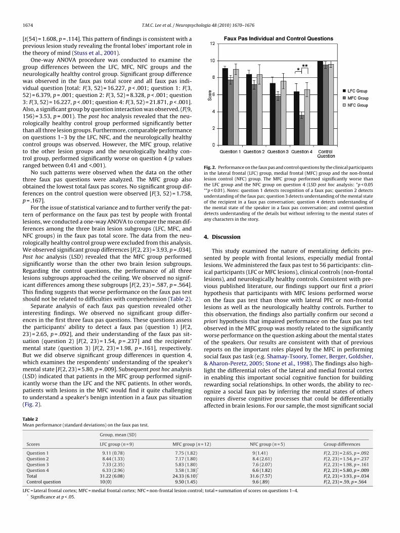
Fig. 2. Performance on the faux pas and control questions by the clinical participantsin the lateral frontal (LFC) group, medial frontal (MFC) group and the non-frontallesion control (NFC) group. The MFC group performed significantly worse thanthe LFC group and the NFC group on question 4 (LSD post hoc analysis: *p < 0.05**p < 0.01). Notes: question 1 detects recognition of a faux pas; question 2 detectsunderstanding of the faux pas; question 3 detects understanding of the mental state
TM
L
674 T.M.C. Lee et al. / Neurops
t(54) = 1.608, p = .114]. This pattern of findings is consistent with arevious lesion study revealing the frontal lobes’ important role inhe theory of mind (Stuss et al., 2001).
One-way ANOVA procedure was conducted to examine theroup differences between the LFC, MFC, NFC groups and theeurologically healthy control group. Significant group differenceas observed in the faux pas total score and all faux pas indi-
idual question [total: F(3, 52) = 16.227, p < .001; question 1: F(3,2) = 6.379, p = .001; question 2: F(3, 52) = 8.328, p < .001; question: F(3, 52) = 16.227, p < .001; question 4: F(3, 52) = 21.871, p < .001].lso, a significant group by question interaction was observed. (F(9,56) = 3.53, p = .001). The post hoc analysis revealed that the neu-ologically healthy control group performed significantly betterhan all three lesion groups. Furthermore, comparable performancen questions 1–3 by the LFC, NFC, and the neurologically healthyontrol groups was observed. However, the MFC group, relativeo the other lesion groups and the neurologically healthy con-rol group, performed significantly worse on question 4 (p valuesanged between 0.41 and <.001).
No such patterns were observed when the data on the otherhree faux pas questions were analyzed. The MFC group alsobtained the lowest total faux pas scores. No significant group dif-erences on the control question were observed [F(3, 52) = 1.758,= .167].
For the issue of statistical variance and to further verify the pat-ern of performance on the faux pas test by people with frontalesions, we conducted a one-way ANOVA to compare the mean dif-erences among the three brain lesion subgroups (LFC, MFC, andFC groups) in the faux pas total score. The data from the neu-
ologically healthy control group were excluded from this analysis.e observed significant group differences [F(2, 23) = 3.93, p = .034].
ost hoc analysis (LSD) revealed that the MFC group performedignificantly worse than the other two brain lesion subgroups.egarding the control questions, the performance of all three
esions subgroups approached the ceiling. We observed no signif-cant differences among these subgroups [F(2, 23) = .587, p = .564].his finding suggests that worse performance on the faux pas testhould not be related to difficulties with comprehension (Table 2).
Separate analysis of each faux pas question revealed othernteresting findings. We observed no significant group differ-nces in the first three faux pas questions. These questions assesshe participants’ ability to detect a faux pas (question 1) [F(2,3) = 2.65, p = .092], and their understanding of the faux pas sit-ation (question 2) [F(2, 23) = 1.54, p = .237] and the recipients’ental state (question 3) [F(2, 23) = 1.98, p = .161], respectively.
ut we did observe significant group differences in question 4,hich examines the respondents’ understanding of the speaker’sental state [F(2, 23) = 5.80, p = .009]. Subsequent post hoc analysis
LSD) indicated that patients in the MFC group performed signif-cantly worse than the LFC and the NFC patients. In other words,atients with lesions in the MFC would find it quite challengingo understand a speaker’s benign intention in a faux pas situationFig. 2).
able 2ean performance (standard deviations) on the faux pas test.
Group, mean (SD)
Scores LFC group (n = 9) MFC group (n =
Question 1 9.11 (0.78) 7.75 (1.82)Question 2 8.44 (1.33) 7.17 (1.80)Question 3 7.33 (2.35) 5.83 (1.80)Question 4 6.33 (2.96) 3.58 (1.38)*
Total 31.22 (6.08) 24.33 (6.10)*
Control question 10 (0) 9.50 (1.45)
FC = lateral frontal cortex; MFC = medial frontal cortex; NFC = non-frontal lesion control;* Significance at p < .05.
of the recipient in a faux pas conversation; question 4 detects understanding ofthe mental state of the speaker in a faux pas conversation; and control questiondetects understanding of the details but without inferring to the mental states ofany characters in the story.
4. Discussion
This study examined the nature of mentalizing deficits pre-sented by people with frontal lesions, especially medial frontallesions. We administered the faux pas test to 56 participants: clin-ical participants (LFC or MFC lesions), clinical controls (non-frontallesions), and neurologically healthy controls. Consistent with pre-vious published literature, our findings support our first a priorihypothesis that participants with MFC lesions performed worseon the faux pas test than those with lateral PFC or non-frontallesions as well as the neurologically healthy controls. Further tothis observation, the findings also partially confirm our second apriori hypothesis that impaired performance on the faux pas testobserved in the MFC group was mostly related to the significantlyworse performance on the question asking about the mental statesof the speakers. Our results are consistent with that of previousreports on the important roles played by the MFC in performingsocial faux pas task (e.g. Shamay-Tsoory, Tomer, Berger, Goldsher,& Aharon-Peretz, 2005; Stone et al., 1998). The findings also high-light the differential roles of the lateral and medial frontal cortex
in enabling this important social cognitive function for buildingrewarding social relationships. In other words, the ability to rec-ognize a social faux pas by inferring the mental states of othersrequires diverse cognitive processes that could be differentiallyaffected in brain lesions. For our sample, the most significant social12) NFC group (n = 5) Group differences
9 (1.41) F(2, 23) = 2.65, p = .0928.4 (2.61) F(2, 23) = 1.54, p = .2377.6 (2.07) F(2, 23) = 1.98, p = .1616.6 (1.82) F(2, 23) = 5.80, p = .009
31.6 (7.57) F(2, 23) = 3.93, p = .0349.6 (.89) F(2, 23) = .59, p = .564
total = summation of scores on questions 1–4.

cholo
dfsti
qoiaisuGdtomirdtsot
wttcVpcoaWg(moueswost
caFWpdtriach(chtBe
T.M.C. Lee et al. / Neuropsy
eficit for people with MFC lesions is their impaired ability to dif-erentiate the individual’s intention to hurt the feelings of others inocial interactions. Subsequent social behaviors affected by misin-erpreting the intention to hurt will likely affect the quality of socialnteractions and relationships for people with MFC lesions.
Examining the pattern of performance across the four faux pasuestions reveals that people with MFC lesions performed worsen all four questions, but the worst on question 4. Hence, perform-ng a social faux pas task may involve load of cognitive processest least at two levels of difficulty: identification and understand-ng of a social faux pas as well as the understanding to the mentaltate of the recipient being at a lower level of difficulty, relative tonderstanding the mental state and intentionality of the speaker.uessing the mental representation of the speakers could be a moreemanding task for it involves the complex meta-representation ofhe value of actions of others that enables inference of the intentionf the speakers. Following this line of thoughts, the poor perfor-ance on question 4 by people with MFC lesion may relate to
ncrease complexity and difficulty as demanded by question 4,elative to the other questions. Future studies controlling for thisifficulty confound would help verify if deficit of inferring the inten-ion of the speaker observed in people with MFC lesion is functionpecific or cognitive loading specific. Furthermore, generalizationf the findings of this study should take into the consideration ofhe varying levels of difficulty of the questions of the faux pas test.
Krueger et al. (2009) have proposed a functional segregationithin the MFC for mediating social event knowledge. Specifically,
he more dorsal MFC is more involved with actions relative tohe ventral MFC, which is more involved with feelings and out-omes (Amodio & Frith, 2006). Further along this line of thought,an Overwalle (2009) suggests that inferences about self-schematareferentially activate the ventral MFC and the medial orbitofrontalortex, but that inferences about schemata relating to the judgmentf other people preferentially activate the dorsal MFC. Amodiond Frith (2006) proposed functional divisions within the MFC.e compared performance on the faux pas test between sub-
roups within the MFC region based on their respective lesion sitesDamasio & Damasio, 1989). No significant differences in perfor-
ance on all faux pas questions between the MFC subgroups werebserved. However, the sample size of each group was small andnbalanced, which may cause significant limitation to the gen-ralizability of this observation. Future lesion and neuroimagingtudies using a larger MFC sample to delineate the specific rolesithin the sub-regions of the MFC as well as the functional effect
f the size of MPF lesions would provide data essential for under-tanding the neural mechanisms behind mentalizing and otherheory-of-mind abilities.
Some studies have suggested that the superior temporal sul-us, temporal–parietal junction, and temporal poles may also playn important role in specific tasks of mentalizing (e.g. Amodio &rith, 2006; David et al., 2008; Saxe & Kanwisher, 2003; Saxe &exler, 2005). In a meta-analytic review, Van Overwalle (2009)
roposed that the MFC and the temporal–parietal junction haveifferent functional roles; specifically, the temporal–parietal junc-ion is engaged in inferring temporary goals and intentions at aelatively more perceptual level of representation, and the MFCntegrates social information across time and allows for reflectionnd representation of traits, norms, and intentionality at a moreognitive level. Mitchell, Mason, Macrae, and Banaji (2006) discussow the mirror system in the frontal temporal–parietal regionsVan Overwalle, 2009) is important for tracking the continually
hanging states of the emotions and intentions of others. Othersave also commented on the relationships between the mirror sys-em and mentalizing (e.g. Grezes, Frith, & Passingham, 2004; Hynes,aird, & Grafto, 2006; Walter et al., 2004). However, Shamay-Tsooryt al. (2006) argue that previous studies of patients with temporo-gia 48 (2010) 1670–1676 1675
parietal junction lesions may have recruited patients suffering fromlarge lesions, including the superior temporal and the angular gyri,that might have caused them to have language deficits. Hence, ageneral reasoning deficit owing to attentional deficits and languageimpairment might explain the impaired performance in theory-of-mind tasks.
People with lateral FC lesions performed less well on the fauxpas test compared with the neurologically healthy controls. Buttheir performances at recognizing the occurrence of a faux pas insocial situations, making inferences about others’ emotional men-tal states, and understanding people’s intentions were comparablewith those observed in people with non-frontal lesions. The com-parable performance between the LFC and the non-frontal lesiongroups is consistent with previous observations (Saver & Damasio,1991; Stone et al., 1998). Damasio (2005) suggested that patientswith LFC lesions may also have impaired social functioning thatmay be understood as part of a larger picture of cognitive defects.This speculation awaits verification in future research. In this con-nection, the interrelationships between other cognitive skills thatcontribute to intact mentalizing, such as logical reasoning, mem-ory and learning, and reward and punishment processing, wouldbe worth studying in future research.
The different pathologies presented by the frontal lesionpatients may raise concerns about possible confounding effect onthe findings. Klein et al. (2002) observed that patients sufferingfrom low-grade glioma performed worse on cognitive domainsthan those with vascular diseases. Since all patients in the dorsolat-eral group suffered from glioma and should have performed worsethan the medial group, the observation that the latter performedworse than the former group on the faux pas test provides reas-suring evidence that the findings were not related to the impact ofglioma on cognitive functioning. After all, the near ceiling perfor-mance on the control questions of the entire lesion group indicatesan unimpaired performance in comprehending the stories. Hence,the results obtained in this study should be considered a fair rep-resentation of the social cognitive deficits experienced by peoplewith frontal lesions.
The constraint on time for assessment set by the hospital wherethe data were acquired greatly limits the number of neuropsy-chological tests that could be administered for obtaining a morecomprehensive cognitive profile for each participant. This time con-straint together with the small sample size of the study may affectthe generalizability of the findings. Future research may considerincreasing the sample size by adopting a multi-center design. Ameta-analytic review of lesion studies in social cognition shouldyield important insights into the neural mechanisms underlyingmentalizing. Due to the process of cortical reorganization, thevarying duration of post-onset of illness among the three lesionsubgroups might have confounded the results of this study. Accord-ing to literature, maximum recovery usually occurs in the very earlystages post-injury. Hence other studies have used 1.3 months post-injury as the cut-off (see Stuss, Binns, Murphy, & Alexander, 2002).It is accepted that there might be some continuing albeit smallerpost-injury recovery during the time period post-injury covered byour participants. In order to control for the confounding variance,exploratory analysis of covariate analysis procedure was performedon the faux pas test results with the days post-onset as a covari-ate. The results suggested that duration in days post-lesions wasnot a significant covariate in this study, suggesting that chronic-ity of lesion is not related to the faux pas test performance in thissample. However, the generalizability of this observation should
be reaffirmed in future studies using a larger sample of varyingpost-lesion duration.Our findings shed light on the unique nature of social deficitsthat challenge the quality of life for people with MFC lesions. Fur-ther neuroimaging and behavioral studies should be performed

1 ycholo
tcbotoifos
F
sSit
R
A
A
B
B
B
CC
D
D
D
F
F
FF
G
H
H
676 T.M.C. Lee et al. / Neurops
o decode the neural correlates and mechanisms as well as theognitive variables affecting mentalizing. The data acquired wille essential for eventually constructing a neurocognitive modelf mentalizing for verification in future experimentation. Fromhe perspective of clinical practice, our findings support clinicalbservations regarding the heterogeneous profiles of impairmentn social functioning in people suffering from frontal lesions at dif-erent locations. This information enables a better understandingf the challenges encountered by these individuals in their dailyocial interactions.
unding
This project was supported by the May Endowed Professor-hip of The University of Hong Kong and the National Naturalcience Foundation of China (#30828012 and #30670706). We arendebted to Professor Donald T. Stuss for his helpful comments onhe manuscript.
eferences
lexander, M. P., Stuss, D. T., Shallice, T., Picton, T. W., & Gillingham, S. (2005).Impaired concentration due to frontal lobe damage from two distinct lesionsites. Neuorology, 65, 572–579.
modio, D. M., & Frith, C. D. (2006). Meeting of minds: The medial frontal cortex andsocial cognition. Nature Reviews: Neuroscience, 7, 268–277.
aron-Cohen, S., Leslie, A. M., & Frith, U. (1985). Does the autistic child have a theoryof mind? Cognition, 21, 37–46.
aron-Cohen, S., O’Riordan, M., Stone, V. E., Jones, R., & Plaisted, K. (1999). Recog-nition of faux pas by normally developing children with Asperger’s syndromeor high-functioning autism. Journal of Autism and Developmental Disorders, 29,407–418.
rothers, L. (1990). The social brain: A project for integrating primate behavior andneurophysiology in a new domain. Concepts in Neuroscience, 1, 27–61.
ohen, S. (2004). Social relationships and health. American Psychologist, 59, 676.ritchley, H. D., Mathias, C. J., Josephs, O., O’Doherty, J., Zanini, S., Dewar, B. K., et
al. (2003). Human cingulated cortex and autonomic control: Converging neu-roimaging and clinical evidence. Brain, 126, 2139–2152.
amasio, H. (2005). Disorders of social conduct following damage to prefrontal cor-tices. In J. P. Changeux, A. R. Damasio, W. Singer, & Y. Christen (Eds.), Neurobiologyof human values (pp. 37–46). Berlin: Springer.
amasio, H., & Damasio, A. R. (1989). Lesion analysis in neuropsychology. Oxford:Oxford University Press.
avid, N., Aumann, C., Santos, N. S., Bewernick, B. H., Eickhoff, S. B., Newen, A., etal. (2008). Differential involvement of the posterior temporal cortex in mental-izing but not perspective taking. Social Cognitive and Affective Neuroscience, 3,279–289.
olstein, M. F., Folstein, S. E., & McHugh, P. R. (1975). Mini mental state: A practicalmethod for grading the cognitive state of patients for the clinician. Journal ofPsychiatric Research, 12, 189–198.
rith, C. D. (2000). The role of dorsolateral prefrontal cortex in the selection of action,as revealed by functional imaging. In S. Monsell, & J. Driver (Eds.), Control ofcognitive processes: Attention and performance (pp. 269–386). Cambridge: MITPress.
rith, C. D., & Frith, U. (2006). The neural basis of mentalizing. Neuron, 50, 531–534.rith, U., & Frith, C. D. (2003). Development and neurophysiology of mentalizing.
Philosophical transactions of the Royal Society of London. Series B, Biological science,29, 459–473.
rezes, J., Frith, C., & Passingham, R. E. (2004). Brain mechanisms for inferring deceitin the actions of others. Journal of Neuroscience, 24, 5500–5505.
amilton, M. (1960). A rating scale for depression. Journal of Neurology, Neurosurgery,and Psychiatry, 23, 56–62.
appaney, K., Zelazo, P. D., & Stuss, D. T. (2004). Development of orbitofrontal func-tion: current themes and future directions. Brain and Cognition, 55, 1–10.
gia 48 (2010) 1670–1676
Huettel, S. A., & McCarthy, G. (2004). What is odd in the oddball task? Prefrontalcortex is activated by dynamic changes in response strategy. Neuropsychologia,42, 379–386.
Hynes, C. A., Baird, A. A., & Grafto, S. T. (2006). Differential role of the orbital frontallobe in emotional versus cognitive perspective-taking. Neuropsychologia, 44,374–383.
Klein, M., Heimans, J. J., Aaronson, N. K., van der Ploeg, H. M., Grit, J., Muller, M., et al.(2002). Effect of radiotherapy and other treatment-related factors on mid-termto long-term cognitive sequelae in low-grade gliomas: A comparative study.Lancet, 360, 1361–1368.
Kringelbach, M. L. (2005). The human orbitofrontal cortex: Linking reward to hedo-nic experience. Nature Reviews Neuroscience, 6, 691–702.
Krueger, F., Barbey, A. K., & Grafman, J. (2009). The medial prefrontal cortex mediatessocial event knowledge. Trends in Cognitive Sciences, 13, 103–109.
Lhermitte, F. (1986). Human autonomy and the frontal lobes. Part II. Patient behaviorin complex and social situations: The “environmental dependency syndrome”.Annals of Neurology, 4, 335–343.
Mazza, M., Costagliola, C., Di Michele, V., Magliani, V., Pollice, R., Ricci, A., et al. (2007).Deficit of social cognition in subjects with surgically treated frontal lobe lesionsand in subjects affected by Schizophrenia. European Archives of Psychiatry andClinical Neuroscience, 257, 12–22.
Mesulam, M. M. (2002). The human frontal lobes: Transcending the default modethrough contingent encoding. In D. T. Stuss, & R. T. Knight (Eds.), Princi-ples of Frontal Lobe Function (pp. 8–30). Oxford, England: Oxford UniversityPress.
Mitchell, J. P., Macrae, C. N., & Banaji, M. R. (2004). Encoding specific effects of socialcognition on the neural correlates of subsequent memory. Journal of Neuro-science, 24, 4912–4917.
Mitchell, J. P., Mason, M. F., Macrae, C. N., & Banaji, M. R. (2006). Thinking aboutothers: The neural substrates of social cognition. In J. T. Cacioppo, P. S. Visser,& C. L. Pickett (Eds.), Social neuroscience: People thinking about thing people (pp.269–386). London, England: The MIT Press.
Öngür, D., & Price, J. L. (2000). The organization of networks within the orbitaland medial prefrontal cortex of rats, monkeys and human. Cerebral Cortex, 10,206–219.
Ranganath, C., & Rainer, G. (2003). Neural mechanisms for detecting and remember-ing novel events. Nature Review Neuroscience, 4, 193–202.
Robinson, G., Blair, J., & Cipolotti, L. (1998). Dynamic aphasia: An inability to selectbetween competing verbal responses? Brain, 121, 77–89.
Saver, J. L., & Damasio, A. R. (1991). Preserved access and processing of social knowl-edge in a patient with acquired sociopathy due to ventromedial frontal damage.Neuropsychologia, 29, 1241–1249.
Saxe, R., & Kanwisher, N. (2003). People thinking about thinking people: The role ofthe temporo-parietal junction in “theory of mind. Neuroimage, 19, 1835–1842.
Saxe, R., & Wexler, A. (2005). Making sense of another mind: the role of the righttemporo-parietal junction. Neuropsychologia, 43, 1391–1399.
Shamay-Tsoory, S. G., Tibi-Elhanany, Y., & Aharon-Perertz, J. (2006). The ventrome-dial prefrontal cortex is involved in understanding affective but not cognitivetheory of mind stories. Social Neuroscience, 1, 149–166.
Shamay-Tsoory, S. G., Tomer, R., Berger, B. D., Goldsher, D., & Aharon-Peretz, J. (2005).Impaired “affective theory of mind” is associated with right ventromedial pre-frontal damage. Cognitive and Behavioral Neurology, 18, 55–67.
Stone, V. E., Baron-Cohen, S., & Knight, R. T. (1998). Frontal lobe contributions totheory of mind. Journal of Cognitive Neuroscience, 10, 640–656.
Stone, V. E., Baron-Cohen, S., Young, A., Calder, A., & Green, J. (1999). Patients withamygdalectomy show impairments on theory of mind. Unpublished manuscript.University of Denver.
Stuss, D. T., Gallup, G. G., & Alexander, M. P. (2001). The frontal lobes are necessaryfor “theory of mind”. Brain, 124, 279–286.
Stuss, D. T., Binns, M. A., Murphy, K. J., & Alexander, M. P. (2002). Dissociationswithin the anterior attention system: Effects of task complexity and irrelevantinformation on reaction time speed and accuracy. Neuropsychology, 16, 500–513.
Van Overwalle, F. (2009). Social cognition and the brain: a meta-analysis. HumanBrain Mapping, 30, 829–858.
Walter, H., Adenzato, M., Ciaramidaro, A., Enrici, I., Pia, L., & Bara, B. G. J. (2004).Understanding intention in social interactions: The role of the anterior cingulatecortex. Cognitive Neuroscience, 16, 1854–1863.
Zhu, C. Y., Lee, T. M. C., Li, X. S., Jing, S. C., Wang, Y. G., & Wang, K. (2007). Impair-ments of social cues recognition and social functioning in Chinese people withschizophrenia. Psychiatry and Clinical Neurosciences, 61, 149–158.