Fast and sensitive detection of genetically modified...
27
1 1 2 3 4 Fast and sensitive detection of genetically modified 5 yeasts in wine 6 7 Carlos León 1 , Virginia García-Cañas 1 , Ramón González 2 , Pilar Morales 2 , Alejandro Cifuentes 1,* 8 9 1 Laboratory of Foodomics, Institute of Food Science Research (CSIC), Nicolas Cabrera 9, Campus 10 de Cantoblanco, 28049 Madrid, Spain. 11 2 Instituto de Ciencias de la Vid y del Vino (CSIC-UR-CAR), Madre de Dios 51, 26006 Logroño, 12 Spain 13 14 15 16 17 18 19 20 21 22 23 24 *Corresponding author, Tel: 34-91-5618806 Fax#: 34-91-5644853, e-mail: [email protected] 25 26 Con formato: Español (alfab. internacional) Con formato: Español (alfab. internacional)
Transcript of Fast and sensitive detection of genetically modified...

1
1
2
3
4
Fast and sensitive detection of genetically modified 5
yeasts in wine 6
7
Carlos León1, Virginia García-Cañas
1, Ramón González
2, Pilar Morales
2, Alejandro Cifuentes
1,* 8
9
1 Laboratory of Foodomics, Institute of Food Science Research (CSIC), Nicolas Cabrera 9, Campus 10
de Cantoblanco, 28049 Madrid, Spain. 11
2 Instituto de Ciencias de la Vid y del Vino (CSIC-UR-CAR), Madre de Dios 51, 26006 Logroño, 12
Spain 13 14
15
16
17
18
19
20
21
22
23
24
*Corresponding author, Tel: 34-91-5618806 Fax#: 34-91-5644853, e-mail: [email protected] 25
26
Con formato: Español (alfab.internacional)
Con formato: Español (alfab.internacional)

2
ABSTRACT 27
28
In this work, a novel screening methodology based on the combined use of multiplex polymerase 29
chain reaction (PCR) and capillary gel electrophoresis with laser induced fluorescence (CGE-LIF) 30
is developed for the fast and sensitive detection of genetically modified yeasts in wine. As model, a 31
recombinant EKD-13 Saccaromyces cerevisiae strain was selected and different wines were 32
prepared using either recombinant or conventional yeasts. Special emphasis is put on the yeast 33
DNA extraction step, exploring different commercial and non-commercial methods, in order to 34
overcome the important difficulty of obtaining amplifiable DNA from wine samples. To 35
unequivocally detect the transgenic yeast, two specific segments of the transgenic construction were 36
amplified. In addition, a third primer pair was used as amplification control to confirm the quality of 37
the yeast DNA obtained from the extraction step. CGE-LIF provides high sensitivity, good analysis 38
speed and impressive resolution of DNA fragments, making this technique very convenient to 39
optimize multiplex PCR parameters and to analyze the amplified DNA fragments. Thus, the CGE-40
LIF method provided %RSD values for DNA migration times lower than 0.82% (n=10) with the 41
same capillary and lower than 1.92% (n=15) with three different capillaries, allowing the adequate 42
size determination of the PCR products with an error lower than 4% compared to the theoretically 43
expected. The whole method developed in this work requires less than one working day and grants 44
the sensitive detection of transgenic yeasts in wine samples. 45
46
47
48
49
50
Keywords: transgenic yeast, multiplex PCR, capillary gel electrophoresis, genetically modified 51
organisms, wine 52
53

3
1. INTRODUCTION 54
55
The adoption of DNA recombinant technology has been considered the fastest growing trend in the 56
history of agriculture, and, over recent years, the full potential of this modern biotechnology has 57
been exploited for its application in modern plant breeding [1,2]. In addition to genetically modified 58
(GM) crops, yeasts and lactic acid bacteria with a long history of use for food production have been 59
subjected to genetic modification by genetic engineering mainly for improving the industrial 60
processing or the quality of the final product [3-5]. The development of transgenic yeast strains 61
using recombinant DNA technology has been the most recent step used by microbiologists to 62
improve specific properties of wine [5]. Recently, two transgenic Saccharomyces cerevisiae strains 63
have been commercialized in United States and Canada [6,7] to avoid the need of bacterial 64
malolactic fermentation and the associated risk of bacterial spoilage in case of uncontrolled process 65
(ML01 strain, [8,9]) and to reduce ethyl carbamate content (ECMo01 strain, [10]). Other genetic 66
modifications in wine strains have been directed to: (i) reduce ethanol production [11-13]; (ii) 67
release volatile aroma aglycones from grape glycosylated precursors [14-16]; (iii) improve the 68
production of desirable volatile esters [17], the chemical stability of wine [18], and the yeast 69
autolysis during the second fermentation of sparkling wines [19]. 70
71
The development and use of genetically modified organisms (GMOs) for food applications are 72
issues of intense debate and public concern that have pushed the European Union and other 73
countries to establish strict regulations concerning different aspects of GMOs, including risk 74
assessment, marketing, labeling and traceability. To verify the application of such regulations, it is 75
necessary to develop analytical methodologies that can effectively detect GMOs in the food chain. 76
In general, analytical procedures for GMO screening in food are based on DNA amplification by 77
the polymerase chain reaction (PCR) technique. The ability of PCR to amplify specific DNA 78
sequences in a complex DNA extract will depend on the integrity, quantity and purity of the DNA 79
extract. These limiting factors define the amplificability of target DNA sequences by PCR-based 80

4
methods, and are considered critical issues for GMO analysis in highly processed and complex food 81
samples (chocolate, biscuits, etc.). In the case of wine samples, the presence of tannins, polyphenols 82
and polysaccharides may interfere in DNA extraction and/or inhibit the amplification of isolated 83
DNA [20-25]. In addition to the inhibitory effect of wine matrix on PCR, low quantity of DNA and 84
degradation caused e.g., by biochemical and enzymatic action during fermentation and aging, are 85
important constraints for DNA amplification in wine samples [26]. Strategies based on the use of 86
long incubation periods for DNA precipitation or the use of large sample volumes have been 87
applied to extract sufficient DNA from wine samples for subsequent PCR amplification [26-30]. On 88
the other hand, different approaches have been suggested in order to overcome the inhibitory effect 89
of wine matrix on PCR [25, 31]. In this regard, attenuation of amplification by phenolic compounds 90
has been addressed by the addition of molecules that act as polyphenols removers 91
(polyvinylpyrrolidone, polyvinylpolypyrrolidone, activated charcoal, etc.) during the DNA 92
extraction step. In other cases, separation methods, such as PVP-agarose gel electrophoresis 93
purification, have been tailored to remove inhibitors from the DNA extracts from difficult samples 94
as soil [24]. Therefore, an efficient extraction procedure is critical for DNA analysis from wine 95
samples. 96
97
It has already been demonstrated that multiplex PCR is a suitable methodology for the simultaneous 98
detection of specific targets in GMO-derived materials [32-35]. However, optimization of multiplex 99
PCR is more complex than simplex PCR as the presence of more primer pairs in the reaction system 100
reduces the robustness of the amplification process [32]. In many cases, the optimization and 101
analysis of multiplex PCR reactions requires high sensitivity and resolution. In addition, sensitive 102
analytical methodologies are necessary for the detection of recombinant yeasts in wine, as yeast 103
DNA may be degraded and present in low concentration. In this regard, capillary gel electrophoresis 104
with laser induced fluorescence (CGE-LIF) detection has proven to be a helpful separation 105
technique during the optimization of multiplex PCR methods as well as for the simultaneous 106

5
analysis of multiple GMOs in food samples [36]. The aim of this work is, therefore, to develop a 107
novel methodology, based on the combined use of optimum DNA extraction, multiplex PCR 108
amplification and CGE-LIF analysis for the fast and sensitive detection of genetically modified 109
yeast strains in wine. 110
111
2. MATERIALS AND METHODS. 112
2.1. Chemicals. 113
All chemicals were of analytical reagent grade and used as received. Polyvinylpolypyrrolidone 114
(PVPP) was from Applichem (Darmstadt, Germany); phenol was from LabClinics (Madrid, Spain), 115
2-propanol, chloroform and glucose were purchased to Scharlau (Barcelona, Spain); ethanol and 116
isoamyl alcohol was from Merck (Darmstadt, Germany); and RNAse A was from Roche 117
(Barcelona, Spain). Peptone and yeast extract were purchased from CONDA Pronadisa (Madrid, 118
Spain). AmpliTaq Gold DNA polymerase, including GeneAmp PCR buffer II and, 119
deoxynucleotides, MgCl2, were from Applied Biosystems (Madrid, Spain). Uracil DNA glycosylase 120
was purchased from New England Biolabs (Berverly, MA). Oligonucleotides were purchased from 121
Bonsai Biotechnologies (Alcobendas, Spain). Tris(hydroxymethyl)aminomethano (TRIS) and 122
EDTA were obtained from Sigma (St. Louis, MO, USA); 2-hydroxyethyl cellulose (HEC, MWav 123
90000) was from Aldrich (Milwaukee, WI, USA); YOPRO-1 was from Molecular Probes (Leiden, 124
The Netherlands). Separation buffer was stored at 4 ºC and warmed at room temperature before use. 125
Water was deionized by using a Milli-Q system (Millipore, Bedford, MA, USA). 126
127
2.2. Samples. 128
S. cerevisiae strain EC1118 is a conventional wine yeast strain commercialized by Lallemand Inc. 129
(Montreal Canada). EKD-13 is a genetically modified strain of S. cerevisiae with an improved 130
capacity to release mannoproteins to the media during the fermentation of the must [18]. Both S. 131
cerevisiae strains were grown separately in YPD broth (2% glucose, 2% peptone, 1% yeast extract) 132
6
as negative and positive controls, respectively. 5 mL of YPD culture containing the reference yeasts 133
were incubated for 24 h at 30ºC. Bottled wine, labelled as Petit Verdot, was donated by Laboratorio 134
Agroalimentario de Jerez (Cádiz, Spain). For experimental production of control and recombinant 135
wine samples, precultures were grown in YPD broth. Two types of grape must, including a blend of 136
red wine grapes (musts 1 and 2) and a Graciano monovarietal red wine (must 3) were used for 137
fermentation assays with EC1118 and the recombinant EKD-13 strains. Must were sulphited in 138
origin to 30-50 ppm of potassium metabisulphite. 200 mL of the unclarified musts (musts 1 and 3), 139
or a must clarified by gentle centrifugation (must 2) were inoculated 1% in volume from a 140
preculture in YPD grown for 48 h at 28ºC. All musts were fermented as for white wines (i.e., 141
without maceration of skins and seeds), but the must differed in colour intensity with a deeper 142
colour for the blend of red grapes (musts 1 and 2) than for the Graciano must (must 3). 143
Fermentation was carried out at a controlled temperature of 25ºC in erlemenyer flasks closed with 144
fermentation locks filled with Vaseline oil. Fermentation kinetics was monitored as CO2 formation 145
as estimated by daily recording loss of weight through the fermentation lock. Wine fermentation 146
was considered complete after two days of constant weight. This was confirmed by HPLC analysis 147
of the main fermentation metabolites (glucose, fructose, glycerol and ethanol). No further 148
clarification was performed on the wine samples. 149
150
2.3. DNA extraction and quantification. 151
Different DNA extraction protocols (commercial and non-commercial) were evaluated in this work 152
in order to obtain representative and reliable DNA extracts from the different types of samples. 153
154
2.3.1 DNA extracts from yeasts. 155
Extractions of DNA from cultured yeasts were performed using a commercial kit (MasterPureTM
156
Yeast DNA Purification Kit from Epicentre Biotechnologies, Madison, WI) following the 157
instructions given by the manufacturer. Briefly, 300 μL of Yeast Cell Lysis solution were added to 158

7
the pellet, followed by 150 μL of MPC Protein Precipitation Reagent and 0.5 mL of isopropanol. 159
The DNA extract (50 μL) was incubated with RNase (1 μL) at 60ºC for 30 min. After incubation, 1 160
volume of phenol/chloroform/isoamyl alcohol (25:24:1, v/v/v) was added and homogenized. After 161
centrifugation at 14000 rpm was performed, the upper phase was transferred to another tube and 162
another extraction with chloroform/isoamyl alcohol (24:1, v/v) was done. Then, 0.1 volumes of 3 M 163
sodium acetate (pH 5.2) and 2 volumes of ethanol were added to the upper phase collected. The 164
solution was kept at -20 ºC for 2 h. The precipitate was collected by centrifugation at 14000 rpm for 165
10 min and washed with 70% ethanol. The remaining ethanol was evaporated at room temperature 166
and the pellet was dissolved in 50 μL TE buffer that is composed of 10 mM Tris-HCl and 1 mM 167
EDTA at pH 8.0. 168
169
2.3.2 DNA extracts from wines. 170
Several DNA extraction protocols were evaluated to amplify DNA from wine samples. The 171
experiments were done using Petit Verdot as a model wine sample trying to obtain DNA of good 172
quality and yield in order to get a sensitive amplificability of the mrp2 gene by PCR. Briefly, 15 mL 173
of the wine sample were centrifuged (10000 rpm for 5 min at 5 ºC) to recover the solid parts in 174
suspension. DNA extraction from the solid parts (pellets) was attempted using three different 175
methods (A, B and C, see below). Alternatively, during method optimization, the following 176
procedure was also assayed as sample pre-treatment in order to remove potential inhibitors from the 177
sample. Briefly, the solid part (pellet) was resuspended in 2 mL water and homogenized by 178
pipetting. The suspension was centrifuged (14000 rpm for 5 min at room temperature) and the 179
supernatant was discarded. This washing step was repeated twice. After washing, 1 mL of a 10% 180
(w/v) PVPP aqueous suspension was added to the pellet. After homogenization by vortexing, the 181
tube was incubated at room temperature for 5 min. Then, the suspension was centrifuged (14000 182
rpm for 10 min at room temperature) and the supernatant was discarded. The DNA from the pellet 183
was then subjected to extraction by Method A, B and C (see next). Method A: DNA was extracted 184

8
from the pellet using the aforementioned commercial kit from MasterPureTM
Yeast DNA 185
Purification Kit and following the manufacturer instructions as described above. Method B and C: 186
These methods were based on the protocol described by Querol et al. [37] with some modifications. 187
The pellet was suspended in 0.5 ml of 1 M sorbitol-0.1 M EDTA, pH 7.5. Next, 20 μL of a solution 188
of Zymolyase-20 (2.5 mg/ml) was added. The suspension was incubated at 37 °C for 90 min, and 189
then centrifuged at 14000 rpm for 2 min. Next, the pellet was suspended in 500 μL of 50 mM TE 190
buffer, pH 7.4. After suspension, 10 μL of an aqueous solution containing 20% sodium dodecyl 191
sulfate was added and the mixture was incubated at 65 °C for 30 min. The suspension was 192
incubated with RNase (1 μL) at 60ºC for 15 min. Immediately thereafter, 200 μL of 5 M potassium 193
acetate was added and the tubes were placed on ice for 5 min. Then, the solution was centrifuged at 194
14000 rpm for 15 min. In Method B, supernatant was transferred to a fresh microcentrifuge tube, 195
and the DNA was precipitated by adding 1 volume of 2-propanol. After incubation at room 196
temperature for 5 min, the tubes were centrifuged for 10 min. The DNA was washed with 70% 197
ethanol, dried at room temperature, and dissolved in 50 μL of TE buffer. In Method C, supernatant 198
was extracted with 1 volume of phenol/chloroform/isoamyl alcohol (25:24:1, v/v/v) and then, 199
centrifuged at 14000 rpm for 10 min. The aqueous phase was transferred to a clean microcentrifuge 200
tube and DNA was purified with QIAamp DNA Mini Kit columns from Qiagen (Madrid, Spain) 201
following the manufacturers instructions. Finally, the DNA retained in the column was eluted with 202
50 μL TE buffer. 203
204
2.4. Oligonucleotide primers. 205
Primer pairs were designed using Primer3 online software [38] according to unique and specific 206
DNA sequences to EKD-13 yeast strain. Namely, two primers pairs (indicated as pairs 207
KmPF/KmPR and KmTF/KmTR in Table 1) were designed to amplify short DNA sequences 208
(within the range of 100-200 bp) for the recombinant S. cerevisiae EKD-13. In addition, a third 209
primer pair (SCF1/SCR1 in Table 1) was used as amplification control of a 104 bp DNA fragment 210

9
in mrp2 gene in S. cerevisiae genome [30]. The primer pairs were also tested using the PrimerList 211
software [39] to control their suitability to be used in a multiplex system. The oligonucleotides were 212
purchased from Bonsai Technologies (Alcobendas, Spain). 213
214
2.5. Simplex and multiplex PCR conditions. 215
Amplification reactions were run in a Mastercycler gradient thermocycler, Eppendorf (Madrid, 216
Spain). Initially, the efficiency of the primer pairs to amplify the target sequences were separately 217
tested by performing simplex PCR reactions using 0.5 μM of each primer pair, SCF1/SCR1, 218
KmTF/KmTR and KmPF/KmPR and several genomic DNA extracts from cultured EC1118 or 219
EKD-13 yeast strains. Reactions were carried out using a mixture containing 10 mM Tris-HCl pH 220
8.3, 50 mM KCl, 2.6 mM MgCl2, 0.25 mM dCTP, 0.25 mM dGTP, 0.25 mM dATP, 0.5 mM dUTP, 221
0.25 U of Uracil-DNA Glycosilase (UDG), 1 U AmpliTaq Gold 1 polymerase, 50 ng genomic DNA 222
and the primer pair concentration indicated above in a final volume of 50 μL. PCR reaction was 223
preceded by an incubation period of 10 min at 37ºC for UDG activity. The PCR program consisted 224
of an initial denaturation step at 95ºC for 10 min followed by 10 cycles, which involved a 225
denaturation step at 95 ºC for 30 s, 40 cycles at 95 ºC for 30 s, 60 ºC for 30 s, 72 ºC for 30 s, and a 226
final elongation step at 72 ºC for 10 min. Multiplex reactions were carried out under the same 227
conditions described for simplex PCR but including the three primer pairs at the following 228
concentrations: 0.7 μM SCF1/SCR1, 0.2 μM KmTF/KmTR and 0.1 μM KmPF/KmPR. To confirm 229
the absence of contaminant DNA in simplex and multiplex PCR reactions, blank (without DNA 230
template) reactions were systematically carried out in all experiments. For multiplex PCR, the 231
primers were premixed in order to minimize the analysis-to-analysis variability due to pipetting. 232
233
2.6. CGE-LIF conditions. 234
The analyses of reaction products from simplex and multiplex PCR amplifications were carried out 235
in a PACE-MDQ (Beckman Coulter) equipped with an Ar+ laser working at 488 nm (excitation 236

10
wavelength) and 520 nm (emission wavelength). Bare fused-silica capillaries with 75 µm I.D. were 237
purchased from Composite Metal Services (Worcester, England). Injections were made at the 238
cathodic end using N2 pressure of 0.5 psi for 40 s (1 psi=6894.76 Pa). The PACE-MDQ instrument 239
was controlled by a PC running the 32 Karat Software from Beckman. Before first use, any 240
uncoated capillary was preconditioned by rinsing with 0.1 M HCl for 30 min. The following 241
conditions were used for PCR products separations: Bare fused silica capillary with 60 cm total 242
length, 50 cm effective length and 75 m I.D; separation buffer (20 mM Tris, 10 mM phosphoric 243
acid, 2 mM EDTA, 500 nM YOPRO-1, and 4.5 % HEC at pH 7.3); temperature of separation: 45 244
ºC; running electric field: -217 V/cm. Between injections, capillaries were rinsed using water for 5 245
min followed by 0.1 M HCl for 4 min, and separation buffer for 4 min. At the end of the day, the 246
capillary was rinsed with deionized water for 5 min and stored overnight with water inside. For 247
accurate size determination of the DNA fragments from PCR reactions, an eCAP dsDNA 1000 Test 248
Mix from Beckman Coulter was used in CGE-LIF analysis. This sample was diluted to a final 249
concentration of ca. 200 ng/µL in TE buffer. 250
251
3. RESULTS AND DISCUSSION 252
3.1. Optimization of the DNA extraction method. 253
DNA extraction from pure cultures of EC1118 and EKD-13 strains in YPD media was successfully 254
carried out using the commercial MasterPureTM
kit. A first evaluation of the extracts obtained from 255
1.5 mL of pure culture of yeast using UV spectroscopy gave OD260/280 and OD260/230 values of 1.6 256
and 2.5, corresponding to extraction yields ranging from 200 to 350 ng/µL, respectively. However, 257
in the case of wine samples, the DNA extracts obtained showed very low yield and purity as 258
determined by UV spectroscopy and agarose gel electrophoresis (AGE). To corroborate this point, 259
all DNA extracts were tested to perform the PCR amplification of a specific sequence in S. 260
cerevisiae genome using the SCF1/SCR1 primer pair. Electrophoretic results showed only positive 261
amplification of the control sequence in the extracts obtained from cultured yeasts (Figure 1, lines 7 262
11
and 8), whereas no detectable signals were obtained in the amplification of wine extracts (data not 263
shown). These results demonstrate that in spite of the availability of commercial kits tailored for the 264
extraction of DNA from specific organisms, such as yeast, there are critical factors related to the 265
sample matrix, DNA integrity and abundance that will determine the suitability of an extraction 266
method for a particular (food) sample. 267
268
In order to obtain yeast DNA extracts from wine samples with good yield and quality for the 269
subsequent PCR amplification, three different protocols to extract DNA from bottled wine samples 270
were initially investigated. Namely, the methods tested were a commercial kit (Method A), the 271
Zymolyase method including 2-propanol precipitation (Method B), and the Zymolyase method 272
combined with silica column purification (Method C). Extractions were performed using sample 273
volumes of 15 mL. To study the contribution of PVPP to the quality and yield of the extract, 274
extractions were performed with addition of PVPP to the sample as described under Materials and 275
Methods. As controls, extractions were also performed without PVPP addition. Extracted DNA 276
using the three methods could not be visualized in ethidium bromide-stained agarose gels due to 277
low extraction yields, that were close to or below the detection limit of AGE (~50 ng dsDNA in 278
polydisperse samples). Although Method B provided the highest yields (> 50 ng/µL), a smear band 279
typical of RNA impurities was visualized in the extracts obtained by this method. These results 280
suggested a possible overestimation of DNA concentrations based on UV spectroscopy data, in the 281
extracts obtained by Method B. Also, it could be seen that the extracts obtained without PVPP 282
treatment provided values of OD260/230 lower than 0.4, indicating the presence of contaminants 283
(likely, phenolic compounds). In general, the addition of PVPP had a detrimental effect on the 284
yields, obtaining DNA yield values ca. 10%, 67% and 78% lower in the extracts obtained by 285
Method B, A and C, respectively, than the yields obtained without PVPP. In spite of these results, 286
the addition of PVPP to the wine samples was considered necessary in order to obtain DNA of 287
sufficient quality as can be deduced from lines 1, 3 and 4 in Figure 1. However, partial or complete 288

12
inhibition of the subsequent PCR amplification of the specific sequence in S. cerevisiae was 289
observed in all the cases, probably due to the copurification of inhibitory substances (lines 2, 5 and 290
6 in Figure 1), indicating the complexity and relevance of the DNA extraction step. As the three 291
methods provided similar negative results during the PCR amplification, a further optimization of 292
the DNA extraction conditions was carried out. Method A was chosen for subsequent optimization 293
since it provided better extracts in terms of purity (OD260/230 > 0.7 and non detectable RNA bands in 294
AGE). Higher starting volume of wine sample was then assayed. In this case, increasing the sample 295
volume to 50 mL improved the extraction yield about two-fold (from 20 to 44 ng/µL) with 296
acceptable OD260/280 values (>1.7), and low OD260/230 values (<1.0). Although DNA appeared as a 297
weak smear along the electrophoretic line in AGE, indicating DNA degradation in the wine sample, 298
the specific sequence of S. cerevisiae genomic DNA could be amplified and clearly detected in the 299
extracts obtained under these conditions. Next, Method A was applied to the DNA extraction of the 300
wine samples produced in our lab with EC-1118 and EKD-13 yeast strains to test the suitability of 301
the method with other types of wine. However, all the attempts to amplify the same reference 302
sequence in these new extracts failed. This could be due to the different composition of the wine 303
samples, also noticeable during extraction by the higher size of the pellets. Consequently, Method 304
A was adapted to the extraction of high volumes of sample by using double volume of extraction 305
reagents than those indicated by the manufacturer of the kit. The rest of extraction conditions were 306
the same as described before. Under these conditions, DNA extracts with high yields and purity 307
values were obtained for most of the samples (see Table 2). Although the AGE results also showed 308
some DNA degradation, the suitability of the method to provide PCR-quality extracts from wine 309
samples was corroborated by the positive PCR amplification of the DNA from all the samples. 310
311
3.2. Design and optimization of multiplex PCR for transgenic yeast (EKD-13 strain) detection. 312
The most accepted strategy for GMO detection in food relies on the PCR amplification of a specific 313
DNA sequence. In GM plants, PCR targets used for identification are based on event-specific 314

13
elements that are unique for a transformation event. These are sequences that cover the border of 315
the transgenic insert and the plant genome. These sequences, however, cannot be used for the 316
detection of recombinant yeasts. Insertion of the transforming DNA into the yeast chromosome is 317
easily achieved by homologous recombination between chromosomal sequences and the 318
construction of interest. In contrast to the GM plants, the insertion of recombinant DNA in many 319
recombinant yeast strains is directed to a specific locus by incorporating a cognate homologous 320
genomic sequence into the vector or construction of interest. The size of an edge fragment specific 321
for the target recombinant yeast would be too large for an efficient PCR amplification. In this case, 322
the detection of recombinant yeasts can be approached by targeting junctions of contiguous 323
transgenic elements. Consequently, the primers for the detection of EKD-13 strain were designed 324
according to one of the recombinant constructs used for deletion of KNR4 gene in this recombinant 325
strain. The construct is composed of KanMX4 coding sequence (from Escherichia coli DH5α), 326
which provides resistance to geneticin, flanked by KNR4 promoter and terminator sequences, 327
homologous to those found in non-recombinant S. cerevisiae [40]. For construct-specific detection, 328
a primer pair (KmPF/KmPR) was designed to span a junction segment covering the KNR4 promoter 329
and KanMX4 coding sequence (Table 1). To increase the specificity for the recombinant strain, the 330
forward primer (KmPF) was devised to target the border of KNR4 promoter and a short sequence, 331
derived from the vector used for transformation (pDKNR4-3), located between promoter and 332
coding sequence. The reverse primer (KmPR) was complementary to KanMX4 coding sequence. To 333
unequivocally detect EKD-13 strain in samples, a second primer pair (KmTF/KmTR) was designed 334
to target another specific segment of the same construction (Table 1). For this design, a reverse 335
primer (KmTR) was selected to target a specific sequence derived from pDKNR4-3 vector and 336
located between coding sequence and terminator, whereas the forward primer was designed to 337
recognize KanMX4 coding sequence. In addition, a third primer pair (SCF1/SCR1) was used as 338
amplification control of a 104 bp fragment of mrp2 nuclear gene that codes for a mitochondrial 339
ribosome protein in S. cerevisiae (Table 1). The specificity of this sequence has been evaluated in 340

14
12 species commonly found in wine and must and a total of 23 S. cerevisiae strains by Salinas et al. 341
[30]. 342
343
As can be seen in Figure 2A, a 104 bp well-characterized amplicon of mrp2 gene sequence (peak r) 344
was produced by PCR amplification using SCF1/SCR1 in the presence of genomic DNA from 345
yeast. The specificity of the primer pairs designed in this study was determined by amplifying DNA 346
extracted from cultured EKD-13 and EC-1118 strains with the designed KmPR/KmPF and 347
KmTR/KmTF primers in simplex PCR format. Figures 2B and C show the CGE-LIF analysis of 348
simplex PCR amplifications of each primer pair with genomic DNA extract from cultured EKD-13 349
strain. Moreover, as expected, these primers were not able to amplify DNA from conventional EC-350
1118 strain (data not shown) corroborating the selectivity of this approach. 351
352
The length of the amplified DNA fragments was verified comparing their theoretical and 353
experimental sizes. To do this, an equation was obtained co-injecting a mixture containing the three 354
PCR products with a standard mixture of DNA fragments with known sizes and plotting the 355
logarithm of the length (bp) of these known DNA fragments vs. the inverse of their migration time 356
(log (bp) = 4.27 – 43.86/tm (r = 0.991, n = 5)). Migration times (tm) of the peaks were used to 357
calculate the length of the PCR amplicons. The experimentally calculated length of the PCR 358
products was 100, 170, and 193 bp (for peaks r, t, and p, respectively, see Figure 2). These values 359
were in good agreement with the sizes theoretically expected (104, 174, and 199 bp, respectively), 360
demonstrating that this PCR-CGE-LIF method provides accurate amplification and determination of 361
the DNA fragments (calculated values showed an error < 4% compared to the theoretical values). 362
An extra signal was systematically detected close to peak p in amplifications carried out with the 363
KmPF/KmPR primer pair of samples containing EKD-13 strain genomic DNA. The detection of 364
this extra peak can be explained by different reasons. For instance, minor signals can frequently 365
result from slippage of DNA polymerase that produces either shorter or even longer PCR stutter 366

15
products. More likely, the presence of this minor peak can be originated by impurities of the 367
synthetic oligonucleotides used for amplification. In any case, the presence of this minor peak with 368
a calculated size of 200 bp did not preclude the detection of any specific PCR product. 369
370
Reproducibility of the described CGE-LIF procedure with a standard 100 bp DNA ladder was 371
adequate for the purpose of this application. Calculated %RSD values of up to 0.82% (n=10) with 372
the same capillary and up to 1.92% (n=15) with three different capillaries were obtained for DNA 373
fragments of 100 and 200 bp-length. 374
375
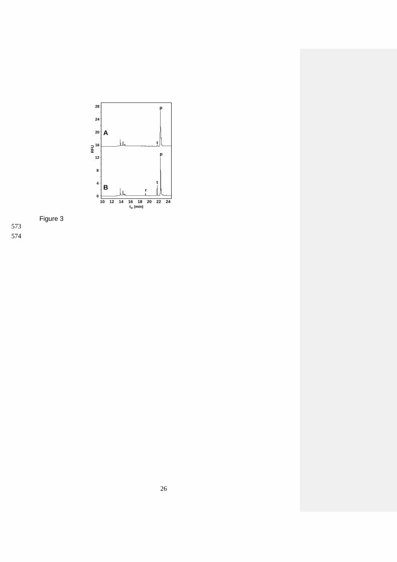
A multiplex PCR method was next developed in order to reduce the number of reactions required to 376
detect the transgenic yeast, trying to determine in a single analyses the three PCR amplicons. To 377
attain this, DNA extracts from cultured yeasts were next amplified in one tube with a mixture of the 378
six primers by multiplex PCR method. Figure 3A shows the CGE-LIF electrophoregram for the 379
multiplex PCR amplification of a DNA extract from cultured EKD-13 strain under the same 380
conditions of simplex PCR reactions, except for the concentration of primers (0.5 μM of 381
SCF1/SCR1, 0.1 μM KmTF/KmTR, and 0.1 μM of KmPF/KmPR). As can be seen, under these 382
conditions, no amplification was detected for the two shorter DNA fragments (i.e., 104 bp and 174 383
bp sequences) in multiplex format. Therefore, a further optimization of the multiplex PCR reaction 384
parameters had to be carried out in order to obtain optimal amplification for all the DNA sequences 385
under study. In this sense, since CGE-LIF provides accurate quantitative information, high 386
sensitivity and high resolution, this technique can be conveniently used to adjust PCR parameters 387
with confidence [41-43]. Accordingly, concentrations of SCF1/SCR1 and KmTR/KmTF primer 388
pairs were adjusted in order to improve co-amplification of these PCR fragments. Figure 3B shows 389
the electropherogram obtained once optimum triplex PCR amplification conditions were obtained 390
(namely, 0.7 μM SCF1/SCR1, 0.2 μM KmTF/KmTR, and 0.1 μM KmPF/KmPR). 391
392
3.3 Analysis of wine samples by multiplex PCR-CGE-LIF. 393

16
The suitability of the multiplex PCR-CGE-LIF method was tested analyzing wine samples obtained 394
in separate vinification processes using conventional or transgenic yeast strains. The results are 395
shown in Figure 4A-F. As can be seen, all the electropherograms of Figure 4 reveal the presence 396
of peak r, which corresponds to the expected 104 bp mrp2 amplicon used as control, assuring that 397
amplifiable DNA from S. cerevisiae has been obtained in all the extracts analyzed. Figures 4B, D 398
and F show that in the electropherograms of the wine samples produced with transgenic EKD-13 399
strain, peaks p and t, which correspond to the expected 174 bp and 199 bp amplicons, could be 400
detected, whereas no peaks other than peak r could be observed in the analysis of the wine samples 401
obtained with the non-recombinant yeast strain (Figure 4A, C and E). With regard to the effect of 402
the sample matrix, as can be seen in Figure 4F, the analysis of wine obtained from non-clarified 403
musts did not show significant differences compared to the corresponding wine samples obtained 404
from centrifuged musts (Figure 4B). These results demonstrate the good performance of the 405
multiplex PCR-CGE-LIF method even in wine obtained from non-clarified musts, in which 406
background DNA is expected to be more abundant and complex. 407
408
In conclusion, a new multiplex PCR-CGE-LIF separation method is proposed for the detection of 409
recombinant yeasts in wine samples. In addition, the extraction methodology described here is new, 410
although based on the adaptation of a commercial kit for the extraction of PCR-quality yeast DNA 411
from wine samples, including the addition of PVPP to obtain detectable amplification products. The 412
usefulness of this multiplex PCR-CGE-LIF method was demonstrated detecting genetically 413
modified yeasts in different wine samples. The method developed in this work allows the sensitive 414
detection of transgenic yeasts in wine samples in less than one working day, with no need for 415
additional yeast culturing steps. 416
417
ACKNOWLEDGEMENTS 418
419

17
This work was supported by a CSIC-Beckman Coulter contract and the CSIC project 200870I185. 420
Authors want to thank Dr J. Barcenilla for his help with the yeast cultures. We are grateful to Rosa 421
López (CIDA, Gobierno de La Rioja), for providing must samples for experimental wine 422
production. We want to thank J. M. Mateo for providing bottled wine samples (Laboratorio 423
Agroalimentario y Estación Enológica de Jerez, Cádiz). C. Leon wants to thank Comunidad 424
Autónoma de Madrid for a grant. 425
426
REFERENCES 427
428
[1] World Health Organization, Food safety (2002). http://www.who.int/fsf/GMfood. 429
430
[2] V. García-Cañas, C. Simó, C. León, E. Ibáñez, A. Cifuentes, Mass Spectrom. Rev. in press. 431
432
[3] I.S. Pretorius, Yeast 16 (2000) 675. 433
434
[4] W. Sybesma, J. Hugenholtz, W.M. De Vos, E.J. Smid, Electron. J. Biotechn. 9 (2006) 1. 435
436
[5] E. Cebollero, D. Gonzalez-Ramos, L. Tabera, R. Gonzalez, Biotechnol. Lett. 29 (2007) 191. 437
438
[6] Food and Drug Administration, FDA Notice 000120 (2003). 439
http://www.accessdata.fda.gov/scripts/fcn/gras_notices/grn000120.pdf. 440
441
[7] Food and Drug Administration, FDA Notice 000175 (2005). 442
http://www.accessdata.fda.gov/scripts/fcn/gras_notices/grn000175.pdf. 443
444
[8] J.I. Husnik, H. Volschenk, J. Bauer, D. Colavizza, Z. Luo, H.J.J. van Vuuren, Metab. Eng. 8 445
(2006) 315. 446
447
[9] J.I. Husnik, P.J. Delaquis, M.A. Cliff, H.J.J. van Vuuren, Am. J. Enol. Vitic. 58 (2007) 42. 448
449
[10] J. Coulon, J.I. Husnik, D.L. Inglis, G.K. Van Der Merwe, A. Lonvaud, D.J. Erasmus, H.J.J. 450
Van Vuuren, Am. J. Enol. Vitic. 57 (2006) 113. 451
452
Con formato: Inglés (Estados Unidos)
Con formato: Español (alfab.internacional)

18
[11] S. Michnick, J.L. Roustan, F. Remize, P. Barre, S. Dequin, Yeast 13 (1997) 783. 453
454
[12] F. Remize, J.M. Sablayrolles, S. Dequin, J. Appl. Microbiol. 88 (2000) 371. 455
456
[13] J.M. Eglinton, A.J. Heinrich, A.P. Pollnitz, P. Langridge, P.A. Henschke, M. de Barros- Lopes, 457
Yeast 19 (2002) 295. 458
459
[14] P. Sánchez-Torres, L. González-Candelas, D. Ramón, FEMS Microbiol. Lett. 145 (1996) 189. 460
461
[15] M.A. Ganga, F. Pinaga, S. Valles, D. Ramon, A. Querol, Int. J. Food Microbiol. 47 (1999) 171. 462
463
[16] P. Manzanares, M. Orejas, J.V. Gil, L.H. De Graaff, J. Visser, D. Ramón, Appl. Environ. 464
Microbiol. 69 (2003) 7558. 465
466
[17] M. Lilly, M.G. Lambrechts, I.S. Pretorius, Appl. Environ. Microbiol. 66 (2000) 744. 467
468
[18] D. Gonzalez-Ramos, E. Cebollero, R. Gonzalez, Appl. Environ. Microbiol. 74 (2008) 5533. 469
470
[19] E. Cebollero, A. Martinez-Rodriguez, A.V. Carrascosa, R. Gonzalez, FEMS Microbiol. Lett. 471
246 (2005) 1. 472
473
[20] A.A. Ogunjimi, P.V. Choudary, FEMS Immunol. Med. Microbiol. 23 (1999) 213. 474
475
[21] D.V. Jobes, D.L. Hurley, L.B. Thien, Taxon. 44 (1995) 379. 476
477
[22] J.E. Maliyakal, Nucleic Acids Res. 20 (1992) 2381. 478
479
[23] Y. Tsai, B.H. Olson, Appl. Environ. Microbiol. 58 (1992) 2292. 480
481
[24] C. Young, R.L. Burghoff, L.G. Keim, V. Minak-Bernero, J.R. Lute, S.M. Hinton, Appl. 482
Environ. Microbiol. 59 (1993) 1972. 483
484
[25] I.G. Wilson, Appl. Environ. Microbiol. 63 (1997) 3741. 485
486
[26] F. Savazzini, L. Martinelli, Anal. Chim. Acta 563 (2006) 274. 487
Con formato: Español (alfab.internacional)
Con formato: Español (alfab.internacional)

19
488
[27] R. Siret, J.M. Boursiquot, M.H. Merle, J.C. Cabanis, P. This, J. Agr. Food Chem. 48 (2000) 489
5035. 490
491
[28] R. Siret, O. Gigaud, J.P. Rosec, P. This, J. Agr. Food Chem. 50 (2002) 3822. 492
493
[29] M.M. Baleiras-Couto, J.E. Eiras-Dias, Anal. Chim. Acta 563 (2006) 283. 494
495
[30] F. Salinas, D. Garrido, A. Ganga, G. Veliz, C. Martínez, Food Microbiol. 26 (2009) 328. 496
497
[31] M.C. Simon, D.I. Gray, N. Cook, Appl. Environ. Microbiol. 62 (1996) 822. 498
499
[32] V. García-Cañas, A. Cifuentes, R. González, Crit. Rev. Food Sci. Nutr. 44 (2004) 425. 500
501
[33] M. Hernández, D. Rodríguez-Lázaro, D. Zhang, T. Esteve, M. Pla, S. Prat, J. Agr. Food Chem. 502
53 (2005) 3333. 503
504
[34] Y. Zhou, Y. Li, X. Pei, Chromatographia 66 (2007) 691. 505
506
[35] J. Xu, S. Zhu, H. Miao, W. Huang, M. Qiu, Y. Huang, X. Fu, Y. Li, J. Agr. Food Chem. 55 507
(2007) 5575. 508
509
[36] V. García-Cañas, R. González, A. Cifuentes, TrAC - Trends Anal. Chem. 23 (2004) 637. 510
511
[37] A. Querol, E. Barrio, T. Huerta, D. Ramon, Appl. Environ. Microbiol. 58 (1992) 2948. 512
513
[38] S. Rozen, H.J. Skaletsky, in S. Krawetz, S. Misener (Eds.), Bioinformatics Methods and 514
Protocols: Methods in Molecular Biology. Humana Press, New Jersey, 2000, pp. 365. 515
516
[39] R. Kalender, Primer Digital (2010). http://primerdigital.com/tools/. 517
518
[40] D. Gonzalez-Ramos, M. Quiros, R. Gonzalez, J. Agr. Food Chem. 57 (2009) 8373. 519
520
[41] V. García-Cañas, R. González, A. Cifuentes, Electrophoresis 25 (2004) 2219. 521
522
Con formato: Español (alfab.internacional)
Con formato: Español (alfab.internacional)
Con formato: Español (alfab.internacional)
Con formato: Italiano (Italia)
Con formato: Inglés (Estados Unidos)
Con formato: Español (alfab.internacional)

20
[42] V. García-Cañas, A. Cifuentes, J. Agr. Food Chem. 56 (2008) 8280. 523
524
[43] V. García-Cañas, M. Mondello, A. Cifuentes, Electrophoresis 31 (2010) 2249. 525
526
Con formato: Inglés (Estados Unidos)

21
FIGURE LEGENDS 527
528
Figure 1 Electrophoretic analysis in a 2% (w/v) agarose gel of the amplifications performed with 529
SCF1/SCR1 primer pair in DNA extracts from 15 mL Petit Verdot wine using different conditions: 530
Lines 1, 3, and 4 correspond to the samples treated with PVPP; and lines 2, 5, and 6 correspond to 531
untreated controls. Lines 1 and 2, samples were extracted using Method A; lines 3 and 5, samples 532
were extracted using Method B; lines 4 and 6, samples extracted using Method C. Lines 7 and 8, 533
amplified DNA extracts obtained from 1.5 mL cultured EC-1118 strain using a commercial kit and 534
manufacturer’s protocol 535
536
Figure 2 CGE-LIF electropherograms of amplified DNA from transgenic EKD-13 cultured yeast 537
using simplex PCR with different primer pairs: A) SCF1/SCR1; B) KmTR/KmTF; and C) 538
KmPR/KmPF. CGE-LIF separation conditions: uncoated fused-silica capillary with 60 cm total 539
length, 50 cm effective length and 75 m I.D.; separation voltage: -13 kV, running temperature 540
45ºC. Injection time 40 s (0.5 psi). BGE: 20 mM Tris, 10 mM phosphoric acid, 2 mM EDTA, 500 541
nM YOPRO-1, and 4.5 % HEC at pH 7.3. 542
543
Figure 3 CGE-LIF analysis of multiplex PCR reactions performed with DNA from transgenic 544
EKD-13 cultured yeast and the three primer pairs at the following concentrations: A) 0.5 M 545
SCF1/SCR1, 0.1 M KmTR/KmTF, 0.1 M KmPF/KmPR; B) 0.7 M SCF1/SCR1, 0.2 M 546
KmTR/KmTF, 0.1 M KmPF/KmPR. CGE-LIF separation conditions as in Figure 2 547
548
Figure 4 Electrophoretic analysis of triplex PCR amplifications from different wines: A, C and E) 549
musts fermented with the conventional EC1118 strain; B, D and F) musts fermented with transgenic 550
EKD-13 strain. Wine produced from must 2 (A and B), must 3 (C and D) and must 1(E and F). 551
CGE-LIF separation conditions as in Figure 2 552
553
554
555
556

22
TABLES 557
558
Table 1. Primers used for simplex and multiplex PCR amplifications of DNA extracts from culture 559
yeasts and wine samples. 560
Name Sequence (5’-3’) Ref.
KmPF ATCCCCCATGGCTATCACGA This work
KmPR GCACGTCAAGACTGTCAAGG
KmTF CAGAAAGTAATATCATGCGTCAATCG This work
KmTR AGCTCGGTACCTCGATGATAAG
SCF1 GGACTCTGGACATGCAAGAT [30]
SCR1 ATACCCTTCTTAACACCTGGC 561
562

23
563
Table 2. Concentration values and UV parameters (ratios at 260/280 and 260/230 nm) of DNA 564
extracts obtained under optimized extraction conditions from different wine samples. 565
566
Yeast strain Wine sample C (g/l) OD260/280 OD260/230
EC1118
(conventional)
Must 1 2.43 1.93 2.10
Must 2 2.72 1.89 2.20
Must 3 2.67 1.90 1.85
EKD-13
(transgenic)
Must 1 0.80 1.99 2.45
Must 2 0.67 1.84 1.80
Must 3 1.72 1.92 2.14
567
568

24
569
570
Figure 1
100 bp
200 bp
300 bp

25
571
572
Figure 2
tm (min)10 12 14 16 18 20 22 24
RF
U
0
40
80
120
160
r
t
p
200 A
B
C
26
573
574
Figure 3
RF
U
0
4
8
12
t
p
10 12 14 16 18 20 22 24
r
t
p
16
20
24
28
A
B
tm (min)

27
575
Figure 4
10 14 18 22
0
1
2
3
rA
10 14 18 22
0
4
8
rt
p
B
10 14 18 22
0
4
8
r t
p
F
D
10 14 18 22
0
2
4
6
r t
p
tm (min) tm (min)
RF
U
RF
U
10 14 18 220
1
2
3
r
10 14 18 220
1
2
3
r
C
E