Fall 2016 ACFAP Quarterlydelta phalanx) Dr. Hannah Park, DPM ... in removing nail products from toes...
28
http://www.acfap.org American College of Foot and Ankle Pediatrics Fall 2016 ACFAP Quarterly
-
Upload
nguyenkhanh -
Category
Documents
-
view
217 -
download
2
Transcript of Fall 2016 ACFAP Quarterlydelta phalanx) Dr. Hannah Park, DPM ... in removing nail products from toes...
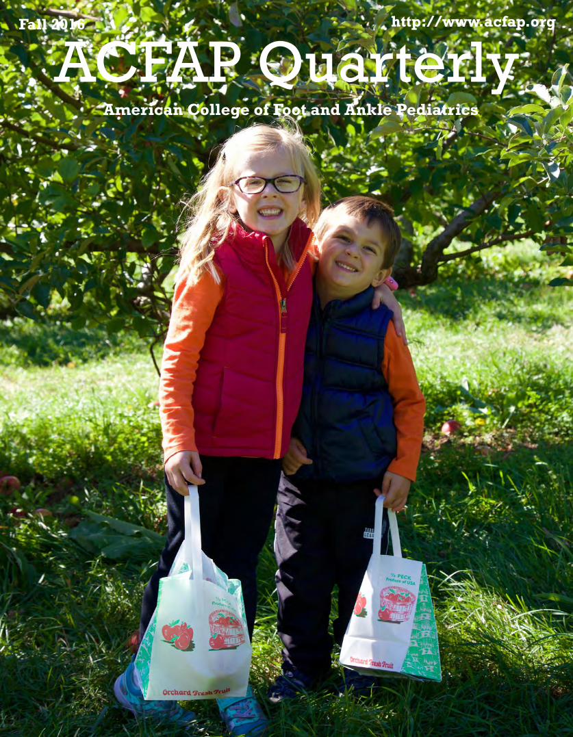
http://www.acfap.org
American College of Foot and Ankle Pediatrics
Fall 2016
ACFAP Quarterly

4 ACFAP Quarterly Fall 2016

5 ACFAP Quarterly Fall 2016
Features:
6 President’s MessageLouis J. DeCaro, DPM
8 Contact Dermatitis: Considerations in the Pediatric Population
Alecia Y. Williams. DPM, DABPM
10 Simple Diagram for Documentingand Explaining RotationalAbnormalities in Children
Nicholas Bolognini,DPM
12 A Case Study: Four limb polydactyly, a combination of simple and complex poly-dactyly and use of autograft for the cor-
rection of longitudinal epiphyseal bracket (delta phalanx)
Dr. Hannah Park, DPMDr. Prasad Gourineni, MD
Sponsor Spotlight: Blue Orchid Marketing/ practiceXeleration24 Editor ; Dr. Mary Clare Zavada
Layout Editor; Andrew Gromada
Contributing Editor; Sara Gromada25 ACFAP Sponsors
The Mass Effect of a Lipoblastoma: A Pediatric Tumor Case Report
Melissa Gulosh DPM, AACFASSarah Daigle DPM
Rachel Balloch DPM, AACFAS
16

6 ACFAP Quarterly Fall 2016
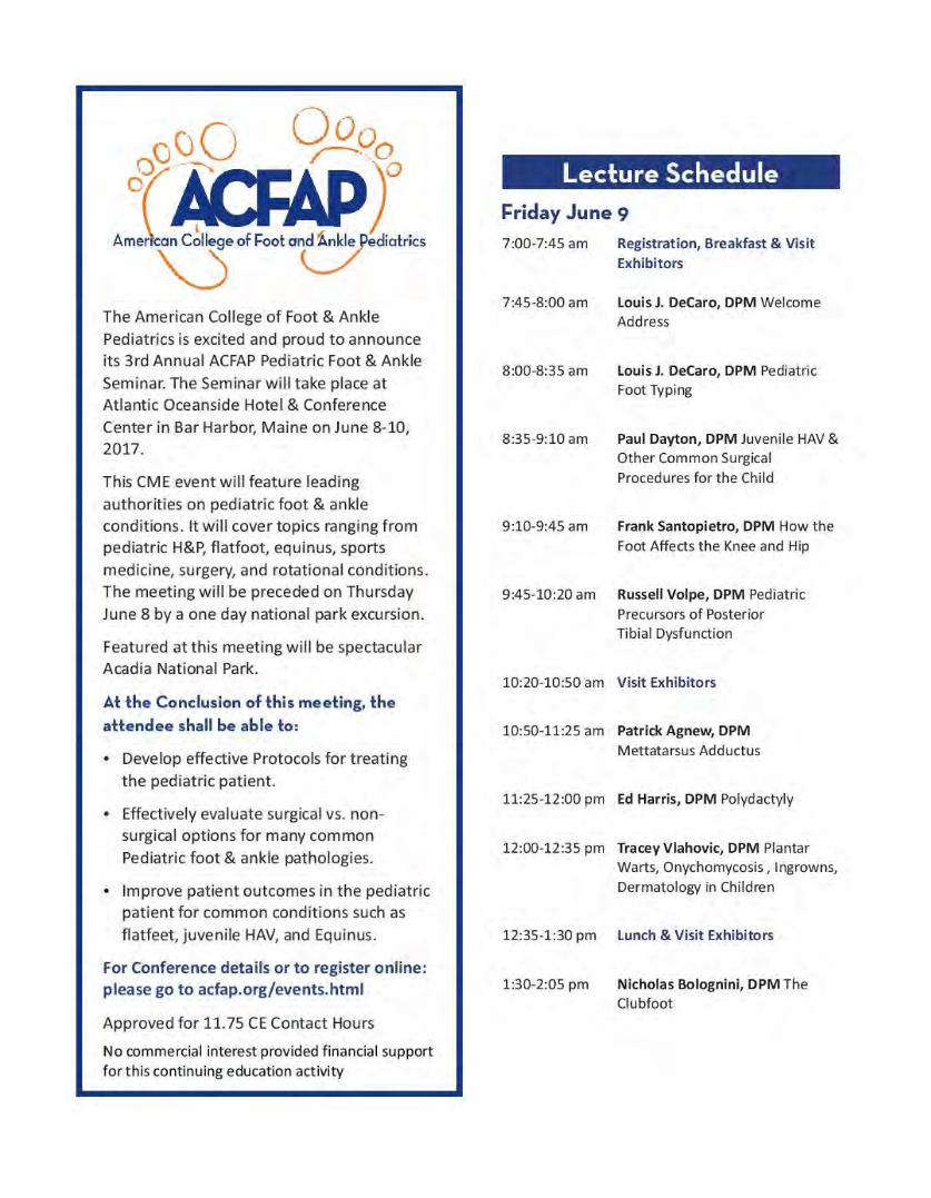
Presidents Message Hippocrates once said “The greatest medicine of all is teaching people how not to need it.” That quote for me embodies the very spirit, purpose, and drive of ACFAP. Our primary focus as physicians is to heal, but also we must never lose site of our obligation to prevent. For those of you who have ever heard me lecture, you know I also always draw attention to what I call our “moral obligation.” I believe our “moral obligation” as physicians, especially those who frequently see kids, is to strive for more prevention. I ask you all to help me get this message out. As 2016 wraps up, I look to make 2017 the “year of pediatric education” throughout our profession. Actively sug-gesting more pediatric education at seminars, filling out conference lecture suggestion forms, and talking to people who organize conferences are ways to advance this cause. Please join me!
We have been keeping up this mission already. Recently ACFAP was represented at the APMA national meeting in Philly, Heartland meeting in Iowa, and Region 7 in Vancouver, WA. Upcoming, ACFAP will be present at the AAPPM in San Antonio as well as SAM 2017 in Orlando.
As you all know ACFAP 2017 Annual Scientific Meeting has been set for the past several months. We are already receiving registrations. I encourage each and every one of you to plan and register early! This seminar is going to sell out quickly! We are continuing the National Park “tradition” at the Atlantic Oceanside Resort, Acadia National Park, Bar Har-bor, ME June 8-10 2017. Once again we will precede the meeting with a group outing in Acadia on the 8th. We have lined up a professional photographer as our tour guide, Mr. Don Toothaker ( toothakerphoto.com ) who has conducted expedi-tions in Acadia over 20 times!
The scientific part of the conference will take place at Acadia National Park on Friday and Saturday June 9-10, 2017. This CME (11.75 CME’s Proposed) event will feature leading authorities on pediatric foot and ankle conditions in-cluding Drs. Russell Volpe, Ed Harris, Tracey Vlahovic and many more! It will cover topics both conservative and surgical. As well the seminar will feature new and exciting panels and workshops. There will be a “Flatfoot Debate” consisting of a panel of docs representing surgical, orthotic, and conservative care point of views. Workshops and panels will include Pon-setti casting, practice management, and how to guides to “measuring” the pediatric patient. Please go to our website acfap.org for more information.
Our meetings are designed with two main missions in mind. The first is to educate in a positive environment that is both professional and fun at the same time. The second mission is to forge camaraderie between pediatric foot and ankle specialists and the companies that offer our patients the best and innovative solutions to manage their conditions…and maybe even foster ideas for new procedures, technology and products by putting the best minds together and learning from each other.
Each year the meeting is getting better and better. It really is the “Can’t Miss” meeting of the year, located in one of those “Can’t Miss” spots!
I want to again welcome all past, future, and current members of the American College of Foot and Ankle pediat-rics to this new era not only in this organization, but also in the education of pediatric foot and ankle medicine. Thank you to each and every one of you for making this all possible!
Louis J. DeCaro, DPM President, ACFAP www.acfap.org

7 ACFAP Quarterly Fall 2016

8 ACFAP Quarterly Fall 2016
Contact Dermatitis: Considerations in the Pediatric Population
Alecia Y. Williams. DPM, DABPM
The current practice environment has always meant extensively tracking acute allergies. With the advent of the EMR, it has gotten easier to catalog not only the allergies, but the onset of the reaction and the type of the reaction. There are many recognized types of allergenic materials. It is important to recognize any potential reaction in this population as initial sensitiza-tion and presenting pathology many now present at an earlier age. It is now recognized that contact sensitiza-tion begins in early childhood via exposures such as vaccinations, piercing, topical medications, and cos-metics.1 It is also important to differentiate its presence as the prevalence of atopic dermatitis in the pediatric population in much of the US is at least 10%..2 With an accurate and timely diagnosis comes the ability to quickly treat or refer to the allergist or dermatologist.
CONTACT DERMATITIS OVERVIEW
Contact dermatitis may is divided into three types: allergic, irritant, and photocontact. The vast majority of cases are irritant-induced. This owes itself to repeated exposure in an industrial setting of the adult population. The trigger substance itself directly damages the skin. Allergic contact dermatitis (ACD) is a delayed type hypersensitivity reaction elicited by the contact of the skin with the offending chemical or sensitizer in individuals who have been previously sensitized to it. 3 When differentiating the other types of dermatitis, especially atopic, lesions and eruptions about other parts of the body, notably the head, scalp neck, cubital fossa or the popliteal fossa. An acute eruption of ACD can consist of pruritic, erythematous, indurated, scaly plaques and in severe cases may have vesiculation or bullae.1 Photocontact is involvement of UV light (present in sunlight) and a normally unharm-ful substance on exposed skin. Neither photocontact nor irritant type of dermatitis involve the immune sys-tem.4 The onset of the dermatitis is within two weeks of exposure to the allergen.
IRRITANT CONTACT DERMATITIS
Solvents, surfactants, cleaners as well as latex are the basis for this type of dermatitis. Solvents used in removing nail products from toes and nails are also a common example. Clinically, the affected area may present with scaling, hyperkeratosis or fissuring. There may be pain, itching or burning along with itching.
TRIGGERS OF ALLERGIC CONTACT DERMATITIS
Sensitizers come from many different groups of materials: plants, metals, preservatives, rubbers, ad-hesives, glues, fabric dyes/finishes, and topical prepa-rations. A common sensitizer is found in poison ivy, poison oak, and poison sumac. Nickel is common with jewelry use as is gold, chromium and cobalt. Topical
SensitizerAnkle Bracelet Nickel, chromiumTattoo Nickel, chromium,
cobaltSandals/Shoes Chromium leather treat-
ment, Vulcanizedrubber/MBT
Socks Vulcanized Rubber/MBT, dye
Adhesive Bandages, tape Colophony (rosin)
Nail Polish FormaldehydePedicure Nail Designs/Artifi-cial Nail
Acrylates
Osseous Procedure Metallic implant
Nickel, Chromium, Cobalt, Iron6
Table 1. Examples of sensitizing products affecting the Lower Extremity

9 ACFAP Quarterly Fall 2016
medications include gentamicin, neomycin, hydro-quinone, and bacitracin. The sensitization can also be enhanced by heat, occlusion, and humidity in the case of shoe gear. 5 Table 1 shows some examples of com-mon products. Image 1 shows chromium as chromate treatment of leather.
DIFFERENTIAL DIAGNOSES
Other etiologies can strongly mimic dermatitis. Bacterial infections such as Impetigo which is a Staph or Strep infection presents as crusting, yellow lesions mainly occurs in the face, neck and hands. Fungal infections can easily be excluded with skin scraping analysis. Bites from insects ranging from fleas, ticks, bed bugs, spiders, chiggers, ants and mites must also be considered. Scabies has intense itching. Some bites may have a linear distribution. Viral exanthems have a larger skin surface distribution affecting multiple parts of the body much like psoriasis or atopic dermatitis.
TREATMENT
Identification of offending agent is most im-portant for future eruption. Wet cool compresses to affected area. Menthol preparation or calamine lotion may also be effective. Weeping lesions can be alleviated with topical astringents, aluminum acetate or alumi-num sulfate under occlusion. Benzocaine and topical
antibiotics with neomycin or bacitracin again should be avoided because they are allergenic. Oral antibiotics are recommended for complicated secondary infec-tion.
SUMMARY
Many products, dressings, bandages, now ex-clude latex, but there may be other potential sensitizer amongst these products. The key is direct contact and representative distribution on the skin. Humidity, heat and occlusion can enhance the sensitization of rubber contact from either socks or shoes causing the erup-tion. If topical antibiotics are the cause of ACD and there is an indication for topical antibiotics, Mupirocin has the lowest rate of sensitization.
REFERENCES
1. Weston WL, Contact Dermatitis in Children, Uptodate.com, 2016
2. Tollefson MM, Bruckner AL. Atopic Dermatitis: Skin-Directed Management. Pediatrics, December 2014, Volume 134/Issue 6 3. Contact dermatitis. (2016, May 25). In Wikipedia, The Free Encyclopedia.
Retrieved 02:05, June 24, 2016, from https://en.wikipedia.org/w/index.php?title=Contact_dermatitis&oldid=722081361
4. Anderson DM, Keith J, Novac P; Elliott MA, eds. (1994). Dorland’s Illustrated Medical Dictionary (28th ed.). W. B. Saun-ders Company.
5. Adams AK, Warshaw EM, Allergic Contact Dermatitis from Mercapto Compounds, Dermatitis. 2006;17(2):56-70
6. Basko-Plluska JL, Thyssen JP; Schalock PC. Cutaneous and Systemic Hypersensitivity Reactions to Metallic Implants. Dermatitis. 2011;22(2):65-79
Dr. Williams is in Private Practice in New York City. She is also board certified by the Ameri-can Board of Podiatric Medicine.
Image 1: Chromate Allergy from Leather Shoe.
Uptodate.com, Courtesy of William L Weston, MD.
10 ACFAP Quarterly Fall 2016
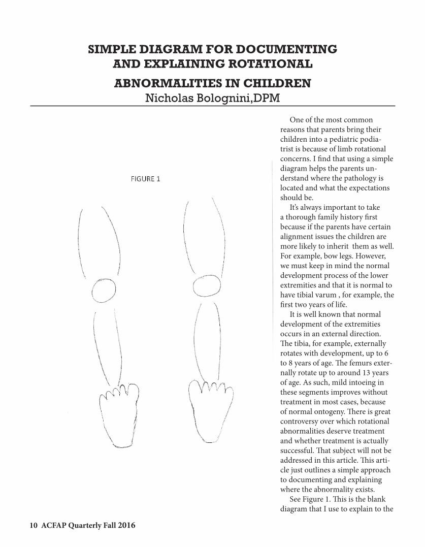
SIMPLE DIAGRAM FOR DOCUMENTING AND EXPLAINING ROTATIONAL
ABNORMALITIES IN CHILDREN Nicholas Bolognini,DPM
One of the most common reasons that parents bring their children into a pediatric podia-trist is because of limb rotational concerns. I find that using a simple diagram helps the parents un-derstand where the pathology is located and what the expectations should be. It’s always important to take a thorough family history first because if the parents have certain alignment issues the children are more likely to inherit them as well. For example, bow legs. However, we must keep in mind the normal development process of the lower extremities and that it is normal to have tibial varum , for example, the first two years of life. It is well known that normal development of the extremities occurs in an external direction. The tibia, for example, externally rotates with development, up to 6 to 8 years of age. The femurs exter-nally rotate up to around 13 years of age. As such, mild intoeing in these segments improves without treatment in most cases, because of normal ontogeny. There is great controversy over which rotational abnormalities deserve treatment and whether treatment is actually successful. That subject will not be addressed in this article. This arti-cle just outlines a simple approach to documenting and explaining where the abnormality exists. See Figure 1. This is the blank diagram that I use to explain to the

11 ACFAP Quarterly Fall 2016
parents where the rotational issues are located. Figure 2 is an example of how I mark the diagram during the examination. For example, the Left lower extremity markings indicate that there is left sided femoral anteversion, causing internal positioning (45 degrees of internal rotation available and only 10 degrees of external rotation), and the tibial and foot segments are normal. The right side markings indicate that the rotation in the femoral segment is normal, but, there is tibial torsion present and meta-tarsus adductus with the heel bisector going through the 3rd webspace. This has been a simple, but, effective tool for me in my practice to explain rotational pathology to the parents. I feel it helps them understand where the rota-tional problems are located and can even reassure them, as we explain normal de-velopment and which components can improve just with maturity. Clinicians can use it in various ways (exact num-bers or not), mark it for frontal plane and/or transverse plane deformities, and use one for sitting and one for lying supine , and ,finally, use it as a tool to help follow their improvement, whether treated or just observed over time.
Nicholas Bolognini,DPMFellow, American College of Foot and Ankle PediatricsFellow, American College of Foot and Ankle Surgeons

12 ACFAP Quarterly Fall 2016
A Case Study: Four limb polydactyly, a combination of simple and complex polydactyly and use of au-
tograft for the correction of longitudinal epiphyseal bracket (delta phalanx)
Dr. Hannah Park, DPMDr. Prasad Gourineni, MD
Abstract: Polydactyly is a common congenital deformity of an extra digit or digits to a hand or foot and oc-curs commonly with syndactyly. There is a variety of polydactyly depending on the duplication of the all or part of the toe and the presence of bone duplication. In this case study, we present a case presentation of a 11 month old female child who had polydactyly of all four limbs as well as a longitudinal bracketed epiphysis (delta phalanx) causing a hallux varus deformity and syndactyly.
Background: A 8 month of female with polydactyly of all four limbs presented to our pediatric orthopedic clinic with the mother concern for the child unable to wear
normal shoes as well as cosmetic appearance. The child had identical simple post axial polydactyly of bilat-eral hands (Figure 1) which had no functional ability. However on the lower extremities were two different pre axial deformities. The right foot, the child had a pre axial complex polydactyly deformity with a well formed hallux with syndactilized digits of 2 and 3 and a hallux varus deformity due to a longitudinal brack-eted epiphysis (Figure 2A). The left foot, the child had a pre axial simple polydactyly attached to an enlarged 2nd digit which was also syndactilized to the 3rd digit which also had a hallux varus deformity and longitudinal bracketed epiphysis of the 1st metatarsal (Figure 3A). The pedia-trician did not make a diagnosis to what congenital
Figure 1 Simple Post axial deformity of the left hand, identical deformity on the right.
Figure 2A. Complex pre-axial deformity with a delta phalanx and hallux varus deformity, syndactilized dig-its of 2 & 3 Right foot

13 ACFAP Quarterly Fall 2016
deformity would be linked with the child’s multiple polydactyly deformities and the wide set eyes and enlarged forehead. The child did not have any other medical history or surgeries and there was no family history of polydactyly or other congenital deformities. Due to the quantity and complexity of the deformities of all four limbs, as well as the necessity of the child needing to wear shoes in the next few months when the child would start walking, decision was made to perform surgery when the patient was 10 months old. The child was put under general anesthesia by the pediatric anesthesiologist and care was made to scrub, prep and drape all four limbs of the patient. No tourni-quet was used. Local anesthetic block using Marcaine 0.25% with epinephrine was used to locally block the incision sites. First attention was drawn to the hands in which two elliptical incision were made at the bases of the ex-tra digit of the hands, bleeding was cauterized using a bovie and the subcutaneous fat was debunked in order to achieve closure. Incision of the hands were closed using chromic suture. This was repeated for bilateral hands without complications. Surgical site was later dressed with antibiotic ointment, xeroform, 2x2 gauze, webril and a light ACE bandage. Then attention was drawn to the right foot, longitudinal incision was made over the medial aspect
of the foot with an elliptical incision around the pre-axial extra digit. Incision was carried down using a bovie with care to avoid any neuromuscular structures. The extra digit was disarticulated at the base. Care was made to save the extra digit as the bone from the enlarged phalanx was used as autografts. It was noted at the head of the 1st metatarsal that the shaped of the metatarsal was enlarged and curved as it articulated with two halluxus and consistent with a typical lon-gitudinal bracketed epiphysis. The cartilage of the 1st metatarsal medially which articulated on the pre-axial extra digit was carefully removed using a rongeur and curette. Care was made sure not to touch the articulat-ing surface of the 2nd hallux. The syndactyly of the 2nd and 3rd digits were left alone in order to prevent hallux varus reoccurrence instead of performing the usual modified farmer’s procedure. It was noted that despite the resection of the polydactyly, there was a hallux varus deformity of the 2nd digit on the 1st metatarsal that was coming from a proximal deformity of the longitudinal bracketed epiphysis due to abnormal closure of the epiphysis which had caused decreased in longitudinal growth but continual transverse growth medially. Using a small osteotome, a perpendicular bone cut was made at the proximal 1/3 of the 1st metatarsal and wedged open to correct the varus deformity. A k-wire was in-serted from the 2nd digit to the 1st metatarsal with the
Figure 2 B. After resection of the pre axial polydactyly, osteotome placed in the area for open wedge oste-otomy. Irregular shaped delta phalanx noted on the 1st metatarsal of the Right foot.
Figure 3A, Complex pre axial polydactyly of the left foot with syndactylized digits of 2 & 3 and a hallux varus deformity with a delta phalanx.

14 ACFAP Quarterly Fall 2016
open wedge osteotomy correction. A autograft from the base of the proximal phalanx of the extra digit was used to also hold the open wedge osteotomy correction and increase bone healing. Then lastly attention was drawn to the left foot in which the complex pre axial deformity was resected using 2 elliptical incisions at the 2nd digit and a longi-tudinal incision was elongated on the medial aspect of the foot in order to correct the hallux varus deformity and longitudinal bracketed epiphysis of the 1st meta-tarsal. The enlarged curved 1st metatarsal had made it impossible for the longitudinal growth of the 1st metatarsal causing a worsened hallux varus deformity than the contralateral foot. Using a small osteotome, a perpendicular bone cut was made at the proximal 1/3 of the 1st metatarsal and wedged open to correct the varus deformity. A k-wire was inserted from the 2nd digit to the 1st metatarsal with the open wedge oste-otomy correction. Another autograft from the base of the proximal phalanx of the right foot extra digit was used to also hold the open wedge osteotomy correction and increase bone healing. Then the surgical sites were copious flushed and closed subcutaneously using vicryl and then sim-ple interrupted sutures with chromic. Surgical site was applied with antibiotic ointment, xeroform, 2x2 gauze,
webril, and a posterior splint was applied with an ACE bandage which was worn about 6 days. Pt returned after 6 days for surgical incision site check which was clean and without infection to all four limbs. Hand incision sites were simply dressed with bandaids. Then an above knee soft cast with webril was applied to bilateral legs to be worn for 3 weeks.
Discussion: Polydactyly is a deformity of an extra digit or digits to a hand or foot and occurs commonly with syndactyly. There is a variety of polydactyly depend-ing on the duplication of the all or part of the toe and the presence of bone duplication. The extra digit may also articulate with a partial or completely duplicated metatarsal which may have a “Y” or “T” shape to articulate with the toe. A simple polydactyly consists of an extra digit of soft tissue only, where as a complex polydactyly consists of a extra digit with bone. Pre-axial polydactyly occurs when the extra digit occurs on the medial side of the foot (9% occurrence) which commonly occurs with longitudinal bracketed epiphy-sis. Central polydactyly occurs especially when there is a partially formed 4th metatarsal (6% occurrence) and syndacctyly or synchondrosis of the of the digit occurs with the 5th proximal phalanx. Post-axial polydactyly
Figure 3B: After removal of the polydactyly deformity, the hallux varus deformity and delta phalanx noted.
Figure 4A: Immediate post op, right foot

15 ACFAP Quarterly Fall 2016
occurs when the extra digit occurs on the lateral side of the foot (85% occurrence).1
Longitudinal bracketed epiphysis is rare growth disorder known as the “delta phalanx” of the metatarsal in which the epiphysis and the physis con-nect on the medial side on the 1st metatarsal causing the diaphysis and the normal proximal epiphysis to be connected with a distal pseudopiphysis (1). The abnormal connection causes the lateral diaphysis with an epiphysis to continue to grow causing a wide meta-tarsal with a varus deformity of the hallux, causing a triangular or “C” shaped epiphyseal plate (2). The deformity most commonly occurs at the 1st metatarsal and is commonly associated with a hallux varus defor-mity, pre-axial polydactyly, syndactyly, clubfoot, apert’s syndrome, and poland’s syndrome . No conservative treatment is available however the resection of the longitudinal bracketed epiphysis with the preservation of normal proximal epiphysis and the distal pseudo epiphysis with soft tissue release for the hallux varus deformity and resection of the pre-axial polydactyly is a surgical treatment with good results and low to no reoccurrence rates (1)(3)(4). In our case we also performed as opening wedge osteotomy with autograft insertion to correct the hallux varus deformity with temporary fixation with a k-wire. This wire would be removed after 3 weeks. We presented an interesting case study of a 10 month old female with polydactyly of all four limbs that was surgically treated. Resection of polydactyly depending on whether it is a simple verses complex de-formity as well as the presence of a longitudinal brack-eted epiphysis causing a hallux varus deformity and the
presence of syndactyly were all part of the equation of the surgical treatment plan for this patient.
References: (1) Mosaca, V. S. Principles and management of pediatric foot and ankle deformities and malformations. 2014. (2) Choo, A and Mubarak, S. J. Longitudinal epiphyseal bracket. Journal of Children’s Orthopaedics. December 2013. 7: 6. 449- 454. (3) Shea, K. G. Mubarak, S. J. Alamin, T. Pre-ossified longi-tudinal epiphyseal bracket of the foot: treatment by partial bracket excision before ossification. Journal of pediatric orthopaedics. 2001. May-June: 21 (3) 360-365. (4) Lampropulous, M. Pugigdevall, M. Zapozko, D. Malva-mez, H. Treatment of first metatarsal longitudinal epiphy-seal bracket by excision before closure. Journal of foot and ankle surgery. 2007. July-August: 46 (4) 297-301.
Dr. Hannah Park, DPMPediatric Orthopedic Foot and Ankle Surgery Fellow at Advocate Christ Medical Center, Oak Lawn, IL.
Dr. Prasad Gourineni, MDPediatric Orthopedic Fellowship Attending at Advo-cate Christ Medical Center, Oak Lawn, IL.
Figure 4B: Immediate post op right foot
Figure 5: Immediate post op left foot

16 ACFAP Quarterly Fall 2016
The Mass Effect of a Lipoblastoma: A Pediatric Tumor Case Report
Melissa Gulosh DPM, AACFASSarah Daigle DPM
Rachel Balloch DPM, AACFAS
I. IntroductionLipoblastomas are rare, benign tumors of embryonic or immature adipose tissue, separated by connective tissue septa of various thicknesses, with focal myx-oid.1,2 These neoplasms are found almost exclusively in infancy and in early childhood, and account for less than 1% of all childhood neoplasms.3 Diagnosis occurs before 3 years of age in 90% of cases, and 40% of cases are diagnosed in the first year of life.4 Lipo-blastomas often present as an asymptomatic, rapidly enlarging, soft lobular mass on the extremity. 3 Most
lipoblastomas occur in the superficial tissues of the arms and legs, but also arise in the head, neck, parotid gland, eyelid, tonsillar fossa, mediastinum, and retro-peritineum regions.5 Although these tumors are be-nign, they can have a significant mass effect, including displacement of bones and compression of soft tissues. There is a slight male predominance with a predilec-tion for the left side of body.1,4,5 This paper reports a case of lipoblastoma found in the plantar left foot of a 2-year old male.
Figure 1: Gross pictures of the left foot in this 2yo male patient Figure 2: AP Xray, Left foot

17 ACFAP Quarterly Fall 2016
Figure 3: Lateral Xray, demonstrating indiscriminate soft tissue mass in plantar left foot
Figure 4: T1 Sagittal view demonstrates hyperintense internal fat signal with multiple internal septations measur-ing up to approximately 1.4mm in thickness. This finding is centered along the quadratus plantae muscle along the plantar aspect of the midfoot and extends posteriorly along the plantar medial aspect of the calcaneus to the level of the sustentaculum
18 ACFAP Quarterly Fall 2016
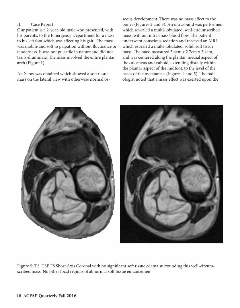
II. Case Report Our patient is a 2-year-old male who presented, with his parents, to the Emergency Department for a mass to his left foot which was affecting his gait. The mass was mobile and soft to palpation without fluctuance or tenderness. It was not pulsatile in nature and did not trans-illuminate. The mass involved the entire plantar arch (Figure 1).
An X-ray was obtained which showed a soft tissue mass on the lateral view with otherwise normal os-
seous development. There was no mass effect to the bones (Figures 2 and 3). An ultrasound was performed which revealed a multi-lobulated, well-circumscribed mass, without intra-mass blood flow. The patient underwent conscious sedation and received an MRI which revealed a multi-lobulated, solid, soft tissue mass. The mass measured 5.4cm x 2.7cm x 2.4cm, and was centered along the plantar, medial aspect of the calcaneus and cuboid, extending distally within the plantar aspect of the midfoot, to the level of the bases of the metatarsals (Figures 4 and 5). The radi-ologist noted that a mass effect was exerted upon the
Figure 5: T2_TSE FS Short Axis Coronal with no significant soft tissue edema surrounding this well-circum-scribed mass. No other focal regions of abnormal soft tissue enhancemen

19 ACFAP Quarterly Fall 2016
flexor digitorum and flexor hallucis longus tendons. On physical exam, this was seen with flexible flexion contracture of the toes.
The final impression of the MRI was that of a lipoblas-toma. An atypical lipoma was felt to be less likely due to the prominence and number of internal septations within the mass as well as the patients demographic. A liposarcoma was also felt to be less likely due to the young age of this patient and lack of significant sur-rounding soft tissue edema adjacent to the mass. It was felt that complete excision would be the most beneficial treatment to prevent further mass effect and potential foot deformity.
A curvilinear, lazy S incision was determined to be most appropriate, as this could easily be extended proximally, as the MRI indicated extension of the mass
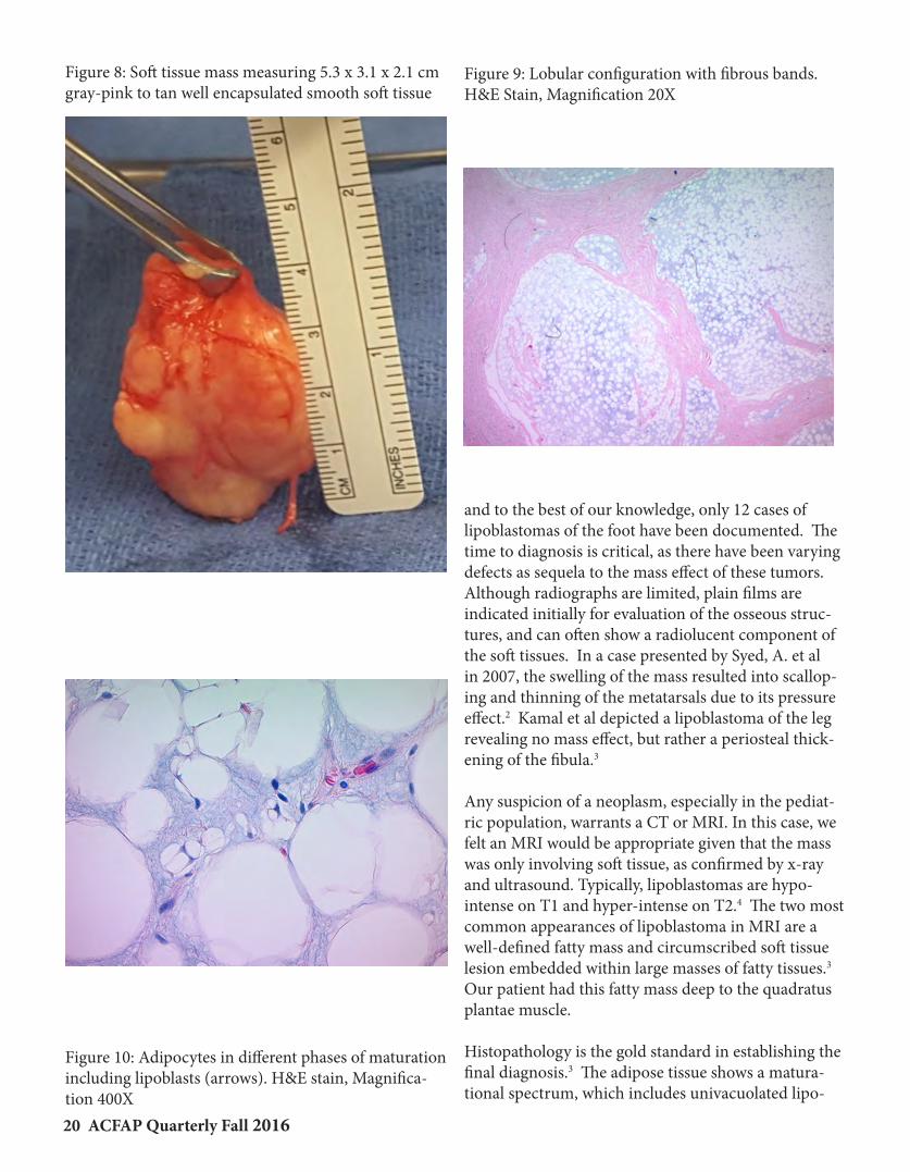
to the level of the sustentaculum tali. This would also allow adequate exposure medial to lateral. The large mass was well encapsulated, and without abnormal protrusions into the surrounding tissues. The mass was sent to pathology whose assessment was a multi-lobulated, lipomatous neoplasm with myxoid changes compatible with a benign lipoblastoma (Figures 9 and 10). The patient’s postoperative healing after 2 weeks is shown in Figure 11. In a two year old child, a plantar incision has its risks, including scar formation. Despite the activity level relayed by the mother, the plantar in-cision healed without incident or dehiscence. We used 4-0 monocryl to avoid suture removal in a child.
III. DiscussionAdipose tumors are not common in children, account-ing for only 6% of pediatric soft tissue lesions.4 There has been multiple case reports regarding these tumors,
Figure 6: Intraoperative gross picture showing the mass within the plantar arch. Notice the flexion of the digits secondary to the mass effect.
Figure 7: Intraoperative gross picture after the mass was removed. Notice the normalization of the digits into a rectus position.
20 ACFAP Quarterly Fall 2016
and to the best of our knowledge, only 12 cases of lipoblastomas of the foot have been documented. The time to diagnosis is critical, as there have been varying defects as sequela to the mass effect of these tumors. Although radiographs are limited, plain films are indicated initially for evaluation of the osseous struc-tures, and can often show a radiolucent component of the soft tissues. In a case presented by Syed, A. et al in 2007, the swelling of the mass resulted into scallop-ing and thinning of the metatarsals due to its pressure effect.2 Kamal et al depicted a lipoblastoma of the leg revealing no mass effect, but rather a periosteal thick-ening of the fibula.3
Any suspicion of a neoplasm, especially in the pediat-ric population, warrants a CT or MRI. In this case, we felt an MRI would be appropriate given that the mass was only involving soft tissue, as confirmed by x-ray and ultrasound. Typically, lipoblastomas are hypo-intense on T1 and hyper-intense on T2.4 The two most common appearances of lipoblastoma in MRI are a well-defined fatty mass and circumscribed soft tissue lesion embedded within large masses of fatty tissues.3 Our patient had this fatty mass deep to the quadratus plantae muscle.
Histopathology is the gold standard in establishing the final diagnosis.3 The adipose tissue shows a matura-tional spectrum, which includes univacuolated lipo-
Figure 8: Soft tissue mass measuring 5.3 x 3.1 x 2.1 cm gray-pink to tan well encapsulated smooth soft tissue
Figure 9: Lobular configuration with fibrous bands. H&E Stain, Magnification 20X
Figure 10: Adipocytes in different phases of maturation including lipoblasts (arrows). H&E stain, Magnifica-tion 400X

21 ACFAP Quarterly Fall 2016
Table 1: Differential Diagnoses1,2,7,8,10,11
DDX LE Tumors Age/Affected Body Part(s)
Histology
Lipoblastoma Occurs typically w/in first 3 yrs of life
Wide anatomic distribution w/ predilection for extremities
Lobulated w/ connective tissue septa containing mature and immature adipose tissue
Connective tissue septa and myxoid mesenchymal tissue
No nuclear atypia PLAG1 gene rearrangement, located on chromosome
8q12
Myxoid Liposarcoma
Occur age 10+ Peak 50-65 yrs Predilection for
extremities and retroperitoneum
Similar histology to lipoblastoma but may also have larger myxoid component, cellular atypia and nuclear hyperchromasia w/ high mitotic count
No PLAG1 gene rearrangement
Primitive myxoid mesenchymal tumor of infancy
First year of life Wide anatomic
distribution
Homogenous oval to spindled mesenchymal cells in myxoid background; No lipoblasts
Open chromatin; Indistinct nucleoli w/ mild atypic No PLAG1 gene rearrangement
Lipoma Occurs at any age Peaks 40+ yrs Wide anatomic
distribution
Well encapsulate lobules of adipose tissue Thin, fibrous capsule Gene rearrangements involve chromosomes 6, 12, and
13
Lipofibromatosis Pediatric Wide anatomic
distribution w/ predilection for hands and feet
Abundant adipose tissue traversed by bundles of spindled fibroblast-like cells
Well organized fibroblastic element within septa May involve S-100 protein in the spindled element
Fibrous Hamartoma of Infancy
First 2 years of life Axilla, inguinal region,
and proximal extremities
Poorly circumscribed; Fibro-fatty component w/ primitive mesenchymal cells
Spindled fibroblastic cells, varying amounts of collagen, nodular aggregates of immature stellate-shaped and spindled cells in myxoid matrix, and mature univacuolated adipocytes
Calcifying Aponeurotic Fibroma
First decade of life Strong predilection for
hands and feet
Nodular foci with calcification and cartilage formation Small epitheloid fibroblasts in corded array and lack of
defined fatty elements
Juvenile (Infantile) Fibromatosis
Pediatric Wide anatomic
distribution w/ predilection for muscles of head/neck, shoulders, upper arm and thigh
3 subtypes: mesenchymal, fibroblastic, and desmoid variants
Morphologically diverse, composed of fibroblast-like cells with histological differences attributed to pt age and level of maturation

22 ACFAP Quarterly Fall 2016
blasts, signet ring cells, multivacuolated lipoblasts, and mature adipocytes. Mature adipocytes typically are centrally located while, at periphery, immature myxoid and stellate mesenchymal cells are seen.6 The mature and immature adipose tissues are typically separated by connective tissue septa of varying thickness, seen in Figure 10 from our patient’s histopathology slides. The observation of myxoid stroma is more prominent in infants.4
Definitive treatment is complete surgical excision.1,8 Lipoblastomas are considered biologically benign and grow locally, which can lead to a significant mass ef-fect. Fortunately for our patient, the mass effect was only affecting the flexor tendons which we feel will resolve now that the tumor has been excised. Local recurrences are reported in 9% to 25% of cases with no metastases reported in the literature.2,3,5
It is important to be aware of the differential diagnoses and understand the importance of early diagnosis and treatment. Table 1 exhibits a breakdown on pertinent
differentials to consider. A sense of urgency is impera-tive when working with the pediatric population. Our patient was seen in the emergency department, ob-tained an MRI, and was taken to surgery within a two week timeframe. Fortunately, this tumor is of benign nature. We educated the parents about the risk of re-currence and follow up is to be scheduled accordingly.
References:1 - Chien, A. et al. Two Young Girls with Lipoblastoma and a Review of the Literature. Pediatric Dermatology. 2006, Vol 23, No 2,152-1562 - Syed, A. et al. Lipoblastoma - A Rare Pediatric Foot Tumor. Acta Orthop. Belg., 2007, 73, 400-4023 - Kamal, A. et al. Lipoblastoma and Lipoblastomatosis of the Lower Leg. Case Reports in Orthopedics. Volume 2014, Article ID 5828764 - Shinkai, T. et al. A Case of unusual Histology of Infantile Lipoblastoma Confirmed by PLAG1 Rearrangement. Surgi-cal Case Reports. (2015) 1:425 - Kerkeni, Y. et al. Lipoblastoma in Childhood: About 10 Cases. Afr J Pediatric Surgery 2014;11:32-346 - Young, R. et al. Acral Lipoblastoma. Dept of Dermatology, Wilford Hall Medical Center, Lackland Air Force Base, San Antonio, TX. Vol 65, April 20007 - Warren M. et al. Undifferentiated Myxoid Lipoblas-toma with PLAG -HAS2 Fusion in an infant; morphologi-cally mimicking primitive myxoid mesenchymal tumor of infancy (PMMTI) - diagnostic importance of cytogenic and molecular testing and literature review. Cancer Genetics 209 (2016) 21-298 - Wilson, T. et al. Evaluation and management of fibrofatty tumors of the extremities: case report. J Neurosurg Pediatric 17:66-69, 20169 – Papendieck, C. et al. Lipoblastoma-Lipoblastomatosis Associated with Unilateral Limb Hypertrophy: A Case Re-port in a Newborn. Lymphology 36 (2003) 69-7310 – Fetsch, J. et al. A Clinicopathologic Study of 45 Pediat-ric Soft Tissue Tumors With an Admixture of Adipose Tis-sue and Fibroblastic Elements, and a Proposal for Classifica-tion as Lipofibromatosis. The American Journal of Surgical Pathology 24(11):1491-1500, 200011 – Deepti, A. et al., Lipofibromatosis: report of a rare pe-diatric soft tissue tumor. Skeletal Radiology (2008) 37:555-558
Figure 11: 2 weeks postoperative healing with Monocryl in place. No signs of dehiscence.

We are not just another Podiatric Pathology Service,
we are a partner and advocate of the profession!
The Bako Difference:Reports with actionable information,
not just a diagnosis!
Bako Pathology Services:Bako Pathology Services:
The Bako Difference:Reports with actionable information,
not just a diagnosis!
Rev. July, 2016
• Precise, but detailed diagnoses
• Standards of care forsurgical management
• Medical therapeutic options
• Photomicrographic imaging
• Pathologists available for consultation7:00 a.m. - 7:00 p.m. EST

24 ACFAP Quarterly Fall 2016
SponSor Spotlight
Ten years ago Blue Orchid Marketing began offering data base internal marketing services to podiatrists to grow their practices. Today the company provides full programs of comprehensive turnkey strategies in both internal and external marketing to all types of medical practices.
Our internal marketing focuses on an opportunity that many practices overlook- leveraging your EHR patient data to drive business, revenue, and new patients to the practice. Using a variety of tested and targeted communication & campaign strategies, the goals are to reactivate inactive patients, promote regularity of visits, accelerate personal referrals, build the brand awareness, and keep you ahead of the competition. We help a prac-tice build and nurture lasting patient relationships that result in the increase of referrals of new patients.
practiceXeleration was formed under the Blue Orchid umbrella to target external marketing for helping businesses increase their online presence. This to builds SEO and improves placement in or-ganic searches. A website for a business today is non-negotiable, but even more important is employ-ing the right external marketing to get found on the internet and drive traffic to the website. To achieve this goal, bundled in our external marketing strategies is full service social media management, pro-fessional organic blog creation, set up and oversight in 70 online directories, and reputation manage-ment.
Blue Orchid Marketing and practiceXeleration provide an experienced team with proven strategies to help grow, promote and drive new patients and revenue to the practice.
Blue Orchid Marketing/ practiceXeleration
www.BlueOrchidMarketing.com www.practiceXeleration.com
Internal and External MarketingWe do it all for you!
Contact: David [email protected]

25 ACFAP Quarterly Fall 2016
A Big Thank You ToACFAP Corporate Sponsors


Barry UniversitySchool of Podiatric Medicine
320 NW 115th Street, Miami,FL 33161
To be held at
Annual1st
is proud to present the
ACFAPPODOPEDIATRICS
SEMINAR