Fall 2015 Thompson Terminal Point Organization...
70
Fall 2015 Thompson Terminal Point Organization and Enhancements Jerry I. Hochman, B.A., D.C., F.I.C.S.
Transcript of Fall 2015 Thompson Terminal Point Organization...

Fall 2015
Thompson Terminal Point
Organization and Enhancements
Jerry I. Hochman, B.A., D.C., F.I.C.S.

Leg Checks and Research

Rothbart BA 2006. Relationship of Functional Leg-Length Discrepancy
to Abnormal Pronation. Journal American Podiatric Medical
Association;96(6):499-507
An Anteriorly Rotated Innominate is Statistically Linked to a Functional
Leg Length Discrepancy
Objective: to determine whether a correlation exists between abnormal pronation and functional
leg-length discrepancies.
Method: Visual assessment and a pelvic thrust maneuver were used to identify the functionally
short leg The Foot Posture Index was used with a modified stance position to identify the more
pronated foot. The posterosuperior iliac spines were used to identify the "relative" position of the
innominate bones.
Results: A significant positive correlation was found between abnormal pronation and hip
position and between hip position and functional leg-length discrepancy.
Conclusion: These results are consistent with a theoretical ascending dysfunctional pelvic model:
Abnormal pronation pulls the innominate bones anteriorly (forward); anterior rotation of the
innominate bones shift the acetabula posteriorly and cephalad (backward and upward); and this
shift in the acetabula hyperextends the knees and shortens the legs, with the shortest leg
corresponding to the most pronated foot.

Friction-reduced table

Chiroslide
www.lafayetteinstrument.com

Allis (Galeazzi) test
Knee higher long tibia
Knee distal long femur

A Galeazzi test suggesting developmental dysplasia of the hip or a
leg-length discrepancy. The test is positive when the knees are at
different heights as the patient lies supine with ankles to buttocks and
hips and knees flexed.

Leg Length Alignment Asymmetry In A Non-clinical Population And Its Correlation To A Decrease In General Health As Measured By The SF-12: A Pilot Study Gary A. Knutson, DC, Edward F. Owens, Jr., MS, DC [November 1, 2004, pp 1-5] JVSR
Conclusion: This pilot study suggests that in this group of volunteers (n=50) from the non-clinical general population, those who demonstrated a commonly used sign of subluxation/joint dysfunction - supine leg length alignment asymmetry - had a significantly (P=0.017) lower measure of general health as determined by the SF-12 survey than those volunteers without such asymmetry.
Most commonly used methods of detecting spinal subluxation and
the preferred term for its description: a survey of chiropractors in
Victoria, Australia.
Walker BF, Buchbinder R.
Department of Public Health and Tropical Medicine, James Cook
University, Townsville, Queensland, Australia. [email protected]
PURPOSE: To determine the most commonly used diagnostic methods
for detecting subluxation.
PARTICIPANTS: All 554 chiropractors registered May 30, 1994, with
the Chiropractors and Osteopaths Registration Board of Victoria.
RESULTS: The response rate was 85%. The most commonly used
method was static palpation. Pain description, orthopedic tests, motion
palpation, visual posture analysis, leg length discrepancy, neurological
tests and plain static X-rays had mean scores greater than 4.0. All of
these methods, as well as functional X-ray views and kinesiological
muscle testing, were considered reliable.

J Manipulative Physiol Ther. 1995 Sep;18(7):448-52.
Comparison of leg length inequality measurement methods as
estimators of the femur head height difference on standing X-ray.
Rhodes DW, Mansfield ER, Bishop PA, Smith JF.
University of Alabama, USA.
OBJECTIVE: To assess the validity and reliability of prone and supine
measurements of leg length inequality and to determine the potential use
of measurements at the iliac crests and patient demographics as
predictors to estimate standing leg length differential.
DESIGN: Repeated prone and supine measurements of leg length
inequality were made by an experienced chiropractor and compared with
iliac crest and femur head measurements made on X-rays of standing
patients. Multiple regression analysis was performed.
PARTICIPANTS: The first 50 new patients with low back pain that were
X-rayed were included in the study. RESULTS:
Intraexaminer reliability was excellent for the prone
measurements. The supine tests were less reliable.
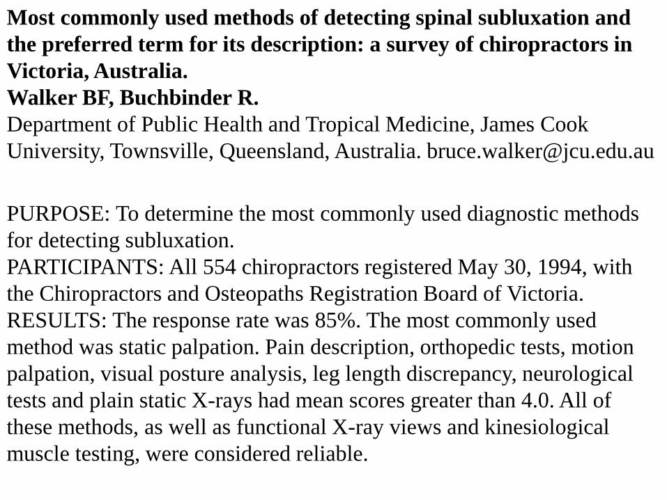
J Manipulative Physiol Ther. 1994 Oct;17(8):530-8. Optoelectric
measurement of changes in leg length inequality resulting from
isolation tests.
De Witt JK, Osterbauer PJ, Stelmach GE, Fuhr AW.
Exercise and Sport Research Institute, Arizona State University, Tempe
85287-0404.
OBJECTIVE: a) Establish a precise, standardized method to assess prone
leg alignment changes (functional "leg length inequality"), which have,
until now, been reported clinically to occur as a result putative
chiropractic subluxation isolation tests [neck flexion (C5) and extension
(C1)]; and b) describe differences in leg alignment changes in a group of
healthy subjects and patients with chronic spinal complaints.
CONCLUSIONS: 1) small leg displacements (< 1 mm) were recorded by
the optoelectric measurement system; 2) heel position changes during
isolation tests were identifiable; 3) as a result of head-up
maneuvers, patients exhibited more asymmetrical heel
movement than controls

Cooperstein R. The Derefield pelvic leg check: a kinesiological
interpretation. Chiropractic Technique 1991 MAY Vol. 3(2) Pgs. 60-
5.
The short leg observed in the legs-flexed position may result from
guarding responses instigated by stretch reflexes involving
asymmetry of quadriceps tone associated with pelvic
asymmetry.

J Manipulative Physiol Ther. 2003 Nov-Dec;26(9):557-66. Validity of
compressive leg checking in measuring artificial leg-length
inequality.
Cooperstein R, Morschhauser E, Lisi A, Nick TG.
Department of Technique, Palmer College of Chiropractic West
OBJECTIVE: To determine the accuracy of instrumented prone
compressive leg checking. DESIGN: Repeated measures (n = 26) on
single subjects (n = 3).
METHODS: A pair of surgical boots were modified to permit continuous
measurement of leg-length inequality (LLI). Multiple prone leg-check
observations of a blinded examiner on 3 subjects were tested against
artificial LLI that was created by randomly inserting 0 to 6 1.6-mm
shims in either boot.
CONCLUSION: Compressive leg checking seems highly
accurate, detecting artificial changes in leg length +/-1.87 mm, and
thus possesses concurrent validity assessed against artificial LLI. Pre-
leg-check and post-leg-check differences should exceed 3.74 mm to be
confident a real change has occurred.
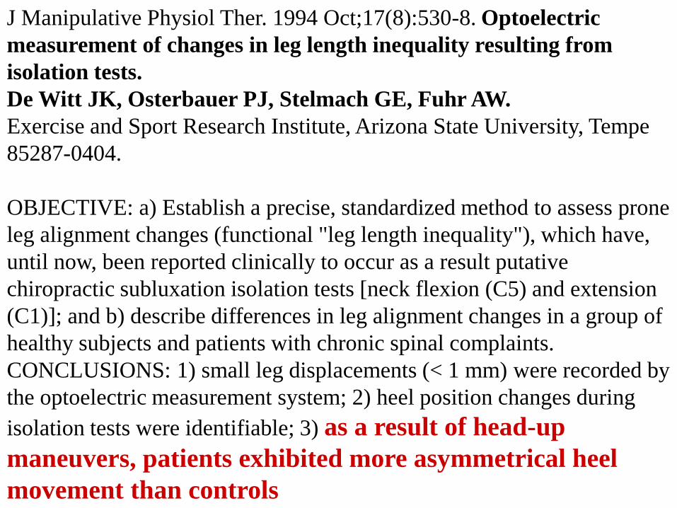
J Manipulative Physiol Ther. 2000 May;23(4):258-75. Are chiropractic
tests for the lumbo-pelvic spine reliable and valid? A systematic
critical literature review.
Hestbaek L, Leboeuf-Yde C.
Nordic Institute for Chiropractic and Clinical Biomechanics, Odense,
Denmark.
Only tests for palpation for pain had consistently acceptable results.
Motion palpation of the lumbar spine might be valid but showed poor
reliability, whereas motion palpation of the sacroiliac joints seemed to be
slightly reliable but was not shown to be valid. Measures of leg-length
inequality seemed to correlate with radiographic measurements but
consensus on method and interpretation is lacking. CONCLUSION:
The detection of the manipulative lesion in the lumbo-pelvic spine
depends on valid and reliable tests. Because such tests have not been
established, the presence of the manipulative lesion remains hypothetical.
Great effort is needed to develop, establish, and enforce valid and
reliable test procedures.

J Manipulative Physiol Ther 1988 Oct;11(5):396-9
Reliability of the Derifield-Thompson test for leg length inequality,
and use of the test to demonstrate cervical adjusting efficacy.
Shambaugh P, Sclafani L, Fanselow D.
New York Chiropractic College Research Division, Glen Head 11545.
Twenty-six subjects walked into five successive examining rooms where
a Derifield leg check was performed, including an estimate of the
millimeters of difference in leg lengths. The subjects then entered a
treatment room where they were randomly given no treatment, cervical
adjusting, or gluteal massage. This process continued for 5 cycles. This
study demonstrated that clinicians could reliably measure
a LLI to less than 3 mm (both inter- and intraobserver),
and also detect a change in LLI when the head was
rotated.

J Manipulative Physiol Ther 1999 Nov-Dec;22(9):565-9
Interexaminer reliability of activator methods' relative leg-
length evaluation in the prone extended position.
Nguyen HT, Resnick DN, Caldwell SG, Elston EW Jr,
Bishop BB, Steinhouser JB, Gimmillaro TJ, Keating JC Jr.
Los Angeles College of Chiropractic, Whittier, Calif, USA.
CONCLUSION: There was good reproducibility between 2
examiners by using the Activator Method to detect leg
length inequality in the prone extended position. This study
does not address the validity or clinical significance of the measurement
method. Future studies should include larger numbers, a wider variety of
subjects, and a diversity of examiners.

J Manipulative Physiol Ther 1992 Nov-Dec;15(9):576-90
Leg length inequality.
Mannello DM.
Research Division, Logan College of Chiropractic, Chesterfield, MO
63006-1065.
CONCLUSION: There appears to be a lack of agreement concerning incidence,
classification and point of clinical significance. However, the manifestations or
consequences of LLI demonstrate greater accordance. Of the three most
commonly utilized evaluation methods, radiographic measures such as the scanograms
are recognized as the most reliable procedure for the evaluation of anatomical LLI.
Much controversy exists with some of the clinical orthopedic methods and the visual
"quick" leg check. Because there is such a vast range in estimates of reliability, few if
any definitive conclusions can be made regarding these methods. Given this, it is
evident that more research is needed before the use of certain orthopedic and visual
checks are considered reliable and valid.

J Manipulative Physiol Ther 1989 Oct;12(5):364-8
Precise measurement of functional leg length inequality and changes
due to cervical spine rotation in pain-free students.
Falltrick DR, Pierson SD.
Research Department, Life Chiropractic College-West, San Lorenzo, CA
94580.
A series of blinded studies to determine the feasibility of documenting functional leg length
inequalities and changes in functional leg length in normal and non-normal patients were
performed.
Results indicated an absence of any significant effect of head rotation, type of table, galvanic
stimulation, or any difference between persons classified as cervically lesioned or not cervically
lesioned.
Failure to obtain subjects with frank pain as well as absence of an applied cephalad
pressure (as is performed clinically) during leg length evaluation were considered
as possible explanations for the failure to detect an effect of head rotation in the leg
lengths. Discussion addresses the need for sensitive leg length inequality
assessment techniques which eliminate subjectivity and contribute to decreased
error variances.

J Manipulative Physiol Ther 1989 Apr;12(2):93-7
Interexaminer reliability of an isolation test for the identification of
cervical subluxation.
Youngquist MW, Fuhr AW, Osterbauer PJ.
Activator Methods, Inc., Phoenix, AZ 85060-0317.
to determine whether prone leg length analysis in association with an
isolation test maneuver was reproducible.
The results indicate good reliability using this method of
analysis for putative upper cervical subluxation in this
patient population.

J Manipulative Physiol Ther 1983 Jun;6(2):61-6
Inter- and intra-examiner reliability of leg-length differential
measurement: a preliminary study.
DeBoer KF, Harmon RO Jr, Savoie S, Tuttle CD.
An inter- and intra-examiner reliability study was performed to validate a prone leg length-
differential test. Naive students (n = 40) were called, in random order, into three adjacent
examining rooms where three experienced chiropractic clinicians measured differential leg
lengths. Using standard placement a tape measure was read to the nearest mm to detect
inequalities at the shoe-sole interface. The leg length differences were recorded, for both the
straight and flexed legs prone positions, twice by each of the three clinicians. Intraclass
correlations were significant for the two independent readings for all three examiners, indicating
high reliability of the test. Good agreement among examiners was indicated as well by significant
intraclass correlation in two of the three possible examiner combinations. These results argue
strongly for the reality of the leg length inequality phenomenon and also that it can
be reliably measured.

J Manipulative Physiol Ther. 1987 Oct;10(5):232-8. Short leg
correction: a clinical trial of radiographic vs. non-radiographic
procedures.
Aspegren DD, Cox JM, Trier KK.
Low Back Pain Clinic, Fort Wayne, Indiana.
Visual leg length insufficiency detection and correction is compared with
established radiographic procedures on 41 consecutive patients
presenting to a chiropractic clinic with low back pain. It is commonly
accepted that the most accurate procedure of short leg demonstration is
the standing X ray. Visual correction, as described by Rene Cailliet, uses
three anatomical points of reference: a) iliac crest levelness, b) vertical
appraisal of the spine from the sacral base (the spine should be
perpendicular to the sacral base) and c) levelness of the posterosuperior
iliac spine (PSIS) dimples.
Continued on next slide:

Lifts of varying thickness were placed under the foot of the short leg in
both leg length corrective procedures. This study found that the
visual method of measurement did not differ significantly
from the X-ray method of measurement for leg length
insufficiency. Further, it was found that when comparing those in
which the visual measure was less than the X-ray measure and those in
which the visual measure was greater, there was a significant relationship
between visual and X-ray measures. Eta (eta 2) demonstrates that there is
a very strong relationship between visual and X-ray methods of
measurement. A review of the literature is presented regarding the
correlation of leg length insufficiency and musculoskeletal disorders, as
well as the discrepancy required to alter biomechanical properties of the
trunk and lower extremity.
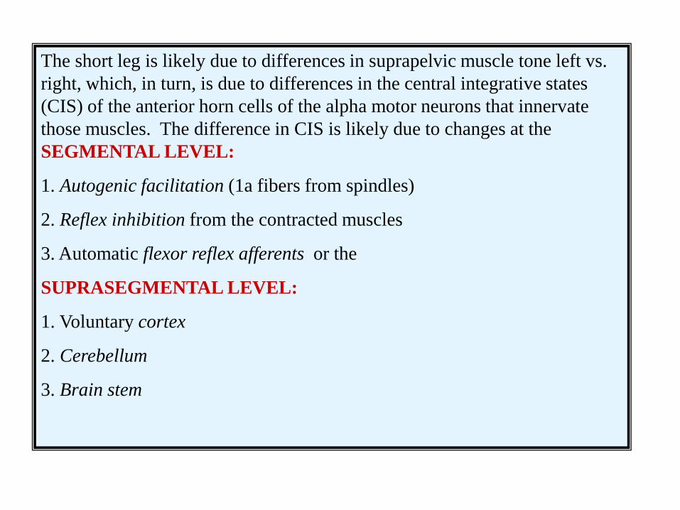
The short leg is likely due to differences in suprapelvic muscle tone left vs.
right, which, in turn, is due to differences in the central integrative states
(CIS) of the anterior horn cells of the alpha motor neurons that innervate
those muscles. The difference in CIS is likely due to changes at the
SEGMENTAL LEVEL:
1. Autogenic facilitation (1a fibers from spindles)
2. Reflex inhibition from the contracted muscles
3. Automatic flexor reflex afferents or the
SUPRASEGMENTAL LEVEL:
1. Voluntary cortex
2. Cerebellum
3. Brain stem

Leg Check Finding
Central Integrated State of AMNs
Segmental Level:
Autogenic Facilitation
Contralateral Inhibition
Flexor Reflex Afferents
Suprasegmental Levels:
Cortex
Cerebellum
Brain Stem

The Neck: Cervical Syndromes
Syndrome: a collection of findings

Why the cervicals before the
pelvis?
THOMPSON’S ORGANIZATION OF LEG-
LENGTH RELATED SPINAL SYNDROMES IS
ONLY USABLE IN THE ABSENCE OF
CERVICAL ROTATATION INTERFERENCE
WITH THE PRONE LEG CHECK.

Leg Check Equations
Prone Patient
Short Left Leg
Long Right Leg

Unilateral Cervical Syndrome
Look for trigger point on side opposite head turn (side of body
rotation). If no trigger point, think Atlas
Legs balance on head
rotation to one side
only

Cervical Syndrome
SCP- LPJ SSP- Zygomatic Arch LOC- L-M, P-A, I-S Rotate the head completely before adjusting. Maintain contact during rotation Chin is 1” above the bottom of the head piece
Cervical
Rotation
This adjustment can also be done from the head of the table, with
more of an S-I line of drive for the lower cervicals, to follow the
plane line of the disc and stay perpendicular to the facets.

Cervical Syndrome ATLAS
Palpation reveals no tender nodulations
If right rotation balances the legs, the listing is LP Atlas
If left rotation balances the legs, the listing is RP Atlas
Legs balance on
head rotation to one
side only
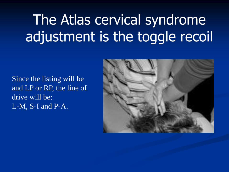
The Atlas cervical syndrome adjustment is the toggle recoil
Since the listing will be
and LP or RP, the line of
drive will be:
L-M, S-I and P-A.

Double Cervical Lock
Legs balance
on head
rotation to
both sides
Palpate for tender nodules bilaterally at different segments.
Adjust the more superior segment 1st, then recheck for a
unilateral cervical syndrome to adjust the more inferior
segment.

Cervical Syndrome- Posterior
Cervical
Legs balance
on head
rotation to
both sides
Palpation reveals tender nodulations on the right and left of the
same segment. X-ray analysis may confirm a military neck

Posterior Cervical Adjustment Double Thumb Method

Xception Derifield Cervical Syndrome
Cervical
Rotation
Cervical
Rotation

Xception Derifield Cervical Syndrome
Unilateral
Cervical
Rotation
Cervical
Rotation
If right cervical rotation, with the legs flexed, creates balanced legs,
you have a RXDCS.
If left cervical rotation, with the legs flexed, creates balanced legs,
you have a LXDCS.
Balanced legs extended.
Cervical rotation has no
effect.
Short leg appears on
flexion. Cervical rotation
balanced legs.

X-Derifield Cervical Syndrome vs.
X-Derifield Negative Derifield
XD CS
Cervical
Rotation
Cervical
Rotation =
= XD -D
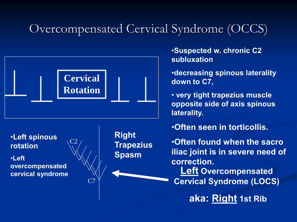
Overcompensated Cervical Syndrome (OCCS)
•Suspected w. chronic C2 subluxation
•decreasing spinous laterality down to C7,
• very tight trapezius muscle opposite side of axis spinous laterality.
•Often seen in torticollis.
•Often found when the sacro iliac joint is in severe need of correction.
Left Overcompensated Cervical Syndrome (LOCS)
aka: Right 1st Rib
Cervical
Rotation
Right Trapezius Spasm
C2
C7
•Left spinous rotation •Left overcompensated cervical syndrome

Over Compensated
Cervical Syndrome
SCP- 1st Rib Head SSP- Zygomatic Arch LOC- L-M, S-I, I-S Number of thrusts: 1 Laterally flex the head; mimic modified cervical set This move eliminates multiple cervical fixations. Effective for torticollis
Patient’s head is rotated fully to her left in this picture. (may be
impossible) Dr’s. stance is on the right (side of trapezius spasm).
Cervical Rotation
Side of Trapezius Spasm
C2
C7
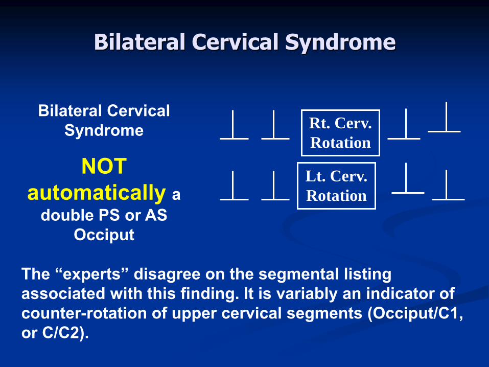
Bilateral Cervical Syndrome
Rt. Cerv.
Rotation
Lt. Cerv.
Rotation
Bilateral Cervical Syndrome
NOT automatically a
double PS or AS Occiput
The “experts” disagree on the segmental listing associated with this finding. It is variably an indicator of counter-rotation of upper cervical segments (Occiput/C1, or C/C2).

Bilateral Cervical Syndrome
Palpatory C2 Spinous Pain SCP- Inferior nuchal line CPs- Bilateral thenars LOC- A-P, I-S scoop with a flip to ceiling Number of thrusts: 1 Tuck the chin in Effective for removal of C1-Occiput fixations
Rt. Cerv. Rotation
Lt. Cerv. Rotation

Bilateral Cervical Syndrome: I-S with a flip

Positive and Negative Derifield

Negative Derifield (-D) The most common leg check
OR -D Left
The short leg in extension either remains short OR
balances out with the other leg in flexion.
With the associated trigger points, this finding suggests an
anterior &/or inferior ipsilateral sacrum.

-Derifield AI Sacrum Trigger Points (Ideally get at least 3 trigger points)
1. Ipsilateral achilles tendon
2. Medial tibial condyle (medial hamstrings)
3. Inferior medial ishial tuberosity
4. Medial and inferior to PSIS
5. Ipsi pubic tubercle
6. Contralateral T2-6 costo-transverse junction and mid-clavicular 1st or 2nd intercostal space pain anteriorly

Negative Derifield Trigger Points
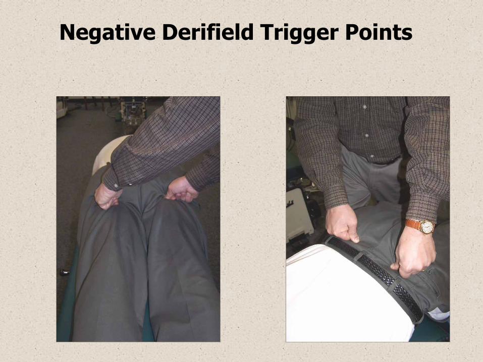
Negative Derifield Trigger Points

Negative D as AI Sacrum Parts 1 & 2
SCP- Ischial Tuberosity SSP- Roll in Toggle LOC- I-S, slight M-L Number of thrusts- 3-5
SCP-Medial to PSIS SSP- Top of knee LOC- Medial to Lateral Number of thrusts- 3-5
Think: Pro
Wrestling
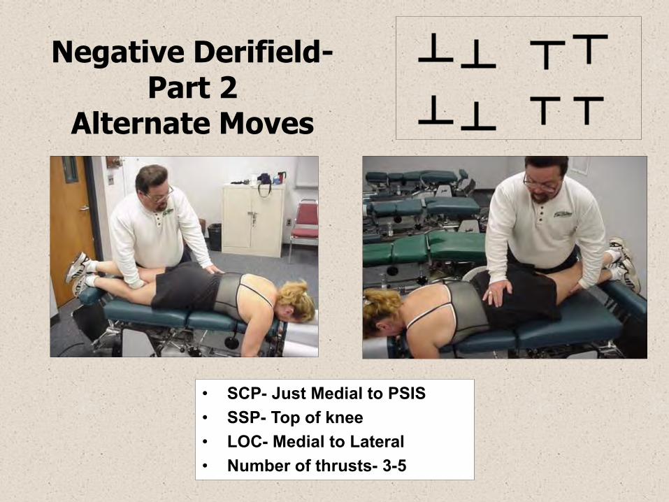
Negative Derifield- Part 2
Alternate Moves
• SCP- Just Medial to PSIS • SSP- Top of knee • LOC- Medial to Lateral • Number of thrusts- 3-5

Negative Derifield Supine(Preferred) Parts 1&2
SCP- Ischial Tuberosity SSP- ASIS LOC- I-S, M-L Number of thrusts- 3-5
SCP- Inguinal Ligament SSP- Top of Knee LOC- Fingers lateral, A-P Number of thrusts- 3-5
Think: Deliver the baby
Think: Torque

If the AI Trigger points are not found, check for:
• Posterior rocked ischium (usually ipsilateral)
• PI ilium (with leg lag on the involved side)
• Sacral apex left or right
• IN or EX
• L5 or L4

Positive Derifield (+D)
When combined with ipsilateral leg lag, this finding indicates a PI ilium.
+D on left

Positive Derifield
The Positive Derfield is often associated with the PI ilium. However, the PI ilium must exhibit the following triggers:
1. Insertion of sartorius on medial anterior knee
2. Palpatory pain on upper inguinal ligament
3. Resistence to flexion of the leg

PI Ilium Trigger Points:
Upper Inguinal Ligament (Sartorius Origin)
Medial Knee (Sartorius Insertion)
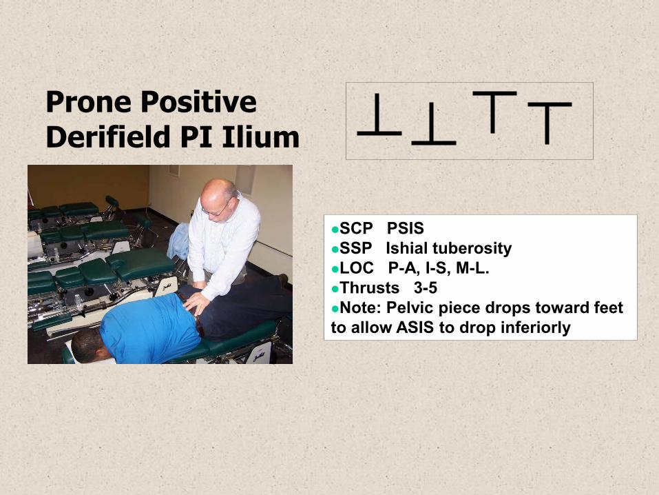
Prone Positive Derifield PI Ilium
SCP PSIS SSP Ishial tuberosity LOC P-A, I-S, M-L. Thrusts 3-5 Note: Pelvic piece drops toward feet to allow ASIS to drop inferiorly

Supine Positive Derifield
SCP Inguinal Ligament one inch above pubic tubercle SSP Roll in like toggle LOC A-P, S-I, L-M Thrusts 3-5 Use arm fossa test for exact SCP
This move helps correct anterior pelvic muscular dysfunction

A closer look at sacrum
SACRAL
APEX LEFT
BASE
RIGHT
INFERIOR
ON RIGHT
SUPERIOR
ON LEFT
Frontal plane sacral rotation

Testing for Sacral Rotation
Sacral Apex
Right
Sacral Apex
Left

Rotated Sacrum Test Results
Right leg is free to lift
higher
Left leg is free to lift
higher
Pt.
Prone

Rotated Sacrum Test Showing Sacral Apex Left (SAL)
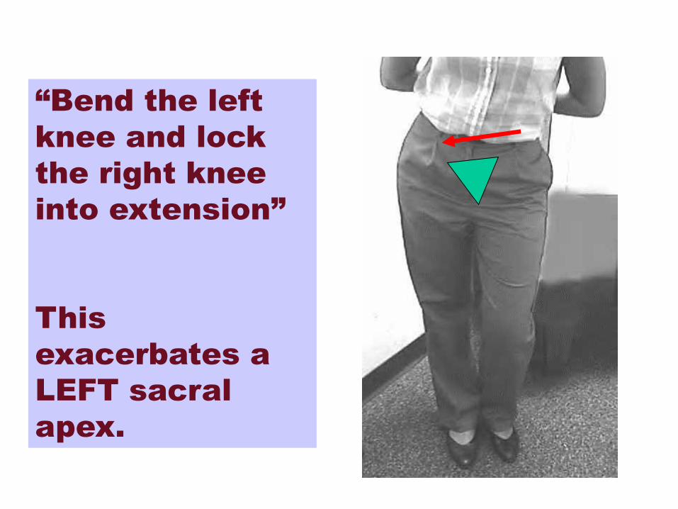
“Bend the left
knee and lock
the right knee
into extension”
This
exacerbates a
LEFT sacral
apex.

“Bend the right
knee and lock the
left knee into
extension”
This accentuates
a right sacral
apex.
SAR

Sacral Base Rotation (for any coronal plane sacral rotation affecting either
S-I joint or L5 disc)
SCPs- Sacral apex and medial PSIS LOC- Scissors Thrust (M-L, and L-M) Number of thrusts- 3-5 Cross Involved( lower) leg over higher leg Done on side of higher leg prone

Posterior Rocked Ischium
SCP Ishial Tuberosity SSP Roll in like toggle LOD P-A, S-I with a roll. Thrusts 3-5
Thompson Indicator:
tenderness at
gastrocnemius. Looks
like AS on film.
or

Opening the IN Closing the EX
Making
the EX
Worse
Making
the EX
Better

Testing for the IN ilium on the left

Testing for an EX on the left

Adjusting the IN Ilium
Thumb web at lateral posterior distal thigh
Stabilizing at the ASIS

Adjusting the EX
Stabilize