FACULTY OF VETERINARY MEDICINE BENHA UNIVERSITY
18
188 Pharmacokinetics and Tissue Residues of Tilmicosin in Normal and Experimentally Mycoplasma Gallisepticum-Infected Broiler Chickens Ashraf Abdelhakim Elkomy 1 , Nora Eltanany 1 , Mohamed Aboubakr 1 , Zeinab Roushdy Mohamed 2 and Mohamed Elbadawy 1 * 1 Department of Pharmacology, Faculty of Veterinary Medicine, Benha University, Moshtohor, Toukh, Elqaliobiya, 13736, Egypt. 2 Mycoplasma department, Animal Health Research Institute, Dokki, Giza, Egypt. *Corresponding author: Dr. Mohamed Elbadawy ([email protected]) A B S T R A C T The present study was conducted to investigate the pharmacokinetics of tilmicosin (25 mg/kg b.wt.) following single and repeated oral administration (once daily for three consecutive days) as well as tissue residues in normal and experimentally Mycoplasma gallisepticum-infected broiler chickens. Following single administration of tilmicosin in normal chickens, the drug reached its maximum serum concentrations (Cmax) after 2.45±0.01 h of administration with value of 0.97±0.04 μg/ml. Absorption half-life (t0.5ab) of tilmicosin was 0.89±0.02 h and the elimination half-life (t0.5el) was 14.73 ±1.24 h. The repeated oral administration of tilmicosin in normal and Mycoplasma gallisepticum- infected chickens revealed a lower significant serum tilmicosin concentration after all times of sampling in infected chickens compared to those of normal chickens. Tilmicosin residues was assayed in lung, kidney, liver, heart, breast muscle, thigh muscle, fat and skin after 24, 48, 72, 96, 120 and 144 h post last dose. The results showed that the highest residue values were recorded in lung followed by liver and kidneys while the lowest values were recorded in heart. Tilmicosin residues were not detected in all tested tissues except in lung till 5 th day after sessation of drug administration. However, all the tested tissues were free from tilmicosin residues after 5 th day after sessation of tilmicosin administration and this suggest a withdrawal period of five days for tilmicosin in broiler chickens. In conclusion, timicosin has rapid absorption, long elimination half-life, rapid and extensive penetration from blood into tissues especially lungs. Additionally, timicosin had a short withdrawal time. Keywords: Pharmacokinetics, tissue residues, tilmicosin, Broiler chickens. (http://www.bvmj.bu.edu.eg) (BVMJ-34(3): 188-205, 2018) BENHA VETERINARY MEDICAL JOURNAL, VOL. 34, NO. 3: 188-205, AUGUST 2018 A SPECIAL ISSUE FOR THE 6 TH SCIENTIFIC INTERNATIONAL CONFERENCE BENHA UNIVERSITY
Transcript of FACULTY OF VETERINARY MEDICINE BENHA UNIVERSITY
188
Pharmacokinetics and Tissue Residues of Tilmicosin in Normal and
Experimentally Mycoplasma Gallisepticum-Infected Broiler Chickens
Ashraf Abdelhakim Elkomy1, Nora Eltanany1, Mohamed Aboubakr1, Zeinab Roushdy
Mohamed2 and Mohamed Elbadawy1*
1Department of Pharmacology, Faculty of Veterinary Medicine, Benha University, Moshtohor,
Toukh, Elqaliobiya, 13736, Egypt. 2Mycoplasma department, Animal Health Research Institute, Dokki, Giza, Egypt.
*Corresponding author: Dr. Mohamed Elbadawy ([email protected])
A B S T R A C T
The present study was conducted to investigate the pharmacokinetics of tilmicosin (25 mg/kg
b.wt.) following single and repeated oral administration (once daily for three consecutive days) as
well as tissue residues in normal and experimentally Mycoplasma gallisepticum-infected broiler
chickens. Following single administration of tilmicosin in normal chickens, the drug reached its
maximum serum concentrations (Cmax) after 2.45±0.01 h of administration with value of 0.97±0.04
µg/ml. Absorption half-life (t0.5ab) of tilmicosin was 0.89±0.02 h and the elimination half-life (t0.5el)
was 14.73 ±1.24 h. The repeated oral administration of tilmicosin in normal and Mycoplasma
gallisepticum- infected chickens revealed a lower significant serum tilmicosin concentration after all
times of sampling in infected chickens compared to those of normal chickens. Tilmicosin residues
was assayed in lung, kidney, liver, heart, breast muscle, thigh muscle, fat and skin after 24, 48, 72,
96, 120 and 144 h post last dose. The results showed that the highest residue values were recorded in
lung followed by liver and kidneys while the lowest values were recorded in heart. Tilmicosin
residues were not detected in all tested tissues except in lung till 5th day after sessation of drug
administration. However, all the tested tissues were free from tilmicosin residues after 5th day after
sessation of tilmicosin administration and this suggest a withdrawal period of five days for tilmicosin
in broiler chickens. In conclusion, timicosin has rapid absorption, long elimination half-life, rapid
and extensive penetration from blood into tissues especially lungs. Additionally, timicosin had a
short withdrawal time.
Keywords: Pharmacokinetics, tissue residues, tilmicosin, Broiler chickens.
(http://www.bvmj.bu.edu.eg) (BVMJ-34(3): 188-205, 2018)
BENHA VETERINARY MEDICAL JOURNAL, VOL. 34, NO. 3: 188-205, AUGUST 2018
A SPECIAL ISSUE FOR THE 6TH SCIENTIFIC INTERNATIONAL CONFERENCE
BENHA UNIVERSITY FACULTY OF VETERINARY MEDICINE
Pharmacokinetics and Tissue Residues of Tilmicosin in Normal and Experimentally Mycoplasma Gallisepticum-Infected
Broiler Chickens
189
biya, 13736, Egypt. 2Mycoplasma department, Animal Health Research Institute, Dokki, Giza, Egypt.
*Corresponding author: Dr. Mohamed Elbadawy ([email protected])
A B S T R A C T
The present study was conducted to investigate the pharmacokinetics of tilmicosin (25 mg/kg
b.wt.) following single and repeated oral administration (once daily for three consecutive days) as
well as tissue residues in normal and experimentally Mycoplasma gallisepticum-infected broiler
chickens. Following single administration of tilmicosin in normal chickens, the drug reached its
maximum serum concentrations (Cmax) after 2.45±0.01 h of administration with value of 0.97±0.04
µg/ml. Absorption half-life (t0.5ab) of tilmicosin was 0.89±0.02 h and the elimination half-life (t0.5el)
was 14.73 ±1.24 h. The repeated oral administration of tilmicosin in normal and Mycoplasma
gallisepticum- infected chickens revealed a lower significant serum tilmicosin concentration after all
times of sampling in infected chickens compared to those of normal chickens. Tilmicosin residues
was assayed in lung, kidney, liver, heart, breast muscle, thigh muscle, fat and skin after 24, 48, 72,
96, 120 and 144 h post last dose. The results showed that the highest residue values were recorded in
lung followed by liver and kidneys while the lowest values were recorded in heart. Tilmicosin
residues were not detected in all tested tissues except in lung till 5th day after sessation of drug
administration. However, all the tested tissues were free from tilmicosin residues after 5th day after
sessation of tilmicosin administration and this suggest a withdrawal period of five days for tilmicosin
in broiler chickens. In conclusion, timicosin has rapid absorption, long elimination half-life, rapid
and extensive penetration from blood into tissues especially lungs. Additionally, timicosin had a
short withdrawal time.
Keywords: Pharmacokinetics, tissue residues, tilmicosin, Broiler chickens.
(http://www.bvmj.bu.edu.eg) (BVMJ-34(3): 188-205, 2018)
1.INTRODUCTION
Tilmicosin is a semisynthetic, broad-
spectrum, bacteriostatic macrolide antibiotic
with a wide range of veterinary uses. It shows
promising prospect of applications in clinical
veterinary field. Tilmicosin is a useful drug for
treatment and control of respiratory diseases
because of its large volume of distribution,
long half-life and preferential high
accumulation in lungs (Debono et al., 1989).
Tilmicosin is used for treatment and control of
respiratory diseases caused by Mycoplasma
gallisepticum, Mycoplasma synoviae,
Ornithobacterium rhinotracheale and
Pasteurella multocida in broilers (Jordan &
Horrocks, 1996; Kempf et al., 1997; EMEA
1998; Prescott 2000; Varga et al., 2001; Abu-
Basha et al., 2007). It has been also licensed
for combating respiratory diseases in pigs,
sheep and cattle (Moore et al., 1996; Hoar et
al., 1998; Christodoulopoulos et al., 2002).
Elkomy et al., (BVMJ-34(2): 188-205, 2018)
190
The pharmacokinetics after parenteral
administration of tilmicosin has been studied
in cow (Ziv et al., 1995; Modric et al., 1998),
goat (Ramadan, 1997), sheep (Modric et al.,
1998), and elk (Clark et al., 2004). However,
limited studies are available concerning the
disposition of tilmicosin following oral
administration to animals including fowl,
swine, broiler chicken and lactating goat
(Keles et al., 2001; Shen et al., 2005; Elsayed
et al., 2014; El-Komy et al., 2016).
Mycoplasma infection can create
inconvenience and economic damage and
continues to be an important cause of loss in
poultry production that should be taken into
consideration in the poultry industry, which is
often not apparent, and it silently damages the
effectiveness of investment (Mohammed et
al., 1987; Kleven, 1990). At the poultry
industry Mycoplasma gallisepticum (Mg) and
Mycoplasma Synovie (Ms) are of the greatest
importance (Mohammed et al., 1987 &
Kleven et al., 1997). Uses of recent anti-
mycoplasmal drugs either in the prophylaxis
and therapy is still recommended in the
eradication programs than application of
sanitary measures or usage of vaccine (Arzey
and Arzey, 1992; Jordan et al., 1999).
Tilmicosin has also been shown to be effective
for the treatment of mastitis in cattle and
sheep, pasteurellosis in rabbits, and
Mycoplasma gallisepticum infections in
chicken (McKay et al., 1996; Kempf et al.,
1997; Croft et al., 2000; Dingwell et al.,
2003).
Administering veterinary medications
to animals without an appropriate withdrawal
period may lead to violative residues in
tissues. Despite the extensive use of tilmicosin
in poultry industry, limited data is currently
available about the disposition and tissues
residues of tilmicosin in broiler chickens.
Therefore, the main purpose of the present
study was to investigate and provide an
overview of the pharmacokinetic profile as
well as tissue residues of tilmicosin in both
normal and experimentally Mycoplasma
gallisepticum-infected broiler chickens
following single and repeated oral
administration and to determine its withdrawal
time.
2.MATERIALS AND METHODS:
1. Drug
Tilmicosin phosphate was obtained as
an oral solution from ATCO Pharma for
Pharmaceutical Industries, Cairo, Egypt, with
a commercial name of tilmicoral®, a 25% oral
solution, 240 ml. Each 100 ml contain
tilmicosin phosphate 29.4 gm (Eq. to 25 gm
tilmicosin base) which applied orally by intra-
crop administration. Tilmicosin has the
chemical name of 20-Deoxo-20-(3, 5-
dimethyl-1-piperidinyl) desmycosin and
molecular weight of 869.15 g/mol and with
molecular formula of C46H80N2O13
(Debono et al., 1989). Tilmicosin is produced
by the removal of mycarose of tylosin A in an
organic solvent such as butyl acetate (Debono
et al., 1989).
2. Experimental Chickens
Forty-one clinically healthy Hubbard
chickens weighting about 1500 to 2000 g.
Birds were chosen randomly from poultry
farm in Qalubia government, Egypt. Chickens
were of both sexes. Chickens were maintained
at a suitable temperature and humidity
according to their ages. The chickens had a
free access to water and feed and the feed was
free from antibacterial drugs.
3. Experimental design
Chickens were divided into 3 groups,
group (1): five clinically healthy broiler
chickens were orally intra-crop administered a
Pharmacokinetics and Tissue Residues of Tilmicosin in Normal and Experimentally Mycoplasma Gallisepticum-Infected
Broiler Chickens
191
single dose of tilmicosin (25 mg/kg b.wt.).
Blood samples (1ml) were collected from the
brachial veins of each bird at 15 and 30 min.
and at 1, 2, 4, 6, 8, 12 and 24 h after dosing for
determination of tilmicosin concentration
using microbiological assay.
Group (2): eighteen clinically healthy
chickens were orally intra-crop administered
of 25 mg tilmicosin/kg b.wt. once daily for
three consecutive days, to determine
pharmacokinetics parameters and tissue
residues of tilmicosin in healthy chickens.
Group (3): eighteen experimentally
Mycoplasma gallisepticum-infected chickens
were orally intra-crop administered of 25 mg
tilmicosin /kg b.wt. once daily for three
consecutive days to determine
pharmacokinetics and tissue residues of
tilmicosin in infected chickens.
4. Experimental infection
Each chicken was experimentally
infected with 100 μl aliquots of PPLO broth
culture equal containing (1x10⁸ CFU/ml of a
pathogenic strain of M. gallisepticum which
was given by automatic micropipette/chick
intra-tracheal to induce infection according to
Belah et al., (2012). After appearance of the
respiratory clinical symptoms as dyspnea,
sneezing, nasal discharges, tracheal rales,
lacrimal discharge, conjunctivitis and lack of
appetite that beginning within 24 hours after
infection and become intensive at 3rd day post
infection with M. gallisepticum, each chicken
was orally intra-crop administered tilmicosin
at a dose rate of 25mg/kg b.wt. once daily for
three consecutive days. After that blood and
tissue samples were taken for assaying of
pharmacokinetics and residues.
5. Collection of samples
5.1. Blood samples
About one ml of blood was taken from
the brachial vein of five birds, following
administration of tilmicosin. Blood samples
were collected at 15 and 30 min. and at 1, 2, 4,
6, 8, 12 and 24 h after each dose for
determination of tilmicosin concentration
using microbiological assay. Blood samples
were collected in sterilized test tubes and
allowed to clot. Sera were separated by
centrifugation at 600 g for 15 min. Sera were
kept frozen at -20°C until assayed.
5.2. Tissue samples
For determination of tissue distribution
and residual contents of tilmicosin, the
microbiological assay technique (Petracca and
Wanner, 1993) was used. Three chickens were
randomly selected and slaughtered from group
(2) and group (3) at 24, 48, 72, 96, 120 and
144 h after the last administered dose of
tilmicosin. From each slaughtered chicken,
tissue samples of lung, heart, liver, kidney,
breast muscle, thigh muscle, fat and skin were
collected for assaying of residues of
tilmicosin. Samples were frozen at -20°C until
assayed.
6. Analytical procedure
Tilmicosin was assayed in chicken’s
serum and distilled water by microbiological
assay method using Bacillus subtilis ATCC
6633 as a test organism for tilmicosin (Arret et
al., 1971). The test organism was obtained
from microbiology department, Animal Health
Research Institute. Dokki, Giza, Egypt. Three
plates were used for each sample. A well in
each plate was filled with reference
concentration (1 µg/ml of tilmicosin in
distilled water or normal chicken's serum).
The plates were incubated at 37º C for 18-24 h
then the diameter of inhibitory zones was
measured. The average diameter of inhibition
zone of the samples was corrected by using
the diameter of the reference concentration.
Elkomy et al., (BVMJ-34(2): 188-205, 2018)
192
From the standard curve, the concentration
corresponding to the correct values of the zone
diameter were obtained.
For assay of tissue samples, two grams
of tissue were homogenized by automatic
homogenizer with 2 ml of distilled water.
Mixtures were centrifuged at 600 g for 10 min.
and supernatant fluid of each sample was
aspirated and directly assayed
microbiologically for tilmicosin concentration.
7. Pharmacokinetic analysis
The pharmacokinetic parameters were
calculated according to Baggot, (1978 a & b).
The pharmacokinetic analysis of data was
done using noncompartmental analysis based
on statistical moment theory as
described by Gibaldi & Perrier (1982).
8. Statistical analysis
The data were calculated as mean ±
standard error (S.E) of observation in PK and
residue analysis. All statistical analysis was
carried out according to (Snedecor and
Cochran, 1980).
Differences of P < 0.05 * Significant, P
< 0. 01 ** highly significant and P < 0.001
*** very highly significant.
3.RESULTS
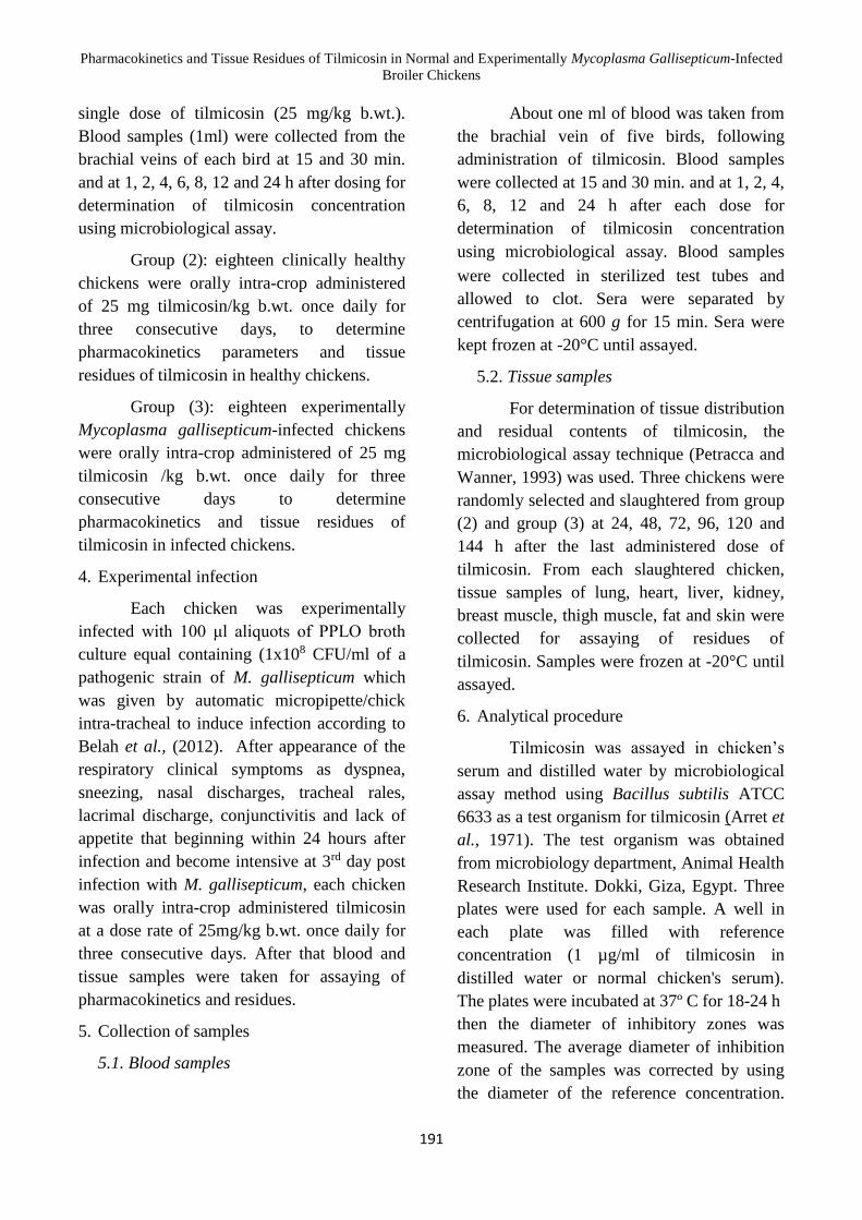
Time versus serum tilmicosin
concentrations following a single oral
administration of 25 mg tilmicosin/kg. b.wt.
were illustrated in table 1 and shown in figure
1. Tilmicosin concentration was firstly
detected in serum at 15 min. with a mean
value of 0.25 ± 0.020 and the drug reached its
maximum serum concentration of 1.23±0.062
µg/ml at about 2 h. The pharmacokinetic
parameters following a single oral
administration of tilmicosin were recorded in
table 2. Tilmicosin was rapidly absorbed after
its oral administration with an apparent first
order absorption rate constant (Kab) of 0.77 ±
0.019/h, while absorption half life time (t0.5ab)
was 0.89 ± 0.02 h. Tilmicosin was eliminated
at rate (Kel) equal to 0.048 ± 0.003/h and the
elimination half-life time (t0.5el) was
14.73±1.24 h. The mean residence time
(MRT) was 8.15 ± 0.15 h and the area under
the serum tilmicosin concentration curve
(AUC) was found to be 9.86 ± 0.59 µg•h/ml.
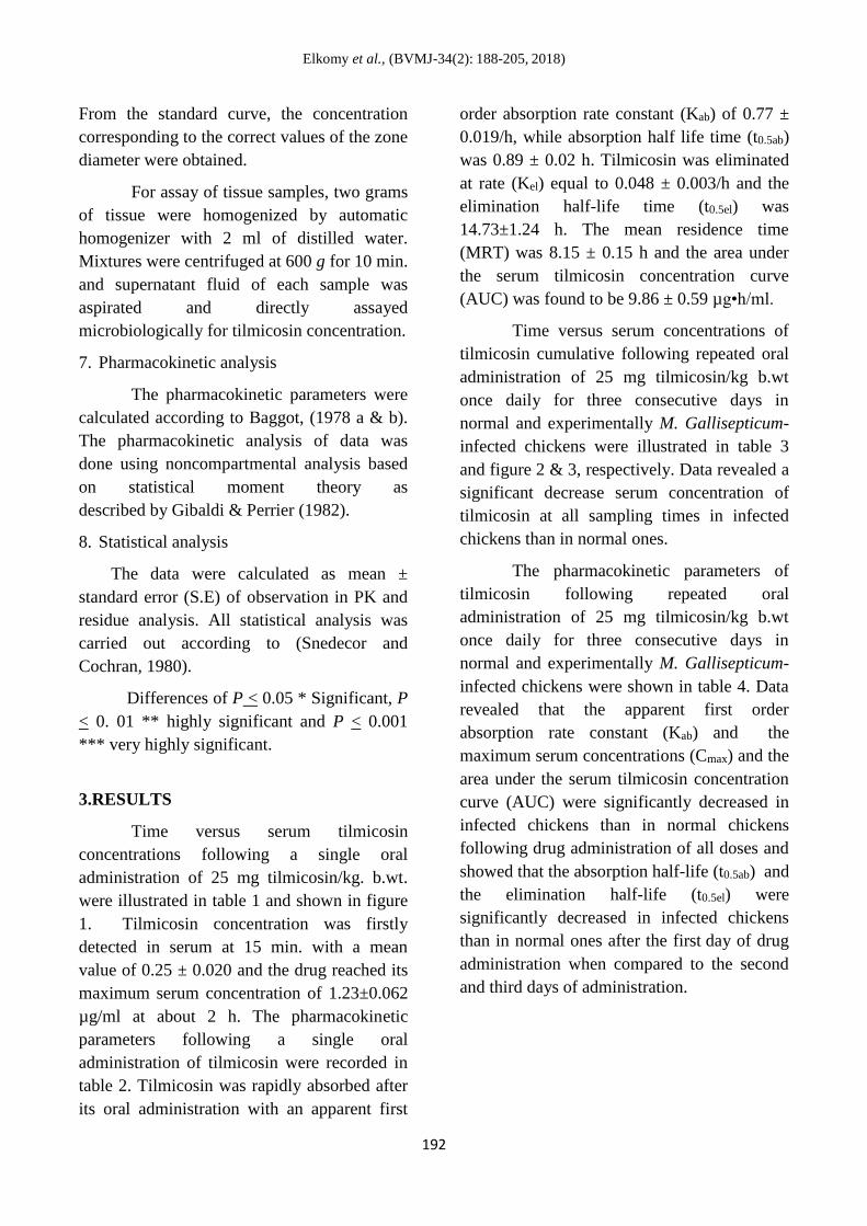
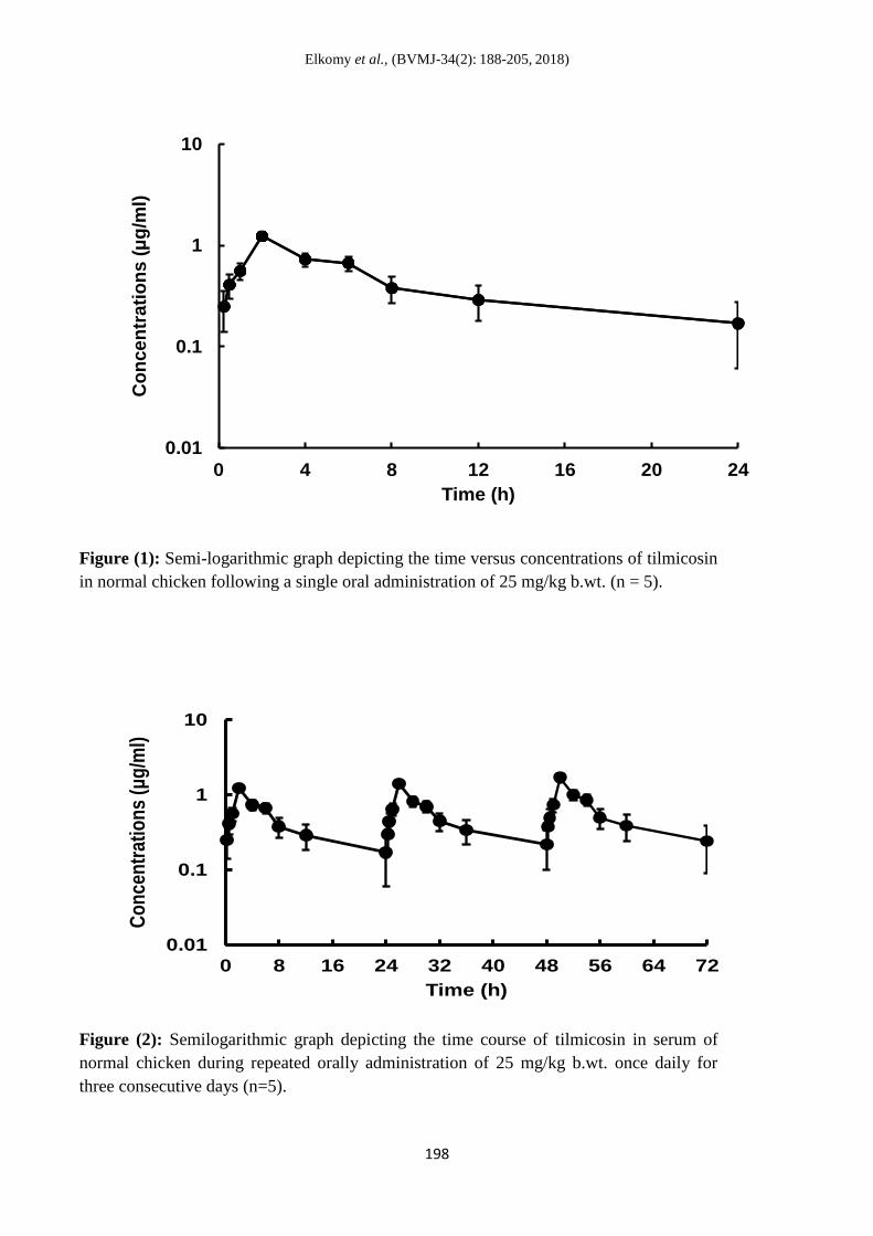
Time versus serum concentrations of
tilmicosin cumulative following repeated oral
administration of 25 mg tilmicosin/kg b.wt
once daily for three consecutive days in
normal and experimentally M. Gallisepticum-
infected chickens were illustrated in table 3
and figure 2 & 3, respectively. Data revealed a
significant decrease serum concentration of
tilmicosin at all sampling times in infected
chickens than in normal ones.
The pharmacokinetic parameters of
tilmicosin following repeated oral
administration of 25 mg tilmicosin/kg b.wt
once daily for three consecutive days in
normal and experimentally M. Gallisepticum-
infected chickens were shown in table 4. Data
revealed that the apparent first order
absorption rate constant (Kab) and the
maximum serum concentrations (Cmax) and the
area under the serum tilmicosin concentration
curve (AUC) were significantly decreased in
infected chickens than in normal chickens
following drug administration of all doses and
showed that the absorption half-life (t0.5ab) and
the elimination half-life (t0.5el) were
significantly decreased in infected chickens
than in normal ones after the first day of drug
administration when compared to the second
and third days of administration.

Pharmacokinetics and Tissue Residues of Tilmicosin in Normal and Experimentally Mycoplasma Gallisepticum-Infected
Broiler Chickens
193
Tissue concentrations of tilmicosin in
slaughtered normal and experimentally M.
Gallisepticum-infected chickens following the
repeated oral dosage regimen of 25 mg/kg
b.wt. once daily for three consecutive days,
were recorded in table 5. The data revealed a
significant decrease in tissue concentrations of
tilmicosin in experimentally M. Gallisepticum-
infected chickens than in normal ones. Lungs
had the highest concentrations of the drug
followed by liver and kidney, while the lowest
concentrations were determined in heart. This
suggests that lung should be the target tissue
for tilmicosin residues in broiler chickens.

Elkomy et al., (BVMJ-34(2): 188-205, 2018)
194
Table (1): Serum concentrations of tilmicosin (μg/ml) in normal chicken following
a single oral administration of 25 mg/kg b.wt. (n = 5).
Table (2): Pharmacokinetic parameters of tilmicosin (μg/ml) in normal chicken following a single oral
administration of 25 mg/kg b.wt. (n =5).
Parameter
Unit
Chicken’s number _
(X±S.E.) (1) (2) (3) (4) (5)
Kab h-1 0.79 0.77 0.82 0.79 0.70 0.77 ± 0.019
t0.5ab H 0.87 0.89 0.84 0.87 0.98 0.89 ± 0.02
Cmax μg/ml 0.96 0.86 1.01 1.11 0.90 0.97 ± 0.04
Tmax H 2.39 2.50 2.47 2.45 2.45 2.45 ± 0.01
Kel h-1 0.049 0.053 0.042 0.036 0.058 0.048 ± 0.003
t0.5el H 13.93 12.87 16.27 18.77 11.83 14.73 ± 1.24
AUC µg• h/ml 9.50 8.80 10.9 11.61 8.53 9.86 ± 0.59
AUMC µg• h/ml 76.34 70.81 92.21 98.8 65.75 80.78 ± 6.32
MRT H 8.03 8.04 8.48 8.50 7.70 8.15 ± 0.15
Time after
administration
(h)
Chicken’s number _
(X±S.E.) (1) (2) (3) (4) (5)
0.25 0.27 0.20 0.27 0.32 0.23 0.25 ± 0.020
0.5 0.43 0.38 0.42 0.48 0.37 0.41 ± 0.019
1 0.57 0.52 0.59 0.63 0.52 0.56 ± 0.021
2 1.22 1.06 1.32 1.42 1.16 1.23 ± 0.062
4 0.71 0.67 0.78 0.87 0.66 0.73 ± 0.039
6 0.66 0.65 0.71 0.75 0.61 0.67 ± 0.020
8 0.36 0.33 0.42 0.45 0.35 0.38 ± 0.022
12 0.28 0.27 0.34 0.34 0.23 0.29 ± 0.021
24 0.16 0.14 0.21 0.24 0.13 0.17 ± 0.021

Pharmacokinetics and Tissue Residues of Tilmicosin in Normal and Experimentally Mycoplasma
Gallisepticum-Infected Broiler Chickens
195
Table (3): Comparison of serum concentrations of tilmicosin (µg/ml) in normal (N) and experimentally
M. gallisepticum infected (I) broiler chickens during repeated oral administration of 25 mg/kg b.wt.
once daily for three consecutive days (n=5).
Days 1stday (1st dose) 2ndday (2nddose) 3rdday (3rd dose)
Time (h) N I N I N I
(X±S.E.) (X±S.E.) (X±S.E.) (X±S.E.) (X±S.E.) (X±S.E.)
0.25 0.25 ± 0.020 0.18 ± 0.01* 0.30 ± 0.02 0.21 ± 0.01** 0.38 ± 0.01 0.27±0.01 ***
0.5 0.41 ± 0.019 0.28 ± 0.02** 0.44 ± 0.03 0.32 ± 0.03* 0.50 ± 0.04 0.37 ± 0.02*
1 0.56 ± 0.021 0.39±0.02*** 0.64 ± 0.03 0.44 ± 0.04** 0.74 ± 0.06 0.52 ± 0.04*
2 1.23 ± 0.062 0.80±0.05*** 1.40± 0.07 1.10 ± 0.06* 1.70 ± 0.09 1.30 ± 0.08*
4 0.73 ± 0.039 0.51±0.04** 0.82 ± 0.08 0.78 ± 0.07 1.00 ± 0.08 0.90 ± 0.06
6 0.67 ± 0.020 0.42±0.02*** 0.70 ± 0.06 0.53 ± 0.05 0.86 ± 0.07 0.64 ± 0.04*
8 0.38 ± 0.022 0.34 ± 0.01 0.45 ± 0.03 0.40 ± 0.04 0.50 ± 0.07 0.46 ± 0.04
12 0.29 ± 0.021 0.26 ± 0.01 0.34 ± 0.02 0.29 ± 0.02 0.39± 0.03 0.35 ± 0.03
24 0.17 ± 0.021 0.14 ± 0.01 0.22± 0.02 0.20 ± 0.01 0.24 ± 0.01 0.22 ± 0.01
*→ Represent the significance in comparison with data of the normal group.
* P > 0.05 ** P > 0.01 *** P >0.001

Elkomy et al., (BVMJ-34(2): 188-205, 2018)
196
Table (4): Comparison of pharmacokinetic parameters of tilmicosinin normal (N) and experimentally M.
Gallisepticum infected (I) broiler chickens during repeated oral administration of 25 mg/kg b.wt. once daily for three
consecutive days (n=5).
*→ Represent the significance in comparison with data of the normal group.
Days 1stday (1stdose) 2ndday (2nddose) 3rdday (3rd dose)
Parameter N I N I N I
(unit) (X±S.E.) (X±S.E.) (X±S.E.) (X±S.E.) (X±S.E.) (X±S.E.)
Kab (h-1) 0.77 ± 0.019 0.70 ± 0.04 0.82 ± 0.02 0.70 ± 0.03* 0.73 ± 0.04 0.70 ± 0.03
T0.5 (ab) (h) 0.89 ± 0.02 0.68±0.03*** 0.84 ± 0.05 0.98 ± 0.06 0.94 ± 0.04 0.97 ± 0.04
Cmax (µg/ml) 0.97 ± 0.04 0.62±0.05 *** 1.07 ± 0.08 0.86±0.08 1.30 ± 0.09 1.01 ± 0.07 *
Tmax (h) 2.45 ± 0.01 2.44±0.09 2.41 ± 0.12 2.66±0.07 2.47 ± 0.15 2.65 ± 0.08
Kel (h-1) 0.048±0.003 0.054±0.002 0.042 ± 0.001 0.040±0.003 0.044 ± 0.002 0.044 ± 0.002
T0.5 ( el ) (h) 14.73 ± 1.24 12.7±1.06 16.20 ± 0.87 17.12 ± 0.73 15.53 ± 0.96 15.61 ± 0.82
AUC (µg•h/ml) 9.86 ± 0.59 7.44±0.61 * 11.25 ± 0.71 9.49 ± 0.63 13.16 ± 0.87 11.12 ± 0.64
AUMC (µg•h/ml) 80.78 ± 6.32 65.15±7.05 94.86 ± 8.42 82.54 ± 7.36 108.09 ± 8.07 95.57 ± 8.13
MRT (h) 8.15 ± 0.15 8.75 ± 0.31 8.43 ± 0.26 8.69 ± 0.38 8.20 ± 0.29 8.59 ± 0.27

Pharmacokinetics and Tissue Residues of Tilmicosin in Normal and Experimentally Mycoplasma Gallisepticum-Infected Broiler Chickens
197
* P > 0.05 ** P > 0.01 *** P > 0.001
Table (5): Comparison of tissue residue concentrations (μg/g) of tilmicosin following oral administration of 25 mg/kg.b.wt. once daily for three
consecutive days in normal (N) and experimentally M. Gallisepticum infected (I) chicken (n=3).
- Not detected
*→ Represent the significance in comparison with data of the normal group.
* P> 0.05 ** P> 0.01
*** P> 0.0
Time after the last dose (h)
24 48 72 96 120 144
Tissues N I N I N I N I N I N I
Kidney 4.53±0.12 3.88±0.17 * 3.12±0.21 2.65±0.11 1.67±0.07 1.32±0.07 ** 0.35±0.01 0.28 ± 0.01 ** - - - -
Lung 9.45±0.34 8.30±0.25 * 6.33±0.36 5.76±0.25 3.52±0.12 2.93±0.09 ** 1.96±0.07 1.75 ± 0.09 0.51 ± 0.02 0.45± 0.01* - -
Heart 4.24±0.17 3.41±0.16 ** 2.15±0.17 1.84±0.09 0.57±0.01 0.48±0.01 *** - - - - - -
Liver 5.32±0.16 4.56±0.14 ** 3.28±0.12 2.73±0.13 * 1.85±0.07 1.53±0.05 ** 0.42±0.02 0.33 ± 0.01 ** - - - -
Breast
muscle
- - - - - - - - - - - -
Thigh
muscle
- - - - - - - - - - - -
Fat - - - - - - - - - - - -
Skin - - - - - - - - - - - -
Elkomy et al., (BVMJ-34(2): 188-205, 2018)
198
Figure (1): Semi-logarithmic graph depicting the time versus concentrations of tilmicosin
in normal chicken following a single oral administration of 25 mg/kg b.wt. (n = 5).
Figure (2): Semilogarithmic graph depicting the time course of tilmicosin in serum of
normal chicken during repeated orally administration of 25 mg/kg b.wt. once daily for
three consecutive days (n=5).
0.01
0.1
1
10
0 4 8 12 16 20 24
Co
nc
en
tra
tio
ns
(μ
g/m
l)
Time (h)
0.01
0.1
1
10
0 8 16 24 32 40 48 56 64 72
Co
nce
ntr
atio
ns
(μg
/ml)
Time (h)

Pharmacokinetics and Tissue Residues of Tilmicosin in Normal and Experimentally Mycoplasma Gallisepticum-Infected
Broiler Chickens
199
Figure (3): Semilogarithmic graph depicting the time course of tilmicosin in serum of
experimentally Mycoplasma gallisepticum-infected chicken during repeated orally
administration of 25 mg/kg b.wt. once daily for three consecutive days (n=5).
4.DISCUSSION
Pharmacokinetics of tilmicosin after
intravenous administration were limited and
unsuccessful due to its considerable
cardiovascular adverse effects and deaths
(Main et al., 1996, Papich & Riviere 2001;
Abu-Basha et al., 2007).
In the present investigation, the
pharmacokinetic properties of tilmicosin are
similar to those of macrolides in general and
characterized by low serum concentrations and
large volumes of distribution, with
accumulation and persistence in many tissues
particularly lungs, which may concentrate the
drug 20- 60-fold compared to serum (Ziv et al.,
1995; Scorneaux and Shryock, 1999; Clark et
al., 2004). Intracellular concentrations have
been shown to be 40 times greater than that of
serum (Ziv et al., 1995; Scorneaux and
Shryock, 1999).
In the present study, following a single
oral administration of 25 mg/kg b.wt. of
timicosin, the drug reached its maximum
serum concentrations (Cmax) after 2.45±0.01 h
of administration with value of 0.97±0.04
µg/ml. Tilmicosin could be detected in serum
in a therapeutic level of 0.17 ± 0.021 µg/ml for
24 h after administration. The obtained result
of Cmax were consistent with those recorded for
tilmicosin in calf (0.976 ± 0.06 μg/ml;
Dimitrova et al., 2012a), in cows (0.86 ± 0.20
μg/ml; Avci and Elmas, 2014) and in lactating
goat (1.07 ± 0.052 μg/ml; El-Komy et al.,
2016). In contrast, the reported Cmax in the
present study were lower than those recorded
for tilmicosin in goat (1.56 ug/ml; Ramadan,
0.01
0.1
1
10
0 8 16 24 32 40 48 56 64 72
Co
nc
en
tra
tio
ns (μ
g/m
l)
Time (h)

Elkomy et al., (BVMJ-34(2): 188-205, 2018)
200
1997), tilmicosin in sheep (1.19 ± 0.30 μg/ml;
Atef et al., 1999), tilmicosin in fowl (1.28 ±
0.04 μg/ml; Keles et al., 2001), tilmicosin in
swine ( 2.03 ± 0.28 μg/ml; Shen et al., 2005),
tilmicosin in broiler chickens (2.09 ± 0.37 and
2.12 ± 0.40 μg/ml for Pulmotil® AC and
Provitil®, respectively; Abu-Basha et al.,
2007), tilmicosin in rabbit (1.31 μg/ml; Gallina
et al., 2010) and tilmicosin in broiler chicken
(1.25 ± 0.092 µg/ml; Elbadawy and Aboubakr,
2017). On the other hand, the obtained result of
Cmax was higher than those recorded for
tilmicosin in adult sheep (0.44 µg/ml;
Cochrane and Thomson, 1990) and for
tilmicosin oral solution in broiler chickens
(0.583 ± 0.03 µg/ml; Dimitrova et al., 2012b).
In the current study, tilmicosin reached
its maximum serum concentration (Tmax) at
about (2.45 ± 0.01 h). This result was shorter
than that reported for tilmicosin in goat (6.39
h; Ramadan, 1997), tilmicosin in sheep (3.9 h;
Modric et al., 1998), tilmicosin in broiler
chicken (3.99± 0.84, 5,82 ± 1.04 h; Abu-Basha
et al., 2007) and tilmicosin in broiler chickens
(3 h; Dimitrova et al., 2012b) and higher than
those reported for tilmicosin in in cattle (0.5 h;
Modric et al., 1998), tilmicosin in calf (1h;
Dimitrova et al., 2012a). tilmicosin in cow (1h;
Avci and Elmas, 2014) and tilmicosin in
lactating goat (1.91 ± 0.19h and 1.46 ± 0.243h;
for healthy and vaccinated ones, respectively
El-Komy et al., 2016).
In the present work, following repeated
oral administrations of tilmicosin, the obtained
serum levels of tilmicosin in Mycoplasma
Gallisepticum-infected broiler chickens were
significantly lower than those in normal
healthy ones. These lower serum
concentrations of tilmicosin in experimentally
Mycoplasma Gallisepticum-infected broiler
chickens might be attributed the
infection/inflammation further improves its
tissue penetration (Modric et al., 1999) and
was similar to data recorded by (Baggot, 1980;
Naccari et al., 2001; Abo El-Ela et al., 2015;
El-Komy et al., 2016).
The obtained results illustrated a
significant decrease in the maximum serum
concentration (Cmax) in Mycoplasma
Gallisepticum-infected broiler chickens than in
normal broiler chickens following all doses.
These results were similar with El-Komy et al.,
(2016) who found a significant decrease in
Cmax in experimentally Pasterulla Multocida-
infected lactating goats than in healthy
lactating goats following a single subcutaneous
injection of 10 mg tilmicosin/kg b.wt. These
existing differences are relatively common and
are frequently related to or attributed to inter-
species variation, used assay methods, dose of
drug, chemical form of drug, amount of time
between blood sampling and/or the health
status, live body weight, age of the animal,
climatic or other conditions related to
experimental designs (Haddad et al., 1985).
In this experiment, the obtained results
of serum and tissue residues of tilmicosin in
slaughtered chickens following its repeated
oral administrations of 25 mg/kg b.wt once
daily for three consecutive days revealed a
good distribution of tilmicosin in serum and in
other tested tissues (lung, liver, kidney and
heart). Tilmicosin appeared to be retained at
higher concentrations and for longer times in
the edible tissue than in serum. The
concentrations of tilmicosin were high in tested
tissues 24 h after stopping drug administration,
then decreased slowly over time and tilmicosin
residues were only detected in the lung till 5th
day after sessation of tilmicosin administration.
Lung had the highest concentration of
tilmicosin followed by liver and kidney, whie
the lowest concentration was determined in
heart. This suggests that lung should be the
target tissue for tilmicosin residues in broiler
chickens.
Pharmacokinetics and Tissue Residues of Tilmicosin in Normal and Experimentally Mycoplasma Gallisepticum-Infected
Broiler Chickens
201
Similar findings were previously
reported for tilmicosin in fowl (Keles et al.,
2001), tilmicosin in broiler chickens (Zhang et
al., 2004) and tilmicosin in broiler chickens
Elsayed et al., 2014).
The concentration of tilmicosin in rat’s
lungs was higher than the serum tilmicosin at
all tested times and rats infected with
Mycoplasma pulmonis had higher lung
tilmicosin concentration than non-infected
ones (Modric et al., 1999). This phenomenon
was also seen in lung tissues of chickens,
swine and cattle (Scorneaux and Shryock,
1998a, b, 1999). The high success rate of
treatment is due to the prolonged presence of
therapeutic concentrations of tilmicosin in the
lung tissues (Papich and Riviere, 2001).
Using the microbiological assay
technique, tilmicosin could not be detected in
all tested tissues except in lung on the 5th day
post last oral administration. In particular the
high clearance of tilmicosin indicated the
reduced possibility of finding residues of
antimicrobial in broiler chickens a few days
after treatment and necessity of shorter
withdrawal time for this antimicrobial. The
withdrawal period in this study was shown to
be five days. The obtained results were similar
to those recorded after oral administration of
tilmicosin (4 days) in broiler chickens at 25
mg/kg b.wt. for 5 days (Elsayed et al., 2014)
and after oral administration of tilmicosin in
broiler chickens at 25 mg/kg b.wt. once daily
for 5 days, withdrawal period of 6 days
(Elbadawy and Aboubakr, 2017). On the other
hand, the obtained result was shorter than that
recorded after oral administration of tilmicosin
to broiler chicken at 37.5 and 75.0 mg/l for 5
days, a pre-slaughter withdrawal time of more
than 9 days is needed to ensure that the drug is
eliminated from the tissues (Zhang et al, 2004).
5.CONCLUSION
Serum concentration of tilmicosin in
normal and Mycoplasma gallisepticum-
infected broiler chickens could be detected in a
therapeutic level for 24 h following oral
administration, exceed MIC of tilmicosin for
Mycoplasma gallisepticum. Tilmicosin was
rapidly absorbed and slowly eliminated after
oral administration in broiler chicken.The
highest concentration of tilmicosin was in lung
tissue, suggesting that tilmicosin is suitable for
treatment of respiratory infection in broiler
chickens. The high concentration of tilmicosin
in kidney tissue, suggest that tilmicosin was
also suitable for treatment of urinary infection
in broiler chickens. Tilmicosin withdrawal
period of 5 days should be adopted.
6.REFERENCES
Abo El-Ela, F.I., El-Banna, H.A., Manal, B.
El-Deen, El-Gendy A.A. and Tohamy
M.A. 2015. Pharmacokinetics of
Tylvalosin Alone or in Combination
with Vitamin E in Broiler Chickens.
As. J. Ani. & Vet. Adv., 10: 556-566.
Abu-Basha, E.A., Idkaidek, N.M. and Al-
Shunnaq, A.F. 2007. Pharmacokinetics
of tilmicosin (Provitil powder and
Pulmotil liquid AC) oral formulations
in chickens. Vet. Res. Commun.; 31(4):
477-485.
Arret, B.,Johnson, D.P. and Kirshboum, A.
1971. Outline of details for
microbiological assay of antibiotics:
2nd revision. J. Pharm. Sci., 60(11):
1689-1694.
Arzey, G.G. and Arzey, K.E. 1992. Successful
treatment of mycoplasmosis in layer
chickens with single dose therapy.
Aust. Vet. J., 69: 126-128.

Elkomy et al., (BVMJ-34(2): 188-205, 2018)
202
Atef, M., Abo el-Sooud, K., Nahed, E. and
Tawfik, M. 1999. Elimination of
tilmicosin in lactating ewes. Dtsch.
Tierarztl. Wochenschr., 106 (7): 291-
294.
Avci, T. and Elmas, M. 2014. Milk and blood
pharmacokinetics of tylosin and tilmicosin
following parenteral administrations to cows.
Sci. World J., vol. 2014, pp: 1-6.
Baggot, J.D. 1978a. Some aspects of clinical
pharmacokinetics in veterinary
medicine. J. Vet. Pharmacol. Ther., 1:
5-18.
Baggot J.D. 1978b. Some aspects of clinical
pharmacokinetics in veterinary
medicine II. J. Vet. Pharmacol. Ther.,
II: 111-118.
Baggot, J.D. 1980. Distribution of
antimicrobial agents in normal and
diseased animals. J. Am. Vet. Med.
Ass., 19(76): 1085-1090.
Belah, S. S., El- Gebaly, L.S., Zoghbi, A.F.
and Galal, Z.M. 2012. Influence of
tilmicosin on some haematological,
biochemical and immunological
parameters in broilers experimentally
infected with mycoplasma. Egypt. J.
Agric. Res., 90(1): 17- 26.
Christodoulopoulos, G., Warnick, L.D.,
Papaioannou, N. and Fthenakis, G.C.
2002. Tilmicosin administration to
young lambs with respiratory infection:
safety and efficacy considerations. J.
Vet. Pharmacol. Ther.,25(5): 393-397.
Clark, C., Woodbury, M., Dowling, P., Ross,
S. and Boison, J.O. 2004. A
preliminary investigation of the
disposition of tilmicosin residues in elk
tissues and serum. J. Vet. Pharmacol.
Ther., 27(5): 385-387.
Cochrane, R.L. and Thomson, T.D. 1990.
Toxicology and pharmacology of
tilmicosin following administration of
subcutaneous and intravenous
injections to sheep. Lilly Research
Study Report T5C768908 (Eli Lilly,
Pers. Comm.: Tilmicosin Injection.
Application to Extend License to
Include Sheep).
Croft, A., Duffield, T., Menzies, P., Leslie, K.,
Bagg, R. and Dick, P. 2000. The effect
of tilmicosin administered to ewes prior
to lambing on incidence of clinical
Mastitis and Subsequent Lamb
Performance. Can. Vet. J., 41(4): 306-
311.
Debono, M., Willard, K.E., Kirst, H.A., Wind,
J.A., Crouse, G.D., Tao, E.V., Vicenzi,
J.T., Counter, F.T., Ott, J.L. and Ose,
E.E. 1989. Synthesis and antimicrobial
evaluation of 20-deoxo-20-(3,5-
dimethylpiperidin-1- yl) desmycosin
(tilmicosin, EL-870) and related cyclic
amino derivatives. J. Antibiot. (Tokyo),
42 (8): 1253-1267.
Dimitrova, D., Georgiev, K. and Tsoneva, D.
2012b. Pharmacokinetics of tilmicosin
in broiler chickens after single oral
application. Institute of Mountain
Animal Stockbreeding and Agriculture,
Troyan Bulgaria., 15(1): 1-12.
Dimitrova, D., Petkov, P., and Tsoneva, D.
2012a. Pharmacokinetics of tilmicosin
in calves after single subcutaneous
application. Agri. Sci. & technol., 4(3):
211-214.
Dingwell, R.T., Leslie, K.E., Duffield, T.F.,
Schukken, Y.H., DesCoteaux, L.,
Keefe, G.P., Kelton, D.F., Lissemore,
K.D., Shewfelt, W., Dick, P. and Bagg,
R. 2003. Efficacy of intramammary
tilmicosin and risk factors for cure of
Pharmacokinetics and Tissue Residues of Tilmicosin in Normal and Experimentally Mycoplasma Gallisepticum-Infected
Broiler Chickens
203
Staphylococcus aureus infection in the
dry period. J. Dairy Sci., 86(1): 159-
168.
Elbadawy, M. and Aboubakr, M. 2017.
Pharmacokinetics, tissue residues of
tilmicosin phosphate (tilmicoral®) and
it's in vitro and in vivo evaluation for
the control of Mycoplasma
gallisepticum infection in broiler
chickens. Int. J. Pharmacol. Toxicol., 5
(1), 11-16.
El-Komy, A. Abd El-Hakim A., El Sayed, M.
Gamal El-Din A., Mobarez, E. A.,
Azoz, H.A. and Afify, A.E. 2016.
Pharmacokinetics of tilmicosin in
healthy and experimentally Pastreulla
Multocida Infected Lactating Goats.
WJPPS., 5(6): 2429-2438.
Elsayed, M., Elkomy, A., Aboubakr, M. and
Morad, M. 2014. Tissue residues,
hematological and biochemical effects
of tilmicosin in broiler chicken. Vet.
Med. Int., Vol. 2014, pp. 1-6.
EMEA. 1998. (The European Agency for the
Evaluation of Medicinal Products
Veterinary Medicines Evaluation Unit)
Committee for veterinary medicinal
products. Tilmicosin (extension to
chicken), summary report 2.
Gallina, G., Lucatello, L., Drigo, I., Cocchi,
M., Scandurra, S., Agnoletti, F. and
Montesissa, C. 2010. Kinetics and
intrapulmonary disposition of
tilmicosin after single and repeated oral
bolus administrations to rabbits. Vet.
Res.; 34(1): S69-72.
Gibaldi, M. & Perrier, D. 1982.
Noncompartmental Method Based on
Statistical Moment In:
Pharmacokinetics. 2nd ed, Marcel
Dekker, NY, Vol. 15, pp: 409-410.
Haddad, N.S., Pedersol, J.P., Ravis, W.R.,
Fazel, M.H. and Carson, R.L. 1985.
Pharmacokinetics of gentamicin at
steady-state in ponies, serum, urine and
endometrial concentration. Am. J. Vet.
Res., 46(6): 1268-1271.
Hoar, B.R., Jelinski, M.D., Ribble, C.S.,
Janzen E.D. and Johnson, J.C. 1998. A
comparison of the clinical field efficacy
and safety of florfenicol and tilmicosin
for the treatment of undifferentiated
bovine respiratory disease of cattle in
western Canada. Can. Vet. J., 39(3):
161-166.
Jordan, F.T.W., Forrester, C.A., Hodge A. and
Reeve-Johonson, L.G. 1999. The
comparison of an aqueous preparation
of tilmicosin with tylosin in the
treatment of Mycoplasma gallisepticum
infection of turkey poults. Avian Dis.,
43(3): 521-525.
Jordan, F.T. and Horrocks, B.K. 1996. The
minimum inhibitory concentration of
tilmicosin and tylosin for Mycoplasma
gallisepticum and Mycoplasma
synoviae and a comparison of their
efficacy in the control of Mycoplasma
gallisepticum infection in broiler
chicks. Avian Dis.; 40 (2):326-334.
Keles, O., Bakirel, T. and Sener, S. 2001.
Pharmacokinetics and tissue levels of
tilmicosin in fowls. Turk. J. Vet. Anim.
Sci., 25(4): 629-634.
Kempf, I., Reeve-Johnson, L., Gesbert, F. and
Guittet, M. 1997. Efficacy of tilmicosin
in the control of experimental
Mycoplasma gallisepticum infection in
chickens. Avian Dis.; 41 (4): 802-807.
Kleven, S.H. 1990. Summary of discussion of
avian mycoplasma team, IRPCM, IOM.
Avian Pathol., 19: 795-800.

Elkomy et al., (BVMJ-34(2): 188-205, 2018)
204
Kleven, S.H., Rowland, G.N. and Olson, N.O.
1997. Mycolpasma synoviae infection.
In: Diseases of poultry, 10th Edn., Iowa
State University Pess. Ames, IA., pp:
220-228.
Main, B.W., Means, J.R., Rinkema, L.E.,
Smith, W.C. and Sarazan, R.D. 1996.
Cardiovascular effects of the macrolide
antibiotic tilmicosin, administered
alone and in combination with
propranolol or dobutamine, in
conscious unrestrained dogs. J. Vet.
Pharmacol. Ther., 19: 225–232.
McKay, S.G., Morck, D.W., Merrill, J.K.,
Olson, M.E., Chan, S.C. and Pap, K.M.
1996. Use of tilmicosin for treatment of
pasteurellosis in rabbits. Am. J. Vet.
Res.;57(8):1180-1184.
Modric, S., Webb, A.I. and Davidson, M.
1999. Effect of respiratory tract disease
on pharmacokinetics of tilmicosin in
rats. Lab. Anim. Sci.; 49(3):248-253.
Modric, S., Webb, A.I. and Derendorf, H.
(1998): Pharmacokinetics and
pharmacodynamics of tilmicosin in
sheep and cattle. J. Vet. Pharmacol.
Ther., 21(6): 444-452.
Mohammed, H.O., Carpenter, T.E. and
Yamamoto, R. 1987. Economic impact
of Mycoplasma Gallisepticum and
Mycoplasma synoviae in commercial
layer flocks. Avian Dis., 31:477-482.
Moore, G.M., Basson, R.P. and Tonkinson,
L.V. 1996. Clinical field trials with
tilmicosin phosphate in feed for the
control of naturally acquired
pneumonia caused by Actinobacillus
pleuropneumoniae and Pasteurella
multocida in swine. Am. J. Vet. Res.,
57: 224-228.
Naccari, F., Giofrè, F., Pellegrino, M., Calò,
M., Licata, P. and Carli, S. 2001.
Effectiveness and kinetic behaviour of
tilmicosin in the treatment of
respiratory infections in sheep. Vet.
Rec., 148 (25):773-786.
Omija, B., Mittema, E.S. and Maitho, T.E.
1994. Oxytetracycline Residue Levels
in Chicken Eggs after Oral
Administration of Medicated Drinking
Water to Laying Hens. Food Additives
and Contaminants, 11:641-647.
Papich, M.G. and Riviere, J.E. 2001.
Chloramphenicol and derivatives,
macrolides, lincosamides and
miscellaneous antimicrobials. J. Vet.
Pharmacol. Ther., 8th ed, (Iowa State
Press, Ames, USA), 880-881.
Petracca, K. and Wanner, M. 1993. The effect
of pregnancy and lactation in sows on
the pharmacokinetics of the gyrase
inhibitor marbofloxacin. Schweiz.
Arch. Tierheilkd., 135(10): 298-304.
Prescott J.F. 2000. Lincosamides, macrolides,
and pleuromutilins. In: Antimicrobial
therapy in veterinary medicine, 3rd ed.
(Iowa State University Press,Ames,IA),
pp. 229-262.
Ramadan, A. 1997. Pharmacokinetics of
tilmicosin in serum and milk of goats.
Res. Vet. Sci., 62 (1):48-50.
Roudaut, B., Moretain, J.P. and Biosseau, J.
1987. Excretion of Oxytetracycline in
Egg after Medication of Laying Hens.
Food Additives and Contaminants,
4:297-307.
Roudaut, B. and Moretain, J.P. 1990. Residues
of Macrolide Antibiotics in Eggs
Following Medication of Laying Hens.
Bri. Poult. Sci., 31: 661-675.

Pharmacokinetics and Tissue Residues of Tilmicosin in Normal and Experimentally Mycoplasma Gallisepticum-Infected
Broiler Chickens
205
Scorneaux, B. and Shryock, T.R. (1998a):
Intracellular accumulation, subcellular
distribution, and efflux of tilmicosin in
chicken phagocytes. Poult. Sci.; 77
(10):1510-1521.
Scorneaux, B. and Shryock, T.R. 1998b.
Intracellular accumulation, subcellular
distribution and efflux of tilmicosin in
swine phagocytes. J. Vet. Pharmacol.
Ther., 21: 257-268.
Scorneaux, B. and Shryock, T.R. 1999.
Intracellular accumulation, subcellular
distribution, and efflux of tilmicosin in
bovine mammary, blood, and lung
cells. J. Dairy‚ Sci.; 82 (6):1202-1212.
Shen, J., Li, C., Jiang, H., Zhang, S., Guo, P.,
Ding, S. and Li, X. 2005.
Pharmacokinetics of tilmicosin after
oral administration in swine. Am. J.
Vet. Res.; 66(6):1071-1074.
Snedecor, G.W. and Cochran, W.G. 1980.
Statistical Methods. 7th Edition, Iowa
State University Press, Ames, IA; 39-
63.
Varga, J., Fodor, L. and Makrai,L. 2001.
Characterisation of some
Ornithobacterium rhinotracheale strains
and examination of their transmission
via eggs. Acta Vet. Hungarica, 49: 125-
130.
Yashimura, M., Osawa, D., Rasa, F.C.S.,
Hermawati, D., Werdiningsihi, S.,
Isriyanthi, N.M.R. and Sugimoto, T.
1991. Residues of Doxycycline and
Oxytetracycline in Eggs after
Medication via Drinking Water to
Laying Hens. Food Additives and
Contaminants, 8: 65-69.
Yoshida, M., Kubota, D., Yonezawa, S.,
Nakamura, H., Azechi, H. and
Terakado, N. 1971. Transfer of Dietary
Spiramycin into the Eggs and Its
Residue in the Liver of Laying Hen.
Jap. Poult. Sci., 8: 103-110.
Yoshida, M., Kubota, D., Yonezawa, S.,
Nakamura, H., Yamaoka, R. and
Yoshimura, H. 1973. Transfer of
Dietary Erythromycin into the Eggs
and Its Residue in the Liver of Laying
Hen. Jap. Poult. Sci., 10: 29 36.
Zhang, Y., Jiang, H., Jin, X., Shen, Z., Shen, J.,
Fu, C. and Guo, J. 2004. Residue
depletion of tilmicosin in chicken
tissues. J. Agric. Food Chem.; 52(9):
2602-2605.
Ziv, G., Shem-Tov, M., Glickman, A.,
Winkler, M. and Saran, A. 1995.
Tilmicosin antibacterial activity and
pharmacokinetics in cows. J. Vet.
Pharmacol. Ther., 18 (5): 340-345.