Faculty of Medical Science, Stip and Clinic of Oncology and Radiotherapy, Skopje R.Macedonia Ewing...
8
Faculty of Medical Science, Stip and Clinic of Oncology and Radiotherapy, Skopje R.Macedonia Ewing sarcoma: a case report D-r Marija Karakolevska - Ilova
-
Upload
rosanna-mcdonald -
Category
Documents
-
view
222 -
download
3
Transcript of Faculty of Medical Science, Stip and Clinic of Oncology and Radiotherapy, Skopje R.Macedonia Ewing...
Faculty of Medical Science, Stip and Clinic of Oncology and Radiotherapy,
Skopje R.Macedonia
Ewing sarcoma: a case report
D-r Marija Karakolevska - Ilova
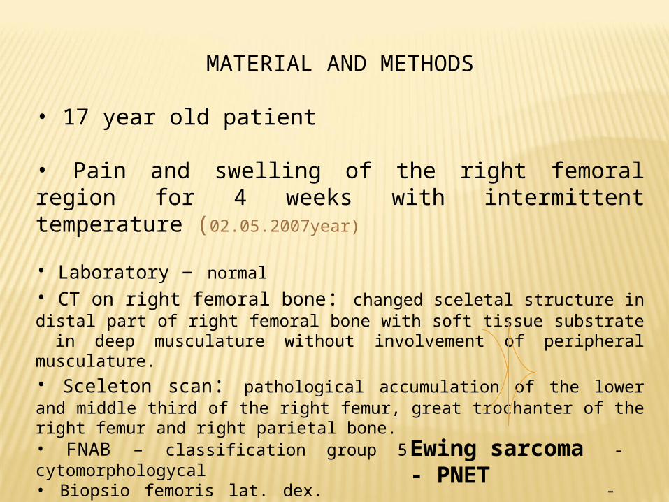
MATERIAL AND METHODS
• 17 year old patient
• Pain and swelling of the right femoral region for 4 weeks with intermittent temperature (02.05.2007year)
• Laboratory – normal
• CT on right femoral bone: changed sceletal structure in distal part of right femoral bone with soft tissue substrate in deep musculature without involvement of peripheral musculature.• Sceleton scan: pathological accumulation of the lower and middle third of the right femur, great trochanter of the right femur and right parietal bone.• FNAB – classification group 5 - cytomorphologycal• Biopsio femoris lat. dex. - histophatologycal - immunohistohemical
Ewing sarcoma - PNET

Initial treatment with:
-Radyotherapy : 3D conformal delivery treatment with TCT (1,25Mev), depth = 6sm, field: 10x30 sm, TTD=50Gy (25fr/2Gy)(11.06.2007 – 13.07.2007 ) followed by
- Chemotherapy :IV courses with : Cyclophosphamide 1000mg 1-3 d, Doxorubicin 30mg 1-3d, Etoposid 200 mg 1-3d, MESNA 4000mg 1-3 d. followed by II courses of Cyclophosphamide 500mg 1-3 d, Etoposid 200mg 1-3 and Vinkristine 2mg -Blood support is achieved with GSF and antifungal drug after each course.
- At the end of chemotherapy the surgeon was consulted for reevaluation for surgery , and no surgery was performed.

Follow up:
2008:Stable disease. ECOG PS =0, X-rays of the lung : normal, ultrasound of abdomen: normal, laboratory: normal ( every 3 months )
2009: Stable disease. ECOG PS =0, X-rays of the lung: normal, ultrasound of abdomen: normal, laboratory: normal ( every 3 months )
2010: ECOG PS=0, X-rays of the lung: normal, ultrasound of abdomen: normal, laboratory: normal ( every 3 months ) until 1.10.2010 when the pacient came with:
- Cephalea- Vomiting and fatigue- Left side facial pain- Egzophtalmus on the left eye
Examinations : CT of brain: Exspansive mass in middle fossa witch is compromasing the temporal lobe and left frontal lobe. It penetrates into the left orbit with dislocation of the eye bulge anteriorly. Scan sceleton(Tc99m): Recidiv localis
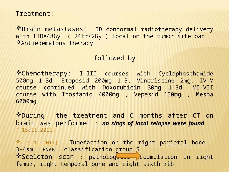
Treatment:
Brain metastases: 3D conformal radiotherapy delivery with TTD=48Gy ( 24fr/2Gy ) local on the tumor site badAntiedematous therapy
followed by
Chemotherapy: I-III courses with Cyclophosphamide 500mg 1-3d, Etoposid 200mg 1-3, Vincristine 2mg, IV-V course continued with Doxorubicin 30mg 1-3d, VI-VII course with Ifosfamid 4000mg , Vepesid 150mg , Mesna 6000mg.
During the treatment and 6 months after CT on brain was performed : no sings of local relapse were found ( 15.11.2011)
( 1.12.2011) – Tumefaction on the right parietal bone – 3-4sm . FNAB – classification group 5Sceleton scan : pathological accumulation in right femur, right temporal bone and right sixth rib
Bisphosphonate therapy was added3D conformal RT locally on tumefaction of the right parietal bone with TTD=39Gy
After 2 months : cephalea - recidiv retrobulbaris ( 28.02.2012 )The patient was treated with palliative chemotherapy with Docetaxel 120 mg ( VI courses )4 months after :
-Egzophtalmus on the left eye - LDH : 889; 913
Treatment : Whole brain irradiation TTD=30Gy with Temozolomide.
At the moment the patient is with deteriorated general condition, with paresis of the left lower limb

DISCUSSION
- Ewing's sarcoma has a propensity to metastasize to the lung, bone and bone marrow. This tumor can also involve the CNS with a relatively low incidence – 1-8%.
- The literature suggests the incidence may be increasing with the increase in use of chemotherapy and with patients living longer.
- Between 20-25% of patients are diagnosed with metastatic disease.
- Studies investigating CNS involvement of Ewing's sarcoma have reported that spread through direct extension from the skull is more frequent than hematogenous spread.
- Prognostic factors that confer poorer outcome are: serum LDH level, axial localization or older age ( > 15 years), size and volume, incomplete or no surgery for local therapy and time of relaps ( < 24 months ).
LDH elevated at time of CNS relapse – LDH has to be evaluated during the whole treatment.
Patients with metastases and recurrent disease still fare poorly, with 5-year survival rates of 20% witch highlights the need for novel chemotherapeutic agents, bisphosphonates and targeted therapy