Faculdade de Ciências Departamento de Biologia Vegetal
50
Universidade de Lisboa Faculdade de Ciências Departamento de Biologia Vegetal Biofilm formation by Azospirillum brasilense – Microbial socialization in the rhizosphere Francisco Diogo de Almeida Cerqueira Dissertação MESTRADO EM MICROBIOLOGIA APLICADA 2014
Transcript of Faculdade de Ciências Departamento de Biologia Vegetal

Universidade de Lisboa
Faculdade de Ciências
Departamento de Biologia Vegetal
Biofilm formation by Azospirillum brasilense – Microbial socialization in
the rhizosphere
Francisco Diogo de Almeida Cerqueira
Dissertação
MESTRADO EM MICROBIOLOGIA APLICADA
2014
Universidade de Lisboa
Faculdade de Ciências
Departamento de Biologia Vegetal
Biofilm formation by Azospirillum brasilense – Microbial socialization in
the rhizosphere
Francisco Diogo de Almeida Cerqueira
Dissertação
Mestrado em Microbiologia Aplicada
Dissertação orientada pela Doutora Cristina Cruz (FCUL, DBV, CBA) e pelo Doutor Luís Carvalho
(FCUL, CBA)
2014

Biofilm formation by Azospirillum brasilense – Microbial socialization in
the rhizosphere
Francisco Diogo de Almeida Cerqueira
2014
This thesis was fully performed at the Center for Environmental Biology (CBA), Faculty of Sciences of the University of Lisbon, under the direct supervision of Doctor Luís Carvalho. Doctor Cristina Cruz was the internal designated supervisor in the scope of the Master in Applied Microbiology of the Faculty of Sciences of the University of Lisbon.

i
Acknowledgements
This work was carried out in fulfillment of the Master in Applied Microbiology at the
Faculty of Sciences of the University of Lisbon, being performed at Center for Environmental
Biology (CBA), Faculty of Sciences of the University of Lisbon. I would like to acknowledge, Dr.
Cristina Cruz, for providing the necessary support and means to perform this work.
I would like to thank my parents and my friends for their immeasurable support which
culminated in the opportunity of accomplishing this dissertation.
I would like to acknowledge the group of Biotic-Abiotic Interactions, for their precious
guidance in the laboratory since the beginning of this work.
Last but definitely not least, I am incredibly grateful to my co-supervisor Luís Carvalho for
his patience and guidance.

ii
Abstract
Azospirillum brasilense is a remarkable Plant-Growth Promoting Rhizobacteria (PGPR),
with great potential as agricultural inoculant for several crops. Several isolates of this species
have been produced as inoculant by some companies and applied in the agriculture as a
biofertilizer. Therefore, there is a great interest in gathering more scientific knowledge that
improves A. brasilense efficacy as a PGPR.
To enhance the beneficial effects, from phytohormone synthesis to nitrogen fixation, it
is essential that A. brasilense colonize plant roots and survive in the soil. For such, a vast array
of traits is required, including biofilm formation and chemotactic response. The rhizosphere is a
hotspot of biodiversity and a place of intensive networking; root exudates are the major plant
tool to modulate the reciprocal interactions between the plant and the rhizosphere. In this work
I focus on the influence of root symbiotic Arbuscular Mycorrhizal Fungi (AMF) and rhizospheric
bacteria on A. brasilense biofilm formation with the following objectives: Firstly, to test if root
exudates from maize plants colonized or not with different AMF species generate chemotactic
response and stimulate biofilm formation of A. brasilense. Secondly, to test the interspecific
influence of several soil bacterial strains in A. brasilense biofilm formation. Moreover, to assess
the effect of Pseudomonas putida X236 and Bradyrhizobium japonicum by growing A. brasilense
with supernatants, in co-inoculation, and with root exudates from maize plants inoculated with
each one of those strains and with A. brasilense itself. Finally, inquire Phytohormone production,
namely indol-3-acetic-acid (IAA), of A. brasilense in biofilm to assess a potential benefit to plants.
Results showed that some mycorrhizal fungal species interact with A. brasilense via root
exudates acting as chemoatractants. The soil borne bacteria P. putida X236 promoted biofilm
formation of A. brasilense, indicating potential to create a co-inoculum with A. brasilense. Root
exudates of maize plants inoculated with A. brasilense provoke a raise in its biofilm. IAA levels
in biofilm cells were generally higher than in planktonic cells. This study highlights the
importance of interspecific relations and of microbial consortia inoculums to maximize functions
of the PGPR A. brasilense.

iii
Resumo
Azospirillum brasilense é uma Rizobactéria Promotora do Crescimento de Plantas, com
grande potencial para uso como inoculante agrícola em diversas culturas de plantas. Foram já
desenvolvidos inóculos com aplicação na agricultura como biofertilizante. Portanto, há um
grande interesse em obter conhecimento que possa melhorar a eficácia de A. brasilense.
Para aumentar os efeitos benéficos potenciais do A. brasilense como a síntese de fito-
hormonas e a fixação de azoto, é essencial que A. brasilense consiga colonizar eficazmente as
raízes, possa perdurar no solo e tenha as condições necessárias para manifestar o fenótipo
adequado o que inclui a formação de biofilme e a resposta quimiotáctica a exsudados
radiculares. A rizosfera é um “hotspot” de biodiversidade e palco de inúmeras relações entre
organismos, sendo os exsudados radiculares o meio pelo qual a raiz influência a estrutura e
função do microbioma rizosférico. Neste contexto o meu trabalho de mestrado foi centrado no
estudo da influência de fungos micorrízicos arbusculares (AMF) e bactérias rizosféricas na
formação de biofilme em A. brasilense e teve como objectivos: Em primeiro lugar, testar
exsudados radiculares de plantas de milho colonizadas ou não por diferentes espécies de AMF,
e analisar a resposta quimiotática e a formação de biofilme por A. brasilense. Em segundo lugar,
avaliar o efeito de interacções de várias estirpes de bactérias presentes no solo na formação de
biofilme de A. brasilense. Adicionalmente, estudar-se mais aprofundadamente o efeito de
Pseudomonas putida X236 e Bradyrhizobium japonicum, crescendo A. brasilense com
sobrenadantes, em co-inoculação e com exsudados radiculares recolhidos de plantas de milho
inoculadas com cada uma dessas estirpes e do próprio A. brasilense. Por último, avaliar a síntese
de fito-hormonas, nomeadamente de indol-3-acético (IAA), por parte de A. brasilense em
biofilme para averiguar a influência de um potencial efeito benéfico nas plantas.
Os resultados deste estudo demonstraram que certas espécies de AMF interagem com
A. brasilense via exsudados, actuando como quimioatratores. A bactéria P. putida X236
estimulou a formação de biofilme de A. brasilense, indicando potencial para formar um co-
inóculo com A. brasilense. Exsudados de plantas de milho inoculadas com A. brasilense
promoveram um aumento do seu biofilme. Níveis de IAA sintetizado em biofilmes foram
geralmente superiores aos das células planctónicas, indicando a importância da colonização
radicular de A. brasilense pela forma de biofilme. Este trabalho realça a importância das relações
interespecíficas e o potencial do uso de consórcios microbianos para maximizar funções de A.
brasilense como PGPR.
iv
Index
Acknowledgements .............................................................................................................. i
Abstract .............................................................................................................................. ii
Resumo .............................................................................................................................. iii
I - Introduction .................................................................................................................... 1
I.1 - Rhizosphere ........................................................................................................................ 1
I.2 - Biofilms ............................................................................................................................... 1
I.2.1 - Quorum sensing .......................................................................................................... 1
I.2.2 - Development ............................................................................................................... 2
I.2.3 - Ecological advantages ................................................................................................. 2
I.2.4 - Biofilms in Plants ......................................................................................................... 3
I.2.5 – Azospirillum brasilense characteristics and biofilm.................................................... 4
I.3 - Mycorrhizal fungi ............................................................................................................... 5
I.4 - Microbial Socialization ....................................................................................................... 5
I.5 - Root Exudates .................................................................................................................... 6
I.6 - Bacterial Inoculants ............................................................................................................ 7
I.6.1 - A. brasilense inoculation ............................................................................................. 8
I.6.2 - Co-inoculation ............................................................................................................. 8
I.7 - Thesis goals and strategy ................................................................................................... 9
II - Materials and Methods ................................................................................................. 11
II.1 - Bacteria and growth conditions ...................................................................................... 11
II.2 - Biofilm growth and quantification .................................................................................. 11
II.3 - Root exudates assay ........................................................................................................ 12
II.3.1 - Plant growth of mycorrhized roots experiment ...................................................... 12
II.3.2 - Plant growth of bacterial roots experiment ............................................................. 13
II.3.3 - Root Exudates preparation ...................................................................................... 13
II.3.4 - Biofilm Formation evaluation................................................................................... 14
II.4 - Chemotaxis assay ............................................................................................................ 14
II.5 - Bacterial supernatant assay ............................................................................................ 14
II.5.1 - Supernatant preparation ......................................................................................... 14
II.5.2 - Biofilm formation evaluation ................................................................................... 15
II.6 - Co-innoculation assay ..................................................................................................... 15
II.6.1 - Co-inoculation .......................................................................................................... 15
II.6.2 - Biofilm formation evaluation ................................................................................... 15

v
II.7 - Indol-3-acetic-acid quantification assay ......................................................................... 15
II.8 - Data analysis ................................................................................................................... 16
III - Results ........................................................................................................................ 17
III.1 - Interactions with AMF .................................................................................................... 17
III.1.1 - Effects of root exudates from mycorrhized plants on biofilm formation ............... 17
III.1.2 - Effects of root exudates from mycorrhized plants on Chemotaxis ........................ 18
III.2 - Interactions with bacteria .............................................................................................. 20
III.2.1 - Bacterial supernatants assay ................................................................................... 20
III.2.2 - Co-inoculations assay .............................................................................................. 23
III.2.3 - Effects of A. brasilense, P. putida X236 and B. japonicum via root exudates assay 26
III.3 - Indol-3-acetic-acid quantification assay ........................................................................ 27
IV - Discussion ................................................................................................................... 29
IV.1 – Microbial Interactions ................................................................................................... 29
IV.2 – IAA ................................................................................................................................. 31
V - Concluding Remarks ..................................................................................................... 33
VI - References .................................................................................................................. 35

1
I - Introduction
I.1 - Rhizosphere
The rhizosphere is the narrow zone of soil that surrounds and is influenced by plant
roots. The rhizosphere is a complex environment with an enormous microbial biodiversity,
where it is possible to find countless and highly dynamic interactions, either positive or negative,
between roots and soil borne microorganisms. These organisms can have profound effects on
the growth, nutrition and plant health, and they can also affect the composition and biomass of
plant communities in natural ecosystems (1).
I.2 - Biofilms
The vast majority of bacteria in their natural habitat tend to persist as a community
named biofilm attached to a surface in a self-produced extracellular matrix, and not as free living
form (2). They usually appear between interfaces, like air-liquid or liquid solid. A biofilm can be
composed by just one species or, more commonly, can be formed by several bacterial species,
forming a multicellular complex that requires coordination and has a complex behavior (3).
I.2.1 - Quorum sensing
Biofilm formation is a controlled process, in which bacteria communicate cell to cell by
Quorum sensing. It involves production, detection and response of the signaling molecules
named autoinducers (AI) (4). In most gram-negative bacteria the AIs fall in the category of
Acylated homoserine lactones (AHLs), and in gram-positive AIs are peptides (5). These molecules
allow bacteria to sense population density, by assessing the accumulation of AIs. AIs diffuse
away, and, therefore, are present at concentrations below the threshold required for detection.
At high cell density, the cumulative production of AIs leads to a local higher concentration,
enabling detection and response (6).
Quorum sensing does not only have a major role in the early stages of biofilms, but it
also regulates the whole process and even cell detachment when the cell density is too high
after the maturation, and they need to disperse for other niches colonization (4).

2
I.2.2 - Development
Biofilms form in response to environmental stimuli and they are not a static entity,
being a part of biological cycle composed by several stages: initiation, maturation, maintenance
and dispersal. During initiation planktonic bacteria adhere to a surface and this initial phase is
dependent on motility, cell to cell interactions and adhesion factors, for attachment. Afterwards
biofilms maturate, with high synthesis of exopolysaccharides (EPS), and increased resistance to
external stress factors. At last bacteria disperse and return to a planktonic growth mode, due to
nutrient starvation, high cell density, and other factors that might act as a signal, therefore
bacteria may colonize new niches (Fig. 1) (3).
Fig. 1: Scheme of model of biofilm development. Planktonic cells receive environmental stimuli and initiate cell-to-
cell communication resulting in the formation of microcolonies, and then maturation begins with synthesis
extracellular matrix followed by maintenance of the biofilm while environmental conditions allow it. Cells in the
biofilm can return to a mobile state to complete the cycle of biofilm development. Adapted from (3).
I.2.3 - Ecological advantages
Biofilms give several ecological advantages for the bacteria, being the most important
protection, cooperation and gene transfer.
Protection is given by a matrix composed mostly by EPS, nucleic acids and proteins (7).
Acts as a physical barrier preventing certain antimicrobial agents going into the biofilm by acting
as an ion exchanger, thereby restricting diffusion of compounds from the surrounding area into
the biofilm (8). This propriety is dependent on the characteristics of both the agent and the EPS
matrix. The effect appears to be most pronounced with antibiotics that are hydrophilic and
positively charged, such as the aminoglycosides (9, 10). Studies have shown that EPS has also

3
been reported to sequester metals, cations, and toxins (11, 12), and to protect from physical
stress from the environment like pH, UV radiation dissecation (2). Bacteria in multispecies
biofilms have some sort of metabolic cooperation, as some bacterial exchange of nutrients,
where some use others metabolites, thus removing them, which can be useful since they could
be toxic (12). Biofilms are also an excellent niche for gene transfer between bacteria, by
conjugation for plasmid transfer. It occurs at a higher rate between cells in biofilms than
between planktonic cells (14, 15).
I.2.4 - Biofilms in Plants
Plants change their surrounding environment including the soil, creating niches for
microorganisms such as bacteria to prosper. Roots exudate macronutrients into the soil, making
them available for bacteria, and they colonize and aggregate as biofilms, although biofilms may
be present in the whole plant surface, from the roots to the leaves (Fig. 2) (16).
Fig. 2: Scheme representing Tomato plant and the wide distribution of biofilms over plant surface. Subtitles: (a)
transmission electron micrograph of sweet corn leaf xylem vessel infected by Pantoea stewartii; (b) fluorescence
microscopy of Agrobacterium tumefaciens microcolony; (c) fluorescence microscopy of bean leaf with Pseudomonas
syringae and Pantoea agglomerans and (d); fluorescence and light microscopy of alfalfa root hairs with Sinorhizobium
melioti. Adapted from (16).

4
I.2.5 - Azospirillum brasilense characteristics and biofilm
One of the most studied rhizobacteria is Azospirillum brasilense due to its role in plant
root system. A. brasilense is a plant growth-promoting rhizobacteria (PGPR) (17). It is a PGPR
included in the class of alpha proteobacteria, which promotes growth and yield of agronomic
and ecological important plant species (18, 19, 20). It can be found in a wide range of habitats
associated with roots of various types of plant species. A. brasilense was found to possess no
obligate specificity in respect to the host plant (21, 22).
A. brasilense is well known for its capability of plant growth promotion by
phytohormone synthesis, particularly the auxin indole-3-acetic acid (IAA) (23), nitrogen fixation,
converting atmospheric nitrogen into ammonium under microaerobic conditions, through the
action of the nitrogenase complex (24). Some studies showed that it is capable of nitric oxide
production, inducing lateral root formation in tomato seedlings (25). There are not much studies
about its biocontrol competencies, nonetheless it has been shown that A. brasilense contributes
for crown gall disease, bacterial leaf blight of mulberry, and bacterial leaf and vascular diseases
of tomato biocontrol (26, 27, 28, 29). Furthermore it synthetizes phenylacetic acid (PAA), which
has antibacterial and antifungic activity (30).
In addition to all these competencies, A. brasilense is capable to colonize and form
biofilm in plant root surface. Root exudates and oxygen concentration create taxis towards the
roots. The carbon compound chemotaxis is strain-specific, reflecting the variability of root
exudation by different host plants (31, 32).
In most gram negative bacteria AHLs are the molecules responsible for quorum sensing,
though most Azospirillum strains do not synthetize AHLs (33, 34). Aerotaxis is also extremely
important for A. brasilense to meet the right oxygen concentration that allows respiration and
nitrogenase function for nitrogen fixation (35).
Afterwards, in attachment and aggregation, A. brasilense uses an arabinose-containing
polysaccharide (36) and outer membrane proteins, as major outer membrane protein (MOMP)
(37, 38). Tad pili is supposed to be involved in attachment to the root surface and biofilm
formation in A. brasilense sp245, instead of type IV pili which is used by most bacteria (39).
Several factors may act as stimuli for A. brasilense biofilm formation, including signaling
molecules synthetized by itself, plants or other microorganisms. Recently it has been shown that
2, 4-diacetylphloroglucinol (DAPG), a secondary metabolite produced by some bacteria of
Pseudomonas genus, has a positive effect on biofilm formation at intermediate concentrations
(40). Other studies shown that NO, a signaling molecule with a wide variety of functions,

5
influence biofilm formation of several bacteria including A. brasilense sp245, possibly acting as
early signal in biofilm development (41).
I.3 - Mycorrhizal fungi
Mycorrhizal fungi are symbionts in the roots of the majority of higher plants, enhancing
plant nutrition, stress resistance and pathogenic resistance (42). Fungi colonize the host plant's
roots, being either intracellularly as in arbuscular mycorrhizal fungi (AMF), or extracellularly as
in Ectomycorrhizae fungi (ECM). All AMF belong to the phylum Glomeromycota (43), while ECM
belong to the phyla Basidiomycota, Ascomycota and Zygomycota (44). Many agriculturally and
horticulturally important crop species are included on the terrestrial plants that form a symbiotic
association with AMF (42).
AMF and bacteria naturally co-exist in the rhizosphere. Interactions between AMF and
PGPR most likely occur, and can modulate plant health and productivity. Many non-symbiotic
rhizosphere bacteria fix atmospheric nitrogen making it available for plants. There are reports
of positive interaction between free-living N2-fixing bacteria and AMF (45), including one study
suggesting that AMF species may have stimulating effects on nitrogen fixer A. brasilense (46).
Although free-living bacteria contribute for nitrogen availability, symbiotic bacteria of plant
nodules are responsible for most of nitrogen fixation in plants and studies have revealed that
nodulation by indigenous rhizobia is greatly improved by AMF (47).
Studies have shown that some bacterial species respond to the presence of certain AMF
suggesting a high degree of specificity between bacteria associated with AMF. One possible
explanation for this stimulation of certain bacterial species by specific AMF may be due to those
bacteria being activated by species-specific fungal exudates (48, 49). Therefore, AMF can
potentially shape soil microbial community composition and function by acting as large suppliers
of carbon to the rhizosphere (50).
I.4 - Microbial Socialization
In the rhizosphere there are interactions between plants and those microorganisms and
amongst themselves. Bacteria may act as a biocontrol agent, inhibiting growth of pathogenic

6
microorganisms, or can have a mutualistic or commensal interaction with other bacteria or
fungus (16).
Most studies have focused on root-secreted compounds on the selection of root-
associated bacteria and the control of their plant-beneficial activities; bacterial secondary
metabolites, including phytohormones, on root growth and plant defense; and quorum-sensing
pheromones in the regulation of microbe–microbe social relationships (40).
Members of Pseudomonadaceae family are known for their role in the rhizosphere.
Various strains of Pseudomonas putida and Pseudomonas fluorescens produce antifungical
metabolites, siderophores and hydrogen cyanide, that inhibit growth of several pathogenic
fungi, especially Fusarium oxysporum (51, 52, 53). Sometimes bacteria not only inhibit growth
of other organism by their secondary metabolites, but by nutrient or niche competition, as it
can occur in P. fluorescens PCL1751 (51, 54). Other strains of P. fluorescens synthetize DAPG, a
phenolic compound that can increase gene expression, particularly the genes responsible for
cell motility, biofilm formation, poly-β-hydroxybutyrate, and auxin production of A. brasilense
(40).
Not only pseudomonads act against phytopathogens, gram positive bacteria as Bacillus
spp. are involved in biocontrol of soilborn pathogens. Strains of Bacillus subtilis, Bacillus
megatherium, Bacillus licheniforms and Bacillus pumilus inhibit several phytopathogenic fungi,
responsible for different diseases in plants with high economic value (55, 56, 57, 58, 59).
Bradyrhizobium japonicum, thought it fixes atmospheric nitrogen, it may contribute by
other means in plant growth. B. japonicum and A. brasilense are able to promote germination
and seedling growth, either singled or combined of maize (Zea mays L.) and soybeans (60).
I.5 - Root Exudates
Soil proprieties such as pH, nutrient availability are important for microbial community
structure and survival. Plant roots release a wide range of compounds, including sugars, ions,
vitamins, amino acids, purines and nucleosides to the soil and consequently, these compounds
are exploited by soil bacteria (61).
Plants deposit up to 40 % of the photosynthetically fixed carbon via their root system in
a processed named rhizodeposition, and there it becomes accessible for organisms in the
rhizosphere (62). This causes a growth in the microbial population in the rhizosphere, and that

7
community composition is considerably distinct from the surrounding bulk soil, a phenomenon
called the rhizospheric effect (63).
To reach and use these nutrients, bacteria need to have certain traits like motility,
chemotaxis, attachment (61), to proceed to root colonization, which is an important step either
for phytopathogenic or benefitial bacteria. Plants can produce molecules excreted to the
rhizosphere via root exudates, and may select distinct microbial communities, depending on the
molecules that constitute the exudates (64). Exudates can act as chemotatic signal containing
molecules that mimics Quorum sensing signals, attracting PGPRs, and inhibiting pathogenic
bacteria, trough niche competition, or antibacterial compounds (Fig. 3). Therefore plants can
interfere with Quorum sensing controlled processes such as biofilm formation (62).
Fig. 3: Scheme of root colonization beneficial bacteria and their functions. (I) Plant roots exudate compounds that act
as chemotactic signal. Some bacterial strains from the soil communities respond specifically to the signals and start
express traits related to root colonization. Local immune responses in host roots are suppressed by PGPR. (II) PGPR
on the root surface starts biofilm formation. PGPR may protect root tissues against pathogens via the production of
antibiotics or niche and nutrient competition. Adapted from (65).
I.6 - Bacterial Inoculants
Bacterial inoculation emerges as an interesting alternative to chemical compounds in
agriculture like pesticides, herbicides and fertilizers (66). The usage of such products has
hazardous environmental effects, polluting the soil and ground water (67). These inoculations

8
have a wide range of utility, like biofertilization with nitrogen-fixing and phosphate solubilizer
bacteria; phytohormone stimulation or production and last but not less important function as
biocontrol agent protecting plants against pathogens (66).
So, the use of bacterial inoculants can have a direct effect on plant growth and
protection, but it can also indirectly influence plants by influencing bacterial community of the
rhizosphere, temporarily or permanently (66).
I.6.1 - A. brasilense inoculation
Inoculations with A. brasilense affect mostly plant performance and rhizosphere
microbiome structure and function, and have already been successful in crop fields as maize
among others (68). Studies have shown that effects on native microbial community vary
according with A. brasilense strain and plant species. For instance, the strain Sp245 does not
have much effect on bacterial community in rhizosphere with maize (69). Other strains as 40M
and 42M change physiological profiles (CLPPs) of microbial communities of Asian rice (Oryza
sativa L.) (70).
I.6.2 - Co-inoculation
Inoculation with more than one strain, have the purpose of getting a greater positive
effect expecting those strains to act synergistically. This can be attempted with bacteria that in
mono-inoculation have distinct effects. Nevertheless, competitive results between strains in
inoculation may happen, consequently having reduced or even null effect on plants or in the
rhizosphere community (66).
As example, co-inoculation of B. subtilis 101 with A. brasilense Sp245 neither had
synergetic effect on plant biomass compared to their mono-inoculations in tomato
(Lycopersicon esculentum) nor had significant effect in bacterial community in the rhizophere
(71). Obtaining successful co-inoculations with synergetic effects of the bacteria represent a
difficult task, thought already several have been discovered. As an example, co-inoculation of
Pseudomonas chlororaphis IDV1 and P. putida RA2, is capable of diminishing the gram-positive
dominant community and increasing the gram-negative community (72). Co-inoculation of
DAPG-producing strain P. fluorescens F113 and A. brasilense Sp245-Rif in wheat resulted in
enhanced root diameter, total root volume, and root number when compared to single

9
inoculations (40). There is research of A. brasilense co-inoculation with AMF proving it can be
successful, like A. brasilense Az39 and Glomus intraradices improving rice growth in well watered
conditions and in drought stress (73). A. brasilense Sp7 co-inoculated with either Glomus clarum
or Glomus fasciculatum have a stimulant effect, increasing weight and root length of tomato
(74). A. brasilense Az39 co-inoculated with B. japonicum E109 promote seed germination and
early seedling growth in corn and soybean (60).
I.7 - Thesis goals and strategy
A. brasilense has been produced as inoculant by some companies, and applied in the
agriculture as a biofertilzer in some countries (67). Therefore, there is a great interest in
gathering more scientific knowledge that improves A. brasilense efficacy as a PGPR.
A. brasilense needs a vast array of traits to colonize plant roots, including chemotactic
response, and biofilm formation. (61) Fungal and bacterial interaction and possible biofilm
formation stimulation were an interesting subject to test.
The purpose of this thesis was to evaluate whether and how rhizospheric microbial
interspecific interactions, namely with root symbiotic AMF and rhizospheric bacteria, could
influence and stimulate biofilm formation in A. brasilense. To achieve this goal the influence of
(i) AMF was evaluated by testing root exudates from maize plants colonized with or without
different AMF species (Glomus mosseae, Glomus claroideum, Glomus intraradices and
Gigaspora sp.) on biofilm formation and chemotactic response, and (ii) of interspecifc bacterial
interactions on biofilm formation by testing (a) bacterial supernatants from several soil borne
strains (Pseudomonas putida X236, Pseudomonas fluorescens 20130311XA1, Mesorhizobium loti
B29, Bacillus pumilus B7, Bacillus subtilis 168, Bacillus licheniformis B6, Bacillus megaterium GY-
23 and B3, Bradyrhizobium japonicum and Azotobacter chroococcum.), (b) bacterial co-
inoculation with P. putida and B. japonicum (species selection based on results obtained) in
several frequencies, and (c) bacterial-influenced root exudates from maize plants inoculated
with A. brasilense, P. putida and B. japonicum.
To test whether biofilm is an important feature to improve A. brasilense as a PGPR, its
function as a phytohormone producer was evaluated by performing IAA quantifications in
biofilm and planktonic cells, and then to inquire any difference of produced IAA for each DL-
tryptophan supplementation by cell type.

10

11
II - Materials and Methods
II.1 - Bacteria and growth conditions
Bacterial strains, used in this work are listed: A. brasilense AMC B1, M. loti AMC B28, B.
subtilis 168, P. putida X236, P. fluorescens 20130311XA1, B. megaterium GY-23, B. japonicum,
A. chrocorum, B. licheniformis AMC B6, B. pumilus AMC B7. AMF inoculum was acquired to
Symbiom, Ltd, and B. licheniformis, M. loti, B. megaterium, B. pumilus and A. brasilense strains
were supplied by AMC Chemicals & Trichodex, SL. The other strains were isolated from work
developed at Faculty of Sciences, University of Lisbon. Bacterial strains were grown in NB
medium (75) at 28 °C and 160 rpm in an orbital shaker, except for B. japonicum, which was grown
in YMB (76).
II.2 - Biofilm growth and quantification
In all assays biofilms developed in microtiter plates of 96 wells, which were involved in
Parafilm M® and incubated for 5 days at 28 °C, in a growth chamber. The medium utilized to
allow biofilm growth and possible stimulation was NB diluted to 1/10 of original concentration.
Diluted NB was chosen after a trial to test if there was any significant difference between usage
of NB, YMB and LB. After testing, no significant difference was detected in biofilm formation
(one-way ANOVA, p>0.05). Then, NB dilution at 10 % was used.
In each well of the 96 well microtiter, 40 µl of root exudate or bacterial supernatant, 20
µl of NB and 130 µl of distilled water were added, and supplemented with 10 µl of NB; pre-grown
culture of A. brasilense or of A. brasilense in co-culture with other bacterial strain at different
proportions. Control wells without root exudates or bacterial supernatant were also performed.
In each plate there was one well corresponding to the blank, in which only diluted NB and
distilled water was added. The number of replicates varied between 3, 5, 8 or 10 according with
the assay.
Biofilm formation was quantified at the end of the assays, using Crystal Violet (CV) (77)
method with slight modifications. Briefly, 200 µl of PBS buffer (NaCl 8 g/l, KCl 0.2 g/l, Na2HPO4
1.44 g/l, KH2PO4 0.24 g/l, pH=7.4) was added drop-wise to wash the wells, then PBS was
removed, and 200 µl of methanol was added. After 15 minutes, methanol was removed by
letting the microtiter plate dry upside-down on tissue paper. Subsequently, the biofilms became
fixed onto the wells surface, and 200 µl of CV 1 % (wt/vol) was added. Fifteen minutes later, the

12
excess of CV was removed by rinsing carefully the wells with tap water. Microtiter plates were
dried upside-down on tissue paper, and then to dissolve attached stained cells from the walls,
200 µl of acetic acid 33 % (vol/vol) was added. Finally, 160 µl of each well were transferred to
another sterilized microtiter plate.
For biofilm quantification, A. brasilense biofilm over walls of wells was measured by
reading the optical density (OD) at 570 nm wavelength in a microtiter reader (Tecan Spectra
Rainbow 1000). Planktonic cells together with the pellicle were transferred to another microter
plate and quantified by reading OD at 600 nm wavelength. Both measurements were used to
calculate later the Specific Biofilm Formation (SBF), formulated as SBF=(B-C)/G, where B is the
OD570 of the attached biofilm cells on wells, C is the OD570 of the stained control wells, and G is
the OD600 of the transferred planktonic cells (78, 79).
II.3 - Root exudates assay
II.3.1 - Plant growth of mycorrhized roots experiment
Maize was the plant chosen for the experiments. Seed sterilization were accomplished
by putting the seeds in 70 % ethanol during 3 minutes, right after they were transferred to
sodium hypochlorite 5 % (vol/vol) solution for another 3 minutes, and then the seeds were
washed 5 times in sterilized distilled water. Seed germination was done in Petri dishes with filter
paper on the bottom and involved around with adherent pellicle. Incubation of seedlings lasted
10 days in a dark chamber at 28 oC. Every 2 days some sterilized water was added to the Petri
dishes, in the flux chamber.
Maize plants were put in 1.5 l pots with a substrate composed by 1 l of sterilized river
sand and Styrofoam spheres (1:2). Plants were inoculated with 15 ml corresponding to 14 000
propagules of AMF inoculum per pot of G. mosseae, G. claroideum, G. intraradices and
Gigaspora sp., except the controls where inoculum was not added. The plants were grown in a
growth chamber at (25 oC, 75 % of relative humidity, 16 h light period). Plants were watered 3
times per week with Hoagland solution ½ (80) and water during the 45 days of development.
Each treatment had 3 replicates.

13
II.3.2 - Plant growth of bacterial roots experiment
Maize was the plant chosen for the experiments. Seed sterilization and seedling growth
was processed the same way as described previously in plant growth of the mycorrhized roots
experiment.
Then plants were put in a sterile system (Fig. 4), composed by UNICORN® bags with 0.2
µm membrane allowing gas exchange, and polystyrene tubes and 3 of these systems were used.
1 ml of previously grown A. brasilense, B. japonicum or P. putida X236 until early exponential
phase (OD600 of 0.12-0.15), were inoculated in the polystyrene tubes filled with 10 ml of NB
medium. Later the plants were put in a growth chamber (25 oC, 75 % humidity, 16 h light period)
and grew for 15 days.
Fig. 4: Scheme of plant growth in the sterile system. The plastic bag is the outer rectangle, allowing sterility for plant
growth. The circles represent the polystyrene tubes, the ones with H20, were filled with water, and plant roots
exudated there. The circles with bacterial strains names were the ones with NB inoculated with A. brasilense, B.
japonicum and P. putida X236, the control circle was just filled with NB. The red rectangle represents the tubes in
which roots of the same plant were immersed in NB inoculated with bacteria and immersed in water.
II.3.3 - Root Exudates preparation
After plant growth, to collect root exudates from the bacterial experiment, plants were
then removed from the tubes, and the water where maize roots had been exudating was
filtrated through Ø 0.22 µm nitrocellulose filters (Millipore®) and solutions were kept at -20 oC
until use. Collection of the root exudates from mycorrhized plants was similar; except that root
system was incubated in Erlenmeyer flasks filled with 100 ml sterilized Milli-Q water for 24 h
(81).

14
II.3.4 - Biofilm Formation evaluation
A total of 3 microtiter plates with each one having 3 replicates of every sort of root
exudate with and without inoculation (A. brasilense, B. japonicum and P. putida X236) were
used, being each replicate a sample of each plant growth system.
The assay with root exudates from mycorrhized maize plants (G. mosseae, G.
claroideum, G. intraradices and Gigaspora sp.), was performed in the same way.
II.4 - Chemotaxis assay
Chemotaxis assay (82) was performed using the “drop” assay with slight modifications,
with the purpose to test the effect of root exudates collected from mycorrhized maize plants,
and compared to some organic acids (malate, succinate) and amino acids (L-aspartate, L-serine,
L-arginine, L-glycine, L-proline, L-cysteine, L-glutamine) normally found in maize root exudates.
Briefly, A. brasilense grown 48h in NB was diluted 100-fold into 150 ml of NB. When A.
brasilense reached the early exponential phase (OD600 of 0.12-0.15), 40 ml samples were
centrifuged (Eppendorf centrifuge 5810R) at 3 000 rpm for 10 minutes and re-suspended in 12
ml of chemotaxis buffer (100 mM potassium phosphate, pH 7.0, 20 μM EDTA). An aqueous
solution of 1 % hydroxypropylmethylcellulose (Sigma-Aldrich), formulated to give a viscosity of
approximately 4 000 cP in a 2 % aqueous solution, was added to the cell suspension to give a
final volume of 15 ml. The resulting suspension was transferred to a Petri dish. In the Petri dish
center, 10 µl of each root exudate, organic acid and amino acid were dropped. After 2 hours of
incubation at room temperature, the plates were analyzed for the appearance of a clear zone,
and measured. The bacteria attracted by the added chemoattractant leave a clear zone where
they were previously sited. Three replicates were used for each compound and their
concentration was 40 mMol.
II.5 - Bacterial supernatant assay
II.5.1 - Supernatant preparation
Bacterial strains of P. putida X236, P. fluorescens 20130311XA1, M. loti B29, B. pumilus
B7, B. subtilis 168, B. licheniformis B6, B. megaterium GY-23 and B3, B. japonicum and A.

15
chroococcum, previously grown until late exponential phase (OD600 of 1.0-1.5) were centrifuged
(Eppendorf centrifuge 5417C) at 13 000 rpm for 10 minutes, and then the supernatants were
collected. For further analysis two bacteria were selected, P. putida X236 and B. japonicum.
Supernatant preparation was performed identically.
II.5.2 - Biofilm formation evaluation
A total of 3 microtiter plates with each one having 5 replicates for each supernatant
treatment and control were used.
II.6 - Co-innoculation assay
II.6.1 - Co-inoculation
P. putida X236 and B. japonicum were inoculated each one with A. brasilense in a
pairwise co-culture system in several proportions, being A. brasilense in 0, 10, 25, 50, 75, 90 and
100 % frequency. Grown until OD600 of 1.0-1.5.
II.6.2 - Biofilm formation evaluation
A total of 3 Microtiter plates with each one having 5 replicates for each co-inoculation
were used. In A. brasilense-P. putida X236 co-inoculations 2 controls were used: A. brasilense,
P. putida X236 growing separately and in A. brasilense-B. japonicum co-inoculations 2 controls
were used, but instead of P. putida X236, B. japonicum was used as control.
II.7 - Indol-3-acetic-acid quantification assay
IAA was quantified by using a modified Salkowski colorimetric assay (83, 84) in microtiter
plates. Briefly, grown cultures of A. brasilense (early exponential phase OD600 of 0.12-0.15) were
inoculated in microtiter plates. After they were grown for 3 days, planktonic cells were removed,
200 µl of NB 1/10 supplemented with DL-tryptophan (0, 10, 25, 50, 100, 200 mg/ml) was added.
Two days later, 170 µl of each well was transferred to a new microtiter plate, then the plates

16
were centrifuged (Eppendorf centrifuge 5810R) at 3 500 rpm for 10 minutes. After centrifugation
150 µl of each well were transferred to a new plate, followed by addition of 100 µl of Salkowski
reagent (FeCl3.6H2O 0.5 M and HClO4 35 %). The reaction took 30 minutes in a dark chamber and
right after microtiter plate reader (Tecan Spectra Rainbow 1000) was used to measure OD530. A
total of 3 plates were used, each one with 10 replicates of every DL-tryptophan supplementation
and the control. The blank corresponded to one well of NB 1/10. Then IAA detected by Salkowski
colorimetric assay in planktonic cells and biofilm were compared.
II.8 - Data analysis
The experiments conducted had a design in which every treatment was replicated 3 to
10 times in every microtiter plate depending on the assay. Average of replicates was calculated,
and then each microtiter plate functioned as a single replicate of a total of 3 replicated plates.
Statistical analysis of the data was done using Softstat Statistica 11® software following one-way
analysis of variance (ANOVA), except for IAA quantification (see below); when the ANOVA was
significant the means were separated using a post-hoc test, Tukey Honest Significant Difference
(HSD) at p≤0.05 level of significance. Two-way ANOVA was utilized merely in IAA quantification
assay testing the effects of cell type and DL-tryptophan concentration, and subsequently
Student’s t-tests were performed to inquire any significant difference between the cell number
of biofilm and planktonic cells, as well in the differences of produced IAA for each DL-tryptophan
supplementation by cell type. When necessary data was transformed by log10(X), before
proceeding to ANOVA.

17
III - Results
III.1 - Interactions with AMF
III.1.1 - Effects of root exudates from mycorrhized plants on biofilm formation
The results of the assay of effects of root exudates from mycorrhized maize plants on
biofilm formation showed no significant differences between AMF treatments and control and
among AMF species (Fig. 5), as confirmed by one-way ANOVA analysis (F5, 12=1.574, p=0.240).
Specific Biofilm Formation (SBF) had the same outcome (one-way ANOVA, F5, 12=0.537, p=0.745)
(Fig. 6).
Fig. 5: A. brasilense biofilm quantification by CV method, from the assay of effects of root exudates from mycorrhized
plants. Subtitles: OD, optic density; X-axis, root exudates origins. Error bars represent the standard deviation of the
mean (n=3).

18
Fig. 6: One-way ANOVA of effects of root exudates from mycorrhized plants assay. Subtitles: % SBF, biofilm
percentage given by Specific Biofilm Formation (SBF) ratio; X-axis, root exudates origins. Error bars represent the
standard deviation of the mean (n=3).
III.1.2 - Effects of root exudates from mycorrhized plants on Chemotaxis
A. brasilense dropped in the middle of the Petri dishes, when attracted by either the
exudates or other compounds added beforehand generated a clear zone. By other words, this
zone corresponds to the migration of bacterial cells where they were previously sited to the
Petri dish center. One-way ANOVAs, comparing organic acids and amino acids (Fig. 7) showed
significant differences between treatments (F8, 18=17.485, p<0.001), as in the comparison of root
exudates (Fig. 8) (F4, 10=97.750, p<0.001). Subsequently post-hoc Tukey HSD tests were
performed.

19
Fig 7: Chemotatic response of A. brasilense to several amino acids and organic acids. Control refers to root exudates
from non-mycorrhized plants. Subtitles Halo (cm), halo in centimeters (clear area created by bacteria where they were
previously sited); X axis, compounds used in the assay. Error bars represent the standard deviation of the mean (n=3).
Fig 8: Chemotatic response of A. brasilense to several root exudates collected from mycorrhized plants. Control refers
to root exudates from non-mycorrhized plants. Subtitles Halo (cm), halo in centimeters (clear area created by bacteria
where they were previously sited); X axis, compounds used in the assay. Error bars represent the standard deviation
of the mean (n=3).

20
Analyzing differences in the compounds tested (Fig. 7) it is perceptible that there are
significant differences among the amino acids tested, comparing amino acids with L-aspartate
significant differences can be found, namely L-arginine, L-glycine and L-proline. Examining root
exudates (Fig. 8), three root exudates did not generate a chemotactic response (control, G.
intraradices and Gigapsora sp.), whereas G. mosseae and G. claroideum did (p<0.05). Moreover,
there was a significant difference between those exudates which created a chemotactic
response, being G. mosseae root exudate the one who formed the largest clear zone in the Petri
dishes.
III.2 - Interactions with bacteria
III.2.1 - Bacterial supernatants assay
A screening with several soil borne bacterial species was performed to assess which
species stimulate A. brasilense biofilm, and whether there is a phylogenetic trend in inter-specifc
interactions influencing the formation of biofilm. The results indicated that only the supernatant
of the strain P. putida X236 stimulate biofilm quantity of A. brasilense compared to without
bacterial supernatant (t-test, p=0.03), and all the other strain supernatants tested did not
influence biofilm formation, that is, there were no significant differences (t-test, p>0.05) in the
quantity of biofilm formed when compared to without supernatant (Fig. 9).

21
Fig. 9: Ratio of A. brasilense development according the supernatant treatment, from the screening of the assay of
effects of bacterial supernatants from several strains. The ratio corresponds to (OD of A. brasilense grown with
bacterial supernatant/OD A. brasilense grown without supernatant). The dashed line represents the growth of A.
brasilense without supernatant. Subtitles: Y-axis, supernatant origins. Error bars represent the standard deviation of
the mean (n=3).
P. putida X236 were then selected for further analyses and assays, together with B.
japonicum as a bacterial strain control and because this nitrogen-fixer strain of B. japonicum was
previously observed to stimulate root exudation and plant growth (60). The one-way ANOVA
comparing A. brasilense biofilm quantification (Fig. 10) of these two strains with the control
(without bacterial supernatant) showed significant differences (F2, 6=9.567, p=0.013), whereas
for SBF one-way ANOVA (Fig. 11) did not show any significant differences (F2, 6=3.076, p=0.120).
Post-hoc Tukey HSD test performed in the biofilm quantification indicated that P. putida
X236 presented significant differences when compared with either to control (without bacterial
supernatant) or to B. japonicum supernatant. Despite similar results to biofilm quantification
with highest value belonging to P. putida X236, the results of SBF were not statistically significant.

22
Fig. 10: A. brasilense biofilm quantification from the assay of effects of bacterial supernatants, using the CV method.
Subtitles: OD, optic density; X-axis, supernatant origins. Error bars represent the standard deviation of the mean (n=3).
Fig. 11: A. brasilense SBF from the assay of effects of bacterial supernatants. Subtitles: X-axis, bacterial supernatant
origin; % SBF, biofilm percentage given by Specific Biofilm Formation (SBF) ratio. Error bars represent the standard
deviation of the mean (n=3).

23
III.2.2 - Co-inoculations assay
The co-inoculation assay was composed by two experiments. In the first one, a co-
inoculation experiment of A. brasilense and P. putida X236, the one-way ANOVA (Fig. 12)
demonstrated significant effects of treatment (F6, 14=7.733, p=0.008). Consequently post-hoc
Tukey test was performed to inquire the significant differences of biofilm formed in different
settings.
Fig. 12: Biofilms quantification, by CV method, from the co-inoculations of A. brasilense and P. putida X236
experiment testing different initial frequencies. Subtitles: OD, optic density; the ratio (X/Y) in which X is A. brasilense
and Y is P. putida X236 initial inocula frequencies. The numbers in (X/Y) are A. brasilense / P. putida X236 proportion.
Error bars represent the standard deviation of the mean (n=3).
Both controls (single strain inoculation) showed significant differences with the co-
inoculations in the proportion of 90/10, 75/25 and 25/75, having a lower OD and therefore less
biofilm formed. Co-inoculation 75/25 also showed a significant difference when was compared
to 10/90. All other co-inoculations did not display any significant difference when compared
between themselves. With this result it is perceptible that the A. brasilense / P. putida X236
initial frequency of 75/25 is the co-inoculation which creates the highest OD value, hence the
highest biofilm quantity. A. brasilense - P. putida X236 co-inoculations had encouraging results,

24
and SBF ratios as well (Fig. 13). All co-inoculations had higher SBF ratios than both single strain
inoculations, with 90/10 (A. brasilense / P. putida X236) with the highest ratio. Also as the
bacterial proportion in the co-inoculations increases in favor to P. putida X236 and A. brasilense
lowers, the SBF ratio had a declining tendency (ANOVA, F6, 14=5.282, p=0.049). Although, in these
co-inoculation experiments it was not possible to distinguish between cells of A. brasilense and
the other species tested in the formed biofilm, the declining biofilm formation (as SBF) with
decreasing initial frequency of A. brasilense (Fig. 12, 13) suggests that A. brasilense biofilm was
influenced by P. putida X236 when in co-culture, as it was found in the supernatant assay (Fig. 9,
10).
Fig. 13: SBF from the experiment of co-inoculations A. bransilense - P. putida X236. Subtitles: the ratio (X/Y), in which
X is A. brasilense and Y is P. putida X236 initial inocula frequencies. The numbers in (X/Y) are A. brasilense / P. putida
X236 proportion. %, biofilm percentage given by Specific Biofilm Formation (SBF) ratio. Error bars represent the
standard deviation of the mean (n=3).
The other experiment, in which A. brasilense and B. japonicum co-inoculation was
evaluated, had a different outcome. After log10(X) transformation, one-way ANOVA did not show
significant effects (F6, 14=0.610, p= 0.718), which indicates that there is no significant difference
in the biofilm formed, independently of the co-inoculations proportions (Fig. 14).
The one-way ANOVA of SBF ratio for A. brasilense-B. japonicum co-inoculation (Fig. 15)
had a significant result (F6, 14=3.954, p=0.015), and Tukey HSD tests indicated that only the
frequency of 50/50 made a significant difference compared to both single strain inoculations.
100/0 90/10 75/25 50/50 25/75 10/90 0/100
A. brasilense/P. putida X236 proportion
0
5
10
15
20
25
30
35
40
45
50
55
60
% S
BF

25
Fig 14: Biofilms quantification, by CV method, from the co-inoculations of A. brasilense and B. japonicum experiment
testing different initial frequencies. Subtitles: OD, optic density; the ratio (X/Y) in which X is A. brasilense and Y is B.
japonicum initial inocula frequencies. The numbers in (X/Y) are A. brasilense / B. japonicum proportion. Error bars
represent the standard deviation of the mean (n=3).
Fig. 15: SBF from the experiment of co-inoculations A. bransilense - B. japonicum. Subtitles: (X/Y) is the ratio, in which
X is A. brasilense and Y is B. japonicum initial inocula frequencies. The numbers in (X/Y) are A. brasilense / B. japonicum
proportion. % SBF, biofilm percentage given by Specific Biofilm Formation (SBF) ratio. Error bars represent the
standard deviation of the mean (n=3).
100/0 90/10 75/25 50/50 25/75 10/90. 0/100
A. brasilense/B. japonicum proportion
0
5
10
15
20
25
30
35
40
45
50
55
60
% S
BF

26
III.2.3 - Effects of A. brasilense, P. putida X236 and B. japonicum via root exudates assay
In this assay testing the indirect effects of bacterial strains in A. brasilense biofilm
formation via maize root exudates, ANOVA results showed that root exudates effects were
significant (F4, 10=8.109, p=0.003), with significant differences (Tukey HSD test) in biofilm
formation between root exudates from planta inoculated with A. brasilense and without
exudate, control exudate or P. putida X236 (Fig. 16). Therefore, there was a significant positive
influence of root exudates from plants inoculated with A. brasilense, but not with P. putida X236
or B. japonicum.
Fig. 16: A. brasilense biofilm quantification by CV method from the assay of effects of root exudates from plants
inoculated with A. brasilense, P. putida X236 and B. japonicum. Subtitles: OD, optic density; X-axis, root exudates
origins. Error bars represent the standard deviation of the mean (n=3).
For root exudates effects on SBF, one-way ANOVA also showed significant results (F4,
10=8.207, p=0.003), with significant increases in SBF (Tukey HSD test) of the root exudates from
plants inoculated with A. brasilense or B. japonicum compared to without exudate (Fig. 17). Root
exudates from plants with inoculated bacteria did not have any significant difference compared
to control exudate.

27
Without exudateControl exudate
A. brasilense P. putida X236
B. japonicum 0
5
10
15
20
25
30
35
40
45
50
55
60%
SB
F
Fig. 17: SBF from the assay of effects of root exudates from plants inoculated with A. brasilense, P. putida X236 and
B. japonicum. Subtitles: %, biofilm percentage given by Specific Biofilm Formation (SBF) ratio; X-axis, root exudate
origins. Error bars represent the standard deviation of the mean (n=3).
III.3 - Indol-3-acetic-acid quantification assay
IAA quantification assay was performed, to confirm the capacity of the A. brasilense to
synthetize this phytohormone, and to inquire any noticeable differences in the amount formed
in biofilm or planktonic lifestyle (Fig. 18). Two-way ANOVA showed that the main effects were
all significant (F1, 48=145.753, p<0.001 for cell lifestyle; F5, 48=699.904, p<0.001 for DL-tryptophan
concentration; F5, 48=165.342, p<0.001 for factor interaction). Subsequently t-tests were
performed to inquire any significant difference among cell number of biofilm and planktonic
cells, also this test served to investigate if there was any significant difference of produced IAA
for each DL-tryptophan supplementation by cell type. Difference in cell number amongst biofilm
and planktonic was significant (p=0.045), being the highest values corresponding to planktonic
cells. Without DL-tryptophan supplementation the IAA produced is 26.5 and 10.5 µg/ml in
biofilm and planktonic form respectively, and t-test result showed significant differences
(p=0.045). As the supplementation increases IAA quantified raises too. For 10, 25 and 50 µg/ml
DL-tryptophan supplementations, biofilms produced significantly more IAA than planktonic cells
(p<0.001). At 100 µg/ml supplementation IAA quantity was similar, around 300 µg/ml and no

28
significant differences were detected (p=0.910). At 200 µg/ml supplementation planktonic cells
produced significantly more IAA than biofilms (p<0.001).
Fig. 18: IAA quantification of A. brasilense biofilms and planktonic cells, as white and blue bars respectively, by a
modified Salkwoski colorimetric method in microtiter plate. Subtitles: the numbers on the x-axis represent the DL-
tryptophan concentration in mg/ml; IAA µg/ml is the amount of IAA synthesized detected by the colorimetric method.
Error bars represent the standard deviation of the mean (n=3).
29
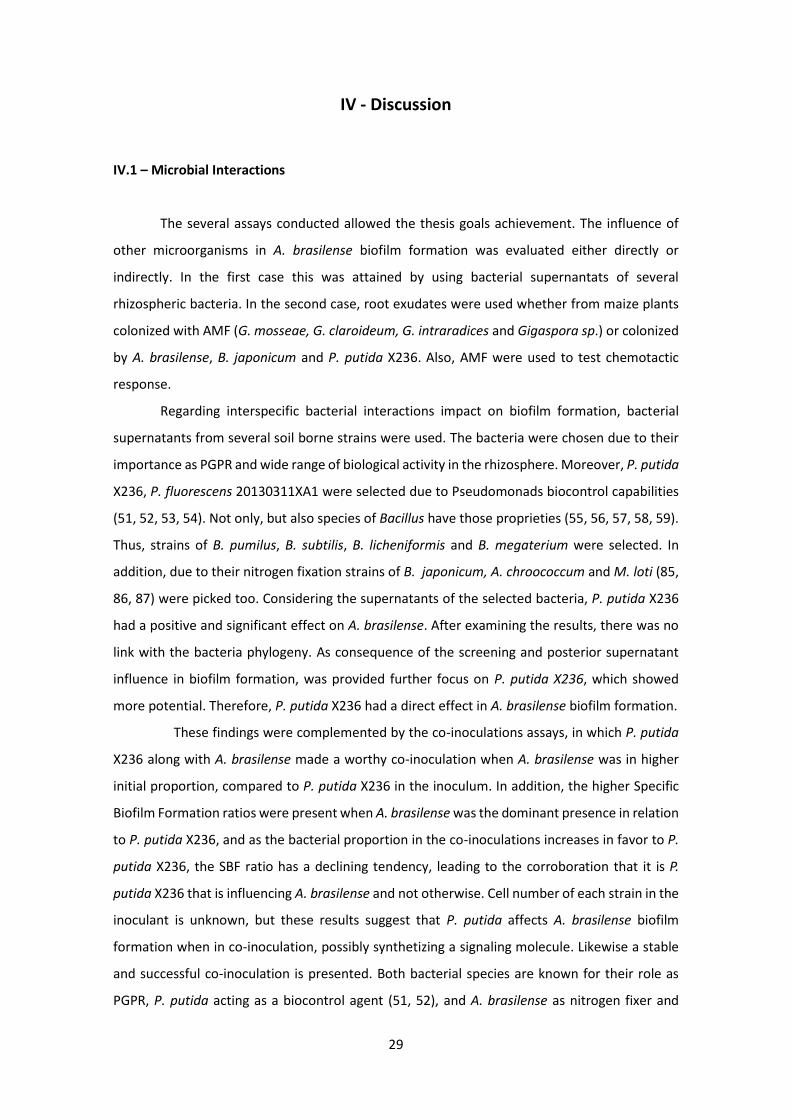
IV - Discussion
IV.1 – Microbial Interactions
The several assays conducted allowed the thesis goals achievement. The influence of
other microorganisms in A. brasilense biofilm formation was evaluated either directly or
indirectly. In the first case this was attained by using bacterial supernantats of several
rhizospheric bacteria. In the second case, root exudates were used whether from maize plants
colonized with AMF (G. mosseae, G. claroideum, G. intraradices and Gigaspora sp.) or colonized
by A. brasilense, B. japonicum and P. putida X236. Also, AMF were used to test chemotactic
response.
Regarding interspecific bacterial interactions impact on biofilm formation, bacterial
supernatants from several soil borne strains were used. The bacteria were chosen due to their
importance as PGPR and wide range of biological activity in the rhizosphere. Moreover, P. putida
X236, P. fluorescens 20130311XA1 were selected due to Pseudomonads biocontrol capabilities
(51, 52, 53, 54). Not only, but also species of Bacillus have those proprieties (55, 56, 57, 58, 59).
Thus, strains of B. pumilus, B. subtilis, B. licheniformis and B. megaterium were selected. In
addition, due to their nitrogen fixation strains of B. japonicum, A. chroococcum and M. loti (85,
86, 87) were picked too. Considering the supernatants of the selected bacteria, P. putida X236
had a positive and significant effect on A. brasilense. After examining the results, there was no
link with the bacteria phylogeny. As consequence of the screening and posterior supernatant
influence in biofilm formation, was provided further focus on P. putida X236, which showed
more potential. Therefore, P. putida X236 had a direct effect in A. brasilense biofilm formation.
These findings were complemented by the co-inoculations assays, in which P. putida
X236 along with A. brasilense made a worthy co-inoculation when A. brasilense was in higher
initial proportion, compared to P. putida X236 in the inoculum. In addition, the higher Specific
Biofilm Formation ratios were present when A. brasilense was the dominant presence in relation
to P. putida X236, and as the bacterial proportion in the co-inoculations increases in favor to P.
putida X236, the SBF ratio has a declining tendency, leading to the corroboration that it is P.
putida X236 that is influencing A. brasilense and not otherwise. Cell number of each strain in the
inoculant is unknown, but these results suggest that P. putida affects A. brasilense biofilm
formation when in co-inoculation, possibly synthetizing a signaling molecule. Likewise a stable
and successful co-inoculation is presented. Both bacterial species are known for their role as
PGPR, P. putida acting as a biocontrol agent (51, 52), and A. brasilense as nitrogen fixer and

30
phytohormone producer (23, 24). However, studies about P. putida X236 were not found in the
literature. Nevertheless, it is a good begin for a future co-inoculant consortium. Field tests would
be necessary as it is already been shown in several studies that in vitro and in vivo (crop field)
results are sometimes different (66) and to ensure a synergism of the strains in inoculant is
accomplished. P. putida strains have the potential to even shift the microbial communities from
being, dominant gram positive to gram negative (70).
In relation to indirect effects in biofilm formation by A. brasilense, specifically via root
exudates, neither P. putida X236 nor B. japonicum had pronounced influence. Nonetheless,
maize root exudates colonized of either AMF or PGPR, produced interesting results. Exudates
from plants inoculated with A. brasilense influenced biofilm formation of itself, whereas
exudates from mycorrhized plants only had effects A. brasilense chemotaxis. Moreover, G.
mosseae created a greater chemotactic response by A. brasilense. Both microorganisms are
recognized to stimulate maize to synthetize several cyclic hydroxamic acids, which may
accumulate in roots and are known for their beneficial effect on plants immune response,
namely against fungal and bacterial pathogens. One of those compounds is 2, 4-dihydroxy-7-
methoxy-2H-1, 4-benzoxazin-3(4 H)-one (DIMBOA). In addition, A. brasilense depending on the
strain and maize cultivar may impact secondary metabolism, either stimulating DIMBOA
synthesis or 6-methoxbenzoxazolin-2-one (MBOA), thereby these secondary metabolites may
be exudated by roots and even in a larger scale in plants inoculated with A. brasilense (88, 89).
Despite of biocontrol capabilities of these molecules, recent research has shown that P.
putida KT240 may be attracted by DIMBOA to the maize roots. Furthermore, studies
demonstrated that at least in wheat, DIMBOA and MBOA may actually alter microbial
communities in the rhizospere, increasing Gram-negative bacteria biomass (90, 91). Even though
this should be taken carefully, as DIMBOA usually degrades quickly in soil into its derivates such
as MBOA (92). Based in the literature, it could be hypothesized that in the present study A.
brasilense may had stimulated DIMBOA synthesis and exudation by the roots and trigger biofilm
formation. Regarding exudates from plants inoculated with G. mosseae, they could contain
DIMBOA, which might had generated a chemotactic response by A. brasilense.
Regarding the organic acids utilized, this strain of A. brasilense did not generate
chemotactic response towards those organic acids. This results are according to previous
studies, which showed that chemotaxis in Azospirillum is strain dependent (32). As for amino
acids results were consistent as previous studies describe, A. brasilense is attracted towards
amino acids commonly found in root exudates (93) and are used as a nitrogen source. However,
amino acid composition may be different among plant species and change throughout their

31
development, also as mentioned in previous research, specific amino acids may have a major
role in colonization of a specific plant species and for nitrogen fixation as well (31).
Summing up, root exudates assays aided to verify that certainly P. putida X236 has a
direct stimulating effect in A. brasilense biofilm formation, which is not through maize root
exudates.
IV.2 – IAA
The IAA production detected raises either in biofilm as in planktonic cells. This is
expected due to several tryptophan dependent pathways like Indole-3-pyruvate pathway, being
this one thought to be one of the major pathway for IAA biosynthesis (94). In the absence of
tryptophan supplementation, IAA synthesis is viable due to tryptophan independent pathway,
which was demonstrated in A. brasilense (94).
Looking at the results, in biofilm IAA was detect in greater concentrations without or
with low supplementation compared to planktonic cells, and the last ones reach their uppermost
synthesis of IAA with high supplementation, consequently is conceivable to claim that in biofilm
A. brasilense is less dependent on tryptophan as precursor for IAA synthesis, using tryptophan
independent pathway to synthetize IAA, and while as free living this bacteria is more prone to
utilize tryptophan dependent pathways. However, these results should be analyzed carefully
since the difference in cell numbers are significant, being higher in planktonic cells, which might
possibly caused the difference in IAA synthesis in the assay. In addition, there is in course a
reviewing process of the independent tryptophan pathway as until now no specific enzymes of
this pathway have been described (94). Since A. brasilense colonizes the roots, being this region,
especially the tip of the root rich in amino acids (95), with a large support of amino acids, it was
expectable that in biofilm the usage of tryptophan pathways was more dominant.

32

33
V - Concluding Remarks
Comprehend what may influence A. brasilense development and function is crucial in
such high potential bacteria to be used in agriculture as a biofertilizer, contributing to reduce
pollutions by in the future being a substitute to chemical fertilizers. Increasing biofilm formation
might help to reduce the costs of a future inoculum and that way encourage its utilization.
Therefore it is important to study how biotic factors influence biofilm formation and chemotaxis
of this species.
With this work it was demonstrated that several biotic factors can positively influence
biofilm formation of the A. brasilense strain used. One bacterium used, namely P. putida X236
has the capability of influencing directly, possibly via secondary metabolites. This strain also
showed to be a good combination for a potential future co-inoculum along with A. brasilense.
Nevertheless, further research is required to verify which compounds produced by P. putida
X236 influenced A. brasilense biofilm formation and further testing ensure positive effects exists
on selected plants. Root exudates from mycorrhized plants did not influenced biofilm formation,
though two mycorrhizal fungal species, G. mosseae and G. claroideum, proved to have a role in
A. brasilense chemotaxis. IAA synthesis, which is one of the major plant benefits attributed to A.
brasilense, and further research is required either in the pathways specifics as in tryptophan
independent pathway or synthesis in biofilm and its differences to planktonic cells.
The main goal was achieved, and interspecific interactions were inquired. This work
highlights the potential for using a microbial inoculum consortium consisting of mycorrhizal
fungi and bacterial species to stimulate biofilm development and function of A. brasilense.

34

35
VI - References
1- Philippot L, Raaijmakers JM, Lemanceau P, van der Putten WH. (2013). Going back to the roots: the microbial ecology of the rhizosphere. Nature Review of Microbiology. 11: 789-799
2- Costerton JW, Cheng KJ, Geesey GG, Ladd TI, Nickel JG, Dasgupta M, Marrie TJ. (1987). Bacterial biofilms in nature and disease. Annual Review of Microbiology. 41: 435-464
3- O’Toole G, Kaplan HB, Kolter R. (2000). Biofilm Formation as Microbial Development. Annual Review of Microbiology. 54: 49-79
4- Solano C, Echeverz M, Lasa I. (2014). Cell regulation biofilm dispersion and quorum sensing. Current Opinion in Microbiology. 18: 96-104
5- Miller MB, Bassler BL. (2001). Quorum sensing in bacteria. Annual Review of Microbiology. 55: 165-199
6- Kaplan HB, Greenberg EP. (1985). Diffusion of autoinducer is involved in regulation of the Vibrio fischeri luminescence system. Journal of Bacteriology. 163: 1210-1214
7- Christensen BE. (1989). The role of extracellular poly-saccharides in biofilms. Journal of Biotechnology. 10: 181-202
8- Gilbert P, Das J, Foley I. (1997). Biofilms susceptibility to antimicrobials. Advances in Dental Research. 11: 160–167
9- Nichols WW, Dorrington SM, Slack MPE, Walmsley HL. (1988). Inhibition of tobramycin diffusion by binding to alginate. Antimicrobial Agents and Chemotherapy. 32: 518-523
10- Nichols WW, Evans MJ, Slack MPE, Walmsley HL. (1989). The penetration of antibiotics into aggregates of mucoid and non-mucoid Pseudomonas aeruginosa. Journal of General Microbiology. 135: 1291-1303
11- Decho AW. (1990). Microbial exopolymer secretions in ocean environments: their role(s) in food webs and marine processes. Oceanography and Marine Biology - an Annual Review. 28: 73-153
12- Flemming HC. (1993). Biofilms and environmental protection. Water Science and Technology. 27: 1-10
13- Costerton JW, Lewandowski Z, Caldwell DE, Korber DR, Lappin-Scott HM. (1995). Microbial biofilms. Annual Review of Microbiology. 49: 711-745
14- Ehlers LJ, Bouwer EJ. (1999). RP4 plasmid transfer among species of Pseudomonas in a biofilm reactor. Water Science and Technology. 7: 163-171.
15- Hausner M, Wuertz S. (1999). High rates of conjugation in bacterial biofilms as determined by quantitative in situ analysis. Applied and Environmental Microbiology. 65: 3710-3713
16- Danhorn T, Fuqua C. (2007). Biofilm formation by plant-associated bacteria. Annual Review of Microbiology. 61: 401-422
17- Kloepper JW, Schroth MN. (1978). Plant growth-promoting rhizobacteria on radishes. Proceedings of the 4th international conference on plant pathogenic bacteria (Angers, France: Station de Pathologie Végétale et Phytobactériologie). Institut National de la Recherche Agronomique. 2: 879–882
18- Kloepper JW, Scher FM, Laliberte B, Tipping B. (1986). Emergence-plant growth promoting rhizobacteria: description and implication for agriculture. Iron, siderophores, and plant diseases. Swinburne TR eds. Springer Life Science, The Netherlands
19- Davison J. (1988). Plant beneficial bacteria. Bio-Technology. 6: 182-286 20- Okon Y, Labandera-Gonzalez CA. (1994). Agronomic applications of Azospirillum: an evaluation
of 20 years of worldwide field inoculation. Soil Biology & Biochemistry. 26: 1591–1601 21- Bashan Y, Levanony H. (1990). Current status of Azospirillum inoculation technology: Azospirillum
as a challenge for agriculture. Canadian Journal of Microbiology. 36: 591–608 22- Bashan Y, Ream Y, Levanony H, Sade A. (1995). Non-specific responses in plant growth, yield and
root colonization of noncereal crop plants to inoculation with Azospirillum brasilense Cd. Canadian Journal of Botany. 67: 1317–1324
23- Baca BE, Elmerich C. (2007). Microbial production of plants hormones by microorganisms. Associative nitrogen-fixation bacteria and cyanobacteria. IV. Series: fitrogen fixation: origins, Applications, and Research Progress. 113–137. Elmerich C, Newton W, eds. Springer Life Science, The Netherlands

36
24- Steenhoudt O, Vanderleyden J. (2000). Azospirillum, a free-living nitrogen-fixing bacterium closely associated with grasses: genetic, biochemical and ecological aspects. Federation of European Microbiological Societies Microbiology Reviews. 24: 487-506
25- Creus CM, Graziano M, Casanovas EM, Pereyra MA, Simontacchi M, Puntarulo S, Barassi CA, Lamattina L. (2005). Nitric oxide is involved in the Azospirillum brasilense-induced lateral root formation in tomato. Planta. 221: 297-303
26- Bakanchikova TI, Lobanok EV, Pavlova-Ivanova LK, Redkina TV, Nagapetyan ZA, Majsuryan AN. (1993). Inhibition of tumor formation process in dicotyledonous plants by Azospirillum brasilense strains. Mikrobiologiya. 62: 515–523
27- Romero AM, Correa O, Moccia S, Rivas JG. (2003). Effect of Azospirillum-mediated plant growth promotion on the development of bacterial diseases on fresh-market and cherry tomato. Journal of Applied Microbiology. 95: 832–838
28- Bashan Y, de-Bashan LE. (2002). Protection of tomato seedlings against infection by Pseudomonas syringae pv. Tomato by using the plant growth-promoting bacterium Azospirillum brasilense. Applied and Environmental Microbiology. 68: 2637–2643
29- Bashan Y, de-Bashan LE. (2002). Reduction of bacterial speck (Pseudomonas syringae pv. tomato) of tomato by combined treatments of plant growth-promoting bacterium, Azospirillum brasilense, streptomycin sulfate, and chemo-thermal seed treatment. European Journal Plant Pathology. 108: 821–829
30- Somers E, Ptacek D, Gysegom P, Srinivasan M, Vanderleyden J. (2005). Azospirillum brasilense produces the auxin-like phenylacetic acid by using the key enzyme for indole-3-acetic acid biosynthesis. Applied and Environmental Microbiology. 71: 1803-10
31- Hartmann A, Fu HA, Burris RH. (1988). Influence of amino acids on nitrogen fixation ability and growth of Azospirillum spp. Applied and Environmental Microbiology. 54: 87-93
32- Reinhold B, Hurek T, and Fendrik I, (1985). Strain specific chemotaxis of Azospirillum spp. Journal of Bacteriology. 162: 190-195
33- Vial L, Cuny C, Gluchoff-Fiasson K, Comte G, Oger PM, Faure D, Dessaux Y, Bally R, Wisniewski-Dyé F. (2006). N-acyl-homoserine lactone-mediated quorum-sensing in Azospirillum: an exception rather than a rule. Federation of European Microbiological Societies Microbiology Ecology. 58: 155–168
34- Somers E, Ptacek D, Gysegom P, Srinivasan M, Vanderleyden J. (2005). Azospirillum brasilense produces the auxin-like phenylacetic acid by using the key enzyme for indole-3-acetic acid biosynthesis. Applied and Environmental Microbiology. 71: 1803-1810
35- Greer-Phillips SE, Stephens BB, Alexandre G. (2004). An energy taxis transducer promotes root colonization by Azospirillum brasilense. Journal of Bacteriology. 186: 6595-6604
36- Burdman S, Okon Y, Jurkevitch E. (2000). Surface characteristics of Azospirillum brasilense in relation to cell aggregation and attachment to plant roots. Critical Reviews in Microbiology. 26: 91-110
37- Burdman S, Jurkevitch E, Schwartsburd B, Okon Y. (1999). Involvement of outermembrane proteins in the aggregation of Azospirillum brasilense. Microbiology. 145:1145–52
38- Burdman S, Dulguerova G, Okon Y, Jurkevitch E. (2001). Purification of the major outer membrane protein of Azospirillum brasilense, its affinity to plant roots, and its involvement in cell aggregation. Molecular Plant-Microbe Interactions. 14: 555-561
39- Wisniewski-Dyé F, Borziak K, Khalsa-Moyers G, Alexandre G, Sukharnikov LO, Wuichet K, Hurst GB, McDonald WH, Robertson JS, Barbe V, Calteau A, Rouy Z, Mangenot S, Prigent-Combaret C, Normand P, Boyer M, Siguier P, Dessaux Y, Elmerich C, Condemine G, Krishnen G, Kennedy I, Paterson AH, González V, Mavingui P, Zhulin IB. (2011). Azospirillum genomes reveal transition of bacteria from aquatic to terrestrial environments. Public Library of Science Genetics. 7(12): e1002430. doi: 10.1371/journal.pgen.1002430
40- Combes-Meynet E, Pothier JF, Moënne-Loccoz Y, Prigent-Combaret C. (2011) The Pseudomonas secondary metabolite 2, 4-diacetylphloroglucinol is a signal inducing rhizoplane expression of Azospirillum genes involved in plant-growth promotion. Molecular Plant-Microbe Interactions. 24: 271-284
41- Di Palma AA, Pereyra CM, Ramirez LM, Xiqui Vazquez ML, Baca BE, Pereyra MA, Lamattina L , Creus CM. (2013). Denitrification-derived nitric oxide modulates biofilm formation in Azospirillum brasilense. Federation of European Microbiological Societies Microbiology Letters. 338: 77-85
http://www.ncbi.nlm.nih.gov/pubmed?term=Casanovas%20EM%5BAuthor%5D&cauthor=true&cauthor_uid=15824907
37
42- Smith SE, Read DJ. (2008). Mycorrhizal Symbiosis 3rd eds. Academic Press, Amsterdam 43- Schüssler A, Schwarzott D, Walker C. (2001). A new fungal phylum, the Glomeromycota:
phylogeny and evolution. Mycological Research. 105:1413–1421 44- Tedersoo L, May TW, Smith ME. (2010). Ectomycorrhizal lifestyle in fungi: global diversity,
distribution, and evolution of phylogenetic lineages. Mycorrhiza. 20: 217-263 45- Fitter AH, Garbays J. (1994). Interactions between mycorrhizal fungi and other soil organisms.
Plant and Soil. 159: 123-132 46- Mishra S, Sharma S, Vasudevan P. (2008). Comparative effect of biofertilizers on fodder
production and quality in guineagrass (Panicum maximum Jacq.). Journal of the Science of Food and Agriculture. 88: 1667-1673
47- Hayman DS. (1982). Influence of soils and fertility on activity and survival of vesicular-arbuscular mycorrhizal fungi. Phytopathology. 72: 1119–1125
48- Andrade G., Mihara KL, Linderman RG, Bethlenfalvay, GJ. (1997). Bacteria from rhizosphere and hyphosphere soils of different arbuscular-mycorrhizal fungi. Plant and Soil. 192: 71-79
49- Artursson V, Finlay RD, Jansson JK. (2005) Combined bromodeoxyuridine immunocapture and terminal restriction fragment length polymorphism analysis highlights differences in the active soil bacterial metagenome due to Glomus mosseae inoculation or plant species. Environmental Microbiology. 7: 1952–1966
50- Drigo B, Pijl AS, Duyts H, Kielak AM, Gamper HA, Houtekamer MJ, Boschker HTS, Bodelier PLE, Whiteley AS, van Veen JA, Kowalchuk GA. (2010). Shifting carbon flow from roots into associated microbial communities in response to elevated atmospheric CO2. Proceedings of the National Academy of Sciences. 107: 10938-10942
51- Validov S, Kamilova F, Qi S, Stephan D, Wang JJ, Makarova N, Lugtenberg B. (2007). Selection of bacteria able to control Fusarium oxysporum f. Sp. Radicis lycopersici in stonewool substrate. Journal of Applied Microbiology. 102: 461-471
52- de Boer M, Bom P, Kindt F, Keurentjes JJ, van der Sluis l, van Loon LC, Bakker PA. (2003). Control of Fusarium Wilt of Radish by Combining Pseudomonas putida Strains that have Different Disease-Suppressive Mechanisms. Phytopathology. 93: 626-632
53- Weller DM, Thomashow LS. (1993). Microbial metabolites with biological activity against plant pathogens, Pest Management: Biologically Based Technologies. 173-180. Lumsden RD, Vaughn JL eds. American Chemical Society, Washington DC
54- Kamilova F, Validov S, Azarova T, Mulders I, Lugtenberg B. (2005). Enrichment for enhanced competitive plant root tip colonizers selects for a new class of biocontrol bacteria. Environmental Microbiology. 7: 1809–1817
55- Cavaglieri L, Orlando J, Rodríguez MI, Chulze S, Etcheverry M. (2005).Biocontrol of Bacillus subtilis against Fusarium verticillioides in vitro and at the maize root level. Research in Microbiology. 156: 748-54
56- Kildea S, Ransbotyn V, Khan MR,Fagan B, Leonard G, Mullins E. (2008). Bacillus megaterium shows potential for the biocontrol of septoria tritici blotch of wheat. Biological Control. 47: 37-45
57- Lee JP, Lee SW, Kim CS, Son JH, Song JH, Lee KY, Kim HJ, Jung SJ, Moon BJ. (2006). Evaluation of formulations of Bacillus licheniformis for the biological control of tomato gray mold caused by Botrytis cinerea. Biological Control. 37: 329-337
58- Huang X, Zhang N, Yong X, Yang X, Shen Q .(2012). Biocontrol of Rhizoctonia solani damping-off disease in cucumber with Bacillus pumilus SQR-N43. Microbiological Research. 167: 135-43
59- Xie ZP, Staehelin C, Vierheilig H, Wiemken A, Jabbouri S, Broughton WJ, Vogeli-Lange R, T Boller. (1995). Rhizobial nodulation factors stimulate mycorrhizal colonization of nodulating and nonnodulating soybeans. Plant Physiology. 108: 1519-1525
60- Cassána F, Perriga D, Sgroya V, Pennab O, Lunaa V. (2009). Azospirillum brasilense Az39 and Bradyrhizobium japonicum E109 inoculated singly or in combination, promote seed germination and early seedling growth in corn (Zea mays L.) and soybean (Glycine max L.). European Journal of Soil Biology. 45: 28-35
61- Paterson E, Gebbing T, Abel C, Sim A, Telfer G. (2007). Rhizodeposition shapes rhizosphere microbial community structure in organic soil. New Phytologist. 173: 600-610
62- Bais HP, Weir TL, Perry LG, Gilroy S, Vivanco JM. (2006). The role of root exudates in rizosphere interactions with plants and other organisms. Annual Review of Plant Biology. 57: 233-66
http://www.ncbi.nlm.nih.gov/pubmed?term=Cavaglieri%20L%5BAuthor%5D&cauthor=true&cauthor_uid=15950130
38
63- Bakker PA, Berendsen RL, Doornbos RF, Wintermans PC, Pieterse CM. (2013). The rhizospere revisited: root microbiomics. Frontiers in Plant Science. 4: 165
64- Haichar F, Marol C, Berge O, Rangel-Castro J, Prosser J. (2008). Balesdent J, Heulin T, Achouak W. Plant host habitat and root exudates shape soil bacterial community structure. International Society for Microbial Ecology. 2: 1221-1230
65- Pieterse CM, Zamioudis C, Berendsen RL, Weller DM, Van Wees SC, Bakker PA. (2014). Induced systemic resistance by beneficial microbes. Annual Review of Phytopathology. 52: 347-375
66- Trabelsi D, Mhamdi R. (2013). Microbial inoculants and their impact on soil microbial communities: A Review Hindawi BioMed Research International. Vol. 2013 Article ID 863240
67- Morrissey JP, Dow JM, Mark GL, O’Gara F. (2004). Are microbes at the root of a solution to world food production? European Molecular Biology Organization reports. 5: 922-926
68- Reis VM, dos Santos Teixeira KR, Pedraza RO. (2011). Bacteria in agrobiology: plant growth responses. 6: 132-134. Maheshwari DK eds. Springer Life Science, The Netherlands
69- Lerner A, Herschkovitz Y, Baudoin E. (2006). Effect of Azospirillum brasilense inoculation on rhizobacterial communities analyzed by denaturing gradient gel electrophoresis and automated ribosomal intergenic spacer analysis. Soil Biology and Biochemistry. 38: 1212-1218
70- de Salamone I, Di Salvo LP, Ortega J, Boa Sorte P, Urquiaga S, Teixeira KR. (2010). Field response of rice paddy crop to Azospirillum inoculation: physiology of rhizosphere bacterial communities and the genetic diversity of endophytic bacteria in different parts of the plants. Plant and Soil. 336: 351-362
71- Felici C, Vettori L, Giraldi E, Forino LM, Toffanin A, Tagliasacchi AM, Nuti M. (2008). Single and co-inoculation of Bacillus subtilis and Azospirillum brasilense on Lycopersicon esculentum: effects on plant growth and rhizosphere microbial community. Applied Soil Ecology. 40: 260-270
72- Kozdrój J, Trevors JT, Van Elsas JD. (2004). Influence of introduced potential biocontrol agents on maize seedling growth and bacterial community structure in the rhizosphere. Soil Biology and Biochemistry. 36: 1775–1784
73- Ruíz-Sánchez M, Armada E, Munoz Y, de Salamone I, Aroca R, Ruíz-Lozano J, Azcón R. (2011). Azospirillum and arbuscular mycorrhizal colonization enhance rice growth and physiological traits under well-watered and drought conditions. Journal of Plant Physiology. 168: 1031-1037
74- Pulido LE, Cabrera A, Medina N. (2003). Biofertilization using rhizobacteria and AMF in the production of tomato (Lycopersicon esculentum Mill.) and onion (Allium cepa L.) seedlings. II. Root colonization and nutritional status. Cultivos Tropicales. 24: 5-13
75- Lapage S, Shelton J, Mitchell T. (1970). Methods in Microbiology. Norris J. and Ribbons D eds. Academic Press, London.
76- Allen EK, Allen ON. (1950). Biochemical and symbiotic properties of the rhizobia. Bacteriological Reviews. 14: 273-330
77- O'Toole GA. (2011). Microtiter dish biofilm formation assay. Journal of Visualized Experiments. 47: 2437
78- Niu C, Gilbert ES. (2004). Colorimetric method for identifying plant essential oil components that affect biofilm formation and structure. Applied and Environmental Microbiology. 70: 6951-6956
79- Donati AJ, Lee HI, Leveau JH, Chang WS. (2013). Effects of indole-3-acetic acid on the transcriptional activities and stress tolerance of Bradyrhizobium japonicum. Public Library of Science ONE. 8(10): e76559. doi:10.1371/journal.pone.0076559
80- Hoagland DR, Arnon DI. (1950). The water-culture method of growing plants without soil. California Agricultural Experiment Station. 347.
81- Pinior A, Wys U, Piche ÂY, Vierheilig H. (1999). Plants colonized by AM fungi regulate further root colonization by AM fungi through altered root exudation. Canadian Journal of Botany 77: 1-7.
82- de Weert S, Kuiper I, Lagendijk EL, Lamers GE, Lugtenberg BJ. (2004). Role of chemotaxis toward fusaric acid in colonization of hyphae of Fusarium oxysporum f. sp. radicis-lycopersici by Pseudomonas fluorescens WCS365. Molecular Plant-Microbe Interactions. 16: 1185-1191
83- Gordon SA, Weber RP. (1951). Colorimetric estimation of indolacetic acid. Plant Physiology. 26: 192-195
84- Sarwar M, Kremer RJ. (1995). Determination of bacterially derived auxins using a microplate method. Letters in Applied Microbiology. 20: 282-285
85- Kaneko T, Nakamura Y, Sato S, Minamisawa K, Uchiumi T, Sasamoto S, Watanabe A, Idesawa K, Iriguchi M, Kawashima K, Kohara M, Matsumoto M, Shimpo S, Tsuruoka H, Wada T, Yamada
http://www.ncbi.nlm.nih.gov/pubmed?term=Berendsen%20RL%5BAuthor%5D&cauthor=true&cauthor_uid=24906124

39
M, Tabata S. (2002). Complete genomic sequence of nitrogen-fixing symbiotic bacterium Bradyrhizobium japonicum USDA110. DNA Research. 9: 189-197
86- Yates MG, de Souza EM, Kahindi JH. (1997). Oxygen, hydrogen and nitrogen fixation in Azotobacter. Soil Biology and Biochemistry. 29: 863-869
87- Shimoda Y, Shinpo S, Kohara M, Nakamura Y, Tabata S, Sato S. (2008). A large scale analysis of protein-protein interactions in the nitrogen-fixing bacterium Mesorhizobium loti. DNA Research. 15: 13-23
88- Walker V, Bertrand C, Bellvert F, Moënne-Loccoz Y, Bally R, Comte G. (2011). Host plant secondary metabolite profiling shows a complex, strain-dependent response of maize to plant growth-promoting rhizobacteria of the genus Azospirillum. New Phytologist. 189: 494-506
89- Song YY, Cao M, Xie LJ, Liang XT, Zeng RS, Su YJ, Huang JH, Wang RL, Luo SM. (2011). Induction of DIMBOA accumulation and systemic defense responses as a mechanism of enhanced resistance of mycorrhizal corn (Zea mays L.) to sheath blight. Mycorrhiza. 21: 721-731
90- Neal AL, Ahmad S, Gordon-Weeks R, Ton J. (2012). Benzoxazinoids in Root Exudates of Maize Attract Pseudomonas putida to the Rhizosphere. Public Library of Science ONE. 7(4): e35498. doi: 10.1371/journal.pone.0035498
91- Chen KJ, Zheng YQ, Kong CH, Zhang SZ, Li J, Liu XG. (2010). 2, 4-Dihydroxy-7-methoxy-1, 4-benzoxazin-3-one (DIMBOA) and 6-Methoxy-benzoxazolin-2-one (MBOA) levels in the wheat rhizosphere and their effect on the soil microbial community structure. Journal of Agricultural and Food Chemistry. 58: 12710-12716
92- Macias FA, Oliveros-Bastidas A, Marin D, Castellano D, Simonet AM, Molinillo JM. (2004). Degradation studies on benzoxazinoids. Soil degradation dynamics of 2, 4-dihydroxy-7-methoxy-(2H)-1, 4-benzoxazin-3(4H)-one (DIMBOA) and its degradation products, phytotoxic allelochemicals from Gramineae. Journal of Agricultural and Food Chemistry. 52: 6402-6413
93- Zhulin IB, Tretyakova SE, Ionatov VV. (1988). Chemotaxis of Azospirillum brasilense towards compounds typical of plant root exudates. Folia Microbiologica. 33: 277-28
94- Spaepen S, Vanderleyden J, R Remans. (2007). Indole-3-acetic acid in microbial and microorganism-plant signaling. Federation of European Microbiological Societies Microbiology Reviews. 31: 425-448
95- Jaeger JH, Lindow SE, Miller S, Clark E, Firestone MK. (1999). Mapping of sugar and amino acid availability in soil around roots with bacterial sensors of sucrose and tryptophan. Applied and Environmental Microbiology. 65: 2685–2690

40

VII - Appendices
Appendix I: Sterile system used in root exudate assay. Plants were inoculated with B. japonicum, P. putida X236 or A.
brasilense.
Appendix II: Microtiter plate of Indol-3-acetic-acid quantification assay, the pink color was caused by reaction with
Salkowski reagent. Afterwards optic density was measured at OD530.
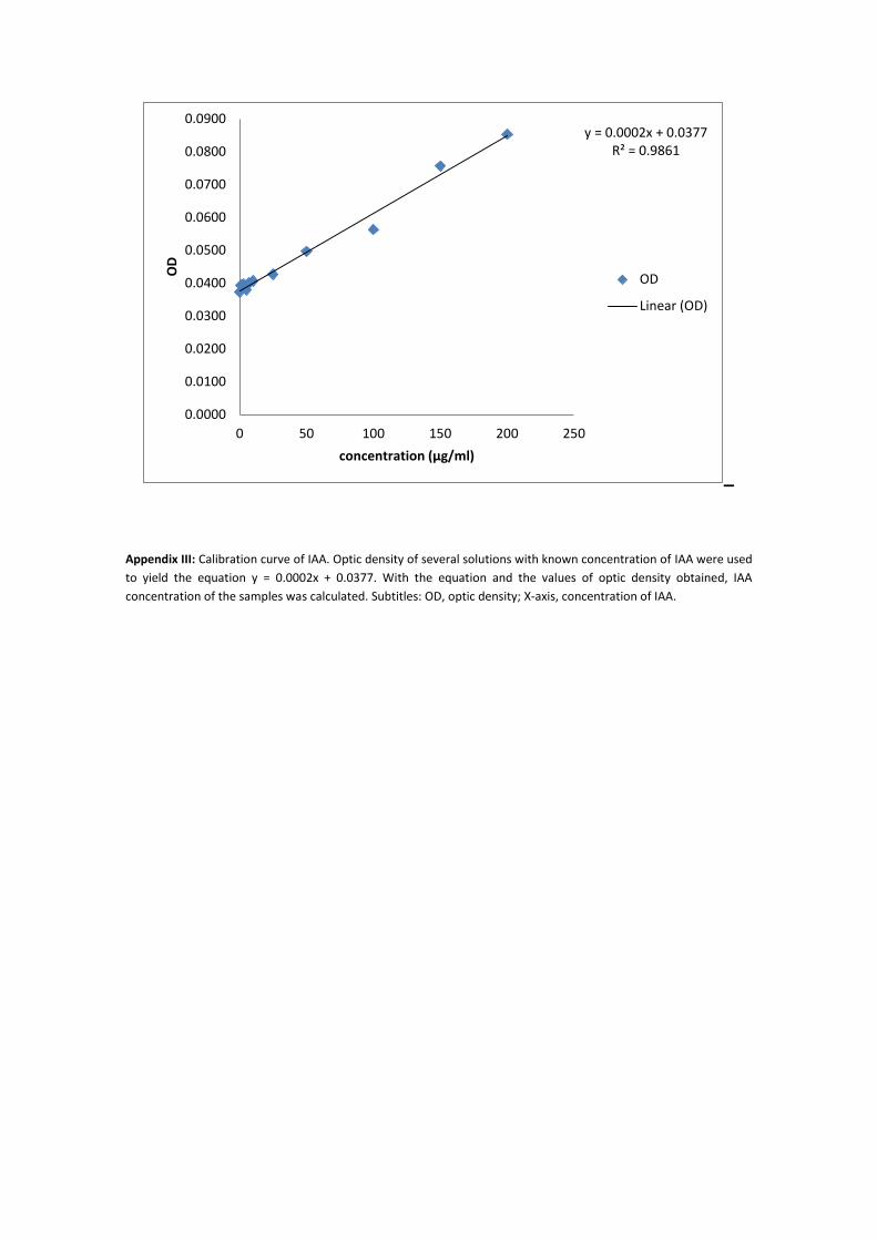
_
Appendix III: Calibration curve of IAA. Optic density of several solutions with known concentration of IAA were used
to yield the equation y = 0.0002x + 0.0377. With the equation and the values of optic density obtained, IAA
concentration of the samples was calculated. Subtitles: OD, optic density; X-axis, concentration of IAA.
y = 0.0002x + 0.0377R² = 0.9861
0.0000
0.0100
0.0200
0.0300
0.0400
0.0500
0.0600
0.0700
0.0800
0.0900
0 50 100 150 200 250
OD
concentration (µg/ml)
OD
Linear (OD)