FACTORS INFLUENCING THE OUTCOME OF ACUTE...
47
FACTORS INFLUENCING THE OUTCOME OF ACUTE LATERAL HUMERAL CONDYLAR FRACTURE IN CHILDREN AND ITS RELATED COMPLICATIONS BY DR WARITH MUHAMMAD BIN SYAFREIN EFFENDI Dissertation Submitted In Partial Fulfilment of The Requirement for The Degree of Master of Medicine (Orthopaedics) UNIVERSITI SAINS MALAYSIA 2015
Transcript of FACTORS INFLUENCING THE OUTCOME OF ACUTE...

FACTORS INFLUENCING THE OUTCOME OF ACUTE
LATERAL HUMERAL CONDYLAR FRACTURE IN
CHILDREN AND ITS RELATED COMPLICATIONS
BY
DR WARITH MUHAMMAD BIN SYAFREIN EFFENDI
Dissertation Submitted In Partial Fulfilment of
The Requirement for
The Degree of Master of Medicine
(Orthopaedics)
UNIVERSITI SAINS MALAYSIA
2015

FACTORS INFLUENCING THE OUTCOME OF ACUTE LATERAL HUMERAL
CONDYLAR FRACTURE IN CHILDREN AND ITS RELATED COMPLICATIONS
DR WARITH MUHAMMAD BIN SYAFREIN EFFENDI
Department of Orthopedic,
School of Medical Sciences, Universiti Sains Malaysia
Health Campus, 16150 Kelantan, Malaysia.
Introduction : Lateral condylar of humerus fractures are among the commonest fracture
in children. Open reduction and internal fixation (ORIF) is the preferred option as it
prevents complications caused by inaccurate reduction, however the outcomes remain
variable.
Objective : The purpose of this study is to determine factors influencing the overall
functional outcome following lateral condylar fractures of humerus in children and to
describe the complications that arise.

Methods: There were children until the age of 14 years old for the girls and 16 years old
for the boys were involved in this study. All of them were treated for lateral condyle
humeral fracture for at least 1 year. They were selected and contacted after reviewing
their radiological and treatment records and were asked to come to HUSM for further
evaluation. During evaluation, the functional ability of the involved elbow was assessed.
A radiological assessment of affected limb was also performed through a proper antero-
posterior radiograph. The functional outcome was assessed based on activity of daily
living, range of motion and carrying angle of the affected elbow with the normal elbow
and graded using Dhillon scoring system into excellent, good, fair and poor outcome. The
amounts of residual displacement after treatment were documented. Data were
statistically analysed using SPSS version 20.
Result: Twenty-seven male and six female patients were involved in this study. The age
at time of fracture was within 2 to12 years old with the mean of 6.3 years old. Thirteen
patients had medial residual displacement between 3mm to 5mm and 20 patients had
2mm or less medial residual displacement. A large numbers of patients attained good
functional outcome scoring (42.4%) followed by excellent score (27.3%). This is
followed by fair score (24.2%). Only a small numbers of patient had poor scoring system
(6.1%). Both residual medial intraarticular displacement and residual lateral cortex
displacement post fracture treatment (both <2mm and 3mm-5mm) did not significantly
affect the early functional outcome (Dhillon score) .( Multiple logistic regression, β = -
0.19; 95% CI=-1.09, 0.40; p-value= 0.034) (Multiple logistic regression, β= -0.12,CI= -

0.67,0.63, p-value 0.94). Age of fracture, type of treatment and method of surgical
fixation are not statistically correlated with Dhillon score. In term of complications, only
one patient (3.0%) had persistent pain, 14 (42.4%) patients clinically had lateral condyle
prominence, 4 (12%),with cubitus varus deformity, 2 (6.1%) had fishtail deformity, 19
(57.6%) had osteophytes and there was no incidence of AVN and non union. All of these
complications are not statistically associated with Dhillon scoring.
Conclusion: This study shows that children who sustained lateral condyle of humerus
fracture have excellent-good outcome (69.7%) after at least 1 year of follow up. The
amount of medial and lateral residual displacement of lateral humeral condyle fracture
did not affect the early functional outcome as long as it is within 5mm. The most
common complications encountered are osteophytes (57.6%) and lateral condyle
overgrowth (42.4%).

ABSTRAK
Latar- belakang: Kepatahan sisi luar tulang siku (humerus)adalah antara patah yang
lazim pada kanak-kanak . Pembedahan (Open reduction Internal Fixation )untuk
memperbetulkan tulang yang patah dan teranjak adalah pilihan yang lebih popular
kerana ia dapat mengelakkan komplikasi yang disebabkan oleh teknik pembedahan yang
tidak tepat, namun hasil akhir dari rawatan ini adalah tidak sama dan sering berubah-
ubah.
Objektif : Tujuan kajian ini adalah untuk menilai hasil akhir fungsi bagi patah sisi luar
tulang siku (humerus)di kalangan kanak-kanak yang menjalani operasi atau rawatan
konservatif dan juga komplikasi-komplikasi yang biasa terjadi. Ia juga bertujuan untuk
mengkaji sama ada tahap anjakan serpihan tulang patah (displacement) dan kualiti redusi
pembedahan dapat mempengaruhi hasil akhir fungsi atau tidak.
Metodologi: Tiga puluh tiga kanak-kanak yang belum mencapai kematangan usia iaitu
14 tahun untuk kanak-kanak perempuan dan 16 tahun untuk kanak-kanak lelaki , terlibat
dalam kajian ini. Kesemua mereka telah dirawat selepas mengalami patah sisi luar di siku
(humerus) untuk sekurang-kurangnya 1 tahun. Mereka telah dipilih dan dihubungi
selepas kajian semula rekod radiologi dijalankan selepas rawatan dan telah diminta untuk
datang ke HUSM untuk penilaian lanjut. Pada masa yang sama , keupayaan hasil akhir
fungsi tangan yang terlibat telah dinilai dan dimarkahkan . Satu penilaian radiologi dari

siku yang terlibat juga dilakukan dengan kaedah radiografi . Hasil akhir fungsi dinilai
berdasarkan aktiviti seharian, sudut dan darjah pergerakan sendi yang terlibat
.Pemarkahan ini dinilai menggunakan kaedah Dhillon Scoring ke dalam kumpulan yang
sangat baik, baik, sederhana dan teruk. Anjakan serpihan yang patah dan kualiti redusi
pembedahan didokumentasikan . Data dianalisis secara statistik menggunakan SPSS versi
20.
Keputusan: Dua puluh tujuh kanak-kanak lelaki dan 6 perempuan terlibat dalam kajian
ini. Usia pada masa kepatahan adalah puratanya berusia 6.3 tahun merangkumi usia dari
2 tahun sampai 12 tahun. Tiga belas pesakit mengalami anjakan kepatahan antara 3mm-
5mm manakala 20 lagi pesakit mengalami anjakan kurang dari 2mm. 14 pesakit
mencapai skor yang bagus (42.4 %) diikuti dengan 9 pesakit skor sangat baik ( 27.3 %).
Ini diikuti dengan 8 pesakit skor sederhana ( 24.2 %). Hanya sejumlah kecil pesakit iaitu
2 orang dikategorikan sebagai teruk (2 (6.1 %). Kajian kami menunjukkan bahawa
anjakan serpihan tulang sebaik selepas rawatan , secara statistiknya tidak mempengaruhi
hasil awal fungsi skor Dhillon bagi anjakan tulang sisi dalam (residual medial
intraarticular displacement) (Multiple regresi logistik , β= -0.12,CI= -0.67,0.63, p-value
0.94) dan anjakan tulang sisi luar (residual lateral cortex displacement) (Multiple regresi
logistik, β= -0.12,CI= -0.67,0.63, p-value 0.94). Juga tidak ada perbezaan yang signifikan
di antara jenis rawatan dan skor Dhillon ( CI: -2.23,0.86 , p-value _0.37 ). Dalam konteks
komplikasi pula , hanya satu pesakit (3.0 %) mempunyai kesakitan yang berkekalan, 14 (
42.4 %) pesakit secara klinikal mempunyai tonjolan tulang , empat pesakit dengan
kebengkokan tulang (Cubitus varus) ( 12%) , dua ( 6.1 %) pesakit mempunyai kecacatan
fishtail , unjuran tulang (osteophytes) sekitar sembilan belas ( 57.6 % ) dan tidak ada
kejadian AVN dan tulang tidak bercantum (non union). Semua komplikasi ini tidak ada
kaitan statistik dengan pemarkahan Dhillon .
Kesimpulan: Kajian ini telah menunjukkan bahawa kebanyakan kanak-kanak yang
mengalami kepatahan tulang sisi luar di siku (humerus) akan mempunyai hasil yang
Cemerlang-Bagus ( 69.7 %) selepas sekurang-kurangnya 1 tahun rawatan susulan.
Jumlah anjakan kepatahan samada dari sisi dalam atau sisi luar tulang tidak menunjukkan
prebezaan dari segi fungsi awal Dhillon selagi anjakan masih dalam lingkungan 5mm.
Komplikasi yang paling biasa dihadapi adalah unjuran tulang (osteophtyes ( 57.6 %) dan
tonjolan tulang (lateral prominence) ( 42.4 %). Tiada sebarang kejadian AVN dan tulang
tidak bercantum ditemukan. Walau bagaimanapun, kami mendapati bahawa tahap
anjakan kepatahan selepas pembedahan dan jenis rawatan tidak mempengaruhi skor
Dhillon.
ii
l
TABLE OF CONTENT
FRONTPIECE I
ACKNOWLEGDEMENTS II
LIST OF CONTENT III
ABSTRAK IV
ABSTRACT V
ABBREVIATION VI
LIST OF FIGURES VII
LIST OF TABLES VIII

iii
II. ACKNOWLEDGEMENT
BISMILLAHIRRAHMANIRRAHIM
Praise to Allah the Almighty to allow the completion of this dissertation
I am very grateful to DR. ISMAIL MUNAJAT, as the supervisor of this study, senior lecturer in
Orthopedics Department Universiti Sains Malaysia, for the never ending support, encouragement,
guidance and patience during the course of the study. He has always been an exemplary figure to me
and at multiple times has solved major problems encountered during the study.
ASSOC PROF IMRAN YUSOF, the Head of Orthopedics Department, Universiti Sains Malaysia for his
encouragement and support for the conduction of the study.
PUAN NURHAZWANI HAMID, for her generous help in triggering the idea for this study and her
assistant in performing the statistical analysis.
My sincere thanks to DR. FAUZLIE YUSOF, Senior Consultant Orthopedic Surgeon of Hospital Melaka
as my co- supervisor for my dissertation who keeps on supporting me during the preparation of this
dissertation.
DR. N. SIVAPATHASUNDARAM, Consultant and the Head of Orthopedics and Traumatology Unit of
Hospital Melaka for his full support.
iv
Not to forget my thanks to all the patients in this study and support staffs in Hospital Universiti Sains
Malaysia (HUSM) for their precious time, support and kindness during the process of data collection
and preparation of this dissertation.
Thanks to all my fellow colleagues and lectures in HUSM for the support, suggestion, guidance and
encouragement during the completion of this dissertation.
Last but not least, my special thanks to my dear parents, SYAFREIN EFFENDI B. USMAN and NORAIN
BTE ISHAK, for their understanding and full support ,my beloved wife NORMA BTE JAMALUDIN , and
my wonderful children ZIYAD ZAYDAN and ZAIRA SOFEA, for their time and patience that they have
sacrificed, encouragement and understanding throughout my life.

v
III. LIST OF CONTENTS
1.0 Introduction 1
2.0 Literature review 3
2.1 Bone 3
2.1.1 Cellular biology 5
2.1.1(a) Osteoblasts 5
2.1.1(b) Osteocytes 7
2.1.1(c) Osteoclasts 7
2.1.2 Bone matrix 8
2.1.3 Blood supply of the bone 9
2.1.4 Fracture healing 11
2.1.5 The growth plate 13
2.2 Elbow fracture in children 16
2.2.1 Functional anatomy of elbow joint 18
2.2.2 Fracture classification 24
2.2.3 Mechanism of injury 27
2.2.4 Patient assessment and radiographic

vi
evaluation 28
2.2.5 Treatment option 32
2.2.6 Complications of the elbow fracture 38
3.0 Objectives of the study 45
4.0 Research methodology 46
4.1 Study design 46
4.2 Ethical approval 46
4.3 Study population 46
4.4 Inclusion criteria 46
4.5 Exclusion criteria 47
4.6 Sampling method and sample size calculation 47
4.6.1 Sampling technique 47
4.6.2 Sample size calculation 47
4.7 Data collection and methodology steps 48

vii
4.8 Statistical analysis 62
4.8.1 Simple Linear Regression (SLR) 62
4.8.2 Multiple Linear Regression (MLR) 63
4.8.3 Data requirements 64
4.8.4 Steps in Multiple Linear Regression (MLR) Analysis 67
5.0 Results and statistical analysis 69
6.0 Discussion of the result 99
6.1 Demographic Interpretation 99
6.2 Association between functional outcome and amount of
residual fracture displacement after treatment 100
6.3 Association of functional outcome with method of reduction 104
6.4 Functional outcome of acute lateral humeral condylar fracture and related
complications 106
7.0 Conclusion 111
8.0 Pitfalls and limitations 112
9.0 Recommendations 114
10.0 References 115
11.0 Appendix 119

viii
11.1 Appendix A- Flow chart 119
11.2 Appendix B- Proforma 120
11.3 Appendix C- Borang Etika-02 122
11.4 Appendix D- Research information and consent 124
11.5 Appendix E- Maklumat kajian dan kebenaran 132

ix
VI ABSTRAK
Latar- belakang: Kepatahan sisi luar tulang siku (humerus)adalah antara patah yang lazim
pada kanak-kanak . Pembedahan (Open reduction Internal Fixation )untuk memperbetulkan
tulang yang patah dan teranjak adalah pilihan yang lebih popular kerana ia dapat mengelakkan
komplikasi yang disebabkan oleh teknik pembedahan yang tidak tepat, namun hasil akhir dari
rawatan ini adalah tidak sama dan sering berubah-ubah. Tujuan kajian ini adalah untuk menilai
hasil akhir fungsi bagi patah sisi luar tulang siku (humerus)di kalangan kanak-kanak yang
menjalani operasi atau rawatan konservatif dan juga komplikasi-komplikasi yang biasa terjadi.
Ia juga bertujuan untuk mengkaji sama ada tahap anjakan serpihan tulang patah (displacement)
dan kualiti redusi pembedahan dapat mempengaruhi hasil akhir fungsi atau tidak.
Metodologi: Tiga puluh tiga kanak-kanak yang belum mencapai kematangan usia iaitu .14
tahun untuk kanak-kanak perempuan dan 16 tahun untuk kanak-kanak lelaki , terlibat dalam
kajian ini. Kesemua mereka telah dirawat selepas mengalami patah sisi luar di siku (humerus)
untuk sekurang-kurangnya 1 tahun. Mereka telah dipilih dan dihubungi selepas kajian semula
rekod radiologi dijalankan selepas rawatan dan telah diminta untuk datang ke HUSM untuk
penilaian lanjut. Pada masa yang sama , keupayaan hasil akhir fungsi tangan yang terlibat telah
dinilai dan dimarkahkan . Satu penilaian radiologi dari siku yang terlibat juga dilakukan dengan
kaedah radiografi . Hasil akhir fungsi dinilai berdasarkan aktiviti seharian, sudut dan darjah
pergerakan sendi yang terlibat .Pemarkahan ini dinilai menggunakan kaedah Dhillon Scoring ke
dalam kumpulan yang sangat baik, baik, sederhana dan teruk. Anjakan serpihan yang patah dan
kualiti redusi pembedahan didokumentasikan . Data dianalisis secara statistik menggunakan
SPSS versi 20.

x
Keputusan: Dua puluh tujuh kanak-kanak lelaki dan 6 perempuan terlibat dalam kajian ini.
Usia pada masa kepatahan adalah puratanya berusia 6.3 tahun merangkumi usia dari 2 tahun
sampai 12 tahun. Tiga belas pesakit mengalami anjakan kepatahan antara 3mm-5mm manakala
20 lagi pesakit mengalami anjakan kurang dari 2mm. 14 pesakit mencapai skor yang bagus
(42.4 %) diikuti dengan 9 pesakit skor sangat baik ( 27.3 %). Ini diikuti dengan 8 pesakit skor
sederhana ( 24.2 %). Hanya sejumlah kecil pesakit iaitu 2 orang dikategorikan sebagai teruk (2
(6.1 %). Kajian kami menunjukkan bahawa anjakan serpihan tulang sebaik selepas rawatan ,
secara statistiknya tidak mempengaruhi hasil awal fungsi skor Dhillon bagi anjakan tulang sisi
dalam (residual medial intraarticular displacement) (Multiple regresi logistik , β= -0.12,CI= -
0.67,0.63, p-value 0.94) dan anjakan tulang sisi luar (residual lateral cortex displacement)
(Multiple regresi logistik, β= -0.12,CI= -0.67,0.63, p-value 0.94). Juga tidak ada perbezaan yang
signifikan di antara jenis rawatan dan skor Dhillon ( CI: -2.23,0.86 , p-value _0.37 ). Dalam
konteks komplikasi pula , hanya satu pesakit (3.0 %) mempunyai kesakitan yang berkekalan, 14
( 42.4 %) pesakit secara klinikal mempunyai tonjolan tulang , empat pesakit dengan
kebengkokan tulang (Cubitus varus) ( 12%) , dua ( 6.1 %) pesakit mempunyai kecacatan
fishtail , unjuran tulang (osteophytes) sekitar sembilan belas ( 57.6 % ) dan tidak ada kejadian
AVN dan tulang tidak bercantum (non union). Semua komplikasi ini tidak ada kaitan statistik
dengan pemarkahan Dhillon .
Kesimpulan: Kajian ini telah menunjukkan bahawa kebanyakan kanak-kanak yang mengalami
kepatahan tulang sisi luar di siku (humerus) akan mempunyai hasil yang Cemerlang-Bagus (

xi
69.7 %) selepas sekurang-kurangnya 1 tahun rawatan susulan. Jumlah anjakan kepatahan
samada dari sisi dalam atau sisi luar tulang tidak menunjukkan prebezaan dari segi fungsi awal
Dhillon selagi anjakan masih dalam lingkungan 5mm. Komplikasi yang paling biasa dihadapi
adalah unjuran tulang (osteophtyes ( 57.6 %) dan tonjolan tulang (lateral prominence) ( 42.4 %).
Tiada sebarang kejadian AVN dan tulang tidak bercantum ditemukan. Walau bagaimanapun,
kami mendapati bahawa tahap anjakan kepatahan selepas pembedahan dan jenis rawatan tidak
mempengaruhi skor Dhillon.

xii
V ABSTRACT
Back ground: Lateral condylar of humerus fractures are among the commonest fracture in
children. Open reduction and internal fixation (ORIF) is the preferred option as it prevents
complications caused by inaccurate reduction, however the outcomes remain variable. The
purpose of this study is to determine factors influencing the overall functional outcome
following lateral condylar fractures of humerus in children and to describe the complications
that arise.
Methods: There were children until the age of 14 years old for the girls and 16 years old for the
boys were involved in this study. All of them were treated for lateral condyle humeral fracture
for at least 1 year. They were selected and contacted after reviewing their radiological and
treatment records and were asked to come to HUSM for further evaluation. During evaluation,
the functional ability of the involved elbow was assessed. A radiological assessment of affected
limb was also performed through a proper antero-posterior radiograph. The functional outcome
was assessed based on activity of daily living, range of motion and carrying angle of the
affected elbow with the normal elbow and graded using Dhillon scoring system into excellent,
good, fair and poor outcome. The amounts of residual displacement after treatment were
documented. Data were statistically analysed using SPSS version 20.
Result: Twenty-seven male and six female patients were involved in this study. The age at time
of fracture was within 2 to12 years old with the mean of 6.3 years old. Thirteen patients had
xiii
medial residual displacement between 3mm to 5mm and 20 patients had 2mm or less medial
residual displacement. A large numbers of patients attained good functional outcome scoring
(42.4%) followed by excellent score (27.3%). This is followed by fair score (24.2%). Only a
small numbers of patient had poor scoring system (6.1%). Both residual medial intraarticular
displacement and residual lateral cortex displacement post fracture treatment (both <2mm and
3mm-5mm) did not significantly affect the early functional outcome (Dhillon score) .( Multiple
logistic regression, β = -0.19; 95% CI=-1.09, 0.40; p-value= 0.034) (Multiple logistic
regression, β= -0.12,CI= -0.67,0.63, p-value 0.94). Age of fracture, type of treatment and
method of surgical fixation are not statistically correlated with Dhillon score. In term of
complications, only one patient (3.0%) had persistent pain, 14 (42.4%) patients clinically had
lateral condyle prominence, 4 (12%),with cubitus varus deformity, 2 (6.1%) had fishtail
deformity, 19 (57.6%) had osteophytes and there was no incidence of AVN and non union. All
of these complications are not statistically associated with Dhillon scoring.
Conclusion: This study shows that children who sustained lateral condyle of humerus fracture
have excellent-good outcome (69.7%) after at least 1 year of follow up. The amount of medial
and lateral residual displacement of lateral humeral condyle fracture did not affect the early
functional outcome as long as it is within 5mm. The most common complications encountered
are osteophytes (57.6%) and lateral condyle overgrowth (42.4%).

xiv
VI ABBREVIATION
AVN : Avascular necrosis
LCH : Lateral condyle humerus
LCOG : Lateral condyle overgrowth
RMD : Residual medial displacement

xv
VII LIST OF FIGURES Page
Fig. 1 Illustration of cortical bone 5
Fig. 2 Blood supply to bone 11
Fig.3 Structure and blood supply of a typical growth plate 15
Fig. 4 Illustration of the average time of appearance of ossification centers at the
distal humerus 22
Fig.5 Anterior and posterior extraosseous vascular anatomy around the elbow 23
Fig.6 Illustrations of the Milch classification of lateral condylar fracture 25
Fig.7 Illustrations of pediatric lateral humeral condylar fractures as described by Jakob. 26
Fig. 8 Hand held protractor goniometer 53
Fig. 9 Assessment of degree of flexion and extension 54

xvi
Fig. 10 The proper positioning of forearm for antero-posterior radiograph view 56
Fig. 11 The proper positioning of elbow for lateral radiograph view 56
Fig 12 Post reduction residual displacement measurement 57
Fig 13: Radiographic carrying angle of elbow 58
Fig 14: An osteophyte formation of the lateral condyle. 60
Fig.15: Pie chart of the distribution for gender to Dhillon score 77
Fig. 16: The histogram of the age of the patients at the time of fracture 78
Fig.17: Pie chart of the distribution for type of treatment 79
Fig.18: Pie chart of the distribution for type of surgery 80
Fig.19: Distribution of the residual medial displacement of patients (mm) 81
Fig. 20: Distribution of the residual lateral displacement of the patients (mm) 20
Fig. 21: Pie chart for Medial Residual Displacement 83
Fig.22: Pie chart of the distribution for Lateral Residual Displacement 84

xvii
Fig.23: Pie chart of the distribution for range of motion 85
Fig.24: Histogram for complications of lateral condyle fracture. 86
VIII LIST OF TABLES
Table 1.Dhillon scoring system 52
Table 2: Summary of the results 69
Table 3: Characteristic to the subjects 72
Table 4: Characteristics of the subjects for categorical variables 73
Table 5 : Summary of the variables 76
Table 6: Associated factors of Dhillon score by Simple Linear Regression 87
Table 7: Independent variable 88
Table 8 Associated factors of Dhillon score 92

1
1. INTRODUCTION
Lateral condylar fracture of humerus is the second most common fracture of the
elbow in children. Majority of them are injuries of growth plate with a Salter-
Harris IV fracture pattern, and they are common in children between 2-14 years
of age.(Wilkins.,1996) Open reduction and internal fixation (ORIF) is the
preferred option for most lateral condylar fractures because it prevents
complications caused by inaccurate reduction, although long arm cast
immobilization, closed reduction, and internal fixation (CRIF) can provide
effective treatment for the undisplaced or minimally displaced fractures. The
treatment goal in lateral condylar fracture is union without residual deformity.
(Landin et al.,1986)
However, complications such as nonunion, avascular necrosis (AVN), premature
epiphyseal fusion, lateral condylar overgrowth, stiffness, cubitus varus or cubitus
valgus, and fishtail deformity have been reported after the operative treatment of
lateral condylar fractures, despite initial anatomic reduction and fixation.(Song et
al.,2008)
One of method to assess the success of the treatment is by measuring the
functional outcome during follow up (Dhillon et al.,1988). Dhillon scoring system
has been widely used to assess the functional outcome after lateral condyle
fracture. There are multiple factors that could influence the functional outcome.
Kyong et al (2010) made a study of an association between Jakob type of

2
classifications and functional outcome of lateral humeral condyle fracture in
children and showed no differences in functional results between fracture types.
Wattenbarger et al (2002) study on lateral humeral condyle in children, and
measured the fracture displacement based on lateral metaphyseal site of the
fragments which is from non articulating surface of the elbow joint rather than
from medial intraarticular metaphyseal fragments. There was lack of literature
studying the associations between functional outcome and residual medial
intraarticular displacement which is more crucial to reflect articular incongruency.
Weiss et al (2009) also concluded from his study that fracture displacement and
articular incongruency predict complications.
Therefore, the current study is designed to determine factors influencing the
functional outcome of acute lateral condyle fracture of humerus particularly the
amount of residual medial displacement at minimum 1 year follow up. Apart from
that, this study also highlights the related complications encountered during
follow up.

3
2 LITERATURE REVIEW
2.1 BONE
Bone is a specialized mineralized connective tissue. It is dynamic ,
well structured and one of the hardest substances in the body and
constantly changes shape in relation to the stresses placed on it. It is
the primary structural framework for support and protection of all
organs in the body. (Sinnathamby.,2006) Bone also gives the
necessary rigidity to function as attachment and lever for muscles and
supports the body against gravity. Bone is a reservoir for several
minerals in the body and stores about ninety percent of body’s
calcium. (Brinker, Miller.,1999)
All of the bones have two basic structural components which are
compact and cancellous bone. Compact or cortical bone is the solid,
dense bone that is presence in the walls of bone shafts and on external
bone surfaces. Cancellous or trabecular bone is more porous,
lightweight and has honeycomb structure. It consists of delicate bars
and sheet of bone, with thin bony spicules (trabeculae) branch and
intersects to form a sponge like network. This bone is found where
tendons are attached, in vertebra bodies, in the ends of long bones and
within flat bones. (Kaplan et al.,1996)
The molecular and cellular compositions of compact and cancellous
bone tissue are identical and only difference in porosity that separates
4
these gross anatomical bones types. Immature bone or woven bone is
the first kind of bone to develop in prenatal life. Existence of this
immature bone usually temporary as it is replaced with mature bone as
growth continues which largely absent from normal bone after age of
four years. Immature bone is usually formed rapidly and characterized
the embryonic skeleton, sites of fracture repair and variety of bone
tumours. Mature or lamellar bone composed of parallel or concentric
lamellae, 3 to 7 micrometer thick. Osteocytes within their lacunae are
dispersed at regular interval between or within lamellae. (Brinker,
Miller.,1999)
Canaliculi, connect neighboring lacunae with each other which form a
network or channels that facilitate the flow of nutrients, hormones and
waste products of osteocytes, permitting these cells to communicate
with each other. (Sinnathamby.2006) The bulk of compact bone is
composed of an abundance of larger longitudinal canals system
(approximately 50 micrometer in diameter), each constitute of
cylinders of lamellae, concentrically arranged around a vascular space
known as the Haversian canals. (Miller.,2008)
The canals and the surrounding lamellae are called a Haversian system
or an osteon and connected to each other by Volkmann’s canal. This
second systems of canals, penetrates the bone more or less
perpendicular to its surface. These canals establish connections with
the inner and outer surfaces of the bone. (Miller.,2008)

5
Fig. 1 Illustration of cortical bone (adapted from Miller, M. D., Review of
Orthopedics, 6th
Edition, Page 2)
2.1.1 CELLULAR BIOLOGY
Bone is an essential and complex biological tissue. It consists of
important cells for its formation, repair, maintenance and mineral
homeostasis. The predominant bone cells are osteoblasts,
osteocytes and osteoclasts. (Miller.,2008)

6
2.1.1 (a) OSTEOBLASTS
Derived from undifferentiated mesenchymal stems cells, the
osteoblasts responsible for the formation and organization of the
cellular matrix of bone and its subsequent mineralization. These
cells are also responsible for the synthesis of collagen and other
bone proteins. Osteoblasts have more Golgi apparatus,
endoplasmic retinaculum and mitochondria compared to other
cells, in view of its function. (Miller.,2008)
Osteoblasts have parathyroid hormone (PTH) receptors on their
cell membranes and these allow them to respond to the PTH and
produce alkaline phosphatase, type 1 collagen, osteocalcin and
bone sialoprotein. When PTHs bind to these receptors, it stimulates
osteoblasts to release a secondary messenger to stimulate
osteoclastic resorption of bone and elevate the serum calcium in
the body. Osteoblasts also have receptor for 1,25-
dihydroxyvitamin D that stimulates matrix alkaline phosphatase
synthesis and production of bone specific proteins such as
osteocalcin. Osteoblasts respond to glucocorticoids which then
inhibit the synthesis of Deoxyribonucleic acid (DNA), osteoblastic
proteins and collagen production. (Miller.,2008)

7
2.1.1 (b) OSTEOCYTES
Originating from osteoblasts, osteocytes are mature bone cells that
are surrounded by calcified bony matrix which not as active in
matrix production as osteoblasts and secrete substances for bone
maintenance. They have long interconnecting cytoplasmic channel
known as canaliculli that connect them to each other and to the
surface of the bone. Ninety percent of the mature skeleton cells are
constituted of osteocytes. Osteocytes have a high nucleus/
cytoplasmic ratio, which flattened nucleus and poor in organelles
such as endoplasmic retinaculum and Golgi apparatus. Osteocytes
are directly stimulated by calcitonin and inhibited by parathyroid
hormones. (Brinker, Miller.,1999)
2.1.1 (c) OSTEOCLASTS
Osteoclasts are responsible for bone resorption. These irregular
shaped giant cells are originated from multiple macrophages that
consolidated and bound to the surfaces of the bone via cell
attachment proteins called integrins. Morphologically, osteoclasts
are motile multinucleated cells that have ruffled border that
increases its surface area which important in bone resorption. Bone
resorption occur in shallow depression called Howship’s lacunae,
region that been occupied by osteoclast. Osteoblasts stimulate the
8
differentiation of macrophages to mature osteoclasts via expression
of the receptor activator of NF-kβ ligand that binds to the receptors
on osteoclasts and increase bone resorption. The bone resorption
activity of osteoclasts is also regulated by parathyroid hormones
and calcitonin which secreted by parathyroid and thyroid glands,
respectively. (Brinker, Miller.,1999)
2.1.2 BONE MATRIX
Bone matrix has organic and inorganic components. The organic
component of the bone matrix constitutes about 40 percent of dry
weight of the bone. It composed of collagen, proteoglycans, non-
collagenous matrix proteins, growth factors and cytokines. The
inorganic component of the bone matrix constitutes about 60
percent of dry weight of the bone. It composed mainly calcium
hydroxyapatite which provides compressive strength to the bone
and calcium phosphate which makes up the remaining inorganic
matrix. (Kaplan et al.,1996)
Main collagen in the bone is type 1 collagen, which makes up 90
percent of the organic component of the bone. It provides tensile
strength to the bone. Proteoglycans are composed of subunits
known as Glycosaminoglycan proteins complexes which
responsible for compressive strength of the bone. Non-
collagenous matrix proteins such as osteocalcin, osteonectin and

9
osteopontin are responsible to promote bone formation and
mineralization. (Miller.,2008)
Growth factors and cytokines as, for example Transforming
Growth Factor-β, Insulin- like Growth Factor, Interleukin-1 and 6
and Bone Morphogenetic proteins aid in bone cell differentiation,
activation, growth and turnover. (Kaplan et al.,1996)
2.1.3 BLOOD SUPPLY OF THE BONE
Bone as a complex biological tissue receives 5 to 10 percent of the
cardiac output. Long bones obtain blood supply from nutrient
artery, metaphyseal- epiphyseal arterial system and perosteal
system. The nutrient arteries derive from the major arteries of
systemic circulation. The nutrient arteries enter the diaphyseal
cortex through the nutrient foramen and enter the medullary canal.
In the medullary canal, arteries branches into arterioles in the
endosteal cortex. This high pressure system supply at least the
inner two third of the mature diaphyseal cortex through the
Haversian system. (Sinnathamby.,2006)
The metaphyseal- epipphyseal arterial system originates from the
periarticular vascular plexus. This arterial system mainly supplies
the cancellous bone of the proximal and distal metaphyseal and
anastomoses with the medullary system.The perosteal source

10
which the low pressure system formed by the vessels in the
periosteum, especially in the area of tendinous and fascial
attachments. These vessels penetrate and supply the outer third of
mature diaphyseal cortex. It absent especially in the joint where the
surface of the area is covered with articular cartilage.
(Miller.,2008)
In normal physiology of bone, the direction of the arterial flow in
mature bone is from the endosteum to the periosteum or
centrifugal pattern. In a complete displaced fracture bone, the
nutrient artery will be disrupted and the periosteal system pressure
will be dominant which the flow will be reverse as a result of
changes in pressure gradient (centripetal pattern). The metaphyseal
or periosteal system will play a major role in vascularisation of the
callous formation in the bone healing process. In this case it is very
important to preserve and respect as much as possible the
periosteum and soft tissue surrounding while reducing and fixating
the fracture in operative procedure. (Miller.,2008)

11
Fig. 2 Blood supply to bone (Adapted from Miller M. D., Review of
Orthopedics, 6th
Edition, Page 7)
2.1.4 FRACTURE HEALING
Fracture healing originally been describe based upon histological observations
consisting of sequential stages with haematoma formation, inflammation, repair
and remodeling. With advancement of cellular and molecular technologies, it is
now discovered that there is a continuum process involving a diversity type of
cells and their receptors, biochemical mediators, endocrines and growth factors
which effects on fracture healing. (Miller.2008)

12
At initial phase, bleeding from the fracture site and surrounding tissues give rise
to haematoma, which provides a source of hematopoietic cells that capable of
secreting growth factors such as Bone Morphogenetic proteins, Transforming
Growth Factor-β, Insulin- like Growth Factor, Platelet Derived Growth Factor and
Fibroblast Growth Factor. Injured tissues and platelets also take part at this
inflammatory phase by releasing the vasoactive mediators, inflammatory
cytokines (Interleukin 1 and 6) and growth factors.Cytokines influence the cell
migration, proliferation, differentiation and matrix synthesis. Macrophages,
polymorphonuclear and mast cells then accumulate at fracture site to begin
process of removing tissue debris and clots. Necrotic bone will be removed by
osteoclasts. Growth factors recruit fibroblasts, mesenchymal cells and
osteoprogenitor cells to fracture site. (Miller.2008)
At the reparative stage, vascularisation process taken place with the presence of
local vasodilatation and neovascularisation. Undifferentiated mesenchymal cells
that originate from inner layer of periosteum and cancellous bone migrate to
fracture site and have the ability to form cells which in turn form cartilage
(chondroblast), woven bone (osteoblast) or fibrous tissue (fibroblast). Fracture
haematoma organize, fibroblasts and chondroblast appear between bone ends and
cartilage is formed (type II collagen). Primary callus response occurs within 2
weeks. At bone ends, the bridging callus (soft callus) will be formed. It later being
replace via the process of enchondral ossification by the woven bone (hard
callus). Another type of callus, medullary callus will forms at the later stage to
augment the bridging callus. Periosteal callus forms directly from the inner

13
periosteal cell layer via the process of intramembranous ossification to form
woven bone. Amount of callus formed is inversely proportional to amount of
immobilization of fracture.Consolidation stage takes place from weeks to months.
Woven bone transformed into lamellar bone with continuing osteoclastic and
osteoblastic activity. (Solomon.,2010)
Remodeling stage begins during middle of the repair phase and continues up to 7
years. Remodeling allows the bone to return to its normal configuration and shape
based on the mechanical stress that applied on it (Wolff’s law). Fracture healing is
complete when there is repopulation of medullary canal. (Solomon.,2010)
In the cortical bone, the remodeling occurs by invasion of osteoclast “cutting
cone”, followed by osteoblasts which lay down new lamellar bone (osteon). In the
cancellous bone, the remodeling occurs on the surface of trabeculae which causes
trabeculae to become thicker. (Solomon.,2010)
2.1.5 THE GROWTH PLATE
The growth plate, or physis, is the essential structure adding bone through
endochondral ossification . The primary function of the physis is rapid, integrated
longitudinal and latitudinal growth. Injuries to this component are unique to
skeletally immature patients. The physis is divided into four zones from the center
of the epiphysis to the metaphysis: germinal, proliferative, hypertrophic, and
provisional calcification (Miller.,2008) (Fig. 3). The germinal and proliferative
zones are the location of cellular proliferation, whereas the hypertrophic and

14
provisional calcification zones are characterized by matrix production, cellular
hypertrophy, apoptosis, and matrix calcification.(Miller.2008)
The physeal cartilage remains radiolucent, except for the final stages of
physiologic epiphysiodesis, its exact location follows the metaphyseal contour.
The region of the ossification center nearby to the physis forms a discrete
subchondral bone plate that the essential epiphyseal blood vessels must penetrate
to reach the physeal germinal zone. Damage to this osseous plate in a fracture may
cause localized physeal ischemia. (Wilkins.,1996)
If a segment of the epiphyseal vasculature is compromised, whether for the short
term or permanently, the zones of cellular growth associated with these particular
vessels cannot go through suitable cell division. In contrast, unaffected regions of
the physis continue longitudinal and latitudinal growth, leaving the affected region
behind . The growth rates of the cells directly adjacent to the affected area are
more mechanically compromised than areas farther away. The inconsistent
growth results in an angular or longitudinal growth deformity, or both . The central
region seems more sensitive to ischemia than the periphery, which may have a
variable capacity to recover through continued latitudinal growth . (Wilkins.,1996)

15
Fig. 3 Structure and blood supply of a typical growth plate. (From Netter FH:
CIBA collection of medical illustrations, vol 8: Musculoskeletal system, part I:
Anatomy, physiology and developmental disorders, Basel, Switzerland, 1987,
CIBA, p 166.

16
2.2 ELBOW FRACTURE IN CHILDREN
Upper-extremity fractures account for 65% to 75% of all fractures in children.
This is because children tend to protect themselves with their outstretched arms
when they fall. Distal forearm is the most common area of the upper extremity
injured; while 7% to 9% of fractures involve the elbow.In the elbow region, the
distal humerus accounts for approximately 86% of fractures. Supracondylar
fractures are the most frequent elbow injuries in children, reported to occur in
55% to 75% of patients with elbow fractures. This is followed by lateral
condylar fractures, and thirdly the medial epicondylar fractures. Olecranon,
radial head and neck, and medial epicondyle and T-condylar fractures are much
less common. (Landin et al .,1986)
Fractures involving the lateral condylar region in the immature skeleton either
cross the physis or follow it for a short distance into the trochlea. 16.9% of distal
humeral fracture constitute fractures of the lateral condylar physis. The
diagnosis of lateral condylar physeal injuries may be less obvious both clinically
and on x-ray than that of supracondylar fractures, especially if the fracture is
minimally displaced. Functional loss of range of motion in the elbow is much
more frequent with lateral condylar physis fractures because the fracture line
often extends into the articular surface (Wilkins.,1996). A poorly treated lateral
condylar physeal injury, however, is likely to result in a significant loss of range
of motion that is not as responsive to surgical correction. The poor outcome of a
17
lateral condylar physeal fracture may not be obvious until months or even years
later (Ippolito et al.,1996)
Fractures of the lateral condylar physis are only occasionally associated with
injuries outside the elbow region. Within the elbow region, the associated
injuries that can occur with this fracture include dislocation of the elbow (which
may be a result of the injury to the lateral condylar physis rather than a separate
injury), radial head fractures, and fractures of the olecranon, which are often
greenstick fractures. Acute fractures involving only the anatomic capitellum are
rare in the immature skeleton. (Landin et al .,1986)
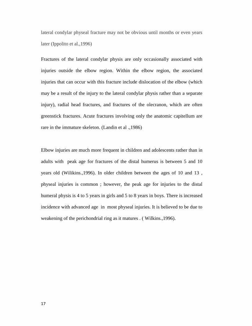
Elbow injuries are much more frequent in children and adolescents rather than in
adults with peak age for fractures of the distal humerus is between 5 and 10
years old (Wilikins.,1996). In older children between the ages of 10 and 13 ,
physeal injuries is common ; however, the peak age for injuries to the distal
humeral physis is 4 to 5 years in girls and 5 to 8 years in boys. There is increased
incidence with advanced age in most physeal injuries. It is believed to be due to
weakening of the perichondrial ring as it matures . ( Wilkins.,1996).

18
2.2.1 FUNCTIONAL ANATOMY OF ELBOW JOINT
The elbow joint acts as a lever arm when positioning the hand. It thus functions as
a fulcrum for forearm lever. In patients using crutches, it functions as aweight
bearing joint. During throwing, there is transfer of energy between the shoulder
and elbow It is crucial for activities of daily living. The elbow is a complex joint
composed of three individual joints which consists of radio-capitellar joint,
ulnahumeral joint and proximal radio-ulna joint . It is made up from the distal
flare of humerus includes the medial and lateral epicondyles the flare accounts for
half of the elbow joint . The trochlea is spool shaped and is located medially
whilst the capitellum is located laterally. ( Morrey et al.,1983).
These joints are contained within a common articular cavity. Elbow is composed
of a hinge joint (the humeroulnar articulation) and a pivot joint (the humeroradial
articulation).The radiohumeral articulation is a pivot joint with the radial head is
covered by cartilage for approximately 240 degrees. However the lateral 120
degrees contains no cartilage which is crucial for internal fixation of radial head
fractures. (Sinnathamby.,2006)
The ulnohumeral articulation is a hinge joint with coronoid fossa on distal
humerus receives the coronoid tip in deeper flexion. The coronoid tip has a
buttress effect in the prevention of posterior dislocations. The sublime tubercle on

19
the ulna is where the anterior bundle of the medial ulnar collateral ligament
attaches distally. ( Wilkins et al 1996).
The elbow joint range of motion for flexion and extension 0 to 150 degrees.
Functional ROM: 30 to 130 degrees to allow daily activities such as feeding and
perform perineal hygiene. Pronation and supination has range of motion at 80
degrees and 85 degrees respectively while functional motion is 50 degrees each.
Axis of rotation for the elbow is centered through the trochlea and capitellum and
passes through a point anteroinferior on the medial epicondyle. ( Beals et al.1976)
The carrying angle of the elbow is the clinical measurement of varus–valgus
angulation of the arm with the elbow fully extended and the forearm fully
supinated. The intersection of a line along the midaxis of the upper part of the arm
and a line along the midaxis of the forearm defines this angle. It has valgus angle
at the elbow. For boys and men: 7 degrees; for girls and women, 13 degrees.
Carrying angle decreases with flexion. ( Beals et al.1976)
The entire articular surface of the distal end of the humerus is intra-articular;
however, the medial and lateral epicondyles are both extra-articular. The elbow
capsule attaches the ulna distal to the olecranon and coronoid process, so these
structures are intra-articular. In addition, the entire radial head is located within
the capsule, thus making it intra-articular. Two elbow fat pads are located
between the capsule and the distal end of the humerus: one anterior and the other
posterior. (Sinnathamby.2006)

20
The process of differentiation and maturation begins at the center of the long
bones and progresses distally. The ossification process begins in the diaphyses of
the humerus, radius, and ulna at the same time. By term, ossification of the
humerus has extended distally to the condyles. In the ulna, it extends to more than
half the distance between the coronoid process and the tip of the olecranon. The
radius is ossified proximally to the level of the neck. The bicipital tuberosity
remains largely unossified . (Chessare et al 1977).
Knowledge of the developmental anatomy of the distal humerus is important in
diagnosing bony injuries around the elbow, especially in the pediatric population.
The distal humerus has four ossification centers (Figure 4). The capitellum is the
first center to appear, at age 6 to 12 months, followed by the medial epicondylar
ossification center at 5 to 7 years of age, and then by the trochlea, between 7 and
10 years of age. The fourth center, the lateral epicondyle, appears at 12 to 14
years of age and serves as the origin of the lateral collateral ligament and
supinatorextensor muscle group.(Gravis et al.1993)
Ossification of the distal humerus proceeds at a predictable rate. In general, the
rate of ossification in girls exceeds that of boys (Chessare et al 1977). In some
areas, such as the olecranon and lateral epicondyle, the difference between boys
and girls in ossification age may be as great as 2 years . During the first 6 months,
the distal humerus' ossification border is symmetric . On average, the ossification

21
center of the lateral condyle appears just before 1 year of age but may be delayed
as late as 18 to 24 months When the lateral condyle's ossific nucleus first appears,
the distal humeral metaphyseal border becomes asymmetric before the end of the
second year, where this border becomes well define and concave. (Bede et al
.1975).
The lateral epicondyle of the humerus is a small, tuberculated eminence, curved a
little forward, and giving attachment to the radial collateral ligament of the elbow-
joint, and to a tendon common to the origin of the supinator and some of the
extensor muscles. Specifically, these extensor muscles include the anconeus
muscle, the supinator, extensor carpi radialis brevis, extensor digitorum, extensor
digiti, and extensor carpi ulnaris. (Sinnathamby.2006)
The vascular anatomy around the elbow is structured in three general arcades:
medial, lateral, and posterior (Figure 5). The medial arcade is formed by superior
ulnar collateral, inferior ulna collateral, anterior and posterior ulnar recurrent
arteries and supply medial condyle and medial aspect of trochlear. Lateral arcade
is formed by radial recurrent, interosseous recurrent , and radial collateral arteries.
These lateral arcade supplied the capitellum, lateral condyle and radial head.
Posterior arcade was formed by the medial and lateral arcade, as well as medial
collateral arteries, and provided blood supply to supracondylar region of humerus
through olecranon. In general the posterior segmental vessels predominately
supplied the lateral column while the medial column is supplied by both anteror

22
and posterior segmental vessels. This suggest that minimal amount of
posterolateral periosteum stripping should be avoided
intraoperatively.(Yamaguchi et al.1997)
FIG. 4: A, Illustration of the average time of appearance of ossification centers at the
distal humerus. B, AP radiograph of a right elbow demonstrating the different
ossification centers. 1 = capitellum, 2 = medial epicondyle, 3 = trochlea,
4 = lateral epicondyle (From Wilkins KE: Fractures involving the epicondylar
apophysis, in Rockwood CA Jr, Wilkins KE, King RE, Fractures in Children, ed 3.
Philadelphia, PA, 1991, pp 509-828.)

23
Fig. 5: Anterior (A) and posterior (B) extraosseous vascular anatomy around the
elbow. B = brachial artery, IR = interosseous recurrent artery, IUC = inferior
ulnar collateral artery, MC = middle collateral artery, PUR = posterior ulnar
recurrent artery, R = radial artery, RC = radial collateral artery, RR = radial
recurrent artery, SUC = superior ulnar collateral artery.
(Adapted from Yamaguchi K, Sweet FA, Bindra R, Morrey BF, Gelberman RH:
The extraosseous and intraosseous arterial anatomy of the adult elbow.
J Bone Joint Surg Am 1997,76(11),1653-1662.)

24
2.2.2 FRACTURE CLASSIFICATION OF LATERAL CONDYLE
HUMERUS
All the physes of the distal humerus are vulnerable to injury, each with a distinct
fracture pattern. Milch et al (1964) described two lateral condylar fracture
patterns (Figure 6). Type I is a simple fracture of the lateral condyle, “resulting
from a force directed along the radius and impacting the head of the radius in the
capitulotrochlear sulcus” according to Milch (1964).
Type I fractures are characterized by a fracture line that courses lateral to the
trochlea and into the capitulotrochlear groove, resulting in a Salter- Harris type IV
fracture. The elbow remains stable because the trochlea is intact. Milch type II
fracture is a fracture-dislocation of the lateral condyle resulting from a force
“directed upward and outward along the ulna, impacting the olecranocoronoid
ridge against the trochlear groove.” (Milch et al.1964)
Type II fracture is characterized by a fracture line that extends into the apex of the
trochlea, resulting in a Salter-Harris type II fracture. The elbow is unstable
because the trochlea is disrupted. This classification system is of little use in
determining fracture management and is largely of historical interest. (Milch et
al.1964)
Jakob et al (1975) described pediatric lateral humeral condylar fracture based on
fracture fragment displacement (Figure 7). Type I fracture is nondisplaced, with
an intact articular surface. The fracture line does not completely traverse the
cartilaginous epiphysis. Type II fracture is complete and extends through the
articular surface. The fracture may be moderately displaced. Type III fracture