Facial Augmentation With Structural Fat Grafting - Tribeca Plastic
11
Facial Augmentation With Structural Fat Grafting Sydney R. Coleman, MD One hundred years ago, surgeons used subcutane- ous fillers extensively for altering facial and body proportions [1,2]. This practice continued until the 1920s when complications resulting from injected paraffin and other substances became obvi- ous. Aesthetic surgeons, disillusioned with available fillers, generally abandoned the restoration of full- ness as a means of rejuvenation after 1920 and be- gan to focus almost entirely on surgical procedures to remove the signs of aging. Advances in autolo- gous fat grafting in the past 2 decades, as well as safer synthetic fillers, give us a chance to reexamine reju- venation by restoring the fullness of youth. Most physicians in the recent past have ap- proached fillers in the face by filling wrinkles, troughs, or holes. But 3-dimensional augmentation of the face for rejuvenation and recontouring is much more complicated than filling a hole. To re- structure with fat grafting, the surgeon must not only be proficient in maximizing fat survival after placement, but also must sculpt in a totally free- form fashion from the facial bones to skin. Development of fat grafting Fat grafting has been used successfully for soft tissue augmentation since 1893 [3]. In 1896, Silex claimed good cosmetic results in the treatment of periorbital scars with grafted fat. He released skin adherent to bone and grafted small pieces of fat between the bone and skin [4]. In 1908, Eugene Holla ¨nder (1867–1932) from Berlin first described a technique for using a needle and syringe to transplant fatty tis- sue [5]. Four years later, Holla ¨nder, who described the first face-lift [6], published photographs of two patients with lipoatrophy of the face, which was cor- rected with infiltration of fat using his method [5]. Charles Conrad Miller in 1926 described grafting fatty tissue through hollow metal cannulas as a sub- stitute for the subcutaneous injection of paraffin CLINICS IN PLASTIC SURGERY Clin Plastic Surg 33 (2006) 567–577 New York University School of Medicine, 44 Hudson Street, New York, NY 10013, USA Conflict of Interest Statement: Sydney Coleman receives royalties from Byron Medical. E-mail address: [email protected] - Development of fat grafting - Structural fat-grafting technique Harvesting Refinement and transfer Placement - Postoperative care - Complications - Patients Patient 1 Patient 2 Patient 3 Patient 4 - Results of facial rejuvenation using fat grafting - Discussion Harvesting and refinement Placement Volumes of placement Levels for placement Intradermal placement Release of adhesions Research - Summary - References 567 0094-1298/06/$ – see front matter ª 2006 Elsevier Inc. All rights reserved. doi:10.1016/j.cps.2006.09.002 plasticsurgery.theclinics.com
Transcript of Facial Augmentation With Structural Fat Grafting - Tribeca Plastic
C L I N I C S I NP L A S T I C
S U R G E R Y
Clin Plastic Surg 33 (2006) 567–577
567
Facial Augmentation With StructuralFat GraftingSydney R. Coleman, MD
- Development of fat grafting- Structural fat-grafting technique
HarvestingRefinement and transferPlacement
- Postoperative care- Complications- Patients
Patient 1Patient 2Patient 3Patient 4
- Results of facial rejuvenation using fatgrafting
- DiscussionHarvesting and refinementPlacementVolumes of placementLevels for placementIntradermal placementRelease of adhesionsResearch
- Summary- References
One hundred years ago, surgeons used subcutane-ous fillers extensively for altering facial and bodyproportions [1,2]. This practice continued untilthe 1920s when complications resulting frominjected paraffin and other substances became obvi-ous. Aesthetic surgeons, disillusioned with availablefillers, generally abandoned the restoration of full-ness as a means of rejuvenation after 1920 and be-gan to focus almost entirely on surgical proceduresto remove the signs of aging. Advances in autolo-gous fat grafting in the past 2 decades, as well as safersynthetic fillers, give us a chance to reexamine reju-venation by restoring the fullness of youth.
Most physicians in the recent past have ap-proached fillers in the face by filling wrinkles,troughs, or holes. But 3-dimensional augmentationof the face for rejuvenation and recontouring ismuch more complicated than filling a hole. To re-structure with fat grafting, the surgeon must notonly be proficient in maximizing fat survival after
0094-1298/06/$ – see front matter ª 2006 Elsevier Inc. All righplasticsurgery.theclinics.com
placement, but also must sculpt in a totally free-form fashion from the facial bones to skin.
Development of fat grafting
Fat grafting has been used successfully for soft tissueaugmentation since 1893 [3]. In 1896, Silex claimedgood cosmetic results in the treatment of periorbitalscars with grafted fat. He released skin adherent tobone and grafted small pieces of fat between thebone and skin [4]. In 1908, Eugene Hollander(1867–1932) from Berlin first described a techniquefor using a needle and syringe to transplant fatty tis-sue [5]. Four years later, Hollander, who describedthe first face-lift [6], published photographs of twopatients with lipoatrophy of the face, which was cor-rected with infiltration of fat using his method [5].Charles Conrad Miller in 1926 described graftingfatty tissue through hollow metal cannulas as a sub-stitute for the subcutaneous injection of paraffin
New York University School of Medicine, 44 Hudson Street, New York, NY 10013, USAConflict of Interest Statement: Sydney Coleman receives royalties from Byron Medical.E-mail address: [email protected]
ts reserved. doi:10.1016/j.cps.2006.09.002
Coleman568
and Vaseline [1]. He reported that infiltrated fat gavea better long-term correction and a more natural-appearing change in facial and body contours thanparaffin. He claimed that using his technique to de-posit fat through a cannula gave better results inmany situations as compared with implanting fatthrough large incisions. Even though Conrad Millerreported good results with the injected fat, the tech-nique he described never became popular.
Soon after the early descriptions of suction curet-tage of fat for body contouring [7,8], Chajchir andBenzaquen [9] described injecting suctioned fatinto the face. At about the same time, Teimourianand Illouz [10–12] described their experiences withthe injection of semiliquid fat into iatrogenic lipo-suction deformities. Although some initial impres-sions of fat grafting were positive [13–16], many ofthe early pioneers of fat grafting were not enthusias-tic about the results with their specific techniques[11,17]. Illouz [11,18] compared the longevity ofinjecting fat into the face as similar to the longevityof injected collagen. In the late 1980s editorials ap-peared in the plastic surgery literature by well-respected plastic surgeons denouncing fat graftingthough injection based on the early negative results[19,20]. Instead of considering why their techniquemight not be working, surgeons such as Ellenbogenand Ersek [21,22] publicly denounced the conceptof fat grafting as an intrinsically faulty procedure.With more experience, they later changed theirtechnique and obtained positive results [23,24].
Gradually, surgeons have come to realize thattransplanted fat can create not only satisfyingchanges in contour, but also long-lasting results[24–27]. As in every surgical procedure, the successof fat grafting is dependent upon many factors: thetechniques and instruments used to harvest, refine,and place fat into the donor site is obviously impor-tant, but also important are the volumes of implan-tation, the sites implanted, the levels of placement,and even the individual patient. In 1926, Dr. Con-rad Miller warned, ‘‘the end-results in free fat trans-plantation depend, aside from various local andgeneral factors, on the method and technique’’ [1].These words are as much a key to successful fat graft-ing (as well as every surgical procedure) now as theywere 80 years ago. Paying careful attention to tech-nique from 1987 to 1994, I developed the followingmethod for the reliable subcutaneous transplanta-tion of autologous fat, emphasizing respect for han-dling tissues and basic sound surgical technique.
Structural fat-grafting technique
Harvesting
These steps used to harvest, prepare, and transplantfragile fat tissue have been previously described in
exhaustive detail [28–30]. Fat should be harvestedas intact tissue parcels that can be inserted througha small cannula but large enough to maintain tis-sue architecture. Harvesting sites are selected onlyto enhance body contour, as no clear correlationbetween donor site location and longevity of theimplanted tissue has been demonstrated [31,32].A No. 11 blade scalpel creates 3- or 4-mm incisionsfor access. Then a blunt Lamis infiltrator distributessolution into the donor sites. The choice of solu-tion depends on the donor areas and on theprojected volume of fat to be removed. For harvest-ing smaller volumes, local anesthesia using 0.5% li-docaine with 1:200,000 epinephrine is adequate.For larger volumes under local anesthesia withsedation, a solution of 0.25% with 1:400,000epinephrine is used. Under general or epidural an-esthesia, Ringer’s lactate with 1:400,000 epineph-rine is infiltrated into the donor sites to aid inhemostasis. Approximately 1 mL of solution is in-filtrated for every mL of fat to be harvested. Super-wet or tumescent techniques should not be usedduring the harvesting phase since the motion ofthe harvested fat through large amounts of liquidmay break up the parcels of fatty tissues andthereby decrease the potential survival of the sub-cutaneous tissues.
Fatty tissue is harvested through the same inci-sions used for infiltration of anesthetic solutions.These incisions are just large enough (usually 2 to3 mm) to permit insertion of the tip of the harvest-ing cannula. Fat harvested with this specific tech-nique is with a 10-mL syringe attached to atwo-holed Coleman harvesting cannula with a blunttip (Fig. 1). Larger syringes are more cumbersomeand may create damaging negative pressures. Dur-ing the removal of fatty tissue, care is taken to min-imize mechanical trauma to the fragile parcels offat. A combination of slight negative pressure anda curetting action allows parcels of fatty tissue tomove through the cannula, through the Luer-Lokaperture, and into the barrel of the syringe withminimal mechanical damage.
Plunger-locking devices should never be usedduring harvesting for transplantation. The high vac-uums created by these devices or by pulling backtoo far on the plunger can increase the negativepressure dangerously. This can bring the pressurein the syringe to the point of vaporization, whichwill obviously damage the fatty parcels of tissue.
Refinement and transfer
After the fat has been harvested, the cannula is re-moved from the syringe and replaced with a Luer-Lok plug to prevent spillage during centrifugation.After the sealing the syringe, the plunger is removedfrom the proximal end of the syringe. The syringe is

Facial Augmentation With Structural Fat Grafting 569
Fig. 1. The Coleman cannula. (From Coleman S. Structural fat grafting. St Louis (MO): Quality Medical Pub; 2004;with permission.)
then centrifuged for 3 minutes at 3000 rpm to sepa-rate the viable from the nonviable components. Theoil layer is decanted and the aqueous componentdrained. Porous paper or neuropads can then beused to wick off the remaining oil. Care should betaken not to allow the material from the wicks toshred off into the refined tissue. The refined, un-washed fat is then transferred to 1-mL Luer-Lok sy-ringes for use in the face or dorsum of the hands.
Placement
Either general anesthesia or combinations of re-gional and local anesthesia are the choices for facialanesthesia during placement. The positioning of in-cisions allows placement from at least two directionsinto each area grafted. A blunt 18- or 17-gauge can-nula with one distal aperture just proximal to thetip is most commonly used for structural fat place-ment. The shape of the tip, the length of the cannula,and the curvature of the cannula can be altered asneeded. Using a blunt Type I Coleman infiltrationcannula and a 3-mL syringe, the local anesthesia isinfiltrated through the same incisions that will beused for placement of the fat. Vasoconstrictionfrom epinephrine lessens bruising, hematomas,and intravascular embolization of the transplantedfat [33]. Sharp needles are avoided in the subcutane-ous planes for injection of local anesthesia or fat.
The blunt infiltration cannula is attached to the1-mL Luer-Lok syringe filled with the refined tissue.The cannula is inserted through the incisions andadvanced through the tissues to the appropriateplane. Unlike the sharp tip of a needle, the bluntcannula tip does not cut a newly defined channelthrough the recipient tissues but follows more nat-ural tissue planes. As the cannula is withdrawn,fatty tissue is injected into the pathway of the re-treating cannula. No fat is placed during advance-ment of the cannula to avoid placement of the fatin clumps and to encourage nutrition and
integration of the grafted tissue by placement insmall aliquots. Fundamental to this technique isthe placement of fat in tiny aliquots from one tenthof a milliliter to as little as one fiftieth of a milliliterwith each pass of the cannula.
Placing the fat in small aliquots maximizes thesurface area of contact between the surrounding tis-sues and the grafted fat. This allows each parcel offat access to a blood supply and a greater possibilityto anchor in a more stable fashion in the new site.Such placement enhances the potential for survival,encourages stability, and minimizes the possibilityof visible or palpable irregularities. Dramatic vol-ume corrections are possible by making manypasses of the tiny aliquots in one procedure, butthis requires time and patience and results in re-markable and prolonged swelling.
Postoperative care
The most expected sequela of fat grafting using thistechnique is swelling. The many passes of a bluntcannula used for placement of the refined tissue re-sults in remarkable tissue edema. Immediately afterthe procedure, plastic adhesive tape or Tegaderm(3M, St. Paul, MN) is placed over the infiltratedareas. This remains in place for 3 or 4 days and ap-pears to reduce bruising and swelling. For 72 hourspostoperatively, cold should be applied frequentlyand all infiltrated sites should be elevated.
Light touch can be instrumental in reducingswelling by encouraging lymphatic drainage. How-ever, deep massage of infiltrated areas should beapproached with caution in the first weeks afterplacement. Although it is difficult to move the re-cently infiltrated fat with gentle manipulation ofthe area, strong directed pressure could move infil-trated fatty tissue.
Other maneuvers such as holistic medications,low-level laser [34], and electromagnetic therapy[35] may also accelerate the resolution of swelling.

Coleman570
Even with all of these maneuvers to reduce edema,postoperative swelling is still a major factor. Al-though some physicians report a few days to 2weeks for recovery from fat grafting [36], my expe-rience is that the recovery can be much longer[37,38].
Complications
A surgeon should be aware of the potential compli-cations be able to avoid them, to minimize unto-ward effects such as swelling, and to provide anadequate informed consent. The immediate poten-tial complications of structural fat grafting are re-lated to surgery itself. Late complications areprimarily aesthetic.
Although the cannulas used are blunt, damage tounderlying structures (nerves, blood vessels, mus-cles, and glands) is possible. The most devastatingis the rare complication of intravascular emboli[33]. However, cannulation of a vessel and perma-nent nerve injury have never been reported usinga blunt cannula.
Bacterial contamination of the fatty tissue can re-sult in infection and resorption of the grafted tissue.Meticulous sterile technique must be observed withcareful attention to preoperative patient prepara-tion with antiseptic soaps and an antiseptic agentsuch as povidone iodine.
Fat grafting appears to be stable after placement inmost situations. However, during the procedure andin the immediate postoperative period, care shouldbe taken to avoid migrations of the fatty tissue intothe surrounding tissues. Forcing too much fatty tis-sue into any area may cause the fat to move into an-other area. Placement of fat grafts into areas ofsignificant intrinsic facial muscle motion, such asthe lateral corrugators, may precipitate migrationfrom the area of placement. In addition, after trans-plantation, fat has the potential to increase or de-crease in size especially related to weight gain or loss.
Most patients find it desirable to remove fat andcontour the rest of the body at the same time asfat grafting. However, a potential complicationwith this technique is the creation of donor site de-formities or problems with incisions. Even the sur-geon who is facile at liposuction may create donorsite deformities. This is not a complete list of com-plications, and more exhaustive descriptions of po-tential complications [38] and untoward effects areavailable.
Patients
Patient 1
This 42-year-old presented after a face-lift and en-doscopic forehead lift (without any eyelid
procedures) 2 years previously. After the proce-dures, she noticed hollowing of her periorbital re-gion (Fig. 2A, C, E). Over a year before, a physicianattempted placement of Restylane (Q-Med, Uppsa-la, Sweden) into her upper and lower eyelids, butthe patient noted lumps and a bluish discolorationin the areas of injection.
Fat was placed at one procedure in the followingamounts: right temple 4 cc; left temple 5 cc; frontalforehead 4 cc; upper eyelids 1.5 cc each side; rightanterior malar fold 1.5 cc; left anterior malar fold2 cc; right infraorbital rim 2.5 cc; left infraorbitalrim 3 cc; lateral eyelid 0.8 cc each side; and each an-terior malar region 1.5 cc. Into selected specificwrinkles of the forehead, 2.7 cc was placed usinga 22-gauge needle for intradermal placement.
The patient returned at 10 months with subtle,but remarkable changes (Fig. 2B, D, F). She hadno topical treatments or change in skin care. Theprimary change noted was in the quality of herskin with filling of the anterior malar folds (teartrough) and ablation of the wrinkles in her lowereyelids and crows’ feet. The pores of the foreheadand the anterior malar region were beginning toline up into wrinkles in the before photos, buthave become smaller and less linear in the afterphotos. There is a correction of the hollowing ofthe upper and lower eyelids, with less upper eyelidshowing and an apparent elevation of the eye-brows. The temples area slightly expanded, whichallows more visualization of the brow from thefront view.
Patient 2
This 48-year-old presented asking for larger lips(Fig. 3A). The following volumes were placed inone procedure: body of the upper lip 2 cc; whiteroll 1 cc; philtrum 0.75 cc; body of the lower lip 6cc; marionette grooves 1.8 cc each; nasolabial folds4 cc on right and 3 cc on left. She returned 2 yearsafter the one procedure (Fig. 3B).
Patient 3
This 22-year-old presented for lip augmentation(Fig. 4A). She had 3.5 cc of fat infiltrated into thebody of her upper lip and 5.5 into the body ofher lower lip with a blunt cannula. Using a 22-gauge needle, 1 cc was placed into the deep vermil-lion along the white roll and another 1 cc along therim of the lower lip. One year after the one proce-dure the patient returned (Fig. 4B).
Patient 4
This 33-year-old presented complaining of having‘‘no chin and a weak jaw,’’ which he felt made him‘‘look weak and unattractive’’ (Fig. 5A, B, C). Hehad a chin implant 8 years earlier and buccal fat
Facial Augmentation With Structural Fat Grafting 571
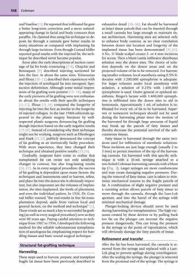
Fig. 2. (A, B, C ) This 42-year-old presented complaining of hollowing of her eyelids after a face-lift and foreheadlift. (D, E, F ) Ten months after diffuse placement over the temples, upper and lower eyelids, the patient returnswith remarkable rejuvenation. The quality of her skin is enhanced with lightening of the lower eyelid skin, ab-lation of most wrinkles, and a decrease in the size of pores over infiltrated areas. She has a restoration of thefullness of her brow with a youthful decrease in upper eyelid show and an apparent elevation in the position ofher eyebrows.
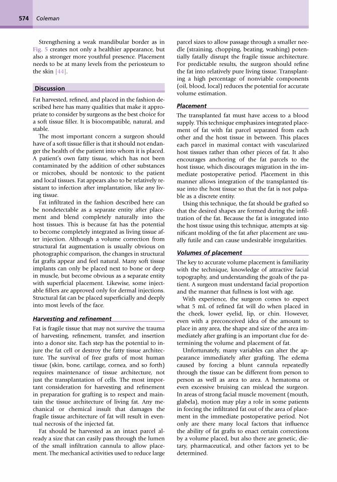
removal 3 years earlier. Along the posterior border,18 cc of refined fat was placed on the right and 19 ccon the left, both feathering up over the masseter; 8cc was placed along each anterior border of themandible feathering up to the lateral chin; and 16cc was placed over the lower body of the chin. Fi-nally, 6.5 c was suctioned from the right jowl area
and 1.5 cc from the left. He returned 1.5 years laterpleased with his results, but asking for reduction ofthe submental region; 11.4 cc was suctioned. Hehad a more athletic and healthier appearancewhen he returned 2 years after the submental suc-tioning and 3.5 years after the only fat grafting tothe jawline (Fig. 5D, E, F).
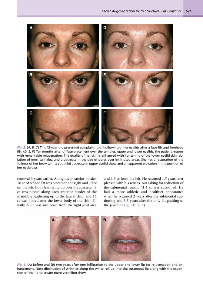
Fig. 3. (A) Before and (B) two years after one infiltration to the upper and lower lip for rejuvenation and en-hancement. Note diminution of wrinkles along the white roll up into the cutaneous lip along with the expan-sion of the lip to create more vermilion show.

Coleman572
Fig. 4. (A, B) This 22-year-old returned 1 year after one procedure to her upper and lower lips. Please note theattempt to maintain the central cleft of the lower lip while attaining central eversion.
Results of facial rejuvenation using fatgrafting
These patients illustrate that a full and structuredperiorbital region, voluptuous lips, and a strong,healthy appearing mandibular border projecthealth and well-being. By using this technique con-sistently, I have observed that the result from fatgrafted by this technique seems to stabilize at about4 to 5 months. Although a large portion of thestructurally grafted fat appears to remain after graft-ing, research quantifying the percentage of survivalwith this particular method is needed to demon-strate accurately the percentage of survival.
The goal in facial rejuvenation should include thefollowing: to restore a youthful fullness to the up-per face; to sculpt an angular but full cheek thatflows into a full lower eyelid; and to create an attrac-tive, straight shadow under the mandible witha well-shaped chin.
Adding support to the midface and lower eyelidsrather than excising and suspending structures re-sults in a much more natural rejuvenation and en-hancement. The cheek, both malar and buccalareas, is relatively easy area to visualize in 3 dimen-sions. The results of cheek augmentation with fatare the most what-you-see-is-what-you-get at theend of the procedure of any area in the face andbody [39]. I recommend this area for learning 3-di-mensional manipulation of the face.
On the other hand, the lower eyelid is one of themost difficult areas for structural fat grafting [39].Areas of excess fullness and even irregularities orsmall lumps can be visible through the skin whenthe swelling resolves. The lower eyelid is not anarea to first learn transplanting fat, but should bedone when the surgeon has performed enoughgrafting in other areas to approach the lower eyelidwith confidence in the technique.
The upper one third of the face is the least under-stood area of the face. Although much easier techni-cally to approach than the lower eyelid, the templesand brow require a complete re-thinking of the area
to rejuvenate [40,41] (Fig. 2). We have misidenti-fied many of the problems of aging in the uppereyelid, brow, and temple, and excision and suspen-sion procedures do not always work in this area torejuvenate, and often make the patient look old orsick.
Changing structure of the lips as in Figs. 3 and 4,is a relatively simple procedure, but often poorlyunderstood by surgeons and patients. The processof aging in the lip and the mechanisms for restoringor creating structures in the lip entails not only anintimate knowledge of the subtle curvatures ina youthful, attractive lip, but also the differences be-tween the shape and size of the upper and lowerlips [42,43]. A youthful aesthetic upper lip has a dis-tinctly protuberant and continuous full white rollthat tapers off from peaks on either side of the Cu-pid’s bow and becomes less obvious laterally. Ce-phalic to the white roll, the skin curves intoa slight concave. The two distinct philtral columnsascend in a similar concave fashion from their ori-gin at the peaks of the Cupid’s bow to the colu-mella. Under the Cupid’s bow, the vermillion ofthe lip has a well-centered, distinct tubercle, whichis the most caudal element of an attractive upperlip. The width of a lip comes from the most lateralfullness, which is significantly less protuberant thanthe central tubercle in the vermilion. Between thecentral tubercle and the lateral fullness is oftena concavity or slight depression.
The lower lip has a slightly protuberant rim,which is less distinctive than the white roll of theupper lip. The fullness and depressions of an attrac-tive lower lip complement the upper lip followingthe shape with opposing components. A distinctcentral cleft is bordered on either side by oblongprotuberances significantly larger than any of the el-ements of the upper lip. The alignment of the ob-long lower lip tubercles is oblique in a fashionthat pushes the most central lip out into a distinctpout. The amount of vermilion visible is muchgreater in a normal, youthful lower lip than in anupper lip.

Facial Augmentation With Structural Fat Grafting 573
Fig. 5. (A, B, C ) This 33-year-old presented complaining of having ‘‘no chin and a weak jaw.’’ He had a chin im-plant 8 years earlier and buccal fat removal 3 years earlier. Along the posterior border, 18 cc was placed on theright and 19 cc on the left; 8 cc was placed along the anterior border of the mandible on each side feathering upinto the lateral chin; and 16 cc was placed over the lower body of the chin. Finally, 6.5 c was suctioned from theright jowl area and 1.5 from the left. He returned 1.5 years later pleased with his results, but asking for reduc-tion of the submental region; 11.4 cc was suctioned. He had a much more athletic and healthier appearancewhen he returned 2 years after the submental suctioning and three and a half years after the only fat graftingto the jawline (D, E, F ).
The lip is not a trough or hole that the surgeoncan just fill; instead, he or she must visualize a 3-dimensional structure and sculpt in free form. Toattain consistent results with lip enhancement,a well-thought-out strategy that aims at a specific
shape for the lip should be devised for every pa-tient. However, the lips are not clay to be molded;they are living flesh with many variables that are dif-ficult to predict: swelling, bleeding, scarring, and soforth.
Coleman574
Strengthening a weak mandibular border as inFig. 5 creates not only a healthier appearance, butalso a stronger more youthful presence. Placementneeds to be at many levels from the periosteum tothe skin [44].
Discussion
Fat harvested, refined, and placed in the fashion de-scribed here has many qualities that make it appro-priate to consider by surgeons as the best choice fora soft tissue filler. It is biocompatible, natural, andstable.
The most important concern a surgeon shouldhave of a soft tissue filler is that it should not endan-ger the health of the patient into whom it is placed.A patient’s own fatty tissue, which has not beencontaminated by the addition of other substancesor microbes, should be nontoxic to the patientand local tissues. Fat appears also to be relatively re-sistant to infection after implantation, like any liv-ing tissue.
Fat infiltrated in the fashion described here canbe nondetectable as a separate entity after place-ment and blend completely naturally into thehost tissues. This is because fat has the potentialto become completely integrated as living tissue af-ter injection. Although a volume correction fromstructural fat augmentation is usually obvious onphotographic comparison, the changes in structuralfat grafts appear and feel natural. Many soft tissueimplants can only be placed next to bone or deepin muscle, but become obvious as a separate entitywith superficial placement. Likewise, some inject-able fillers are approved only for dermal injections.Structural fat can be placed superficially and deeplyinto most levels of the face.
Harvesting and refinement
Fat is fragile tissue that may not survive the traumaof harvesting, refinement, transfer, and insertioninto a donor site. Each step has the potential to in-jure the fat cell or destroy the fatty tissue architec-ture. The survival of free grafts of most humantissue (skin, bone, cartilage, cornea, and so forth)requires maintenance of tissue architecture, notjust the transplantation of cells. The most impor-tant consideration for harvesting and refinementin preparation for grafting is to respect and main-tain the tissue architecture of living fat. Any me-chanical or chemical insult that damages thefragile tissue architecture of fat will result in even-tual necrosis of the injected fat.
Fat should be harvested as an intact parcel al-ready a size that can easily pass through the lumenof the small infiltration cannula to allow place-ment. The mechanical activities used to reduce large
parcel sizes to allow passage through a smaller nee-dle (straining, chopping, beating, washing) poten-tially fatally disrupt the fragile tissue architecture.For predictable results, the surgeon should refinethe fat into relatively pure living tissue. Transplant-ing a high percentage of nonviable components(oil, blood, local) reduces the potential for accuratevolume estimation.
Placement
The transplanted fat must have access to a bloodsupply. This technique emphasizes integrated place-ment of fat with fat parcel separated from eachother and the host tissue in between. This placeseach parcel in maximal contact with vascularizedhost tissues rather than other pieces of fat. It alsoencourages anchoring of the fat parcels to thehost tissue, which discourages migration in the im-mediate postoperative period. Placement in thismanner allows integration of the transplanted tis-sue into the host tissue so that the fat is not palpa-ble as a discrete entity.
Using this technique, the fat should be grafted sothat the desired shapes are formed during the infil-tration of the fat. Because the fat is integrated intothe host tissue using this technique, attempts at sig-nificant molding of the fat after placement are usu-ally futile and can cause undesirable irregularities.
Volumes of placement
The key to accurate volume placement is familiaritywith the technique, knowledge of attractive facialtopography, and understanding the goals of the pa-tient. A surgeon must understand facial proportionand the manner that fullness is lost with age.
With experience, the surgeon comes to expectwhat 5 mL of refined fat will do when placed inthe cheek, lower eyelid, lip, or chin. However,even with a preconceived idea of the amount toplace in any area, the shape and size of the area im-mediately after grafting is an important clue for de-termining the volume and placement of fat.
Unfortunately, many variables can alter the ap-pearance immediately after grafting. The edemacaused by forcing a blunt cannula repeatedlythrough the tissue can be different from person toperson as well as area to area. A hematoma oreven excessive bruising can mislead the surgeon.In areas of strong facial muscle movement (mouth,glabela), motion may play a role in some patientsin forcing the infiltrated fat out of the area of place-ment in the immediate postoperative period. Notonly are there many local factors that influencethe ability of fat grafts to enact certain correctionsby a volume placed, but also there are genetic, die-tary, pharmaceutical, and other factors yet to bedetermined.

Facial Augmentation With Structural Fat Grafting 575
Levels for placement
Surgeons sculpting the face with autologous fatmust address the many different levels, anatomicalcompartments, and structures such as bone, muscle,fascia, major salivary glands, nerves, blood vessels,and skin. I have described in the past placementof fat at all levels from the periosteum, perichon-drium, or mucosa to the subdermis or vermilion.However, I now avoid placement of fat into facialmuscles in most situations as it creates superficialedema, which deceives a surgeon into believingthat a correction has been attained. For instance,placement of fat into the obicularis oris will createsubmucosal and subvermillion swelling that willevert the lip. However, as the superficial swellingsubsides from under the mucosa and vermillion,the lip inverts. This leaves a thicker muscle in thelip without the correction afforded by the eversionof the vermillion and mucosa. Therefore, in a frontphotograph, the lip will not appear to have in-creased in size because it is no longer everted eventhough the fat has survived. Instead, in a profileview the lips will project away from the face in anunnatural fashion.
Intradermal placement
I have specifically discouraged intradermal place-ment in the past [45]; however, in the past 3 yearsas I began to use hyaluronic acid in my practice, Ibecame reacquainted with the advantages of intra-dermal augmentation. With that in mind, for scarsand deep wrinkles, I now use a sharp 22-gauge nee-dle to place small amounts of fat into the deep der-mis to complement the subcutaneous placement.Forcing large parcels of fat through a needle is tech-nically awkward, and the needles block frequently.Particular care should be paid to avoiding place-ment that is isolated since such corrections mayresult in circumscribed, visible corrections. Thelong-term effect of this method of placement doesnot appear to be as reliable as with subcutaneousplacement with a larger bore cannula. However, Ihave observed significant correction for 2 years insome patients; furthermore, the effect of intradermalplacement of fat is different from the effect of place-ment of the same volume into deeper subcutaneousplanes. With intradermal placement, wrinkles can besoftened or even ablated with minimal volume. Scarscan be improved or eliminated that would not be asaffected by simple subcutaneous placement.
Release of adhesions
This intradermal technique of fat placement is spe-cifically different from the technique of injectionwith a sharp needle into a subcutaneous plane de-scribed by Carraway and Mellow [13]. They
described the use of small sharp needles to under-mine before placement with the needle. This maycreate a potential space that can destabilize theplacement of fat and encourage migration and ir-regularities. Therefore, I do not recommend under-mining before injection. If adhesions, scars, orligaments are still present after the initial placementof fat subcutaneously and intradermally, it may benecessary to release them with a ‘‘v-dissector’’ orsharp needle in a ‘‘subcision’’ fashion [46]. In theseinstances, the sharp instrument is used to disrupta connection from the dermis to the underlying an-atomic structures. This type of maneuver is trau-matic and may allow the placed fat to migrate.Such destabilizing approaches should be usedwith caution and should be delayed until after in-tradermal and subcutaneous placement is substan-tially completed.
Research
Fat grafting is technique dependent, and not alltechniques, surgeons, and patients will experiencethe same longevity or effect of grafted fat. One ofthe current problems with analysis of the resultsof fat grafting is that there has been never beena standardization of the methods of fat graftingused. Radically different techniques are often usedin both research and in clinical practices, whichmakes extrapolation of the result of studies or anec-dotal experiences to other situations difficult. In ad-dition, remarkable ranges of the results have beennoted from one human to another and between dif-ferent anatomical sites even when using exactly thesame technique and volumes [47]. Surgeons shouldtry to identify the techniques, patients, and ana-tomic regions that encourage long-term survivaland reliable results with grafted fatty tissue.
Summary
The outcome of grafting fat is dependent upon var-iations in the technique used to harvest, refine,transfer, and place fat as well as the avoidance ofcomplications. In addition, the effects of graftedfat differ between recipient sites and even from pa-tient to patient. Because of this variability and per-haps because of other factors not understood, theresults of fat grafting with some techniques, insome patients, and in some areas can be unpredict-able. Directed research will help us to understandbetter the differences between different techniques,surgeons, and patients for this valuable techniqueand to elucidate the methods and situations thatwill provide surgeons with the most consistentresults.
Fat grafting through a blunt cannula has beenused by plastic surgeons for altering facial contours

Coleman576
for almost 100 years. Autologous tissue is com-pletely biocompatible, and therefore is usually thesafest choice for altering facial volume or contours.Furthermore, fat grafts can be placed in such a fash-ion that they are long lasting, completely inte-grated, and natural appearing. However, only inthe past 20 years have advances in techniques andinstrumentation allowed us to obtain predictableresults that make fat grafting a viable option forsoft tissue augmentation. Concurrent with the de-velopment of fat grafting, our understanding of ag-ing and methods of rejuvenation have developedalso. We now approach rejuvenation and adjust-ment of facial proportion with a better understand-ing of the need for the restoration or adjustment offacial volume.
References
[1] Miller JJ, Popp JC. Fat hypertrophy after autolo-gous fat transfer. Ophthal Plast Reconstr Surg2002;18(3):228–31.
[2] Kolle FS. Plastic and cosmetic surgery. New York:Appleton; 1911.
[3] Neuber F. Fettransplantation. Bericht uber die Ver-handlungen der Deutschen Gesellschaft fur Chir-urgie. Zentralbl Chir 1893;22:66 [in German].
[4] Neuhof H, Hirshfeld S. The transplantation oftissues. New York: D. Appleton and Company;1923.
[5] Hollander E. Plastik und Medizin. Stuttgart: Fer-dinand Enke; 1912.
[6] Rogers BO. The development of aesthetic plasticsurgery: a history. Aesthetic Plast Surg 1976;1(1):3–24.
[7] Kesselring UK, Meyer R. A suction curette for re-moval of excessive local deposits of subcutane-ous fat. Plast Reconstr Surg 1978;62(2):305–6.
[8] Teimourian B, Fisher JB. Suction curettage to re-move excess fat for body contouring. Plast Re-constr Surg 1981;68(1):50–8.
[9] Chajchir A, Benzaquen I. Liposuction fat grafts inface wrinkles and hemifacial atrophy. AestheticPlast Surg 1986;10(2):115–7.
[10] Teimourian B. Repair of soft-tissue contour defi-cit by means of semiliquid fat graft. Plast Re-constr Surg 1986;78(1):123–4.
[11] Illouz YG. The fat cell ‘‘graft’’: a new technique tofill depressions. Plast Reconstr Surg 1986;78(1):122–3.
[12] Illouz Y-G. Liposuction: the Franco-American ex-perience. Beverly Hills (CA): Medical Aesthetic,Inc.; 1985.
[13] Carraway JH, Mellow CG. Syringe aspiration andfat concentration: a simple technique for autolo-gous fat injection. Ann Plast Surg 1990;24(3):293–6.
[14] Lewis CM. Transplantation of autologous fat.Plast Reconstr Surg 1991;88(6):1110–1.
[15] Fournier PF. Facial recontouring with fat grafting.Dermatol Clin 1990;8(3):523–37.
[16] Chajchir A, Benzaquen I, Arellano A. [Compara-tive study on lipoinjection and other methods].Med Cutan Ibero Lat Am 1988;16(6):489–96.[in Spanish].
[17] Silkiss RZ, Baylis HI. Autogenous fat grafting byinjection. Ophthal Plast Reconstr Surg 1987;3(2):71–5.
[18] Illouz YG. Surgical remodeling of the silhouetteby aspiration lipolysis or selective lipectomy.Aesthetic Plast Surg 1985;9(1):7–21.
[19] Goldwyn RM. Unproven treatment: whose bene-fit, whose responsibility? Plast Reconstr Surg1988;81(6):946–7.
[20] Fredricks S. Fat grafting injection for soft-tissueaumgenation [discussion]. Plast Reconstr Surg1989;84:935.
[21] Ellenbogen R. Invited commentary on autologusfat injection. Ann Plast Surg 1990;24:297.
[22] Ersek RA. Transplantation of purified autologousfat: a 3-year follow-up is disappointing. Plast Re-constr Surg 1991;87(2):219–27.
[23] Ersek RA, Chang P, Salisbury MA. Lipo layeringof autologous fat: an improved technique withpromising results. Plast Reconstr Surg 1998;101(3):820–6.
[24] Ellenbogen R, Motykie G, Youn A, et al. Facial re-shaping using less invasive methods. AestheticSurgery Journal 2005;25(2):144–52.
[25] Coleman SR. Long-term survival of fat trans-plants: controlled demonstrations. AestheticPlast Surg 1995;19(5):421–5.
[26] Guerrerosantos J. Long-term outcome of autolo-gous fat transplantation in aesthetic facial recon-touring: sixteen years of experience with 1936cases. Clin Plast Surg 2000;27(4):515–43.
[27] Trepsat F. Periorbital rejuvenation combining fatgrafting and blepharoplasties. Aesthetic PlastSurg 2003;27(4):243–53.
[28] Coleman S. Hand rejuvenation with structuralfat grafting. Plast Reconstr Surg 2002;110(7):1731–43.
[29] Coleman S. Structural fat grafting. St Louis(MO): Quality Medical Pub; 2004.
[30] Coleman SR. Structural fat grafting. In: Nahai F,editor. The art of aesthetic surgery: principles &techniques. St Louis (MO): Quality MedicalPub; 2005. p. 289–363.
[31] Rohrich RJ, Sorokin ES, Brown SA. In search ofimproved fat transfer viability: a quantitativeanalysis of the role of centrifugation and harvestsite. Plast Reconstr Surg 2004;113(1):391–5.
[32] Ullmann Y, Shoshani O, Fodor A, et al. Search-ing for the favorable donor site for fat injection:in vivo study using the nude mice model. Der-matol Surg 2005;31(10):1304–7.
[33] Coleman SR. Avoidance of arterial occlusionfrom injection of soft tissue fillers. Aesthetic Sur-gery Journal 2002;22(6):555–7.
[34] Posten W, Wrone DA, Dover JS, et al. Low-levellaser therapy for wound healing: mechanism

Facial Augmentation With Structural Fat Grafting 577
and efficacy. Dermatol Surg 2005;31(3):334–40.
[35] Kinney BM. Pulsed Electromagnetic Field Ther-apy in Plastic Surgery. Aesthetic Surg Journal2005;25(1):87–92.
[36] Kanchwala SK, Holloway L, Bucky LP. Reliablesoft tissue augmentation: a clinical comparisonof injectable soft-tissue fillers for facial-volumeaugmentation. Ann Plast Surg 2005;55(1):30–5[discussion 35].
[37] Coleman SR. Facial recontouring with lipostruc-ture. Clin Plast Surg 1997;24(2):347–67.
[38] Coleman SR. Problems, Complications andPostprocedure Care: Chapter 4. Structural fatgrafting. St. Louis, Mo.: Quality Medical Pub.;2004. 75–102.
[39] Coleman SR. Infraorbital and Cheek RegionsChapter 11. Structural fat grafting. St. Louis,Mo.: Quality Medical Pub.; 2004. 293–352.
[40] Coleman SR. The technique of periorbital lip-oinfiltration. Operative Techniques in Plasticand Reconstructive Surgery 1994;1:20–6.
[41] Coleman SR. Supraorbital area: brow, upper eye-lids, and temples. In: Coleman S, editor. Struc-tural fat grafting. St Louis (MO): QualityMedical Pub; 2004. p. 353–400.
[42] Coleman SR. Lips. In: Coleman S, editor. Struc-tural fat grafting. St Louis (MO): Quality MedicalPub; 2004. p. 203–36.
[43] Coleman SR. Lipoinfiltration of the upperlip white roll. Aesthetic Surgery Journal 1994;14(4):231–4.
[44] Coleman SR. Chin and jawline. In: Coleman S,editor. Structural fat grafting. St Louis (MO):Quality Medical Pub; 2004. p. 237–70.
[45] Coleman SR. Structural fat grafts: the ideal filler?Clin Plast Surg 2001;28(1):111–9.
[46] Orentreich DS, Orentreich N. Subcutaneous in-cisionless (subcision) surgery for the correctionof depressed scars and wrinkles. Dermatol Surg1995;21(6):543–9.
[47] Coleman SR. Structural fat grafting: more thana permanent filler. Plast Reconstr Surg 2006;118(3 Suppl):108S–20S.