Extremophiles - Department of Biological Sciences, Studies in Life

Extremophiles from unique ecosystems of Kazakhstan as potential
producers of novel antibiotics
Azliyati Azizan, Ph.D. Associate Professor
Lyudmila Trenozhnikova, Ph.D. Institute of Microbiology and Virology, Almaty, Kazakhstan
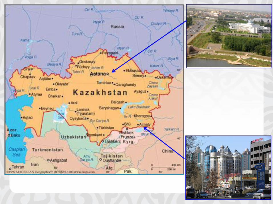

Institute of Microbiology and Virology Almaty, Kazakhstan
Lead Extremophile Collaborator (since 2006) Dr. Lyudmila Trezhnovikova
Virology Collaborator (since 2011) Dr. Vladimir Berezin

Outline
• Background
– Antibiotics and resistance
– Drug Discovery and Natural Products
• Study: goals, approach (screening) and findings
– IMV study
– IMV-USF collaboration (pilot study)
• Preliminary chemical characterization
• Future direction

c
c


Targets of Antimicrobials Copyright © The McGraw-Hill Companies, Inc. Permission required for reproduction or display.
1. Cell wall inhibitors
Block synthesis and repair
Penicillins
Cephalosporins
Carbapenems
Vancomycin
Bacitracin
Fosfomycin
Isoniazid
2. Cell membrane
Causelossofselective permeability
Polymyxins
Daptomycin
3. DNA/RNA
Inhibit replication and transcription
Inhibit gyrase(unwinding enzyme)
Quinolones
Inhibit RNA polymerase
Rifampin
Ribosome
mRNA DNA
4. Protein synthes is inhibitors acting
on ribosomes
Site of action
50S subunit
Erythromycin
Clindamycin
Synercid
Pleuromutilins
Site of action
30S subunit
Aminoglycosides
Gentamicin
Streptomycin
Tetracyclines
Glycylcyclines
Both 30S
and 50S
Blocks initiation of protein
synthesis
Linezolid
5. Folic acid synthesis
Block pathways and inhibit
metabolism
Sulfonamides (sulfa drugs)
Trimethoprim
Substrate
Enzyme
Product

Major Antimicrobial Drug Groups
• About 260 different antimicrobial drugs
• Classified in 20 drug families
• Largest number of antimicrobial drugs are for bacterial infections – Antibiotic Source:
• fungi and bacteria
• semi-synthetic compounds

Spectrum of Activity

Aminoglycoside Drugs
– Products of various species of soil actinomycetes in
the genera Streptomyces and Micromonospora
– Relatively broad spectrum because they inhibit protein synthesis
– Subgroups and uses • Aerobic gram-negative rods and certain gram-positive
bacteria
• Streptomycin: Bubonic plague and tularemia and good anti-tuberculosis agent
• Gentamicin: Less toxic and used for gram-negative rods

Resistance Mechanisms
• Antibiotics are present in nature
• Microbes are capable of adapting quickly to selective pressures
• Drug resistance has arisen for all antibiotics
• ESKAPE pathogens
• Two main strategies employed by microbes – Prevent access of the drug to the target site
– Alter the nature of the target site

Antibiotic Resistance
Not drug-resistant
Drug-resistant mutant
(a) Population of microbial cells (b) Sensitive cells ( ) eliminated by drug;
resistant mutants survive
Remaining
population
grows
overtime
(c) All cells are now resistant
Exposure
to drug
Copyright © The McGraw-Hill Companies, Inc. Permission required for reproduction or display.

Disk Diffusion Assays
• Kirby-Bauer
• Standardized conditions
• Zones of inhibition
• Larger zone indicates more susceptible
• Smaller zone indicates more resistant
S
R
R
I
I
S
1 2 3 4 0 5
Kirby-Bauer Disc Diffusion Test*
Oxytetracycline 30g
(R<17 mm;S 22mm)
Enrofloxacin 5 g
(R < 17 mm;S 22 mm) Gentamicin 10 g
(R < 17 mm; S 21 mm)
OT
30 GN
10
CTX
30
AMP
10
C
30
Cefotaxime 30 g
(R < 14 mm; S 23 mm)
Ampicillin10g
(R<14mm;S22mm) Chloramphenicol 30 g
(R < 21 mm; S 21 mm)
= Zone of Inhibition = Antibiotic carrier (disc)
imprinted with abbreviation
and concentration
= Region of
bacterial growth
Disc Diffusion Test (schematic).
Example and evaluation of asensitivity test, agar diffusion method.
R = resistant, I = intermediate, S = sensitive
(a) *R and S values differ from table 12.7 due to differing concentrations of the antimicrobials. (b)
ENR mm
ENR
5
b: © Kathy Park Talaro
Copyright © The McGraw-Hill Companies, Inc. Permission required for reproduction or display.

E-Test Strips
• Drug gradient used
• Can determine MIC
• Read where the zone touches the strip

DRUG DISCOVERY AND NATURAL PRODUCTS

Starting point
Research and development of drug or vaccines products is - demanding - risky

How are they valued?
$ Market value
Societal value
Vaccines
Drugs

The path toward a drug

• In spite of pessimism; there are many antibacterial agents in clinical trials
– Novel agents on the rise: last 85 years
– Gram positive, gram negative and multi-drug resistant
• 39 antibacterial compounds
– 25 in Phase 2 or Phase 3 Trials
• “Guarded optimism”
– Renewed commitment from companies
– Cubist (acquired Optimer and Trius Therapeutics), Roche


Natural Products
• Natural products originate from bacteria, fungus, plants or other natural sources (marine organisms)
• Scaffold for many effective antibiotics (semi-synthetic) • Screening natural products is complicated
– Complex mixture of secondary metabolites – Rediscovery of known active compounds
• Dereplication to rule out knowns • Competition with HTS of synthesized compounds
– Competition with HTS of synthesized compounds – Purified natural compounds exert challenge
• Success (using in-vitro bioassay) depends on – Proper design, validation and implementation of screening assays

• Natural products (NP) are a rich source of compounds for drug discovery (34%: 1981-2010)
• Their use has decreased in the last 2 decades; barriers: NP screening in HTS against Targets
• Review: technical advances to reduce barriers
– Genomic and metabolomic approaches
– Augment traditional methods of screening
– Increased functional assays and phenotypic screen

NCE: New Chemical entities -NCEs are small molecules NBE: New Biological entities -biological products : -proteins, antibodies, viruses and vaccines. ADC: antibody drug conjugates

Outline
• Background
– Antibiotics
– Natural Products and Drug Discovery
• Study: goals, approach (screening) and findings
– IMV study
– IMV-USF collaboration (pilot study)
• Preliminary chemical characterization
• Future direction

BACKGROUND
• Bacteria in the order Actinomycetales account for 45% of the
bioactive microbial metabolites discovered
• These organisms have played a central role in the
development of the modern pharmaceutical industry.
• The search for novel antibiotics for use in medicine –
important aspect of soil microbial diversity.

Objective of study: 1) Collect soil samples from extreme environments of Kazakhstan
2) Isolate & characterize extremophiles as potential producers of novel antibiotics

Kazakhstan Extremophiles The purpose of this project is the study of biodiversity of extremophiles and screening of
microorganism strains with the industrially valuable properties from the soils of Kazakhstan.
Collections and Selection of samples for analyses
1. The collection of soil and mud samples from extreme geographical zones of Kazakhstan (solonchak, solonets, and solod soils, mineral water sources, anthropogenic area). 2. Research of microorganism diversity in the extreme habitats (natural and anthropogenic). 3. Isolation of pure strains of extremophiles from soils and muds. 4. Study of antimicrobial activity of the extremophiles against gram-positive and gram-negative test-organisms, including clinical resistant strains. 5. Selection of strains with the potential for industrial application and their id with PCR 6. Creating a collection of extremophiles as producers of new industrially valuable biologically active substances.
Some snap-shots of sites (North and South) in Kazakhstan
Site 46-58
Site 19 Site 33
Site 48
Site R Site Z3

Methods and findings
• Soil samples were plated following the dilution plating method on modified Bennett’s agar with the following contents: glucose – 0.2%, peptone – 0.2%, yeast extract -0.1%, agar -2%, рН 7.2.
• Actinomycetes from the natural substrate samples were isolated on the three variants of modified Bennett’s agar:
#1 – modified Bennett’s agar, рН 7.2; #2 - modified Bennett’s agar +5% NaCl, рН 7.2; #3 - modified Bennett’s agar +0.5% Na2CO3, рН 9.0. • Bacterial strains: hospital strain MRSA # 3316 and Escherichia coli
(pMG223) were used in this study. • Actinomycetes isolates screened through 2 stages; disc diffusion
method according to Barry, A.L. and C. Thornsberry*
* Barry, A.L. and C. Thornsberry, 1985. Susceptibility Tests: Diffusion test procedure. In: Manual of Clinical Microbiology, 4th Edn., Ballows, E.A., W.J. Hawsler Jr and H.I. Shadomy (Eds.). American Society of Microbiology, Washington D C., pp: 978-987.

Primary Screening: disc diffusion
1) Actinomycetes from the natural substrate samples were isolated on the three variants of modified Bennett’s agar: #1 – modified Bennett’s agar, рН 7.2; #2 - modified Bennett’s agar +5% NaCl, рН 7.2; #3 - modified Bennett’s agar +0.5% Na2CO3, рН 9.0. 2) Pure culture was obtained and a lawn of bacteria was prepared 3) Using cork-borer, the pure culture was transferred to a lawn of test organism (E.coli and S. aureus)
Media #1
Media #2…etc
Crude screening: use cork-borer to transfer Actinomycetes strains to lawn of test organisms
1) Pure culture of test organism grown as a lawn 2) MRSA # 3316 and Escherichia coli (pMG223) 3) Zone of inhibition noted 4) Extremophile culture condition that produces
antagonistic activities determined
Extraction - crude (not pure compound)…

Crude Extraction

Secondary Screening: disc diffusion
1) Crude extracts (powder) was reconstituted to various concentrations
2) These are applied in an aseptic manner to a set of sterile discs
3) Dried discs are then placed on the lawn of test organisms
1) Pure culture of test organism grown as a lawn 2) MRSA (KZ) and Escherichia coli (KZ), and MRSA
(USA) and Acinetobacter baumannii (USA) 3) Sterile discs containing known concentrations
of extracts overlaid on the lawn 4) Zone of inhibition noted 5) Extracts (and concentrations) that produce
antagonistic activities determined
Chemical characterization, etc
Sterile discs
Crude Extracts Reconstituted

Microbiological characterizations
A. Extracts from Strain 91/1 against G+ pathogenTop – growth on medium 1, рН 7; then clockwise (medium 1, рН 9), (medium 1 + 0,25% Na2CO3),(medium 1 + 0,375% Na2 CO3),
(medium 1 + 0,5% Na2CO3), (medium 1 + 0,75% Na2CO3), (medium 1 + 1% Na2CO3).Test-microorganism – S.aureus 209P, nutrient agar.
Fig 1. Antagonistic properties of extremophile actinomycetes in high salt media, varying pH conditions against Gram positive organisms
B. Extracts Strain 46/15 against G+ pathogensTop – growth on salt-free medium (medium 1, рН 7), then clockwise (medium 1, рН 9), (medium 1 + 5% NaCl), (medium 1 + 7.5% NaCl), (medium 1 + 10% NaCl). Test-microorganism – S.aureus 209P, nutrient agar.
A B

Subgroups of actinomycetes
Group IA Subgroup IBa Subgroup IBc Subgroup ICa
Subgroup IBb Subgroup ICc Subgroup ICb

Subgroup Antagonism in
neutral habitat
Antagonism in saline
habitat
Antagonism in alkaline
habitat
IA + + +
IBa + + -
IBb + - +
IBc - + +
ICa + - -
ICb - + -
ICc - - +
IIAa + + no growth
IIAb + no growth +
IIAc no growth + +
IIBa + - no growth
IIBb - no growth +
IIBc no growth + -
Classification of actinomycetes based on
ability to show antagonism in different habitats

Streptomyces morphogenesis
Developmental life cycle of
Streptomyces coelicolor
• Streptomyces largest genus of Actinobacteria, Order Actinomycetales and the type genus of family Streptomycetaceae
• Gram positive bacilli, about 550 species
• Found in soil and decaying vegetation
• Spores, hyphae, mycelium
• Regulatory genes: afsB, bldA, whiG
• Diverse secondary metabolites • important role in life cycle in nature
• Produce over 2/3 of the clinically useful antibiotics of natural origin
• Chloramphenicol (from S. venezuelae)
• Daptomycin (from S. roseosporus)
• Neomycin (from S. fradiae)
• Puromycin (from S. alboniger)
• Streptomycin (from S. griseus)
• Tetracycline (from S. rimosus and S. aureofaciens)
• Antifungal of medicinal importance • nystatin (from S. noursei),
• amphotericin B (from S. nodosus)
Slide culture of a Streptomyces species


Results
• Actinomycetes strains analyzed based on their ability to show antagonism in the
conditions of saline or alkaline environment;
• 415 strains with antagonistic properties were selected:
– antagonism against clinical MRSA strains (100%):
– 21.6% had activities against E. coli
– 28.4% against A. niger.
• Changes in growth, morphogenesis, and antagonism of 415 strains of extremophile
actinomycetes were determined in the habitats: neutral, saline, and alkaline.
– The actinomycetes were classified into groups, subgroups, and variants.
– The "cosmopolitan" (55.4%) dominated: grow and show antagonism in all studied habitats.
• Two variants of actinomycetes whose antagonism is inversely related to their
morphogenesis were determined.
– The variant Q “quitters” (39.8%) antagonism correlated to good growth and formation of aerial mycelium
– The variant F “fighters” (60.2%) - antagonism correlated to the inhibition of growth and aerial mycelium.

Pilot study (IMV - USF Collaboration)

Project Focus and Goals
Long term Focus of Collaboration
Characterization of extremophiles obtained through screening of the producers of valuable antibiotics from unusual (extreme) ecosystems of Kazakhstan
• The goal of the pilot project is to characterize a small sample of the extracts from Actinomycetes strains.
– Compare susceptibility of extracts for antibiotics activity: Kazakhstan vs U.S. HAI pathogens (MRSA and Acinetobacter)
– Chemical characterization and identification of putative active components (CDDI proteomics)
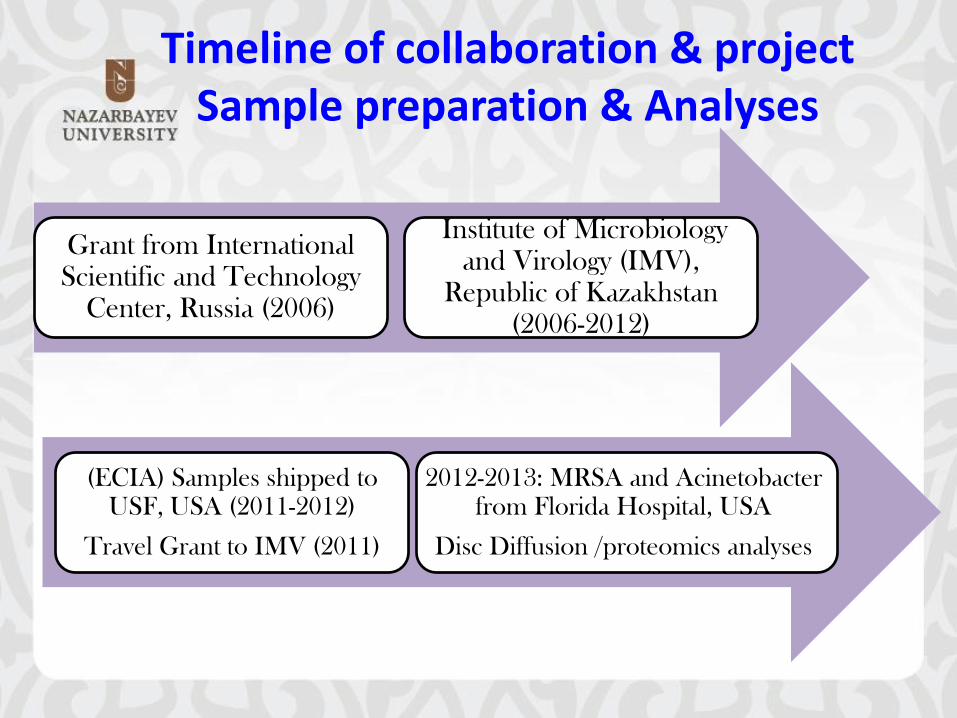
Timeline of collaboration & project Sample preparation & Analyses
Grant from International Scientific and Technology
Center, Russia (2006)
Institute of Microbiology and Virology (IMV),
Republic of Kazakhstan (2006-2012)
(ECIA) Samples shipped to USF, USA (2011-2012)
Travel Grant to IMV (2011)
2012-2013: MRSA and Acinetobacter from Florida Hospital, USA
Disc Diffusion /proteomics analyses

Background
• Methicillin-resistant Staphylococcus aureus (MRSA) is the leading cause of multidrug resistant (MDR) hospital-associated infections (HAI) in the U.S
• MDR infections result in increased morbidity, mortality, cost of care, length of hospital stay, and increasingly insusceptible to known antimicrobials
• Multidrug-resistant (MDR) Acinetobacter baumannii is becoming an important healthcare- acquired pathogen in hospitals and other health care settings.
• Acinetobacter baumannii is an increasing cause of HAI in Intensive Care Units (ICUs) in the United States with it being the fifth most frequent cause of pneumonia
• Most antibiotics are natural products or semisynthetic derivatives from soil actinomycetes

Methods and findings
MRSA Isolate 48-29: zone of inhibition- 23mmMRSA Controls
Sulfa/Trimethoprim : zone of inhibition-36mmTobramicin- no zone
MRSA Isolate 19-25: zone of inhibition18mm
Fig. 3. Zone of inhibition of U.S. MRSA isolates against discs withKZ extracts
Fig. 4. AcinetobacterType AN controlsAmpicillin: zone of inhibition-25mmTetracycline:no zone

Overall summary of MRSA susceptibility to extremophile extracts
a Growth Medium 1 = Modified Bennett’s pH=7.2 b Growth Medium 2 = Modified Bennett’s pH=7.2, 5% NaCl c Growth Medium 3 = Modified Bennett’s pH=9.0, 0.5% Na2CO3 d NG = No growth of the Actinomycetes producer eZone of Inhibition for US HA-MRSA reported is an average of multiple DDAs.
Inhibition of Kazakhstan and US HA-MRSA by Actinomycetes Antagonists
Antagonist #
and Source ( pH)
Zone of Inhibition (Kazakhstan HA-MRSA) mm United States HA-MRSA
Zone of Inhibitione Growth Media
1a 2
b 3
c
2-2 mud (9.1) 0 18 NG 0
6-12 rhizosphere (8.6) 0 25 NG 0
18-7 sandy soil (10.0) 13 44 NG 0
19-25 soil (9.3) 0 35 0 11.5
33-1 mud (9.6) NGd 49 39 10.0
36-3 meadow soil (8.3) 10 24 23 0
41-8 saline soil (10.0) 11 29 15 7.0
48-29 sandy soil (10.0) 0 32 22 20.5
51-9 rhizosphere (9.5) 10 46 10 0
58-22 rhizosphere (8.9) 0 18 14 22.5
72-1 soil (9.6) 0 50 NG 0
96-1 soil (8.6) 11 26 19 0
Q4-39 soil (10.0) 0 16 15 0
Y-45 rhizosphere (9.8) 0 20 16 0

Methods and findings
http://www.research.usf.edu/cddi/

USF Center for Drug Discovery and Innovation (CDDI)
• LCMS analysis summary • Mass Spectrometry (MS): Agilent
6120 single quadrupole • Ion Mode: electrospray (ESI), positive
and negative • Mass range acquisition: m/z 100-
1200 amu • Software: chemstation
• High Performance Liquid
Chromatography (HPLC): Agilent 1100 Binary Pump, Well Plate autosampler, (2mL vial) Diode array detector (DAD), Thermostatted Column Compartment.
• Column: Phenomenex kinetex C18 2.6um, 3.0 x 100 mm
Sample Preparation Actinomycetes extracts diluted in Ethanol for a concentration of 1mg/mL in a 100 mL insert

Antimarin
Quick Dereplication Search: (using mass (MS) data observed and source taxonomic information)
For microbes:
• Antimarin database (available at the chemodiversity lab)
Note (1) the search was based on combined taxonomic and mass information (2) antimarin gathers pure compounds from marine
and marine/terrestrial
micro-organisms
• Complementary online data:
- Scifinder (exhaustive and
fully updated data base)
- Dictionary of Natural products
(not covered by USF)

19-25
Complex mixture : m/z+: 381 m/z-: 377
related +/- ions observed: m/z+: 213, 227 (fragments), 498, 516, 538 m/z-: 512 (fragment), 550, 560, MW= 515
Pure compound: m/z+: 512, 530, 552; m/z-: 564, 574, MW= 529
Mixture: m/z+: 544, 552, 566; m/z-: 588, MW= 543
Mixture m/z+: 171, 200, 258, 363; m/z-: low abundance, possible MW= 170
Mixture m/z+: 217, 457, 515; m/z-: 316, 360, 512, 671

I- possible structures for MW= 515 from sample 19-25 (antimarin)
m/z(+): [M-H2O+H]+= 498.3, [M+H]+= 516.2, [M+Na]+= 538.3 Possible fragment or impurity Need to be identified (pending)
m/z (-): [M+Cl]-= 549.9, [M+HCOO]-= 560.2

Summary and Conclusions
• Five extracts from Actinomycetes showed distinct inhibition zones when tested against US MRSA: promising candidates for novel antibiotics
• Initial chemical characterization was performed to investigate their antagonistic properties further
• The established approach and methodologies will be expanded and applied in future studies

Future directions
• Screen for producers of antibiotics active against Acinetobacter spp. and other gram negative pathogens and other important pathogens (MDRTB)
• Approach from several angles--- – bioactive guided fractionation to test which fractions contain active components – chemical characterization to identify novel compound
• Use of PCR and traditional culture to identify producer organism - for scale up
• Identification and gene expression studies of dormant genes
• Differential identification of peaks
– Prepare large batch culture from different growth conditions – Compare extracts from conditions where antibody produced and not producing – Identify peaks corresponding to active components


Acid Fast Stain-MDRTB

Acknowledgements
Former USF Students
• Lylah Seaton
• Ami Patel
• Colton Faza
• Magda Baksh
• Stefanie Albert
USF Colleagues/CDDI
• Dr. Boo Kwa
• Dr. Jill Roberts
• Dr. Laurent Calcul
Florida
Hospital Collaborators
Jill Whitaker
Christen Mayer
Microbiology Lab
The USF College of Public Health for the ECIA and Travel Awards
IMV Collaborator • Dr. Lyudmila Trezhnovikova • Faculty & Staff at IMV • ISTC for funding

Workshop on Biodiversity and Climate Change

Спасибо..!!

Спасибо..!!
Tien Shan Mountains
Kaindy-Lake
Mountains and Lakes