Extraoral and Intraoral Exam - Dentalelle Tutoring · Extraoral and Intraoral Exam Dentalelle...
69
Extraoral and Intraoral Exam Dentalelle Tutoring 1
Transcript of Extraoral and Intraoral Exam - Dentalelle Tutoring · Extraoral and Intraoral Exam Dentalelle...

Extraoral and Intraoral ExamDentalelle Tutoring
1
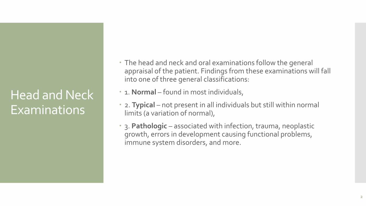
Head and Neck Examinations
The head and neck and oral examinations follow the general appraisal of the patient. Findings from these examinations will fall into one of three general classifications:
1. Normal – found in most individuals,
2. Typical – not present in all individuals but still within normal limits (a variation of normal),
3. Pathologic – associated with infection, trauma, neoplastic growth, errors in development causing functional problems, immune system disorders, and more.
2
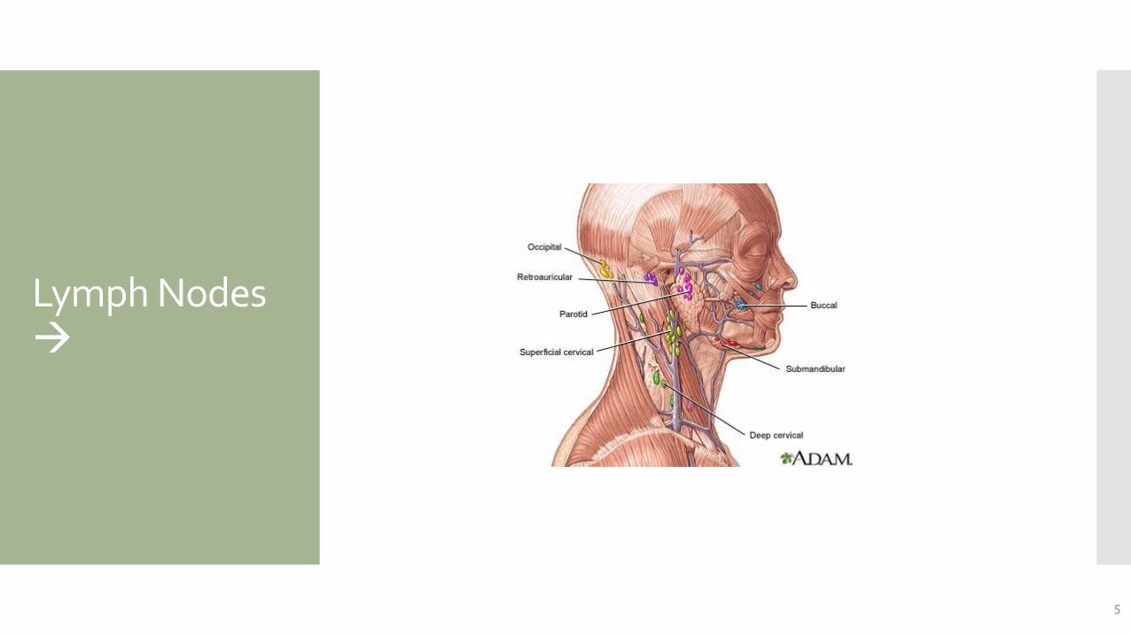
Lymph Nodes
The major lymph nodes of the head and neck area should be palpated with the patient in an upright position. Findings which should be noted include enlarged palpable nodes, fixed nodes, tender nodes and whether the palpable nodes are single or present in groups.
Findings which include single or multiple, tender, and fixed nodes are very suspicious for malignancy.
Groups of tender nodes usually occur in conjunction with some type of infection. Occasionally nodes will remain enlarged and palpable after an infection. This is a relatively common occurrence especially within the submandibular group of lymph nodes. When examined, these nodes should be small (less than 1 cm), non-tender and mobile. Remember to correlate findings from the medical history and general appraisal of the patient to the observations made during the head and neck examination.
3
Nodes
Occipital nodes - Palpate the occipital nodes about one inch above and below the hairline.
Auricular - Palpate the pre and post auricular nodes bilaterally using the pads of the index, middle and ring fingers.
Cervical Chain - Palpate the nodes medial to the sternocleidomastoid muscle using a bidigital technique and the nodes posterior to the muscle with a bimanual technique.
Supraclavicular - These nodes are examined using digital compressions just superior to the clavicle.
Submandibular - Palpate the submandibular nodes by pulling or rolling the tissues under the chin up and over the inferior border of the mandible. Ask the patient to touch the roof of the mouth with the tongue, pressing firming against the roof will allow you to assess the muscles and any pathology associated with the submandibular lymph node areas.
4
Lymph Nodes
5
Palpation
Digital – single finger
Bidigital – finger and thumb of the same hand
Bimanual – use of finger or fingers and thumb from each hand
Bilateral – two hands used at the same time
6
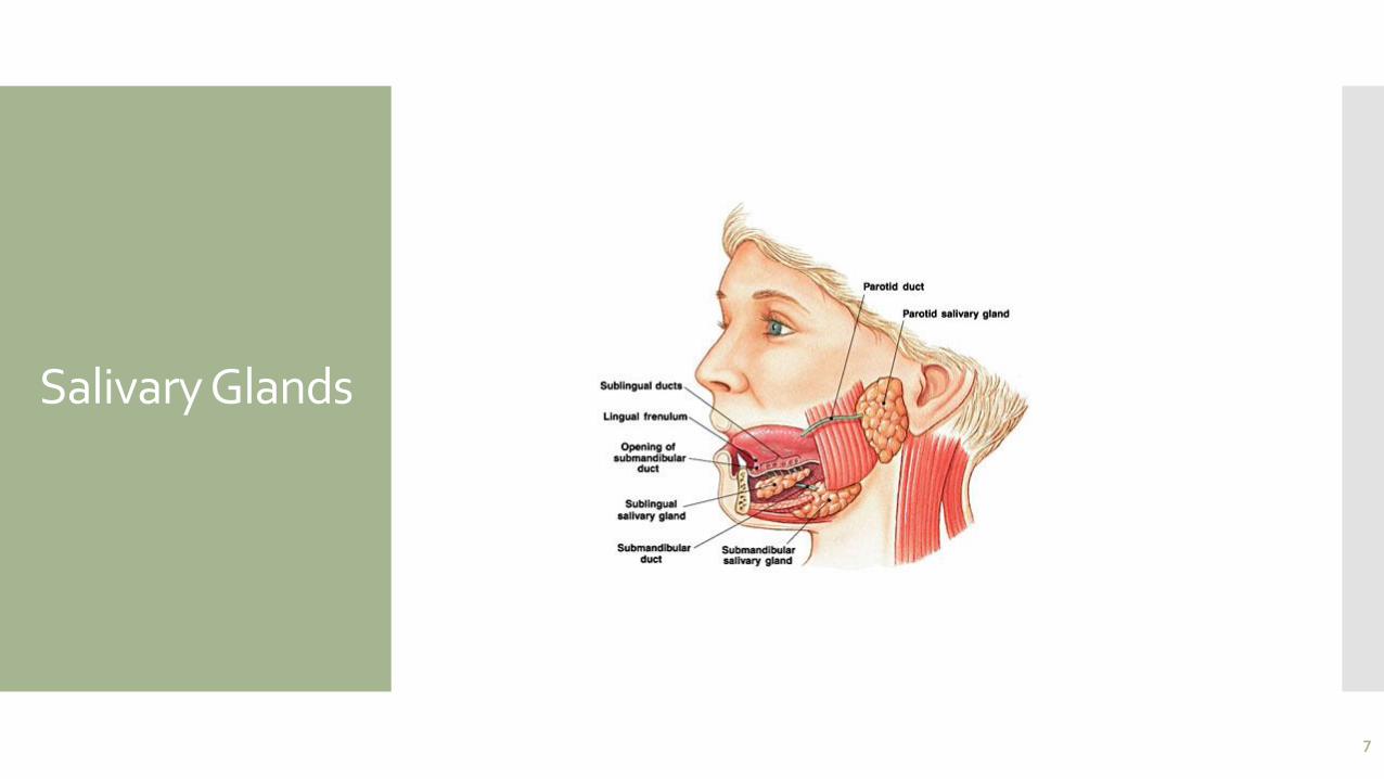
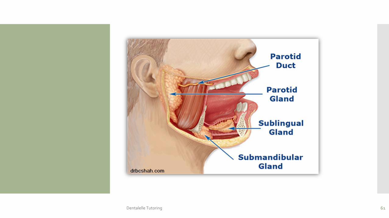
Salivary Glands
7
Floor of the Mouth
The floor of the mouth is examined using direct and indirect vision followed by bimanual palpation of the entire area. The patient should be asked to raise the tongue making direct visual examination of the tissues toward the midline of the floor of the mouth possible.
The mirror should be used to examine the areas near the inferior border of the mandible. The tissues should appear moist and very vascular. The normal anatomy of the area should be identified including:
Sublingual caruncle – small rounded projection at the base of the lingual frenum which houses Wharton’s duct from the submandibular salivary gland
Sublingual folds – two oblique elevations found radiating laterally away from the lingual frenum on either side of the caruncle which house the ducts from the sublingual salivary gland
Lingual frenum – muscle attachment from the ventral surface of the tongue to the floor of the mouth. This attachment varies in length from person to person.
8
The Tongue
The clinician should identify the normal anatomy of the tongue including:
Dorsal surface – papillae (filiform, fungiform, circumvallate), median sulcus, sulcus terminalis
Lateral borders – foliate papillae
Ventral surface – lingual veins, plica fimbriata, lingual frenum
Atypical findings on the dorsal surface of the tongue are common. They include: fissuring, scalloping, benign migratory glossitis and enlarged papillae, among others. A lingual thyroid may rarely be found on the posterior dorsal surface at the foramen cecum. Lingual varicosities are a common finding on the ventral surface of the tongue, especially in older patients.
9
The Tongue
The tongue is the most common intraoral site for oral cancer. Therefore, any sign of pathology should be investigated thoroughly. Some of the pathological findings that are found on the tongue include:
Hairy tongue – filiform papilla become elongated due to a variety of reasons from overuse of mouth rinses to not cleaning the tongue adequately.
Candidiasis – fungal infection of the tongue often associated with deeply fissured tongues.
Glossitis – inflammation of the tongue due to anemia, nutritional deficiencies and others.
10

Oral Cancer
11
Oral Cancer
Oral cancer is the sixth most form of common cancer in the world with a 5-year survival rate of less than 50%. The most common causes of oral cancer are tobacco and alcohol, factors that can be controlled. Increasingly the Human Papilloma Virus 16 (HPV16) is a recognized cause of carcinomas in the oropharynx and base of tongue.
The presentation of oral cancer is typically as an ulcer, red patch or white lesion.
It is important for the dental health care provider to inspect the mouth for suspicious lesions and take a biopsy to establish the diagnosis. Since smaller oral cancers have a better prognosis than larger ones, early detection plays an important role in reducing both the treatment associated morbidity and death rate from the disease.
Oral cancer is a common disease mostly associated with tobacco and alcohol use. An emerging form of cancer is that occurring in the base of tongue and oropharynx where the disease is linked to HPV16 infection. All forms of oral cancer are amenable to detection by dentists and diagnosis by biopsy.
12
Risks
The use of tobacco and alcohol are the two most important risk factors for cancer of the mouth. Smoking is the single most important risk factor for oral cancer. The use of tobacco and alcohol synergistically increase the risk of oral cancer.
Although it has been suggested that chewing tobacco is a significant cause of oral cancer, epidemiological studies have shown that the risk is small.1
By contrast, the use of pan (areca nut and tobacco leaves soaked in an alkali solution) is significantly associated with the development of oral cancer. Although a number of other factors have been implicated in the development of oral cancer, such as various bacterial and viral infections, for most oral cancers to date, none have been conclusively proven.
For cancer of the lip, the single most important risk factor is heavy exposure to ultraviolet rays of the sun
13
Viruses
Herpes GroupThe Herpes group of viruses has been implicated in the development of several cancers in man. Epstein-Barr virus (EBV) has been strongly associated with African Burkitt’s lymphoma and nasopharyngeal carcinoma. In oral carcinoma, some authors have detected Herpes simplex viral proteins in malignant cells whilst others have shown a co-carcinogenic effect of this virus with other carcinogens in cell culture studies. The precise oncogenic effects of HSV on the pathogenesis of oral cancers however, have not been established.
Human Papilloma Virus (HPV)Over 100 types of human papilloma virus have been identified. The wart (verruca vulgaris) is caused by HPV 2 and 4, whilst condylomata accuminata (genital warts) are caused by HPV 6 and 11. Heck’s disease (Focal epithelial hyperplasia) is associated with HPV 13 and 32. Of interest is the finding of HPV, particularly types 16 and and less frequently type 18, associated with squamous cell carcinoma of the oropharynx (tonsil).
14
Treatment
If the pathologist reports the lesion as cancer, then the patient needs to be referred promptly to a cancer treatment centre for assessment and treatment. An important component of the assessment is to establish the stage of the disease. Similar to cancers at other sites in the body, cancer of the mouth is staged using the TNM system where T stands for tumor size, N for the presence or absence of tumor involvement in regional lymph nodes and M for the presence or absence of distant metastases.
Generally, the prognosis is better when the tumor is smaller and has not metastasized to either lymph nodes or other organs. Staging also plays an important role in determining the most appropriate therapy for cancer of the oral cavity.
15
Treatment
Cancer of the oral cavity is treated by: Surgery Radiation therapy Chemotherapy is often added to either treatment.
For cancers that are restricted to the oral cavity, surgery is generally the preferred treatment modality. Depending on the site, small lesions can be excised and closed primarily. Larger lesions may require more complicated surgical reconstruction using grafting material taken from the arm or the leg.
For patients with bulky tumors or for those with tumor in regional lymph nodes, the preferred treatment is radiotherapy. In these instances, radiation is administered in small daily doses (fractions) of about 2 Grays until a total dose of about 70 Gray has been administered.
Radiation therapy has the effect of destroying the salivary glands and reducing the vascularity to the mandible and maxilla. Hence, patients who have had radiation therapy to the head and neck have a life-time risk of developing a form of osteomyelitis, termed osteoradionecrosisof the jaws.
16
Brush Biopsy
The brush biopsy is considered an adjunct diagnostic tool which uses a patented spiral shaped stiff brush to remove epithelial cells for examination by a pathologist.
The brush is firmly rolled over the lesion until bleeding points are observed signaling the epithelial tissues have been penetrated. The cells caught up on the brush are wiped off on a glass slide and sent to the laboratory where pathologists use both computer assisted and standard methods to evaluate the sample for the presence of abnormal cells.
The brush biopsy cannot render a definitive diagnosis, but will give the practitioner more conclusive evidence of further abnormalities such as abnormal cells that have been detected.
17
Cytology
A new cytology technique being used by many hospitals and gynecology practices in place of the traditional scraped Pap smear may be used with oral specimens as well.
Instead of transferring the specimen onto a glass slide, the brush used to collect the cell specimen is twirled in a vial containing an alcohol-based fixative/preservative liquid.
The brush head remains in the fixative so that no cell is lost during this process and the entire vial is sent to a laboratory for microscopic evaluation of the cells.
18
Velscope
The VELscope is a hand held device that excites the tissue causing it to fluoresce. Normal, healthy tissue will appear pale green when exposed to the emitted blue light, and when the tissue is not normal, the color will be darker green to black. No pre-rinse is used with this adjunct.
The device has been found useful especially to delineate surgical margins that may extend beyond the visible lesion margins.
When there is a high degree of inflammation, such as in the case of lichen planus, mucous membrane pemphigoid, pemphigus vulgaris, lupus, etc. the tissue may appear dark; thereby, producing a false positive result. Any type of inflammatory process such as cheek chewing, etc. will also exhibit a darker color change. (www.velscope.com)
19
ViziLite
The ViziLite device has several components. The patient rinses with one percent acetic acid for 30-60 seconds followed by the use of a Chemiluminescence light.
The light stick is especially beneficial in identifying white and erythroleukoplakia lesions. After the light stick is broken and shaken, it is placed next to all visible oral soft tissue surfaces. The device will cause illumination of any leukoplakia and/ or erythroleukoplakia lesions causing them to appear bright white.
T-Blue is a part of the plus system and contains pharmaceutical grade tolonium chloride, which is a toluidine-blue dye that stains the nuclear cell material blue. The use of the T-Blue assists in further delineating the extent/margins of any detected lesions. This is an FDA approved product. (www.vizilite.com)
20
Simple Oral Pathology
Radiographs and oral photography are important adjuncts which can be utilized to compare changes in tissue. This is especially beneficial in cases where severe inflammation is present. Many mucosal diseases have an inflammatory component and some adjunct devices do not present any conclusive evidence supporting anything other than inflammation with a failure to render a clinical diagnosis.
Often tissue specimens may return as diagnosed “non-specific ulcerative tissue” and crucial areas may be missed due to excessive inflammation. Being able to compare digital images of the lesion provides the practitioner the added benefit of actually seeing the progression or extension of the lesion.
An added benefit is being able to show the patient any progression when suggesting they need to have a biopsy.
21
Exfoliative Cytology
The microscopic examination of desquamated cells for diagnosticpurposes. The cells are obtained from lesions, sputum, secretions,urine, and other material by aspiration, scraping, a smear of tissue.
Steps: Prepare the lesion – irrigate and wipe
Scrape the lesion – scrape with metal spatula several times
Smear on the glass slide – start at center of slide
Fix the cells – flood with alcohol on the slide or spray
Obtain second smear – duplicate process
Complete the fixation – leave slides for 30 minutes, after 20 minutes tip slide to slide the remaining alcohol off
Prepare History or Data Sheet – dentists name (address), patients name (address), lesion details, other info.
22
Red Lesion
Erythroplakia – “Red Lesion”
Etiology – a significant risk factor to the oral mucosa is chronic exposure to carcinogenic components found in all types of tobacco. The other common risk factor is chronic alcohol exposure. Even worse is a combination of both.
Typical Visual Cues – a circumscribed, or ill-defined, erythematous plaque that varies in size, thickness and surface configuration. It has a velvety appearance and occurs most frequently on the floor of the oral cavity, ventral area of the tongue and the soft palate.
Useful Clinical Information – a painless and persistent lesion, found more commonly in adult males and patients who report tobacco exposure.
Treatment Recommendations – the patient should be counseled in a tobacco cessation program if biopsies reveal that the lesion is premalignant. Then, a more extensive therapy is indicated and the patient should be re-evaluated at regular intervals for other oral mucosal changes.
Clinical Significance – erythroplakia occurs less frequently than leukoplakia, but it is much more likely to exhibit microscopic evidence of premaligancy.
23
Squamous Cell Carcinoma
Etiology – idiopathic (unknown). Risk factors include: tobacco use, alcohol use, sun radiation, genetic predisposition, nutritional deficiency, immunosuppression, and infections, such as candidal leukoplakia and human papillomavirus.
Typical Visual Cues – a deep-seated ulcerated mass, fungating ulcerated mass, ulcer margins commonly elevated, adjacent tissues commonly firm to palpation, and may have residual leukoplakia and/or erythroplakia.
Useful Clinical Information – more common in adult males, continuous enlargement, local pain, referred pain often to the ear, and paresthesia often of the lower lip.
Treatment Recommendations – patient should be counseled to stop tobacco use and given a referral to a local medical treatment facility for appropriate treatment (surgery, radiation therapy, chemotherapy), and a careful periodic re-evaluation.
Clinical Significance – early diagnosis is essential for cure, presence of lymph node metastasis greatly worsens the prognosis, approximately 50% of patients have evidence of lymph node metastasis at time of diagnosis (that is why the extraoral examination is so critical at the time of each intraoral examination), and patients who have had one cancer are at greater risk of having a second oral cancer. The overall 5-year survival rate is 45-50%.
24
Oral Management
Bacterial Plaque Control
Start oral hygiene instruction at first appointment and emphasize preventive infection control procedures and potential oral side effects associated with cancer therapy. Toothbrushing instructions include using a soft or extrasoft toothbrush; a flavored dentifrice may not be tolerated, but fluoride is essential. Mouthrinses include saline solution to moisten the mucosa or baking soda rinses for managing oral mucositis. Chlorhexidine gluconate rinsing may also be recommended for antibacterial properties. Commercial mouthrinses that contain alcohol should be avoided.
Daily Fluoride Therapy
Daily fluoride therapy is indicated for patients about to undergo head and neck radiation therapy, if the salivary glands are in the field of radiation. Make impressions and fabricate a custom fluoride tray, advise the patient to apply custom trays lined with prescription neutral sodium fluoride gel to the teeth for four minutes once daily or use brush-on sodium fluoride varnish. Advise patient to refrain from eating, drinking, or rinsing for 30 minutes following fluoride application.
Dietary Instructions
Instruct the patient in the preparation of foods. Avoid highly cariogenic foods such as carbohydrates and also spicy foods. A soft, bland diet is recommended. Water is recommended throughout the day to moisten the oral cavity. Saliva substitutes are also recommended to decrease the risk of caries.
Avoid Alcohol and Tobacco Products
If the patient uses tobacco products, a logical step for the cancer patient is to start tobacco cessation counseling. See the ADAA continuing education course on tobacco cessation for further
25

The Dentition
26
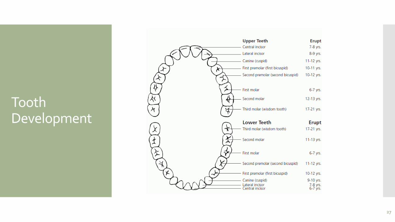
Tooth Development
27
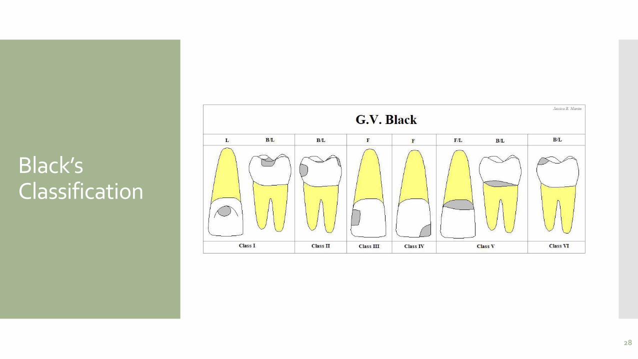
Black’s Classification
28
Caries
Early childhood caries, also known as baby bottle caries, baby bottle tooth decay, is a disease characterized by severe decay in the teeth of infants or young children. Early childhood caries (ECC) is a very common bacterial infection. Its prevalence is epidemic; in the US its rate is highest in minority and rural populations, at times infecting over 70% of the children. A large body of scientific evidence indicates that ECC is an infectious and transmissible disease, with Streptococcus mutans the primary microbiological agent in the disease. The disease process begins with the transmission of the bacteria to the child, usually from the primary caretaker. Caretakers with untreated dental disease present a very high risk to their children.
Root caries – soft, progressive lesion of cementum and dentin. This can increase with age and gingival recession is necessary.
29
Non-Carious
Enamel Hypoplasia – organic enamel hasn’t formed properly. Enamel is partially or entirely missing. AMELOGENESIS IMPERFECTA
Attrition – wearing away of the tooth from constant contact
Erosion – loss of tooth substance by a chemical process
Abrasion – mechanical wearing away of tooth substance by forces other than mastication. At ROOT SURFACES and incisal edges.
30
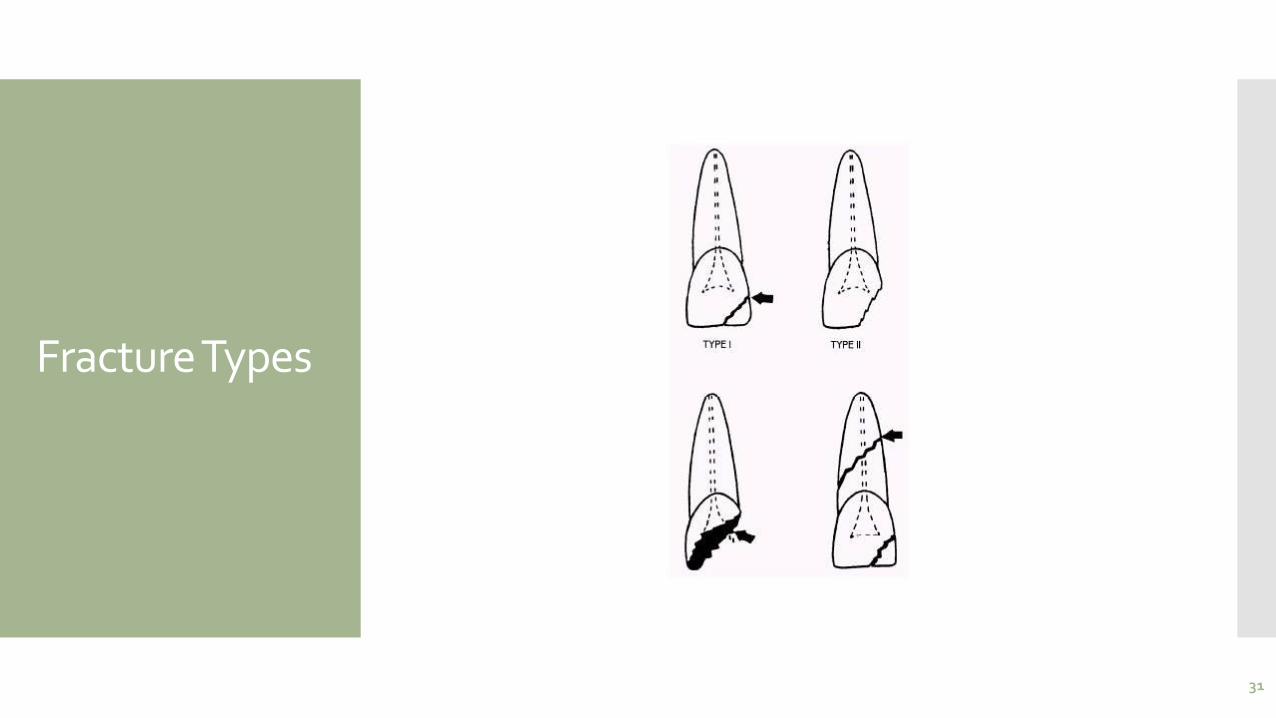
Fracture Types
31
Pulp Testing
A pulp test is a form of dental treatment. Its primary purpose is tooth physical health evaluation, particularly, the dentist seats a tool on the tooth that transmits an electric current, high or cold temperature.
Certain pulp assessment techniques demand complete usage of the tooth’s surface area, therefore tooth encased fully with crowns could be harder to assess compared to uncovered tooth.
The level to which this electric current generates a reaction in the tooth can assess possible problems within the tooth. Pulp vitality tests are important elements of the endodontic evaluation and assist in revealing the condition of the tooth pulp.
These types of pulp tests can easily confirm the demand for dental interventions such as root canals or tooth extraction. The tooth pulp test determines the physical health of a tooth’s pulp.
Once pulp vitality test of a affected tooth implies that pulp is not active, dentists engage in root canal treatment to eliminate the lifeless cells and protect against abscess.
A dead pulp gives NO response. Lingering pain means irreversible pulpitis, and when pain subsides it means reversible pulpitis.**
32
Caries Development
A carious lesion develops in three stages of demineralization. The first stage in demineralization of enamel is called the incipient lesion or “white spot”. This beginning carious lesion can be reversed with the daily use of fluoride or calcium and phosphate, persistent oral hygiene care, and a reduction of refined carbohydrates. The second stage involves the progression of demineralization leading to the DEJ and into the dentinal layer. The third stage is the actual cavitation in the dentinal layer. Neither of the last two stages can be reversed and require mechanical removal of dental caries.
There are three levels of preventive dentistry that the dental professional should understand when educating patients in the dental caries process. The first step is primary prevention. This prevents the transmission of S. mutans and delays the establishment of bacteria in infants, toddlers, and young children. The second step is secondary prevention, which prevents, arrests, or reverses the microbial shift before any clinical signs of the disease occur. The third step focuses on limiting or stopping the progression of the caries process by initiating remineralization therapy of existing lesions.
33
Prevention
Once S. mutans and lactobacilli bacteria are established in the oral cavity, the greater the risk for future caries to develop.
Where biofilm (plaque) accumulates, the bacterial count is considered to be higher, as in areas in the oral cavity that are difficult to reach during oral hygiene, such as pit and fissures.
Newly erupted teeth are deficient in mineral content (calcium and phosphate), making them more susceptible to bacteria. By introducing antimicrobial agents, such as fluoride the bacterial count may be significantly reduced.
34
Demineralization
When refined carbohydrates are introduced into the oral cavity, lactic acid production occurs as an end product of S. mutans, causing the saliva pH to drop from a neutral pH of approximately 7 to an acidic pH of 4.5-5.0. This metabolizing acidogenic bacteria’s lactic acid production begin to demineralize the enamel.
The more acidogenic bacteria present, the more lactic acid produced.
When saliva is released into the oral cavity via the salivary glands, the pH of the saliva returns to normal or an approximate pH of 7 and a period of remineralization (repair) occurs. This process is facilitated if fluoride or calcium and phosphate ions are present locally. The balance between demineralization and remineralization is crucial. If the balance is not maintained and demineralization occurs too frequently, then an incipient lesion will occur. This incipient or ‘white spot’ lesion may take up to approximately 9 months or more to be seen via digital imaging or radiographically as a radiolucency or dark spot on a bite-wing radiograph.
35
Low Acidogenic Foods
Raw vegetables, such as broccoli, cucumber, lettuce, carrots, peppers.
Meat, fish, poultry.
Beans, peas, nuts, natural peanut butter.
Milk, cheeses, flavored yogurts.
Corn chips, peanuts, popcorn.
Fats, oils, butter, margarine.
Non-sugar sweeteners, such as xylitol®, NutraSweet®, aspartame®, saccharin®, sucralose®.
Soft drinks, sport drinks, and energy drinks containing sugar are big business in the United States. Frequent consumption of sugary drinks has long been known to contribute to dental caries. According to the Centers for Disease Control (CDC), consumption of soft drinks in the United States has increased over the last 30 years with both adults and children. Soft drinks have been linked to obesity and type 2 diabetes. Teenagers and young adults consume more sugar drinks than other age groups. Males consume more soft drinks than females and low-income Americans consume more soft drinks than those with higher incomes.
A non-diet soft drink is made from carbonated water, added sugar and flavors. Each can of soda contains the equivalent of about ten teaspoons, or 40 grams, of sugar. Mountain Dew® is so popular in the United States that the coined phrase “Mountain Dew Mouth” is a recognized term used by the dental profession for patients diagnosed with rampant caries and/or erosion.
36
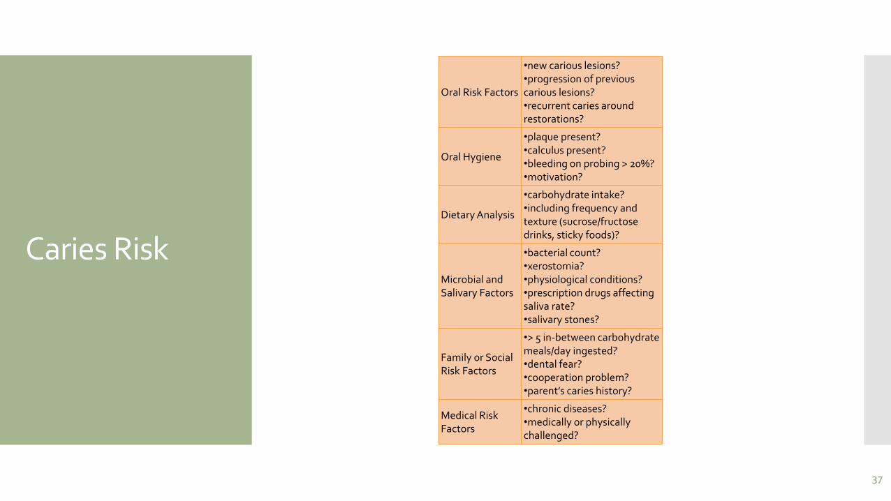
Caries Risk
Oral Risk Factors
•new carious lesions?•progression of previous carious lesions?•recurrent caries around restorations?
Oral Hygiene
•plaque present?•calculus present?•bleeding on probing > 20%?•motivation?
Dietary Analysis
•carbohydrate intake?•including frequency and texture (sucrose/fructose drinks, sticky foods)?
Microbial and Salivary Factors
•bacterial count?•xerostomia?•physiological conditions?•prescription drugs affecting saliva rate?•salivary stones?
Family or Social Risk Factors
•> 5 in-between carbohydrate meals/day ingested?•dental fear?•cooperation problem?•parent’s caries history?
Medical Risk Factors
•chronic diseases?•medically or physically challenged?
37

Stages of Calculus Formation
38
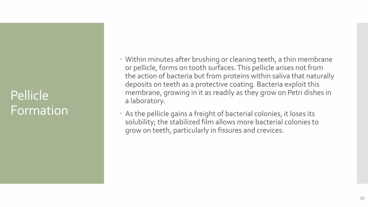
Pellicle Formation
Within minutes after brushing or cleaning teeth, a thin membrane or pellicle, forms on tooth surfaces. This pellicle arises not from the action of bacteria but from proteins within saliva that naturally deposits on teeth as a protective coating. Bacteria exploit this membrane, growing in it as readily as they grow on Petri dishes in a laboratory.
As the pellicle gains a freight of bacterial colonies, it loses its solubility; the stabilized film allows more bacterial colonies to grow on teeth, particularly in fissures and crevices.
39
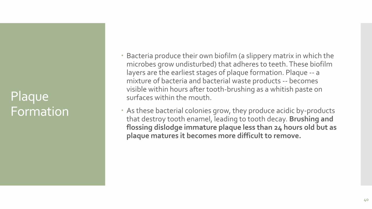
Plaque Formation
Bacteria produce their own biofilm (a slippery matrix in which the microbes grow undisturbed) that adheres to teeth. These biofilm layers are the earliest stages of plaque formation. Plaque -- a mixture of bacteria and bacterial waste products -- becomes visible within hours after tooth-brushing as a whitish paste on surfaces within the mouth.
As these bacterial colonies grow, they produce acidic by-products that destroy tooth enamel, leading to tooth decay. Brushing and flossing dislodge immature plaque less than 24 hours old but as plaque matures it becomes more difficult to remove.
40
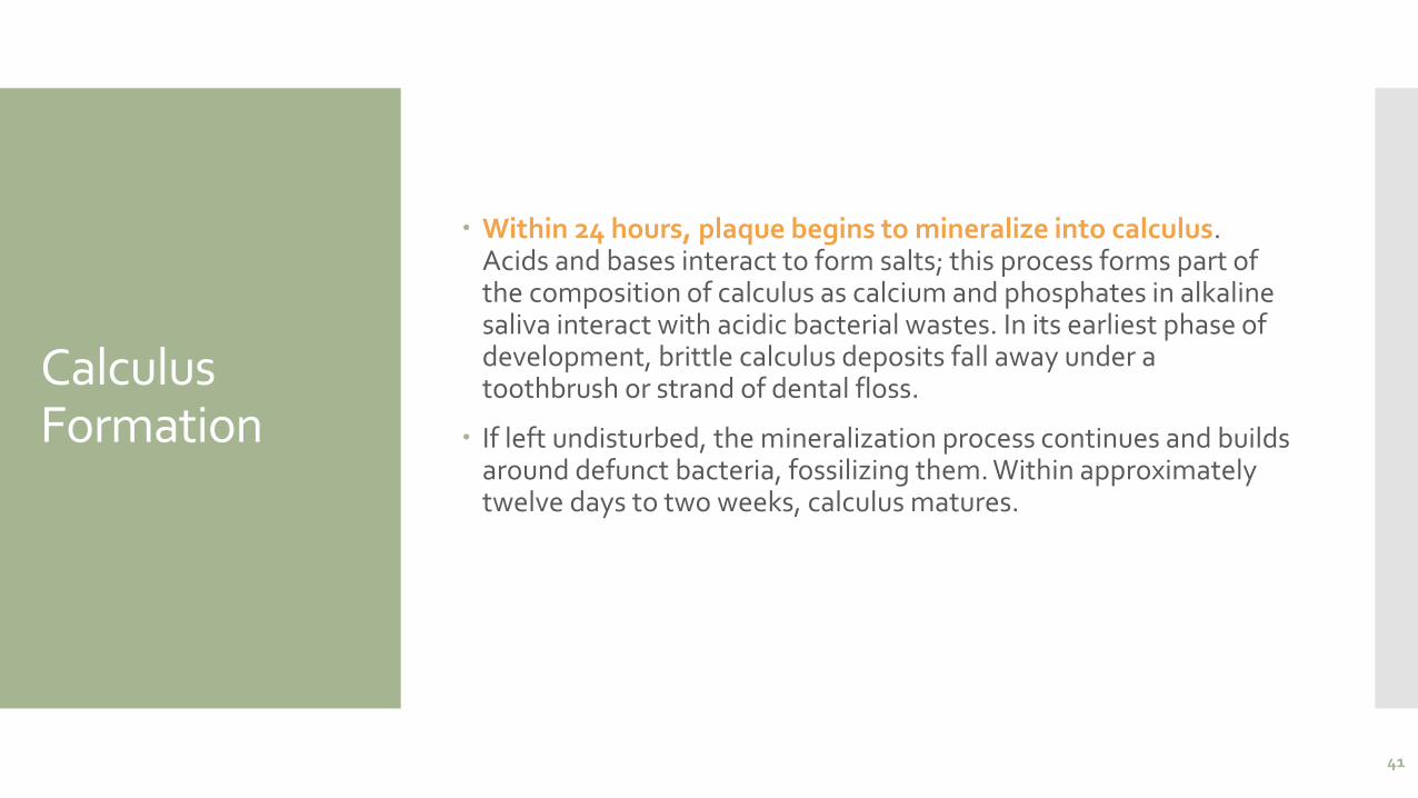
Calculus Formation
Within 24 hours, plaque begins to mineralize into calculus. Acids and bases interact to form salts; this process forms part of the composition of calculus as calcium and phosphates in alkaline saliva interact with acidic bacterial wastes. In its earliest phase of development, brittle calculus deposits fall away under a toothbrush or strand of dental floss.
If left undisturbed, the mineralization process continues and builds around defunct bacteria, fossilizing them. Within approximately twelve days to two weeks, calculus matures.
41
Maturation
Dentists can readily see mature calculus formations as whitish or pale yellow structures affixed to teeth. The largest calculus deposits typically occur on the lingual (nearest the tongue) surfaces of the lower front teeth but tartar can occur anywhere in the mouth.
Sub-gingival calculus grows below the gums, while supra-gingival calculus grows on visible tooth surfaces above the gums. Although it feels solid, porous calculus plays host to multiple colonies of bacteria that thrive on the increased surface area that the mineralized plaque provides.
42
Flossing
Another question patients ask frequently is, “Do I floss before or after brushing?” Again, it doesn’t matter. As long as they are flossing properly, dental professionals are thrilled. The point of brushing and flossing is plaque removal. By removing the irritant plaque and reducing the bacterial count in the saliva, the patient is making a significant difference in preventing caries and periodontal disease.
There are two flossing methods available to teach your patients. One is the circle or loop method and the other is the spool method.
The circle or loop method is preferred for children or any patient with low manual dexterity. A piece of floss approximately 18 inches long is tied at the ends to form a loop or circle. The patient uses the thumb and index finger of each hand in various combinations to guide the floss interproximally through the contacts. When inserting floss, it is gently eased between the teeth with a seesaw motion at the contact point, making sure not to snap the floss and cause trauma to the gingival papilla. Once through the contact area, gently slide the floss up and down the mesial and distal marginal ridges in a C-shape around the tooth directing the floss subgingivally to remove the debris.
The spool method utilizes a piece of floss approximately 18-24 inches long where the majority of the floss is loosely wound around the middle finger of one hand and a small amount of floss around the middle finger of the opposite hand. The same procedure is followed as the loop method when positioning the floss interproximally. After each marginal ridge is cleaned, the used floss is moved or spooled to the other hand until all supragingival and subgingival areas have been cleaned, including the distal areas of the posterior teeth.
43
Tooth Picks
If your patients have large diastemas or food impaction areas, they should be encouraged to utilize interproximal sticks such as Stim-U-Dents®. Made of balsa wood, Stim-U-Dents® are used to remove debris and plaque, and are preferred by dental professionals over standard toothpicks because toothpicks can splinter into the gingiva and damage the gingival tissue.
If patients do not have access to floss, they can use the wooden balsa sticks to remove plaque and stimulate the gingiva.
InterproximalThese small interproximal brushes are attached to handles and are used for large spaced interproximal areas and for orthodontic patients to use between their brackets to remove debris. There are a variety of brushes available, including travel sizes for pockets and purses. The brushes are tapered for easy access to difficult areas and patients seem to adapt well to instructional use.
44

Stats
45
Injuries
Soft Tissue Injuries
The face is often the most exposed part of the body in athletic competition and injuries to the soft tissues of the face are frequent. Abrasions, contusions, and lacerations are common and should be evaluated to rule out fractures or other significant underlying injury. These usually occur over a bony prominence of the facial skeleton such as the brow, cheek, and chin. Lip lacerations are also common.
Fractures
Fractures of the facial bones present an even more complex problem. One of the most frequent sites of bony injury is the zygoma (cheekbone). Fractures of the zygoma, occurring as a result of direct blunt trauma from a fall, elbow or fist, account for approximately 10% of the maxillofacial fractures seen in sports injuries. Like the zygoma, the prominent shape and projection of the mandible cause it to be frequently traumatized. Approximately 10% of maxillofacial fractures resulting from sporting activities occur in the mandible when the athlete strikes a hard surface, another player or equipment. In a mandibular fracture, airway management is the most important aspect of immediate care.
In both children and adults, the condyle is the most vulnerable part of the mandible. Fractures in this region have the potential for long-term facial deformity. Recent data suggest that condylar fractures in children can alter growth of the lower face.
46
Continued
TMJ Injuries
Most blows to the mandible do not result in fractures, yet significant force can be transmitted to the temporomandibular disc and supporting structures that may result in permanent injury. In both mild and severe trauma, the condyle can be forced posteriorly to the extent that the retrodiscal tissue is compressed. Inflammation and edema can result, forcing the mandibular condyle forward and down in acute malocclusion. Occasionally this trauma will cause intracapsular bleeding, which could lead to ankylosis of the joint.
Tooth Intrusion
Tooth intrusion occurs when the tooth has been driven into the alveolar process due to an axially directed impact. This is the most severe form of displacement injury. Pulpal necrosis occurs in 96% of intrusive displacements and is more likely to occur in teeth with fully formed roots. Immature root development will usually mean spontaneous re-eruption. Mature root development will require repositioning and splinting or orthodontic extrusion.
Crown and Root Fractures
Crown fractures are the most common injury to the permanent dentition and may present in several different ways. The simplest form is crown infraction. This is a crazing of enamel without loss of tooth structure. It requires no treatment except adequate testing of pulpal vitality. Fractures extending into the dentin are usually very sensitive to temperature and other stimuli. The most severe crown fracture results in the pulp being fully exposed and contaminated in a closed apex tooth or a horizontal impact may result in a root fracture. The chief clinical sign of root fracture is mobility. Radiographic evaluation and examination of adjacent teeth must be performed to determine the location and severity of the fracture as well as the possibility of associated alveolar fracture. Treatment is determined by the level of injury.
47
Avulsion
Certainly one of the most dramatic sports-related dental injuries is the complete avulsion of a tooth. Two to sixteen percent of all injuries involving the mouth result in an avulsed tooth. A tooth that is completely displaced from the socket may be replaced with varying degrees of success depending, for the most part, on the length of time it is outside the tooth socket.
If the periodontal fibers attached to the root surface have not been damaged by rough handling, an avulsed tooth may have a good chance of recovering full function. After two hours, the chance for success is greatly diminished. The fibers become necrotic and the replaced tooth will undergo resorption and ultimately be lost.
48
Mouthguards
When considering recommendations, an ideal mouthguard:
protects the teeth, soft tissue, bone structure, and temporomandibular joints
diminishes the incidence of concussions and neck injuries
exhibits protective properties that include high power absorption and power distribution throughout the expansion
provides a high degree of comfort and fit to the maxillary arch
remains securely and safely in place during action
allows speaking and does not limit breathing
is durable, resilient, tear resistant, odorless, and tasteless
49
Types
Stock MouthguardsStock mouthguards may be purchased from a sporting goods store or pharmacy. They are made of rubber, polyvinyl chloride or a polyvinyl acetate copolymer. The advantage is that this mouthguard is relatively inexpensive, but the disadvantages far outweigh the advantages. They are available only in limited sizes, do not fit very well, inhibit speech and breathing and require the jaws to be closed to hold the mouthguard in place. Because the stock mouthguards do not fit well, the player may not wear the mouthguard due to discomfort and irritation. The Academy of Sports Dentistry has stated that the stock mouthguard is unacceptable as an orofacial protective device.
Mouth-Formed ProtectorsThere are two types of mouth-formed protectors: the shell-liner and the thermoplastic mouthguard. The shell-liner type is made of a preformed shell with a liner of plastic acrylic or silicone rubber. The lining material is placed in the player’s mouth, molds to the teeth and then is allowed to set. The preformed thermoplastic lining (also known as “boil and bite”) is immersed in boiling water for 10 to 45 seconds, transferred to cold water and than adapted to the teeth. This mouthguard seems to be the most popular of the three types and is used by more than 90 percent of the athletic population
Custom Made Mouth ProtectorsThis is the superior of the three types and the most expensive to the athlete. But isn’t it worth the cost to protect an athlete’s teeth from injury? Most parents will spend quite a bit of money on athletic shoes, but might not think about protecting their child’s teeth. This mouthguard is made of thermoplastic polymer and fabricated over a model of the athlete’s dentition. The mouthguard is made by the dentist and fits exactly to the athlete’s mouth. The advantages include: fit, ease of speech, comfort and retention. By wearing a protective mouthguard, the incidence of a concussion by a blow to the jaw is significantly reduced because the condyle is separated form the base of the skull by placing the mandible in a forward position.
50

Landmarks of the Oral CavityIndividual Tutoring
Dentalelle Tutoring 51
Landmarks
Masticatory Mucosa: Heavily keratinized tissue that lines the hard palate and tongue.
Alveolar Mucosa: Lightly keratinized tissue that lines floor of the mouth and covers the alveolar processes.
Labial and Buccal Mucosa: Thinly keratinized tissue that lines the inner surface of the lips and cheeks
Oral Vestibule: A pocket formed by the soft tissue of the lips/cheeks and the gingiva, its deepest point the “muccobuccal fold”.
Frenum: Raised lines of oral mucosa that extend from the alveolar mucosa to the labial and buccal mucosa.
Fordyce Granules/Spots: Yellowish sebaceous glands found on the facial mucosa near the corners of the mouth.
Linea Alba: A raised white line of keratinized tissue on the buccal mucosa that runs parallel to the line of the occlusal plane
Parotid Papilla: Flap of tissue found opposite the maxillary 2nd molar on the buccal mucosa and contains the terminal end of the Parotid Duct (Stenson’s Duct).
Dentalelle Tutoring 52
Dentalelle Tutoring 53
Landmarks
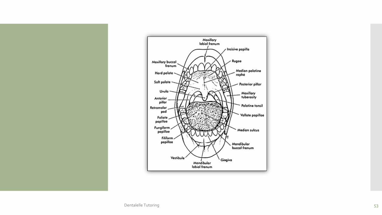
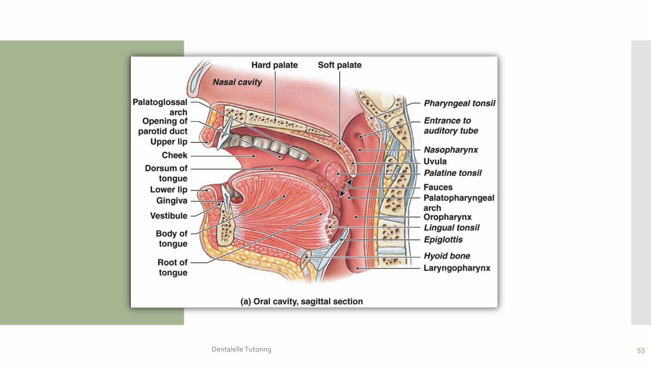
Hard Palate: Bony structure that separates oral cavity from nasal cavity.
Incisive Papilla: Thick keratinized tissue that covers the incisive foramen.
Palatine Raphe: Midline ridge of tissue that covers the bony suture of the palate.
Palatine Rugae: Irregular ridges or folds of masticatory mucosa that extend horizontally from either side of the palatine raphe.
Soft Palate: Forms the posterior section of the palate and is not supported by underlying bone. It can be lifted to meet the posterior pharyngeal wall to seal the nasopharynx during swallowing and speech.
Uvula: Small conical mass of tissue that hangs from the palatine velum
Fauces: The passageway from the oral cavity to the pharynx (throat).
Pillars of Fauces: Two arches of muscle tissue surrounding posterior oral cavity. The Anterior Pillar of Fauces is also called palatoglossal arch, the Posterior Pillar of Fauces is also called the palatopharyngeal arch. They contract and narrow the fauces during deglutition (swallowing).
Dentalelle Tutoring 54
Dentalelle Tutoring 55
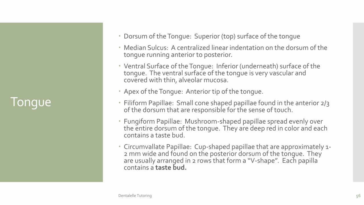
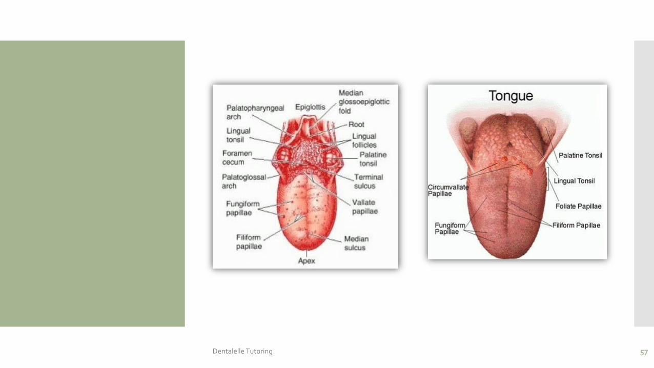
Tongue
Dorsum of the Tongue: Superior (top) surface of the tongue
Median Sulcus: A centralized linear indentation on the dorsum of the tongue running anterior to posterior.
Ventral Surface of the Tongue: Inferior (underneath) surface of the tongue. The ventral surface of the tongue is very vascular and covered with thin, alveolar mucosa.
Apex of the Tongue: Anterior tip of the tongue.
Filiform Papillae: Small cone shaped papillae found in the anterior 2/3 of the dorsum that are responsible for the sense of touch.
Fungiform Papillae: Mushroom-shaped papillae spread evenly over the entire dorsum of the tongue. They are deep red in color and each contains a taste bud.
Circumvallate Papillae: Cup-shaped papillae that are approximately 1-2 mm wide and found on the posterior dorsum of the tongue. They are usually arranged in 2 rows that form a “V-shape”. Each papilla contains a taste bud.
Dentalelle Tutoring 56
Dentalelle Tutoring 57
The Face
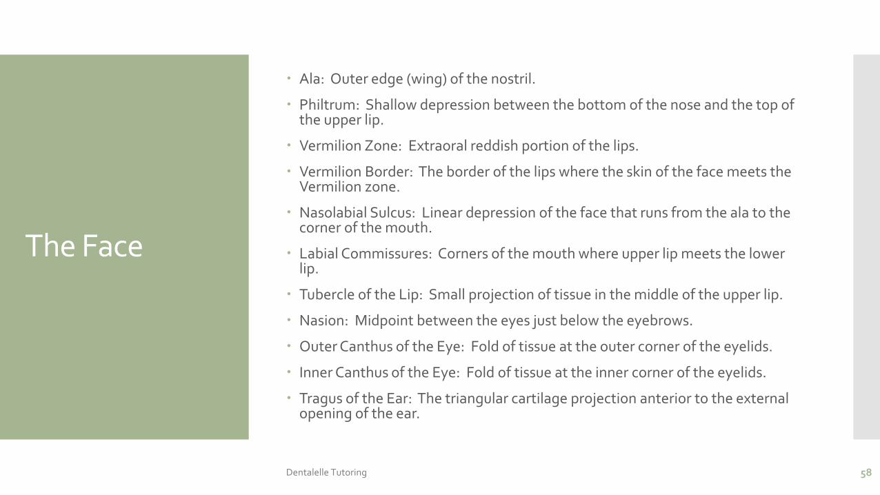
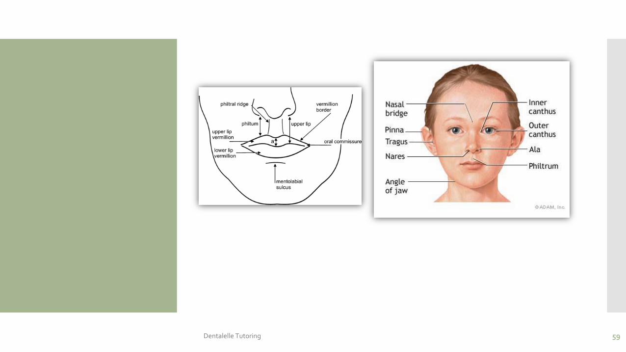
Ala: Outer edge (wing) of the nostril.
Philtrum: Shallow depression between the bottom of the nose and the top of the upper lip.
Vermilion Zone: Extraoral reddish portion of the lips.
Vermilion Border: The border of the lips where the skin of the face meets the Vermilion zone.
Nasolabial Sulcus: Linear depression of the face that runs from the ala to the corner of the mouth.
Labial Commissures: Corners of the mouth where upper lip meets the lower lip.
Tubercle of the Lip: Small projection of tissue in the middle of the upper lip.
Nasion: Midpoint between the eyes just below the eyebrows.
Outer Canthus of the Eye: Fold of tissue at the outer corner of the eyelids.
Inner Canthus of the Eye: Fold of tissue at the inner corner of the eyelids.
Tragus of the Ear: The triangular cartilage projection anterior to the external opening of the ear.
Dentalelle Tutoring 58
Dentalelle Tutoring 59
Floor of the Mouth
Lingual Frenum: Midline fold of tissue between ventral surface of the tongue and floor of the mouth.
Sublingual Caruncles: Two small, raised folds of tissue found on either side of the lingual frenum. They each contain a salivary duct opening for Wharton’s Duct (duct leading from the Submandibular Salivary gland).
Sublingual Folds: Folds of tissue that begin at the Sublingual Caruncles on either side of the lingual frenum and run backward toward the base of the tongue. They contain the many ducts from the sublingual salivary gland.
Dentalelle Tutoring 60
Dentalelle Tutoring 61
Function of Teeth - Incisors
Incisors
There are four incisors in each arch. Two central incisors and two lateral incisors.
Shape – single rooted, crowns are arched and angle toward one sharp incisal edge.
Function – to cut or incise food with their thin edges.
Dentalelle Tutoring 62

Canines
There are two canines in each arch. They are sometimes referred to as cuspids.
Shape – anchored with the longest root, one pointed cusp.
Function – used for holding, grasping, and tearing food. Referred to as the cornerstone of the mouth.
Dentalelle Tutoring 63
Premolars
There are for premolars in each arch. Two first premolars and two second premolars. They are sometimes referred to as bicuspids. There are no premolars in the primary dentition.
Shape – maxillary first premolars have a bifurcated root, all others have one root, one prominent cusp with one or two lesser lingual cusps.
Function – holding food, like canines because they have cusps; also to crush food.
Dentalelle Tutoring 64
Molars
There are three molars in each arch of the permanent dentition. Two first molars, two second molars and two third molars. Third molars are sometimes called wisdom teeth. There are two molars in each arch of the primary dentition. Two first molars and two second molars.
Shape – bifurcated or trifurcated roots, broad chewing surfaces with four to five cusps.
Function – grinding food.
Dentalelle Tutoring 65
Primary
Dentalelle Tutoring 66
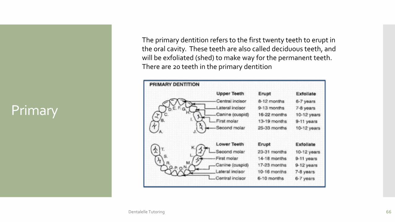
The primary dentition refers to the first twenty teeth to erupt in the oral cavity. These teeth are also called deciduous teeth, and will be exfoliated (shed) to make way for the permanent teeth. There are 20 teeth in the primary dentition
Permanent Teeth
Dentalelle Tutoring 67
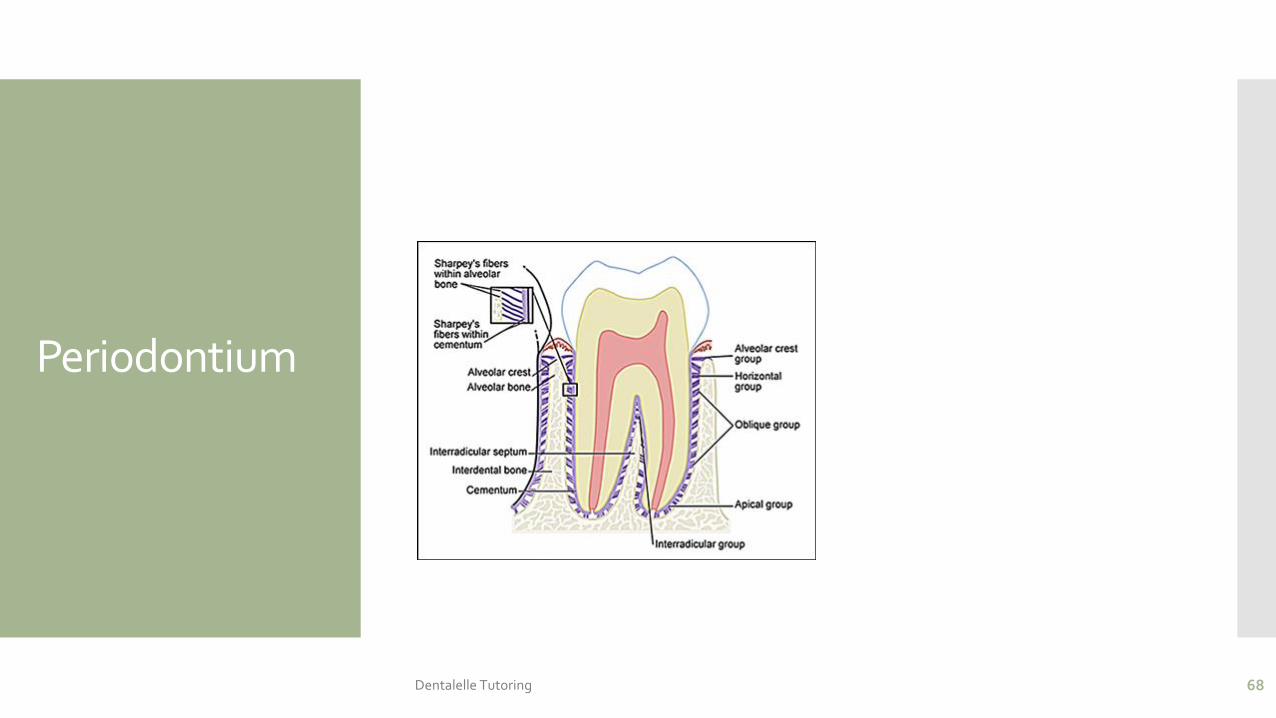
Periodontium
Dentalelle Tutoring 68
References
www.Dentalcare.com
Clinical Practice of the Dental Hygienist
http://medicalimages.allrefer.com/large/lymph-tissue-in-the-head-and-neck.jpg
Darby and Walsh
http://graphics8.nytimes.com/images/2007/08/01/health/adam/1095.jpg
Applegate EJ. The Anatomy and Physiology Learning System, 2nd Edition. Philadelphia: Saunders. p333.
69