Extracellular ATP and the P2X7 receptor in astrocyte-mediated
9
JOURNAL OF NEUROINFLAMMATION Gandelman et al. Journal of Neuroinflammation 2010, 7:33 http://www.jneuroinflammation.com/content/7/1/33 Open Access RESEARCH © 2010 Gandelman et al; licensee BioMed Central Ltd. This is an Open Access article distributed under the terms of the Creative Com- mons Attribution License (http://creativecommons.org/licenses/by/2.0), which permits unrestricted use, distribution, and reproduc- tion in any medium, provided the original work is properly cited. Research Extracellular ATP and the P2X 7 receptor in astrocyte-mediated motor neuron death: implications for amyotrophic lateral sclerosis Mandi Gandelman 1,2 , Hugo Peluffo 1,2 , Joseph S Beckman 3 , Patricia Cassina 2 and Luis Barbeito* 1,4 Abstract Background: During pathology of the nervous system, increased extracellular ATP acts both as a cytotoxic factor and pro-inflammatory mediator through P2X 7 receptors. In animal models of amyotrophic lateral sclerosis (ALS), astrocytes expressing superoxide dismutase 1 (SOD1 G93A ) mutations display a neuroinflammatory phenotype and contribute to disease progression and motor neuron death. Here we studied the role of extracellular ATP acting through P2X 7 receptors as an initiator of a neurotoxic phenotype that leads to astrocyte-mediated motor neuron death in non- transgenic and SOD1 G93A astrocytes. Methods: We evaluated motor neuron survival after co-culture with SOD1 G93A or non-transgenic astrocytes pretreated with agents known to modulate ATP release or P2X 7 receptor. We also characterized astrocyte proliferation and extracellular ATP degradation. Results: Repeated stimulation by ATP or the P2X 7 -selective agonist BzATP caused astrocytes to become neurotoxic, inducing death of motor neurons. Involvement of P2X 7 receptor was further confirmed by Brilliant blue G inhibition of ATP and BzATP effects. In SOD1 G93A astrocyte cultures, pharmacological inhibition of P2X 7 receptor or increased extracellular ATP degradation with the enzyme apyrase was sufficient to completely abolish their toxicity towards motor neurons. SOD1 G93A astrocytes also displayed increased ATP-dependent proliferation and a basal increase in extracellular ATP degradation. Conclusions: Here we found that P2X 7 receptor activation in spinal cord astrocytes initiated a neurotoxic phenotype that leads to motor neuron death. Remarkably, the neurotoxic phenotype of SOD1 G93A astrocytes depended upon basal activation the P2X 7 receptor. Thus, pharmacological inhibition of P2X 7 receptor might reduce neuroinflammation in ALS through astrocytes. Background Amyotrophic lateral sclerosis (ALS) is characterized by the progressive degeneration of motor neurons in the spi- nal cord, brainstem and motor cortex, leading to respira- tory failure and death of affected patients within a few years of diagnosis [1]. The discovery of mutations in the gene encoding the antioxidant enzyme Cu/Zn superoxide dismutase-1 (SOD1) in a subset of patients with familial ALS has led to the development of transgenic animal models expressing different SOD1 mutations [2]. These animal models recapitulate the human disease, exhibiting aberrant oxidative chemistry [3,4], neuroinflammation [5], endoplasmic reticulum stress [6], glutamate excito- toxicity [7], mitochondrial dysfunction [8] and protein misfolding and aggregation [9]. However, the mecha- nisms behind motor neuron death are unknown. Accumulating evidence indicates that non-neuronal cells contribute to motor neuron dysfunction and death in ALS by the maintenance of a chronic inflammatory response [10-12]. Activated microglia accumulate in the spinal cord, producing inflammatory mediators and reac- tive oxygen and nitrogen species [11]. Astrocytes, the most abundant cells in the adult nervous system, also * Correspondence: [email protected] 1 Neurodegeneration Laboratory, Institut Pasteur, Montevideo, Uruguay Full list of author information is available at the end of the article
Transcript of Extracellular ATP and the P2X7 receptor in astrocyte-mediated

JOURNAL OF NEUROINFLAMMATION
Gandelman et al. Journal of Neuroinflammation 2010, 7:33http://www.jneuroinflammation.com/content/7/1/33
Open AccessR E S E A R C H
ResearchExtracellular ATP and the P2X7 receptor in astrocyte-mediated motor neuron death: implications for amyotrophic lateral sclerosisMandi Gandelman1,2, Hugo Peluffo1,2, Joseph S Beckman3, Patricia Cassina2 and Luis Barbeito*1,4
AbstractBackground: During pathology of the nervous system, increased extracellular ATP acts both as a cytotoxic factor and pro-inflammatory mediator through P2X7 receptors. In animal models of amyotrophic lateral sclerosis (ALS), astrocytes expressing superoxide dismutase 1 (SOD1G93A) mutations display a neuroinflammatory phenotype and contribute to disease progression and motor neuron death. Here we studied the role of extracellular ATP acting through P2X7
receptors as an initiator of a neurotoxic phenotype that leads to astrocyte-mediated motor neuron death in non-transgenic and SOD1G93A astrocytes.
Methods: We evaluated motor neuron survival after co-culture with SOD1G93A or non-transgenic astrocytes pretreated with agents known to modulate ATP release or P2X7 receptor. We also characterized astrocyte proliferation and extracellular ATP degradation.
Results: Repeated stimulation by ATP or the P2X7-selective agonist BzATP caused astrocytes to become neurotoxic, inducing death of motor neurons. Involvement of P2X7 receptor was further confirmed by Brilliant blue G inhibition of ATP and BzATP effects. In SOD1G93A astrocyte cultures, pharmacological inhibition of P2X7 receptor or increased extracellular ATP degradation with the enzyme apyrase was sufficient to completely abolish their toxicity towards motor neurons. SOD1G93A astrocytes also displayed increased ATP-dependent proliferation and a basal increase in extracellular ATP degradation.
Conclusions: Here we found that P2X7 receptor activation in spinal cord astrocytes initiated a neurotoxic phenotype that leads to motor neuron death. Remarkably, the neurotoxic phenotype of SOD1G93A astrocytes depended upon basal activation the P2X7 receptor. Thus, pharmacological inhibition of P2X7 receptor might reduce neuroinflammation in ALS through astrocytes.
BackgroundAmyotrophic lateral sclerosis (ALS) is characterized bythe progressive degeneration of motor neurons in the spi-nal cord, brainstem and motor cortex, leading to respira-tory failure and death of affected patients within a fewyears of diagnosis [1]. The discovery of mutations in thegene encoding the antioxidant enzyme Cu/Zn superoxidedismutase-1 (SOD1) in a subset of patients with familialALS has led to the development of transgenic animalmodels expressing different SOD1 mutations [2]. These
animal models recapitulate the human disease, exhibitingaberrant oxidative chemistry [3,4], neuroinflammation[5], endoplasmic reticulum stress [6], glutamate excito-toxicity [7], mitochondrial dysfunction [8] and proteinmisfolding and aggregation [9]. However, the mecha-nisms behind motor neuron death are unknown.
Accumulating evidence indicates that non-neuronalcells contribute to motor neuron dysfunction and deathin ALS by the maintenance of a chronic inflammatoryresponse [10-12]. Activated microglia accumulate in thespinal cord, producing inflammatory mediators and reac-tive oxygen and nitrogen species [11]. Astrocytes, themost abundant cells in the adult nervous system, also
* Correspondence: [email protected] Neurodegeneration Laboratory, Institut Pasteur, Montevideo, UruguayFull list of author information is available at the end of the article
© 2010 Gandelman et al; licensee BioMed Central Ltd. This is an Open Access article distributed under the terms of the Creative Com-mons Attribution License (http://creativecommons.org/licenses/by/2.0), which permits unrestricted use, distribution, and reproduc-tion in any medium, provided the original work is properly cited.

Gandelman et al. Journal of Neuroinflammation 2010, 7:33http://www.jneuroinflammation.com/content/7/1/33
Page 2 of 9
become reactive and display inflammatory features[12,13]. Remarkably, astrocytes carrying SOD1 mutationsrelease soluble factors that selectively induce the death ofmotor neurons [14-18]. Astrocytes carrying theSOD1G93A mutation display mitochondrial dysfunction,increased nitric oxide and superoxide production andaltered cytokine liberation profile [14,17,19-22]. Thus,SOD1 mutation causes astrocytes to display a neurotoxicphenotype dependent on autocrine/paracrine pro-inflammatory signaling and increased oxidative andnitrative stress [14,19,23].
In the central nervous system, extracellular adenosine-5'-triphosphate (ATP) has physiological roles in neu-rotransmission, glial communication, neurite outgrowthand proliferation [24]. Extracellular ATP levels markedlyincrease in the nervous system in response to ischemia,trauma and inflammatory insults [25-28]. In these cases,ATP is a potent immunomodulator regulating the activa-tion, migration, phagocytosis and release of pro-inflam-matory factors in immune and glial cells.
Extracellular ATP effects are mediated by metabotropic(P2Y) and ionotropic (P2X) receptors, both widelyexpressed in the nervous system [24]. The P2X7 receptor(P2X7r) is a ligand-gated cation channel that elicits arobust increase in intracellular calcium [29]. Of all P2receptors, P2X7r has the highest EC50 (>100 μM) for ATP.The high extracellular concentrations of ATP needed toactivate P2X7r are most likely to arise under pathologicalconditions. In the normal rodent brain, P2X7r expressionin astrocytes is generally low, but quickly upregulated inresponse to brain injury or pro-inflammatory stimulationin cell culture conditions [30-32]. In astrocytes, P2X7ractivation can potentiate pro-inflammatory signaling, asit enhances IL-1β-induced activation of NF-κB and AP-1,leading to increased production of nitric oxide as well asincreased production of the chemokines MCP-1 and IL-8[33,34].
Inhibition of P2X7r and other P2X receptors is neuro-protective in animal models of experimental autoimmuneencephalomyelitis and Alzheimer's and Huntington's dis-ease [35-37]. In addition, P2X7r mediates motor neurondeath after traumatic spinal cord injury, and systemicinhibition in vivo protects motor neurons and promotesfunctional recovery [25,38]. In ALS patients as well asSOD1G93A animals, increased immunoreactivity forP2X7r has been found in spinal cord microglia [39,40].Furthermore, SOD1G93A microglia in culture display anincreased sensitivity to ATP, and P2X7r activation drives apro-inflammatory activation that leads to decreased sur-vival of neuronal cell lines [41].
Despite the recognized detrimental role of extracellularATP and P2X7r signaling during nervous system pathol-
ogy, little is known about its effects on astrocytes or itspossible role in ALS. We investigated whether ATP actingthrough P2X7r could trigger a neurotoxic transformationof astrocytes leading to motor neuron death. We alsoexplored whether ATP signaling in SOD1G93A astrocytesis involved in the maintenance of their neurotoxic pheno-type towards motor neurons.
MethodsChemicals and reagentsCell culture media and reagents, 5-bromo-2-deoxyuri-dine (BrdU), primary antibody against BrdU and second-ary antibodies were purchased from Invitrogen (LifeTechnologies). The Malachite Green Phosphate Assay kitwas purchased from Cayman Chemical. All otherreagents were from Sigma.
AnimalsProcedures using laboratory animals were in accordancewith the international guidelines for the use of live ani-mals and were approved by the Institutional Animal CareOrganization of the School of Medicine, Universidad dela República (Uruguay) and by the Oregon State Univer-sity IACUC.
Primary astrocyte culturesAstrocytes were prepared from spinal cords of 1 day oldrat pups as previously described [42]. Astrocytes wereplated at a density of 2 × 10⁴ cells/cm2 and maintained inDulbecco's modified Eagle's medium supplemented with10% fetal bovine serum, HEPES (3.6 g/L), penicillin (100IU/mL) and streptomycin (100 μg/mL). Monolayers were>98% pure as determined by GFAP immunoreactivity anddevoid of OX42-positive microglial cells. TransgenicSOD1G93A and non-transgenic astrocytes were preparedin parallel using littermate pups previously genotyped byPCR.
Primary motor neuron culturesMotor neurons were prepared from embryonic day 15 ratspinal cords as previously described [42,43]. Briefly, thedorsal horns of spinal cords were dissected and incubatedin 0.05% trypsin for 15 minutes at 37°C, followed bymechanical dissociation. Motor neurons were then puri-fied by centrifugation on an Optiprep cushion, followedby isolation of p75NTR expressing motor neurons byimmunoaffinity selection with the IgG 192 monoclonalantibody. For co-culture experiments, astrocyte monolay-ers were washed twice with phosphate buffered saline(PBS) after experimental treatments and then non-trans-genic motor neurons were plated on top at a density of350 cells/cm2. Co-cultures were maintained for 48 hoursin L15 medium supplemented with 0.63 mg/ml sodiumbicarbonate, 5 μg/ml insulin, 0.1 mg/ml conalbumin, 0.1

Gandelman et al. Journal of Neuroinflammation 2010, 7:33http://www.jneuroinflammation.com/content/7/1/33
Page 3 of 9
mM putrescine, 30 nM sodium selenite, 20 nM proges-terone, 20 mM glucose, 100 IU/ml penicillin, 100 μg/mlstreptomycin, and 2% horse serum [42,43]. Pure motorneuron cultures were cultured for 48 hours on a polyorni-thine-laminin substrate in Neurobasal media supple-mented with 2% horse serum, 25 μM L-glutamate, 25 μM2-mercaptoethanol, 500 μM L-glutamine, and 2% B-27supplement [42,43]. Survival was maintained by the addi-tion of GDNF (1 ng/ml).
Astrocyte treatmentsAll astrocyte treatments were performed in DMEM 2%FBS for 48 hours unless otherwise stated. Stock solutionswere prepared as 100× and added directly to the well aftermedia change. Inhibitors were added 1 hour prior to sub-sequent treatment. As noted in Figure 1A, to determinethe time-dependency of ATP exposure, media wasreplenished every 48 hours and 100 μM ATP was added.Thus, astrocytes treated for one day received a singleATP addition while astrocytes treated for three and fivedays correspondingly received 2 and 3 ATP additions.
Production of conditioned media and treatment of pure motor neuron culturesTo produce conditioned media, astrocytes were treatedwith ATP 3 times during the course of 5 days. Twenty-four hours after the last treatment, monolayers werewashed 3 times with PBS and then incubated for 48 hourswith Neurobasal media supplemented with 2% horseserum. Conditioned media was centrifuged to removedebris, aliquoted and stored at -80°C until use. Puremotor neuron cultures were exposed to astrocyte condi-tioned media 3 hours after plating by replacing 50% oftheir complete media with conditioned media. GDNFwas then added to a final concentration of 1 ng/ml.
Motor neuron survival assessmentMotor neuron survival was assessed after 48 hours bycounting all cells displaying intact neurites longer than 4cells in diameter [42]. Counts were performed over anarea of 0.9 cm2 in 24-well plates. In pure cultures, motorneurons were counted under phase contrast. In co-cul-tures cells were fixed, immunostained for p75NTR (Figure1A) and counted [42]. In primary motor neuron cultures,the range of motor neuron death is generally limited to asubpopulation of 40 to 50% [44].
GFAP immunofluorescenceAstrocytes grown on coverslips were fixed with ice-cold4% paraformaldehyde in PBS for 15 minutes. Cultureswere permeabilized with 0.1% Triton X-100 in PBS for 15min and blocked for 1 hour with 10% goat serum, 2%bovine serum albumin, and 0.1% Triton X-100 in PBS.Anti-GFAP monoclonal antibody diluted in blockingsolution (1:400) was incubated overnight at 4°C. After
washing, cultures were incubated for 1 hour at room tem-perature with Alexa Fluor 488-conjugated goat anti-mouse antibody (1:500). Nuclei were stained with DAPI(1 μg/mL).
Assessment of astrocyte proliferationConfluent astrocyte monolayers were treated with apy-rase for 48 hours in DMEM 2% FBS. At the end of thefirst 24 hours, BrdU (10 μg/mL) was added. BrdU immu-nofluorescence was performed as described for GFAPwith the addition of a DNA denaturalization step with 1M hydrochloric acid (30 min at room temperature) afterpermeabilization. Percentage of BrdU nuclei was calcu-lated as the number of coincident BrdU and DAPI stainednuclei over the total number of DAPI-stained nuclei.
Determination of ATP degradation by phosphate measurementTo determine extracellular ATP hydrolysis, extracellularphosphate production was measured with the MalachiteGreen Phosphate Assay kit. After the treatment, astro-cyte cultures in 24 well plates were washed 3 times with aphosphate free buffer (2 mM CaCl2, 120 mM NaCl, 5 mMKCl, 10 mM glucose, 20 mM Hepes, pH 7.4) and incu-bated in 500 μl with 3 mM ATP as described [45]. After10 minutes, an aliquot of each well was removed andphosphate was immediately measured following the man-ufacturer's instructions.
StatisticsEach experiment was repeated at least three times anddata are reported as mean ± SEM. Statistical analysis wasperformed by one-way analysis of variance, followed by aStudent-Newman-Keuls test. Differences were declaredstatistically significant if p < 0.05. Statistics were per-formed using SigmaStat (Jandel Scientific, San Rafael,CA, USA).
ResultsATP caused non-transgenic astrocytes to induce motor neuron deathExposure to extracellular ATP caused a neurotoxic acti-vation of spinal cord astrocytes, which lead to death ofco-cultured motor neurons in a time dependent-manner.Because ATP is quickly hydrolyzed in the extracellularmedia and to mimic pathological conditions with persis-tent ATP stimuli, we treated astrocytes repeatedly asshown in diagram in Figure 1B. Before plating motor neu-rons on top, astrocyte monolayers were thoroughlywashed to remove any traces of the treatment. After 2days of co-culture, motor neuron survival was assessed.Astrocytes exposed to a single addition of ATP 24 hoursbefore co-culture showed no significant toxicity to motorneurons (Figure 1B). However, astrocytes exposed to two

Gandelman et al. Journal of Neuroinflammation 2010, 7:33http://www.jneuroinflammation.com/content/7/1/33
Page 4 of 9
additions of ATP (3 and 1 days before co-culture)decreased motor neuron survival by 27 ± 17%, and astro-cytes treated with three ATP additions (5, 3 and 1 daysbefore co-culture) decreased motor neuron survival by 36± 1.4% (Figure 1B). In addition, conditioned media fromthese astrocytes applied to purified motor neuron cul-tures plated on a laminin substrate induced a 20%decrease in survival (Figure 1C), suggesting ATP leads to
the release of a diffusible factor from astrocytes able toinduce motor neuron death. Immunocytochemical analy-sis of these astrocytes evidenced morphological changesassociated with activation, displaying long and thin pro-cesses with intense GFAP immunoreactivity as comparedto the typical polygonal shape of resting astrocytes (Fig-ure 1D).
Figure 1 ATP induced a neurotoxic phenotype in non-transgenic astrocytes. (A) Motor neuron stained for p75NTR cultured on the top of an as-trocyte monolayer (B) Motor neuron survival in coculture with astrocytes pretreated with ATP (100 μM, top graph) as described in the diagram (bot-tom). Astrocytes treated for 5, 3 or 1 day(s) received 3, 2 or 1 ATP addition(s) correspondingly. (C) Survival of motor neurons in pure cultures exposed to conditioned media from control or ATP-pretreated astrocytes (100 μM, 5 days, 3 additions). (D) GFAP immunofluorescence of control and ATP-treat-ed astrocytes (100 μM, 5 days, 3 additions) (E) Motor neuron survival in co-culture with astrocytes pretreated with ATP and apyrase (5 U/ml), ADP, AMP or Adenosine (ADO, 0.1 μM, 5 days) on motor neuron survival. Data are expressed as percentage of control, mean ± SEM from at least three indepen-dent experiments. *p < 0.05, significantly different from untreated control.
Gandelman et al. Journal of Neuroinflammation 2010, 7:33http://www.jneuroinflammation.com/content/7/1/33
Page 5 of 9
To confirm that the effects seen on astrocytes werecaused by ATP and not its degradation products ADP,AMP or adenosine (ADO), we treated astrocytes withATP in combination with the enzyme apyrase (5 U/mL),which rapidly degrades ATP to AMP and phosphate. Inthis condition, the death of co-cultured motor neuronswas completely prevented. Moreover, motor neuron sur-vival increased above controls to 134 ± 8% (Figure 1E).Pretreatment of astrocytes directly with the products ofATP degradation ADP, AMP or adenosine (ADO) (0.1μM added 3 times over five days as was done with ATP)caused an equivalent increase in astrocytic trophic sup-port to motor neurons (Figure 1E).
P2X7r activation causes astrocytes to promote motor
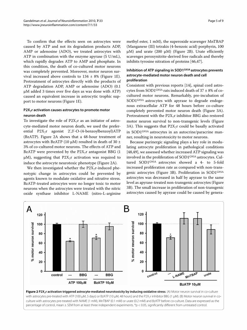
neuron deathTo investigate the role of P2X7r as an initiator of astro-cyte-mediated motor neuron death, we used the prefer-ential P2X7r agonist 2',3'-O-(4-benzoylbenzoyl)ATP(BzATP). Figure 2A shows that a 48-hour treatment ofastrocytes with BzATP (10 μM) resulted in death of 30 ±3% of co-cultured motor neurons. The effects of ATP andBzATP were prevented by the P2X7r antagonist BBG (1μM), suggesting that P2X7r activation was required toinduce the astrocyte neurotoxic phenotype (Figure 2A).
We then investigated whether the P2X7r-induced phe-notypic change in astrocytes could be prevented byagents known to modulate oxidative and nitrative stress.BzATP-treated astrocytes were no longer toxic to motorneurons when the astrocytes were treated with the nitricoxide synthase inhibitor L-NAME (nitro-L-arginine
methyl ester, 1 mM), the superoxide scavenger MnTBAP(Manganese (III) tetrakis (4-benzoic acid) porphyrin, 100μM) and urate (200 μM) (Figure 2B). Urate efficientlyscavenges peroxynitrite-derived free radicals and therebyinhibits tyrosine nitration of proteins [46,47].
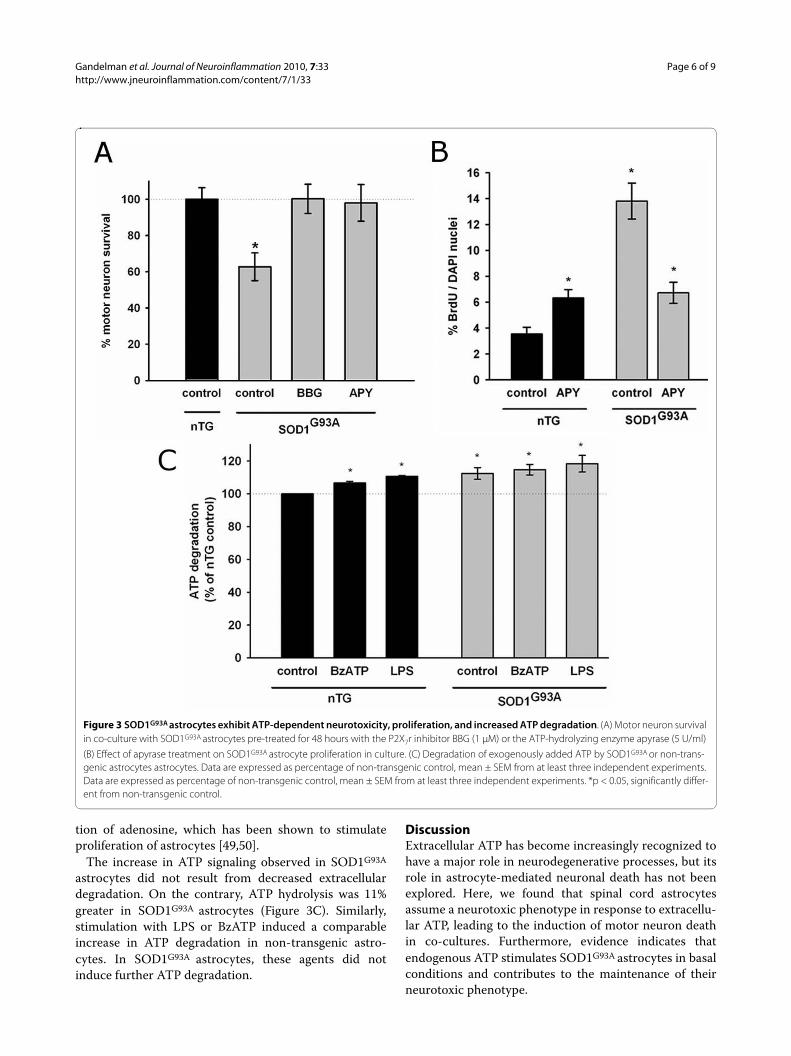
Inhibition of ATP signaling in SOD1G93A astrocytes prevents astrocyte-mediated motor neuron death and cell proliferationConsistent with previous reports [14], spinal cord astro-cytes from SOD1G93A rats induced death of 37 ± 8% of co-cultured motor neurons. Remarkably, pre-incubation ofSOD1G93A astrocytes with apyrase to degrade endoge-nous extracellular ATP for 48 hours before co-culturecompletely prevented motor neuron death (Figure 3A).Pretreatment with the P2X7r inhibitor BBG also restoredmotor neuron survival to non-transgenic levels (Figure3A). This suggests that P2X7r could be basally activatedin SOD1G93A astrocytes in an autocrine/paracrine man-ner, resulting in neurotoxicity to motor neurons.
Because purinergic signaling plays a key role in modu-lating astrocyte proliferation in pathological conditions[48,49], we assessed whether increased ATP signaling wasinvolved in the proliferation of SOD1G93A astrocytes. Cul-tured SOD1G93A astrocytes showed a 4- to 5-foldincreased proliferation rate as compared with non-trans-genic astrocytes (Figure 3B). Proliferation in SOD1G93A
astrocytes was decreased in half by apyrase to the samelevel as apyrase-treated non-transgenic astrocytes (Figure3B). The small increase in proliferation of non-transgenicastrocytes caused by apyrase could be caused by genera-
Figure 2 P2X7r activation triggered astrocyte-mediated neurotoxicity by inducing oxidative stress. (A) Motor neuron survival in co-culture with astrocytes pre-treated with ATP (100 μM, 5 days) or BzATP (10 μM, 48 hours) and the P2X7r inhibitor BBG (1 μM). (B) Motor neuron survival in co-culture with astrocytes pre-treated with NAME (1 mM), MnTBAP (0.1 mM) or urate (0.2 mM) and BzATP before co-culture. Data are expressed as the percentage of control, mean ± SEM from at least three independent experiments. *p < 0.05, significantly different from untreated control.
Gandelman et al. Journal of Neuroinflammation 2010, 7:33http://www.jneuroinflammation.com/content/7/1/33
Page 6 of 9
tion of adenosine, which has been shown to stimulateproliferation of astrocytes [49,50].
The increase in ATP signaling observed in SOD1G93A
astrocytes did not result from decreased extracellulardegradation. On the contrary, ATP hydrolysis was 11%greater in SOD1G93A astrocytes (Figure 3C). Similarly,stimulation with LPS or BzATP induced a comparableincrease in ATP degradation in non-transgenic astro-cytes. In SOD1G93A astrocytes, these agents did notinduce further ATP degradation.
DiscussionExtracellular ATP has become increasingly recognized tohave a major role in neurodegenerative processes, but itsrole in astrocyte-mediated neuronal death has not beenexplored. Here, we found that spinal cord astrocytesassume a neurotoxic phenotype in response to extracellu-lar ATP, leading to the induction of motor neuron deathin co-cultures. Furthermore, evidence indicates thatendogenous ATP stimulates SOD1G93A astrocytes in basalconditions and contributes to the maintenance of theirneurotoxic phenotype.
Figure 3 SOD1G93A astrocytes exhibit ATP-dependent neurotoxicity, proliferation, and increased ATP degradation. (A) Motor neuron survival in co-culture with SOD1G93A astrocytes pre-treated for 48 hours with the P2X7r inhibitor BBG (1 μM) or the ATP-hydrolyzing enzyme apyrase (5 U/ml)
(B) Effect of apyrase treatment on SOD1G93A astrocyte proliferation in culture. (C) Degradation of exogenously added ATP by SOD1G93A or non-trans-genic astrocytes astrocytes. Data are expressed as percentage of non-transgenic control, mean ± SEM from at least three independent experiments. Data are expressed as percentage of non-transgenic control, mean ± SEM from at least three independent experiments. *p < 0.05, significantly differ-ent from non-transgenic control.
Gandelman et al. Journal of Neuroinflammation 2010, 7:33http://www.jneuroinflammation.com/content/7/1/33
Page 7 of 9
Non-transgenic astrocytes required multiple stimuliwith ATP over several days to induce the neurotoxic phe-notype, while a single stimulus with the P2X7r-selectiveagonist BzATP was sufficient to activate astrocytes toinduce the same extent of motor neuron death. BzATP ismost potent as an agonist for P2X7r, but it is also a weakeragonist of P2X1r and P2X3r [51-53]. The involvement ofP2X7r was further implicated in the activation of astro-cyte neurotoxicity by the antagonist BBG, as it com-pletely inhibited the action of ATP and BzATP. BBG is aselective antagonist for both P2X7r and P2X5r. [51-53].Thus, P2X7r appears to be the most likely receptorresponsible for inducing the neurotoxic phenotype inastrocytes.
We have previously shown that oxidative stress inducedby superoxide and nitric oxide forming peroxynitrite innon-transgenic astrocytes leads to a neurotoxic pheno-type [19,42]. Here we found that oxidative stress inducedby BzATP stimulation mediated the transition of non-transgenic astrocytes to a neurotoxic phenotype, as NOSinhibitors as well as superoxide and peroxynitrite scaven-gers prevented their neurotoxicity towards motor neu-rons. In a similar way, Skaper et al showed that P2X7ractivation in microglia stimulated peroxynitrite produc-tion and led to death of co-cultured neurons [54]. Thus,amplification of oxidative stress by P2X7r signaling inmicroglia and astrocytes could lead to the generation ofan adverse environment for vulnerable neurons duringneurodegenerative processes.
Because SOD1G93A astrocytes in culture display a neu-rotoxic phenotype that is maintained by chronic oxidativestress and autocrine pro-inflammatory signaling[14,17,19,20,22], we investigated whether they also pre-sented alterations in extracellular ATP signaling. Indeed,our results indicate that SOD1G93A astrocytes displaybasally augmented extracellular ATP signaling as evi-denced by an ATP-dependent neurotoxic phenotype,increased ATP-dependent proliferation, and increasedextracellular ATP metabolism. Thus, ATP emerges as anextracellular factor that could chronically maintain theSOD1G93A astrocyte aberrant phenotype in an autocrine/paracrine manner.
We found that SOD1G93A astrocytes degraded ATPfaster than non-transgenic astrocytes, ruling out thattheir basal alteration in ATP signaling could be caused bya decrease in its extracellular degradation, thereby allow-ing ATP to accumulate near receptors. An increase inATP degradation could also be induced in non-transgenicastrocytes exposed to BzATP or LPS. We have previouslyshown that LPS induces a neurotoxic phenotype in astro-cytes, leading to motor neuron death [42]. Increased ATPdegradation and/or ectonucleotidase upregulation has
been previously described in neural tissue after corticalstab wound and acute ischemia [55,56]. This phenome-non might reflect a cellular attempt to prevent over-acti-vation of purinergic receptors during increases inextracellular ATP, thus promoting the return of extracel-lular ATP signaling to homeostasis.
Degradation of ATP by ectonucleotidases cannot onlyterminate deleterious ATP signaling, but also initiatesADP and adenosine signaling through P2Y and P1 recep-tors. To our surprise, in non-transgenic astrocytes, ATPdegraded with apyrase, ADP, AMP, or adenosine led to~35% more motor neuron attachment and survival com-pared to untreated controls. Because survival is deter-mined 48 hours after plating of the motor neurons freshlyisolated from spinal cords, any treatment that increasesattachment of motor neurons will result in an increase ofmotor neuron survival above the untreated control.These results illustrate how the astrocyte phenotype canbe modulated from toxic to highly trophic by changingthe balance between ATP, ADP and adenosine signalingthrough P2X, P2Y or adenosine receptors.
In animal models of ALS, proliferative activated astro-cytes interact with microglia to accelerate disease pro-gression [57]. Remarkably, we found that modulatingATP signaling in SOD1G93A astrocytes with apyrase orBBG blocked their neurotoxic phenotype, completelypreventing astrocyte-mediated death of motor neurons.A role for ATP and P2X7r in the SOD1G93A model wasrecently proposed by D'Ambrosi et al [41], who showedthat SOD1G93A microglia are sensitized to BzATP activa-tion. A combination of aberrant ATP signaling in astro-cytes and microglia could generate a positive feedbackloop driving a sustained inflammatory response in thespinal cord. The results presented here and the findingsin SOD1G93A microglia [41] suggest that P2X7r inhibitionin ALS could slow disease progression by decreasingastrocyte and microglial activation.
Taken together, the present work supports the idea thatextracellular ATP acting through P2X7r causes astrocytesto develop a neurotoxic phenotype. In SOD1G93A astro-cytes evidence suggests that P2X7r is basally activatedand contribute to their toxicity towards motor neurons.Thus, modulation of astrocyte P2X7r during diseasecould lead to decreased oxidative stress and inflamma-tory signaling and in turn the switch to a more trophicphenotype towards neurons. A better understanding ofATP and P2X7r signaling in astrocytes could contributeto the development of novel protective therapies in ALSand other neurodegenerative diseases where astrocytesare involved.
Competing interestsThe authors declare that they have no competing interests.

Gandelman et al. Journal of Neuroinflammation 2010, 7:33http://www.jneuroinflammation.com/content/7/1/33
Page 8 of 9
Authors' contributionsMG, PC, LB participated in the design of the study. MG, HP and PC preparedastrocyte and motor neuron cultures and co-cultures. MG collected the co-cul-ture data and carried out all other experiments. All authors reviewed the dataand contributed to the preparation of the manuscript. All authors have readand approved the final manuscript.
AcknowledgementsWe wish to thank the Cell Biology Unit at Institut Pasteur Montevideo for pro-viding cell culture and microscopy facilities, Verónica Abudara and Mauricio Garré for providing reagents, Mark Levy for critical reading of this manuscript and Laura Martínez Palma, Raquel Castellanos, Pablo Díaz-Amarilla and Andrés de Leon for excellent technical help and support. This work was financially sup-ported in part by funding from the National Institute of Health grants NS058628, AT002034 and ES00240 and by Comision Sectorial de Investigacion Cientifica (CSIC, Principal investigator Patricia Cassina).
Author Details1Neurodegeneration Laboratory, Institut Pasteur, Montevideo, Uruguay, 2Departamento de Histología, Facultad de Medicina, Universidad de la República, Montevideo, Uruguay, 3Linus Pauling Institute, Oregon State University, Corvallis, Oregon 97331, USA and 4Instituto de Investigaciones Biológicas Clemente Estable, Montevideo, Uruguay
References1. Rowland LP, Shneider NA: Amyotrophic lateral sclerosis. N Engl J Med
2001, 344:1688-1700.2. Rosen DR: Mutations in Cu/Zn superoxide dismutase gene are
associated with familial amyotrophic lateral sclerosis. Nature 1993, 364:362.
3. Beckman JS, Estevez AG, Crow JP, Barbeito L: Superoxide dismutase and the death of motoneurons in ALS. Trends Neurosci 2001, 24:S15-20.
4. Harraz MM, Marden JJ, Zhou W, Zhang Y, Williams A, Sharov VS, Nelson K, Luo M, Paulson H, Schoneich C, Engelhardt JF: SOD1 mutations disrupt redox-sensitive Rac regulation of NADPH oxidase in a familial ALS model. J Clin Invest 2008, 118:659-670.
5. Papadimitriou D, Le Verche V, Jacquier A, Ikiz B, Przedborski S, Re DB: Inflammation in ALS and SMA: Sorting out the good from the evil. Neurobiol Dis 2009.
6. Kikuchi H, Almer G, Yamashita S, Guegan C, Nagai M, Xu Z, Sosunov AA, McKhann GM, Przedborski S: Spinal cord endoplasmic reticulum stress associated with a microsomal accumulation of mutant superoxide dismutase-1 in an ALS model. Proc Natl Acad Sci USA 2006, 103:6025-6030.
7. Rothstein JD, Tsai G, Kuncl RW, Clawson L, Cornblath DR, Drachman DB, Pestronk A, Stauch BL, Coyle JT: Abnormal excitatory amino acid metabolism in amyotrophic lateral sclerosis. Ann Neurol 1990, 28:18-25.
8. Wong PC, Pardo CA, Borchelt DR, Lee MK, Copeland NG, Jenkins NA, Sisodia SS, Cleveland DW, Price DL: An adverse property of a familial ALS-linked SOD1 mutation causes motor neuron disease characterized by vacuolar degeneration of mitochondria. Neuron 1995, 14:1105-1116.
9. Bruijn LI, Houseweart MK, Kato S, Anderson KL, Anderson SD, Ohama E, Reaume AG, Scott RW, Cleveland DW: Aggregation and motor neuron toxicity of an ALS-linked SOD1 mutant independent from wild-type SOD1. Science 1998, 281:1851-1854.
10. Ilieva H, Polymenidou M, Cleveland DW: Non-cell autonomous toxicity in neurodegenerative disorders: ALS and beyond. J Cell Biol 2009, 187:761-72.
11. McGeer PL, McGeer EG: Inflammation and the degenerative diseases of aging. Ann N Y Acad Sci 2004, 1035:104-116.
12. Barbeito LH, Pehar M, Cassina P, Vargas MR, Peluffo H, Viera L, Estevez AG, Beckman JS: A role for astrocytes in motor neuron loss in amyotrophic lateral sclerosis. Brain Res Brain Res Rev 2004, 47:263-274.
13. Bruijn LI, Miller TM, Cleveland DW: Unraveling the mechanisms involved in motor neuron degeneration in ALS. Annu Rev Neurosci 2004, 27:723-749.
14. Vargas MR, Pehar M, Cassina P, Beckman JS, Barbeito L: Increased glutathione biosynthesis by Nrf2 activation in astrocytes prevents
p75NTR-dependent motor neuron apoptosis. J Neurochem 2006, 97:687-696.
15. Di Giorgio FP, Boulting GL, Bobrowicz S, Eggan KC: Human embryonic stem cell-derived motor neurons are sensitive to the toxic effect of glial cells carrying an ALS-causing mutation. Cell Stem Cell 2008, 3:637-648.
16. Di Giorgio FP, Carrasco MA, Siao MC, Maniatis T, Eggan K: Non-cell autonomous effect of glia on motor neurons in an embryonic stem cell-based ALS model. Nat Neurosci 2007, 10:608-614.
17. Marchetto MC, Muotri AR, Mu Y, Smith AM, Cezar GG, Gage FH: Non-cell-autonomous effect of human SOD1 G37R astrocytes on motor neurons derived from human embryonic stem cells. Cell Stem Cell 2008, 3:649-657.
18. Nagai M, Re DB, Nagata T, Chalazonitis A, Jessell TM, Wichterle H, Przedborski S: Astrocytes expressing ALS-linked mutated SOD1 release factors selectively toxic to motor neurons. Nat Neurosci 2007, 10:615-622.
19. Cassina P, Cassina A, Pehar M, Castellanos R, Gandelman M, de Leon A, Robinson KM, Mason RP, Beckman JS, Barbeito L, Radi R: Mitochondrial dysfunction in SOD1G93A-bearing astrocytes promotes motor neuron degeneration: prevention by mitochondrial-targeted antioxidants. J Neurosci 2008, 28:4115-4122.
20. Cassina P, Pehar M, Vargas MR, Castellanos R, Barbeito AG, Estevez AG, Thompson JA, Beckman JS, Barbeito L: Astrocyte activation by fibroblast growth factor-1 and motor neuron apoptosis: implications for amyotrophic lateral sclerosis. J Neurochem 2005, 93:38-46.
21. Vargas MR, Pehar M, Cassina P, Martinez-Palma L, Thompson JA, Beckman JS, Barbeito L: Fibroblast growth factor-1 induces heme oxygenase-1 via nuclear factor erythroid 2-related factor 2 (Nrf2) in spinal cord astrocytes: consequences for motor neuron survival. J Biol Chem 2005, 280:25571-25579.
22. Hensley K, Abdel-Moaty H, Hunter J, Mhatre M, Mou S, Nguyen K, Potapova T, Pye QN, Qi M, Rice H, Stewart C, Stroukoff K, West M: Primary glia expressing the G93A-SOD1 mutation present a neuroinflammatory phenotype and provide a cellular system for studies of glial inflammation. J Neuroinflammation 2006, 3:2.
23. Pehar M, Vargas MR, Robinson KM, Cassina P, England P, Beckman JS, Alzari PM, Barbeito L: Peroxynitrite transforms nerve growth factor into an apoptotic factor for motor neurons. Free Radic Biol Med 2006, 41:1632-1644.
24. Burnstock G: Purinergic signalling and disorders of the central nervous system. Nat Rev Drug Discov 2008, 7:575-590.
25. Wang X, Arcuino G, Takano T, Lin J, Peng WG, Wan P, Li P, Xu Q, Liu QS, Goldman SA, Nedergaard M: P2X7 receptor inhibition improves recovery after spinal cord injury. Nat Med 2004, 10:821-827.
26. Phillis JW, O'Regan MH, Perkins LM: Adenosine 5'-triphosphate release from the normoxic and hypoxic in vivo rat cerebral cortex. Neurosci Lett 1993, 151:94-96.
27. Melani A, Turchi D, Vannucchi MG, Cipriani S, Gianfriddo M, Pedata F: ATP extracellular concentrations are increased in the rat striatum during in vivo ischemia. Neurochem Int 2005, 47:442-448.
28. Piccini A, Carta S, Tassi S, Lasiglie D, Fossati G, Rubartelli A: ATP is released by monocytes stimulated with pathogen-sensing receptor ligands and induces IL-1beta and IL-18 secretion in an autocrine way. Proc Natl Acad Sci USA 2008, 105:8067-8072.
29. North RA: Molecular physiology of P2X receptors. Physiol Rev 2002, 82:1013-1067.
30. Franke H, Gunther A, Grosche J, Schmidt R, Rossner S, Reinhardt R, Faber-Zuschratter H, Schneider D, Illes P: P2X7 receptor expression after ischemia in the cerebral cortex of rats. J Neuropathol Exp Neurol 2004, 63:686-699.
31. Lovatt D, Sonnewald U, Waagepetersen HS, Schousboe A, He W, Lin JH, Han X, Takano T, Wang S, Sim FJ, Goldman SA, Nedergaard M: The transcriptome and metabolic gene signature of protoplasmic astrocytes in the adult murine cortex. J Neurosci 2007, 27:12255-12266.
32. Narcisse L, Scemes E, Zhao Y, Lee SC, Brosnan CF: The cytokine IL-1beta transiently enhances P2X7 receptor expression and function in human astrocytes. Glia 2005, 49:245-258.
33. John GR, Simpson JE, Woodroofe MN, Lee SC, Brosnan CF: Extracellular nucleotides differentially regulate interleukin-1beta signaling in primary human astrocytes: implications for inflammatory gene expression. J Neurosci 2001, 21:4134-4142.
Received: 29 April 2010 Accepted: 9 June 2010 Published: 9 June 2010This article is available from: http://www.jneuroinflammation.com/content/7/1/33© 2010 Gandelman et al; licensee BioMed Central Ltd. This is an Open Access article distributed under the terms of the Creative Commons Attribution License (http://creativecommons.org/licenses/by/2.0), which permits unrestricted use, distribution, and reproduction in any medium, provided the original work is properly cited.Journal of Neuroinflammation 2010, 7:33
http://www.ncbi.nlm.nih.gov/entrez/query.fcgi?cmd=Retrieve&db=PubMed&dopt=Abstract&list_uids=8332197
http://www.ncbi.nlm.nih.gov/entrez/query.fcgi?cmd=Retrieve&db=PubMed&dopt=Abstract&list_uids=2375630
http://www.ncbi.nlm.nih.gov/entrez/query.fcgi?cmd=Retrieve&db=PubMed&dopt=Abstract&list_uids=7605627
http://www.ncbi.nlm.nih.gov/entrez/query.fcgi?cmd=Retrieve&db=PubMed&dopt=Abstract&list_uids=9743498
Gandelman et al. Journal of Neuroinflammation 2010, 7:33http://www.jneuroinflammation.com/content/7/1/33
Page 9 of 9
34. Panenka W, Jijon H, Herx LM, Armstrong JN, Feighan D, Wei T, Yong VW, Ransohoff RM, MacVicar BA: P2X7-like receptor activation in astrocytes increases chemokine monocyte chemoattractant protein-1 expression via mitogen-activated protein kinase. J Neurosci 2001, 21:7135-7142.
35. Ryu JK, McLarnon JG: Block of purinergic P2X(7) receptor is neuroprotective in an animal model of Alzheimer's disease. Neuroreport 2008, 19:1715-1719.
36. Matute C, Torre I, Perez-Cerda F, Perez-Samartin A, Alberdi E, Etxebarria E, Arranz AM, Ravid R, Rodriguez-Antiguedad A, Sanchez-Gomez M, Domercq M: P2X(7) receptor blockade prevents ATP excitotoxicity in oligodendrocytes and ameliorates experimental autoimmune encephalomyelitis. J Neurosci 2007, 27:9525-9533.
37. Diaz-Hernandez M, Diez-Zaera M, Sanchez-Nogueiro J, Gomez-Villafuertes R, Canals JM, Alberch J, Miras-Portugal MT, Lucas JJ: Altered P2X7-receptor level and function in mouse models of Huntington's disease and therapeutic efficacy of antagonist administration. FASEB J 2009, 23:1893-1906.
38. Peng W, Cotrina ML, Han X, Yu H, Bekar L, Blum L, Takano T, Tian GF, Goldman SA, Nedergaard M: Systemic administration of an antagonist of the ATP-sensitive receptor P2X7 improves recovery after spinal cord injury. Proc Natl Acad Sci USA 2009, 106:12489-12493.
39. Yiangou Y, Facer P, Durrenberger P, Chessell IP, Naylor A, Bountra C, Banati RR, Anand P: COX-2, CB2 and P2X7-immunoreactivities are increased in activated microglial cells/macrophages of multiple sclerosis and amyotrophic lateral sclerosis spinal cord. BMC Neurol 2006, 6:12.
40. Casanovas A, Hernandez S, Tarabal O, Rossello J, Esquerda JE: Strong P2X4 purinergic receptor-like immunoreactivity is selectively associated with degenerating neurons in transgenic rodent models of amyotrophic lateral sclerosis. J Comp Neurol 2008, 506:75-92.
41. D'Ambrosi N, Finocchi P, Apolloni S, Cozzolino M, Ferri A, Padovano V, Pietrini G, Carri MT, Volonte C: The proinflammatory action of microglial P2 receptors is enhanced in SOD1 models for amyotrophic lateral sclerosis. J Immunol 2009, 183:4648-4656.
42. Cassina P, Peluffo H, Pehar M, Martinez-Palma L, Ressia A, Beckman JS, Estevez AG, Barbeito L: Peroxynitrite triggers a phenotypic transformation in spinal cord astrocytes that induces motor neuron apoptosis. J Neurosci Res 2002, 67:21-29.
43. Henderson CE, Bloch-Gallego E, Camu W: Purification and culture of embryonic motor neurons Oxford: IRL Press; 1995.
44. Estevez AG, Sahawneh MA, Lange PS, Bae N, Egea M, Ratan RR: Arginase 1 regulation of nitric oxide production is key to survival of trophic factor-deprived motor neurons. J Neurosci 2006, 26:8512-8516.
45. Wink MR, Braganhol E, Tamajusuku AS, Casali EA, Karl J, Barreto-Chaves ML, Sarkis JJ, Battastini AM: Extracellular adenine nucleotides metabolism in astrocyte cultures from different brain regions. Neurochem Int 2003, 43:621-628.
46. Teng RJ, Ye YZ, Parks DA, Beckman JS: Urate produced during hypoxia protects heart proteins from peroxynitrite-mediated protein nitration. Free Radic Biol Med 2002, 33:1243-1249.
47. Santos CX, Anjos EI, Augusto O: Uric acid oxidation by peroxynitrite: multiple reactions, free radical formation, and amplification of lipid oxidation. Arch Biochem Biophys 1999, 372:285-294.
48. Neary JT, Kang Y: Signaling from P2 nucleotide receptors to protein kinase cascades induced by CNS injury: implications for reactive gliosis and neurodegeneration. Mol Neurobiol 2005, 31:95-103.
49. Rathbone MP, Middlemiss PJ, Kim JK, Gysbers JW, DeForge SP, Smith RW, Hughes DW: Adenosine and its nucleotides stimulate proliferation of chick astrocytes and human astrocytoma cells. Neurosci Res 1992, 13:1-17.
50. Ciccarelli R, Di Iorio P, D'Alimonte I, Giuliani P, Florio T, Caciagli F, Middlemiss PJ, Rathbone MP: Cultured astrocyte proliferation induced by extracellular guanosine involves endogenous adenosine and is raised by the co-presence of microglia. Glia 2000, 29:202-211.
51. Cotrina ML, Nedergaard M: Physiological and pathological functions of P2X7 receptor in the spinal cord. Purinergic Signal 2009, 5:223-232.
52. Surprenant A: Functional properties of native and cloned P2X receptors. Ciba Found Symp 1996, 198:208-219. discussion 219-222
53. Bianchi BR, Lynch KJ, Touma E, Niforatos W, Burgard EC, Alexander KM, Park HS, Yu H, Metzger R, Kowaluk E, Jarvis MF, van Biesen T: Pharmacological characterization of recombinant human and rat P2X receptor subtypes. Eur J Pharmacol 1999, 376:127-138.
54. Skaper SD, Facci L, Culbert AA, Evans NA, Chessell I, Davis JB, Richardson JC: P2X(7) receptors on microglial cells mediate injury to cortical neurons in vitro. Glia 2006, 54:234-242.
55. Braun N, Zhu Y, Krieglstein J, Culmsee C, Zimmermann H: Upregulation of the enzyme chain hydrolyzing extracellular ATP after transient forebrain ischemia in the rat. J Neurosci 1998, 18:4891-4900.
56. Nedeljkovic N, Bjelobaba I, Lavrnja I, Stojkov D, Pekovic S, Rakic L, Stojiljkovic M: Early temporal changes in ecto-nucleotidase activity after cortical stab injury in rat. Neurochem Res 2008, 33:873-879.
57. Yamanaka K, Chun SJ, Boillee S, Fujimori-Tonou N, Yamashita H, Gutmann DH, Takahashi R, Misawa H, Cleveland DW: Astrocytes as determinants of disease progression in inherited amyotrophic lateral sclerosis. Nat Neurosci 2008, 11:251-253.
doi: 10.1186/1742-2094-7-33Cite this article as: Gandelman et al., Extracellular ATP and the P2X7 recep-tor in astrocyte-mediated motor neuron death: implications for amyotrophic lateral sclerosis Journal of Neuroinflammation 2010, 7:33
http://www.ncbi.nlm.nih.gov/entrez/query.fcgi?cmd=Retrieve&db=PubMed&dopt=Abstract&list_uids=1314349
http://www.ncbi.nlm.nih.gov/entrez/query.fcgi?cmd=Retrieve&db=PubMed&dopt=Abstract&list_uids=8879827