External measurements of implanted pacemaker ...
6
British Heart,Journal, I97I, 33, I85-I90. External measurements of implanted pacemaker characteristics A clinical appraisal Olof Edhag, Stefan Hofvendahl, Ingvar Karlof, and Lars Mogensen From the Department of Medicine, Serafimerlasarettet, and the Clinical Physiological Laboratory, Thoracic Clinics, Karolinska Sjukhuset, Stockholm, Sweden A technique for determining the characteristics of impulses from implanted cardiac pacemakers with external methods is reviewed, and its value in detecting pacemaker failures is discussed. The results from a 2-year application with 6ii controls in clinical routine are presented. Sixteen out of b*42 defective pacemakers could be detected before symptoms or electrocardiographic signs of pacing failure developed. The reliability and specificity of the procedure justify its application in clinical routine. Patients with cardiac pacemakers need regular check-ups for the control of the pacemaker .unit. Clinical tools of importance are a careful history, electrocardiograms, and equipment for the external measurement of characteristics of the pacing impulse. The impulse from an ' implanted pacemaker generator can be visualized in a voltage-time plot and photo- graphed with a polaroid camera, yielding an impulsogram. From this, the amplitude, i.e. voltage, time duration, and slope of the purve can readily be evaluated. For accurately working pacemaker genera- -4tors it is important to assess the expected lifetime of the mercury cells as safely as poss- ible at each control. Accurate assessment would minimize the risk for the patient from a ""failing pacemaker function, and reduce the number of operations and thus patient dis- -Zomfort. Furthermore, costs for pacing equip- _ment and nursing will be reduced. For failing pacemakers it is often possible to localize the fault through external measure- ments - whether it is due to electronic failure, battery exhaustion, or a disconnected or damaged electrode. I* These techniques have been used routinely since the spring of I967 on all patients with subcutaneous pacemakers at our hospitals. The experiences over a two-year period are reported, with emphasis on the relative value of impulse analysis, electrocardiogram, and Received 22 May 1970. 2 history in detecting imminent pacemaker failures. Impulse recording for determination of pacemaker characteristics was described in I964 by Davies and Sowton, and its use for routine controls has been reported by Knuckey, McDonald, and Sloman (I965), Nickel (i964), Preston et al. (I966), Ryden (i969), and Thalen et al. (I969). Methods and stimulating principles The pacemaker system consists of an impulse generator, an endocardial electrode (Elema EMT 588), and an indifferent electrode (Elema EMT 564) subcutaneously implanted below the left costal margin. The generators (fixed rate genera- tors, Elema EM I39 and 142; atrial synchronized, Elema EM 141; and QRS synchronized, Elema EM 143) are implanted subcutaneously in the abdomen. At the control performed every third to fourth month, and otherwise when symptoms indicate, the history and electrocardiogram are taken, a physical examination is performed, and an impulse recording undertaken. The impulse frequency was calculated from the electrocardiogram (Elema-Schonander Mingo- graf 42). The generator impulses detected from electrodes on the left arm and leg (lead III) were displayed on an oscilloscope (Tektronix 502 A). The horizontal sweep was triggered by the pace- maker impulses. The amplification was usually 50 millivolts per centimetre on the vertical axis, and the speed of the sweep o 5 msec.lcm. A photograph of the oscilloscope display has been taken at every check-up, and the peak amplitude and duration of the impulse have been measured. on March 11, 2022 by guest. Protected by copyright. http://heart.bmj.com/ Br Heart J: first published as 10.1136/hrt.33.2.185 on 1 March 1971. Downloaded from
Transcript of External measurements of implanted pacemaker ...

British Heart,Journal, I97I, 33, I85-I90.
External measurements of implantedpacemaker characteristicsA clinical appraisal
Olof Edhag, Stefan Hofvendahl, Ingvar Karlof, and Lars MogensenFrom the Department of Medicine, Serafimerlasarettet, and theClinical Physiological Laboratory, Thoracic Clinics, KarolinskaSjukhuset, Stockholm, Sweden
A technique for determining the characteristics of impulses from implanted cardiac pacemakerswith external methods is reviewed, and its value in detecting pacemaker failures is discussed. Theresultsfrom a 2-year application with 6ii controls in clinical routine are presented. Sixteen out of
b*42 defective pacemakers could be detected before symptoms or electrocardiographic signs of pacingfailure developed. The reliability and specificity of the procedure justify its application in clinicalroutine.
Patients with cardiac pacemakers need regularcheck-ups for the control of the pacemaker.unit. Clinical tools of importance are a carefulhistory, electrocardiograms, and equipmentfor the external measurement of characteristicsof the pacing impulse. The impulse from an
' implanted pacemaker generator can bevisualized in a voltage-time plot and photo-graphed with a polaroid camera, yielding animpulsogram. From this, the amplitude, i.e.voltage, time duration, and slope of thepurve can readily be evaluated.
For accurately working pacemaker genera--4tors it is important to assess the expectedlifetime of the mercury cells as safely as poss-ible at each control. Accurate assessmentwould minimize the risk for the patient from a
""failing pacemaker function, and reduce thenumber of operations and thus patient dis-
-Zomfort. Furthermore, costs for pacing equip-_ment and nursing will be reduced.
For failing pacemakers it is often possibleto localize the fault through external measure-ments - whether it is due to electronic failure,battery exhaustion, or a disconnected ordamaged electrode.I* These techniques have been used routinelysince the spring of I967 on all patients withsubcutaneous pacemakers at our hospitals.The experiences over a two-year period are
reported, with emphasis on the relative valueof impulse analysis, electrocardiogram, and
Received 22 May 1970.2
history in detecting imminent pacemakerfailures.
Impulse recording for determination ofpacemaker characteristics was described inI964 by Davies and Sowton, and its use forroutine controls has been reported byKnuckey, McDonald, and Sloman (I965),Nickel (i964), Preston et al. (I966), Ryden(i969), and Thalen et al. (I969).
Methods and stimulating principlesThe pacemaker system consists of an impulsegenerator, an endocardial electrode (Elema EMT588), and an indifferent electrode (Elema EMT564) subcutaneously implanted below the leftcostal margin. The generators (fixed rate genera-tors, Elema EM I39 and 142; atrial synchronized,Elema EM 141; and QRS synchronized, ElemaEM 143) are implanted subcutaneously in theabdomen. At the control performed every third tofourth month, and otherwise when symptomsindicate, the history and electrocardiogram aretaken, a physical examination is performed, andan impulse recording undertaken.The impulse frequency was calculated from the
electrocardiogram (Elema-Schonander Mingo-graf 42). The generator impulses detected fromelectrodes on the left arm and leg (lead III) weredisplayed on an oscilloscope (Tektronix 502 A).The horizontal sweep was triggered by the pace-maker impulses. The amplification was usually50 millivolts per centimetre on the vertical axis,and the speed of the sweep o 5 msec.lcm. Aphotograph of the oscilloscope display has beentaken at every check-up, and the peak amplitudeand duration of the impulse have been measured.
on March 11, 2022 by guest. P
rotected by copyright.http://heart.bm
j.com/
Br H
eart J: first published as 10.1136/hrt.33.2.185 on 1 March 1971. D
ownloaded from

x86 Edhag, Hofvendahl, Karlof, and Mogensen
The photographs and electrocardiograms fromearlier controls of the same patient with the sameimpulse generator have been available for com-parison at check-ups.A pacemaker manufactured by Elema-Schonan-
der delivers impulses by discharging a capacitor.With no load on the pacemaker circuit, i.e. whenthe resistance between the output electrodes isinfinitely high, the impulse is a square wave onthe voltage time plot. When the impulse generatoris connected to a patient, the resistance betweenthe output electrodes is decreased and the shapeof the impulse is changed. Fig. i shows a typicalimpulse recording from a patient (lead III) withan adequate functioning generator, and Fig. 2shows a recording from a patient with a generatorwith reduced voltage of the batteries.
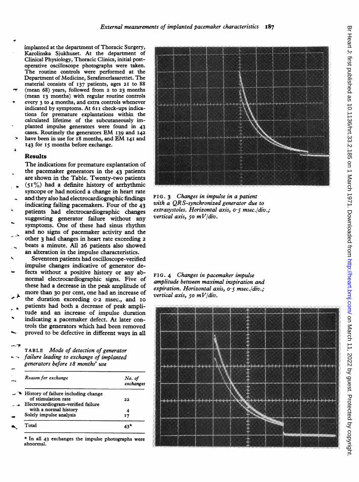
Amplitude, duration, and shape of the impulseare constant parameters in a patient as long as hehas the same pacemaker with an adequate voltageofthe batteries and a functioning electrode system.This is true if the pacemaker is of a fixed rate type.Synchronized generators will decrease the ampli-tude and shorten the duration of the impulse withincreasing frequency (Fig. 3). Synchronous pace-maker generators are therefore checked at aboutthe same impulse rate from time to time in orderto get comparable data.The amplitude varies with respiration and
especially during deep breaths; the difference maybe considerable (Fig. 4). In normal breathingdifferences are small and can usually be neglected.These variations are estimated at every controlsince the oscilloscope display is observed duringseveral respiratory cycles.
Indications for generator exchangeIndications for exchange of the generator havebeen a history of possible pacemaker failure, analteration in the electrocardiogram or the impulsecharacteristics, and are listed below.
(I) A case history indicating pacemaker failureincludes either syncopal (Adams-Stokes) attacks,or careful description of undue arrhythmia, oraccurate observations of palpated arterial pulserate changes not otherwise explained (as forexample by competition).
(2) Electrocardiographic verification of pacingimpulses failing to stimulate the heart whenoccurring in a non-refractory phase.
(3) Failing trigger mechanisms in synchron-ized generators (pacing).
(4) A decrease of the peak amplitude of morethan 30 per cent from the initial value. (At thesame stimulation rate when dealing with triggeredpacing.)
(5) An increase of the impulse duration ofmorethan 0-2 msec. from the initial value. (At the samestimulation rate when dealing with triggeredpacing.)
(6) A change of stimulation rate exceeding 2impulses a minute from the initial rate (only fixed-rate generators).
SubjectsThe electrodes and pacemaker generators were
FIG. I Impulse from a pacemaker derivedfrom lead III, in a voltage time plot. Ampli-tude voltage, impulse duration, and curveshape are the important features of suchimpulsograms. Horizontal axis, o*5 msec./div.;vertical, 50 mV/div.FIG. 2 Pathological changes of an impulso-gram, with decrease in voltage, increase induration but unchanged relative speed ofvoltage decrease. This is consistent with re-duced voltage sum of the batteries and un-changed resistance of intracorporeal circuits.The initial findings were identical with thosein Fig. i. Horizontal axis, o05 msec./div.;vertical, 50 mV/div.
on March 11, 2022 by guest. P
rotected by copyright.http://heart.bm
j.com/
Br H
eart J: first published as 10.1136/hrt.33.2.185 on 1 March 1971. D
ownloaded from
External measurements of implanted pacemaker characteristics 187
implanted at the department of Thoracic Surgery,Karolinska Sjukhuset. At the department ofClinical Physiology, Thoracic Clinics, initial post-operative oscilloscope photographs were taken.The routine controls were performed at theDepartment of Medicine, Serafimerlasarettet. Thematerial consists of 137 patients, ages 2I to 88
- (mean 68) years, followed from 2 to 23 months(mean I3 months) with regular routine controlsevery 3 to 4 months, and extra controls wheneverindicated by symptoms. At 6ii check-ups indica-tions for premature explantations within thecalculated lifetime of the subcutaneously im-planted impulse generators were found in 43cases. Routinely the generators EM I39 and 142have been in use for i8 months, and EM 14I and143 for I5 months before exchange.
ResultsThe indications for premature explantation ofthe pacemaker generators in the 43 patientsare shown in the Table. Twenty-two patients(5I%) had a definite history of arrhythmicsyncope or had noticed a change in heart rateand they also had electrocardiographic findingsindicating failing pacemakers. Four of the 43patients had electrocardiographic changessuggesting generator failure without anysymptoms. One of these had sinus rhythmand no signs of pacemaker activity and theother 3 had changes in heart rate exceeding 2beats a minute. All 26 patients also showedan alteration in the impulse characteristics.
Seventeen patients had oscilloscope-verifiedimpulse changes indicative of generator de-fects without a positive history or any ab-normal electrocardiographic signs. Five ofthese had a decrease in the peak amplitude ofmore than 30 per cent, one had an increase ofthe duration exceeding 0-2 msec., and I0patients had both a decrease of peak ampli-tude and an increase of impulse durationindicating a pacemaker defect. At later con-trols the generators which had been removedproved to be defective in different ways in all
TABLE Mode of detection of generatorfailure leading to exchange of implantedgenerators before I8 months' use
Reason for exchange No. ofexchanges
- History of failure including changeof stimulation rate 22
Electrocardiogram-verified failurewith a normal history 4
Solely impulse analysis I7
Total 43*
FIG. 3 Changes in impulse in a patientwith a QRS-synchronized generator due toextrasystoles. Horizontal axis, o 5 msec./div.;vertical axis, 5o mV/div.
FIG. 4 Changes in pacemaker impulseamplitude between maximal inspiration andexpiration. Horizontal axis, o-5 msec./div.;vertical axis, 50 mV/div.
* In all 43 exchanges the impulse photographs wereabnormal.
on March 11, 2022 by guest. P
rotected by copyright.http://heart.bm
j.com/
Br H
eart J: first published as 10.1136/hrt.33.2.185 on 1 March 1971. D
ownloaded from

x88 Edhag, Hofvendahl, Karlof, and Mogensen
7 5
50
2 5
00 k1 20 30 40 50Weeks
FIG. S Observations of relative impulse amplitude changes with time after implantation, forpacers EM 1421. First postoperative measurements are set at 0OO per cent and repeatedmeasurements from the same pacemaker in the same patient are connected with a line.1 Elema-Schonander Company, Sweden.
cases but one. The oscilloscope measure-ments from this patient were incorrectlyinterpreted as indicative of a deficient pace-maker, due to erroneous amplification in thevoltage-time plot.
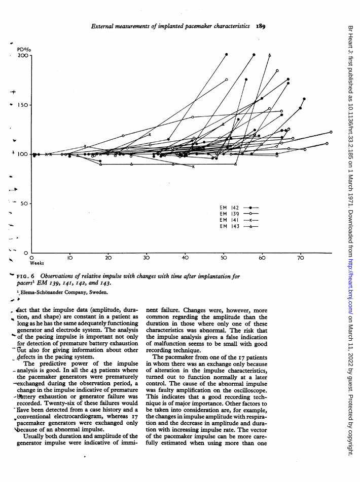
Fig. 5 shows the change with time ofpacing impulse amplitudes for generators oftype EM 142. Fig. 6 shows the change withtime in the impulse duration for generators oftypeEM 139, EM 14i, EM i42, and EM I43.The initially measured value has been set toIoo per cent. Only values for defective pace-makers are included, but both from patientswith and without a positive history and/orelectrocardiographic signs.
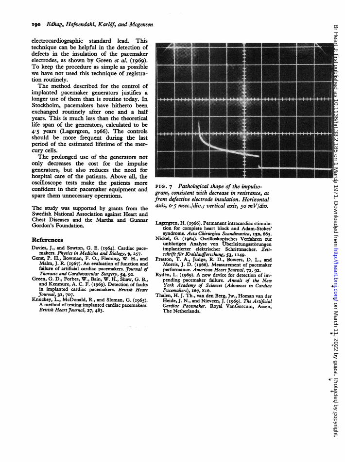
If the anode and cathode electrodes are in-verted at the exchange of a generator this willbe observed on the oscilloscope as an inversionof the impulse. The impulse in Fig. 7 showssigns of an extremely low resistance betweenthe 2 electrodes. The cause of this was ashort circuit between the stimulating and in-different electrode caused by an insulationdefect on the wire ofthe stimulating electrode,
where it passed close to the indifferent elec-trode plate.
DiscussionA patient with atrioventricular conductiondisturbances treated with an artificial cardiacpacemaker is depending on perfect functionin the pacemaker system, not only in order tomaintain an adequate cardiac output but alsofor his mental well-being. He will never haveconfidence in a pacemaker system if hesuffers alarming symptoms before the ex-change of the old pacemaker generator. Toavoid this, a routine needs to be establishedfor the control of these patients giving asexact data as possible concerning actual and2- to 3-month future functioning of thegenerators (Gerst et al., i967). The presentinvestigation indicates that an analysis of thepacing impulse gives such good informationabout the condition of the pacing system thatit justifies its use as a control routine. Thevalue of impulse recording is based on the
on March 11, 2022 by guest. P
rotected by copyright.http://heart.bm
j.com/
Br H
eart J: first published as 10.1136/hrt.33.2.185 on 1 March 1971. D
ownloaded from
External measurements of implanted pacemaker characteristics 189
EM 142 --*EM 139 -0-EM 141 -x-EM 143 6
0 10 20 30 40 50Weeks
%FIG. 6 Observations of relative impulse with changes with time after implantation forpacers' EM 139, 141, 142, and 143.
l.Elema-Schonander Company, Sweden.
tact that the impulse data (amplitude, dura-tion, and shape) are constant in a patient aslong as he has the same adequately functioninggenerator and electrode system. The analysisof the pacing impulse is important not onlyfor detection of premature battery exhaustion-ut also for giving information about otherdefects in the pacing system.The predictive power of the impulse
analysis is good. In all the 43 patients wherethe pacemaker generators were prematurely
--exchanged during the observation period, achange in the impulse indicative of premature
-l'ttery exhaustion or generator failure wasrecorded. Twenty-six of these failures woulditave been detected from a case history and a
conventional electrocardiogram, whereas I7pacemaker generators were exchanged onlybecause of an abnormal impulse.
Usually both duration and amplitude of thegenerator impulse were indicative of immi-
nent failure. Changes were, however, more
common regarding the amplitude than theduration in those where only one of thesecharacteristics was abnormal. The risk thatthe impulse analysis gives a false indicationof malfunction seems to be small with goodrecording technique.The pacemaker from one of the I7 patients
in whom there was an exchange only becauseof alteration in the impulse characteristics,turned out to function normally at a latercontrol. The cause of the abnormal impulsewas faulty amplification on the oscilloscope.This indicates that a good recording tech-nique is of major importance. Other factors tobe taken into consideration are, for example,the changes in impulse amplitude with respira-tion and the decrease in amplitude and dura-tion with increasing impulse rate. The vectorof the pacemaker impulse can be more care-fully estimated when using more than one
0
on March 11, 2022 by guest. P
rotected by copyright.http://heart.bm
j.com/
Br H
eart J: first published as 10.1136/hrt.33.2.185 on 1 March 1971. D
ownloaded from
9go Edhag, Hofvendahl, Karljf, and Mogensen
electrocardiographic standard lead. Thistechnique can be helpful in the detection ofdefects in the insulation of the pacemakerelectrodes, as shown by Green et al. (i969).To keep the procedure as simple as possiblewe have not used this technique of registra-tion routinely.The method described for the control of
implanted pacemaker generators justifies alonger use of them than is routine today. InStockholm, pacemakers have hitherto beenexchanged routinely after one and a halfyears. This is much less than the theoreticallife span of the generators, calculated to be4-5 years (Lagergren, i966). The controlsshould be more frequent during the lastperiod of the estimated lifetime of the mer-cury cells.The prolonged use of the generators not
only decreases the cost for the impulsegenerators, but also reduces the need forhospital care of the patients. Above all, theoscilloscope tests make the patients moreconfident in their pacemaker equipment andspare them unnecessary operations.
The study was supported by grants from theSwedish National Association against Heart andChest Diseases and the Martha and GunnarGordon's Foundation.
ReferencesDavies, J., and Sowton, G. E. (I964). Cardiac pace-
makers. Physics in Medicine and Biology, 9, 257.Gerst, P. H., Bowman, F. O., Fleming, W. H., and
Malm, J. R. (I967). An evaluation of function andfailure of artificial cardiac pacemakers. Journal ofThoracic and Cardiovascular Surgery, 54, 92.
Green, G. D., Forbes, W., Bain, W. H., Shaw, G. B.,and Kenmure, A. C. F. (I969). Detection of faultsin implanted cardiac pacemakers. British HeartJournal, 3I, 707.
Knuckey, L., McDonald, R., and Sloman, G. (I965).A method of testing implanted cardiac pacemakers.British Heart_Journal, 27, 483.
FIG. 7 Pathological shape of the impulso-gram, consistent with decrease in resistance, asfrom defective electrode insulation. Horizontalaxis, o'5 msec./div.; vertical axis, 50 mV/div.
Lagergren, H. (1966). Permanent intracardiac stimula-tion for complete heart block and Adam-Stokes'syndrome. Acta Chirurgica Scandinavica, 132, 663.
Nickel, G. (I964). Oszilloskopisches Verfahren zurunblutigen Analyse von Uberleitungsstbrungenimplantierter elektrischer Schrittmacher. Zeit-schrift fur Kreislaufforschung, 53, I I49.
Preston, T. A., Judge, R. D., Bowers, D. L., andMorris, J. D. (I966). Measurement of pacemakerperformance. American Heart Journal, 71, 92.
Ryddn, L. (I969). A new device for detection of im-pending pacemaker failure. Annals of the NewYork Academy of Sciences (Advances in CardiacPacemakers), I67, 8I6.
Thalen, H. J. Th., van den Berg, Jw., Homan van derHeide, J. N., and Nieveen, J. (1969). The ArtificialCardiac Pacemaker. Royal VanGorcum, Assen,The Netherlands.
4i
on March 11, 2022 by guest. P
rotected by copyright.http://heart.bm
j.com/
Br H
eart J: first published as 10.1136/hrt.33.2.185 on 1 March 1971. D
ownloaded from