Extensive graft-derived dopaminergic ... - neurobiology.lu.se · Extensive graft-derived...
6
Extensive graft-derived dopaminergic innervation is maintained 24 years after transplantation in the degenerating parkinsonian brain Wen Li a , Elisabet Englund b , Håkan Widner c , Bengt Mattsson d , Danielle van Westen e , Jimmy Lätt e , Stig Rehncrona f , Patrik Brundin g , Anders Björklund d,1,2 , Olle Lindvall c,h,1,2 , and Jia-Yi Li a,1,2 a Neural Plasticity and Repair Unit, Wallenberg Neuroscience Center, Department of Experimental Medical Science, Lund University, 221 84 Lund, Sweden; b Division of Oncology and Pathology, Lund University Hospital, 221 85 Lund, Sweden; c Division of Neurology, Lund University Hospital, 221 85 Lund, Sweden; d Neurobiology Unit, Wallenberg Neuroscience Center, Department of Experimental Medical Science, Lund University, 221 84 Lund, Sweden; e Center for Medical Imaging and Physiology, Lund University Hospital, 221 85 Lund, Sweden; f Division of Neurosurgery, Lund University Hospital, 221 85 Lund, Sweden; g Center for Neurodegenerative Science, Van Andel Research Institute, Grand Rapids, MI 49503; and h Lund Stem Cell Center, Lund University Hospital, 221 84 Lund, Sweden Contributed by Anders Björklund, April 1, 2016 (sent for review February 17, 2016; reviewed by Lorenz Studer and Jun Takahashi) Clinical trials using cells derived from embryonic ventral mesen- cephalon have shown that transplanted dopaminergic neurons can survive and function in the long term, as demonstrated by in vivo brain imaging using 18 F-fluorodopa and 11 C-raclopride pos- itron emission tomography. Here we report the postmortem anal- ysis of a patient with Parkinson’s disease who 24 y earlier underwent unilateral transplantation of embryonic dopaminergic neurons in the putamen and subsequently exhibited major motor improvement and recovery of striatal dopaminergic function. Histopathological analysis showed that a dense, near-normal graft-derived dopaminergic reinnervation of the putamen can be maintained for a quarter of a century despite severe host brain pathology and with no evidence of immune response. In addition, ubiquitin- and α-synuclein–posi- tive inclusions were seen, some with the appearance of typical Lewy bodies, in 11–12% of the grafted dopaminergic neurons, reflecting the spread of pathology from the host brain to the transplants. Because the clinical benefits induced by transplantation in this pa- tient were gradually lost after 14 y posttransplantation, our findings provide the first reported evidence, to our knowledge, that even a viable dopaminergic graft giving rise to extensive striatal reinnerva- tion may lose its efficacy if widespread degenerative changes de- velop in the host brain. Parkinson’s disease | transplantation | autopsy | synucleinopathy | reinnervation A fundamental question for cell therapies in neurodegenera- tive disorders is whether transplanted neurons can survive and integrate their terminal network with the host brain for many years despite an ongoing disease process. In patients with Parkinson’s disease (PD) implanted with human embryonic mes- encephalic tissue into the striatum, varying numbers of grafted dopaminergic neurons have survived for up to 2 decades (1–3); however, in some patients, characteristic Lewy bodies were de- tected in a small fraction (1–5%) of grafted dopaminergic neurons at 12–22 y after transplantation (2–5). Other studies have shown that grafted dopaminergic neurons remain healthy up to 14 y posttransplantation (1, 6). Whether the disease process affects the density and extension of a well-developed graft-derived dopami- nergic innervation is unknown. Here we report the histopathological analysis of a unique patient with PD who exhibited major clinical improvement for more than a decade after undergoing unilateral transplantation into the putamen, associated with complete restoration of puta- minal dopaminergic function as demonstrated by positron emission tomography (7). We demonstrate, for the first time to our knowl- edge, that a dense graft-derived dopaminergic reinnervation of the putamen can be maintained for a quarter of a century despite widespread host brain pathology and α-synucleinopathy eventu- ally affecting 11–12% of grafted neurons. Results and Discussion Patient History and Clinical Follow-Up. In 1989, at age 59 y, the patient underwent transplantation with ventral mesencephalic tissue from four human embryos obtained at routine suction abortions (8). Using a computed tomography-guided stereotactic technique, dissociated tissue was implanted along three tracts in the anterior, middle, and posterior parts of the right putamen. The patient had developed typical PD in 1980 and was started on L-dopa treatment in 1982. He did well until 1986, when he de- veloped “on–off” symptoms, with “off” periods characterized by resting tremor, hypokinetic movements, and rigidity, particularly on the left side of the body, and only mild parkinsonian symp- toms during “on” periods. Up to 10 y after transplantation, the patient was monitored according to the Core Assessment Program for Intracerebral Transplantations protocol (9) as part of a scientific program. He Significance Parkinson’s disease is the most common movement disorder. Here we describe the histopathological analysis of a unique patient with Parkinson’s disease who underwent unilateral cell transplantation in the putamen with human embryonic mes- encephalic tissue at 24 y before death. The patient enjoyed major clinical benefits for at least a decade after transplantation. After a quarter of a century, complete graft-derived dopami- nergic reinnervation was still evident in the transplanted puta- men. α-Synuclein–positive inclusions, some with the appearance of typical Lewy bodies, were present in 11–12% of the grafted dopaminergic neurons, reflecting spread of pathology from the host brain to the transplant. The clinical improvements were gradually lost from 14 y posttransplantation, indicating that even extensive graft-derived dopaminergic reinnervation loses its efficacy in a severely degenerating brain. Author contributions: W.L., A.B., O.L., and J.-Y.L. designed research; W.L., E.E., H.W., B.M., D.v.W., J.L., S.R., P.B., O.L., and J.-Y.L. performed research; W.L., H.W., A.B., O.L., and J.-Y.L. analyzed data; and W.L., A.B., O.L., and J.-Y.L. wrote the paper. Reviewers: L.S., Memorial Sloan Kettering Cancer Center; and J.T., Center for iPS Cell Research and Application, Kyoto University. The authors declare no conflict of interest. Freely available online through the PNAS open access option. 1 A.B., O.L., and J.-Y.L. contributed equally to this work. 2 To whom correspondence may be addressed. Email: [email protected], Olle. [email protected], or [email protected]. This article contains supporting information online at www.pnas.org/lookup/suppl/doi:10. 1073/pnas.1605245113/-/DCSupplemental. www.pnas.org/cgi/doi/10.1073/pnas.1605245113 PNAS Early Edition | 1 of 6 NEUROSCIENCE
Transcript of Extensive graft-derived dopaminergic ... - neurobiology.lu.se · Extensive graft-derived...

Extensive graft-derived dopaminergic innervation ismaintained 24 years after transplantation in thedegenerating parkinsonian brainWen Lia, Elisabet Englundb, Håkan Widnerc, Bengt Mattssond, Danielle van Westene, Jimmy Lätte, Stig Rehncronaf,Patrik Brunding, Anders Björklundd,1,2, Olle Lindvallc,h,1,2, and Jia-Yi Lia,1,2
aNeural Plasticity and Repair Unit, Wallenberg Neuroscience Center, Department of Experimental Medical Science, Lund University, 221 84 Lund, Sweden;bDivision of Oncology and Pathology, Lund University Hospital, 221 85 Lund, Sweden; cDivision of Neurology, Lund University Hospital, 221 85 Lund,Sweden; dNeurobiology Unit, Wallenberg Neuroscience Center, Department of Experimental Medical Science, Lund University, 221 84 Lund, Sweden;eCenter for Medical Imaging and Physiology, Lund University Hospital, 221 85 Lund, Sweden; fDivision of Neurosurgery, Lund University Hospital, 221 85Lund, Sweden; gCenter for Neurodegenerative Science, Van Andel Research Institute, Grand Rapids, MI 49503; and hLund Stem Cell Center, Lund UniversityHospital, 221 84 Lund, Sweden
Contributed by Anders Björklund, April 1, 2016 (sent for review February 17, 2016; reviewed by Lorenz Studer and Jun Takahashi)
Clinical trials using cells derived from embryonic ventral mesen-cephalon have shown that transplanted dopaminergic neuronscan survive and function in the long term, as demonstrated byin vivo brain imaging using 18F-fluorodopa and 11C-raclopride pos-itron emission tomography. Here we report the postmortem anal-ysis of a patient with Parkinson’s disease who 24 y earlier underwentunilateral transplantation of embryonic dopaminergic neurons in theputamen and subsequently exhibited major motor improvement andrecovery of striatal dopaminergic function. Histopathological analysisshowed that a dense, near-normal graft-derived dopaminergicreinnervation of the putamen can be maintained for a quarter ofa century despite severe host brain pathology and with no evidenceof immune response. In addition, ubiquitin- and α-synuclein–posi-tive inclusionswere seen, somewith the appearance of typical Lewybodies, in 11–12% of the grafted dopaminergic neurons, reflectingthe spread of pathology from the host brain to the transplants.Because the clinical benefits induced by transplantation in this pa-tient were gradually lost after 14 y posttransplantation, our findingsprovide the first reported evidence, to our knowledge, that even aviable dopaminergic graft giving rise to extensive striatal reinnerva-tion may lose its efficacy if widespread degenerative changes de-velop in the host brain.
Parkinson’s disease | transplantation | autopsy | synucleinopathy |reinnervation
Afundamental question for cell therapies in neurodegenera-tive disorders is whether transplanted neurons can survive
and integrate their terminal network with the host brain for manyyears despite an ongoing disease process. In patients withParkinson’s disease (PD) implanted with human embryonic mes-encephalic tissue into the striatum, varying numbers of grafteddopaminergic neurons have survived for up to 2 decades (1–3);however, in some patients, characteristic Lewy bodies were de-tected in a small fraction (1–5%) of grafted dopaminergic neuronsat 12–22 y after transplantation (2–5). Other studies have shownthat grafted dopaminergic neurons remain healthy up to 14 yposttransplantation (1, 6). Whether the disease process affects thedensity and extension of a well-developed graft-derived dopami-nergic innervation is unknown.Here we report the histopathological analysis of a unique
patient with PD who exhibited major clinical improvement formore than a decade after undergoing unilateral transplantationinto the putamen, associated with complete restoration of puta-minal dopaminergic function as demonstrated by positron emissiontomography (7). We demonstrate, for the first time to our knowl-edge, that a dense graft-derived dopaminergic reinnervation of theputamen can be maintained for a quarter of a century despite
widespread host brain pathology and α-synucleinopathy eventu-ally affecting 11–12% of grafted neurons.
Results and DiscussionPatient History and Clinical Follow-Up. In 1989, at age 59 y, thepatient underwent transplantation with ventral mesencephalictissue from four human embryos obtained at routine suctionabortions (8). Using a computed tomography-guided stereotactictechnique, dissociated tissue was implanted along three tracts inthe anterior, middle, and posterior parts of the right putamen.The patient had developed typical PD in 1980 and was started onL-dopa treatment in 1982. He did well until 1986, when he de-veloped “on–off” symptoms, with “off” periods characterized byresting tremor, hypokinetic movements, and rigidity, particularlyon the left side of the body, and only mild parkinsonian symp-toms during “on” periods.Up to 10 y after transplantation, the patient was monitored
according to the Core Assessment Program for IntracerebralTransplantations protocol (9) as part of a scientific program. He
Significance
Parkinson’s disease is the most common movement disorder.Here we describe the histopathological analysis of a uniquepatient with Parkinson’s disease who underwent unilateral celltransplantation in the putamen with human embryonic mes-encephalic tissue at 24 y before death. The patient enjoyedmajor clinical benefits for at least a decade after transplantation.After a quarter of a century, complete graft-derived dopami-nergic reinnervation was still evident in the transplanted puta-men. α-Synuclein–positive inclusions, some with the appearanceof typical Lewy bodies, were present in 11–12% of the grafteddopaminergic neurons, reflecting spread of pathology from thehost brain to the transplant. The clinical improvements weregradually lost from 14 y posttransplantation, indicating thateven extensive graft-derived dopaminergic reinnervation losesits efficacy in a severely degenerating brain.
Author contributions: W.L., A.B., O.L., and J.-Y.L. designed research; W.L., E.E., H.W., B.M.,D.v.W., J.L., S.R., P.B., O.L., and J.-Y.L. performed research; W.L., H.W., A.B., O.L., and J.-Y.L.analyzed data; and W.L., A.B., O.L., and J.-Y.L. wrote the paper.
Reviewers: L.S., Memorial Sloan Kettering Cancer Center; and J.T., Center for iPS CellResearch and Application, Kyoto University.
The authors declare no conflict of interest.
Freely available online through the PNAS open access option.1A.B., O.L., and J.-Y.L. contributed equally to this work.2To whom correspondence may be addressed. Email: [email protected], [email protected], or [email protected].
This article contains supporting information online at www.pnas.org/lookup/suppl/doi:10.1073/pnas.1605245113/-/DCSupplemental.
www.pnas.org/cgi/doi/10.1073/pnas.1605245113 PNAS Early Edition | 1 of 6
NEU
ROSC
IENCE

improved dramatically during the first 3 y, and L-dopa was with-drawn at 32 mo (8, 10). Immunosuppressive treatment (prednis-olone, azathioprine, and cyclosporine) was slowly tapered andthen stopped at 64 mo (11). Low-dose L-dopa (approximatelyone-third of the preoperative dose) was reintroduced at 74 moowing to progression of symptoms axially and in the limbs ipsilateralto the graft (7). The patient responded well to this medication, andmotor function and L-dopa dose remained unchanged thereafter.At 10 y after transplantation, he showed a marked clinical benefit,with virtually no rigidity, minor hypokinesia, intermittent and mildresting tremor, and no on–off fluctuations (7). Off-medicationdyskinesias were not observed on clinical examinations during the
first decade posttransplantation, except for a slight nonprogressiveactivity-related dyskinesia in the left foot that appeared during thethird posttransplantation year (7, 8, 10, 11). Post hoc analysis ofvideo recordings revealed nontroublesome, very mild off-medicationdyskinesias on the left side of the body (contralateral to thegraft) at 28 mo and 11 y after transplantation, but not at 12 moafter transplantation (12, 13).Between 10 and 24 y after transplantation, the patient was
followed as a regular patient at the neurology outpatient clinic. Aroutine neurologic examination, including assessments of rigid-ity, tremor, hypokinesia, and cognitive function, was performedby the same neurologist every 6–9 mo. Motor function remained
Fig. 1. Survival of transplanted dopaminergic neuronsin the three graft deposits. (A, B, and F) The graft de-posits were rich in TH-positive neurons, arranged inclusters at the periphery of the grafts. (C–E) Cellcounting revealed a total of ∼43,000 surviving dopa-minergic neurons identified by their TH-positive immu-nostaining and/or neuromelanin content. The survivingneurons were categorized into three different pop-ulations: cells with very dense TH staining and anelaborate healthy morphology (arrow in C), cells withweaker TH staining and poorly stained processes (ar-rows in D and E), and cells with neuromelanin gran-ules but no detectable TH staining (arrowheads in E).The first and second types accounted for 76% of thetotal number of surviving neurons. (Scale bars: 1 mmin A, 0.5 mm in B, 20 μm in E.)
2 of 6 | www.pnas.org/cgi/doi/10.1073/pnas.1605245113 Li et al.
unchanged until 12 y posttransplantation, when a low-dosedopamine agonist was added for 2 y and the L-dopa dose wasincreased to the preoperative level because of worseninghypokinesia. The increased L-dopa doses were accompaniedby slight bilateral dyskinesias, but no off-medication dyskinesiaswere noted.The patient initially responded well to this change in medication
regimen, but starting in year 14 began to exhibit progressivelyincreasing rigidity and hypokinesia and a gradual loss of thebeneficial L-dopa response. Also in year 14, cognitive impairmentwas first noted, and progressive dementia ensued. His conditioncontinued to deteriorate, and no graft-related motor improvementremained at 18 y posttransplantation. At that time, the patient wasunable to walk and had lost the ability to swallow and speak. Hedied of cardiac insufficiency at 24 y after transplantation. In-formed consent for an autopsy was obtained in June 2009.Detailed descriptions of the preoperative history, the trans-
plantation procedure, and the clinical course in this patient up to10 y after surgery have been published previously (7, 8, 10).
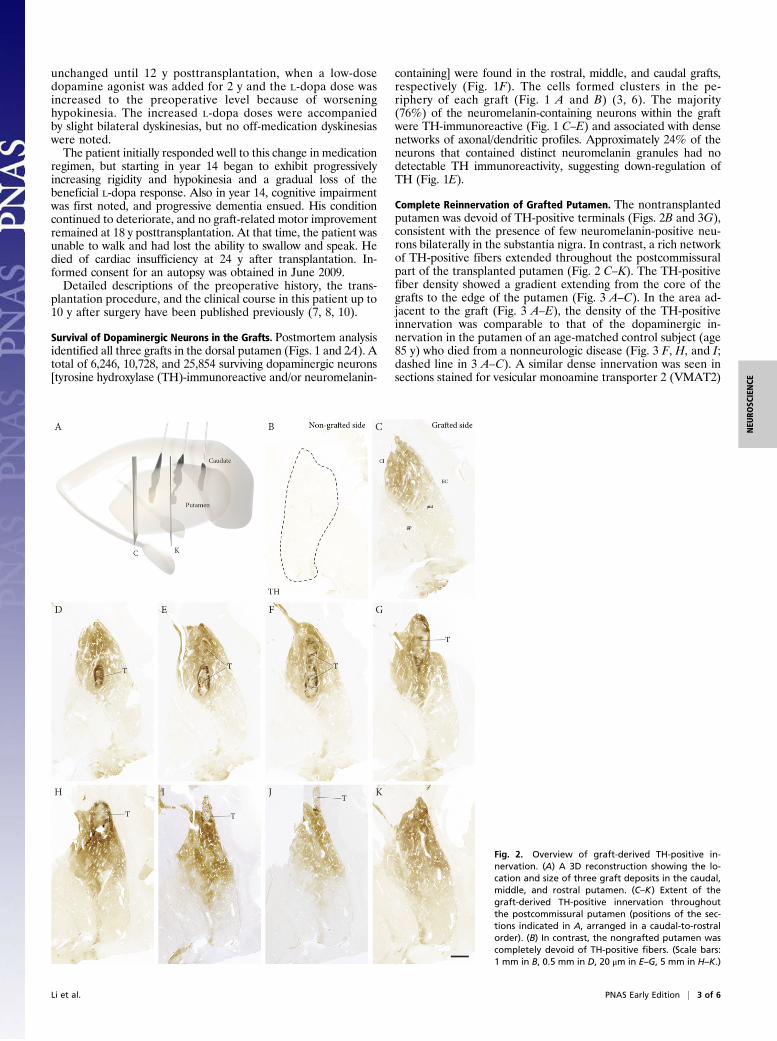
Survival of Dopaminergic Neurons in the Grafts. Postmortem analysisidentified all three grafts in the dorsal putamen (Figs. 1 and 2A). Atotal of 6,246, 10,728, and 25,854 surviving dopaminergic neurons[tyrosine hydroxylase (TH)-immunoreactive and/or neuromelanin-
containing] were found in the rostral, middle, and caudal grafts,respectively (Fig. 1F). The cells formed clusters in the pe-riphery of each graft (Fig. 1 A and B) (3, 6). The majority(76%) of the neuromelanin-containing neurons within the graftwere TH-immunoreactive (Fig. 1 C–E) and associated with densenetworks of axonal/dendritic profiles. Approximately 24% of theneurons that contained distinct neuromelanin granules had nodetectable TH immunoreactivity, suggesting down-regulation ofTH (Fig. 1E).
Complete Reinnervation of Grafted Putamen. The nontransplantedputamen was devoid of TH-positive terminals (Figs. 2B and 3G),consistent with the presence of few neuromelanin-positive neu-rons bilaterally in the substantia nigra. In contrast, a rich networkof TH-positive fibers extended throughout the postcommissuralpart of the transplanted putamen (Fig. 2 C–K). The TH-positivefiber density showed a gradient extending from the core of thegrafts to the edge of the putamen (Fig. 3 A–C). In the area ad-jacent to the graft (Fig. 3 A–E), the density of the TH-positiveinnervation was comparable to that of the dopaminergic in-nervation in the putamen of an age-matched control subject (age85 y) who died from a nonneurologic disease (Fig. 3 F, H, and I;dashed line in 3 A–C). A similar dense innervation was seen insections stained for vesicular monoamine transporter 2 (VMAT2)
Fig. 2. Overview of graft-derived TH-positive in-nervation. (A) A 3D reconstruction showing the lo-cation and size of three graft deposits in the caudal,middle, and rostral putamen. (C–K) Extent of thegraft-derived TH-positive innervation throughoutthe postcommissural putamen (positions of the sec-tions indicated in A, arranged in a caudal-to-rostralorder). (B) In contrast, the nongrafted putamen wascompletely devoid of TH-positive fibers. (Scale bars:1 mm in B, 0.5 mm in D, 20 μm in E–G, 5 mm in H–K.)
Li et al. PNAS Early Edition | 3 of 6
NEU
ROSC
IENCE
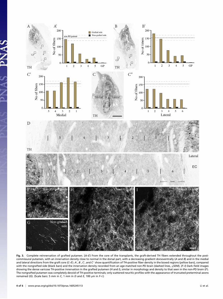
Fig. 3. Complete reinnervation of grafted putamen. (A–E) From the core of the transplants, the graft-derived TH fibers extended throughout the post-commissural putamen, with an innervation density close to normal in the dorsal part, with a decreasing gradient dorsoventrally (A and B) and in the medialand lateral directions from the graft core (C–E). A′, B′, C′, and C′′ show quantification of TH-positive fiber density in the boxed regions (yellow bars), comparedwith the nongrafted side (black bars) and the innervation density recorded from an age-matched non-PD brain (dashed lines, ±SEM). (F–I) Dark-field imagesshowing the dense varicose TH-positive innervation in the grafted putamen (H and I), similar in morphology and density to that seen in the non-PD brain (F).The nongrafted putamen was completely devoid of TH-positive terminals; only scattered neuritic profiles with the appearance of truncated preterminal axonsremained (G). (Scale bars: 5 mm in C, 1 mm in D and E, 100 μm in F–I.)
4 of 6 | www.pnas.org/cgi/doi/10.1073/pnas.1605245113 Li et al.
(Fig. S1). The precommissural part of the putamen, containing therostral graft, was paraffin-embedded. In the 4-μm-thick paraffinsections through this part, TH immunostaining allowed visualiza-tion of cells, but not of axons or terminals.
Microglia Reaction and Graft and Host Brain Pathology. Althoughimmunosuppression was terminated at 5 y posttransplantation, at24 y there was no evidence of an ongoing immune/inflammatoryresponse. Quantitative analyses showed no difference betweentransplanted putamen, nontransplanted putamen, and putamenin the control subject in terms of numbers of IBA-1–positive(recognizing both activated and nonactivated microglia) (Fig. S2A–C) or CD68-positive cells (recognizing activated microglia)(Fig. S2H) or in the morphology of microglia (14) (Fig. S2 D–G).The patient exhibited a dense, near-normal dopaminergic
reinnervation derived from the grafts at 24 y posttransplantation.Two main mechanisms likely underlie the gradual loss of theL-dopa response and dopaminergic graft effects on motor func-tion, as well as the progressive cognitive decline starting at 14 yposttransplantation. First, a considerable number of synuclein-opathy-related phenotypes were detected in the transplantedneurons, reflecting pathology spread from the host brain to the
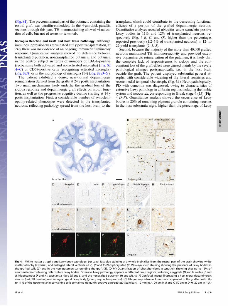
transplant, which could contribute to the decreasing functionalefficacy of a portion of the grafted dopaminergic neurons.Quantitative analyses revealed ubiquitin- and α-synuclein–positiveLewy bodies in 11% and 12% of transplanted neurons, re-spectively (Fig. 4 B, C, and Q), higher than the percentagesreported previously (1.2–5% of transplanted neurons) in 12- to22-y-old transplants (2, 3, 5).Second, because the majority of the more than 40,000 grafted
neurons maintained TH immunoreactivity and provided exten-sive dopaminergic reinnervation of the putamen, it is likely thatthe complete lack of responsiveness to L-dopa and the con-comitant loss of the graft effect were caused mainly by the severepathological changes postsynaptically, i.e., in the host brainoutside the graft. The patient displayed substantial general at-rophy, with considerable widening of the lateral ventricles andsevere medial temporal lobe atrophy (Fig. 4A). Neuropathologically,PD with dementia was diagnosed, owing to characteristics ofextensive Lewy pathology in all brain regions including the limbicsystem and neocortex, corresponding to Braak stage 6 (15) (Fig.4 D–P). Quantitative analysis showed the occurrence of Lewybodies in 20% of remaining pigment granule-containing neuronsin the host substantia nigra, higher than the percentage of Lewy
Fig. 4. White matter atrophy and Lewy body pathology. (A) Luxol fast blue staining of a whole brain slice from the rostral part of the brain showing whitematter atrophy (asterisks) and enlarged lateral ventricles (LV). (B and C) Phosphorylated (S129) α-synuclein staining showing the presence of Lewy bodies inthe grafted cells (C) and in the host putamen surrounding the graft (B). (D–M) Quantification of phosphorylated α-synuclein showing that up to 12% ofneuromelanin-containing cells contain Lewy bodies. Extensive Lewy pathology appears in different brain regions, including amygdala (D and I), cortex (E andJ), hippocampus (F and K), substantia nigra (G and L) and the nongrafted putamen (H and M). (N–P) Confocal images illustrating a host nigral dopaminergicneuron (red, TH positive) containing a typical Lewy body (green, α-synuclein positive). (Q) Ubiquitin-positive inclusions also appeared in the grafted cells. Upto 11% of the neuromelanin-containing cells contained ubiquitin-positive aggregates. (Scale bars: 10 mm in A, 20 μm in B and C, 50 μm in D–H, 20 μm in I–Q.)
Li et al. PNAS Early Edition | 5 of 6
NEU
ROSC
IENCE
bodies in the grafted dopaminergic neurons (11–12%). α-Synu-clein–positive neurites were widespread in the brain, commonlyinvolving limbic and neocortical regions, accompanied by de-generation of white matter, which also was macroscopicallyatrophic.
ConclusionsOur results demonstrate a complete graft-derived dopaminergicreinnervation of the postcommissural putamen maintained for 24 yafter transplantation in a patient with PD. This finding is consistentwith observations in the same patient at 10 y posttransplantation,when positron emission tomography revealed normalization of 18F-dopa uptake and 11C-raclopride binding in the grafted putamen (7).These findings provide the first evidence, to our knowledge, that arich dopaminergic innervation that develops over the first 3 y aftertransplantation and gives rise to major motor improvement can bepreserved for a quarter of a century in PD. However, the clinicaloutcome in this patient demonstrates that even such a viable graftmay lose its efficacy if widespread α-synucleinopathy and majordegenerative changes develop in the host brain.
Materials and MethodsThe study protocol was approved by the Regional Ethical Review Board ofLund University (nr2014/631).
Postmortem Brain Preparation. The patient’s brain (obtained at 4 d post-mortem) was fixed in 6% buffered formaldehyde solution for 1 mo and thentransferred to 20% sucrose solution. Regions were cut into coronal whole brainsections and small specimens. The brain regions in sucrose were sliced into blockscontaining basal ganglia (four blocks from rostral to caudal), mesencephalon,pons, cerebral cortex, hippocampus, and some other brain regions. The mostrostral block of the basal ganglia was paraffin-embedded, and the other threeblocks were used for frozen section preparation. All brain regions not set asidefor quantitative assessments were used for diagnostic analysis, including assess-ment of synuclein pathology and viability of the tissue.
Immunohistochemistry. Basal ganglia blocks were cut into 40-μm-thick free-floating frozen sections and 4-μm-thick slide-mounted paraffin sections. Fordiaminobenzidine (DAB, Vector Laboratories) staining, free-floating sectionswere quenched with 3% H2O2 in 10%methanol for 15 min before blocking (5%normal horse or goat serum and 0.25% Triton X-100 in 0.1M PBS, pH 7.4) for 1 h.Sections were incubated in primary antibodies (diluted in 3% serum and 0.25%Triton X-100 in PBS) overnight at 4 °C or room temperature. The antibodies usedin these experiments are listed in Table S1. After rinsing, biotinylated secondaryantibodies (horse anti-mouse, horse anti-rabbit, goat anti-rat; Jackson Immuno-Research Laboratories) were applied for 2 h at room temperature, followed byincubation with ABC complex (Vector Laboratories) for 30 min, rinsing, and thenincubation with DAB (Vector Laboratories) for 30 s or longer.
Paraffin sections were deparaffinized with xylene and then sequentialdecreasing concentrations of ethanol before use in the immunohistochem-istry procedure described above.
For immunofluorescence double labeling, two different primary anti-bodies raised in rabbit and mouse followed by Alexa Fluor 488 anti-mouseand Cy3-conjugated anti-rabbit secondary antibodies raised in donkey(Jackson ImmunoResearch Laboratories) were added. AutofluorescenceEliminator reagent (EMD Millipore) was applied before mounting.
Before blocking, both free-floating frozen sections and slide-mounted paraffinsections were treated with 10 mM citrate buffer (pH 6.4) to achieve effectiveantigen retrieval. Free-floating sections were incubated in citrate buffer at 80 °Cfor 30 min. Paraffin sections were boiled in citrate buffer in a microwave ovenfor 14 min at 800 W. Sections were rinsed in PBS before staining.
Imaging and Quantification. Bright-field images were captured using anOlympus DP73 camera, driven by CellSens software. Dark-field images wereobtained using an Olympus AX70 camera, driven by Leica DMI 6000Bsoftware.
The numbers of surviving neuromelanin- and/or TH-positive graftedneurons were assessed along each injection track in both paraffin and frozenTH-stained sections. The Abercrombie formula was used to estimate thenumber of cells in each graft. In this formula, cell diameter is assessed from anaverage of 45 neurons in each graft (16).
The density of the TH-positive innervation was assessed using a modified“sphere counting” technique of Mouton et al. (17, 18). Using a 100× oil-immersion objective, in a bright-field microscope (Olympus AX70), a z-stackwas captured throughout the entire thickness of the section at 1-μm inter-vals, using Velocity v5.4.2 image analysis software (PerkinElmer). Next, a19-μm-diameter sphere (the “probe”) was created to measure fiber densitywithin the z-stack, and the points at which fibers passed through the cir-cumference of the sphere at each z-level were counted. A score of 1 wasassigned for each fiber crossing. For each probe, the total number of fiberspassing through the sphere was expressed as a measure of fiber densitywithin the measured volume.
To quantify the VMAT2-positive fibers, staining intensity was measuredusing ImageJ. Ten images from both grafted and nongrafted putamen werechosen for analysis. Images were adjusted to gray scale, and a threshold wasset to exclude the background area. Intensity measurements were performedwithin regions of interest and within the range of threshold limit (19).
For quantification of microglia, eight image fields were chosen fromsections of grafted and nongrafted putamen and from the putamen of anormal subject immunostained with IBA1 antibody. The cell numbers in eachfield were counted, and the IBA1-positive cells classified into four differentmorphological phenotypes. In addition, five images were chosen in CD68-stained sections and the total cell numbers in all fields were counted.
ACKNOWLEDGMENTS. We thank A. Persson and A. Flasch for their excellenttechnical support, and A. C. McCourt for the language editing. This work wassupported by grants from the Swedish Research Council, BAGADILICO-Excellence in Parkinson and Huntington Research, the Swedish ParkinsonFoundation, the Torsten Söderberg Foundation, and the Strategic ResearchArea MultiPark (multidisciplinary research focus on Parkinson’s disease atLund University).
1. Hallett PJ, et al. (2014) Long-term health of dopaminergic neuron transplants inParkinson’s disease patients. Cell Reports 7(6):1755–1761.
2. Kurowska Z, et al. (2011) Signs of degeneration in 12- to 22-year old grafts of mes-encephalic dopamine neurons in patients with Parkinson’s disease. J Parkinsons Dis1(1):83–92.
3. Kordower JH, Chu Y, Hauser RA, Freeman TB, Olanow CW (2008) Lewy body-likepathology in long-term embryonic nigral transplants in Parkinson’s disease. Nat Med14(5):504–506.
4. Li JY, et al. (2008) Lewy bodies in grafted neurons in subjects with Parkinson’s diseasesuggest host-to-graft disease propagation. Nat Med 14(5):501–503.
5. Li JY, et al. (2010) Characterization of Lewy body pathology in 12- and 16-year-oldintrastriatal mesencephalic grafts surviving in a patient with Parkinson’s disease. MovDisord 25(8):1091–1096.
6. Mendez I, et al. (2005) Cell type analysis of functional fetal dopamine cell suspensiontransplants in the striatum and substantia nigra of patients with Parkinson’s disease.Brain 128(Pt 7):1498–1510.
7. Piccini P, et al. (1999) Dopamine release from nigral transplants visualized in vivo in aParkinson’s patient. Nat Neurosci 2(12):1137–1140.
8. Lindvall O, et al. (1992) Transplantation of fetal dopamine neurons in Parkinson’sdisease: One-year clinical and neurophysiological observations in two patients withputaminal implants. Ann Neurol 31(2):155–165.
9. Langston JW, et al. (1992) Core assessment program for intracerebral transplantations(CAPIT). Mov Disord 7(1):2–13.
10. Lindvall O, et al. (1994) Evidence for long-term survival and function of dopaminergic
grafts in progressive Parkinson’s disease. Ann Neurol 35(2):172–180.11. Wenning GK, et al. (1997) Short- and long-term survival and function of unilateral
intrastriatal dopaminergic grafts in Parkinson’s disease. Ann Neurol 42(1):95–107.12. Hagell P, et al. (2002) Dyskinesias following neural transplantation in Parkinson’s
disease. Nat Neurosci 5(7):627–628.13. Piccini P, et al. (2005) Factors affecting the clinical outcome after neural trans-
plantation in Parkinson’s disease. Brain 128(Pt 12):2977–2986.14. Thored P, et al. (2009) Long-term accumulation of microglia with proneurogenic
phenotype concomitant with persistent neurogenesis in adult subventricular zone
after stroke. Glia 57(8):835–849.15. Braak H, Del Tredici K (2009) Neuroanatomy and pathology of sporadic Parkinson’s
disease. Adv Anat Embryol Cell Biol 201:1–119.16. Abercrombie M (1946) Estimation of nuclear population from microtome sections.
Anat Rec 94:239–247.17. Mouton PR, Gokhale AM, Ward NL, West MJ (2002) Stereological length estimation
using spherical probes. J Microsc 206(Pt 1):54–64.18. Grealish S, et al. (2014) Human ESC-derived dopamine neurons show similar preclinical
efficacy and potency to fetal neurons when grafted in a rat model of Parkinson’s
disease. Cell Stem Cell 15(5):653–665.19. Jensen EC (2013) Quantitative analysis of histological staining and fluorescence using
ImageJ. Anat Rec (Hoboken) 296(3):378–381.
6 of 6 | www.pnas.org/cgi/doi/10.1073/pnas.1605245113 Li et al.