Expression Profiling of Gastrointestinal Stromal Tumors ... · PDF fileterapi av GIST. Den...
57
i Expression Profiling of Gastrointestinal Stromal Tumors Biomarkers for Prognosis and Therapy Gabriella Arne Sahlgrenska Cancer Center Department of Pathology Sahlgrenska Academy at the University of Gothenburg Sweden 2012
Transcript of Expression Profiling of Gastrointestinal Stromal Tumors ... · PDF fileterapi av GIST. Den...
i
Expression Profiling of
Gastrointestinal Stromal Tumors
Biomarkers for Prognosis and Therapy
Gabriella Arne
Sahlgrenska Cancer Center
Department of Pathology
Sahlgrenska Academy at the University of Gothenburg
Sweden
2012

ii
Front cover illustrations:
Left) Gene expression microarray of GIST. Middle) Immunohistochemical staining of PROM1
(CD133) protein in GIST biopsy on tissue microarray (TMA) Right) Primary small intestinal
GIST with multiple abdominal and liver metastases visualized by octreotide scintigraphy.
ISBN 978-91-628-8437-6
2012 Gabriella Arne
Printed by Ineko AB, Gothenburg
http://hdl.handle.net/2077/28261

iii
Till min underbara familj

iv

v
ABSTRACT
Expression profiling of Gastrointestinal Stromal Tumors
Biomarkers for Prognosis and Therapy
Gabriella Arne
Sahlgrenska Cancer Center, Department of Pathology,
Sahlgrenska Academy at the University of Gothenburg, Gothenburg, Sweden
Gastrointestinal stromal tumor (GIST) is a mesenchymal tumor of the gastrointestinal tract with a clinical spectrum ranging from indolent tumors to tumors with aggressive behavior and poor patient survival. The established model for prediction of prognosis for GIST is the NIH
risk score, which is based on tumor size and mitotic index. Even so, there are difficulties in predicting the clinical outcome for individual GIST patients, which may lead to inadequate treatment. The majority of GISTs have activating mutations in the genes encoding the tyrosine kinase receptors KIT, or PDGFRA, which are considered to be pathogenic events in tumor development. Imatinib, a tyrosine kinase inhibitor (TKI) that inhibits KIT, has become an important therapeutic option in addition to surgery.
To identify biomarkers that accurately predict clinical outcome in GIST patients, global gene expression profiling was performed based on KIT mutations associated with poor prognosis.
Tumor material from 16 GISTs was analyzed with expression microarray for identification of multiple candidate genes with differential expression related to mutational status. PROM1 was
shown to be highly expressed in GIST with KIT exon 11 mutations. Detection of PROM1
protein with immunohistochemical staining of 204 GISTs arranged in a tissue microarray (TMA) showed that PROM1 expression was predominant in gastric GISTs of high-risk type. Multivariate Cox analysis showed that PROM1 expression was significantly associated with poor prognosis and short patient survival, independently of NIH risk score. To evaluate the usefulness of immunohistochemical biomarkers for prognostication of GIST, we performed a comprehensive study of 14 biomarkers in 205 GISTs in a TMA. There was a significant correlation between expression of CA2, CDKN2A, CXCL12, EPHA4, FHL1, and DPP4 protein and survival. Furthermore, survival analysis using Cox regression showed that CA2, EPHA4, and FHL1 provided prognostic information additional to that from the NIH risk score. Construction of a decision-tree model combining NIH risk and expression of biomarkers further improved the prediction of patient survival. GISTs are effectively treated with surgery and imatinib, but some patients are refractory and develop drug resistance. We have investigated the prerequisites for alternative treatment strategies with peptide receptor-
mediated radiotherapy (PRRT), by analyzing the expression of somatostatin receptors (SSTRs) and uptake of radiolabeled somatostatin analogs in GIST. Analysis of 34 GISTs with pPCR and immunohistochemistry showed expression of SSTR1 and SSTR2. Primary cultures established from GIST showed specific binding and internalization of 177Lu-octreotate. Diagnostic imaging with 111In-octreotide showed tumor uptake of 111In in 3/6 GIST patients in
vivo. Tumor-to-blood activity ratios for 111In measured in biopsies from excised tumor tissue
showed ratios that may be adequate for therapy.
We conclude that the expression of PROM1 in GIST may be used as a prognosticator of patient survival and may provide a therapeutic target. Several immunohistochemical biomarkers provide additional prognostic information in addition to NIH risk score and may be useful in constructing decision-trees for improved prognostic accuracy for GIST patients.
Binding and uptake of radiolabeled somatostatin analogs via SSTR enable tumor imaging and targeted therapy in selected GIST patients.
Key words: Gastrointestinal stromal tumor (GIST); KIT; Biomarker; PROM1 (CD133); Somatostatin
receptor (SSTR); Peptide receptor-mediated radiotherapy (PRRT); Expression profiling;
Immunohistochemistry; Tissue microarray (TMA); Survival analysis
vi
POPULÄRVETENSKAPLIG SAMMANFATTNING
Cancer är ett globalt hälsoproblem och en ledande orsak till dödsfall i västvärlden. Cancer
orsakas av att något går snett i kontrollen över kroppen egna celler. En cells förmåga att växa
okontrollerat beror på genetiska förändringar i arvsmassan (DNA), vilket kan få till följd att
tumörfrämjande proteiner produceras av dessa förändrade gener. Genom framsteg inom
tumörbiologin har vi fått en ökad förståelse för hur cancer uppstår och utvecklas, och ett stort
antal av nya biomarkörer som bidrar till att förbättra diagnos, riskbedömning och terapi av
cancerpatienter.
Sarkom är relativt ovanliga tumörer som uppstår i ben, brosk och mjukdelar. GIST är det
vanligaste sarkomet i mag-tarmkanalen och uppstår oftast i magsäcken eller tunntarmen.
GIST är en heterogen tumörform som uppvisar ett varierat kliniskt förlopp, från
långsamväxande tumörer till tumörer med aggressivt växtsätt och dålig patientöverlevnad. I
denna avhandling har vi undersökt biomarkörer som har ett värde vid prognosbedömning och
terapi av GIST.
Den mest etablerade modellen för att förutsäga prognos för GIST patienter är baserad på
tumörstorlek och celldelningsfrekvens (kallad NIH risk). Överlevnaden för en väsentlig andel
av patienterna avviker dock från angiven riskbedömning, vilket kan få till följd att de inte
erbjuds optimal cancerbehandling. I delarbete I ämnade vi att identifiera nya biomarkörer som
förutsäger aggressiv tumörväxt och dålig prognos av GIST. Genom att använda microarray,
en avancerad DNA-teknik som studerar hela genuttrycket i samma analys, identifierade vi
flera kandidatgener som var intressanta i ett prognos-sammanhang. Speciellt genen PROM1
(även kallad CD133) och dess proteinprodukt visade sig kunna ge mer information om
patienters överlevnad än vad NIH riskgradering gör som ensam variabel. Vi drar därför
slutsatsen att PROM1 kan användas som ett prognostiskt verktyg för GIST-patienter.
För att bedöma värdet av såväl nya som redan kända biomarkörer vid prognosbedömning av
GIST utförde vi i delarbete II en jämförande studie av 14 olika proteinmarkörer. Genom
statistiska undersökningar jämförde vi proteinuttrycket av dessa biomarkörer med
patientöverlevnad, NIH riskgradering, och andra kliniska variabler, och fann att
proteinuttrycket för 6 av dessa markörer gav information om GIST-patienters överlevnad i
vårt material (CA2, CDKN2A, CXCL12, DPP4, EPHA4 och FHL1). Vi föreslår dessutom att
genom att konstruera ett beslutsträd som inkluderar såväl NIH riskgradering som utvalda
biomarkörer (CA2 och EPHA4) kan vi göra en mer korrekt prognosbedömning av GIST
patienters överlevnad än vad den etablerade NIH riskgradering ger ensamt.
Hälften av GIST-patienterna botas med kirurgi, men somliga går dock inte att operera radikalt
beroende på tumörens utbredning. För dessa patienter är läkemedlet Imatinib en viktig
tilläggsbehandling genom att den dämpar tumörtillväxten. Emellertid, utvecklar flertalet
patienter resistens mot Imatinib och därför finns att behov av andra behandlingsalternativ. Vi
har undersökt förutsättningarna för en alternativ behandlingsmetod med radioterapi via
specifika receptorer på cellytan, s.k. somatostatinreceptorer (SSTR). I delarbete III har vi
studerat i vilken omfattning GIST-celler uttrycker SSTR och dessutom om det är möjligt för
cellen att ta upp radionuklider via dessa receptorer. Vi kunde visa att GIST uttrycker två olika
varianter av SSTR (SSTR1 & 2) både på gennivå och som protein. Genom studier både på
cellkultur (in vitro) och på patienter (in vivo) kunde vi även visa att bindning och upptag av
radionuklider in i GIST-celler är möjligt. Vår slutsats är därför att radioterapi via SSTR skulle
kunna bli en ny möjlighet till behandling för utvalda GIST-patienter.

vii
LIST OF PAPERS
This thesis is based on the following papers, referred to in the text by their roman
numerals (I-III):
I. Arne G, Kristiansson E, Nerman O, Kindblom LG, Ahlman H,
Nilsson B, and Nilsson O. Expression profiling of GIST: CD133 is
associated with KIT exon 11 deletions, gastric location and poor
prognosis. International Journal of Cancer 2011; 129(5): 1149-1161.
II. Arne G, Kristiansson E, Nilsson B, Ahlman H, and Nilsson O.
Comparative analysis of biomarkers as prognosticators for survival in
GIST patients. In manuscript.
III. Arne G, Nilsson B, Dalmo J, Kristiansson E, Arvidsson Y, Forssell-
Aronsson E, Nilsson O, and Ahlman H. Gastrointestinal stromal
tumors (GISTs) express somatostatin receptors and bind radiolabeled
somatostatin analogs. Submitted to Acta Oncologica 2011.
viii
TABLE OF CONTENTS
Abbreviations 1
Introduction 2
Cancer 2
Cancer development and genomic instability 2
Cancer Stem Cells 5
Characteristics of the cancer cell 5
Biomarkers 7
Gastrointestinal stromal tumor (GIST) 8
Incidence of GIST 8
Clinical presentation and histopathological characteristics 9
Diagnostic biomarkers in GIST 10
Molecular pathology in GIST 11
Prognostic biomarkers in GIST 15
Treatment of GIST 18
Objectives of the thesis 21
Materials and Methods 22
Results and Discussion 25
Summary and Conclusions 34
Future Perspectives 35
Acknowledgements 37
References 39
Papers I-III

1
ABBREVIATIONS
ANO1 anoctamin 1 (also DOG1)
CSC cancer stem cell
DNA deoxyribonucleic acid
DOTA 1,4,7,10-tetraazaciclododecane- N,N´,N´´,N´´´- tetraacetic acid
DTPA diethylene triamine pentaacetic acid
ETV1 ETS variant 1
GAPDH glyceraldehyd-3-phosphate dehydrogenase
GIST gastrointestinal stromal tumor
HDACI histone deacetylase inhibitor
ICC interstitial cells of Cajal
%IA/g percent of injected activity per gram of tissue 111In indium-111 177Lu lutetium-177
mRNA messenger RNA
MSC mesenchymal stem cell
NE neuroendocrine
NET neuroendocrine tumor
NF1 neurofibromatosis type 1
NIH National Institute of Health
PDGFRA platelet-derived growth factor receptor α
PDGF platelet-derived growth factor (PDGFRA ligand)
PROM1 prominin-1 (also CD133)
PRRT peptide receptor-mediated radiotherapy
qPCR quantitative real-time polymerase chain reaction
R0 totally resected tumor, no residual tumor
RFS recurrence-free survival
RNA ribonucleic acid
SCF stem cell factor (KIT ligand)
SSTR somatostatin receptor
T/B tumor-to-blood activity concentration ratio
TK tyrosine kinase
TKI tyrosine kinase inhibitor
TMA tissue microarray
wt wild type

2
INTRODUCTION
CANCER
Cancer is a global health problem and a leading cause of deaths in industrialized
countries. The incidence of cancer is increasing due to an aging population in the
western world. Improvements in surgery and development of new cancer
therapies have prolonged the survival of patients. Advances in molecular biology
and genetics have given insight into basic principles of cancer initiation and
development. Cancer may occur as hereditary or sporadic tumors. Genetic
alterations in tumors include chromosomal alterations as well as mutations in
specific genes (Weinberg, 2007). These advances in cancer genetics have provided
a molecular classification of tumors that help to improve tumor diagnosis,
prognostication, and therapy. Introduction of high-throughput techniques in
tumor biology has increased the multitude of novel biomarkers predicting
diagnosis, prognosis and therapeutic response. However, application of
biomarkers remains a challenge in translational medicine (Brooks, 2012).
Cancer development and genomic instability
Uncontrolled growth is a characteristic feature of a cancer cell resulting from
changes occurring in the tumor cell or in its microenvironment. The changes in
the tumor cell are due to the accumulation of somatic gene aberrations, or
through epigenetic alterations. The most common mechanism to induce
mutations in the genome includes spontaneous errors in DNA replication and
repair. The majority of mutations does not affect the function of the cell and have
accordingly no consequence for tumor development. However, mutations
involving genes that control growth or the integrity of the genome may give rise
to transformed cells that proliferate abnormally and may have the ability to
invade surrounding tissues (Vogelstein and Kinzler, 1993; Yokota, 2000; Hahn
and Weinberg, 2002). The clonal multistep model for tumor development
assumes a series of randomly occurring mutations and epigenetic alterations of
the DNA (Nowell, 1976; Klein and Klein, 1985; Vogelstein and Kinzler, 1993). A
first mutation may transform a normal cell into a new cell clone with proliferative
advantage, leading to a clonal expansion at the expense of neighboring cells. A
second mutation occurs in one of the clones, resulting in yet another cell clone
with even greater proliferative ability and survival advantage. As this clonal
expansion repeats itself, new stronger populations develop that will drive tumor
progression towards a fully developed malignant phenotype.
Chromosomal aberrations, epigenetic alterations, and mutations in specific genes
all cooperate in carcinogenesis and tumor development. There are three classes of
genes, in which genetic alterations contribute to the pathogenesis of cancer:
oncogenes, tumor suppressor genes, and DNA repair genes.

3
Oncogenes promote cell proliferation
Oncogenes arise by mutations in normal genes, which are known as proto-
oncogenes (Bishop, 1991; Weinberg, 1994; Vogelstein and Kinzler, 2004). Proto-
oncogenes are normally strictly regulated and encode a wide range of proteins
including signal transducers (SRC, RAS family), transcription factors (MYC,
ETV1), growth factors (SCF, PDGF, EGF), growth factor receptors (KIT,
PDGFRA, RET), and inhibitors of apoptosis (MDM2, BCL2) (Croce, 2008).
Proto-oncogenes can be activated into oncogenes by dominant gain-of-function
mutations (Tabin et al., 1982) or to increased expression by chromosomal
amplification or translocation (Slamon, 1987). Activated oncogenes may lead to
tumorigenesis by elevated cell proliferation and inhibition of cell death. Novel
cancer drugs have been designed to target proteins encoded by oncogenes,
including the tyrosine kinase inhibitor (TKI) imatinib mesylate (Glivec®) against
the fusion protein BCR-ABL found in chronic myeloid leukemia (CML), as well
as the receptor tyrosine kinases KIT and PDGFRA activated in gastrointestinal
stromal tumors (GISTs) (Buchdunger et al., 2000; Joensuu et al., 2001).
Tumor suppressor genes control cell growth and apoptosis
Tumor suppressor genes have the opposite function of oncogenes, by acting as
negative regulators of cell proliferation (Klein, 1987). Tumor suppressor genes
encode proteins involved in many cellular functions including cell cycle
inhibition, transcriptional regulation, apoptosis, and genetic stability (e.g. TP53,
RB1, NF1, and CDKN2A) (Sherr, 2004). The inactivation of a tumor suppressor
gene requires that both alleles are affected by chromosomal deletions, point
mutations, or promoter hypermethylation (Knudson, 1971; Sherr, 2004). The loss
of a tumor suppressor gene and its encoding protein may result in loss of response
to external growth-inhibitory signals and thus increased likelihood of cancer
development (Weinberg, 2007).
DNA repair genes protect the integrity of the genome
DNA repair genes encode proteins involved in maintaining the integrity of the
genome, by participating in the cellular response to DNA damage (Peltomäki,
2001; Friedberg, 2003). DNA repair genes (e.g. BRCA1) are considered as
caretakers of the genome since they detect DNA-damage, repair damaged DNA,
and inactivate mutagenic molecules that may damage the DNA (Kastan, 2008;
Negrini et al., 2008). Mutations in such a gene may cause loss of DNA repair
function, which results in genomic instability and an elevated mutational rate in
the genome. Hence, defects in the DNA repair mechanisms allow the successive
accumulation of mutations in oncogenes and tumor suppressor genes, which
promote tumor development. (Kinzler and Vogelstein, 1997, Friedberg, 2003)
4
Epigenetic regulators of gene transcription
Unlike genetic alterations, epigenetic aberrations are chemical modifications of
the DNA or chromatin proteins that may result in changes in gene expression
without altering the DNA sequence (Jones and Baylin, 2002). DNA promoter
methylation is known to have profound effects on gene expression.
Hypermethylation of promoter regions causes gene silencing through
transcriptional inactivation. In cancer, both DNA hypomethylations and
hypermethylations may occur, causing inactivation of tumor suppressor genes
(Herman and Baylin, 2003). Another epigenetic event regulating gene expression
is histone modifications. The genetic information is packaged as chromosomes in
the cell nucleus. The chromatin is composed of DNA wrapped around histones.
The chromatin may be in a transcription-competent or -incompetent state, thus
controlling accessibility of the genome. The state of the chromatin is mainly
controlled by post-translational modifications of histone proteins, e.g.
acetylations, methylations, and phosphorylations (Sharma et al., 2010). Unlike
genetic changes, epigenetic changes are potentially reversible and represent
promising target molecules and predictive biomarkers in tumor treatment
(Sharma et al., 2010). Histone deacetylase inhibitors (HDACIs), inducing cell
apoptosis and/or cell cycle arrest, have already been shown to have selective
toxicity against tumor cells. HDACIs (e.g. valproic acid, vorinostat) in
combination with conventional therapy (e.g. chemo- or radiotherapy) have been
tested with encouraging results in Phase I and II clinical trials of hematological
malignancies and solid tumors (Marks et al., 2001; Johnstone, 2002; Tan et al.,
2010).
MicroRNAs are short non-coding RNA molecules regulating gene expression of
proteins involved in various biological processes, including proliferation,
differentiation, and cell death (Ambros, 2004). MicroRNA inhibits translation of
DNA by degrading mRNA transcripts. Aberrant microRNA expression may have
profound influence on cellular consequences, since a single microRNA can bind
and regulate multiple genes. MicroRNAs are often deregulated in cancer and
have been shown to be involved in tumor initiation, as well as tumor progression.
Increased expression of oncogenic microRNAs can repress targets such as tumor
suppressor genes, whereas loss of tumor suppressive microRNAs may enhance
the expression of target oncogenes (Ambros, 2004; Volinia et al., 2006). Targeting
microRNAs has been proposed to be a novel strategy in cancer therapy, and
experimental studies have shown that inhibition of certain microRNAs (e.g. miR-
21) reduces tumor growth (Negrini et al., 2009; Bonci, 2010).
5
Cancer stem cells
According to the cancer stem cell (CSC) theory, solid tumors are composed of
hierarchies of tumor cells with different functions. CSCs represent a minority of
cells in the tumor and have the ability to produce large numbers of descendant
tumor cells and are responsible for tumor growth and metastasis formation
(Alison et al., 2011). CSCs are self-renewing cells that may divide into one
daughter cell that becomes a new CSC, and another cell that becomes a tumor
progenitor cell (a rapid-amplifying cell). The progenitor cell may undergo a large
series of cell divisions giving rise to the bulk of tumor cells (Al-Hajj and Clarke,
2004). Chemotherapy is generally effective in killing tumor cells, but CSCs are
usually resistant due to expression of cytoprotective enzymes (e.g. ABC-
transporters, acetyl dehydrogenase (ALDH)) (Alison et al., 2011). Thus, CSCs
may persist after chemotherapy (Guzman et al., 2002) causing tumor relapse.
Curative treatment requires elimination of all CSCs. Targeting CSCs is therefore
a promising therapeutic principle. CSCs are believed to be dependent on a
restricted set of signaling pathways (e.g. those associated with KIT, Wnt, sonic
hedgehog, and Notch), all of which are promising candidates for CSC targeted
therapy (Marotta and Polyak, 2009). Furthermore, CSCs express unique cell
surface markers (e.g. CD44, CD90, and CD133 (PROM1)), which may also be
used for CSC targeted therapy (Alison et al., 2011). There are growing
experimental evidence for the existence of CSC subpopulations in malignant
tumors (e.g. in leukemia, glioma, prostate and breast cancer, and Ewing sarcoma)
(Collins et al., 2005; Charafe-Jauffret et al., 2009; Suvà et al., 2009; Alison et al.,
2011).
Characteristics of the cancer cell
Tumor cells arise from normal cells by a multistep process known as tumor
progression. During this process cancer cells acquire a multitude of different
properties, which are common to all types of cancer. These properties are
identified as the “Hallmarks of cancer” (Hanahan and Weinberg, 2011).
The proposed characteristics of a cancer cell are functions of specific control
systems that govern the transformation of normal cells into cancer cells. Normal
cells require growth signals from the extracellular environment to proceed into an
active proliferative state in the cell cycle. Tumor cells develop the ability to sustain
proliferative signaling on its own, either by self production of growth signals or by
constitutive activation of growth signaling pathways. There are several cellular
processes limiting proliferation to keep the delicate balance of homeostasis in a
tissue. Normal cells are regulated by antigrowth signals, including tumor
suppressors, which may force the cell to enter a non-proliferative state or even
undergo apoptosis to maintain the balance. Tumor cells acquire the ability to
evade growth suppressors and/or resist cell death in order to remain proliferative.

6
Several mechanisms may be involved in these circumvention strategies, including
the loss of TP53 tumor suppressor function.
Telomere shortening limits the replicative potential in normal cells to a fixed
number of multiplications. Tumor cells overcome this limitation which enables
replicative immortality. By the deregulation of telomerase, which protects the
chromosome ends, the tumor cell harbors a capacity of unlimited replicative
potential that progresses tumor growth.
Formation of new blood vessels is a vital process in normal tissues as well as in
tumors, for the critical supply of nutrients and oxygen. Tumor cells induce an
angiogenic switch to harbor unrestricted ability to induce angiogenesis.
Furthermore, the contact with blood and lymphatic vessels allows the tumor to
enter the circulation and disseminate. All malignant tumors have the potential to
invade and metastasize. The invasion-metastasis cascade is a multistep process
describing how a cancer cell acquires the ability to penetrate surrounding tissue
and finally colonize vital organs in distant sites. The ability to invade and
metastasize is a characteristic feature of malignant tumors, as opposed to benign
tumors. The ability of a tumor to metastasize and invade vital organs is
responsible for the vast majority of cancer deaths (Weinberg, 2007).
Another characteristic involved in the pathogenesis of cancer is the reprogramming
of energy metabolism. Rapid cell division and growth increase the need of energy
to survive and progress. The capacity to modify the cellular metabolism allows
tumor cells to adapt to both aerobic and hypoxic environments to enable effective
growth. Further, the immune system may eliminate tumor cells by the action of
natural killer (NK) cells and cytotoxic T lymphocytes (CTLs) (Pagés et al., 2010).
Hence, the tumor cell needs to avoid the immune surveillance and evade immune
destruction in order to stay vital.
These hallmarks of cancer explain the acquired functional capabilities that drive
the transformation of a normal cell into a tumor cell. Furthermore, Hanahan and
Weinberg (2011) also addressed two key characteristics that are important for the
initiation of tumor development. The surrounding microenvironment that
nurtures the tumor with bioactive molecules is supported by tumor-promoting
inflammation in the tissue. Finally, cancer has a genetic basis and is primarily
induced by genome instability and mutations as described earlier.
7
BIOMARKERS
“A biomarker is defined as a characteristic that is objectively measured and
evaluated as an indicator of normal biological processes, pathogenic processes,
or pharmacological responses to a specified therapeutic intervention”
- Biomarker Definitions Working Group (2001)
A biomarker is a biological feature used as an indicator of a biological state. The
term biomarker is used in many scientific fields, including cell biology and
medicine. In medicine a biomarker can be a molecule that detects a particular cell
type or a substance that correlates to a particular disease state, but biomarkers are
not necessarily molecules. A biomarker can be any kind of measurable quantity
which may have clinical relevance, e.g. a protein that indicate a stem cell
phenotype, the presence of an antibody that indicate an infection, or a specific
DNA sequence that indicates susceptibility to therapy. In oncology, detection of
biomarkers may provide important information on diagnosis, tumor progression,
or effects of cancer treatment (Brooks, 2012).
Diagnostic biomarkers
Diagnostic biomarkers are used as tools for the identification of patients that have
a specific disease or an abnormal medical condition. To identify a specific cancer,
the expression pattern of certain tumor-specific proteins is often used as
diagnostic biomarkers, together with clinical information as tumor location and
morphology (e.g. PSA expression to diagnose prostate cancer) (DeMatteis, 1992).
The importance of a correct diagnosis is at time a matter of life and death for a
patient, due to choice of and response to therapy.
Prognostic biomarkers
Prognostic biomarkers give information on disease outcome for a patient and
correlates to tumor recurrence. Clinical parameters like tumor size, number of
metastatic sites, or tumor risk grade can serve this purpose. A prognosticator can
also be an elevated expression of a certain protein or lack of expression of the
same. A genetic alteration may carry prognostic value for a patient, e.g.
amplification of MYCN indicates poor prognosis in neuroblastoma (Schwab,
1997).
Predictive biomarkers
Predictive biomarkers can be used to characterize the patient´s disease in order to
determine whether that individual is a suitable candidate for a certain treatment
modality. With an increasing awareness of the heterogeneity among tumors
(Reya et al., 2001), the need for improved selection of patients for a given
anticancer treatment is evident. Individual patients within a tumor disease may be
treated with different therapies in order to obtain optimal outcome.

8
Predictive biomarkers, i.e. specific gene mutations or expression of certain
proteins, might function as tools in the search for such tailored therapies.
Furthermore, the term predictive biomarker is also used when referring to a
patient´s response to a drug, and may further be used as a term for the therapy
target itself. In this sense, the predictive biomarker could be referred to as a
therapeutic biomarker (e.g. breast cancer with ERBB2 amplifications respond to
therapy with monoclonal antibodies (i.e. Herceptin®) that targets the growth
factor receptor protein ERBB2 (Baselga et al., 1998).
Biomarkers are evaluated in order to acquire relevant knowledge about a disease
entity and translate that information into clinical practice. In the search for novel
biomarkers, single factors are often found to correlate to the biological state
investigated. The ultimate biomarker would be one that allows unequivocal
distinction of that state. However, in tumor biology where multiple cellular
processes might be essential for a certain tumor entity, it might be too simple to
rely on a single biomarker to predict prognosis or response to treatment. Instead,
a set of biomarkers may provide a more accurate prediction. High- throughput
technologies including global genome analysis and proteomics may provide an
expression profile of several factors that may be a signature for such a prediction
(Oldenhuis et al., 2008; Brooks, 2012).
GASTROINTESTINAL STROMAL TUMOR (GIST)
Incidence of GIST
Tumors may arise from almost any dividing cell in the body. Most human tumors
(80%) originate from epithelial tissues. Sarcomas are derived from cells of
connective and supporting tissues, i.e. muscle, nerves, fat, bone, cartilage,
synovial tissue, or blood vessels. Sarcomas are also named mesenchymal tumors
and represent 1% of all adult tumors (Weinberg, 2007). Gastrointestinal stromal
tumor (GIST) is the most common mesenchymal tumor in the gastrointestinal
tract with an estimated incidence of approximately 10-20 cases per million
inhabitants annually (Nilsson et al., 2005; Tryggvason et al., 2005; Tzen et al.,
2007). GISTs are rare compared with other tumors in the gastrointestinal tract,
and account for about 2% of gastric malignancies. GIST usually affects the
elderly, but they are also seen in younger age-groups, e.g. so called pediatric
GIST (preferentially seen in young women) (Benesch et al., 2009). The median
age for sporadic GIST has been reported to be about 60-70 years and affects men
and women with equal frequency (Nilsson et al., 2005; Joensuu et al., 2011; Rossi
et al., 2011).

9
Clinical presentation and histopathological characteristics of GIST
GIST arises in the muscular wall along the entire digestive system. The most
frequent primary sites are the stomach (about 60%) and the small intestine (25-
30%), followed by the colo-rectum (5%), and the esophagus (3%). On rare
occasions, primary GISTs are reported in extragastrointestinal locations (e.g.
omentum, mesentery, or retroperitoneum) (Corless & Heinrich, 2008; Joensuu et
al., 2011; Rossi et al., 2011). The clinical spectrum of GIST is divergent, including
indolent tumors and tumors with aggressive behavior. Tumor size may vary from
small tumors less than 2 cm, to large tumors exceeding 30 cm. The diagnosis of
GIST is established on their characteristic morphology and expression of the KIT
protein. GISTs show variable cellularity regardless of malignancy and may be
composed of spindle-shaped cells (60-70%), epitheloid cells (15%), or a mixture of
both (15-20%) (Rossi et al., 2011) (Figure 1).
Figure 1. Hematoxylin & eosin staining of GIST.
Left) Spindle cell GIST. Right) Epitheloid GIST.
Previously, the majority of GISTs were regarded as benign tumors. However,
most GISTs, including small incidentally detected tumors, have been shown to
have metastatic capability (Corless et al., 2002). In fact, up to 50% of all GISTs
have shown to be metastatic at diagnosis. The most common metastatic sites for
GIST are the peritoneum and the liver, and rarely the lymph nodes, lung or bone
(DeMatteo et al., 2000). GISTs give rise to symptoms due to local effects of the
primary or its metastases. The most frequent symptoms are abdominal pain,
gastrointestinal obstruction or bleeding. Since GISTs normally grow non-
invasively, the tumors may become large until a palpable mass has developed.
The overall 5-year survival rate of GIST is about 54%, and the 5-year disease-free
survival rate is about 45% after radical surgery (R0 resection) (DeMatteo et al.,
2000, Gold et al., 2009). However, about half of all GISTs are metastatic at
presentation, and the median overall survival for these patients was reported to 19
months prior to targeted therapy. With the introduction of tyrosine kinase
inhibitors (TKIs), e.g. imatinib, the median overall survival for metastatic GIST
patients has extended to more than 50 months (Van Glabbeke et al., 2007).

10
Diagnostic biomarkers in GIST
The two most specific and sensitive diagnostic biomarkers for GIST are protein
expression of KIT (CD177) (Hornick and Fletcher, 2002) and anoctamin 1
(ANO1) (also named DOG1) (Espinosa et al., 2008), which are positive in about
95% of all GIST (Miettinen et al., 2009) (Figure 2).
Figure 2. Left) KIT and right) ANO1 immunohistochemical staining of GIST
A small subset of GIST (less than 3% of the tumors) stain negatively for both KIT
and ANO1, preferentially gastric epitheliod GISTs (Miettinen et al., 2009), which
makes the diagnosis more difficult for this group. Therefore, additional
immunohistochemical markers to improve GIST diagnosis have been searched
for. CA2 (carbonic anhydrase II) is one proposed novel marker which is
expressed in 95% of GISTs (Parkkila et al., 2009). CA2 expression was
independent of tumor site and did not show positive staining in other tested
malignancies, indicating an additional diagnostic value. PKCΘ (protein kinase C
theta), is another biomarker frequently expressed in GISTs and therefore
proposed as a diagnostic marker (Blay et al., 2004, Duensing et al., 2004). CD34
has previously been used to diagnose GIST. However, this marker has lower
sensitivity and is only expressed in 70-80% of the tumors, mainly in gastric GIST
(Miettinen and Lasota, 2006). GIST can be difficult to distinguish from other
abdominal soft tissue tumors, which may show positive immunoreactivity for
KIT and/or ANO1, including schwannomas, angiosarcomas, peritoneal
leiomyomatosis, uterine type retroperitoneal leiomyomas, metastatic melanomas,
and synovial sarcomas (Miettinen et al., 2009). Hence, a panel of biomarkers (e.g.
KIT, ANO1, S-100, SMA, desmin, and CD34) is often used to establish the final
diagnosis of GIST.
GISTs show neuroendocrine phenotype and express peptide receptors
Neuroendocrine tumor (NET) cells share a set of common properties including
expression of storage vesicles, which can be divided into two types: large dense-
core vesicles (LDCV) and synaptic-like microvesicles (SLMV). These vesicles
contain peptide hormones and biologically active amines (Rindi et al., 2004).
Release of hormones from NET cells is frequently regulated by G-protein coupled
receptors. The diagnosis of neuroendocrine (NE) differentiation is based on

11
typical morphology and the expression of vesicle proteins, e.g. chromogranin A
(CHGA) and synaptophysin (Feldman and Eiden, 2003). NE differentiation may
also include expression of hormones and peptide receptors. Bümming et al. (2007)
identified the expression of several synaptic vesicle proteins (e.g. SV2, synapsin 1,
synaptobrevin, and amphiphysin) in GIST, indicating a possible NE phenotype in
these tumors (Jakobsen et al., 2001; Bümming et al., 2007). The search for a
hormonal activity in GIST has revealed production of the appetite-stimulating
peptide hormone ghrelin (Ekeblad et al., 2006). GIST has also been demonstrated
to express peptide receptors including bombesin subtype 2 receptor,
cholecystokinin subtype 2 receptor, vasoactive intestinal peptide subtype 2, and
somatostatin receptors (SSTR) (Reubi et al., 2004; Palmieri et al., 2007). SSTRs
are G-protein coupled membrane receptors occurring in five different subtypes
(SSTR1-5) (Patel, 1999). SSTR2 is the most widely expressed SSTR subtype in
certain NETs (e.g. midgut carcinoids) (Nilsson et al., 1998). SSTR2 & 5 are
frequently used as targets for both diagnostic and therapeutic purposes in NETs,
utilizing the binding and internalization of radiolabeled somatostatin analogs (e.g. 111In-DTPA-octreotide and 177Lu-DOTA-octreotate) to these receptors
(Kwekkeboom et al., 2010; Swärd et al., 2008). In GIST, SSTRs have been
demonstrated with variable protein expression in subsets of tumors (Reubi et al.,
2004; Palmieri et al., 2007). The SSTR expression pattern in GIST may enable
peptide receptor-mediated radiotherapy (PRRT) as a treatment option for certain
patients.
Molecular pathology in GIST
GISTs have a phenotype similar to ICC
GISTs have a phenotype similar to the interstitial cells of Cajal (ICC) (e.g.
expression of KIT) and are therefore thought to be derived from ICC, or from a
precursor cell (Kindblom et al., 1998). ICC progenitor cells have been identified in
the murine stomach and shown to be KITlow, CD44+
, CD34+
(Bardsley et al.,
2010). ICCs form an intimate network within the intestinal wall, transducing
signals from the nervous system to muscle cells to control motility. ICCs are
therefore referred to as “pacemaker cells” (Faussone-Pellegrini, 1992). ICC and
GIST have a close phenotypic resemblance, e.g. strong expression of KIT
receptor tyrosine kinase protein (Kindblom et al., 1998; Sircar et al., 1999). ICCs
are dependent on a regulated KIT proto-oncogene expression for their normal
development from a mesenchymal progenitor cell into a gastrointestinal
pacemaker cell (Faussone-Pellegrini, 1992). In a pioneering publication, Hirota et
al. (1998) showed that gain of function mutations in KIT was a pathogenic event
in the development of GIST. Later studies have indicated that interactions
between KIT and ETS variant 1 (ETV1) are necessary for both ICC and GIST
development (Chi et al., 2010) (Figure 3).

12
Figure 3. KIT and ETV1 cooperate
in the development of ICC and
GIST. ICC develop from
mesenchymal stem cell (MSC)
precursors (ICC precursors) as a
result of physiological KIT signaling
and ETV1 expression. GISTs
develop from MSC precursors (ICC
precursors) as a result of constitutive
activation of KIT signaling and
ETV1 expression.
ETV1 is a critical regulator of oncogenesis in GIST
ETV1 is a member of the ETS gene family acting as a transcriptional activator by
binding to consensus DNA sequences. Several fusion genes have been identified
with ETV1 as one of the partners (e.g. EWS-ETV1). Formation of an ETV1 fusion
gene is considered to be the oncogenetic event in the development of Ewing
sarcoma and prostate cancer (Im et al., 2000; Tomlins et al., 2007). Furthermore,
full-length ETV1 has been reported to be over expressed in GIST, as well as in
melanoma and prostate cancer (Chi et al., 2010; Jané-Valbuena et al., 2010; Gasi
et al., 2011). ETV1 protein may bind enhancer elements in promoter regions of
several target genes, and thus regulates biological processes such as cell
proliferation, differentiation, and migration. A recent study by Sawyers and
colleagues identified several genes, normally overexpressed in GIST and ICC, to
be dependent on ETV1 expression. Knockdown of ETV1 in GIST cell lines
reduced the expression of e.g. PROM1, DUSP6, and TIMP3, and caused reduction
of cell proliferation (Chi et al., 2010). ETV1 was suggested to be a key regulator in
the development of ICC, as well as the formation of GIST from ICC precursor
cells, by cooperating with activated KIT. Mutated KIT activates MAPK signaling
and thus inhibits proteasomal degradation of ETV1, which in turn is required for
GIST development (Chi et al., 2010; Rubin, 2010) (Figure 3). Together with KIT,
ETV1 has been proposed to be a lineage survival factor in GIST.
Receptor tyrosine kinases - KIT and PDGFRA
Receptor tyrosine kinases (TKs) are receptors for growth factors and have the
ability to induce proliferation in normal cells. KIT and platelet derived growth
factor receptor alfa (PDGFRA) are evolutionary homologues of the type III
receptor tyrosine kinase family. Their natural ligands are stem cell factor (SCF)
and platelet-derived growth factor (PDGF), respectively. These receptor TKs are
complex proteins with a similar structure, consisting of a cytoplasmic domain, a
transmembrane domain, and a unique extracellular ligand-binding domain. The

13
intracellular part comprises two TK domains including one ATP-binding region
(TK1) and one activation loop (TK2). When the receptor is bound to its ligand,
two subunits of the receptor dimerize and allow TK domains to
autophosphorylate. This promotes a catalytic cleft in the juxtamembrane domain
(close to the plasma membrane) to open up with direct access to substrate
molecules. Further phosphorylations of the TKs activate downstream signaling
pathways causing cell proliferation (Lennartsson et al., 2005). Activation of the
KIT receptor engages downstream signaling pathways, including PI3K/AKT,
RAS/MAPK, and JAK/STAT, which promotes cell cycle activation,
proliferation, and inhibition of apoptosis (Lennartsson et al., 2005; Corless et al.,
2011) (Figure 4). Normal KIT receptor function is essential for the development
of ICC, melanocytes, germ cells, and hematopoetic cells (Fleischman, 1993). The
PDGFRA receptor activates signaling pathways similar to those of KIT, but also
phospholipase Cγ (PLCγ), which promotes cell growth and motility (Andrae et
al., 2008).
Figure 4. KIT and PDGFRA signaling pathways in GIST. Phosphorylated KIT or
PDGFRA activates PI3K/AKT/mTOR, JAK/STAT, and RAS/MAPK signaling and
stabilization of ETV1, causing cell cycle activation, cell proliferation, and inhibition of
apoptosis.
14
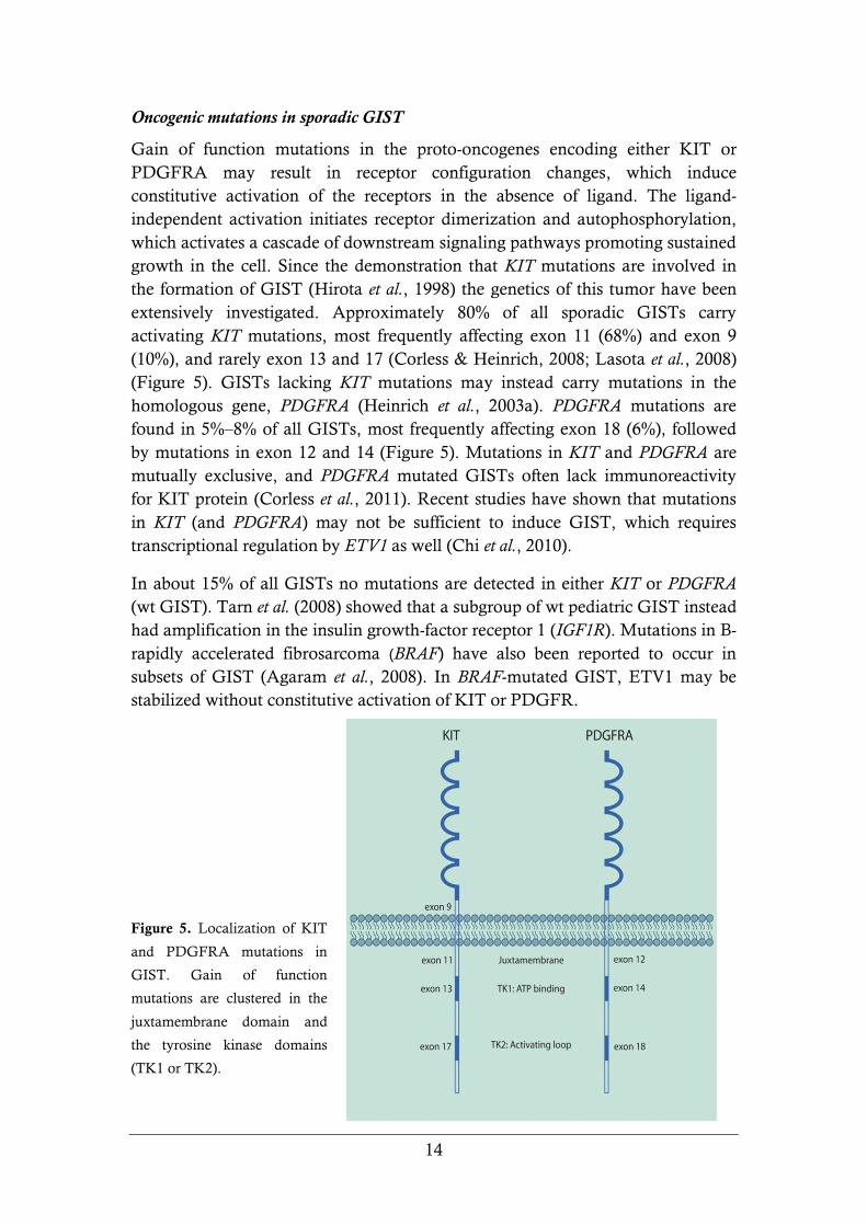
Oncogenic mutations in sporadic GIST
Gain of function mutations in the proto-oncogenes encoding either KIT or
PDGFRA may result in receptor configuration changes, which induce
constitutive activation of the receptors in the absence of ligand. The ligand-
independent activation initiates receptor dimerization and autophosphorylation,
which activates a cascade of downstream signaling pathways promoting sustained
growth in the cell. Since the demonstration that KIT mutations are involved in
the formation of GIST (Hirota et al., 1998) the genetics of this tumor have been
extensively investigated. Approximately 80% of all sporadic GISTs carry
activating KIT mutations, most frequently affecting exon 11 (68%) and exon 9
(10%), and rarely exon 13 and 17 (Corless & Heinrich, 2008; Lasota et al., 2008)
(Figure 5). GISTs lacking KIT mutations may instead carry mutations in the
homologous gene, PDGFRA (Heinrich et al., 2003a). PDGFRA mutations are
found in 5%–8% of all GISTs, most frequently affecting exon 18 (6%), followed
by mutations in exon 12 and 14 (Figure 5). Mutations in KIT and PDGFRA are
mutually exclusive, and PDGFRA mutated GISTs often lack immunoreactivity
for KIT protein (Corless et al., 2011). Recent studies have shown that mutations
in KIT (and PDGFRA) may not be sufficient to induce GIST, which requires
transcriptional regulation by ETV1 as well (Chi et al., 2010).
In about 15% of all GISTs no mutations are detected in either KIT or PDGFRA
(wt GIST). Tarn et al. (2008) showed that a subgroup of wt pediatric GIST instead
had amplification in the insulin growth-factor receptor 1 (IGF1R). Mutations in B-
rapidly accelerated fibrosarcoma (BRAF) have also been reported to occur in
subsets of GIST (Agaram et al., 2008). In BRAF-mutated GIST, ETV1 may be
stabilized without constitutive activation of KIT or PDGFR.
Figure 5. Localization of KIT
and PDGFRA mutations in
GIST. Gain of function
mutations are clustered in the
juxtamembrane domain and
the tyrosine kinase domains
(TK1 or TK2).
15
Although, the majority of GISTs are sporadic, GISTs may also occur in familiar
settings, e.g. neurofibromatosis type 1 (NF1) or as part of Carney Triad or
Carney-Stratakis Syndrome (Bümming et al., 2006). In patients with Carney
Triad, or Carney-Stratakis Syndrome, GIST lack KIT and PDGFRA mutations
and may have reduced activity of mitochondrial complex II. In some patients this
is due to germ-line mutations inactivating the succinate dehydrogenase (SDH)
enzymes (Janeway et al., 2011).
Prognostic biomarkers in GIST
Several prognostic factors have been proposed for GIST, e.g. morphological
features such as tumor size, pleomorphism, mitotic count, high micro-vessel
density, invasive growth, and tumor necrosis, but also genetic factors and
molecular biomarkers (Miettinen et al., 2002).
NIH risk score and tumor location
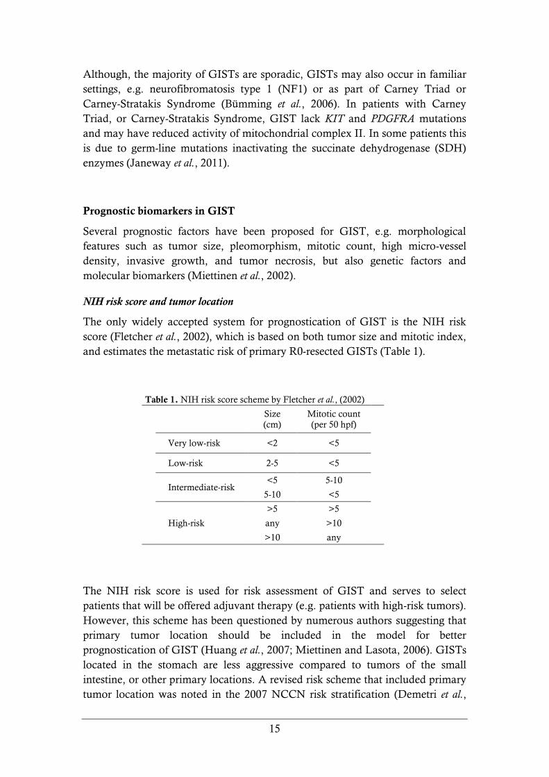
The only widely accepted system for prognostication of GIST is the NIH risk
score (Fletcher et al., 2002), which is based on both tumor size and mitotic index,
and estimates the metastatic risk of primary R0-resected GISTs (Table 1).
Table 1. NIH risk score scheme by Fletcher et al., (2002)
Size
(cm)
Mitotic count
(per 50 hpf)
Very low-risk <2 <5
Low-risk 2-5 <5
Intermediate-risk
<5 5-10
5-10 <5
High-risk
>5 >5
any >10
>10 any
The NIH risk score is used for risk assessment of GIST and serves to select
patients that will be offered adjuvant therapy (e.g. patients with high-risk tumors).
However, this scheme has been questioned by numerous authors suggesting that
primary tumor location should be included in the model for better
prognostication of GIST (Huang et al., 2007; Miettinen and Lasota, 2006). GISTs
located in the stomach are less aggressive compared to tumors of the small
intestine, or other primary locations. A revised risk scheme that included primary
tumor location was noted in the 2007 NCCN risk stratification (Demetri et al.,

16
2007). Comparing the two risk score schemes on a large set of GIST patients,
Goh et al (2008) proved the revised risk score to predict patient outcome more
effectively. However, variations were observed for the recurrence rates in the
high-risk group of GIST. Another model to predict prognosis in GIST was
suggested by Nilsson et al. (2005), which included proliferative activity (Ki67) and
tumor size as prognosticators. The Ki76/size model showed distinct prognostic
value.
Cytogenetic factors
Besides clinical and morphological biomarkers, genetic abnormalities have shown
to provide prognostic information for GIST patients. A limited number of
chromosomal abnormalities are observed in GIST tumors, including monosomy
of chromosome 14, partial losses of 14q or 22q are the most frequent cytogenetic
findings (Yang et al., 2008). Gunawan et al (2007) found that loss of 14q
characterized gastric tumors with stable karyotypes and favorable clinical course.
In contrast, loss on chromosome 1p characterized small intestinal GISTs with a
more aggressive course. Loss of heterozygocity (LOH) on chromosome 9p has
also been shown to associate with a malignant phenotype, possibly due to loss of
the tumor suppressor gene CDKN2A (Sabah et al., 2004; Wozniac et al., 2007;
Corless et al., 2011).
KIT and PDGFRA mutations
Mutational status of KIT and PDGFRA has been shown to influence patient
survival, although several research groups report conflicting results. Furthermore,
the correlation between type of mutation and patient outcome is influenced by the
introduction of tyrosine kinase inhibitors (TKIs) in the treatment of advanced
GIST. Several groups have observed a correlation between poor prognosis and
KIT exon 11 mutations. Especially tumors with KIT exon 11 deletions (primarily
involving codons Trp557 and/or Lys558) have been reported to associate with
poor prognosis (Andersson et al., 2006, Martin et al., 2005, Singer et al., 2002).
Other studies failed to confirm such results (DeMatteo et al., 2008). Poor
prognosis has also been demonstrated for tumors with KIT exon 9 and KIT exon
13 mutations (Lasota et al., 2008, Antonescu et al., 2003), whereas PDGFRA
mutant GISTs have been reported to be less aggressive (Lasota et al., 2006).
Although mutational analysis may have prognostic impact, the importance of
KIT and PDGFRA mutations as prognostic indicators remains to be determined.
On the other hand, mutational status has been shown to be useful as a predictive
biomarker for the response to TKI (Corless et al., 2011).
17
18
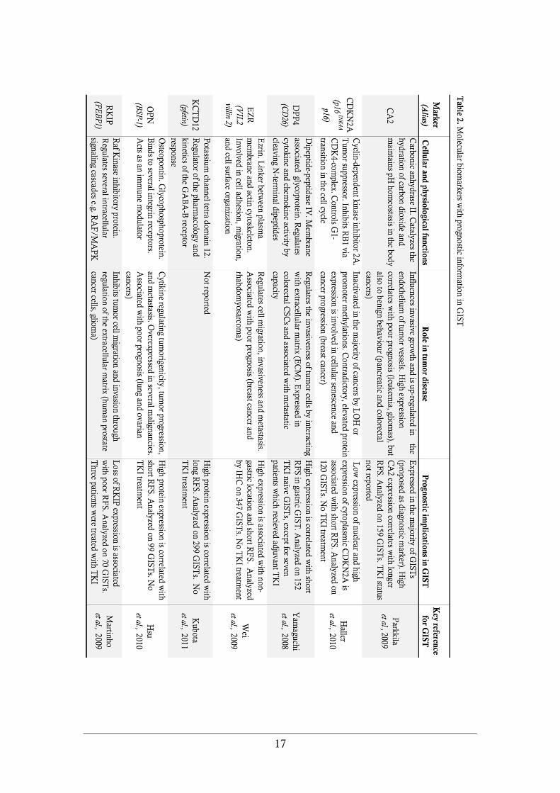
Molecular biomarkers for prognosis
A number of molecular biomarkers have been shown to provide information
regarding GIST patient survival. Reported markers with prognostic relevance in
GIST include CA2, CDKN1B, CDKN2A, DPP4, EZR, HIF1A, KCTD12, NES,
PTGS2, RKIP, SKP2, and VEGF. A summary of biomarkers with survival data
in GIST is presented in Table 2. Comparative studies on biomarker performance
in GIST have not been carried out and influence of TK inhibition on the
usefulness has not been evaluated. None of these proposed molecular biomarkers
have to date been widely introduced into clinical practice.
Treatment of GIST
Surgery is the primary treatment of GIST and approximately half of all patients
are cured with surgery only. Since radiation and chemotherapy are largely
ineffective in GIST, other treatment options must be explored for patients with
unresectable or metastatic tumors. Today, many of these patients are treated with
imatinib as first-line therapy resulting in prolonged patient survival. However,
resistance to TKI is an increasing clinical problem, as well as non-responsive
tumors, which urges the development of novel treatment.
Imatinib and sunitinib
Inhibition of activated KIT, or PDGFRA, by palliative therapy with TKIs has
dramatically improved the survival of patients with high-risk GIST. Imatinib
(Gleevec ™) is the first-line option for unresectable GIST (Bümming et al., 2003;
Van Glabbeke et al., 2007). Imatinib binds to the ATP binding site of the
intracellular tyrosine kinase domain of KIT (or PGFRA). This prevents the
kinase from transferring phosphate from ATP to tyrosine residues of the substrate
complex (e.g. SHC, GRB2, SOS), which leads to the inactivation of downstream
signaling pathways (Lennartsson et al., 2005). Approximately 80% of all GISTs
show primary response to imatinib treatment. However, response rates relate to
mutational status. Tumors with KIT exon 11 mutations are most responsive to
imatinib (70–85% response rate) due to favorable conformation changes of the
juxtamembrane part of the receptor (Corless et al., 2011). Tumors with KIT exon
9 mutations demonstrate an intermediate responsiveness (25–48% response rate),
while tumors with KIT exon 13 or 17 mutations or no mutations respond poorly.
Consequently, the prognosis is better for imatinib-treated patients with KIT exon
11 mutations than for patients with KIT exon 9, or wt GIST (Heinrich, 2003b;
Corless et al., 2011). Primary resistance to imatinib (i.e. resistance within 6 months
of treatment) is seen in 10–15% of all GISTs, including wt tumors and tumors
with PDGFRA exon 18 (D842V) mutations. Secondary resistance to imatinib (i.e.
resistance 6 months after initial response to treatment) develops in 40% of the
patients. The most important mechanism for secondary resistance to imatinib
19
involves acquired mutations in KIT and PDGFRA (Faivre et al., 2007; Corless &
Heinrich, 2008; Liegl et al., 2008; Wang WL et al., 2011).
Sunitinib (Sutent™) was introduced as second-line therapy (Younus et al., 2010).
Sunitinib has broader activity profile than imatinib (KIT and PDGFRA), and
inhibits other receptor TKs such as VEGFR1-3, RET, and FLT3. Inactivation of
these pathways leads to inhibition of cell proliferation and angiogenesis (Chow
and Eckardt, 2007). However, the duration of response is often limited for
sunitinib-treated patients (approximately one year) (Wang WL et al., 2011).
Resistance to imatinib and sunitinib emphasizes the need for alternative
therapeutic strategies.
Therapeutic biomarkers
Novel TKIs have been developed and investigated as treatment of patients that
develop resistance to imatinib and sunitinib. Dasatinib, nilotinib, and sorafenib
have shown advantageous activity profiles related to a PDGFRA mutant (D842V)
and wt GIST (Kim and Zalupski, 2011). Several other therapeutic biomarkers
have been investigated for GIST, including molecules belonging to the
downstream signaling pathways that are activated by KIT and PDGFRA. Drugs
targeting these pathways have been evaluated with promising results in
experimental studies. Several inhibitors of the PI3K/AKT/mTOR signaling
cascade have been investigated, with the most promising effects observed for
mammalian target of rapamycin (mTOR) inhibitors (everolimus) (Bauer et al.,
2007; Schöffski et al., 2010).
Insulin-like growth factor 1 receptor (IGF1R), like KIT and PDGFRA, activates
signaling pathways as RAS/MAPK and PI3K/AKT/mTOR. IGF1R has been
demonstrated to have a significant potential as a therapeutic target for IGF1R-
driven tumors, which has been investigated experimentally with promising results
(Tognon and Sorensen, 2011). IGF1R has been suggested as a treatment option in
wt GIST (Tarn et al., 2008, Braconi et al., 2008). However, the pathogenetic role
of IGF1R in GIST remains to be elucidated.
The conformation of a constitutively activated KIT is stabilized by a chaperon
molecule, heat shock protein 90 (HSP90), which have been suggested as a
therapeutic target in GIST and other tumors (Bauer et al., 2006). Several HSP90
inhibitors have been developed and proved to have anti-tumor effects in
experimental studies. By combining the HSP90 inhibitor retaspimycin
hydrochloride (IPI-504) and imatinib/sunitinib in xenograft GIST, treatment
effects were shown to be enhanced (Floris et al., 2011). However, IPI-504 was
recently suspended from phase III trials for to safety reasons.
Combinations of different TKIs, or TKIs together with other drugs have been
suggested to be advantageous treatment option for specific GIST mutants (e.g.
mTOR inhibitors combined with imatinib) (Nilsson et al., 2009).
20
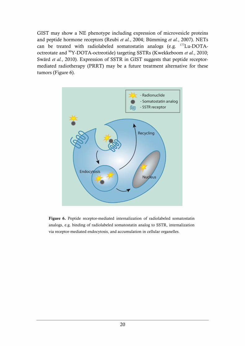
GIST may show a NE phenotype including expression of microvesicle proteins
and peptide hormone receptors (Reubi et al., 2004; Bümming et al., 2007). NETs
can be treated with radiolabeled somatostatin analogs (e.g. 177Lu-DOTA-
octreotate and 90Y-DOTA-octreotide) targeting SSTRs (Kwekkeboom et al., 2010;
Swärd et al., 2010). Expression of SSTR in GIST suggests that peptide receptor-
mediated radiotherapy (PRRT) may be a future treatment alternative for these
tumors (Figure 6).
Figure 6. Peptide receptor-mediated internalization of radiolabeled somatostatin
analogs, e.g. binding of radiolabeled somatostatin analog to SSTR, internalization
via receptor-mediated endocytosis, and accumulation in cellular organelles.

21
OBJECTIVES OF THE THESIS
The general aim of this study was to characterize expression profiles for GIST in
order to identify novel biomarkers for prognosis and therapy.
The specific aims were:
to characterize the gene expression profiles of GISTs in relation to
mutational status in KIT and PDGFRA in order to identify genes
involved in tumor progression and aggressive behavior.
to evaluate the usefulness and prognostic power of immuno-
histochemical biomarkers as predictors of survival in patients with
GIST.
to analyze the expression of somatostatin receptors (SSTRs) in GIST,
and evaluate SSTR as a therapeutic target for peptide receptor-
mediated radiotherapy (PRRT).

22
MATERIALS AND METHODS
Tumor material
Paraffin embedded tumor material of GIST (Paper I, II, III). A total of 263 well
characterized GISTs were used in immunohistochemical analyses in the three
studies included in this thesis. In Paper I we used tumor biopsies from 204
patients with mutational status on KIT and PDGFRA, including 180 patients with
R0-resected tumors and complete survival data and follow-up. In Paper II, tumor
biopsies from 205 patients with R0-resected tumors and complete survival data
and follow-up were used. The paraffin-embedded tumor material in Paper I and
II, were arranged in a tissue microarray (TMA). These patients were only treated
surgically for their tumor disease. The TMA is based on the population-based
study of GISTs (1983–2001) by Nilsson et al. (2005). Mutational status and
survival data have been published previously by Andersson et al. (2006). In Paper
III we used paraffin-embedded tumor material from 34 patients that underwent
resection of GIST at the Sahlgrenska University Hospital, Gothenburg, Sweden
(1997–2008). Some of these patients were given TKI therapy.
Frozen tumor biopsies from GIST (Paper I, II). In Paper I, tumor biopsies from 16
patients (7 with gastric, 7 with small intestinal, and 2 with rectal GIST) were used
for both expression microarray and quantitative real-time PCR (qPCR). These
patients did not receive imatinib treatment before surgery (imatinib naïve
patients). In Paper III, tumor biopsies from 34 patients (16 with gastric, 15 with
small intestinal, and 3 with rectal GIST) were analyzed with qPCR.
Primary cell culture of GIST (Paper III). Tumor tissues from two patients were
used to establish primary cell culture for radionuclide uptake studies in Paper III,
including one gastric and one small intestinal GIST. Cultured tumor cells were
characterized and found to express KIT and DOG-1 by immunofluorescence.
GIST patients for diagnostic imaging and activity ratios of 111In (Paper III). Seven
GIST patients received 170–240 MBq 111In-DTPA-D-Phe1-octreotide (111In-
octreotide) by intravenous injection (Paper III). Diagnostic imaging (scintigraphy)
was performed on 6 patients within 24 h after injection. Tumor samples together
with blood samples drawn during surgery were collected from five patients and
tumor-to-blood 111In activity concentration ratio (T/B) was measured 2–22 days
after injection of 111In-octreotide.

23
Methods
Methods used in this thesis are well established, including gene expression
analyses by microarray and qPCR, immunohistochemistry, tumor cell culture,
radioactivity measurements and scintigraphy, and a short summary follows
below:
Gene expression analysis (Paper I, III). In Paper I, gene expression analysis was
performed on 55k whole genome oligonucleotide microarrays (Swegene DNA
Microarray Resource Center, Lund, Sweden). In order to extend the patient
material, these data were combined with two other published gene expression
datasets in a meta-analysis. The probes in all three datasets were processed to
share a common annotation and further analyzed for their gene expression fold
change. In Paper I and III, qPCR assays were performed in
96-well optical plates using TaqMan® Reverse Transcription Reagents (Applied
Biosystems, CA, USA) and analyzed with an ABI Prism® 7500 Fast System SDS.
Immunohistochemical analysis (Paper I, II, III). Immunohistochemical analyses
were performed on paraffin embedded tumors. Bound antibodies were visualized
using Dako EnVision+ detection systems (DakoCytomation, Denmark) with
HRP-labeled polymer and DAB substrate. For the immunohistochemical scoring
of biomarkers, a dilution series of each antibody was evaluated on TMA sections.
The dilution that resulted in the greatest discrimination in staining pattern
between tumor biopsies was chosen for further analysis. In Paper II, each biopsy
was scored according to the following criteria: 0, when <10% of tumor cells were
labeled; 1+, when 10%-90% of tumor cells were labeled; and 2+, when >90% of
tumor cells were labeled.
Cell culture and confocal microscopy (Paper III). Unlike a cell line, a primary cell
culture consists of a mixed population of cell types and cells with advantageous
growth properties will increase more rapidly in vitro. The primary cell cultures of
GIST (Paper III) were set on collagen-coated Biocoat® Multiwell Plates (BD
Biosciences, MA, USA) with RPMI 1640 medium supplemented with 10% fetal
calf serum, L-glutamine, and PEST. Uptake experiments were performed within
4 days in culture and cell quality were again characterized by KIT and ANO1
expression and SSTR1-5 expression by immunofluorescence detection of Alexa
Flour conjugated antibodies (Molecular Probes Inc., OR, USA) using confocal
microscopy (Zeiss LSM 510 META system).
24
Binding and internalization of radiolabeled somatostatin analogs (Paper III). The
evaluation of binding and internalization (uptake) of the radiolabeled
somatostatin analogs 177Lu-octreotate and 111In-octreotide was performed by three
different methods. Binding and internalization of 177Lu was investigated in
primary cell culture of GIST, where the cells were incubated with 177Lu-octreotate
for 48 hours (control cultures were also supplemented with unlabeled octreotide).
Amount of surface-bound and internalized 177Lu was measured in a gamma
counter (Wallac 1480 WIZARD™; Wallac Oy, Finland). Scintigraphy of 111In-
octreotide in GIST patients was performed by a gamma camera (General Electric
400 AC/T; General Electric, London, UK). 111In activity in tumor biopsies and
blood samples was measured in the gamma counter, and tumor-to-blood 111In
activity concentration ratios were determined.
Statistics (Paper I, II, III). Statistical analyses used in this thesis were performed in
the statistical language R (www.r-project.org) or in SPSS (IBM company, NY,
USA). Gene expression microarray data was normalized by lowess
normalization, and ranked according to average log-fold change and the
moderated t-statistic (Paper I). Meta-analysis, combining gene expression profiles
from three different data sets, was performed by re-annotating all probes to a
common annotation and compare transcripts according to the average fold
change (Paper I). Regression based survival analysis was performed using Cox
proportional hazards model (Paper I, II). The decision-tree model was calculated
using cross-validation (Paper II). Binding efficiencies in studies on cultured cells
were evaluated by linear regression (Paper III).
Ethical approval (Paper I, II, III). For the use of clinical materials in Paper I-III,
we obtained consent from the patients and approval from the Regional Ethical
Review board in Gothenburg, Sweden.
25
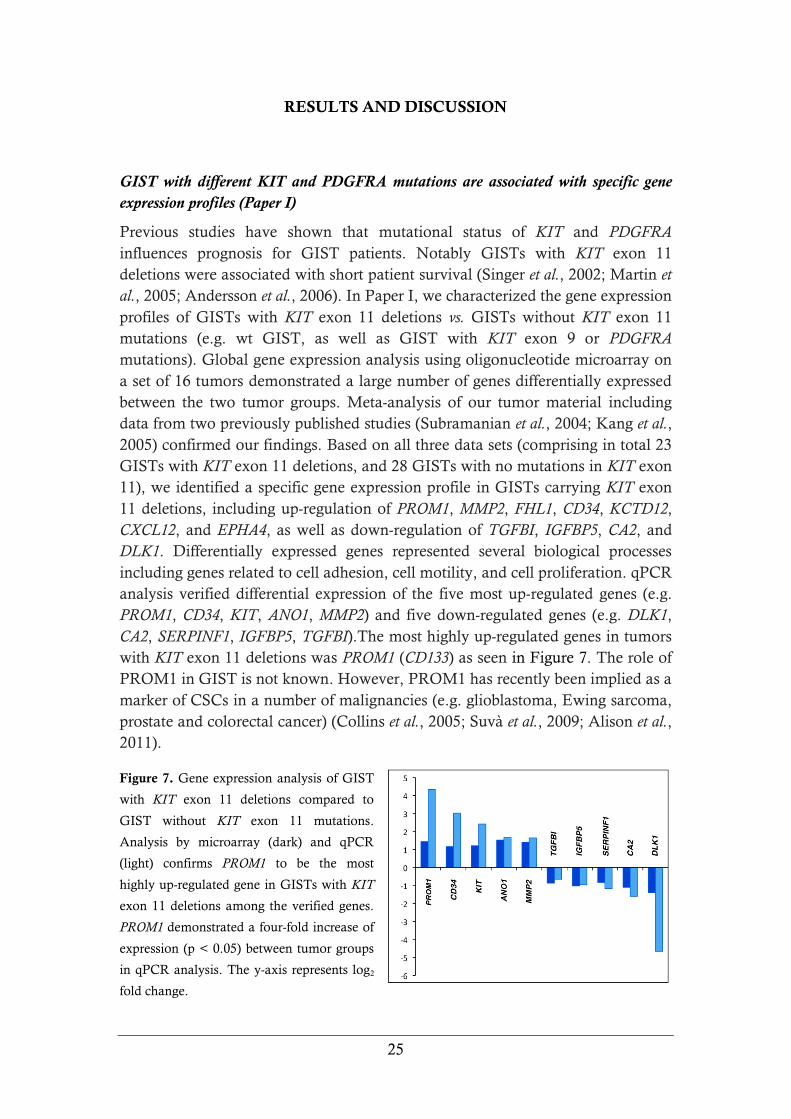
RESULTS AND DISCUSSION
GIST with different KIT and PDGFRA mutations are associated with specific gene
expression profiles (Paper I)
Previous studies have shown that mutational status of KIT and PDGFRA
influences prognosis for GIST patients. Notably GISTs with KIT exon 11
deletions were associated with short patient survival (Singer et al., 2002; Martin et
al., 2005; Andersson et al., 2006). In Paper I, we characterized the gene expression
profiles of GISTs with KIT exon 11 deletions vs. GISTs without KIT exon 11
mutations (e.g. wt GIST, as well as GIST with KIT exon 9 or PDGFRA
mutations). Global gene expression analysis using oligonucleotide microarray on
a set of 16 tumors demonstrated a large number of genes differentially expressed
between the two tumor groups. Meta-analysis of our tumor material including
data from two previously published studies (Subramanian et al., 2004; Kang et al.,
2005) confirmed our findings. Based on all three data sets (comprising in total 23
GISTs with KIT exon 11 deletions, and 28 GISTs with no mutations in KIT exon
11), we identified a specific gene expression profile in GISTs carrying KIT exon
11 deletions, including up-regulation of PROM1, MMP2, FHL1, CD34, KCTD12,
CXCL12, and EPHA4, as well as down-regulation of TGFBI, IGFBP5, CA2, and
DLK1. Differentially expressed genes represented several biological processes
including genes related to cell adhesion, cell motility, and cell proliferation. qPCR
analysis verified differential expression of the five most up-regulated genes (e.g.
PROM1, CD34, KIT, ANO1, MMP2) and five down-regulated genes (e.g. DLK1,
CA2, SERPINF1, IGFBP5, TGFBI).The most highly up-regulated genes in tumors
with KIT exon 11 deletions was PROM1 (CD133) as seen in Figure 7. The role of
PROM1 in GIST is not known. However, PROM1 has recently been implied as a
marker of CSCs in a number of malignancies (e.g. glioblastoma, Ewing sarcoma,
prostate and colorectal cancer) (Collins et al., 2005; Suvà et al., 2009; Alison et al.,
2011).
Figure 7. Gene expression analysis of GIST
with KIT exon 11 deletions compared to
GIST without KIT exon 11 mutations.
Analysis by microarray (dark) and qPCR
(light) confirms PROM1 to be the most
highly up-regulated gene in GISTs with KIT
exon 11 deletions among the verified genes.
PROM1 demonstrated a four-fold increase of
expression (p < 0.05) between tumor groups
in qPCR analysis. The y-axis represents log2
fold change.

26
PROM1 protein is predominantly expressed in GIST with KIT exon 11 mutations,
gastric location, and poor patient survival (Paper I)
In order to further characterize the expression of PROM1 in GIST, we evaluated
tumor biopsies from 204 GIST patients by TMA and immunohistochemistry
using monoclonal antibodies against PROM1 (clone AC133). Out of 195 tumor
biopsies evaluated for PROM1 staining, 55 showed positive labeling (28%).
Comparing PROM1 protein expression and mutational status, we found the
highest proportion of PROM1-positive tumors in GISTs with KIT exon 11
mutations (41%), with significant correlation between positive PROM1 and KIT
exon 11 deletions (p = 1.6 x 10⁻⁵). Tumors with other KIT or PDGFRA mutations,
or wt tumors, had lower proportion of PROM1-positive tumors (0–17%).
Comparing PROM1 protein expression and tumor location, we found higher
frequency of positive tumors in stomach (correlation p = 3.1 x 10⁻¹¹), as compared
to small intestine, colon, and rectum. In total, 51/106 (48%) patients with gastric
GISTs were positive for PROM1. No association between PROM1-labeling and
NIH risk score, tumor size, mitotic count, Ki67-labeling, or histopathological
growth patterns was observed. There was a positive correlation between PROM1-
and CD34-labeling, possibly due to higher expression of both PROM1 and CD34
in gastric GISTs, compared to GISTs in other locations.
We further evaluated PROM1 as a biomarker to predict survival in GIST
patients. Patients with R0-resected tumors and clinical follow-up (n=180) were
included in our survival analysis using Cox regression model. Univariate analysis
including PROM1 labeling, NIH risk score, and mutational status in KIT and
PDGFRA showed that patients with PROM1-positive GIST had shorter survival
compared to patients with PROM1-negative tumors (Figure 8). Patients with
high-risk GIST and patients with KIT exon 11 deletions also showed shorter
overall and recurrence-free survival at univariate analysis. Multivariate analysis
including the variables age, sex, NIH risk group, mutational status, CD34, and
PROM1 labeling, demonstrated that PROM1 expression provided additional
information regarding patient survival compared to all other variables.
Figure 8. PROM1 protein expression is
associated with shorter overall patient
survival. Survival curves are calculated
from survival data on GIST patients with
R0 surgery and estimated by Cox
regression (adjusted for age and sex)
(p<0.05).
27
Survival analysis was also performed on gastric GISTs alone. Patients with
PROM1 expression had significantly shorter survival in the univariate survival
analysis. Multivariate Cox analysis demonstrated PROM1 expression and NIH
risk score as the only significant variables in the gastric GISTs. Thus, PROM1
labeling provided additional information regarding patient survival compared
with age, sex, NIH risk group, and mutational status in all GISTs, as well as in
gastric GISTs. Expression of PROM1 in GIST may have therapeutic
implications. Patients with PROM1 positive tumors and TKI resistance may be
subjected to targeted therapies as indicated by experimental studies using anti-
PROM1 (anti-CD133) monoclonal antibodies (Smith et al., 2008; Wang CH et al.,
2011).
We showed that the PROM1 protein was associated with shorter patient survival.
The fact that PROM1 is a marker of CSCs in a number of solid tumors raised the
question whether PROM1 also was a marker for CSCs in GIST. Two studies
have addressed this question (Bozzi et al., 2011; Chen et al., 2011). Both studies
confirmed high expression of PROM1 in GIST as compared to other sarcomas.
However, PROM1 and CD44 were shown to be universally expressed in a
majority of GIST with higher mRNA value in gastric tumors compared to small
intestinal tumors (Chen et al., 2011). Therefore, expression of PROM1 was
suggested to be a lineage marker in GIST rather than a marker for CSCs. This
hypothesis was further supported by studies on the transcriptional regulator
ETV1, which acts as a lineage survival factor in GIST. ETV knockdown
experiments in GIST cell lines demonstrated down-regulation of lineage markers
including PROM1 (Chi et al., 2010). Characterization of sorted GIST cells
showed enrichment of PROM1- cells in side populations, most likely representing
CSCs. Also, PROM1- cells were shown to form more colonies and were more
invasive than PROM1+
cells in matrigel assays (Bozzi et al., 2011; Chen et al.,
2011). These data suggest that PROM1 is not a CSC marker of GIST.

28
Evaluation of immunohistochemical biomarkers with prognostic relevance in GIST
(Paper II)
The selection of biomarkers investigated for their prognostic value in Paper II was
based on previously published studies on prognostic biomarkers in GIST as
well as differentially expressed genes provided by the gene expression profile in
Paper I. We designed a comprehensive study on protein labeling of these
biomarkers in 205 GIST tumors arranged in a TMA. All immunohistochemical
stainings were evaluated according to one single scoring system on tumor biopsies
incubated at antibody-dilutions that discriminated negative and positive tumors
on the TMA. We evaluated a series of biomarkers for their prognostic relevance
in GIST patients: CA2, CDKN2A (p16), CXCL12, DPP4 (CD26), EPHA4,
EZR, FHL1, KCTD12 (pfetin), MMP2, and PROM1 (CD133). Univariate
analysis showed that expression of CA2, CDKN2A, CXCL12, DPP4, EPHA4,
and FHL1 correlated with recurrence-free survival (RFS) in GIST patients.
Expression of PROM1 only showed marginally statistic significance at univariate
analysis in this series. PROM1 and the other biomarkers that not reached
statistical significance in univariate analysis were excluded from further
evaluation. Multivariate analysis showed that expression patterns of the
biomarkers CA2, EPHA4, and FHL1 provided information on patient survival in
addition to NIH risk score. CDKN2A and DPP4 on the other hand, showed
strong association with NIH risk score. Since primary tumor location is a
prognosticator in GIST (i.e. gastric GIST are less aggressive than tumors with
other locations), we investigated the confounding effects between NIH risk score,
tumor location and expression patterns for the individual biomarkers. Four
biomarkers provided additional information to patient survival in the multivariate
analysis: CA2, DPP4, EPHA4, and FHL1. Expression patterns of the
immunohistochemical biomarkers in relation to NIH risk score and survival are
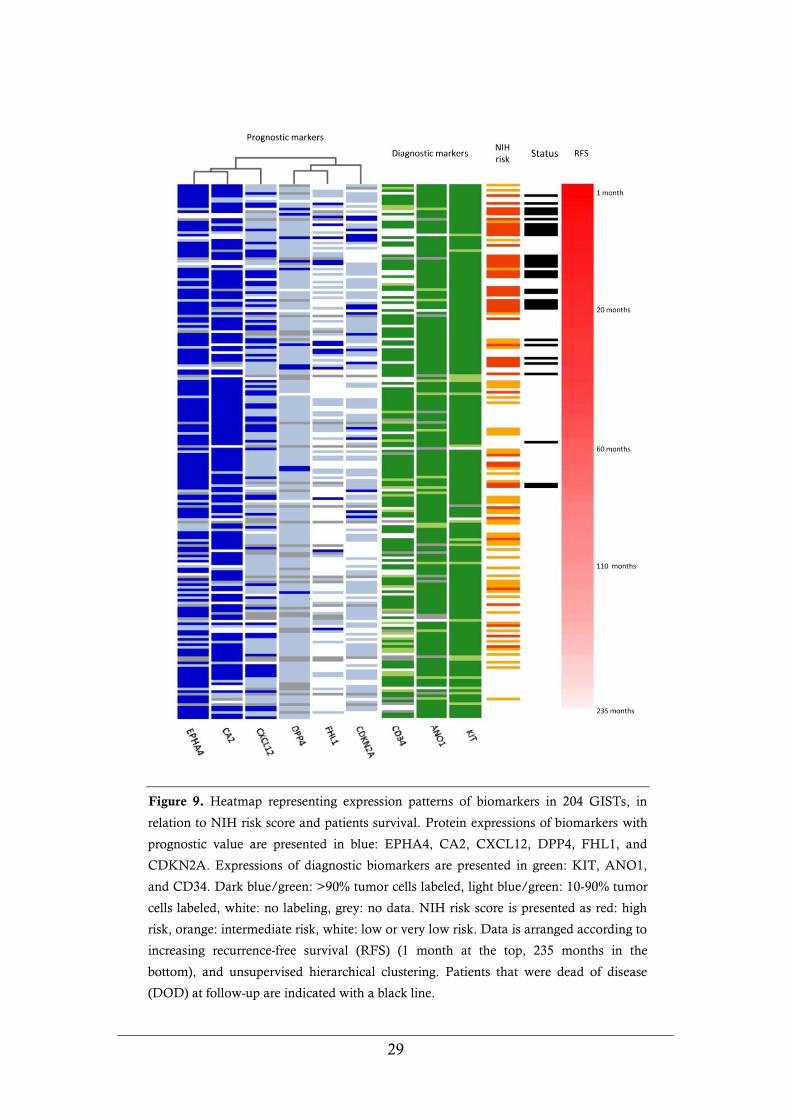
visualized in a heatmap (Figure 9).
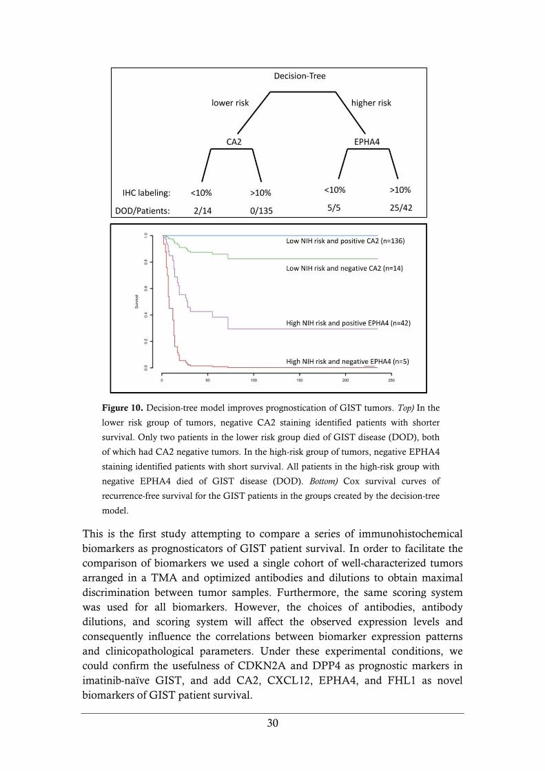
To optimize the prediction of survival in GIST patients, we constructed decision-
tree models combining NIH risk score and expression of immunohistochemical
biomarkers. NIH risk was divided in a high-risk group and into a group of lower
risks (very low-, low-, and intermediate-risk). NIH risk score was found to be the
single most effective prognosticator in the model. In Figure 10, we present a
decision-tree model sorted by NIH risk score, in which the addition of CA2 and
EPHA4 expression patterns were shown to predict patient survival more
accurately than the score alone.
29
Figure 9. Heatmap representing expression patterns of biomarkers in 204 GISTs, in
relation to NIH risk score and patients survival. Protein expressions of biomarkers with
prognostic value are presented in blue: EPHA4, CA2, CXCL12, DPP4, FHL1, and
CDKN2A. Expressions of diagnostic biomarkers are presented in green: KIT, ANO1,
and CD34. Dark blue/green: >90% tumor cells labeled, light blue/green: 10-90% tumor
cells labeled, white: no labeling, grey: no data. NIH risk score is presented as red: high
risk, orange: intermediate risk, white: low or very low risk. Data is arranged according to
increasing recurrence-free survival (RFS) (1 month at the top, 235 months in the
bottom), and unsupervised hierarchical clustering. Patients that were dead of disease
(DOD) at follow-up are indicated with a black line.
30
Figure 10. Decision-tree model improves prognostication of GIST tumors. Top) In the
lower risk group of tumors, negative CA2 staining identified patients with shorter
survival. Only two patients in the lower risk group died of GIST disease (DOD), both
of which had CA2 negative tumors. In the high-risk group of tumors, negative EPHA4
staining identified patients with short survival. All patients in the high-risk group with
negative EPHA4 died of GIST disease (DOD). Bottom) Cox survival curves of
recurrence-free survival for the GIST patients in the groups created by the decision-tree
model.
This is the first study attempting to compare a series of immunohistochemical
biomarkers as prognosticators of GIST patient survival. In order to facilitate the
comparison of biomarkers we used a single cohort of well-characterized tumors
arranged in a TMA and optimized antibodies and dilutions to obtain maximal
discrimination between tumor samples. Furthermore, the same scoring system
was used for all biomarkers. However, the choices of antibodies, antibody
dilutions, and scoring system will affect the observed expression levels and
consequently influence the correlations between biomarker expression patterns
and clinicopathological parameters. Under these experimental conditions, we
could confirm the usefulness of CDKN2A and DPP4 as prognostic markers in
imatinib-naïve GIST, and add CA2, CXCL12, EPHA4, and FHL1 as novel
biomarkers of GIST patient survival.
31
GISTs regularly express SSTR1 and SSTR2 (Paper III)
GISTs have been proposed to have a NE phenotype due to their expression of
peptide hormones (Ekeblad et al., 2006), synaptic vesicle proteins (Reubi et al.,
2004; Bümming et al., 2007), and peptide receptors (Reubi et al., 2004; Palmieri et
al., 2007). Expression of SSTR in GIST has been reported, but data on SSTR
subtypes and expression levels are limited (Reubi et al., 2004; Palmieri et al.,
2007). In order to further characterize the expression of SSTRs in GIST, we
performed expression profiling of SSTR subtypes 1–5 on biopsies from 34 GISTs.
qPCR analysis demonstrated expression of SSTR subtype 1 & 2 in the majority of
tumors, while SSTR3–5 were only expressed at low levels. Median mRNA
expression levels of SSTR1 & 2 were similar, but SSTR1 showed a more variable
expression pattern with high expression levels in individual tumors. The same
tumors were also analyzed by immunohistochemical staining for SSTR1–5
receptor proteins. SSTR1 & 2 showed positive staining in all GISTs and SSTR3–5
in a subset of tumors. Altogether, qPCR or immunohistochemistry thus detected
expression of SSTR1 & 2 in all analyzed tumors and SSTR3–5 in a subset of
GISTs. The divergent results in individual tumors between qPCR and
immunohistochemical analyses most likely reflect intra-tumoral heterogeneity or
differences in sensitivity between the methods. From these data we conclude that
GISTs express multiple SSTR subtypes. However, GISTs mainly express
SSTR1 & 2, as opposed to gastrointestinal carcinoids which mainly express
SSTR2 & 5 (Nilsson et al., 1998), suggesting differences in receptor profiles that
need to be considered when designing SSTR-mediated radiotherapy. We
identified individual patients in our series that might have been candidates for
SSTR-mediated therapy, including tumors of both high-risk and low-risk. Patients
with high SSTR expression, who had developed primary or secondary resistance
to imatinib, could have been suitable for SSTR-mediated therapy.
GIST cells bind and internalize 177Lu–octreotate in vitro (Paper III)
Binding and internalization of a radiolabeled somatostatin analog 177Lu-octreotate
in GIST tumor cells were studied in primary cell cultures from one gastric GIST
and one small intestinal GIST. GIST cells in primary culture expressed SSTR1 &
2, as determined by immunocytochemistry and confocal laser microscopy.
Cultured cells demonstrated specific binding and uptake of 177Lu, as shown in
Figure 11. The amount of internalized 177Lu increased continuously during the
incubation period, whereas surface-bound 177Lu increased marginally, indicating
receptor saturation. The relative amount of internalized vs. surface-bound 177Lu
was 8-fold at 48 hours. In control experiments, both binding and internalization
of 177Lu were effectively reduced by addition of unlabeled octreotide to the culture
media. The two primary cell cultures established from GIST patients bound and
internalized 177Lu to the same degree, suggesting that these patients could have
been candidates for SSTR-mediated radiotherapy.

32
Figure 11. Binding and internalization of 177Lu-octreotate in primary cell culture of
GIST. Experiments of a gastric GIST (left) and a small intestinal GIST (right) showed
significant binding and internalization of 177Lu–octreotate, as compared to control
experiments. Error bars are smaller than symbols.
GISTs can be visualized by scintigraphy using 111In–octreotide (Paper III)
To assess the binding of radiolabeled somatostatin analogs and the possibility of
visualizing SSTR-expressing tumors by scintigraphy, GIST patients were injected
with the radiolabeled somatostatin analog 111In-octreotide preoperatively. Positive
tumor imaging was obtained in three out of six GIST patients, indicating that
radiolabeled somatostatin analogs localize to GIST tumors in significant amounts
(Figure 12). Thus, octreotide scintigraphy via SSTR may be a possible diagnostic
tool for GIST patients, which necessitates a future prospective study. To assess
the amount of internalized 111In in GIST tumors, the 111In activity concentration
(%IA/g tissue) was measured in excised tumor biopsies and blood samples from
five patients. Measurements confirmed specific uptake of 111In, but the tumor-to-
blood activity concentration ratio (T/B) varied between the five patients (range 8
to 96). T/B values in GIST were similar to those obtained in medullary thyroid
carcinoma (Forssell-Aronsson et al., 2000 and 2004), but lower than those
obtained in midgut carcinoids (Hashemi et al., 2003; Forssell-Aronsson et al.,
2004). Relatively high values were observed in individual GIST tumors,
indicating high expression of SSTR, internalization of radionuclide, and rapid
clearance of 111In-octreotide. The need for new treatment strategies in GIST is
evident for those patients that develop primary or secondary resistance to
imatinib and sunitinib. Our experimental results indicate that SSTR-targeted
radiotherapy may be a future therapeutic option for certain patients.

33
Figure 12. Octreotide scintigraphy of a GIST patient injected with 111In-
octreotide, demonstrating an unresectable primary small intestinal GIST, and
multiple abdominal (arrows) and liver (double arrow) recurrences.
Our present data on SSTR1 & 2 expression indicate, heterogeneity between GIST
lesions in the same patient. Good tumor visualization was obtained by
scintigraphy in two patients despite low SSTR mRNA expression in the analyzed
biopsies. Such tumor heterogeneity, with marked differences in 111In binding has
also been observed in patients with NETs, e.g. carcinoids and endocrine
pancreatic tumors (Forssell-Aronsson et al., 2004).
The somatostatin analogs used in this study (i.e. octreotide and octreotate) are
preferentially designed to bind to SSTR2 & 5. NET cells mainly express SSTR2
and have been demonstrated to bind and internalize radiolabeled octreotide in
vitro (Andersson et al., 1996). A majority of NETs are also positive by 111In-
octreotide scintigraphy (Teunissen et al., 2011). In contrast, GISTs showed a
relatively lower expression of SSTR2, which may explain a lower frequency of
scintigraphically positive tumors. The high uptake of 111In and 177Lu observed in
GIST most likely represents internalization together with octreotide/octreotate
after binding to SSTR2. To optimize the diagnostic imaging in GIST, the
somatostatin analog should instead be tailored for binding to SSTR1, or to all
SSTR subtypes (pan analogs).

34
SUMMARY AND CONCLUSIONS
The main observations and conclusions of these investigations can be
summarized as follows.
GISTs with different mutations in KIT and PDGFRA showed distinct
gene expression profiles. PROM1 (CD133) is highly expressed in
GIST with KIT exon 11 mutations, gastric location, and poor
prognosis. PROM1 is a prognostic biomarker and may serve as a
therapeutic target in GIST.
Several immunohistochemical biomarkers predict survival in GIST
patients. Some of these correlate with NIH risk score (e.g. CDKN2A,
DPP4), while others (e.g. CA2, EPHA4, and FHL1) provided
additional prognostic information to this score. A decision-tree model
combining NIH risk score and the expression of CA2 and EPHA4
improves the prediction of patient survival.
Several GISTs express SSTR1 & 2. Primary tumor cell cultures bind
and internalize 177Lu when incubated with radiolabeled somatostatin
analog 177Lu-octreotate. Diagnostic imaging by 111In-octreotide
visualizes GIST in individual patients. Tumor-to-blood activity ratios
in excised tumor biopsies indicated 111In uptake ratios, adequate for
radiotherapy, which may be a novel treatment strategy in case of
resistance to TKI.
35
FUTURE PERSPECTIVES
Identification of cancer stem cells (CSCs) in GIST
CSCs are promising targets for curative cancer therapy. CSCs may be targeted by
interfering with self renewal pathways such as Wnt/β-catenin, sonic hedgehog,
and Notch. Alternatively, CSCs can be targeted by epigenetic manipulations (e.g.
microRNA modifications and HDAC inhibition) or by induced cell
differentiation (e.g. by BMP or retinoic acid stimulation) (Alison et al., 2011).
Targeted therapy in GIST requires identification of CSCs and characterization of
their signaling pathways. Our gene expression profiling study (Paper I) has
identified genes associated with KIT mutational status, including PROM1
(CD133), which is often regarded as a marker for CSCs in several tumor types,
including sarcomas (Suvà et al., 2009; Tirino et al., 2011). However, recent studies
by Bozzi et al. (2011) and Chen et al. (2011) have suggested that PROM1 is a
lineage marker for GIST, as supported by studies in GIST cell lines (Chi et al.,
2010). ETV1 was identified as a lineage survival factor for ICC and GIST. A
molecular signature was associated with ETV1 activation, including high
expression of PROM1, DUSP6, TIMP3, CTSL1, and PTPRE (Chi et al., 2010). In
our profiling study on tumor biopsies from GIST, we did not find any correlation
between ETV1 and PROM1 expression, suggesting that PROM1 may be regulated
by alternative mechanisms. Characterization of CSC and ICC phenotype in GIST
is therefore essential for the development of CSC targeted therapy.
Identification of predictive biomarkers for therapy in GIST
Biomarkers in oncology may be used to assist in diagnosis, prognostication, or
prediction of response to therapy. In GIST, several diagnostic biomarkers have
been proposed (e.g. KIT, ANO1, PKCΘ, CA2, and CD34), which have greatly
facilitated the diagnosis of GIST. In this thesis, we have evaluated a number of
prognostic biomarkers using a cohort of GIST patients treated with surgery only.
We were able to identify several prognostic biomarkers and study their
relationship to survival and clinicopathological variables. These biomarkers need
to be validated prospectively. Biomarkers for therapeutic response are rare, but
KIT and PDGFRA mutations have been correlated to TKI resistance.
Interestingly, one of the prognostic biomarkers identified in this study, i.e.
PROM1, was recently shown to correlate to imatinib response on isolated GIST
cells (i.e. imatinib-sensitive GIST cell lines expressed PROM1, while imatinib-
resistant cell lines did not) (Chen et al., 2011). Further studies evaluating
prognostic biomarkers in relation to the response to TKI inhibition should be
performed.
36
Optimizing peptide receptor mediated radiotherapy (PRRT) in GIST
Resistance to TKIs develops in the majority of GIST patients and many patients
lack therapeutic alternatives despite a good performance status. The results
presented in this thesis suggest that SSTR-mediated therapy may become a
treatment option in GIST, in selected patients. The optimal setting for PPRT may
be as adjuvant treatment after intentionally curative surgery, in a situation where
residual small, or microscopic (R1), tumors may be present. Optimizing peptide
receptor-mediated radiotherapy in GIST will require implementation of
innovative approaches, including the use of new targeting molecules and novel
delivery systems. Optimization of radiolabeled somatostatin analogs including
affinity studies, as well as studies on biodistribution and dosimetry, will be
needed before clinical trials of PRRT via SSTR can be initiated in GIST patients.
An alternative approach would be to develop PRRT using other peptide receptors
that are highly expressed in GIST (e.g. bombesin 2, CCK2, VPAC2).
Radiolabeled bombesin analogs are available (Faintuch et al., 2008) and should be
evaluated in GIST.

37
ACKNOWLEDGEMENTS
I wish to express my sincere gratitude to everyone who has contributed to this
thesis in one way or another. My warmest thanks to all of you!
I would particularly like to thank:
My supervisor, Professor Ola Nilsson, for giving me the opportunity and
inspiration to complete this thesis in your group. Your expertise and enthusiasm
for science have been invaluable. Also, thank you for hours of interesting
discussions by the microscope (‘etta, nolla, tvåa, bom’). I greatly appreciate you
for always being so supportive and encouraging in topics concerning both work
and life.
My extended group and co-authors. My co-supervisor Associate professor Bengt
Nilsson, for sharing your clinical expertise and assistance. Professor Håkan
Ahlman and Professor Eva Forssell-Aronsson, for interesting discussions and
bringing new energy to this project. Johanna Dalmo, for waiting for me in the
culvert.
My closest group of colleagues, including Ellinor, Gülay, Yvonne, Pauline, Linda,
Malin, Anki, Siw, and Anders, for your experienced lab skills, and for
contributions and engagement in my research. I especially value your friendship,
and all lunches talking about cooking and house renovations. I also thank Ann
and Lilian, for helping me in the isotope-lab and for always being so friendly.
Erik Kristiansson, my amazing statistical expert and co-author, for your never-
ending positive energy and for always taking time for my questions. I appreciate
our inspiring lunches and discussions regarding everything from statistical
methods to ice-hockey. I will always value our friendship.
Members of my first group. Professor Lars-Gunnar Kindblom for introducing me
to the field of cancer research during my initial year. Johanna, for letting me
choose green color at the test tube racks. Sibylle, Anna-Carin, and Carina for
‘daring’ to handle the TMA block. Thank you for always being so nice!
All other friends and co-workers at Lundberg Laboratory for Cancer Research and
Sahlgrenska Cancer Center for always making it a pleasure to go to work. Special
thanks to my dear friends Marta, Annika, and Stina for laughs and valuable
discussions, and for sharing all the greatest things in life at the same time. Fredrik
(den onde) and Fredrik (den gode) for always making me feel special. Emman,
Gittan, Mattias, Christer, Pernilla, Ywonne, Barbro, Karin, Tajana, Anna, Birgitta,
and Camilla for nice coffe breaks, great ‘Kräftskivor’, and many laughs.
Ulric for brilliant assistance with color and print, and Ivan for helping me with my
computer (especially when the text is very small).

38
My wonderful friends outside of work, you make my life rich. I hope you know
how much you mean to me! Tove, for your wise reflections of the essence of life.
Annica, Sandra, and Susanna for everything that we share and discussions about
everything but science. All friends in Göteborg - Anisi-Eriksson, Asting, Claesson,
Ehnar, Högberg, Larsson, Persson, Vänerhav, and Wickström-Andersson for making
Göteborg the best place on earth. Thank you for dinners, play dates, and ski-trips!
And all my other wonderful friends who give me positive energy, no one
forgotten!
All lovely members of my crazy family in Linköping - cousins, aunts, and uncles,
for your never-ending love and energy. You mean the world to me!
My closest family, for your endless support through the years.
My wonderful Mamma & Pappa, I love you so much for always believing in me
and always caring for our best. Ni är världsklass! My parents in law, Håkan &
Gunnel for your warmth and support with our children, and Gunilla & Lennart for
care and encouragements. Johanna & Edvard for your love and generosity, and
for making us laugh. Katarina, Lars, Erika and My for the joy you give me and
my family when we meet.
Finally, all my love to Torbjörn, Elmer, and Melvin. You are the best family I
could ever wish for! Thank you for showing me the true meaning of life. Jag
älskar er!
This work was supported by grants from: the Swedish Cancer Society, the Swedish
Research Council, Sahlgrenska Academy (the government ALF agreement), the Assar
Gabrielsson Research Foundation, the Johan Jansson Research Foundation, the
Sahlgrenska University Hospital Foundations, the Royal Society of Arts and Sciences in
Gothenburg, and BioCARE - a National Strategic Research Program at University of
Gothenburg.
39
REFERENCES
Agaram NP, Wong GC, Guo T, Maki RG, Singer S, Dematteo RP, Besmer P,
Antonescu CR. 2008. Novel V600E BRAF mutations in imatinib-naive and
imatinib-resistant gastrointestinal stromal tumors. Genes Chromosomes Cancer
47(10):853-9.
Al-Hajj M, Clarke MF. 2004. Self-renewal and solid tumor stem cells. Oncogene
23(43):7274-82.
Alison MR, Lim SM, Nicholson LJ. 2011. Cancer stem cells: problems for therapy? J
Pathol 223(2):147-61.
Ambros V. 2004. The functions of animal microRNAs. Nature 431(7006):350-5.
Andersson J, Bümming P, Meis-Kindblom JM, Sihto H, Nupponen N, Joensuu H, Oden
A, Gustavsson B, Kindblom LG, Nilsson B. 2006. Gastrointestinal stromal tumors
with KIT exon 11 deletions are associated with poor prognosis. Gastroenterology
130(6):1573-81.
Andersson P, Forssell-Aronsson E, Johanson V, Wängberg B, Nilsson O, Fjälling M,
Ahlman H. 1996. Internalization of indium-111 into human neuroendocrine tumor
cells after incubation with indium-111-DTPA-D-Phe1-octreotide. J Nucl Med
37(12):2002-6.
Andrae J, Gallini R, Betsholtz C. 2008. Role of platelet-derived growth factors in
physiology and medicine. Genes Dev 22(10):1276-312.
Antonescu CR, Sommer G, Sarran L, Tschernyavsky SJ, Riedel E, Woodruff JM,
Robson M, Maki R, Brennan MF, Ladanyi M and others. 2003. Association of KIT
exon 9 mutations with nongastric primary site and aggressive behavior: KIT
mutation analysis and clinical correlates of 120 gastrointestinal stromal tumors. Clin
Cancer Res 9(9):3329-37.
Bardsley MR, Horvath VJ, Asuzu DT, Lorincz A, Redelman D, Hayashi Y, Popko LN,
Young DL, Lomberk GA, Urrutia RA and others. 2010. Kitlow stem cells cause
resistance to Kit/platelet-derived growth factor alpha inhibitors in murine
gastrointestinal stromal tumors. Gastroenterology 139(3):942-52.
Baselga J, Norton L, Albanell J, Kim YM, Mendelsohn J. 1998. Recombinant
humanized anti-HER2 antibody (Herceptin) enhances the antitumor activity of
paclitaxel and doxorubicin against HER2/neu overexpressing human breast cancer
xenografts. Cancer Res 58(13):2825-31.
Bauer S, Duensing A, Demetri GD, Fletcher JA. 2007. KIT oncogenic signaling
mechanisms in imatinib-resistant gastrointestinal stromal tumor: PI3-kinase/AKT is
a crucial survival pathway. Oncogene 26(54):7560-8.
Bauer S, Yu LK, Demetri GD, Fletcher JA. 2006. Heat shock protein 90 inhibition in
imatinib-resistant gastrointestinal stromal tumor. Cancer Res 66(18):9153-61.
Benesch M, Wardelmann E, Ferrari A, Brennan B, Verschuur A. 2009. Gastrointestinal
stromal tumors (GIST) in children and adolescents: A comprehensive review of the
current literature. Pediatr Blood Cancer 53(7):1171-9.

40
Biomarker Definitions Working Group. 2001. Biomarkers and surrogate endpoints:
preferred definitions and conceptual framework. Clin Pharmacol Ther 69(3):89-95.
Bishop JM. 1991. Molecular themes in oncogenesis. Cell 64(2):235-48.
Blay P, Astudillo A, Buesa JM, Campo E, Abad M, Garcia-Garcia J, Miquel R, Marco
V, Sierra M, Losa R and others. 2004. Protein kinase C theta is highly expressed in
gastrointestinal stromal tumors but not in other mesenchymal neoplasias. Clin
Cancer Res 10(12 Pt 1):4089-95.
Bonci D. 2010. MicroRNA-21 as therapeutic target in cancer and cardiovascular disease.
Recent Pat Cardiovasc Drug Discov 5(3):156-61.
Bozzi F, Conca E, Manenti G, Negri T, Brich S, Gronchi A, Pierotti MA, Tamborini E,
Pilotti S. 2011. High CD133 expression levels in gastrointestinal stromal tumors.
Cytometry B Clin Cytom 80(4):238-47.
Braconi C, Bracci R, Bearzi I, Bianchi F, Sabato S, Mandolesi A, Belvederesi L, Cascinu
S, Valeri N, Cellerino R. 2008. Insulin-like growth factor (IGF) 1 and 2 help to
predict disease outcome in GIST patients. Ann Oncol 19(7):1293-8.
Brooks JD. 2012. Translational genomics: The challenge of developing cancer
biomarkers. Genome Res 22(2):183-7.
Buchdunger E, Cioffi CL, Law N, Stover D, Ohno-Jones S, Druker BJ, Lydon NB. 2000.
Abl protein-tyrosine kinase inhibitor STI571 inhibits in vitro signal transduction
mediated by c-kit and platelet-derived growth factor receptors. J Pharmacol Exp
Ther 295(1):139-45.
Bümming P, Nilsson O, Ahlman H, Welbencer A, Andersson MK, Sjölund K, Nilsson
B. 2007. Gastrointestinal stromal tumors regularly express synaptic vesicle proteins:
evidence of a neuroendocrine phenotype. Endocr Relat Cancer 14(3):853-63.
Bümming P, Andersson J, Meis-Kindblom JM, Klingenstierna H, Engstrom K, Stierner
U, Wängberg B, Jansson S, Ahlman H, Kindblom LG and others. 2003.
Neoadjuvant, adjuvant and palliative treatment of gastrointestinal stromal tumours
(GIST) with imatinib: a centre-based study of 17 patients. Br J Cancer 89(3):460-4.
Bümming P, Nilsson B, Sörensen J, Nilsson O, Ahlman H. 2006. Use of 2-tracer PET to
diagnose gastrointestinal stromal tumour and pheochromocytoma in patients with
Carney triad and neurofibromatosis type 1. Scand J Gastroenterol 41(5):626-30.
Charafe-Jauffret E, Ginestier C, Iovino F, Wicinski J, Cervera N, Finetti P, Hur MH,
Diebel ME, Monville F, Dutcher J and others. 2009. Breast cancer cell lines contain
functional cancer stem cells with metastatic capacity and a distinct molecular
signature. Cancer Res 69(4):1302-13.
Chen J, Guo T, Zhang L, Qin LX, Singer S, Maki RG, Taguchi T, Dematteo R, Besmer
P, Antonescu CR. 2011. CD133 and CD44 are universally overexpressed in GIST
and do not represent cancer stem cell markers. Genes Chromosomes Cancer
51(2):186-95.
Chi P, Chen Y, Zhang L, Guo X, Wongvipat J, Shamu T, Fletcher JA, Dewell S, Maki
RG, Zheng D and others. 2010. ETV1 is a lineage survival factor that cooperates
with KIT in gastrointestinal stromal tumours. Nature 467(7317):849-53.

41
Chow LQ, Eckhardt SG. 2007. Sunitinib: from rational design to clinical efficacy. J Clin
Oncol 25(7):884-96.
Collins AT, Berry PA, Hyde C, Stower MJ, Maitland NJ. 2005. Prospective
identification of tumorigenic prostate cancer stem cells. Cancer Res 65(23):10946-51.
Corless CL, Barnett CM, Heinrich MC. 2011. Gastrointestinal stromal tumours: origin
and molecular oncology. Nat Rev Cancer 11(12):865-78.
Corless CL, Heinrich MC. 2008. Molecular pathobiology of gastrointestinal stromal
sarcomas. Annu Rev Pathol 3:557-86.
Corless CL, McGreevey L, Haley A, Town A, Heinrich MC. 2002. KIT mutations are
common in incidental gastrointestinal stromal tumors one centimeter or less in size.
Am J Pathol 160(5):1567-72.
Croce CM. 2008. Oncogenes and cancer. N Engl J Med 358(5):502-11.
de Matteis A. 1992. Tissue markers in the diagnosis and prognosis of prostatic
carcinoma. Eur Urol 21 Suppl 1:66-70.
Dematteo RP, Gold JS, Saran L, Gonen M, Liau KH, Maki RG, Singer S, Besmer P,
Brennan MF, Antonescu CR. 2008. Tumor mitotic rate, size, and location
independently predict recurrence after resection of primary gastrointestinal stromal
tumor (GIST). Cancer 112(3):608-15.
DeMatteo RP, Lewis JJ, Leung D, Mudan SS, Woodruff JM, Brennan MF. 2000. Two
hundred gastrointestinal stromal tumors: recurrence patterns and prognostic factors
for survival. Ann Surg 231(1):51-8.
Demetri GD, Benjamin RS, Blanke CD, Blay JY, Casali P, Choi H, Corless CL, Debiec-
Rychter M, DeMatteo RP, Ettinger DS and others. 2007. NCCN Task Force report:
management of patients with gastrointestinal stromal tumor (GIST)--update of the
NCCN clinical practice guidelines. J Natl Compr Canc Netw 5 Suppl 2:S1-29.
Duensing A, Joseph NE, Medeiros F, Smith F, Hornick JL, Heinrich MC, Corless CL,
Demetri GD, Fletcher CD, Fletcher JA. 2004. Protein Kinase C theta (PKCtheta)
expression and constitutive activation in gastrointestinal stromal tumors (GISTs).
Cancer Res 64(15):5127-31.
Ekeblad S, Nilsson B, Lejonklou MH, Johansson T, Stålberg P, Nilsson O, Ahlman H,
Skogseid B. 2006. Gastrointestinal stromal tumors express the orexigen ghrelin.
Endocr Relat Cancer 13(3):963-70.
Espinosa I, Lee CH, Kim MK, Rouse BT, Subramanian S, Montgomery K, Varma S,
Corless CL, Heinrich MC, Smith KS and others. 2008. A novel monoclonal
antibody against DOG1 is a sensitive and specific marker for gastrointestinal stromal
tumors. Am J Surg Pathol 32(2):210-8.
Faintuch BL, Teodoro R, Duatti A, Muramoto E, Faintuch S, Smith CJ. 2008.
Radiolabeled bombesin analogs for prostate cancer diagnosis: preclinical studies.
Nucl Med Biol 35(4):401-11.
Faivre S, Demetri G, Sargent W, Raymond E. 2007. Molecular basis for sunitinib
efficacy and future clinical development. Nat Rev Drug Discov 6(9):734-45.
42
Faussone-Pellegrini MS. 1992. Histogenesis, structure and relationships of interstitial
cells of Cajal (ICC): from morphology to functional interpretation. Eur J Morphol
30(2):137-48.
Feldman SA, Eiden LE. 2003. The chromogranins: their roles in secretion from
neuroendocrine cells and as markers for neuroendocrine neoplasia. Endocr Pathol
14(1):3-23.
Fleischman RA. 1993. From white spots to stem cells: the role of the Kit receptor in
mammalian development. Trends Genet 9(8):285-90.
Fletcher CD, Berman JJ, Corless C, Gorstein F, Lasota J, Longley BJ, Miettinen M,
O'Leary TJ, Remotti H, Rubin BP and others. 2002. Diagnosis of gastrointestinal
stromal tumors: A consensus approach. Hum Pathol 33(5):459-65.
Floris G, Debiec-Rychter M, Wozniak A, Stefan C, Normant E, Faa G, Machiels K,
Vanleeuw U, Sciot R, Schoffski P. 2011. The heat shock protein 90 inhibitor IPI-504
induces KIT degradation, tumor shrinkage, and cell proliferation arrest in xenograft
models of gastrointestinal stromal tumors. Mol Cancer Ther 10(10):1897-908.
Forssell-Aronsson E, Bernhardt P, Nilsson O, Tisell LE, Wängberg B, Ahlman H. 2004.
Biodistribution data from 100 patients i.v. injected with 111In-DTPA-D-Phe1-
octreotide. Acta Oncol 43(5):436-42.
Forssell-Aronsson EB, Nilsson O, Bejegard SA, Kölby L, Bernhardt P, Mölne J,
Hashemi SH, Wängberg B, Tisell LE, Ahlman H. 2000. 111In-DTPA-D-Phe1-
octreotide binding and somatostatin receptor subtypes in thyroid tumors. J Nucl
Med 41(4):636-42.
Friedberg EC. 2003. DNA damage and repair. Nature 421(6921):436-40.
Gasi D, van der Korput HA, Douben HC, de Klein A, de Ridder CM, van Weerden
WM, Trapman J. 2011. Overexpression of full-length ETV1 transcripts in clinical
prostate cancer due to gene translocation. PLoS One 6(1):e16332.
Goh BK, Chow PK, Yap WM, Kesavan SM, Song IC, Paul PG, Ooi BS, Chung YF,
Wong WK. 2008. Which is the optimal risk stratification system for surgically
treated localized primary GIST? Comparison of three contemporary prognostic
criteria in 171 tumors and a proposal for a modified Armed Forces Institute of
Pathology risk criteria. Ann Surg Oncol 15(8):2153-63.
Gold JS, Gonen M, Gutierrez A, Broto JM, Garcia-del-Muro X, Smyrk TC, Maki RG,
Singer S, Brennan MF, Antonescu CR and others. 2009. Development and
validation of a prognostic nomogram for recurrence-free survival after complete
surgical resection of localised primary gastrointestinal stromal tumour: a
retrospective analysis. Lancet Oncol 10(11):1045-52.
Gunawan B, von Heydebreck A, Sander B, Schulten HJ, Haller F, Langer C, Armbrust
T, Bollmann M, Gasparov S, Kovac D and others. 2007. An oncogenetic tree model
in gastrointestinal stromal tumours (GISTs) identifies different pathways of
cytogenetic evolution with prognostic implications. J Pathol 211(4):463-70.
Guzman ML, Swiderski CF, Howard DS, Grimes BA, Rossi RM, Szilvassy SJ, Jordan
CT. 2002. Preferential induction of apoptosis for primary human leukemic stem
cells. Proc Natl Acad Sci U S A 99(25):16220-5.

43
Hahn WC, Weinberg RA. 2002. Modelling the molecular circuitry of cancer. Nat Rev
Cancer 2(5):331-41.
Haller F, Agaimy A, Cameron S, Beyer M, Gunawan B, Happel N, Langer C, Ramadori
G, von Heydebreck A, Fuzesi L. 2010. Expression of p16INK4A in gastrointestinal
stromal tumours (GISTs): two different forms exist that independently correlate with
poor prognosis. Histopathology 56(3):305-18.
Hanahan D, Weinberg RA. 2011. Hallmarks of cancer: the next generation. Cell
144(5):646-74.
Hashemi SH, Benjegard SA, Ahlman H, Wängberg B, Forssell-Aronsson E, Billig H,
Nilsson O. 2003. 111In-labelled octreotide binding by the somatostatin receptor
subtype 2 in neuroendocrine tumours. Br J Surg 90(5):549-54.
Heinrich MC, Corless CL, Duensing A, McGreevey L, Chen CJ, Joseph N, Singer S,
Griffith DJ, Haley A, Town A and others. 2003a. PDGFRA activating mutations in
gastrointestinal stromal tumors. Science 299(5607):708-10.
Heinrich MC, Corless CL, Demetri GD, Blanke CD, von Mehren M, Joensuu H,
McGreevey LS, Chen CJ, Van den Abbeele AD, Druker BJ and others. 2003b.
Kinase mutations and imatinib response in patients with metastatic gastrointestinal
stromal tumor. J Clin Oncol 21(23):4342-9.
Herman JG, Baylin SB. 2003. Gene silencing in cancer in association with promoter
hypermethylation. N Engl J Med 349(21):2042-54.
Hirota S, Isozaki K, Moriyama Y, Hashimoto K, Nishida T, Ishiguro S, Kawano K,
Hanada M, Kurata A, Takeda M and others. 1998. Gain-of-function mutations of c-
kit in human gastrointestinal stromal tumors. Science 279(5350):577-80.
Hornick JL, Fletcher CD. 2002. Immunohistochemical staining for KIT (CD117) in soft
tissue sarcomas is very limited in distribution. Am J Clin Pathol 117(2):188-93.
Hsu KH, Tsai HW, Lin PW, Hsu YS, Shan YS, Lu PJ. 2010. Osteopontin expression is
an independent adverse prognostic factor in resectable gastrointestinal stromal tumor
and its interaction with CD44 promotes tumor proliferation. Ann Surg Oncol
17(11):3043-52.
Huang HY, Li CF, Huang WW, Hu TH, Lin CN, Uen YH, Hsiung CY, Lu D. 2007. A
modification of NIH consensus criteria to better distinguish the highly lethal subset
of primary localized gastrointestinal stromal tumors: a subdivision of the original
high-risk group on the basis of outcome. Surgery 141(6):748-56.
Im YH, Kim HT, Lee C, Poulin D, Welford S, Sorensen PH, Denny CT, Kim SJ. 2000.
EWS-FLI1, EWS-ERG, and EWS-ETV1 oncoproteins of Ewing tumor family all
suppress transcription of transforming growth factor beta type II receptor gene.
Cancer Res 60(6):1536-40.
Jakobsen AM, Andersson P, Saglik G, Andersson E, Kölby L, Erickson JD, Forssell-
Aronsson E, Wängberg B, Ahlman H, Nilsson O. 2001. Differential expression of
vesicular monoamine transporter (VMAT) 1 and 2 in gastrointestinal endocrine
tumours. J Pathol 195(4):463-72.

44
Jane-Valbuena J, Widlund HR, Perner S, Johnson LA, Dibner AC, Lin WM, Baker AC,
Nazarian RM, Vijayendran KG, Sellers WR and others. 2010. An oncogenic role for
ETV1 in melanoma. Cancer Res 70(5):2075-84.
Janeway KA, Kim SY, Lodish M, Nose V, Rustin P, Gaal J, Dahia PL, Liegl B, Ball
ER, Raygada M and others. 2011. Defects in succinate dehydrogenase in
gastrointestinal stromal tumors lacking KIT and PDGFRA mutations. Proc Natl
Acad Sci U S A 108(1):314-8.
Joensuu H, Roberts PJ, Sarlomo-Rikala M, Andersson LC, Tervahartiala P, Tuveson D,
Silberman S, Capdeville R, Dimitrijevic S, Druker B and others. 2001. Effect of the
tyrosine kinase inhibitor STI571 in a patient with a metastatic gastrointestinal
stromal tumor. N Engl J Med 344(14):1052-6.
Joensuu H, Vehtari A, Riihimaki J, Nishida T, Steigen SE, Brabec P, Plank L, Nilsson B,
Cirilli C, Braconi C and others. 2011. Risk of recurrence of gastrointestinal stromal
tumour after surgery: an analysis of pooled population-based cohorts. Lancet Oncol.
Johnstone RW. 2002. Histone-deacetylase inhibitors: novel drugs for the treatment of
cancer. Nat Rev Drug Discov 1(4):287-99.
Jones PA, Baylin SB. 2002. The fundamental role of epigenetic events in cancer. Nat Rev
Genet 3(6):415-28.
Kang HJ, Nam SW, Kim H, Rhee H, Kim NG, Hyung WJ, Noh SH, Kim JH, Yun CO,
Liu ET. 2005. Correlation of KIT and platelet-derived growth factor receptor alpha
mutations with gene activation and expression profiles in gastrointestinal stromal
tumors. Oncogene 24(6):1066-74.
Kastan MB. 2008. DNA damage responses: mechanisms and roles in human disease:
2007 G.H.A. Clowes Memorial Award Lecture. Mol Cancer Res 6(4):517-24.
Kim EJ, Zalupski MM. 2011. Systemic therapy for advanced gastrointestinal stromal
tumors: beyond imatinib. J Surg Oncol 104(8):901-6.
Kindblom LG, Remotti HE, Aldenborg F, Meis-Kindblom JM. 1998. Gastrointestinal
pacemaker cell tumor (GIPACT): gastrointestinal stromal tumors show phenotypic
characteristics of the interstitial cells of Cajal. Am J Pathol 152(5):1259-69.
Kinzler KW, Vogelstein B. 1997. Cancer-susceptibility genes. Gatekeepers and
caretakers. Nature 386(6627):761, 763.
Klein G. 1987. The approaching era of the tumor suppressor genes. Science
238(4833):1539-45.
Klein G, Klein E. 1985. Evolution of tumours and the impact of molecular oncology.
Nature 315(6016):190-5.
Knudson AG, Jr. 1971. Mutation and cancer: statistical study of retinoblastoma. Proc
Natl Acad Sci U S A 68(4):820-3.
Kubota D, Orita H, Yoshida A, Gotoh M, Kanda T, Tsuda H, Hasegawa T, Katai H,
Shimada Y, Kaneko K and others. 2011. Pfetin as a prognostic biomarker for
gastrointestinal stromal tumor: validation study in multiple clinical facilities. Jpn J
Clin Oncol 41(10):1194-202.
Kwekkeboom DJ, Kam BL, van Essen M, Teunissen JJ, van Eijck CH, Valkema R, de
Jong M, de Herder WW, Krenning EP. 2010. Somatostatin-receptor-based imaging

45
and therapy of gastroenteropancreatic neuroendocrine tumors. Endocr Relat Cancer
17(1):R53-73.
Lasota J, Corless CL, Heinrich MC, Debiec-Rychter M, Sciot R, Wardelmann E,
Merkelbach-Bruse S, Schildhaus HU, Steigen SE, Stachura J and others. 2008.
Clinicopathologic profile of gastrointestinal stromal tumors (GISTs) with primary
KIT exon 13 or exon 17 mutations: a multicenter study on 54 cases. Mod Pathol
21(4):476-84.
Lasota J, Stachura J, Miettinen M. 2006. GISTs with PDGFRA exon 14 mutations
represent subset of clinically favorable gastric tumors with epithelioid morphology.
Lab Invest 86(1):94-100.
Lennartsson J, Jelacic T, Linnekin D, Shivakrupa R. 2005. Normal and oncogenic forms
of the receptor tyrosine kinase kit. Stem Cells 23(1):16-43.
Liegl B, Hornick JL, Antonescu CR, Corless CL, Fletcher CD. 2008.
Rhabdomyosarcomatous Differentiation in Gastrointestinal Stromal Tumors After
Tyrosine Kinase Inhibitor Therapy: A Novel Form of Tumor Progression. Am J
Surg Pathol.
Marks P, Rifkind RA, Richon VM, Breslow R, Miller T, Kelly WK. 2001. Histone
deacetylases and cancer: causes and therapies. Nat Rev Cancer 1(3):194-202.
Marotta LL, Polyak K. 2009. Cancer stem cells: a model in the making. Curr Opin
Genet Dev.
Martin J, Poveda A, Llombart-Bosch A, Ramos R, Lopez-Guerrero JA, Garcia del Muro
J, Maurel J, Calabuig S, Gutierrez A, Gonzalez de Sande JL and others. 2005.
Deletions affecting codons 557-558 of the c-KIT gene indicate a poor prognosis in
patients with completely resected gastrointestinal stromal tumors: a study by the
Spanish Group for Sarcoma Research (GEIS). J Clin Oncol 23(25):6190-8.
Martinho O, Gouveia A, Silva P, Pimenta A, Reis RM, Lopes JM. 2009. Loss of RKIP
expression is associated with poor survival in GISTs. Virchows Arch 455(3):277-84.
Miettinen M, El-Rifai W, L HLS, Lasota J. 2002. Evaluation of malignancy and
prognosis of gastrointestinal stromal tumors: a review. Hum Pathol 33(5):478-83.
Miettinen M, Lasota J. 2006. Gastrointestinal stromal tumors: review on morphology,
molecular pathology, prognosis, and differential diagnosis. Arch Pathol Lab Med
130(10):1466-78.
Miettinen M, Wang ZF, Lasota J. 2009. DOG1 antibody in the differential diagnosis of
gastrointestinal stromal tumors: a study of 1840 cases. Am J Surg Pathol 33(9):1401-
8.
Negrini M, Nicoloso MS, Calin GA. 2009. MicroRNAs and cancer--new paradigms in
molecular oncology. Curr Opin Cell Biol 21(3):470-9.
Negrini S, Gorgoulis VG, Halazonetis TD. 2008. Genomic instability--an evolving
hallmark of cancer. Nat Rev Mol Cell Biol 11(3):220-8.
Nilsson B, Bümming P, Meis-Kindblom JM, Odén A, Dortok A, Gustavsson B,
Sablinska K, Kindblom LG. 2005. Gastrointestinal stromal tumors: the incidence,
prevalence, clinical course, and prognostication in the preimatinib mesylate era--a
population-based study in western Sweden. Cancer 103(4):821-9.

46
Nilsson B, Nilsson O, Ahlman H. 2009. Treatment of gastrointestinal stromal tumours:
imatinib, sunitinib -- and then? Expert Opin Investig Drugs 18(4):457-68.
Nilsson O, Kölby L, Wängberg B, Wigander A, Billig H, William-Olsson L, Fjälling M,
Forssell-Aronsson E, Ahlman H. 1998. Comparative studies on the expression of
somatostatin receptor subtypes, outcome of octreotide scintigraphy and response to
octreotide treatment in patients with carcinoid tumours. Br J Cancer 77(4):632-7.
Nowell PC. 1976. The clonal evolution of tumor cell populations. Science 194(4260):23-
8.
Oldenhuis CN, Oosting SF, Gietema JA, de Vries EG. 2008. Prognostic versus
predictive value of biomarkers in oncology. Eur J Cancer 44(7):946-53.
Pages F, Galon J, Dieu-Nosjean MC, Tartour E, Sautes-Fridman C, Fridman WH. 2010.
Immune infiltration in human tumors: a prognostic factor that should not be
ignored. Oncogene 29(8):1093-102.
Palmieri G, Montella L, Aiello C, Barbieri F, Di Vizio D, Schulz S, Beninati S, Budillon
A, Caraglia M, Insabato L and others. 2007. Somatostatin analogues, a series of
tissue transglutaminase inducers, as a new tool for therapy of mesenchimal tumors
of the gastrointestinal tract. Amino Acids 32(3):395-400.
Parkkila S, Lasota J, Fletcher JA, Ou WB, Kivela AJ, Nuorva K, Parkkila AK,
Ollikainen J, Sly WS, Waheed A and others. 2009. Carbonic anhydrase II. A novel
biomarker for gastrointestinal stromal tumors. Mod Pathol 23(5):743-50.
Patel YC. 1999. Somatostatin and its receptor family. Front Neuroendocrinol 20(3):157-
98.
Peltomäki P. 2001. DNA mismatch repair and cancer. Mutat Res 488(1):77-85.
Reubi JC, Korner M, Waser B, Mazzucchelli L, Guillou L. 2004. High expression of
peptide receptors as a novel target in gastrointestinal stromal tumours. Eur J Nucl
Med Mol Imaging 31(6):803-10.
Reya T, Morrison SJ, Clarke MF, Weissman IL. 2001. Stem cells, cancer, and cancer
stem cells. Nature 414(6859):105-11.
Rindi G, Leiter AB, Kopin AS, Bordi C, Solcia E. 2004. The "normal" endocrine cell of
the gut: changing concepts and new evidences. Ann N Y Acad Sci 1014:1-12.
Rossi S, Miceli R, Messerini L, Bearzi I, Mazzoleni G, Capella C, Arrigoni G, Sonzogni
A, Sidoni A, Toffolatti L and others. 2011. Natural history of imatinib-naive GISTs:
a retrospective analysis of 929 cases with long-term follow-up and development of a
survival nomogram based on mitotic index and size as continuous variables. Am J
Surg Pathol 35(11):1646-56.
Rubin BP. 2010. Bioinformatic mining of gene expression datasets identifies ETV1 as a
critical regulator of oncogenesis in gastrointestinal stromal tumors. Cancer Cell
18(5):407-8.
Sabah M, Cummins R, Leader M, Kay E. 2004. Loss of heterozygosity of chromosome
9p and loss of p16INK4A expression are associated with malignant gastrointestinal
stromal tumors. Mod Pathol 17(11):1364-71.
Schwab M. 1997. MYCN Amplification in Neuroblastoma: a Paradigm for the Clinical
Use of an Oncogene. Pathol Oncol Res 3(1):3-7.

47
Schöffski P, Reichardt P, Blay JY, Dumez H, Morgan JA, Ray-Coquard I, Hollaender
N, Jappe A, Demetri GD. 2010. A phase I-II study of everolimus (RAD001) in
combination with imatinib in patients with imatinib-resistant gastrointestinal stromal
tumors. Ann Oncol 21(10):1990-8.
Sharma S, Kelly TK, Jones PA. 2010. Epigenetics in cancer. Carcinogenesis 31(1):27-36.
Sherr CJ. 2004. Principles of tumor suppression. Cell 116(2):235-46.
Singer S, Rubin BP, Lux ML, Chen CJ, Demetri GD, Fletcher CD, Fletcher JA. 2002.
Prognostic value of KIT mutation type, mitotic activity, and histologic subtype in
gastrointestinal stromal tumors. J Clin Oncol 20(18):3898-905.
Sircar K, Hewlett BR, Huizinga JD, Chorneyko K, Berezin I, Riddell RH. 1999.
Interstitial cells of Cajal as precursors of gastrointestinal stromal tumors. Am J Surg
Pathol 23(4):377-89.
Slamon DJ. 1987. Proto-oncogenes and human cancers. N Engl J Med 317(15):955-7.
Smith LM, Nesterova A, Ryan MC, Duniho S, Jonas M, Anderson M, Zabinski RF,
Sutherland MK, Gerber HP, Van Orden KL and others. 2008. CD133/prominin-1 is
a potential therapeutic target for antibody-drug conjugates in hepatocellular and
gastric cancers. Br J Cancer 99(1):100-9.
Subramanian S, West RB, Corless CL, Ou W, Rubin BP, Chu KM, Leung SY, Yuen ST,
Zhu S, Hernandez-Boussard T and others. 2004. Gastrointestinal stromal tumors
(GISTs) with KIT and PDGFRA mutations have distinct gene expression profiles.
Oncogene 23(47):7780-90.
Suvá ML, Riggi N, Stehle JC, Baumer K, Tercier S, Joseph JM, Suva D, Clement V,
Provero P, Cironi L and others. 2009. Identification of cancer stem cells in Ewing's
sarcoma. Cancer Res 69(5):1776-81.
Swärd C, Bernhardt P, Ahlman H, Wängberg B, Forssell-Aronsson E, Larsson M,
Svensson J, Rossi-Norrlund R, Kölby L. 2010. [177Lu-DOTA 0-Tyr 3]-octreotate
treatment in patients with disseminated gastroenteropancreatic neuroendocrine
tumors: the value of measuring absorbed dose to the kidney. World J Surg
34(6):1368-72.
Swärd C, Bernhardt P, Johanson V, Schmitt A, Ahlman H, Stridsberg M, Forssell-
Aronsson E, Nilsson O, Kölby L. 2008. Comparison of [177Lu-DOTA0,Tyr3]-
octreotate and [177Lu-DOTA0,Tyr3]-octreotide for receptor-mediated radiation
therapy of the xenografted human midgut carcinoid tumor GOT1. Cancer Biother
Radiopharm 23(1):114-20.
Tabin CJ, Bradley SM, Bargmann CI, Weinberg RA, Papageorge AG, Scolnick EM,
Dhar R, Lowy DR, Chang EH. 1982. Mechanism of activation of a human
oncogene. Nature 300(5888):143-9.
Tan J, Cang S, Ma Y, Petrillo RL, Liu D. 2010. Novel histone deacetylase inhibitors in
clinical trials as anti-cancer agents. J Hematol Oncol 3:5.
Tarn C, Rink L, Merkel E, Flieder D, Pathak H, Koumbi D, Testa JR, Eisenberg B, von
Mehren M, Godwin AK. 2008. Insulin-like growth factor 1 receptor is a potential
therapeutic target for gastrointestinal stromal tumors. Proc Natl Acad Sci U S A
105(24):8387-92.

48
Teunissen JJ, Kwekkeboom DJ, Valkema R, Krenning EP. 2011. Nuclear medicine
techniques for the imaging and treatment of neuroendocrine tumours. Endocr Relat
Cancer 18 Suppl 1:S27-51.
Tirino V, Desiderio V, Paino F, De Rosa A, Papaccio F, Fazioli F, Pirozzi G, Papaccio
G. 2011. Human primary bone sarcomas contain CD133+ cancer stem cells
displaying high tumorigenicity in vivo. FASEB J 25(6):2022-30.
Tognon CE, Sorensen PH. 2011. Targeting the insulin-like growth factor 1 receptor
(IGF1R) signaling pathway for cancer therapy. Expert Opin Ther Targets.
Tomlins SA, Laxman B, Dhanasekaran SM, Helgeson BE, Cao X, Morris DS, Menon
A, Jing X, Cao Q, Han B and others. 2007. Distinct classes of chromosomal
rearrangements create oncogenic ETS gene fusions in prostate cancer. Nature
448(7153):595-9.
Tryggvason G, Gislason HG, Magnusson MK, Jonasson JG. 2005. Gastrointestinal
stromal tumors in Iceland, 1990-2003: the icelandic GIST study, a population-based
incidence and pathologic risk stratification study. Int J Cancer 117(2):289-93.
Tzen CY, Wang JH, Huang YJ, Wang MN, Lin PC, Lai GL, Wu CY. 2007. Incidence
of gastrointestinal stromal tumor: a retrospective study based on
immunohistochemical and mutational analyses. Dig Dis Sci 52(3):792-7.
Van Glabbeke MM, Owzar K, Rankin C, C R. GIST Meta-analysis Group (MetaGIST).
Comparison of two doses of imatinib for the treatment of gastrointestinal stromal
tumors (GIST): a meta-analysis based on 1640 patients; 2007. Journal of Clinical
Oncology.
Wang CH, Chiou SH, Chou CP, Chen YC, Huang YJ, Peng CA. 2011a.
Photothermolysis of glioblastoma stem-like cells targeted by carbon nanotubes
conjugated with CD133 monoclonal antibody. Nanomedicine 7(1):69-79.
Wang WL, Conley A, Reynoso D, Nolden L, Lazar AJ, George S, Trent JC. 2011b.
Mechanisms of resistance to imatinib and sunitinib in gastrointestinal stromal
tumor. Cancer Chemother Pharmacol 67 Suppl 1:S15-24.
Wei YC, Li CF, Yu SC, Chou FF, Fang FM, Eng HL, Uen YH, Tian YF, Wu JM, Li
SH and others. 2009. Ezrin overexpression in gastrointestinal stromal tumors: an
independent adverse prognosticator associated with the non-gastric location. Mod
Pathol 22(10):1351-60.
Weinberg RA. 1994. Oncogenes and tumor suppressor genes. CA Cancer J Clin
44(3):160-70.
Weinberg RA. The biology of cancer. Garland Science, Taylor & Francis Group, LLC,
New York, NY, USA. 2007.
Vogelstein B, Kinzler KW. 1993. The multistep nature of cancer. Trends Genet 9(4):138-
41.
Vogelstein B, Kinzler KW. 2004. Cancer genes and the pathways they control. Nat Med
10(8):789-99.
Volinia S, Calin GA, Liu CG, Ambs S, Cimmino A, Petrocca F, Visone R, Iorio M,
Roldo C, Ferracin M and others. 2006. A microRNA expression signature of human
solid tumors defines cancer gene targets. Proc Natl Acad Sci U S A 103(7):2257-61.

49
Wozniak A, Sciot R, Guillou L, Pauwels P, Wasag B, Stul M, Vermeesch JR,
Vandenberghe P, Limon J, Debiec-Rychter M. 2007. Array CGH analysis in
primary gastrointestinal stromal tumors: cytogenetic profile correlates with anatomic
site and tumor aggressiveness, irrespective of mutational status. Genes
Chromosomes Cancer 46(3):261-76.
Yamaguchi U, Nakayama R, Honda K, Ichikawa H, Hasegawa T, Shitashige M, Ono
M, Shoji A, Sakuma T, Kuwabara H and others. 2008. Distinct gene expression-
defined classes of gastrointestinal stromal tumor. J Clin Oncol 26(25):4100-8.
Yang J, Du X, Lazar AJ, Pollock R, Hunt K, Chen K, Hao X, Trent J, Zhang W. 2008.
Genetic aberrations of gastrointestinal stromal tumors. Cancer 113(7):1532-43.
Yokota J. 2000. Tumor progression and metastasis. Carcinogenesis 21(3):497-503.
Younus J, Verma S, Franek J, Coakley N. 2010. Sunitinib malate for gastrointestinal
stromal tumour in imatinib mesylate-resistant patients: recommendations and
evidence. Curr Oncol 17(4):4-10.