Expression of Wnt genes during mouse preimplantation development
4
Expression of Wnt genes during mouse preimplantation development Susan Lloyd a,b , Tom P. Fleming c , Jane E. Collins a, * a School of Medicine, Mailpoint 813, Southampton General Hospital, Southampton SO16 6YD, UK b Leukaemia Research, Hammersmith Hospital, Du Cane Road, London W12 0NN, UK c School of Biological Sciences, University of Southampton, Bassett Crescent East, Southampton SO167 PX, UK Received 24 September 2002; received in revised form 15 November 2002; accepted 10 February 2003 Abstract Pattern formation in the mouse preimplantation embryo is tightly regulated and essential for successful development. Wnt genes are known to regulate cell interactions and cell fate in invertebrates and vertebrates and, therefore, may play a role in the specification of cell lineages and cellular interactions that occur in preimplantation development. Using degenerate primers based on conserved protein sequences in Wnt coding regions, we have found evidence for Wnt gene expression at the blastocyst stage of mouse preimplantation development. We have identified sequences encoding Wnts3a and 4 and confirmed that these are present as transcripts in early development by using reverse transcriptase-polymerase chain reaction (RT-PCR) with specific primers located in the 5 0 half of these Wnt genes. Studies on the timing of expression showed that Wnt3a transcripts were present in 2-cell embryos which may represent maternally or embryonically derived transcripts since the major transition of maternal to zygotic gene expression occurs during the late 2-cell stage. Both Wnt3a and 4 transcripts were detected in some precompact 4/8-cell stages with consistent expression detected in all compact 8-, 16-cell and blastocyst stages. To our knowledge, expression of Wnt genes has not been previously described at such an early stage of mammalian development. q 2003 Elsevier Science B.V. All rights reserved. Keywords: Wnt genes; Blastocyst stage; Zygotic gene expression; preimplantation 1. Results and discussion Mouse preimplantation development results in the formation of a blastocyst with an outer polarised epithelium, the trophectoderm and an inner cell mass, from which the foetus will develop (Fleming et al., 2001; Collins and Fleming, 1995b). Little is known about the mechanisms regulating these fundamental processes of pattern formation. Wnt proteins form a family of conserved, secreted molecules that regulate cell-to-cell interactions during embryogenesis and postnatal development from nematodes to mammals (Wodarz and Nusse, 1998; Hecht and Kemler, 2000; Kuhl et al., 2000; Huelsken and Birchmeier, 2001; Miller, 2001). To date at least 19 Wnt genes have been discovered in mouse and vertebrates, with seven in invertebrates (Gavin et al., 1990; Bergstein et al., 1997; Sidow, 1992; Graba et al., 1995). Wnt proteins show diverse functional effects between cell types that may be exerted via distinct intracellular signalling pathways (Moon et al., 1997; Brandon et al., 2000; Wilson et al., 2001). The onset of epithelial differentiation in the mouse early embryo, with activation of E-cadherin adhesion and the acquisition of cell polarity, bears strong similarities to other mesenchymal epithelial transitions known to involve Wnt signalling. In epithelial transformation of metanephric mesenchyme (Stark et al., 1994; Yoshino et al., 2001; Kispert et al., 1998), simple epithelial bodies are induced in a time frame similar to that seen in epithelial induction in mouse blastocyst formation. It is, therefore, surprising that this important gene family has not been studied in preimplanta- tion development. We used a reverse transcriptase-polymerase chain reaction (RT-PCR) strategy with degenerate oligonucleo- tide primers to conserved amino acid sequences (Gavin et al., 1990) to amplify cDNA products from mouse morula and blastocyst mRNA (Fig. 1). Faint , 400 bps products were purified and reamplified (Fig. 2) for cloning and sequencing. Randomly picked cDNA clones containing an insert were sequenced. Wnts4, 3a and 7a were identified as 1567-133X/03/$ - see front matter q 2003 Elsevier Science B.V. All rights reserved. doi:10.1016/S1567-133X(03)00046-2 Gene Expression Patterns 3 (2003) 309–312 www.elsevier.com/locate/modgep * Corresponding author. Tel.: þ 44-23-80796447; fax: þ 44-23- 80795025. E-mail address: [email protected] (J.E. Collins).
-
Upload
susan-lloyd -
Category
Documents
-
view
212 -
download
0
Transcript of Expression of Wnt genes during mouse preimplantation development

Expression of Wnt genes during mouse preimplantation development
Susan Lloyda,b, Tom P. Flemingc, Jane E. Collinsa,*
aSchool of Medicine, Mailpoint 813, Southampton General Hospital, Southampton SO16 6YD, UKbLeukaemia Research, Hammersmith Hospital, Du Cane Road, London W12 0NN, UK
cSchool of Biological Sciences, University of Southampton, Bassett Crescent East, Southampton SO167 PX, UK
Received 24 September 2002; received in revised form 15 November 2002; accepted 10 February 2003
Abstract
Pattern formation in the mouse preimplantation embryo is tightly regulated and essential for successful development. Wnt genes are known
to regulate cell interactions and cell fate in invertebrates and vertebrates and, therefore, may play a role in the specification of cell lineages
and cellular interactions that occur in preimplantation development. Using degenerate primers based on conserved protein sequences in Wnt
coding regions, we have found evidence for Wnt gene expression at the blastocyst stage of mouse preimplantation development. We have
identified sequences encoding Wnts3a and 4 and confirmed that these are present as transcripts in early development by using reverse
transcriptase-polymerase chain reaction (RT-PCR) with specific primers located in the 50 half of these Wnt genes. Studies on the timing of
expression showed that Wnt3a transcripts were present in 2-cell embryos which may represent maternally or embryonically derived
transcripts since the major transition of maternal to zygotic gene expression occurs during the late 2-cell stage. Both Wnt3a and 4 transcripts
were detected in some precompact 4/8-cell stages with consistent expression detected in all compact 8-, 16-cell and blastocyst stages. To our
knowledge, expression of Wnt genes has not been previously described at such an early stage of mammalian development.
q 2003 Elsevier Science B.V. All rights reserved.
Keywords: Wnt genes; Blastocyst stage; Zygotic gene expression; preimplantation
1. Results and discussion
Mouse preimplantation development results in the
formation of a blastocyst with an outer polarised epithelium,
the trophectoderm and an inner cell mass, from which the
foetus will develop (Fleming et al., 2001; Collins and
Fleming, 1995b). Little is known about the mechanisms
regulating these fundamental processes of pattern
formation.
Wnt proteins form a family of conserved, secreted
molecules that regulate cell-to-cell interactions during
embryogenesis and postnatal development from nematodes
to mammals (Wodarz and Nusse, 1998; Hecht and Kemler,
2000; Kuhl et al., 2000; Huelsken and Birchmeier, 2001;
Miller, 2001). To date at least 19 Wnt genes have been
discovered in mouse and vertebrates, with seven in
invertebrates (Gavin et al., 1990; Bergstein et al., 1997;
Sidow, 1992; Graba et al., 1995). Wnt proteins show diverse
functional effects between cell types that may be exerted via
distinct intracellular signalling pathways (Moon et al., 1997;
Brandon et al., 2000; Wilson et al., 2001). The onset of
epithelial differentiation in the mouse early embryo, with
activation of E-cadherin adhesion and the acquisition of cell
polarity, bears strong similarities to other mesenchymal
epithelial transitions known to involve Wnt signalling. In
epithelial transformation of metanephric mesenchyme
(Stark et al., 1994; Yoshino et al., 2001; Kispert et al.,
1998), simple epithelial bodies are induced in a time frame
similar to that seen in epithelial induction in mouse
blastocyst formation. It is, therefore, surprising that this
important gene family has not been studied in preimplanta-
tion development.
We used a reverse transcriptase-polymerase chain
reaction (RT-PCR) strategy with degenerate oligonucleo-
tide primers to conserved amino acid sequences (Gavin
et al., 1990) to amplify cDNA products from mouse morula
and blastocyst mRNA (Fig. 1). Faint ,400 bps products
were purified and reamplified (Fig. 2) for cloning and
sequencing. Randomly picked cDNA clones containing an
insert were sequenced. Wnts4, 3a and 7a were identified as
1567-133X/03/$ - see front matter q 2003 Elsevier Science B.V. All rights reserved.
doi:10.1016/S1567-133X(03)00046-2
Gene Expression Patterns 3 (2003) 309–312
www.elsevier.com/locate/modgep
* Corresponding author. Tel.: þ44-23-80796447; fax: þ44-23-
80795025.
E-mail address: [email protected] (J.E. Collins).

the most abundantly represented (Table 1). The positioning
of the degenerate primers at the most conserved sites in the
30 half of Wnt gene coding regions leads to the amplification
of highly conserved regions of cDNA that have no introns in
the corresponding genomic DNAs making it possible that
the presence of contaminating genomic DNA could lead to
the amplification of Wnt sequences that are not actually
transcribed in the cells. To exclude the possibility that
genomic sequences were detected in the absence of introns,
nested Wnt primers specific for Wnts3a, 4 and 7a were
designed to amplify the 50 half of mRNA sequences in
regions that span exon splice sites, predicted from BLAST
comparisons of mouse and human Wnt cDNA sequences
with the human genome (Batzoglou et al., 2000). This
approach facilitates the amplification of specific mouse Wnt
mRNAs and allows the identification of mRNA sequences
that are distinguished from genomic DNA sequences.
Primers were tested, using mouse genomic DNA templates
(see Table 2) under the same PCR reaction conditions used
on first strand cDNA, to verify the absence of products (data
not shown). Using this approach, only Wnt3a and 4 mRNA
could then be detected in blastocyst cDNA. Results were
confirmed by sequencing. Wnt7a transcripts were not
detected using three sets of nested primers (data not shown).
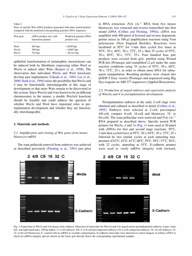
Timing of expression was then assessed from 2-cell to
blastocyst (Fig. 3). Wnt expression was monitored in
three or more mRNA samples per stage. Wnt4 was absent
in 2-cell embryos but the onset of expression was detected in
4/8-cell stages with strong expression in compact 8- and
16-cell embryos and in blastocysts. Wnt3a was detected in
2-cell embryos, less strongly in 4/8-cell stages, with robust
expression in compact 8-, 16-cell and early blastocysts. The
source of the Wnt 3a transcripts in 2-cell embryos may be
maternal or embryonic as the major maternal to zygotic
transcriptional transition occurs at the late 2-cell stage
(Schultz, 2002). Further analysis of oocyte mRNA com-
bined with studies of embryos undergoing mRNA synthesis
inhibition during the 2- to 4-cell transition would be
required to adequately differentiate between these
possibilities.
The demonstration that Wnt3a and Wnt4 transcripts are
detected just prior to the phase where the first epithelial cells
differentiate de novo suggests that Wnts have a role in
compaction and preimplantation development, although
clearly, we cannot exclude the possibilities that transcrip-
tion is occurring in the absence of translation (Kidder and
McLachlin, 1985; Sheth et al., 1997) or that embryo
isolation and culture might lead to differences in transcrip-
tion that do not occur in vivo (Natale et al., 2001). Indeed,
Fig. 1. Schematic diagram representing Wnt family member mRNAs
showing the relative positions of primers used in degenerate and specific
PCR reactions. The degenerate primers were based on the amino acid
codes, shown as single letters, and all possible combinations at positions of
redundancy in the genetic code were included in the degenerate
oligonucleotide mixtures. The specific Wnt3a and Wnt4 primers were
used in nested PCR reactions in combinations of primers 1 with 2 in the first
reaction and primers 3 with 4 in the second reaction. Sequences of these
primers are shown below.
Fig. 2. RT-mediated PCR on morula and blastocyst mRNA using consensus Wnt primers. TBE agarose gels (2%) showing DNA products obtained using
degenerate oligonucleotide mix based on consensus Wnt family sequences on morula (16) and blastocyst (32) cDNA in initial PCR reactions (left-hand gel)
and in PCR re-amplification of purified products (right-hand gel), marker lane (M) 100 bp ladder (Life Technologies).
Table 1
Identity and number of clones sequenced from degenerate PCR reactions
using consensus Wnt primers
Wnt Number of clones
3a 7
4 9
7a 6
10b 1
Miscellaneous non-Wnt 7
S. Lloyd et al. / Gene Expression Patterns 3 (2003) 309–312310
epithelial transformation of metanephric mesenchyme can
be induced both by fibroblasts expressing either Wnt4 or
Wnt3a or indeed other Wnts (Kispert et al., 1998). The
observation that individual Wnt3a and Wnt4 knockouts
develop past implantation (Takada et al., 1994; Lee et al.,
2000; Stark et al., 1994) raises the possibility that Wnt3a and
4 may be functionally interchangeable at this stage of
development or that more Wnts remain to be discovered in
this system. Since Wnts3a and 4 are known to be on different
chromosomes in the mouse, a double Wnt3a/4 knockout
should be feasible and could address the question of
whether Wnt3a and Wnt4 have important roles in pre-
implantation development and whether they are function-
ally interchangeable.
2. Materials and methods
2.1. Amplification and cloning of Wnt genes from mouse
blastocyst mRNA
The zona pellucida removal from embryos was achieved
as described previously (Fleming et al., 1991) just prior
to RNA extraction. Poly (A) þ RNA from five mouse
blastocysts was extracted and reverse transcribed into first
strand cDNA (Collins and Fleming, 1995a). cDNA was
amplified with 400 pmol of forward and reverse degenerate
primer mixes in 100 ml amplification reactions using Vent
polymerase (New England Biolabs). Reactions were
incubated at 958C for 3 min then cycled five times at
958C, 30 s; 468C, 30 s; 728C, 25 s, then 35 cycles of 958C,
30 s; 608C, 30 s; 728C, 25 s. Four hundred base pair
products were excised from gels, purified using Wizard
PCR kits (Promega) and reamplified (2 ml) under the same
reaction conditions using 35 cycles of 958C, 30 s; 608C,
30 s; 728C, 25 s, in order to obtain more DNA for subse-
quent manipulation. Resulting products were cloned into
pGEM T Easy vectors (Promega) and sequenced using Big
Dye reagents on ABI 377 sequencers (Applied Biosystems).
2.2. Production of staged embryos and expression analysis
of Wnts3a and 4 in preimplantation development
Preimplantation embryos at the early 2-cell stage were
obtained and cultured as described in detail (Collins et al.,
1995). Embryos were selected at 2-cell, precompact
4/8-cell, compact 8-cell, 16-cell and blastocyst, 32- to
64-cells. The zona pellucidae were removed and Poly (A) þ
RNA prepared as described above. Specific nested PCR
primers for Wnt3a, 4 and 7a (Fig. 1) were used at 50 pmol
with cDNAs for first and second stage reactions: 958C,
3 min then cycled twice at 958C, 30 s; 648C, 30 s; 728C, 25 s
followed by two similar cycles at each annealing tem-
perature of 638C, 628C, 618C, 608C, 598C, 588C, 578C, 568C,
with 22 cycles, annealing at 558C. E-cadherin primers
were used to verify mRNA integrity with forward,
Table 2
Sizes of specific Wnt cDNA products generated with inner nested primers
compared with the predicted corresponding genomic DNA sequences
Wnt gene cDNA product size with
innermost primers
Predicted genomic DNA
product size
Wnt4 401 bps .8438 bps
Wnt3a 389 bps ,43607 bps
Wnt7a 353 bps ,20,141 bps
Fig. 3. Expression of Wnt3a and 4 in mouse early embryos. Detection of transcripts for Wnts3a and 4 in staged mouse preimplantation embryos. Lanes: outer
left- and right-hand lanes, 100 bp ladder; 2, 2-cell embryos; 4/8, 4- to 8-cell precompacted embryos, C8, 8-cell compacted embryos; 16, 16-cell embryos; 32,
32- to 64-cell blastocysts; C, control with no mRNA to exclude contamination. E-cadherin transcripts were detected in control aliquots of embryo cDNA to
check for mRNA integrity and are shown on the lower gels directly below the corresponding experimental samples.
S. Lloyd et al. / Gene Expression Patterns 3 (2003) 309–312 311

CCATTTTCACGCGCGCTG and reverse CGCGAGCTT-
GAGATGGAT primers to give a 396 bp product.
Acknowledgements
This work was funded by the Medical Research Council,
UK and The Wellcome Trust. We thank Kate Hayes for
assistance in preparing the figures.
References
Batzoglou, S., Pachter, L., Mesirov, J.P., Berger, B., Lander, E.S., 2000.
Human and mouse gene structure: comparative analysis and application
to exon prediction. Genome Res. 10, 950–958.
Bergstein, I., Eisenberg, L.M., Bhalerao, J., Jenkins, N.A., Copeland, N.G.,
Osborne, M.P., Bowcock, A.M., Brown, A.M., 1997. Isolation of two
novel WNT genes, WNT14 and WNT15, one of which (WNT15) is
closely linked to WNT3 on human chromosome 17q21. Genomics 46,
450–458.
Brandon, C., Eisenberg, L.M., Eisenberg, C.A., 2000. WNT signaling
modulates the diversification of hematopoietic cells. Blood 96,
4132–4141.
Collins, J.E., Fleming, T.P., 1995a. Specific mRNA detection in single
lineage-marked blastomeres from preimplantation embryos. Trends
Genet. 11, 5–7.
Collins, J.E., Fleming, T.P., 1995b. Epithelial differentiation in the mouse
preimplantation embryo: making adhesive cell contacts for the first
time. Trends Biochem. Sci. 20, 307–312.
Collins, J.E., Lorimer, J.E., Garrod, D.R., Pidsley, S.C., Buxton, R.S.,
Fleming, T.P., 1995. Regulation of desmocollin transcription in mouse
preimplantation embryos. Development 121, 743–753.
Fleming, T.P., Garrod, D.R., Elsmore, A.J., 1991. Desmosome biogenesis
in the mouse preimplantation embryo. Development 112, 527–539.
Fleming, T.P., Sheth, B., Fesenko, I., 2001. Cell adhesion in the
preimplantation mammalian embryo and its role in trophectoderm
differentiation and blastocyst morphogenesis. Front. Biosci. 6,
D1000–D1007.
Gavin, B.J., McMahon, J.A., McMahon, A.P., 1990. Expression of multiple
novel Wnt-1/int-1-related genes during fetal and adult mouse develop-
ment. Genes Dev. 4, 2319–2332.
Graba, Y., Gieseler, K., Aragnol, D., Laurenti, P., Mariol, M.C., Berenger,
H., Sagnier, T., Pradel, J., 1995. DWnt-4, a novel Drosophila Wnt gene
acts downstream of homeotic complex genes in the visceral mesoderm.
Development 121, 209–218.
Hecht, A., Kemler, R., 2000. Curbing the nuclear activities of beta-catenin.
Control over Wnt target gene expression. EMBO Rep. 1, 24–28.
Huelsken, J., Birchmeier, W., 2001. New aspects of Wnt signaling
pathways in higher vertebrates. Curr. Opin. Genet. Dev. 11, 547–553.
Kidder, G.M., McLachlin, J.R., 1985. Timing of transcription and protein
synthesis underlying morphogenesis in preimplantation mouse
embryos. Dev. Biol. 112, 265–275.
Kispert, A., Vainio, S., McMahon, A.P., 1998. Wnt-4 is a mesenchymal
signal for epithelial transformation of metanephric mesenchyme in the
developing kidney. Development 125, 4225–4234.
Kuhl, M., Sheldahl, L.C., Park, M., Miller, J.R., Moon, R.T., 2000. The
Wnt/Ca2þ pathway: a new vertebrate Wnt signaling pathway takes
shape. Trends Genet. 16, 279–283.
Lee, S.M., Tole, S., Grove, E., McMahon, A.P., 2000. A local Wnt-3a
signal is required for development of the mammalian hippocampus.
Development 127, 457–467.
Miller, J.R., 2001. The Wnts. Genome Biol. 3: REVIEWS3001.1–3001.1S.
Moon, R.T., Brown, J.D., Yang-Snyder, J.A., Miller, J.R., 1997.
Structurally related receptors and antagonists compete for secreted
Wnt ligands. Cell 88, 725–728.
Natale, D.R., De Sousa, P.A., Westhusin, M.E., Watson, A.J., 2001.
Sensitivity of bovine blastocyst gene expression patterns to culture
environments assessed by differential display RT-PCR. Reproduction
122, 687–693.
Schultz, R.M., 2002. The molecular foundations of the maternal to zygotic
transition in the preimplantation embryo. Hum. Reprod. Update 8,
323–331.
Sheth, B., Fesenko, I., Collins, J.E., Moran, B., Wild, A.E., Anderson, J.M.,
Fleming, T.P., 1997. Tight junction assembly during mouse blastocyst
formation is regulated by late expression of ZO-1 alpha þ isoform.
Development 124, 2027–2037.
Sidow, A., 1992. Diversification of the Wnt gene family on the ancestral
lineage of vertebrates. Proc. Natl. Acad. Sci. USA 89, 5098–5102.
Stark, K., Vainio, S., Vassileva, G., McMahon, A.P., 1994. Epithelial
transformation of metanephric mesenchyme in the developing kidney
regulated by Wnt-4. Nature 372, 679–683.
Takada, S., Stark, K.L., Shea, M.J., Vassileva, G., McMahon, J.A.,
McMahon, A.P., 1994. Wnt-3a regulates somite and tailbud formation
in the mouse embryo. Genes Dev. 8, 174–189.
Wilson, S.I., Rydstrom, A., Trimborn, T., Willert, K., Nusse, R., Jessell,
T.M., Edlund, T., 2001. The status of Wnt signalling regulates neural
and epidermal fates in the chick embryo. Nature 411, 325–330.
Wodarz, A., Nusse, R., 1998. Mechanisms of Wnt signaling in develop-
ment. Annu. Rev. Cell Dev. Biol. 14, 59–88.
Yoshino, K., Rubin, J.S., Higinbotham, K.G., Uren, A., Anest, V., Plisov,
S.Y., Perantoni, A.O., 2001. Secreted Frizzled-related proteins can
regulate metanephric development. Mech. Dev. 102, 45–55.
S. Lloyd et al. / Gene Expression Patterns 3 (2003) 309–312312