Expression of Ifnlr1 on intestinal epithelial cells is ...
16
Washington University School of Medicine Digital Commons@Becker Open Access Publications 2017 Expression of Ifnlr1 on intestinal epithelial cells is critical to the antiviral effects of IFN-lambda against norovirus and reovirus Megan T. Baldridge Washington University School of Medicine in St. Louis Sanghyun Lee Washington University School of Medicine in St. Louis Judy J. Brown Vanderbilt University Medical Center Nicole McAllister University of Pisburgh School of Medicine Kelly Urbanek University of Pisburgh School of Medicine See next page for additional authors Follow this and additional works at: hps://digitalcommons.wustl.edu/open_access_pubs is Open Access Publication is brought to you for free and open access by Digital Commons@Becker. It has been accepted for inclusion in Open Access Publications by an authorized administrator of Digital Commons@Becker. For more information, please contact [email protected]. Recommended Citation Baldridge, Megan T.; Lee, Sanghyun; Brown, Judy J.; McAllister, Nicole; Urbanek, Kelly; Dermody, Terence S.; Nice, Timothy J.; and Virgin, Herbert W., ,"Expression of Ifnlr1 on intestinal epithelial cells is critical to the antiviral effects of IFN-lambda against norovirus and reovirus." Journal of Virology.91,7. e02079-16. (2017). hps://digitalcommons.wustl.edu/open_access_pubs/5500
Transcript of Expression of Ifnlr1 on intestinal epithelial cells is ...

Washington University School of MedicineDigital Commons@Becker
Open Access Publications
2017
Expression of Ifnlr1 on intestinal epithelial cells iscritical to the antiviral effects of IFN-lambda againstnorovirus and reovirusMegan T. BaldridgeWashington University School of Medicine in St. Louis
Sanghyun LeeWashington University School of Medicine in St. Louis
Judy J. BrownVanderbilt University Medical Center
Nicole McAllisterUniversity of Pittsburgh School of Medicine
Kelly UrbanekUniversity of Pittsburgh School of Medicine
See next page for additional authors
Follow this and additional works at: https://digitalcommons.wustl.edu/open_access_pubs
This Open Access Publication is brought to you for free and open access by Digital Commons@Becker. It has been accepted for inclusion in OpenAccess Publications by an authorized administrator of Digital Commons@Becker. For more information, please contact [email protected].
Recommended CitationBaldridge, Megan T.; Lee, Sanghyun; Brown, Judy J.; McAllister, Nicole; Urbanek, Kelly; Dermody, Terence S.; Nice, Timothy J.; andVirgin, Herbert W., ,"Expression of Ifnlr1 on intestinal epithelial cells is critical to the antiviral effects of IFN-lambda against norovirusand reovirus." Journal of Virology.91,7. e02079-16. (2017).https://digitalcommons.wustl.edu/open_access_pubs/5500

AuthorsMegan T. Baldridge, Sanghyun Lee, Judy J. Brown, Nicole McAllister, Kelly Urbanek, Terence S. Dermody,Timothy J. Nice, and Herbert W. Virgin
This open access publication is available at Digital Commons@Becker: https://digitalcommons.wustl.edu/open_access_pubs/5500

Expression of Ifnlr1 on IntestinalEpithelial Cells Is Critical to the AntiviralEffects of Interferon Lambda againstNorovirus and Reovirus
Megan T. Baldridge,a* Sanghyun Lee,a Judy J. Brown,b Nicole McAllister,c
Kelly Urbanek,d Terence S. Dermody,c,d Timothy J. Nice,e Herbert W. Virgina
Department of Pathology and Immunology, Washington University School of Medicine, St. Louis, Missouri,USAa; Department of Pathology, Microbiology, and Immunology, Vanderbilt University Medical Center,Nashville, Tennessee, USAb; Department of Microbiology and Molecular Genetics, University of PittsburghSchool of Medicine, Pittsburgh, Pennsylvania, USAc; Department of Pediatrics, University of Pittsburgh Schoolof Medicine, Pittsburgh, Pennsylvania, USAd; Department of Molecular Microbiology and Immunology, OregonHealth and Science University, Portland, Oregon, USAe
ABSTRACT Lambda interferon (IFN-�) has potent antiviral effects against multipleenteric viral pathogens, including norovirus and rotavirus, in both preventing andcuring infection. Because the intestine includes a diverse array of cell types, how-ever, the cell(s) upon which IFN-� acts to exert its antiviral effects is unclear. Here,we sought to identify IFN-�-responsive cells by generation of mice with lineage-specific deletion of the receptor for IFN-�, Ifnlr1. We found that expression of IFNLR1on intestinal epithelial cells (IECs) in the small intestine and colon is required for en-teric IFN-� antiviral activity. IEC Ifnlr1 expression also determines the efficacy ofIFN-� in resolving persistent murine norovirus (MNoV) infection and regulates fecalshedding and viral titers in tissue. Thus, the expression of Ifnlr1 by IECs is necessaryfor the response to both endogenous and exogenous IFN-�. We further demonstratethat IEC Ifnlr1 expression is required for the sterilizing innate immune effects ofIFN-� by extending these findings in Rag1-deficient mice. Finally, we assessedwhether our findings pertained to multiple viral pathogens by infecting mice spe-cifically lacking IEC Ifnlr1 expression with reovirus. These mice phenocopied Ifnlr1-null animals, exhibiting increased intestinal tissue titers and enhanced reovirusfecal shedding. Thus, IECs are the critical cell type responding to IFN-� to controlmultiple enteric viruses. This is the first genetic evidence that supports an essentialrole for IECs in IFN-�-mediated control of enteric viral infection, and these findingsprovide insight into the mechanism of IFN-�-mediated antiviral activity.
IMPORTANCE Human noroviruses (HNoVs) are the leading cause of epidemic gas-troenteritis worldwide. Type III interferons (IFN-�) control enteric viral infections inthe gut and have been shown to cure mouse norovirus, a small-animal model forHNoVs. Using a genetic approach with conditional knockout mice, we identified IECsas the dominant IFN-�-responsive cells in control of enteric virus infection in vivo.Upon murine norovirus or reovirus infection, Ifnlr1 depletion in IECs largely recapitu-lated the phenotype seen in Ifnlr1�/� mice of higher intestinal tissue viral titers andincreased viral shedding in the stool. Moreover, IFN-�-mediated sterilizing immunityagainst murine norovirus requires the capacity of IECs to respond to IFN-�. Thesefindings clarify the mechanism of action of this cytokine and emphasize the thera-peutic potential of IFN-� for treating mucosal viral infections.
KEYWORDS innate immunity, interferons, mucosal immunity, norovirus, reovirus
Received 27 October 2016 Accepted 6January 2017
Accepted manuscript posted online 11January 2017
Citation Baldridge MT, Lee S, Brown JJ,Mcallister N, Urbanek K, Dermody TS, Nice TJ,Virgin HW. 2017. Expression of Ifnlr1 onintestinal epithelial cells is critical to theantiviral effects of interferon lambda againstnorovirus and reovirus. J Virol 91:e02079-16.https://doi.org/10.1128/JVI.02079-16.
Editor Susana López, Instituto deBiotecnologia/UNAM
Copyright © 2017 American Society forMicrobiology. All Rights Reserved.
Address correspondence to Timothy J. Nice,[email protected], or Herbert W. Virgin,[email protected].
* Present address: Megan T. Baldridge,Department of Medicine, WashingtonUniversity School of Medicine, St. Louis,Missouri, USA.
M.T.B. and S.L. contributed equally to this work.
PATHOGENESIS AND IMMUNITY
crossm
April 2017 Volume 91 Issue 7 e02079-16 jvi.asm.org 1Journal of Virology
on May 16, 2017 by W
ashington University in S
t. Louishttp://jvi.asm
.org/D
ownloaded from

Norovirus and rotavirus are viral pathogens that infect at mucosal surfaces andinduce gastroenteritis, characterized by vomiting, diarrhea, and malaise (1, 2). Viral
gastroenteritis causes significant morbidity and mortality in children, the elderly, andimmunocompromised persons, thus representing a substantial health care burden (3,4). Treatments for these illnesses have been limited thus far to symptomatic care,including rehydration, because currently there is no specific antiviral therapy for theseviral pathogens. Lambda interferon (IFN-�; also called type III IFN) is an antiviralcytokine that regulates viral infection at mucosal surfaces and in the liver and brain(5–8). Administration of recombinant IFN-� can prevent and resolve viral infections inthe gastrointestinal tract (8, 9) and at other sites in mice (10). These effects are observedfor murine norovirus (MNoV) in mice lacking adaptive immunity, thus representingsterilizing innate immunity in the intestine (8). These studies indicate the potential forIFN-� as a therapeutic for viral infections, including those causing gastroenteritis, inhumans, including immunocompromised hosts (11). Better understanding of the mech-anisms by which this antiviral cytokine functions is essential to understanding basicmechanisms of intestinal control of viral infection and for potential therapeutic appli-cation in humans.
Binding of IFN-� to its receptor, a heterodimer of interleukin-10R2 (IL-10R2) andIFNLR1 (12, 13), induces an antiviral gene expression program similar to that inducedby type I IFN, with substantial overlap in gene sets in vitro (10, 14, 15). However, typeI and III IFNs exhibit unique antiviral properties in vivo. Ifnlr1�/� mice exhibit elevatedintestinal tissue replication and enhanced fecal shedding of a persistent strain of MNoV(8, 16), a model virus which allows for more tractable in vitro and in vivo analyses thanhuman norovirus (reviewed in references 17 and 18). Recombinant IFN-� treatment issufficient to prevent and cure MNoV infection (8). In contrast, mice deficient for Ifnar1(the receptor for type I IFNs) show enhanced extraintestinal spread of virus, but levelsof MNoV fecal shedding are comparable to those of wild-type mice (8, 16). Similarly,IFNLR1 restricts growth in the epithelium and fecal shedding of reovirus, while IFNAR1instead regulates reovirus growth in the lamina propria (19). IFN-� exhibits an antiviralrole exclusive of type I IFNs against a murine rotavirus strain (9) but cooperates withtype I IFNs to limit intestinal replication of a heterologous simian strain in neonatalbut not adult mice (20). These findings indicate the likely importance of tissuecompartment-, development-, and cell type-specific effects of type I and III IFNs in vivo.These effects may be secondary to unique virulence factors that counter specific IFNsor to differential expression of the IFN receptors (21, 22).
IFNAR1 is thought to be expressed ubiquitously and at especially high levels on cellsof hematopoietic origin (reviewed in references 23 and 24), whereas expression ofdetectable IFNLR1 appears to be limited to mucosal epithelial cells (25), humanhepatocytes (6), and neutrophils (26). Although IFNLR1 expression on peripheral leu-kocytes has also been reported, it does not appear to be functional (27). Upon IFN-�treatment, IFN-stimulated genes accumulate in intestinal epithelial cells (IECs), indicat-ing functional IFNLR1 expression (9, 19, 20). In contrast, in IECs of adult mice, IFNAR1may be expressed at lower levels or alternately trafficked, such as only to the apicalportion of the cell (9, 20). Differential receptor expression thus could account forcomplementary roles for different IFNs in protection against systemic infection (type I)and infection of mucosal (type III) sites. Importantly, however, it has been reported thatcells that do not express detectably high levels of IFNLR1, such as the endothelial cellsof the blood-brain barrier, may still respond to endogenous and exogenous IFN-� withprotective antiviral effects (10). Thus, to successfully identify the cell types required forthe antiviral response to IFN-�, analysis of receptor expression levels may be insuffi-cient, and definitive resolution requires a genetic approach to selectively delete recep-tor expression in specific cell types.
To identify the cell types that respond to IFN-� in vivo in the intestine, we generatedmice with a conditional mutant allele for Ifnlr1 and crossed them to mice expressing Crerecombinase via the action of different cell type-specific promoters (Table 1). Ifnlr1 wastargeted in cell types expected to express high receptor levels (intestinal epithelial cells
Baldridge et al. Journal of Virology
April 2017 Volume 91 Issue 7 e02079-16 jvi.asm.org 2
on May 16, 2017 by W
ashington University in S
t. Louishttp://jvi.asm
.org/D
ownloaded from

[25] and neutrophils [26]) and cells that are known to be permissive for MNoVreplication in tissue culture (macrophages and dendritic cells [35]). Of all the cell typestested, only intestinal epithelial cells (IECs) required expression of Ifnlr1 for the antiviraleffects of IFN-� against MNoV. To show the generality of our findings, we demonstratedthe importance of IEC expression of this receptor for control of reovirus infection. Thisis the first study to genetically define IFN-�-responsive cells in vivo in the context of twoindependent mucosal viral infections. This study also confirms that the cells required forresponding to endogenous IFN-� to attenuate MNoV infection are the same as thosethat respond to exogenous IFN-� administration, including in the elicitation of steril-izing innate immunity.
RESULTSIfnlr1 is expressed in the epithelial fraction along the length of the gastroin-
testinal tract. Tissue from adult mice homozygous for a null mutation in Ifnlr1 (28) orwild-type controls was collected from sites along the intestine, lung, mesenteric lymphnode (MLN), or spleen (Fig. 1A). The small intestine was also dissociated into epithelialand lamina propria fractions as previously described (36), and RNA was isolated from
TABLE 1 Mouse lines, nomenclature, and cell types targeted by specific Cre lines
Ifnlr1 and Cre mouse line(s) Line namea
Cell type(s) targeted(reference)
Ifnlr1tm1Palu; no Cre line Ifnlr1�/� All cells (28)Ifnlr1tm1a(EUCOMM)Wtsi; Villin-Cre Ifnlr1f/f-Villincre Intestinal epithelial cells (29)Ifnlr1tm1a(EUCOMM)Wtsi; MRP8-Cre Ifnlr1f/f-MRP8cre Neutrophils (30)Ifnlr1tm1a(EUCOMM)Wtsi; CD11c-Cre Ifnlr1f/f-CD11ccre Dendritic cells and alveolar
macrophages (31)Ifnlr1tm1a(EUCOMM)Wtsi; LysM-Cre Ifnlr1f/f-LysMcre Macrophages, neutrophils, some
dendritic cells (32, 33)Ifnlr1tm1a(EUCOMM)Wtsi; Deleter-Cre Ifnlr1f�/� All cells (34)aA conditional allele of Ifnlr1 (Ifnlr1f/f) was crossed to multiple different Cre lines for lineage-specific deletionof Ifnlr1 in the specific cell types.
**
Log1
0 Ifn
lr1 m
RN
A co
pies
/ug
RN
A(N
orm
aliz
ed to
Rps
29)
Stomach
Duodenum
Jejunum
Ileum
Cecum
Prox C
olon
Dist C
olonMLN
Lung
Spleen
1
2
3
4
5
6
****
**
****
**
**
****
Epitheli
um LP1
2
3
4
5
6
**
ns
Stomach
IleumJejunumDuodenum
Cecum
ProximalColon
DistalColon
A
B
Lung
MesentericLymph Node
Spleen
Epithelium
Laminapropria
Ifnlr1-/-Wild-type
FIG 1 Ifnlr1 is expressed in the epithelial fraction along the length of the intestine. (A) RNA was isolated from sites along theintestine and the lung, indicated by red boxes, whole mesenteric lymph node (MLN) and spleen, and epithelial and laminapropria (LP) fractions of the small intestine from Ifnlr1-sufficient and -deficient mice. (B) Ifnlr1 expression was quantified byquantitative real-time PCR of RNA from sites depicted in panel A. n � 4 to 6 samples per group, from two independentexperiments, analyzed by Mann-Whitney test. **, P � 0.01; ns, not significant. Prox, proximal; dist, distal.
Epithelial Cell Ifnlr1 Determines Antiviral Activity Journal of Virology
April 2017 Volume 91 Issue 7 e02079-16 jvi.asm.org 3
on May 16, 2017 by W
ashington University in S
t. Louishttp://jvi.asm
.org/D
ownloaded from
these fractions and tissues. Expression of Ifnlr1 was detected by quantitative real-timePCR of cDNA generated from these RNA samples. We found that Ifnlr1 was expressedalong the length of the intestine and in the lung, as well as in systemic tissues,including MLN and spleen (Fig. 1B). Intestinal Ifnlr1 expression was substantially en-riched (at least 30-fold; P � 0.0381) in the epithelial fraction compared to the laminapropria fraction (Fig. 1B), consistent with previous reports (9, 25). As expected, notranscript was detected in any tissue in Ifnlr1�/� mice (Fig. 1B).
Ifnlr1 expression in the small and large intestine is significantly diminished inIfnlr1f/f-Villincre mice. Embryonic stem (ES) cells targeted with a construct containingsequences homologous to Ifnlr1, an FLP recombinant target (FRT)-flanked lacZ andneomycin cassette, and loxP sites flanking exon 2 were provided by the Wellcome TrustSanger Institute (Fig. 2A). Mice derived from these ES cells were crossed with miceexpressing Flp recombinase for deletion of the FRT-flanked cassette (38), leaving aconditional allele of Ifnlr1, referred to as Ifnlr1f (Fig. 2A). Following removal of the floxedregion in cells expressing Cre, the resulting transcript is predicted to produce atruncated protein product (Fig. 2B). For disruption in specific cell lineages, Ifnlr1f/f micewere crossed with various Cre mouse lines (Table 1). For each line, Cre(�) Ifnlr1f/f micewere compared to Cre(�) Ifnlr1f/f littermates to assess the effects of cell type-specificdeletion upon Ifnlr1 expression along the intestine and in extraintestinal tissues (Fig.1A). Ifnlr1f/f-Villincre mice showed significantly diminished Ifnlr1 expression in the smalland large intestine (Fig. 2C). Fractionation of the small intestine into epithelial andlamina propria fractions revealed efficient deletion of Ifnlr1 in the epithelium of thesemice (Fig. 2C). In contrast, Ifnlr1f/f-MRP8cre, Ifnlr1f/f-LysMcre, and Ifnlr1f/f-CD11ccre miceshowed no alterations in intestinal Ifnlr1 expression at the level of the whole tissuestested (Fig. 2D, E, and F). Ifnlr1f/f-MRP8cre, Ifnlr1f/f-LysMcre, and Ifnlr1f/f-CD11ccre micedid exhibit substantial depletion of Ifnlr1 in their respectively targeted cell types ofneutrophils (�85%), macrophages (�91%), and dendritic cells (�85%), consistent witha previous report (39) (Fig. 2D, E, and F). Expression of Ifnlr1 remained unchanged inlung, MLN, spleen, stomach, and duodenum in Ifnlr1f/f-Villincre mice, indicating expres-sion of Cre specific to distal small intestine and colon (Fig. 2C), consistent with previousreports (29, 40).
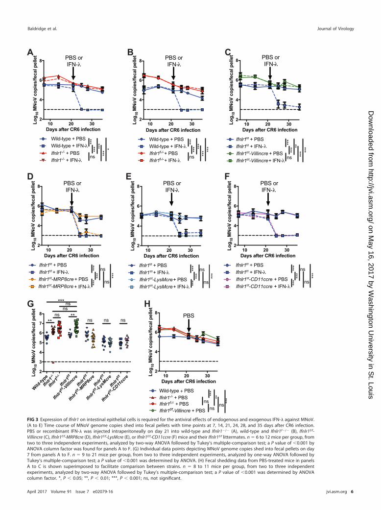
Expression of Ifnlr1 in intestinal epithelium regulates MNoV shedding andresponse to recombinant IFN-�. Ifnlr1�/� mice and wild-type controls were inocu-lated with CR6, a persistent strain of MNoV that replicates well in the intestine, is shedinto the feces at readily detectable levels, and is sensitive to treatment with IFN-� (8,41). As described previously (8), Ifnlr1�/� mice allow higher levels of fecal MNoVshedding than do wild-type mice at early time points (Fig. 3A and G) and are insensitiveto IFN-� treatment, although this treatment terminates MNoV replication in wild-typemice (Fig. 3A). These results were also observed in a novel Ifnlr1-deficient mouse model(Ifnlr1f�/�) (Table 1 and Fig. 3B). This assay was next applied to the four mouse strainswith lineage-specific deletion of Ifnlr1 (Table 1). Ifnlr1f/f-Villincre mice phenocopiedIfnlr1�/� and Ifnlr1f�/� mice, exhibiting both elevated fecal shedding of MNoV andresistance to IFN-� treatment (Fig. 3C). In contrast, Ifnlr1f/f-MRP8cre, Ifnlr1f/f-LysMcre, andIfnlr1f/f-CD11ccre mice exhibited viral loads and response to IFN-� equivalent to thoseof Ifnlr1f/f controls (Fig. 3D, E, and F). At day 7 postinoculation, IFNLR1 regulated fecalshedding of MNoV, as seen by comparing wild-type and Ifnlr1�/� levels (Fig. 3G).Ifnlr1f/f-Villincre mice allowed fecal shedding equivalent to Ifnlr1�/� and Ifnlr1f�/� mice,suggesting that control of MNoV fecal shedding can be fully accounted for by IFNLR1in Villin-expressing cells (Fig. 3G). Similarly, Ifnlr1f/f-Villincre mice exhibited no differencein comparison to Ifnlr1�/� and Ifnlr1f�/� mice along the full time course of infection(Fig. 3H).
Expression of Ifnlr1 in intestinal epithelium is essential for induction of IFN-�-mediated sterilizing innate immunity to MNoV infection. We previously reportedthat recombinant IFN-� can cure persistently infected mice in the absence of adaptiveimmunity (8). To determine whether expression of Ifnlr1 in IECs is required for IFN-�-mediated sterilizing innate immunity to persistent MNoV infection, we established
Baldridge et al. Journal of Virology
April 2017 Volume 91 Issue 7 e02079-16 jvi.asm.org 4
on May 16, 2017 by W
ashington University in S
t. Louishttp://jvi.asm
.org/D
ownloaded from

Dendrit
ic ce
lls0
2 103
4 103
6 103
8 103
Ifnlr1
mR
NA
copi
es/u
g R
NA
(Nor
mal
ized
to R
ps29
) *
Log1
0 Ifn
lr1 m
RN
A co
pies
/ug
RN
A(N
orm
aliz
ed to
Rps
29)
Stomach
Duodenum
Jejunum
Ileum
Cecum
Prox C
olon
Dist C
olonMLN
Lung
Spleen
1
2
3
4
5
6 ns
ns** **
ns**
**
ns
nsns
Epitheli
um LP1
2
3
4
5
6
**
ns
Log1
0 Ifn
lr1 m
RN
A co
pies
/ug
RN
A(N
orm
aliz
ed to
Rps
29)
Stomach
Duodenum
Jejunum
Ileum
Cecum
Prox C
olon
Dist C
olonMLN
Lung
Spleen
1
2
3
4
5
6 ns
nsns ns
nsns ns
ns
nsns
Epitheli
um LP1
2
3
4
5
6
ns
ns
Log1
0 Ifn
lr1 m
RN
A co
pies
/ug
RN
A(N
orm
aliz
ed to
Rps
29)
Stomach
Duodenum
Jejunum
Ileum
Cecum
Prox C
olon
Dist C
olonMLN
Lung
Spleen
1
2
3
4
5
6ns
nsns ns
nsns ns
ns
nsns
Epitheli
um LP1
2
3
4
5
6
ns
ns
Log1
0 Ifn
lr1 m
RN
A co
pies
/ug
RN
A(N
orm
aliz
ed to
Rps
29)
Stomach
Duodenum
Jejunum
Ileum
Cecum
Prox C
olon
Dist C
olonMLN
Lung
Spleen
1
2
3
4
5
6ns
nsns ns
nsns ns
ns
nsns
Epitheli
um LP1
2
3
4
5
6
ns
ns
A
B
lacZ neo
FRT FRTloxP loxP loxP
Exon1
Exon2
Exon3
FRT loxP loxP
Exon1
Exon2
Exon3
+ Flprecombinase
MWRADRWAPLLLFLLQSALGRPRLAMWRADRWAPLLLFLLQSALA I SKPV
PPR NVTLFSQNFT VYLTGDQWSIVQVSRLWCVP*
1 10 20 30 40
Cre(-)Cre(+)
FRT loxP
Exon1
Exon3
+ Crerecombinase
C
Ifnlr1f/f
Ifnlr1f/f-Villincre
Ifnlr1f/f
Ifnlr1f/f-MRP8cre
Ifnlr1f/f
Ifnlr1f/f-LysMcre
Ifnlr1f/f
Ifnlr1f/f-CD11ccre
D
E
F
Neutro
phils0
2 105
4 105
6 105
8 105
Ifnlr1
mR
NA
copi
es/u
g R
NA
(Nor
mal
ized
to R
ps29
)
**
Macro
phages
0
1 103
2 103
3 103
Ifnlr1
mR
NA
copi
es/u
g R
NA
(Nor
mal
ized
to R
ps29
)
*
FIG 2 Ifnlr1 expression is decreased in the small and large intestines of Ifnlr1f/f-Villincre mice. (A) Schematic depicting the Ifnlr1 gene locusin Ifnlr1tm1a(EUCOMM)Wtsi mice. After crossing with mice expressing Flp recombinase (�Flp recombinase), the region between the two FRT siteswas deleted, leaving conditional-ready Ifnlr1f/f mice. In the absence of Cre, all exons are present. With the addition of Cre recombinase, thefloxed exon 2 is deleted. (B) In the absence of Cre [Cre(�)], the IFNLR1 protein is expressed. In the presence of Cre [Cre(�)], the proteinsequence is altered at amino acid 20 and a premature stop codon is introduced at amino acid 42. (C to F) Ifnlr1 expression was assessedby quantitative real-time PCR of sites along the intestine and the lung, MLN and spleen, and epithelial and LP fractions from Ifnlr1f/f-Villincre(C), Ifnlr1f/f-MRP8cre (D), Ifnlr1f/f-LysMcre (E), and Ifnlr1f/f-CD11ccre (F) mice compared to their Ifnlr1f/f littermates. Ifnlr1 expression was alsoassessed by quantitative real-time PCR of isolated bone marrow neutrophils from Ifnlr1f/f-MRP8cre (D), splenic macrophages from Ifnlr1f/f-LysMcre (E), and splenic dendritic cells from Ifnlr1f/f-CD11ccre (F) mice compared to their Ifnlr1f/f littermates. n � 4 to 7 samples per group,from two independent experiments, analyzed by Mann-Whitney test. *, P � 0.05; **, P � 0.01; ns, not significant.
Epithelial Cell Ifnlr1 Determines Antiviral Activity Journal of Virology
April 2017 Volume 91 Issue 7 e02079-16 jvi.asm.org 5
on May 16, 2017 by W
ashington University in S
t. Louishttp://jvi.asm
.org/D
ownloaded from
10 20 302
4
6
8
Days after CR6 infection
Log 10
MN
oV c
opie
s/fe
cal p
elle
t
Ifnlr1f/f + PBS
Ifnlr1f/f + IFN-
Ifnlr1f/f-CD11ccre + PBS
Ifnlr1f/f-CD11ccre + IFN-
PBS or IFN-
ns
******
***
***
ns
10 20 302
4
6
8
Days after CR6 infection
Log 10
MN
oV c
opie
s/fe
cal p
elle
t
Ifnlr1f/f + PBS
Ifnlr1 f/f + IFN-
Ifnlr1f/f-LysMcre+ PBS
Ifnlr1f/f-LysMcre+ IFN-
PBS or IFN-
ns
******
***
***
ns
10 20 302
4
6
8
Days after CR6 infection
Log 10
MN
oV c
opie
s/fe
cal p
elle
t
Ifnlr1f/f + PBS
Ifnlr1f/f + IFN-
Ifnlr1f/f-MRP8cre + PBS
PBS or IFN-
ns***
******
***ns
Ifnlr1f/f-MRP8cre + IFN-
10 20 302
4
6
8
Days after CR6 infection
Log 10
MN
oV c
opie
s/fe
cal p
elle
t
Ifnlr1f/f + PBS
Ifnlr1f/f + IFN-
Ifnlr1f/f-Villincre + PBS
Ifnlr1f/f-Villincre+ IFN-
PBS or IFN-
ns
******
*****
***
D E F
10 20 302
4
6
8
Days after CR6 infectionLo
g 10 M
NoV
cop
ies/
feca
l pel
let
Wild-type + PBS
Wild-type + IFN-
Ifnlr1f-/- + PBS
Ifnlr1f-/- + IFN-
PBS or IFN-
ns
******
******
***
10 20 302
4
6
8
Days after CR6 infection
Log 10
MN
oV c
opie
s/fe
cal p
elle
t
Wild-type + PBS
Wild-type + IFN-
Ifnlr1-/- + PBS
Ifnlr1-/- + IFN-
PBS or IFN-
ns
******
******
*
A B C
Wild-ty
pe
Ifnlr1
-/-
Ifnlr1
f-/-
Ifnlr1
f/f
Ifnlr1
f/f -Villincre
Ifnlr1
f/f
Ifnlr1
f/f -MRP8c
re
Ifnlr1
f/f
Ifnlr1
f/f -LysMcre
Ifnlr1
f/f
Ifnlr1
f/f -CD11
ccre
2
3
4
5
6
7
8
Log 10
MN
oV c
opie
s/fe
cal p
elle
t
**
***
**nsns ns ns
nsns
10 20 302
4
6
8
Days after CR6 infection
Log 10
MN
oV c
opie
s/fe
cal p
elle
t
Ifnlr1f/f-Villincre + PBS
Wild-type + PBS
PBS
ns
*** ** ***Ifnlr1-/- + PBSIfnlr1f-/- + PBS ns
ns
G H
FIG 3 Expression of Ifnlr1 on intestinal epithelial cells is required for the antiviral effects of endogenous and exogenous IFN-� against MNoV.(A to E) Time course of MNoV genome copies shed into fecal pellets with time points at 7, 14, 21, 24, 28, and 35 days after CR6 infection.PBS or recombinant IFN-� was injected intraperitoneally on day 21 into wild-type and Ifnlr1�/� (A), wild-type and Ifnlr1f�/� (B), Ifnlr1f/f-Villincre (C), Ifnlr1f/f-MRP8cre (D), Ifnlr1f/f-LysMcre (E), or Ifnlr1f/f-CD11ccre (F) mice and their Ifnlr1f/f littermates. n � 6 to 12 mice per group, fromtwo to three independent experiments, analyzed by two-way ANOVA followed by Tukey’s multiple-comparison test; a P value of �0.001 byANOVA column factor was found for panels A to F. (G) Individual data points depicting MNoV genome copies shed into fecal pellets on day7 from panels A to F. n � 9 to 21 mice per group, from two to three independent experiments, analyzed by one-way ANOVA followed byTukey’s multiple-comparison test; a P value of �0.001 was determined by ANOVA. (H) Fecal shedding data from PBS-treated mice in panelsA to C is shown superimposed to facilitate comparison between strains. n � 8 to 11 mice per group, from two to three independentexperiments, analyzed by two-way ANOVA followed by Tukey’s multiple-comparison test; a P value of �0.001 was determined by ANOVAcolumn factor. *, P � 0.05; **, P � 0.01; ***, P � 0.001; ns, not significant.
Baldridge et al. Journal of Virology
April 2017 Volume 91 Issue 7 e02079-16 jvi.asm.org 6
on May 16, 2017 by W
ashington University in S
t. Louishttp://jvi.asm
.org/D
ownloaded from

Rag1�/� Ifnlr1f/f-Villincre conditional double knockout mice. Rag1�/� Ifnlr1f/f-Villincremice were orally inoculated with CR6, and viral shedding in the stool was quantified byquantitative PCR (qPCR). Rag1�/� Ifnlr1f/f-Villincre mice showed increased viral shed-ding throughout the infection time course (Fig. 4A). Injection of recombinant IFN-�terminated MNoV replication in Rag1�/� Ifnlr1f/f mice but did not affect MNoV loads inRag1�/� Ifnlr1f/f-Villincre mice (Fig. 4A). At 7 days postinoculation, Rag1�/� Ifnlr1f/f-Villincre mice had significantly higher viral shedding than Rag1�/� Ifnlr1f/f mice, and thelevel of viral shedding in Rag1�/� Ifnlr1f/f-Villincre mice was comparable to the level ofviral shedding in Rag1�/� Ifnlr1�/� mice (Fig. 4B). Therefore, IFN-� responses in IECslimited persistent MNoV infection in the absence of adaptive immunity, and IFN-�signaling in IECs was essential for clearance of persistently infected MNoV by IFN-�-mediated sterilizing innate immunity.
Control of reovirus in intestinal tissue by IFN-� depends upon the expressionof Ifnlr1 in epithelial cells. To assess whether Ifnlr1 expression on IECs was requiredfor control of other enteric pathogens, Ifnlr1�/� and Ifnlr1f/f-Villincre mice were orallyinoculated with 108 PFU of reovirus strain type 1 Lang (T1L). At 4 days postinfection,viral titers in small intestinal tissues, including duodenum, jejunum, and ileum, as wellas viral shedding in stools, were significantly higher in Ifnlr1�/� mice (Fig. 5A and B),consistent with a previous report using another strain of reovirus, type 3 Dearing (19).Control of reovirus was predominantly through the expression of IFNLR1 on IECs, asIfnlr1f/f-Villincre mice displayed increased titers of reovirus in small intestinal tissues aswell as enhanced fecal shedding (Fig. 5A and B). These results demonstrate thatexpression of IFNLR1 in epithelial cells is essential for the control of reovirus infectionby IFN-� in the gut and indicate that IFN-� signaling in IECs is an antiviral mechanismcommon to multiple enteric viral pathogens.
Interferon-stimulated gene expression in the intestine depends upon theexpression of Ifnlr1 in epithelial cells. Ileum and proximal colon tissues were isolatedfrom wild-type (WT), Ifnlr1�/�, Ifnlr1f/f, and Ifnlr1f/f-Villincre mice 1 day posttreatmentwith either PBS or IFN-�. These tissues were then assessed for expression of canonicalantiviral interferon-stimulated genes (ISGs) Oas1a (42), Ifit1 (43), and Ifi44 (44) (Fig. 6A
10 20 302
4
6
8
Days after CR6 infection
Log 10
MN
oV c
opie
s/fe
cal p
elle
tPBS or IFN-
Rag1-/-Ifnlr1f/f + PBSRag1-/-Ifnlr1f/f + IFN-Rag1-/-Ifnlr1f/f-Villincre + PBSRag1-/-Ifnlr1f/f-Villincre + IFN- ns
******
******
***
A B
Rag1-/-
Rag1-/- Ifn
lr1-/-
Rag1-/- Ifn
lr1f/f
Rag1-/- Ifn
lr1f/f -Villi
ncre2
3
4
5
6
7
8
Log 10
MN
oV c
opie
s/fe
cal p
elle
t
******
FIG 4 Expression of Ifnlr1 on intestinal epithelial cells is required for the antiviral effects of IFN-� againstMNoV in the absence of adaptive immunity. (A) Time course of MNoV genome copies shed into fecalpellets with time points at 7, 14, 21, 24, 28, and 35 days after CR6 infection. PBS or recombinant IFN-�was injected intraperitoneally on day 21 and day 23 into Rag1�/� Ifnlr1f/f-Villincre or Rag1�/� Ifnlr1f/f mice.n � 6 to 14 mice per group, combined from three independent experiments, analyzed by two-wayANOVA followed by Tukey’s multiple-comparison test; a P value of �0.001 was found by ANOVA columnfactor. (B) Individual data points depicting MNoV genome copies shed into fecal pellets on day 7 fromRag1�/�, Rag1�/� Ifnlr1�/� double knockouts or mice depicted in panel A. n � 6 to 22 mice per group,combined from two to three independent experiments, analyzed by Mann-Whitney test. ***, P � 0.001;ns, not significant.
Epithelial Cell Ifnlr1 Determines Antiviral Activity Journal of Virology
April 2017 Volume 91 Issue 7 e02079-16 jvi.asm.org 7
on May 16, 2017 by W
ashington University in S
t. Louishttp://jvi.asm
.org/D
ownloaded from

to C). While intestinal tissues from WT and Ifnlr1f/f mice exhibited robust ISG inductionin response to IFN-� treatment, tissues from Ifnlr1�/� and Ifnlr1f/f-Villincre mice failed tosignificantly upregulate these ISGs in response to IFN-�. These data correlate with theimpaired antiviral response against MNoV in Ifnlr1�/� and Ifnlr1f/f-Villincre mice afterIFN-� treatment (Fig. 3A and C), consistent with a potentially critical role for IFNLR1expression on epithelial cells for induction of antiviral ISGs in response to IFN-�treatment.
DISCUSSION
In this study, we found that IECs are the predominant cell type expressing Ifnlr1 inthe small intestine and colon and that this cell type plays a major role in IFN-�-mediated antiviral immunity in the intestine. Antiviral immunity elicited by IFN-� toenteric reovirus and norovirus infection depends upon IFNLR1 signaling in Villin-positive IECs. Using four mouse strains with lineage-specific deletion of Ifnlr1 to studypersistent infection and IFN-�-mediated clearance, we found that only Ifnlr1f/f-Villincremice exhibited a complete phenocopy of Ifnlr1�/� mice. Targeting Ifnlr1 in other cells,including dendritic cells, macrophages, and neutrophils, had no detectable effect onbasal levels of viral shedding or IFN-�-mediated clearance of MNoV. The dominantIFN-�-dependent antiviral contribution by IECs also was confirmed with reovirus infec-tion. In studies using reovirus T1L, we observed viral titers in the small intestine ofIfnlr1f/f-Villincre mice increased comparably to those detected in Ifnlr1�/� mice, al-though we cannot rule out a minor role for other IFN-�-responding cells in the ileum.Therefore, IECs are the functionally dominant IFN-�-responding cells for endogenousand exogenous IFN-� control of viruses in the intestine. There is a clear correlationbetween IFN-�-mediated induction of antiviral ISGs and IEC expression of IFNLR1,suggesting that induction of ISGs in IECs is the mechanism by which IFN-� exerts itsantiviral effects.
Expression of Ifnlr1 mRNA throughout the gut and in other extraintestinal tissues(MLN, lung, and spleen) was quantified by qPCR analysis. In lamina propria cells, therewere fewer than 500 copies of Ifnlr1 mRNA per 1 �g total RNA. In contrast, IECs express
A B
Wild-ty
pe
Ifnlr1
-/-
Ifnlr1
f/f
Ifnlr1
f/f -Villincre
Wild-ty
pe
Ifnlr1
-/-
Ifnlr1
f/f
Ifnlr1
f/f -Villincre
Wild-ty
pe
Ifnlr1
-/-
Ifnlr1
f/f
Ifnlr1
f/f -Villincre
4
5
6
7
Reo
viru
s vi
ral t
iters
(L
og10
PFU
)
******
*****
******
******
******
***
***
nsns ns
nsns
**Duodenum Jejunum Ileum Stool
Wild-ty
pe
Ifnlr1
-/-
Ifnlr1
f/f
Ifnlr1
f/f -Villincre
4
5
6
7
Reo
viru
s vi
ral t
iters
(L
og10
PFU
)
*****
*** *
nsns
FIG 5 Ifnlr1 expression on intestinal epithelial cells limits reovirus infection. (A and B) Titers of reovirus strain T1Lwere assessed at day 4 postinoculation in the different compartments of the small intestine (A) and stool (B) fromwild-type, Ifnlr1�/�, Ifnlr1f/f-Villincre, and Ifnlr1f/f littermate control mice. The small intestine was resected from thepylorus to the cecum and sectioned into three equal parts, representing the duodenum, jejunum, and ileum. Titersare expressed as PFU per milliliter of tissue homogenate or gram of stool. n � 6 to 8 mice per group, combinedfrom two independent experiments, analyzed by one-way ANOVA followed by Tukey’s multiple-comparison test;a P value of �0.001 was determined by ANOVA column factor for all tissues and stool. *, P � 0.05; **, P � 0.01;***, P � 0.001; ns, not significant.
Baldridge et al. Journal of Virology
April 2017 Volume 91 Issue 7 e02079-16 jvi.asm.org 8
on May 16, 2017 by W
ashington University in S
t. Louishttp://jvi.asm
.org/D
ownloaded from

WT
Ifnlr1
-/-
Ifnlr1
f/f
Ifnlr1
f/f -Villincre WT
Ifnlr1
-/-
Ifnlr1
f/f
Ifnlr1
f/f -Villincre
0
1 106
2 106
3 106
4 106
Oas
1a m
RN
A co
pies
/ug
RN
A(N
orm
aliz
ed to
Rps
29)
Oas1aIleum Prox Colon
***
***
ns ns
*** ***
nsns
- + - + - + - + - + - + - + - +IFN-
WT
Ifnlr1
-/-
Ifnlr1
f/f
Ifnlr1
f/f -Villincre WT
Ifnlr1
-/-
Ifnlr1
f/f
Ifnlr1
f/f -Villincre
0
5 106
1 107
Ifit1
mR
NA
copi
es/u
g R
NA
(Nor
mal
ized
to R
ps29
)
Ifit1
***
***
ns ns
***
***
ns ns
Ileum Prox Colon
- + - + - + - + - + - + - + - +IFN-
WT
Ifnlr1
-/-
Ifnlr1
f/f
Ifnlr1
f/f -Villincre WT
Ifnlr1
-/-
Ifnlr1
f/f
Ifnlr1
f/f -Villincre
0
1 106
2 106
3 106
Ifi44
mR
NA
copi
es/u
g R
NA
(Nor
mal
ized
to R
ps29
)
Ifi44
***
***
ns ns
*** ***
ns ns
Ileum Prox Colon
- + - + - + - + - + - + - + - +IFN-
A
B
C
FIG 6 Ifnlr1 expression on intestinal epithelial cells is necessary for induction of interferon-stimulatedgenes. Oas1a (A), Ifit1 (B), and Ifi44 (C) expression was assessed by quantitative real-time PCR of RNA fromdistal ileum and proximal colon tissue from wild-type (WT), Ifnlr1�/�, Ifnlr1f/f, and Ifnlr1f/f-Villincre mice at1 day posttreatment with PBS or recombinant IFN-�. n � 5 to 9 mice per group, combined from twoindependent experiments, analyzed by one-way ANOVA followed by Tukey’s multiple-comparison test;a P value of �0.001 was determined by ANOVA column factor for all tissues. ***, P � 0.001; ns, notsignificant.
Epithelial Cell Ifnlr1 Determines Antiviral Activity Journal of Virology
April 2017 Volume 91 Issue 7 e02079-16 jvi.asm.org 9
on May 16, 2017 by W
ashington University in S
t. Louishttp://jvi.asm
.org/D
ownloaded from

more than 10,000 copies of Ifnlr1 mRNA per 1 �g total RNA throughout the smallintestine. Thus, IECs are the dominant Ifnlr1-expressing cells and function as the majorIFN-�-responding cells for antiviral immunity in the intestine. In MLN, spleen, and lung,we detected comparable expression of Ifnlr1 mRNA. Villin-positive cells were not themajor cell type responsible for Ifnlr1 expression in these tissues, and neither wereneutrophils, dendritic cells, or macrophages. Therefore, there may exist some other celltypes that are important for IFN-� responses in these tissues. Lung epithelial cells,which do not express Villin, likely reflect a major source of Ifnlr1 in that tissue (29). B orT cells, which have been reported to express Ifnlr1 even though they lack a robustresponse to IFN-�, may account for Ifnlr1 expression in the MLN and spleen (27).Another possible cellular source for this expression is endothelial cells, based on thereport that blood-brain barrier endothelial cells respond to IFN-� to restrict West Nilevirus neuroinvasion (10). Thus, it would be interesting to study the role of IFN-� inextraintestinal tissues in control of other pathogens and define the IFN-�-responsivecell types in these contexts.
In some tissues, such as lung and vagina, there is redundancy between type I andIII IFN-mediated antiviral responses. IFN-� controls influenza virus, severe acute respi-ratory syndrome (SARS) coronavirus, respiratory syncytial virus infection in the lung(45–47), and herpes simplex virus 2 (HSV-2) infection in the genital tract (48), redun-dantly with type I IFNs. In the intestine, however, IFN-�-mediated antiviral immunitydoes not redundantly overlap type I IFNs (8, 9, 19). Adult IECs have polarized apicalIFNAR1 expression only at low levels (9), and although IECs in neonates exhibit robustSTAT1 activation after type I IFN treatment, in adult mice they are largely unresponsiveto type I IFN treatment in vivo (9, 19, 20). Moreover, the expression level of Ifnlr1 mRNAis highly enriched in IECs but minimally detectable in other compartments of intestinaltissue (Fig. 1 and 2). This study, bolstering previous findings of alternate cellularexpression patterns for type I and III IFN receptors, helps explain why IFN-�-mediatedimmunity in the intestine is nonredundant with IFN-�/� in adult mice, even thoughthey may stimulate transcription of highly overlapping sets of antiviral genes (7, 20).Our data support a role for IECs as sentinels for enteric virus infection via their responseto compartment-specific IFN-� signaling (19, 20).
One of the important features of IFN-�-mediated immunity is its sterilizing activityagainst persistent MNoV infection in the absence of adaptive immunity (8). We ob-served that persistent MNoV infection of Rag1�/� Ifnlr1f/f-Villincre mice was not re-solved by IFN-� treatment and showed increased viral titers in the stool, similar to ourobservations with Rag1�/� Ifnlr1�/� mice. Thus, IFN-�-mediated sterilizing innateimmunity requires IEC expression of the receptor. Since only macrophages, dendriticcells, and B cells are known to be susceptible to MNoV infection in vitro (35, 49), it is notclear how the IFN-� response in IECs ablates persistent MNoV infection in the absenceof adaptive immunity. One possible explanation is that there is a secondary trans-actingmolecule induced by IFN-� in IECs that clears MNoV in other cell types. A related studyhas demonstrated that rotavirus can be terminated by injecting IL-22 and IL-18 intoRag1�/� mice, but this IL-22- and IL-18-mediated viral clearance does not induce IFN-�or Stat1 activation (50). Thus, there may be multiple innate immunological mechanismsto resolve persistent viral infection in the absence of adaptive immunity. Identifying theeffectors of IFN-�-mediated sterilizing immunity is an important area to pursue in IFN-�immunology.
This study reveals that Ifnlr1 expression in IECs is required for control of entericMNoV and reovirus infections. Using a genetic approach with conditional knockoutmice, we identified IECs as the dominant cell type that responds to endogenous andexogenous IFN-� to control enteric viruses. Understanding the identity of IFN-�-responsive cell types provides further insight into mechanisms that control entericviruses and will enhance future development of IFN-�-mediated antiviral therapeu-tics.
Baldridge et al. Journal of Virology
April 2017 Volume 91 Issue 7 e02079-16 jvi.asm.org 10
on May 16, 2017 by W
ashington University in S
t. Louishttp://jvi.asm
.org/D
ownloaded from

MATERIALS AND METHODSGeneration of MNoV stocks and determination of titers. Stocks of MNoV strain CR6 were
generated from a molecular clone as previously described (51). Briefly, a plasmid encoding the CR6genome was transfected into 293T cells to generate infectious virus, which was subsequently passagedon BV2 cells. After two passages, BV2 cultures were frozen and thawed to liberate virions. Cultures thenwere cleared of cellular debris and virus was concentrated by ultracentrifugation through a 30% sucrosecushion. Titers of virus stocks were determined by plaque assay on BV2 cells (51).
Generation of reovirus stocks and determination of titers. Spinner-adapted murine L929 (L) cellswere grown in either suspension or monolayer cultures in Joklik’s modified Eagle’s minimal essentialmedium (SMEM; Lonza) supplemented to contain 5% fetal bovine serum (Gibco), 2 mM L-glutamine, 100U/ml penicillin, 100 �g/ml streptomycin (Gibco), and 25 ng/ml amphotericin B (Sigma). BHK-T7 cells weregrown in Dulbecco’s modified Eagle’s minimal essential medium (DMEM; Gibco) supplemented tocontain 5% fetal bovine serum, 2 mM L-glutamine, 1 mg/ml Geneticin (Gibco), and nonessential aminoacids (Sigma).
Recombinant reoviruses were generated using plasmid-based reverse genetics (52). Recombinantstrain type 1 Lang (T1L) is a stock generated by plasmid-based rescue from cloned T1L cDNAs (53). After3 to 5 days of incubation, cells were frozen and thawed three times, and virus was isolated by plaquepurification using monolayers of L cells (54). Purified reovirus virions were generated from second- orthird-passage L-cell lysate stocks (55). Viral particles were extracted from infected cell lysates usingVertrel XF (Dupont), layered onto 1.2- to 1.4-g/cm3 CsCl gradients, and centrifuged at 62,000 � g for 16h. Bands corresponding to virions (1.36 g/cm3) were collected and dialyzed in virion storage buffer (150mM NaCl, 15 mM MgCl2, and 10 mM Tris-HCl [pH 7.4]) (56). The concentration of reovirus virions inpurified preparations was determined from the following equivalence: one optical density (OD) unit at260 nm equals 2.1 � 1012 virions (56). Viral titer was determined by plaque assay using L cells (54).
For analysis of reoviral titers in organs, mice were euthanized at various intervals postinoculation, andorgans were harvested into 1 ml of PBS and homogenized by freeze-thaw and bead beating. For analysisof viral titer in stool, samples were harvested at various intervals, weighed, stored in 1 ml of PBS, andhomogenized by freeze-thaw and bead beating. Viral titers in organs and stool homogenates weredetermined by plaque assay using L cells (54). Titers are expressed as PFU per milliliter of tissuehomogenate or per gram of stool.
Mice, infections, and IFN-� treatment. Wild-type (WT) C57BL/6J mice (stock number 000664) werepurchased from Jackson Laboratories (Bar Harbor, ME) and housed at the Washington University Schoolof Medicine under specific-pathogen-free conditions (57) according to university guidelines. Ifnlr1�/�
(B6.Cg-Ifnlr1tm1Palu) mice were obtained from Bristol-Myers Squibb (Seattle, WA) and backcrossed usingspeed congenics onto a C57BL/6J background (28).
To generate mice conditionally deficient for Ifnlr1, Ifnlr1tm1a(EUCOMM)Wtsi ES cells on a C57BL/6Nbackground were provided by the Wellcome Trust Sanger Institute. A conditional-ready (floxed) allele inwhich exon 2 is flanked by loxP sites, designated Ifnlr1f/f, was created (Fig. 2A) (38). Ifnlr1f/f mice werecrossed to Villin-Cre (intestinal epithelial cells [29]), LysM-Cre (macrophages and neutrophils, as well assome dendritic cells [32, 33]), CD11c-Cre (dendritic cells and alveolar macrophages [31]), and MRP8-Cre(neutrophils [30]) lines for selective disruption of Ifnlr1 in different cell types in vivo. Ifnlr1f/f mice were alsocrossed to a Deleter-Cre line (34) to generate an alternate Ifnlr1�/� line, here designated Ifnlr1f�/�.Ifnlr1f�/� mice were backcrossed using speed congenics onto a C57BL/6J background. Mouse lines andnaming conventions are summarized in Table 1.
For MNoV infections, mice were inoculated with a dose of 106 PFU of strain CR6 at 6 to 8 weeks ofage by the oral route in a volume of 25 �l. For reovirus infections, mice were orally gavaged with a doseof 108 PFU of strain T1L virus at 6 to 8 weeks in a volume of 100 �l.
Recombinant IFN-� was provided by Bristol-Myers Squibb (Seattle, WA) as a monomeric conjugatecomprised of 20-kDa linear polyethylene glycol (PEG) attached to the amino terminus of murine IFN-�,as previously reported (8). For treatment of mice, 25 �g of IFN-� diluted in PBS was injected intraperi-toneally.
Stool and tissues were harvested into 2-ml tubes (Sarstedt, Germany) with 1-mm-diameter zirconia/silica beads (Biospec, Bartlesville, OK). Tissues were flash frozen in a bath of ethanol and dry ice and eitherprocessed on the same day or stored at �80°C.
Isolation of epithelial and lamina propria fractions of small intestine. Epithelial and laminapropria fractions were prepared as previously described (36). In brief, after mice were euthanized, smallintestines were collected. Intestinal tissues were washed with cold PBS twice and then chopped andtransferred to new tubes. The tissues were incubated with stripping buffer (10% bovine calf serum, 15mM HEPES, 5 mM EDTA, 5 mM dithiothreitol [DTT] in 1� Hanks’ balanced salt solution [HBSS]) for 20 minat 37°C. The dissociated cells were collected as the epithelial fraction, consisting predominantly of IECs.The remaining tissue was used as the lamina propria fraction.
Isolation of neutrophils, macrophages, and dendritic cells. Neutrophils were isolated fromIfnlr1f/f-MRP8cre mice and Ifnlr1f/f littermates by collecting bone marrow from femurs and tibias. Redblood cells were lysed using red blood cell lysis buffer (Sigma, St. Louis, MO), and neutrophils wereisolated using the mouse neutrophil isolation kit (Miltenyi Biotec, Germany). Isolated neutrophils wereconfirmed to be 95 to 98% double positive for CD11b-allophycocyanin (APC) and Ly6G-fluoresceinisothiocyanate (FITC) (BioLegend, San Diego, CA) (data not shown). Macrophages were isolated fromIfnlr1f/f-LysMcre mice and Ifnlr1f/f littermates by collecting and homogenizing spleens, lysing redblood cells (RBCs), and enriching for macrophages using mouse anti-F4/80 UltraPure MicroBeads(Miltenyi Biotec). Isolated macrophages were confirmed to be 70 to 85% positive for F4/80-AF488
Epithelial Cell Ifnlr1 Determines Antiviral Activity Journal of Virology
April 2017 Volume 91 Issue 7 e02079-16 jvi.asm.org 11
on May 16, 2017 by W
ashington University in S
t. Louishttp://jvi.asm
.org/D
ownloaded from

(Thermo Fisher Scientific) as well as CD11b-APC and CD45.2-phycoerythrin (PE) (BioLegend) (datanot shown). Dendritic cells were isolated from Ifnlr1f/f-CD11ccre mice and Ifnlr1f/f littermates bycollecting and homogenizing spleens, lysing RBCs, and enriching for dendritic cells using the mousepan-dendritic cell isolation kit (Miltenyi Biotec). Isolated dendritic cells were confirmed to be 70 to85% CD11c-AF488 (BioLegend) single positive or CD11c-AF488 and B220-PE (BD Bioscience) doublepositive (data not shown).
Quantitative reverse transcription-PCR. RNA from stool was isolated using a ZR-96 viral RNA kit(Zymo Research, Irvine, CA). RNA from tissues or cells was isolated using TRI Reagent (Invitrogen) and adirect-zol-96 RNA kit (Zymo Research, Irvine, CA) according to the manufacturer’s protocol. Five micro-liters of RNA from stool or 1 �g of RNA from tissue was used as a template for cDNA synthesis with theImPromII reverse transcriptase system (Promega, Madison, WI). DNA contamination was removed usingthe DNAfree kit (Life Technologies).
MNoV TaqMan assays were performed, using a standard curve for determination of absolute viralgenome copies, as described previously (58). Quantitative PCR for housekeeping gene Rps29 wasperformed with forward primer 5=-GCAAATACGGGCTGAACATG-3=, reverse primer 5=-GTCCAACTTAATGAAGCCTATGTC-3=, and probe 5=-/5HEX/CCTTCGCGT/ZEN/ACTGCCGGAAGC/3IABkFQ/-3= (where 3IABkFQis 3= Iowa Black fluorescence quencher; Integrated DNA Technologies), each at a concentration of 0.2 �M,using AmpliTaq gold DNA polymerase (Applied Biosystems). Quantitative PCRs for Ifnlr1 (Mm.PT.58.10781457), Oas1a (Mm.PT.58.30459792), Ifi44 (Mm.PT.58.12162024), and Ifit1 (Mm.PT.58.32674307)were similarly performed using PrimeTime qPCR assays (Integrated DNA Technologies). Standard curvesfor quantitative PCR assays were generated to facilitate absolute quantification of transcript copynumbers. For Rps29, the PCR product using the above-described primers was cloned into the p-ENTR/D-TOPO vector (Thermo Fisher Scientific), and for Ifnlr1 a full-length Ifnlr1 cDNA clone (5036481; OpenBiosystems) was used. Plasmids were Sanger sequenced to confirm the identity of the inserts. For Oas1a,Ifit1, and Ifi44, absolute transcripts were quantitated based on target sequence-containing gBlocks(Integrated DNA Technologies). Cycling parameters for Rps29, Ifnlr1, Oas1a, Ifit1, and Ifi44 were identicalto those for MNoV TaqMan. Absolute values of Ifnlr1, Oas1a, Ifit1, and Ifi44 per microgram of RNA werenormalized to the within-tissue average of housekeeping gene Rps29. No significant changes in absolutecopy number of Rps29 were detected between comparison groups (data not shown).
Statistical analysis. Data were analyzed with Prism 7 software (GraphPad Software, San Diego, CA).In all graphs, three asterisks indicate a P value of �0.001, two asterisks indicate a P value of �0.01, oneasterisk indicates a P value of �0.05, and ns indicates not significant (P � 0.05) as determined byMann-Whitney test, one-way analysis of variance (ANOVA), or two-way ANOVA with Tukey’s multiple-comparison test, as specified in the relevant figure legends.
ACKNOWLEDGMENTSWe thank D. Kreamalmeyer for animal care and breeding, S. Peterson for
technical assistance, members of the Virgin laboratory for manuscript review anddiscussion, S. Doyle and Bristol-Myers Squibb for providing Ifnlr1tm1Palu mice, andBill Skarnes and Wendy Bushell at the Wellcome Trust Sanger Institute for providingIfnlr1tm1a(EUCOMM)Wtsi ES cells. Experimental support was provided by the Speed Con-genics Facility of the Rheumatic Diseases Core Center.
H.W.V. was supported by National Institutes of Health (NIH) grant U19 AI109725 andthe Crohn’s and Colitis Foundation grant 326556. M.T.B. was supported by NIH traininggrant 5T32CA009547 and the W. M. Keck Fellowship from Washington University. T.J.N.was supported by NIH training grant 5T32A100716334 and postdoctoral fellowshipsfrom the Cancer Research Institute and American Cancer Society. S.L. was supported bythe Basic Science Research Program through the National Research Foundation ofKorea (NRF), funded by the Ministry of Education (NRF-2016R1A6A3A03012352). J.B.was supported by NIH training grant 5T32HL007751 and predoctoral fellowshipF31DK108562. T.S.D. was supported by NIH grant R01 AI038296. Research reported inthis publication was supported by NIH award number P30AR048335. The content issolely the responsibility of the authors and does not necessarily represent the officialviews of the National Institutes of Health.
REFERENCES1. Karst SM. 2010. Pathogenesis of noroviruses, emerging RNA viruses.
Viruses 2:748 –781. https://doi.org/10.3390/v2030748.2. Glass RI, Parashar UD, Estes MK. 2009. Norovirus gastroenteritis. N Engl
J Med 361:1776 –1785. https://doi.org/10.1056/NEJMra0804575.3. Koo HL, Ajami N, Atmar RL, DuPont HL. 2010. Noroviruses: the leading
cause of gastroenteritis worldwide. Discov Med 10:61–70.4. Scallan E, Hoekstra RM, Angulo FJ, Tauxe RV, Widdowson MA, Roy SL,
Jones JL, Griffin PM. 2011. Foodborne illness acquired in the United
States–major pathogens. Emerg Infect Dis 17:7–15. https://doi.org/10.3201/eid1701.P11101.
5. Lazear HM, Nice TJ, Diamond MS. 2015. Interferon-lambda: immunefunctions at barrier surfaces and beyond. Immunity 43:15–28. https://doi.org/10.1016/j.immuni.2015.07.001.
6. Hermant P, Michiels T. 2014. Interferon-lambda in the context of viralinfections: production, response and therapeutic implications. J InnateImmun 6:563–574. https://doi.org/10.1159/000360084.
Baldridge et al. Journal of Virology
April 2017 Volume 91 Issue 7 e02079-16 jvi.asm.org 12
on May 16, 2017 by W
ashington University in S
t. Louishttp://jvi.asm
.org/D
ownloaded from

7. Egli A, Santer DM, O’Shea D, Tyrrell DL, Houghton M. 2014. The impactof the interferon-lambda family on the innate and adaptive immuneresponse to viral infections. Emerg Microbes Infect 3:e51. https://doi.org/10.1038/emi.2014.51.
8. Nice TJ, Baldridge MT, McCune BT, Norman JM, Lazear HM, Artyomov M,Diamond MS, Virgin HW. 2015. Interferon-lambda cures persistent mu-rine norovirus infection in the absence of adaptive immunity. Science347:269 –273. https://doi.org/10.1126/science.1258100.
9. Pott J, Mahlakoiv T, Mordstein M, Duerr CU, Michiels T, Stockinger S,Staeheli P, Hornef MW. 2011. IFN-lambda determines the intestinalepithelial antiviral host defense. Proc Natl Acad Sci U S A 108:7944 –7949.https://doi.org/10.1073/pnas.1100552108.
10. Lazear HM, Daniels BP, Pinto AK, Huang AC, Vick SC, Doyle SE, Gale M, Jr,Klein RS, Diamond MS. 2015. Interferon-lambda restricts West Nile virusneuroinvasion by tightening the blood-brain barrier. Sci Transl Med7:284ra259.
11. Muir AJ, Arora S, Everson G, Flisiak R, George J, Ghalib R, Gordon SC, GrayT, Greenbloom S, Hassanein T, Hillson J, Horga MA, Jacobson IM, JeffersL, Kowdley KV, Lawitz E, Lueth S, Rodriguez-Torres M, Rustgi V, Shem-anski L, Shiffman ML, Srinivasan S, Vargas HE, Vierling JM, Xu D, Lopez-Talavera JC, Zeuzem S, EMERGE Study Group. 2014. A randomized phase2b study of peginterferon lambda-1a for the treatment of chronic HCVinfection. J Hepatol 61:1238 –1246. https://doi.org/10.1016/j.jhep.2014.07.022.
12. Kotenko SV, Gallagher G, Baurin VV, Lewis-Antes A, Shen M, Shah NK,Langer JA, Sheikh F, Dickensheets H, Donnelly RP. 2003. IFN-lambdasmediate antiviral protection through a distinct class II cytokine receptorcomplex. Nat Immunol 4:69 –77. https://doi.org/10.1038/ni875.
13. Sheppard P, Kindsvogel W, Xu W, Henderson K, Schlutsmeyer S, Whit-more TE, Kuestner R, Garrigues U, Birks C, Roraback J, Ostrander C, DongD, Shin J, Presnell S, Fox B, Haldeman B, Cooper E, Taft D, Gilbert T, GrantFJ, Tackett M, Krivan W, McKnight G, Clegg C, Foster D, Klucher KM. 2003.IL-28, IL-29 and their class II cytokine receptor IL-28R. Nat Immunol4:63– 68. https://doi.org/10.1038/ni873.
14. Bolen CR, Ding S, Robek MD, Kleinstein SH. 2014. Dynamic expressionprofiling of type I and type III interferon-stimulated hepatocytes revealsa stable hierarchy of gene expression. Hepatology 59:1262–1272.https://doi.org/10.1002/hep.26657.
15. Kohli A, Zhang X, Yang J, Russell RS, Donnelly RP, Sheikh F, Sherman A,Young H, Imamichi T, Lempicki RA, Masur H, Kottilil S. 2012. Distinct andoverlapping genomic profiles and antiviral effects of Interferon-lambdaand -alpha on HCV-infected and noninfected hepatoma cells. J ViralHepat 19:843– 853. https://doi.org/10.1111/j.1365-2893.2012.01610.x.
16. Baldridge MT, Nice TJ, McCune BT, Yokoyama CC, Kambal A, WheadonM, Diamond MS, Ivanova Y, Artyomov M, Virgin HW. 2015. Commensalmicrobes and interferon-lambda determine persistence of enteric mu-rine norovirus infection. Science 347:266 –269. https://doi.org/10.1126/science.1258025.
17. Karst SM, Wobus CE, Goodfellow IG, Green KY, Virgin HW. 2014. Ad-vances in norovirus biology. Cell Host Microbe 15:668 – 680. https://doi.org/10.1016/j.chom.2014.05.015.
18. Wobus CE, Thackray LB, Virgin HW. 2006. Murine norovirus: a modelsystem to study norovirus biology and pathogenesis. J Virol 80:5104 –5112. https://doi.org/10.1128/JVI.02346-05.
19. Mahlakoiv T, Hernandez P, Gronke K, Diefenbach A, Staeheli P. 2015.Leukocyte-derived IFN-alpha/beta and epithelial IFN-lambda constitutea compartmentalized mucosal defense system that restricts enteric virusinfections. PLoS Pathog 11:e1004782. https://doi.org/10.1371/journal.ppat.1004782.
20. Lin JD, Feng N, Sen A, Balan M, Tseng HC, McElrath C, Smirnov SV, PengJ, Yasukawa LL, Durbin RK, Durbin JE, Greenberg HB, Kotenko SV. 2016.Distinct roles of type I and type III interferons in intestinal immunity tohomologous and heterologous rotavirus infections. PLoS Pathog 12:e1005600. https://doi.org/10.1371/journal.ppat.1005600.
21. Visel A, Thaller C, Eichele G. 2004. GenePaint.org: an atlas of geneexpression patterns in the mouse embryo. Nucleic Acids Res 32:D552–D556. https://doi.org/10.1093/nar/gkh029.
22. Diez-Roux G, Banfi S, Sultan M, Geffers L, Anand S, Rozado D, Magen A,Canidio E, Pagani M, Peluso I, Lin-Marq N, Koch M, Bilio M, Cantiello I,Verde R, De Masi C, Bianchi SA, Cicchini J, Perroud E, Mehmeti S, DagandE, Schrinner S, Nurnberger A, Schmidt K, Metz K, Zwingmann C, BrieskeN, Springer C, Hernandez AM, Herzog S, Grabbe F, Sieverding C, FischerB, Schrader K, Brockmeyer M, Dettmer S, Helbig C, Alunni V, Battaini MA,Mura C, Henrichsen CN, Garcia-Lopez R, Echevarria D, Puelles E, Garcia-
Calero E, Kruse S, Uhr M, Kauck C, Feng G, Milyaev N, Ong CK, Kumar L,Lam M, Semple CA, Gyenesei A, Mundlos S, Radelof U, Lehrach H,Sarmientos P, Reymond A, Davidson DR, Dolle P, Antonarakis SE, YaspoML, Martinez S, Baldock RA, Eichele G, Ballabio A. 2011. A high-resolutionanatomical atlas of the transcriptome in the mouse embryo. PLoS Biol9:e1000582. https://doi.org/10.1371/journal.pbio.1000582.
23. Randall RE, Goodbourn S. 2008. Interferons and viruses: an interplaybetween induction, signalling, antiviral responses and virus countermea-sures. J Gen Virol 89:1– 47. https://doi.org/10.1099/vir.0.83391-0.
24. de Weerd NA, Samarajiwa SA, Hertzog PJ. 2007. Type I interferonreceptors: biochemistry and biological functions. J Biol Chem 282:20053–20057. https://doi.org/10.1074/jbc.R700006200.
25. Sommereyns C, Paul S, Staeheli P, Michiels T. 2008. IFN-lambda (IFN-lambda) is expressed in a tissue-dependent fashion and primarily acts onepithelial cells in vivo. PLoS Pathog 4:e1000017. https://doi.org/10.1371/journal.ppat.1000017.
26. Blazek K, Eames HL, Weiss M, Byrne AJ, Perocheau D, Pease JE, Doyle S,McCann F, Williams RO, Udalova IA. 2015. IFN-lambda resolves inflam-mation via suppression of neutrophil infiltration and IL-1beta produc-tion. J Exp Med 212:845– 853. https://doi.org/10.1084/jem.20140995.
27. Witte K, Gruetz G, Volk HD, Looman AC, Asadullah K, Sterry W, Sabat R,Wolk K. 2009. Despite IFN-lambda receptor expression, blood immunecells, but not keratinocytes or melanocytes, have an impaired responseto type III interferons: implications for therapeutic applications of thesecytokines. Genes Immun 10:702–714. https://doi.org/10.1038/gene.2009.72.
28. Ank N, Iversen MB, Bartholdy C, Staeheli P, Hartmann R, Jensen UB,Dagnaes-Hansen F, Thomsen AR, Chen Z, Haugen H, Klucher K, PaludanSR. 2008. An important role for type III interferon (IFN-lambda/IL-28) inTLR-induced antiviral activity. J Immunol 180:2474 –2485. https://doi.org/10.4049/jimmunol.180.4.2474.
29. Madison BB, Dunbar L, Qiao XT, Braunstein K, Braunstein E, Gumucio DL.2002. Cis elements of the villin gene control expression in restricteddomains of the vertical (crypt) and horizontal (duodenum, cecum) axesof the intestine. J Biol Chem 277:33275–33283. https://doi.org/10.1074/jbc.M204935200.
30. Passegue E, Wagner EF, Weissman IL. 2004. JunB deficiency leads to amyeloproliferative disorder arising from hematopoietic stem cells. Cell119:431– 443. https://doi.org/10.1016/j.cell.2004.10.010.
31. Stranges PB, Watson J, Cooper CJ, Choisy-Rossi CM, Stonebraker AC,Beighton RA, Hartig H, Sundberg JP, Servick S, Kaufmann G, Fink PJ,Chervonsky AV. 2007. Elimination of antigen-presenting cells and auto-reactive T cells by Fas contributes to prevention of autoimmunity.Immunity 26:629 – 641. https://doi.org/10.1016/j.immuni.2007.03.016.
32. Clausen BE, Burkhardt C, Reith W, Renkawitz R, Forster I. 1999.Conditional gene targeting in macrophages and granulocytes usingLysMcre mice. Transgenic Res 8:265–277. https://doi.org/10.1023/A:1008942828960.
33. Jakubzick C, Bogunovic M, Bonito AJ, Kuan EL, Merad M, Randolph GJ.2008. Lymph-migrating, tissue-derived dendritic cells are minor constit-uents within steady-state lymph nodes. J Exp Med 205:2839 –2850.https://doi.org/10.1084/jem.20081430.
34. Schwenk F, Baron U, Rajewsky K. 1995. A cre-transgenic mouse strain forthe ubiquitous deletion of loxP-flanked gene segments including dele-tion in germ cells. Nucleic Acids Res 23:5080 –5081. https://doi.org/10.1093/nar/23.24.5080.
35. Wobus CE, Karst SM, Thackray LB, Chang KO, Sosnovtsev SV, Belliot G,Krug A, Mackenzie JM, Green KY, Virgin HW. 2004. Replication of Noro-virus in cell culture reveals a tropism for dendritic cells and macro-phages. PLoS Biol 2:e432. https://doi.org/10.1371/journal.pbio.0020432.
36. Lefrancois L, Lycke N. 2001. Isolation of mouse small intestinal intraepi-thelial lymphocytes, Peyer’s patch, and lamina propria cells. Curr ProtocImmunol Chapter 3:Unit 3.19.
37. Reference deleted.38. Kanki H, Suzuki H, Itohara S. 2006. High-efficiency CAG-FLPe deleter mice
in C57BL/6J background. Exp Anim 55:137–141. https://doi.org/10.1538/expanim.55.137.
39. Abram CL, Roberge GL, Hu Y, Lowell CA. 2014. Comparative analysis ofthe efficiency and specificity of myeloid-Cre deleting strains using ROSA-EYFP reporter mice. J Immunol Methods 408:89 –100. https://doi.org/10.1016/j.jim.2014.05.009.
40. Zhong Z, Baker JJ, Zylstra-Diegel CR, Williams BO. 2012. Lrp5 and Lrp6play compensatory roles in mouse intestinal development. J CellBiochem 113:31–38. https://doi.org/10.1002/jcb.23324.
Epithelial Cell Ifnlr1 Determines Antiviral Activity Journal of Virology
April 2017 Volume 91 Issue 7 e02079-16 jvi.asm.org 13
on May 16, 2017 by W
ashington University in S
t. Louishttp://jvi.asm
.org/D
ownloaded from

41. Nice TJ, Strong DW, McCune BT, Pohl CS, Virgin HW. 2013. A single-amino-acid change in murine norovirus NS1/2 is sufficient for colonictropism and persistence. J Virol 87:327–334. https://doi.org/10.1128/JVI.01864-12.
42. Elkhateeb E, Tag-El-Din-Hassan HT, Sasaki N, Torigoe D, Morimatsu M,Agui T. 2016. The role of mouse 2=,5=-oligoadenylate synthetase 1paralogs. Infect Genet Evol 45:393– 401. https://doi.org/10.1016/j.meegid.2016.09.018.
43. Reynaud JM, Kim DY, Atasheva S, Rasalouskaya A, White JP, DiamondMS, Weaver SC, Frolova EI, Frolov I. 2015. IFIT1 differentially interfereswith translation and replication of alphavirus genomes and promotesinduction of type I interferon. PLoS Pathog 11:e1004863. https://doi.org/10.1371/journal.ppat.1004863.
44. Carlton-Smith C, Elliott RM. 2012. Viperin, MTAP44, and protein kinase Rcontribute to the interferon-induced inhibition of Bunyamwera Or-thobunyavirus replication. J Virol 86:11548 –11557. https://doi.org/10.1128/JVI.01773-12.
45. Crotta S, Davidson S, Mahlakoiv T, Desmet CJ, Buckwalter MR, Albert ML,Staeheli P, Wack A. 2013. Type I and type III interferons drive redundantamplification loops to induce a transcriptional signature in influenza-infected airway epithelia. PLoS Pathog 9:e1003773. https://doi.org/10.1371/journal.ppat.1003773.
46. Mordstein M, Kochs G, Dumoutier L, Renauld JC, Paludan SR, Klucher K,Staeheli P. 2008. Interferon-lambda contributes to innate immunity ofmice against influenza A virus but not against hepatotropic viruses. PLoSPathog 4:e1000151. https://doi.org/10.1371/journal.ppat.1000151.
47. Mordstein M, Neugebauer E, Ditt V, Jessen B, Rieger T, Falcone V,Sorgeloos F, Ehl S, Mayer D, Kochs G, Schwemmle M, Gunther S, DrostenC, Michiels T, Staeheli P. 2010. Lambda interferon renders epithelial cellsof the respiratory and gastrointestinal tracts resistant to viral infections.J Virol 84:5670 –5677. https://doi.org/10.1128/JVI.00272-10.
48. Ank N, West H, Bartholdy C, Eriksson K, Thomsen AR, Paludan SR. 2006.Lambda interferon (IFN-lambda), a type III IFN, is induced by viruses andIFNs and displays potent antiviral activity against select virus infectionsin vivo. J Virol 80:4501– 4509. https://doi.org/10.1128/JVI.80.9.4501-4509.2006.
49. Jones MK, Watanabe M, Zhu S, Graves CL, Keyes LR, Grau KR, Gonzalez-Hernandez MB, Iovine NM, Wobus CE, Vinje J, Tibbetts SA, Wallet SM,
Karst SM. 2014. Enteric bacteria promote human and mouse norovirusinfection of B cells. Science 346:755–759. https://doi.org/10.1126/science.1257147.
50. Zhang B, Chassaing B, Shi Z, Uchiyama R, Zhang Z, Denning TL, CrawfordSE, Pruijssers AJ, Iskarpatyoti JA, Estes MK, Dermody TS, Ouyang W,Williams IR, Vijay-Kumar M, Gewirtz AT. 2014. Viral infection. Preventionand cure of rotavirus infection via TLR5/NLRC4-mediated production ofIL-22 and IL-18. Science 346:861– 865.
51. Strong DW, Thackray LB, Smith TJ, Virgin HW. 2012. Protruding domainof capsid protein is necessary and sufficient to determine murine noro-virus replication and pathogenesis in vivo. J Virol 86:2950 –2958. https://doi.org/10.1128/JVI.07038-11.
52. Kobayashi T, Antar AA, Boehme KW, Danthi P, Eby EA, Guglielmi KM,Holm GH, Johnson EM, Maginnis MS, Naik S, Skelton WB, Wetzel JD,Wilson GJ, Chappell JD, Dermody TS. 2007. A plasmid-based reversegenetics system for animal double-stranded RNA viruses. Cell HostMicrobe 1:147–157. https://doi.org/10.1016/j.chom.2007.03.003.
53. Kobayashi T, Ooms LS, Ikizler M, Chappell JD, Dermody TS. 2010. Animproved reverse genetics system for mammalian orthoreoviruses. Vi-rology 398:194 –200. https://doi.org/10.1016/j.virol.2009.11.037.
54. Virgin HWT, Bassel-Duby R, Fields BN, Tyler KL. 1988. Antibody protectsagainst lethal infection with the neurally spreading reovirus type 3(Dearing). J Virol 62:4594 – 4604.
55. Furlong DB, Nibert ML, Fields BN. 1988. Sigma 1 protein of mamma-lian reoviruses extends from the surfaces of viral particles. J Virol62:246 –256.
56. Smith RE, Zweerink HJ, Joklik WK. 1969. Polypeptide components ofvirions, top component and cores of reovirus type 3. Virology 39:791– 810. https://doi.org/10.1016/0042-6822(69)90017-8.
57. Cadwell K, Patel KK, Maloney NS, Liu TC, Ng AC, Storer CE, Head RD,Xavier R, Stappenbeck TS, Virgin HW. 2010. Virus-plus-susceptibility geneinteraction determines Crohn’s disease gene Atg16L1 phenotypes inintestine. Cell 141:1135–1145. https://doi.org/10.1016/j.cell.2010.05.009.
58. Baert L, Wobus CE, Van Coillie E, Thackray LB, Debevere J, Uyttendaele M.2008. Detection of murine norovirus 1 by using plaque assay, transfec-tion assay, and real-time reverse transcription-PCR before and after heatexposure. Appl Environ Microbiol 74:543–546. https://doi.org/10.1128/AEM.01039-07.
Baldridge et al. Journal of Virology
April 2017 Volume 91 Issue 7 e02079-16 jvi.asm.org 14
on May 16, 2017 by W
ashington University in S
t. Louishttp://jvi.asm
.org/D
ownloaded from