Expression of a Herpes Simplex Virus 1 Open Reading Frame Antisense to the Yi34.5 Gene and
8
JOURNAL OF VIROLOGY, Sept. 1994, p. 6021-6028 Vol. 68, No. 9 0022-538X/94/$04.00 + 0 Copyright © 1994, American Society for Microbiology Expression of a Herpes Simplex Virus 1 Open Reading Frame Antisense to the Yi34.5 Gene and Transcribed by an RNA 3' Coterminal with the Unspliced Latency-Associated Transcript MICHAEL LAGUNOFF AND BERNARD ROIZMAN* The Marjorie B. Kovler Viral Oncology Laboratories, The University of Chicago, Chicago, Illinois 60637 Received 14 April 1994/Accepted 13 June 1994 Sensory neurons harboring latent herpes simplex virus 1 express viral RNAs derived from one or more transcriptional units contained in the inverted repeats which flank the long unique sequence but which terminate in the inverted repeats flanking the small unique sequences of viral DNA. These transcripts are also made in productively infected cells. We have identified 16 potential open reading frames (ORFs) predicted to encode 50 or more codons within the domain of the largest reported unspliced transcript and examined 5 ORFs by in-frame insertion of a sequence encoding an epitope reacting with a monoclonal antibody against a human cytomegalovirus protein. One ORF (ORF P), coincident with but antisense to the -y,34.5 gene, was expressed but only under conditions in which ICP4 was not functional. To ensure the authenticity of the expression, a second degenerate sequence encoding the same epitope was inserted into a distant site of the same ORF. The protein expressed by the ORF P with two insertions migrated more slowly than the one carrying one insertion only, indicating that ORF P is expressed. A remarkable feature of herpes simplex virus 1 and 2 (HSV-1 and HSV-2) is their capacity to cause both productive infection resulting in cell death and latent infection in which the virus is maintained without apparent injury to the cell for the life of the host (32). Specifically, upon entry into its host, the virus infects, multiplies in, and destroys the cells at the portal of entry into the body. Either the infecting virus or the progeny of the productive infection may infect nerve endings and be transported to the nuclei of sensory neurons (reviewed in references 33 and 34). In experimental animal model systems, some of the neurons undergo productive infection, whereas in other neurons, viral replicative functions are re- stricted and a latent infection is established (33, 34). The neurons harboring the latent virus are not intrinsically nonper- missive inasmuch as explantation of neurons harboring latent virus results in termination of the latent state, productive infection, and cell death (40). Virus also reactivates in neurons in situ following stress, damage to the cells innervated by the neurons, or hormonal imbalance (33). A key question is whether HSV-1 encodes functions which enable the establishment of the latent state. The literature published to date is ambiguous. Thorough analyses of sensory neurons have shown that the only detectable viral expression in these cells consists of RNAs now known as the latency- associated transcripts (LATs), but their function is far from clear (12, 41, 42). The original LAT was described as a 2-kb, nuclear, non- polyadenylated and uncapped RNA (11, 41, 42). The RNA was found by in situ hybridization of latently infected mouse, rabbit, and human trigeminal ganglia (21, 31, 37, 39, 41). A transcript of this size and sequence content is also present in productively infected cells (42). A 1.5-kb LAT species is also present in sensory neurons of infected animals harboring latent virus, but this transcript has not been detected in productively infected cells (43). In subsequent studies, (i) a TATA box * Corresponding author. Mailing address: The Marjorie B. Kovler Viral Oncology Laboratories, The University of Chicago, 910 E. 58th St., Chicago, IL 60637. Phone: (312) 702-1898. Fax: (312) 702-1631. which punctuates the initiation of transcription of the 2-kb LAT was mapped to a TATA box approximately 700 bases upstream from its 5' end inasmuch as the deletion of 203 bases around the TATA abolished the appearance of the 2-kb transcript (12), (ii) the region between the 2-kb LAT and the predicted cap site was also transcribed (12, 28, 49) and (iii) it was concluded that the 2-kb transcript is a stable intron spliced from a larger, approximately 8.3-kb polyadenylated transcript from the observation that the first polyadenylation signal 3' to the promoter is approximately 8.3 kb away and the 2-kb LAT is flanked by canonical, functional splice donor and acceptor sites (15, 49). The approximately 8.3-kb transcript is hence- forth designated the putative unspliced LAT. The 1.5-kb RNA therefore either is a further spliced species from the 2-kb LAT or arises from alternate splicing of the 8.3-kb RNA in latently infected neurons. The sequences 5' and 3' of the 2-kb RNA were detected by in situ hybridization of trigeminal ganglia harboring latent virus, but the quantities detected were con- siderably smaller than those of the 2-kb LAT (28, 42, 49). The 8.3-kb transcript was detected by hybridization of electro- phoretically separated RNA extracted from productively in- fected cells and from rabbit trigeminal ganglia harboring latent virus (49). The spliced 6-kb transcript has never been detected or cloned as cDNA. The observation that the 2-kb LAT was antisense to the 3' domain of the cr0 gene led to the speculation that the LAT transcript enabled the establishment of latency by suppressing the expression of the ox0 gene (15, 41). However, mutants from which the sequences encoding the LAT or its promoter have been deleted establish latency and are maintained in sensory neurons in a latent state (19, 22, 36, 38). Another hypothesis, that the function of LATs is to enable reactivation from the latent state, is supported by reports from several laboratories that the incidence of reactivation of virus from explanted sensory neurons of mice and rabbits and the kinetics of reactivation of these deletion mutants decreased (4, 17, 22). In our hands, a mutant from which the promoter and approxi- mately 1.4 kb of the transcribed 5' domain of the LAT sequences had been deleted reactivated from explanted gan- 6021 Downloaded from https://journals.asm.org/journal/jvi on 15 December 2021 by 58.236.246.216.
Transcript of Expression of a Herpes Simplex Virus 1 Open Reading Frame Antisense to the Yi34.5 Gene and
JOURNAL OF VIROLOGY, Sept. 1994, p. 6021-6028 Vol. 68, No. 90022-538X/94/$04.00 + 0Copyright © 1994, American Society for Microbiology
Expression of a Herpes Simplex Virus 1 Open Reading FrameAntisense to the Yi34.5 Gene and Transcribed by an RNA 3'Coterminal with the Unspliced Latency-Associated Transcript
MICHAEL LAGUNOFF AND BERNARD ROIZMAN*The Marjorie B. Kovler Viral Oncology Laboratories, The University of Chicago, Chicago, Illinois 60637
Received 14 April 1994/Accepted 13 June 1994
Sensory neurons harboring latent herpes simplex virus 1 express viral RNAs derived from one or moretranscriptional units contained in the inverted repeats which flank the long unique sequence but whichterminate in the inverted repeats flanking the small unique sequences of viral DNA. These transcripts are alsomade in productively infected cells. We have identified 16 potential open reading frames (ORFs) predicted toencode 50 or more codons within the domain of the largest reported unspliced transcript and examined 5 ORFsby in-frame insertion of a sequence encoding an epitope reacting with a monoclonal antibody against a humancytomegalovirus protein. One ORF (ORF P), coincident with but antisense to the -y,34.5 gene, was expressedbut only under conditions in which ICP4 was not functional. To ensure the authenticity of the expression, asecond degenerate sequence encoding the same epitope was inserted into a distant site of the same ORF. Theprotein expressed by the ORF P with two insertions migrated more slowly than the one carrying one insertiononly, indicating that ORF P is expressed.
A remarkable feature of herpes simplex virus 1 and 2(HSV-1 and HSV-2) is their capacity to cause both productiveinfection resulting in cell death and latent infection in whichthe virus is maintained without apparent injury to the cell forthe life of the host (32). Specifically, upon entry into its host,the virus infects, multiplies in, and destroys the cells at theportal of entry into the body. Either the infecting virus or theprogeny of the productive infection may infect nerve endingsand be transported to the nuclei of sensory neurons (reviewedin references 33 and 34). In experimental animal modelsystems, some of the neurons undergo productive infection,whereas in other neurons, viral replicative functions are re-stricted and a latent infection is established (33, 34). Theneurons harboring the latent virus are not intrinsically nonper-missive inasmuch as explantation of neurons harboring latentvirus results in termination of the latent state, productiveinfection, and cell death (40). Virus also reactivates in neuronsin situ following stress, damage to the cells innervated by theneurons, or hormonal imbalance (33).A key question is whether HSV-1 encodes functions which
enable the establishment of the latent state. The literaturepublished to date is ambiguous. Thorough analyses of sensoryneurons have shown that the only detectable viral expression inthese cells consists of RNAs now known as the latency-associated transcripts (LATs), but their function is far fromclear (12, 41, 42).The original LAT was described as a 2-kb, nuclear, non-
polyadenylated and uncapped RNA (11, 41, 42). The RNA wasfound by in situ hybridization of latently infected mouse,rabbit, and human trigeminal ganglia (21, 31, 37, 39, 41). Atranscript of this size and sequence content is also present inproductively infected cells (42). A 1.5-kb LAT species is alsopresent in sensory neurons of infected animals harboring latentvirus, but this transcript has not been detected in productivelyinfected cells (43). In subsequent studies, (i) a TATA box
* Corresponding author. Mailing address: The Marjorie B. KovlerViral Oncology Laboratories, The University of Chicago, 910 E. 58thSt., Chicago, IL 60637. Phone: (312) 702-1898. Fax: (312) 702-1631.
which punctuates the initiation of transcription of the 2-kbLAT was mapped to a TATA box approximately 700 basesupstream from its 5' end inasmuch as the deletion of 203 basesaround the TATA abolished the appearance of the 2-kbtranscript (12), (ii) the region between the 2-kb LAT and thepredicted cap site was also transcribed (12, 28, 49) and (iii) itwas concluded that the 2-kb transcript is a stable intron splicedfrom a larger, approximately 8.3-kb polyadenylated transcriptfrom the observation that the first polyadenylation signal 3' tothe promoter is approximately 8.3 kb away and the 2-kb LATis flanked by canonical, functional splice donor and acceptorsites (15, 49). The approximately 8.3-kb transcript is hence-forth designated the putative unspliced LAT. The 1.5-kb RNAtherefore either is a further spliced species from the 2-kb LATor arises from alternate splicing of the 8.3-kb RNA in latentlyinfected neurons. The sequences 5' and 3' of the 2-kb RNAwere detected by in situ hybridization of trigeminal gangliaharboring latent virus, but the quantities detected were con-siderably smaller than those of the 2-kb LAT (28, 42, 49). The8.3-kb transcript was detected by hybridization of electro-phoretically separated RNA extracted from productively in-fected cells and from rabbit trigeminal ganglia harboring latentvirus (49). The spliced 6-kb transcript has never been detectedor cloned as cDNA.The observation that the 2-kb LAT was antisense to the 3'
domain of the cr0 gene led to the speculation that the LATtranscript enabled the establishment of latency by suppressingthe expression of the ox0 gene (15, 41). However, mutants fromwhich the sequences encoding the LAT or its promoter havebeen deleted establish latency and are maintained in sensoryneurons in a latent state (19, 22, 36, 38). Another hypothesis,that the function of LATs is to enable reactivation from thelatent state, is supported by reports from several laboratoriesthat the incidence of reactivation of virus from explantedsensory neurons of mice and rabbits and the kinetics ofreactivation of these deletion mutants decreased (4, 17, 22). Inour hands, a mutant from which the promoter and approxi-mately 1.4 kb of the transcribed 5' domain of the LATsequences had been deleted reactivated from explanted gan-
6021
Dow
nloa
ded
from
http
s://j
ourn
als.
asm
.org
/jour
nal/j
vi o
n 15
Dec
embe
r 20
21 b
y 58
.236
.246
.216
.

6022 LAGUNOFF AND ROIZMAN
R7512AGCATG I Tog ccC
TC rI GtC
G:1
Hj
R7520 R7519CACCAG!G!Tasx GTG GAG T CGTTACTGGTGE iJGTCCAC CTOCSAATG Toi GAC
---I.---l Plj IK N
Minor Int.I
Maorlntronw LAT
(34.5-i= }H~~a
5A4
-a4 3
2
1
FIG. 1. Schematic representation of the arrangements of transcripts and ORFs in the LAT region. Line 1, orientation 1 of HSV-1; lines 2 to6, the domain of the LAT unit present in two copies per genome; line 2, a transcript encompassing y34.5 and oxO (7, 19a); line 3, the 3' end of a4,the y34.5 and the A0 transcripts, and coding domains; line 4, the 8.3-kb unspliced LAT, with the position of the major 2-kb intron noted; line 5,the major 2-kb intron (Int.) with the alternative, additionally spliced 1.5-kb species as well as the new transcript that is 3' coterminal with LAT;line 6, the 16 ORFs in the unspliced LAT described in the text; line 7, the sites of insertion and the linker sequences used for the insertion of theepitope tags into ORFs A, B, M, 0, and P and the corresponding recombinant virus numbers.
glia of mice harboring latent HSV-1 with an efficiency approx-imately 60 to 70% of that of the wild-type virus (21a). Thedecrease in reactivation frequency, while reproducible, is notimpressive, since reductions in the frequency of reactivation ofexplanted neurons are frequently seen in the case of mutantsfrom which a variety of coding and noncoding domains of theviral genome have been deleted (reviewed in reference 35).A central issue regarding the domain from which the LAT
RNA arises concerns the overall transcription and codingdomains of that region of the viral genome. To reiterate, onlyone transcript, the 2-kb transcript from this region of the viralDNA, is so abundant as to be detected readily and unequivo-cally in neurons harboring latent virus (37, 41). The unspliced8.3-kb RNA is present in a much smaller abundance, and thespliced species which gives rise to the 2.0- and 1.5-kb LATS isnot present in amounts sufficiently high to be detected (28, 42,49; reviewed in reference 16). Nevertheless, the domainsencoding the 8.3-kb RNA contains many open reading frames(ORFs A to P in Fig. 1). If domains of the 8.3-kb transcrip-tional unit are expressed, they may be below the level ofdetection. To test whether the ORFs are expressed, we epitopi-cally tagged specific ORFs to determine whether they areexpressed in infected cells in culture.
In this article, we report that of the five ORFs tested, onewas expressed under special conditions in infected cells inculture. Relevant to our results are the following.
(i) Inspection of the domain of the unspliced 8.3-kb LATshows the existence of at least 16 ORFs greater than 50 aminoacids in length designated by the letters A through P (Fig. 1,line 6). ORFs A and B are defined by the first two methionineinitiation codons downstream of the TATA box of LAT and
upstream of the 2-kb splice donor site (Fig. 1, line 4). In HSV-1strain 17 [HSV-1(17)], there is a third AUG between theTATA box and the splice donor site which is not present inHSV-1(F), and therefore it is not shown here (26, 46). ORF Bwould be spliced into a large ORF by the removal of the 2-kbstable intron. In the unspliced RNA, ORF B would terminateone codon past the splice donor site. ORFs A and B are notwell conserved among HSV-1(F), HSV-1(17), and HSV-1(KOS), though all three strains have both start codons (26,46).
(ii) The HSV-1(F) ORFs C, D, E, and F are the HSV-1(F)ORFs contained within the 2-kb LAT, but only ORFs E and Fare conserved among different strains of HSV-1 (46). ORFs Cand D would be spliced out in the 1.5-kb species. Doerig et al.(13) reported that ORF E is expressed in that an antibody toa fusion polypeptide containing part of the ORF E fused to thebacterial TrpE proteins reacted with electrophoretically sepa-rated proteins from cells infected with wild-type virus but notfrom cells infected with a deletion mutant. However, theprotein identified by the antibody is over 2.5 times the size ofthe predicted coding capacity of ORF E. This report has notbeen confirmed, and no additional data on the products of thisORF have been published.
(iii) The reported sequence of HSV-1(17) shows the pres-ence of seven ORFs (G, H, I, J, K, L, and N; Fig. 1, line 6)antisense to aoO. ORF M, which is partially antisense to the 5'end of oaO, contains 222 codons with three in-frame startcodons, all of which have good translation initiation context(20, 26).
(iv) ORFs 0 and P are antisense to the -y34.5 transcript(Fig. 1, lines 2 and 3), and in HSV-1(F) they contain 173 and
7
6
lI-
ar..
J. VIROL.
- ao
I
ON---
a 0 L a
Dow
nloa
ded
from
http
s://j
ourn
als.
asm
.org
/jour
nal/j
vi o
n 15
Dec
embe
r 20
21 b
y 58
.236
.246
.216
.
HSV-1 PROTEIN FROM A GENE ANTISENSE TO -y,34.5 6023
248 codons, respectively. Both ORF 0 and ORF P areextremely well conserved among HSV-1 strains (8). The singlemajor difference is due to the variability in the number ofnine-nucleotide repeats encoding the triplet Ala-Thr-Pro inthe -y34.5 gene (1, 8). While the studies described here were inprogress, Bohenzky et al. (5) reported that the promoterdomain of the otO gene acts as a promoter in an oppositeorientation. Yeh and Schaffer (48) reported that an RNAinitiates shortly before the 3' terminus of the -y34.5 gene andis coterminal with the 8.3-kb mRNA. They also reported thatthe shorter 3' coterminal transcript was found to be mostabundant 12 or more h after infection and could be detectedonly in cells infected with mutants lacking a functional ICP4(48). The methionine initiation codon of ORF P is 171 basesdownstream from the cap site of this transcript and hence iscompletely contained within the transcript described by Bo-henzky et al. (5). The methionine initiation codon of ORF 0has a start codon four nucleotides 3' of the predicted TATAbox, and therefore it is unlikely that ORF 0 would betranslated from this mRNA. Yeh and Schaffer (48) suggestedthat ICP4 inhibits the expression of the ORF P transcript bybinding to an ICP4 binding site 22 bases downstream from theTATA box and 4 bases upstream of the cap site or the ORF Ptranscript. Inasmuch as the transcript initiates from a siteupstream of the otO gene and extends the length of the -Y,34.5gene, none of the published deletions made in the promoterand transcribed domains of the LATs would be expected toaffect the expression of this transcript (19, 22, 36, 38).
MATERIALS AND METHODS
Cells and viruses. HSV-1(F), a limited-passage isolate, is theprototype strain used in this laboratory (14). The recombinantviruses described previously were as follows. HSV-1(F)A305lacks the 501-bp BglII-SacI fragment from the domain of thethymidine kinase (tk) gene (29). In the R3616 genome, approx-imately 1,000 bp were deleted from both copies of the -y34.5gene (6). In the R4002 genome, a tk gene driven by the cx27gene promoter (ot27-tk) was inserted into the 5' coding se-quences of both copies of the Yi34.5 gene (6). R7405 contain-ing a UL20 ORF tagged with a sequence encoding an epitopeof the glycoprotein B of human cytomegalovirus (CMV) wasdescribed elsewhere (45). This virus served as a positivecontrol for the reactivity of the monoclonal antibody with theepitopically labeled protein. Titrations and preparations ofrecombinant virus stocks were done in Vero cells (AmericanType Culture Collection). Transfections of viral DNA weredone in rabbit skin cells originally obtained from J. McClaren.Selection for tk-plus viruses was done in 143TK- cells overlaidin HAT medium (Dulbecco modified Eagle medium contain-ing 5% fetal bovine serum, hypoxanthine, aminopterin, andthymidine). Selections for tk-minus viruses were done on143TK- cells overlaid with Dulbecco modified Eagle mediumcontaining 5% newborn calf serum and 40 Fig of bromode-oxyuridine (BUdR) per ml of medium. Viral DNA was isolatedfrom infected cells and purified on an Nal gradient as de-scribed previously (44).
Antibodies. The mouse monoclonal antibodies CH28-2 to anepitope of the glycoprotein B of human CMV (23), H640 toICP4, and H745 to ICP35 were obtained from GoodwinBiotechnology Inc., Plantation, Fla.
Plasmids. The ot27-tk gene from pRB3621 was inserted byblunt-end ligation into the Notl site of pRB4784, whichcontains the Sacl-Sall subfragment of BamHI-B (pRB112)cloned into pGEM 3zf+. In this plasmid, designated aspRB4786, oa27-tk was placed 335 bp upstream of the LAT
TATA box. To insert the 20-amino-acid CMV epitope, anoligonucleotide with the sequence GTCAAGGGTCAGAAGCCTAATCTTCTAGATAGATTAAGGCATCGTAAGAACGGATATCGACAT and its complement were synthesized,annealed to form an RsrII fragment, and cloned into the RsrIIsite of pRB4784 to generate pRB4787. ORF B was tagged byinsertion of the fragment generated by annealing the oligonu-cleotide CGGTCCGTGCATGCAAAGGGTCAGAAGCCTAATCTTCTAGATAGATTAAGGCATCGTAAGAACGGATATCGACATCGGTCCGAGCATGCCATG and its com-plement into a partial SphI digest of pRB4785 to yieldpRB4788. pRB4785 is identical to pRB4784 except that theSphI site in the vector pGEM 3z was destroyed by cleavagewith SphI, digestion with T4 DNA polymerase, and ligation. Totag ORF M, the BamHI S fragment cloned into pGEM 3z withthe SphI site removed as pRB4789 was digested with SphI, andthe fragment generated by cleavage with this enzyme wasreplaced with the SphI fragment generated by annealing theoligonucleotide AAGGGTCAGAAGCCTAATCTTCTAGATAGATTAAGG CACCG TAAGAACGGATATCGACACCGATG and its complement to yield pRB4790. The taggedORF 0 was generated by inserting the fragment generated byannealing oligonucleotide AAGGGTCAGAAGCCTAATCTTC TAGATAGATTAAG GCACCGTAAGAACGGATATCGACACCAG and its complement into the Dralll site ofpRB143 (BamHI-S) to yield pRB4791. The tagged ORF P(ORF P1) was generated by inserting the fragment generatedby annealing the oligonucleotide GTTACCAAGGGTCAGAAGCCTAATCTTCTAGATAGATTAAGGCACCGTAAGAACGGATATCGACAC and its complement into theBstEII site of BamHI-S (pRB143) to yield pRB4792. To inserttwo tags into ORF P (ORF P2) a DrallI fragment generated byannealing the oligonucleotide GTGGAAAGGACAAAAGCCCAACCTTCTAGACCGACTCCGACATAGAAAGAATGGGTACCGACATGGCAG and its complement was insertedinto the DrallI site of pRB4792 to yield pRB4793. All of theinserts were sequenced to ensure that the tags were in framewith the ORF sequences into which they were inserted.The DNA probes used to confirm the sites of insertion of
the tags were cloned as pRB4805 and pRB4794. To clonepRB4805, the EcoRV-SalI fragment containing the LATTATA box and LAT 5' sequences in pRB112 was inserted intothe SmaI and Sall sites of pGEM 3zf+. pRB4794 was made bysubcloning the NcoI fragment from pRB143 into pGEM 3zf+.The recombinant viruses were constructed as described inResults.
Analyses of viral DNAs. Viral DNAs were digested withappropriate restriction enzymes, subjected to electrophoresison 28-cm 0.8% agarose gels, and transferred to a Zeta probe asrecommended by the manufacturer (Bio-Rad, Richmond, Ca-lif.). The blot was rinsed in 2x SSC (lx SSC is 0.15 M NaClplus 0.015 sodium citrate), soaked in a prehybridization solu-tion containing 30% formamide, 6x SSC, 1% milk, 1% sodiumdodecyl sulfate, and 100 ,ug of single-stranded calf thymusDNA per ml for at least 30 min at 65°C, and then hybridized inthe same solution and temperature with 1 x 10⁶ to 5 x 10⁶cpm of the appropriate denatured 32P-labeled probe as previ-ously described (1). Autoradiographic images were made byovernight exposure to Kodak XAR5 film.
Immunoblots. Infected cells were rinsed, scraped into phos-phate-buffered saline, and spun down in microcentrifuge tubes.The cell pellet was resuspended in disruption buffer, denaturedby boiling, subjected to electrophoresis in denaturing poly-acrylamide gels of 9.3% (R7507 lysates), 12% (R7512, R7519,R7520, and R7522 lysates), and 17% (R7515 lysates), electri-cally transferred to nitrocellulose and blocked, reacted with the
VOL. 68, 1994
Dow
nloa
ded
from
http
s://j
ourn
als.
asm
.org
/jour
nal/j
vi o
n 15
Dec
embe
r 20
21 b
y 58
.236
.246
.216
.

6024 LAGUNOFF AND ROIZMAN
1 2 3 4
tr,:A r- r r-:r; C4 X c
5 6 7 8 9
fh, .-r
VI r- r- r r----~ -) n:!
LA P1J40
FEb
D MKF
0 R
FIG. 2. Autoradiographic image of restriction enzyme digests ofthe recombinant virus DNAs. Viral DNAs were digested, electro-phoretically separated on 0.8% agarose gels, transferred to Zeta probemembranes, hybridized to radiolabeled probes, and exposed to KodakXAR5 film. Lanes 1 to 4 contain DraI, EcoNI, and XbaI triple digestsof the DNAs of HSV-1(F) or of the recombinant viruses hybridizedwith an EcoRV-SalI probe in BamHI fragment B that contains theLAT promoter and the 5' region of LAT (Figure 3, left). Lanes 5 to 9contain BamHI, DraI, and XbaI triple digests of the DNAs ofHSV-1(F) or of the recombinant viruses hybridized with an NcoIsubclone of BamHI-S (Fig. 3, right). The letters refer to the expectedDNA bands for each virus shown in Fig. 3.
a b UL b a c Us ca
C;5I=,
1 ,Dl No R5 R S Sa En B
1 2
monoclonal antibody, and processed as recommended bymanufacturer of either the enhanced chemiluminescence (Am-ersham International, Amersham, England) or alkaline phos-phatase colorimetric (Bio-Rad) detection kit.
RESULTSConstruction of recombinant viruses with tagged ORFs. We
chose to examine the expression of five ORFs, i.e., ORFs A, B,M, 0, and P. To accomplish this objective, we inserted intoeach ORF a sequence encoding a 20-amino-acid sequencereactive with a known monoclonal antibody (CH28-2) to anepitope from the glycoprotein B of human CMV as describedin Materials and Methods (23). Figure 2 shows the results ofanalyses of electrophoretically separated digests of wild-typeand recombinant virus DNAs. Figure 3 shows the predictedsizes of the restriction endonuclease fragments generated as aresult of insertion of the sequences encoding the epitopes. Theprocedures were as follows.
(i) The recombinant R7501 was constructed by cotransfec-tion of plasmid pRB4786 carrying the oa27-tk gene in the NotIrestriction endonuclease cleavage site near the LAT promoterwith intact HSV-1(F)A305 (tk-minus virus) into rabbit skincells. The progeny of the transfection was passaged in 143TK-cells in HAT medium for selection of tk-plus virus. The NotIsite is in the inverted repeats flanking the unique long (UL)sequences. As shown by the DraI-EcoNI-XbaI digest (Fig. 2),the selected recombinant virus contained an insert containingan EcoNI site in both copies of the inverted repeat.
(ii) To recombine the epitopically tagged ORF A and ORFB, intact R7501 DNA was cotransfected with either pRB4787or pRB4788. The progeny of the transfection were plated on143TK- cells in the presence of BUdR. As previously de-scribed, only the viral enzyme is available to phosphorylateBUdR, and therefore only spontaneous tk-minus mutants orrecombinant viruses lacking the tk gene replicate under theseconditions (30). The tk gene of these recombinants was subse-quently repaired. The DNAs of R7505 and R7507 recombi-
a b UL b ac' Us ca
f~~~~~~~~~-- 11
D1 N B2 D3 Bam S SS N BI II II p-L-I I 9
(A) 3.8 Kbp
(B) 4.2 Kbp
3
4
Xbal(D) 1.1 Kbp dI> (C) 2.8 Kbp
Xbal(F) 1.3 Kbp (E) 2.5 Kbp
R5
HSU- I (F)
ORF M1(R751 2)%VI I.0 ._,
S ORF O(R7520)
6 (ORF P(R7519) °'
ORF P2 ((R7522) 0.2
Probe
Sa
(1) 2.5 KbpXbal
(J) 1.5 Kbp ,z, 7 (K) 0.95 Kbp
Xbal(M) 0.7 Kbp ' J (L) 1.8 Kbp
Xbal(0) -J7 (N) 2.3 Kbp2 Kbp
Xbal Xbal
2 Kbp 0.5Kbp
I 9
10
111
l 12
I 13
14N N
FIG. 3. Schematic representation of sequence arrangements and expected bands of wild-type and relevant genomic domains of recombinantviruses digested with restriction endonucleases. Line 1, the HSV-1(F) genome with the sequence between the UL region and the BamHI site inthe b repeat shown in line 2 and the remainder of the b repeat or BamHI S fragment shown in line 8. Lines 3 to 6, the expected DNA bandsgenerated by restriction endonuclease digestion and shown in Fig. 2, lanes 1 to 4. Lines 9 to 13, the expected DNA bands generated by restrictionendonuclease digestion and shown in Fig. 2, lanes 5 to 9. The letters in lines 3 to 6, and 9 to 13 correspond to the labels of the bands in Fig. 2.Lines 7 and 14 show the locations of the probes used in lanes 1 to 4 and 9 to 13, respectively, of Fig. 2.
HSU- I (F)
R7501
ORF R(R7505)
ORF B(R7507)
Probe
J. VIROL.
Dow
nloa
ded
from
http
s://j
ourn
als.
asm
.org
/jour
nal/j
vi o
n 15
Dec
embe
r 20
21 b
y 58
.236
.246
.216
.
HSV-1 PROTEIN FROM A GENE ANTISENSE TO -y,34.5 6025
2 3 4 5 6 7 8 9 10 11 12
r~- r>41- FU -
L<-.T-
_-
_ _ ~ _.
0 l _ ;> _ _ ;;;;> 4r; ;> U^r C
CK = DC X X _J cl
R7522CACCGTCC GAGTGCG1TACCTG
-Dra 11 BslE 1
_I____g ORF P
Yl 34.5
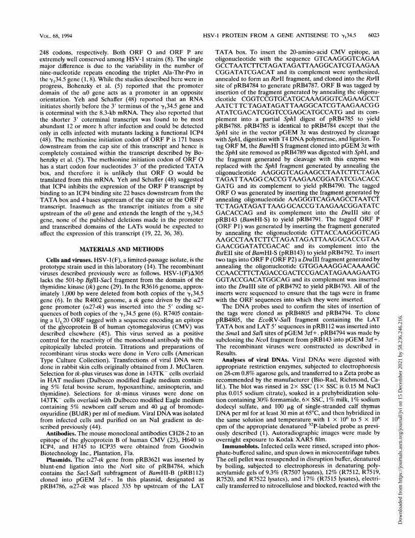
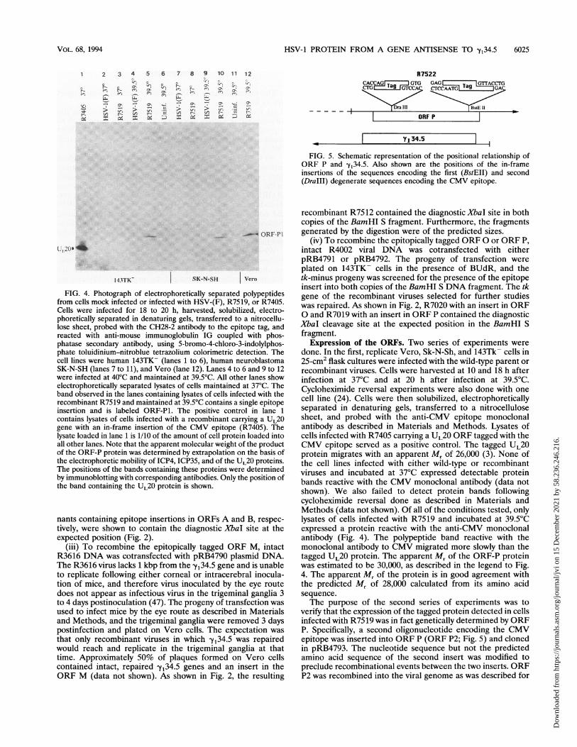
FIG. 5. Schematic representation of the positional relationship ofORF P and -y34.5. Also shown are the positions of the in-frameinsertions of the sequences encoding the first (BstEII) and second(DraIII) degenerate sequences encoding the CMV epitope.
L1' 20Oi 4'4
143TKr
FIG. 4. Photograph of electrophoretifrom cells mock infected or infected withCells were infected for 18 to 20 h, haphoretically separated in denaturing gelslose sheet, probed with the CH28-2 antireacted with anti-mouse immunoglobuphatase secondary antibody, using 5-biphate toluidinium-nitroblue tetrazoliumcell lines were human 143TK- (lanes 1SK-N-SH (lanes 7 to 11), and Vero (lanewere infected at 40°C and maintained atelectrophoretically separated lysates of c
band observed in the lanes containing lysrecombinant R7519 and maintained at 39insertion and is labeled ORF-P1. Thecontains lysates of cells infected with a rgene with an in-frame insertion of thelysate loaded in lane 1 is 1/10 of the amouall other lanes. Note that the apparent mcof the ORF-P protein was determined bythe electrophoretic mobility of ICP4, ICPThe positions of the bands containing theby immunoblotting with corresponding anthe band containing the UL20 protein is
nants containing epitope insertions itively, were shown to contain the d:expected position (Fig. 2).
(iii) To recombine the epitopicalR3616 DNA was cotransfected withThe R3616 virus lacks 1 kbp from theto replicate following either cornealtion of mice, and therefore virus in(does not appear as infectious virus i:to 4 days postinoculation (47). The piused to infect mice by the eye routeand Methods, and the trigeminal gar
postinfection and plated on Vero c
that only recombinant viruses in wwould reach and replicate in the ttime. Approximately 50% of plaqucontained intact, repaired -Yi34.5 gcORF M (data not shown). As shou
recombinant R7512 contained the diagnostic XbaI site in bothcopies of the BamHI S fragment. Furthermore, the fragments
ORF-P1 generated by the digestion were of the predicted sizes.(iv) To recombine the epitopically tagged ORF 0 or ORF P,
intact R4002 viral DNA was cotransfected with eitherpRB4791 or pRB4792. The progeny of transfection wereplated on 143TK- cells in the presence of BUdR, and the
SK-N-SH Vern tk-minus progeny was screened for the presence of the epitopeinsert into both copies of the BamHI S DNA fragment. The tk
ically separated polypeptides gene of the recombinant viruses selected for further studiesiHSV-(F), R7519,or R7405- was repaired. As shown in Fig. 2, R7020 with an insert in ORFs, transferred to a nitrocellue 0 and R7019 with an insert in ORF P contained the diagnosticibody to the epitope tag, and XbaI cleavage site at the expected position in the BamHI Slin IG coupled with phos- fragment.romo-4-chloro-3-indolylphos- Expression of the ORFs. Two series of experiments werecolorimetric detection. The done. In the first, replicate Vero, Sk-N-Sh, and 143Tk- cells in
to 6), human neuroblastoma 25-cm2 flask cultures were infected with the wild-type parent or12). Lanes 4 to 6 and 9 to 12 recombinant viruses. Cells were harvested at 10 and 18 h after39.5°C. All other lanes show infection at 37°C and at 20 h after infection at 39.5°C.-ells maintained at 37°C. Theesatna*T Cycloheximide reversal experiments were also done with one;ates of cells infected with the . s>.50C contains a single epitope cell line (24). Cells were then solubilized, electrophoreticallypositive control in lane 1 separated in denaturing gels, transferred to a nitrocellulose
recombinant carrying a UL20 sheet, and probed with the anti-CMV epitope monoclonalCMV epitope (R7405). The antibody as described in Materials and Methods. Lysates ofLint of cell protein loaded into cells infected with R7405 carrying a UL20 ORF tagged with the)lecular weight of the product CMV epitope served as a positive control. The tagged UL20extrapolation on the basis of protein migrates with an apparent Mr of 26,000 (3). None of
'35, and of theUL20 proteins. the cell lines infected with either wild-type or recombinantese proteins were determined viruses and incubated at 37°C expressed detectable proteinitibodies. Only the position of bands reactive with the CMV monoclonal antibody (data notshown. shown). We also failed to detect protein bands following
cycloheximide reversal done as described in Materials andMethods (data not shown). Of all of the conditions tested, only
in ORFs A and B, respec- lysates of cells infected with R7519 and incubated at 39.5°CLiagnostic XbaI site at the expressed a protein reactive with the anti-CMV monoclonal
antibody (Fig. 4). The polypeptide band reactive with theLly tagged ORF M, intact monoclonal antibody to CMV migrated more slowly than theIpRB4790 plasmid DNA. tagged UL20 protein. The apparent M, of the ORF-P proteinYy34.5 gene and is unable was estimated to be 30,000, as described in the legend to Fig.
I or intracerebral inocula- 4. The apparent Mr of the protein is in good agreement withoculated by the eye route the predicted Mr of 28,000 calculated from its amino acidn the trigeminal ganglia 3 sequence.rogeny of transfection was The purpose of the second series of experiments was toas described in Materials verify that the expression of the tagged protein detected in cellsnglia were removed 3 days infected with R7519 was in fact genetically determined by ORFells. The expectation was P. Specifically, a second oligonucleotide encoding the CMV,hich -y,34.5 was repaired epitope was inserted into ORF P (ORF P2; Fig. 5) and cloned:rigeminal ganglia at that in pRB4793. The nucleotide sequence but not the predictedies formed on Vero cells amino acid sequence of the second insert was modified to-nes and an insert in the preclude recombinational events between the two inserts. ORFvn in Fig. 2, the resulting P2 was recombined into the viral genome as was described for
VOL. 68, 1994
Dow
nloa
ded
from
http
s://j
ourn
als.
asm
.org
/jour
nal/j
vi o
n 15
Dec
embe
r 20
21 b
y 58
.236
.246
.216
.

6026 LAGUNOFF AND ROIZMAN
0 0 ~~0 0
~~ ~~ - r (
LIfN N NN
N
ORF-P2 .ORF-PIl
FIG. 6. Photograph of electrophoretically separated polypeptidesfrom Vero cells infected with HSV-1(F), R7519, R7520, or R7522 andincubated at 37 or 39.5°C, as indicated. The procedures for processingthe infected cell lysates were done as described in the legend to Fig. 4.The band obtained in cells infected with the virus carrying a single tagin ORF P is labeled ORF-Pl, whereas the band obtained with thedouble tag is labeled ORF-P2.
R7519 to yield the recombinant virus R7522, which is shown tocontain the two inserts, as evidenced by the two diagnosticXbaI sites in Fig. 2. As shown in Fig. 6, the lysates of Vero cellsinfected with R7522 express a protein with a mobility slowerthan that of ORF P1 expressed by R7519 and which reacts withthe CMV antibody. This same result was also seen in rabbitskin cells (data not shown). Our results indicate that theprotein band tagged by the CMV epitope is genetically deter-mined by the sequence of ORF P and that this ORF isexpressed and encodes a protein.
AHSV- 1(F)HSV-1(17)HSV -1 (CVG)HSV- 1(MGH10)Consensus
HSV-1(F)HSV-1(17)HSV-1(CVG)HSV- 1(MGH10)Consensus
HSV-1(F)HSV- 1(17)HSV- 1(CVG)HSV- 1(MGH10)Consensus
HSV- 1(F)HSV-1(17)HSV-1(CVG)HSV- 1(MGH10)Consensus
HSV-1(F)HSV-1(17)HSV-1(CVG)HSV- 1(MGH10)Consensus
MTASASATRR RNRARSARSR AHEPRRARRA AEAQTTRWRT RTWGEKRTRMTASASATRR RNRARSARSR AHEPRRARRA AEAQTTRWRT RTWGEKRTRMTASASATRR RNRARSARSR AHEPRRARRA AEAQTTRWRT RTWGEKRTRMTASASATRR RNRARSARSR AHEPRRARRA AEAQTTRWRT RTWGEKRTRMTASASATRR RNRARSARSR AHEPRRARRA AEAQTTRWRT RTWGEKRTR
(AGV)lo AGGSGAPS PPARRRRRRA RCSAVTRRRR ARRGGRRKGR EGGWE(AGV)5 AGGSGAPS PPARRRRRRA RCSAVTRRRR ARRGGRRKGR EGGWE(AGV)6 AGGSGAPS PPARRRRRRA RCSAVTRRRR ARRGGRRKGR EGGWE(AGV)6 AGGfGAPS PPARRRRRRA RCSAVTRRRR ARRGGRRKGR EGGWE(AGV)n AGGSGAPS PPARRRRRRA RCSAVTRRRR ARRGGRRKGR EGGWE
GIAPPPGPAPGGGDR GRGAAAVGRA SGAGSGGGLS GQSSSSSSSD ADSGGsAPPPGPtPGGGgR GRGAAAVGRA SGAdSGGGLS GQSSSSSSSD ADSGGsAPPPGPAPGGGDR GRGAAAVGRA SGAGSGGGLS GQSSSSSSSD ADSGGlAPPPGPAPGGGDR GRGAAAVGRA SGAGSGGGLS GQSSSSSSSD ADSGG-APPPGPAPGGGDR GRGAAAVGRA SGAGSGGGLS GQSSSSSSSD ADSG
TWSHWRSSSEQEGGGP 1AGGGGGAAA GALLTaGSEL GVEVTWDCAV GTATWSHWRSSSEQEGGGP PAGGGGGAAA GALLTaGSEL GVEVTWDCAV GTATWSHcRSSSEQEGGGP PAGGGGGAAA GALLTtGSEL GVEVTWDCAV GTATWSHWRSSSEQEGGGP P....... AA GALLTtGSEL GVEVTWDCAV GTATWSHWRSSSEQEGGGP PAGGGGGAAA GALLT-GSEL GVEVTWDCAV GTA
PVGPGGRGRRGPRW.RR RRAMETESVP GW*PVGPGGRGRRGPRW.RR RRAMETESVP GW*PVGPGGRGRRGPRWRRR RRAMETESVP GW*PVGPGGRGRRGPRWRRR RRAMETESVP GW*PVGPGGRGRRGPRWRRR RRAMETESVP GW*
DISCUSSION
The salient features of the results presented in this reportare as follows.
(i) We have chosen for these studies five ORFs among the16 predicted to encode more than 50 codons as more likely toexpress and allow detection of translated gene products. Thesestudies do not exclude the possibility that some of the otherORFs are expressed.
(ii) Of the five ORFs tested, only one yielded a translationproduct. The protocol that we have chosen to demonstrategene expression unambiguously identifies the polypeptide seenin the gel with ORF P inasmuch as we show that the migrationof ORF P is genetically determined by the epitopic inserts intoits coding domain. The fact that the other four tagged ORFsdid not yield a translation product adds to the cumulativeevidence that insertion of the epitope 3' to the translationinitiation site does not endow transcripts with a capacity to betranslated.While a negative result does not constitute compelling
evidence, particularly since expression could be tissue specific,we should note that the procedures that we have used areextremely sensitive. For example, the amount of CMV epitopeattributed to ORF P protein detected in these studies consti-tutes approximately 1% of the amount of UL20 produced ininfected cells.
(iii) The observation that ORF P is expressed was notexpected because it is nearly completely antisense to the y1 34.5ORF. Only 8 codons of ORF P are not antisense to y34.5, andonly 23 codons of yi34.5 are not antisense to ORF P. As such,this is the first instance in which two ORFs in the HSV genomeare quasi-totally antisense to each other. Because of theoverlap, the sequence of ORF P is well conserved amongHSV-1 strains (Fig. 7A): the only major variability noted todate is in the number of triplet Ala-Gly-Val repeats corre-sponding to the Ala-Thr-Pro repeats of the 1y34-5 gene (8).An ORF also exists in HSV-1(HG52) at approximately the
BHSV-1(F) MTASAsATRR RNRaRSARSR AhEPRRARRA AeaQtTRWRT rTWGEKrT..HSV-2(HG52) MTASAaATRR RNRsRSARSR AqDPRRARRA AvsQaTRWRT cTRGEKhTcgConsensus MTASA-ATRR RNR-RSARSR A-- PRRARRA A- -Q-TRWRT -T-GEK-T--
HSV-1(F) ..raGVaGva .... GVaGva GvaGVaGva. ...GVaGvaG vaGgsGapspHSV-2(HG52) rgdtGVgGas ggrrGVrGse GrqGVgGasg grrGVrGseG rqGvgGeaylConsensus .... GV-G---.-GV-G-- G--GV ------GGV-G--G --G- -G----
HSV-1(F) PARRrRrRaR csa...... vtRRRRHSV-2(HG52) PARRvRgRgR ggpppaqaqa rqvlrrgaqp *rqvgrkgal rpallaRRRRConsensus PARR-R-R-R-R-------- ---------- ---------- ----- RRRR
HSV-1(F) arrGGRRkgR eggWeglApp pGp...... aPggHSV-2(HG52) gagGGRRgvR girRlppAla rGavrplcvv vagvvrvvav vrlghqqPapConsensus - - -GGRR- -R ......A.-G.A.- P--
HSV-1(F) gDRGRGaaAv gRaSgagsgg ..GlsGqsss ssssdadsGt WshWrssseqHSV-2(HG52) qERGRGrrAl dRgSrvvrrd hlGvcGwera garhgwsaGa RhgWsagarpConsensus --RGRGA R S--A----G--G --------G- ---W------
HSV-1(F) egGgPlAGgg ggaaaGaLlt Agselgvevt WDcavGT.Ap VgpggrgrrgHSV-2(HG52) apGtPaAGtp aaghgGrLgs A*arsrsasg REsslGThAr VtaspclpseConsensus - -G-P-AG .- ....G-L-- A--------- ----GT-A- V .-------
HSV-1(F) prWrrRRAme tesvpgw*HSV-2(HG52) ltRpgWRAqp gpccgps*Consensus . RA -- -.------
FIG. 7. Comparison of the amino acid sequences predicted for HSV-1 (A) and HSV-2 (B) ORF P. (A) Sequence alignment of ORF P inHSV-1(F), HSV-1(17), HSV-1(CVG2), and HSV-1(MGH10); (B) sequence alignment of ORF P in HSV-1(F) and an ORF in the correspondingposition in HSV-2(HG52). The comparison was done with the aid of the Gap program of the University of Wisconsin Genetics Computer Grouppackage.
J. VIROL.
Dow
nloa
ded
from
http
s://j
ourn
als.
asm
.org
/jour
nal/j
vi o
n 15
Dec
embe
r 20
21 b
y 58
.236
.246
.216
.

HSV-1 PROTEIN FROM A GENE ANTISENSE TO -y34.5 6027
same place in the genome (25). Thus, the HSV-2 sequencecontains a TATA box, upstream sequences encoding se-quences similar to SPi response elements, and a binding sitefor ICP4 at position +22. Moreover, the first 50 predictedamino acids of HSV-1 and HSV-2 ORF P show good homol-ogy (Fig. 7B). However, downstream sequences beginning withthe triplet repeat AGV in HSV-1 show little or no homology tothe published HSV-2 sequence. The extent of the homology ofHSV-1 and HSV-2 predicted proteins can be improved byassuming sequencing errors involving frameshifts. This wouldnot be surprising since this region of the HSV-2 genomecontains a very high G+C content. The possibility that thereported sequence of HSV-2 ORF P requires reassessment issuggested by the report that whereas the HSV-1 antisense gene-Yi34.5 yields an unspliced mRNA, the HSV-2 equivalent wasreported to yield a spliced mRNA (25). HSV genes which yieldspliced mRNAs are few in number and well conserved amongHSV-1 and HSV-2 strains (2).
(iv) The regulation of the expression of ORF P is puzzlingand remains to be determined. Bohenzky et al. reported thatthe RNA was present in infected cells at 10 h after infectionand found the RNA by nuclear run-on experiments undercycloheximide treatment (5). Yeh and Schaffer failed to detectthe RNA in cells infected and maintained in the presence ofcycloheximide (48). The finding of an ICP4 binding site closeto the transcription initiation of ORF P DNA suggested thatORF P is inhibited by ICP4 but requires other at proteins for itsexpression. At face value, our results are concordant with butnot necessarily supportive of this conclusion. Both(x4 andORF P contain ICP4 binding sites near the transcriptioninitiation sites, and in the case of the a4 gene, it has beendemonstrated that ICP4, the product of thea4 gene, inhibitstranscription ofao4 (27). This has not been shown as yet forORF P, but the close proximity of the binding site to thetranscription initiation sites raises the possibility that in thiscase ICP4 can also block transcription of ORF P, and theobservation that cells infected with recombinants derived fromHSV-1(F), a wild-type virus temperature sensitive in ICP4express ORF P is consistent with this conclusion. The distinc-tion between the expression of thea4 gene and ORF P is basedon the observation that(x4 is expressed in the presence ofcycloheximide whereas ORF P is not (18). Such findings, whilepotentially significant, are not compelling evidence since theORF P transcript may be unstable in the presence of cyclo-heximide.
Perhaps more significant is the report by Yeh and Schaffer(48) that they failed to detect the ORF P mRNA in murinetrigeminal ganglia harboring latent virus. At face value, thisobservation suggests that ORF P is not a member of the familyof the 3' coterminal LATs. However, in the same assays, Yehand Schaffer (48) failed to detect the 8.3-kb unspliced LAT,raising the possibility that the assays were not sensitive enoughto detect small amounts of ORF P mRNA which could bepresent only in the small fraction of total neurons harboringlatent virus (34).A more satisfying scenario would be if ORF P were to be
expressed during latency and if its transcription were to besuppressed by newly synthesized ICP4. Its accumulation late ininfection would reflect loss of activity of ICP4 due to posttrans-lational modification late in infection. The evidence to satisfythis scenario, and to support a role for ORF P in latentlyinfected cells, is not yet available. We should note, however,that none of the deletions previously reported in the domain ofthe LAT transcript encompassed ORF P (19, 22, 36, 38). Theonly mutations introduced into the domain of ORF P are thosegenetically engineered in the Yi34.5 gene (6). It is noteworthy
that the deletion mutants in theYi34.5 gene exhibit a reducedcapacity to establish latency or reactivate from the latent state(47). Studies designed to determine whether the reducedcapacity to establish latency reflects the mutations in y,34.5 orthe ORF P are in progress. The phenotype ascribed to-Yi34.5,i.e., blocking the stress response which results in the prematuretotal shutoff of protein synthesis (9, 10), is indeed that of-Y,34.5inasmuch as the human neuroblastoma cell line SK-N-SHexpressing only the -Yi34.5 gene does not exhibit the stressresponse associated with infection of the parental line with the-y134.5- (5a).
ACKNOWLEDGMENTS
We thank Saul Silverstein and Joany Chou for useful discussions.These studies were aided by Public Health Service grants from the
National Cancer Institute (CA47451) and the National Institute forAllergy and Infectious Diseases (AI124009) and by an unrestrictedgrant from the Bristol-Myers Squibb Program in Infectious Diseases.
REFERENCES1. Ackermann, M., J. Chou, M. Sarmiento, R. A. Lerner, and B.
Roizman. 1986. Identification by antibody to a synthetic peptide ofa protein specified by a diploid gene located in the terminalrepeats of the L component of herpes simplex virus genome. J.Virol. 58:843-850.
2. Baines, J. D., and B. Roizman. 1992. The cDNAof UL15, a highlyconserved herpes simplex virus 1 gene, effectively replace the twoexons of the wild-type virus. J. Virol. 66:5621-5626.
3. Baines, J. D., P. L. Ward, G. Campadelli-Fiume, and B. Roizman.1991. The UL20 gene of herpes simplex virus 1 encodes a functionnecessary for viral egress. J. Virol. 65:6414-6424.
4. Block, T. M., S. Deshmane, J. Masonis, J. Maggioncalda, T.Valyi-Nagi, and N. W. Fraser. 1993. An HSV LAT null mutantreactivates slowly from latent infection and makes small plaqueson CV-1 monolayers. Virology 192:618-630.
5. Bohenzky, R. A., A. G. Papavassiliou, I. H. Gelman, and S.Silverstein. 1993. Identification of a promoter mapping within thereiterated sequences that flank the herpes simplex virus type 1 ULregion. J. Virol. 67:632-642.
5a.Chou, J., and B. Roizman. Unpublished data.6. Chou, J., E. R. Kern, R. J. Whitley, and B. Roizman. 1990.
Mapping of herpes simplex virus-i neurovirulence to -Yi34.5, agene nonessential for growth in culture. Science 250:1262-1266.
7. Chou, J., and B. Roizman. 1986. The terminal a sequence ofherpes simplex virus genome contains the promoter of a genelocated in the repeat sequences of the L component. J. Virol.57:629-637.
8. Chou, J., and B. Roizman. 1990. The herpes simplex virus 1 genefor ICP34.5, which maps in inverted repeats, is conserved inseveral limited-passage isolates but not in strain 17syn+. J. Virol.64:1014-1020.
9. Chou, J., and B. Roizman. 1992. The -y34.5 gene of herpes simplexvirus 1 precludes neuroblastoma cells from triggering total shutoffof protein synthesis characteristic of programmed cell death inneuronal cells. Proc. Natl. Acad. Sci. USA 89:3266-3270.
10. Chou, J., and B. Roizman. 1994. The herpes simplex virus 1 -y34.5gene function which blocks the host response to infection maps inthe homologous domain of the genes expressed during growtharrest and DNA damage. Proc. Natl. Acad. Sci. USA 91:5247-5251.
11. Devi-Rao, G. B., S. A. Goodart, L. M. Hecht, R. Rochford, M. K.Rice, and E. K. Wagner. 1991. Relationship between polyade-nylated and nonpolyadenylated herpes simplex virus type 1 laten-cy-associated transcripts. J. Virol. 65:2179-2190.
12. Dobson, A. T., F. Sederati, G. Devi-Rao, W. M. Flanagan, M. J.Farrell, J. G. Stevens, E. K. Wagner, and L. T. Feldman. 1989.Identification of the latency-associated transcript promoter byexpression of rabbit beta-globin mRNA in mouse sensory nerveganglia latently infected with a recombinant herpes simplex virus.J. Virol. 63:3844-3851.
13. Doerig, C., L. I. Pizer, and C. L. Wilcox. 1991. An antigen encoded
VOL. 68, 1994
Dow
nloa
ded
from
http
s://j
ourn
als.
asm
.org
/jour
nal/j
vi o
n 15
Dec
embe
r 20
21 b
y 58
.236
.246
.216
.
6028 LAGUNOFF AND ROIZMAN
by the latency-associated transcript in neuronal cell cultureslatently infected with herpes simplex virus type 1. J. Virol. 65:2724-2727.
14. Ejercito, P. M., E. D. Kieff, and B. Roizman. 1968. Characteriza-tion of herpes simplex virus strains differing in their effects onsocial behavior of infected cells. J. Gen. Virol. 2:357-364.
15. Farrell, M. J., A. T. Dobson, and L. T. Feldman. 1991. Herpessimplex virus latency-associated transcript is a stable intron. Proc.Natl. Acad. Sci. USA 88:790-794.
16. Fraser, N. W., T. M. Block, and J. G. Spivaclk 1992. Thelatency-associated transcripts of herpes simplex virus: RNA insearch of function. Virology 191:1-8.
17. Hill, J. M., F. Sedarati, R. T. Javier, E. K. Wagner, and J. G.Stevens. 1990. Herpes simplex virus latent phase transcriptionfacilitates in vivo reactivation. Virology 174:117-125.
18. Honess, R. W., and B. Roizman. 1975. Regulation of herpesvirusmacromolecular synthesis: sequential transition of polypeptidesynthesis requires functional viral polypeptides. Proc. Natl. Acad.Sci. USA 72:1276-1280.
19. Javier, R. T., J. G. Stevens, V. B. Dissette, and E. K. Wagner. 1988.A herpes simplex virus transcript abundant in latently infectedneurons is dispensable for establishment of the latent state.Virology 166:254-257.
19a.Johnson, J., J. Chou, and B. Roizman. Unpublished data.20. Kozak, M. 1989. The scanning model for translation: an update. J.
Cell Biol. 108:229-241.21. Krause, P. R., K. D. Croen, S. E. Straus, and J. M. Ostrove. 1988.
Detection and preliminary characterization of herpes simplexvirus type 1 transcripts in latently infected human trigeminalganglia. J. Virol. 62:4819-4823.
21a.Lagunoff, M., and B. Roizman. Unpublished data.22. Leib, D. A., C. L. Bogard, M. Kosz-Vnenchak, K. A. Hicks, D. M.
Coen, D. M. Knipe, and P. A. Schaffer. 1989. A deletion mutant ofthe latency-associated transcript of herpes simplex virus type 1reactivates from the latent state with reduced frequency. J. Virol.63:2893-2900.
23. Liu, F., and B. Roizman. 1991. The promoter, transcriptional unit,and coding sequence of herpes simplex virus family 35 proteins arecontained within and in frame with the UL26 open reading frame.J. Virol. 65:206-212.
24. Mavromara-Nazos, P., M. Ackermann, and B. Roizman. 1986.Construction and properties of a viable herpes simplex virus 1recombinant lacking coding sequences of the a47 gene. J. Virol.60:807-812.
25. McGeogh, D. J., C. Cunningham, G. McIntyre, and A. Dolan.1991. Comparative sequence analysis of the long repeat regionsand adjoining parts of the long unique regions in the genomes ofherpes simplex viruses types 1 and 2. J. Gen. Virol. 72:3057-3075.
26. McGeoch, D. J., M. A. Dalrymple, A. J. Davison, A. Dolan, M. C.Frame, D. McNab, L. J. Perry, J. E. Scott, and P. Taylor. 1988. Thecomplete DNA sequence of the long unique region in the genomeof herpes simplex virus type 1. J. Gen. Virol. 69:1531-1574.
27. Michael, N., and B. Roizman. 1993. Repression of the herpessimplex virus 1 a4 gene by its gene product occurs within thecontext of the viral genome and is associated with all threeidentified cognate sites. Proc. Natl. Acad. Sci. USA 90:2286-2290.
28. Mitchell, W. J., R. P. Lirette, and N. W. Fraser. 1990. Mapping oflow abundance latency-associated RNA in the trigeminal gangliaof mice latently infected with herpes simplex virus type 1. J. Gen.Virol. 71:125-132.
29. Post, L. E., S. Mackem, and B. Roizman. 1981. Regulation of agenes of herpes simplex virus: expression of chimeric genesproduced by fusion of thymidine kinase with a gene promoters.Cell 24:555-565.
30. Post, L. E., and B. Roizman. 1981. A generalized technique fordeletion of specific genes in large genomes: ao gene 22 of herpessimplex virus 1 is not essential for growth. Cell 25:227-232.
31. Rock, D. L., A. B. Nesburn, H. Ghiasi, J. Ong, T. L. Lewis, J. R.
Lokensgard, and S. L. Wechsler. 1987. Detection of latency-related viral RNAs in trigeminal ganglia of rabbits latently in-fected with herpes simplex virus type 1. J. Virol. 61:3820-3826.
32. Roizman, B. 1966. An inquiry into the mechanisms of recurrentherpes infections of man, p. 283-304. In M. Pollard (ed.). Perspec-tives in virology, vol. IV. Harper & Row (Hoeber MedicalDivision), New York.
33. Roizman, B., and A. E. Sears. 1987. An inquiry into the mecha-nisms of herpes simplex virus latency. Annu. Rev. Microbiol.41:543-571.
34. Roizman, B., and A. E. Sears. 1990. Herpes simplex viruses andtheir replication, p. 1795-1841. In: B. Fields (ed.), Virology. RavenPress, New York.
35. Roizman, B., and A. E. Sears. 1993. The replication herpes simplexvirus, p. 11-68. In B. Roizman, C. Lopez, and R. J. Whitley (ed.),Human herpes viruses. Raven Press, New York, New York.
36. Sedarati, F., K. M. Izumi, E. K. Wagner, and J. G. Stevens. 1989.Herpes simplex virus type 1 latency-associated transcription playsno role in establishment or maintenance of a latent infection inmurine sensory neurons. J. Virol. 63:4455-4458.
37. Spivack, J. G., and N. W. Fraser. 1987. Detection of herpessimplex virus type 1 transcripts during latent infection in mice. J.Virol. 61:3841-3847.
38. Steiner, I., J. G. Spivack, R. P. Lirette, S. M. Brown, A. R.MacLean, J. H. Subak-Sharpe, and N. W. Fraser. 1989. Herpessimplex virus type 1 latency-associated transcripts are evidently notessential for latent infection. EMBO J. 8:505-511.
39. Steiner, I., J. G. Spivack, D. R. O'Boyle II, E. Lavi, and N. W.Fraser. 1988. Latent herpes simplex virus type 1 transcription inhuman trigeminal ganglia. J. Virol. 62:3493-3496.
40. Stevens, J. G., and M. L. Cook. 1971. Latent herpes simplex virusin spinal ganglia of mice. Science 173:843-844.
41. Stevens, J. G., E. K. Wagner, G. B. Devi-Rao, M. L. Cook, and L. T.Feldman. 1987. RNA complementary to a herpesvirus a genemRNA is prominent in latently infected neurons. Science 235:1056-1059.
42. Wagner, E. K., G. Devi-Rao, L. T. Feldman, A. T. Dobson, Y.Zhang, W. M. Flanagan, and J. G. Stevens. 1988. Physicalcharacterization of the herpes simplex virus latency-associatedtranscript in neurons. J. Virol. 62:1194-1202.
43. Wagner, E. K., W. M. Flanagan, G. Devi-Rao, Y. Zhang, J. M. Hill,K. P. Anderson, and J. G. Stevens. 1988. The herpes simplex viruslatency-associated transcript is spliced during the latent phase ofinfection. J. Virol. 62:4577-4585.
44. Walboomers, J. M. M., and J. T. Schegget. 1976. A new method forthe isolation of herpes simplex virus type 2 DNA. Virology74:256-258.
45. Ward, P. L., G. Campadelli-Fiume, E. Avitabile, and B. Roizman.The localization and putative function of the UL20 membraneprotein in cells infected with herpes simplex virus 1. Submitted forpublication.
46. Wechsler, S. L., A. B. Nesburn, J. Zwaagstra, and H. Ghiasi. 1989.Sequence of the latency-related gene of herpes simplex virus type1. Virology 168:168-172.
47. Whitley, R. J., E. R. Kern, S. Chatterjee, J. Chou, and B. Roizman.1993. Replication, establishment of latency, and induced reactiva-tion of herpes simplex virus -yi34.5 deletion mutants in rodentmodels. J. Clin. Invest. 91:2837-2843.
48. Yeh, L., and P. A. Schaffer. 1993. A novel class of transcriptsexpressed with late kinetics in the absence of ICP4 spans thejunction between the long and short segments of the herpessimplex virus type 1 genome. J. Virol. 67:7373-7382.
49. Zwaagstra, J. C., H. Ghiasi, S. M. Slanina, A. B. Nesburn, S. C.Wheatley, K. Lillycrop, J. Wood, D. S. Latchman, K. Patel, andS. L. Wechsler. 1990. Activity of herpes simplex virus type 1latency-associated transcript (LAT) promoter in neuron-derivedcells: evidence for neuron specificity and for a large LAT tran-script. J. Virol. 64:5019-5028.
J. VIROL.
Dow
nloa
ded
from
http
s://j
ourn
als.
asm
.org
/jour
nal/j
vi o
n 15
Dec
embe
r 20
21 b
y 58
.236
.246
.216
.