Expression and beta-glucan binding properties of Scots pine (Pinus ...
40
http://www.diva-portal.org Postprint This is the accepted version of a paper published in Plant Molecular Biology. This paper has been peer- reviewed but does not include the final publisher proof-corrections or journal pagination. Citation for the original published paper (version of record): Sooriyaarachchi, S., Jaber, E., Covarrubias, A., Ubhayasekera, W., Asiegbu, F. et al. (2011) Expression and beta-glucan binding properties of Scots pine (Pinus sylvestris L.) antimicrobial protein (Sp-AMP). Plant Molecular Biology, 77(1-2): 33-45 http://dx.doi.org/10.1007/s11103-011-9791-z Access to the published version may require subscription. N.B. When citing this work, cite the original published paper. Permanent link to this version: http://urn.kb.se/resolve?urn=urn:nbn:se:uu:diva-158571
Transcript of Expression and beta-glucan binding properties of Scots pine (Pinus ...
http://www.diva-portal.org
Postprint
This is the accepted version of a paper published in Plant Molecular Biology. This paper has been peer-reviewed but does not include the final publisher proof-corrections or journal pagination.
Citation for the original published paper (version of record):
Sooriyaarachchi, S., Jaber, E., Covarrubias, A., Ubhayasekera, W., Asiegbu, F. et al. (2011)
Expression and beta-glucan binding properties of Scots pine (Pinus sylvestris L.) antimicrobial
protein (Sp-AMP).
Plant Molecular Biology, 77(1-2): 33-45
http://dx.doi.org/10.1007/s11103-011-9791-z
Access to the published version may require subscription.
N.B. When citing this work, cite the original published paper.
Permanent link to this version:http://urn.kb.se/resolve?urn=urn:nbn:se:uu:diva-158571
1
Expression and β-glucan binding properties of Scots pine (Pinus sylvestris L.) antimicrobial
protein (Sp-AMP)
Sanjeewani Sooriyaarachchi1&, Emad Jaber2&, Adrian Suárez Covarrubias3, Wimal
Ubhayasekera1§, Frederick O. Asiegbu2, Sherry L. Mowbray1, 3*
1 Department of Molecular Biology, Swedish University of Agricultural Sciences, Box 590,
Biomedical Center, SE-751 24, Uppsala, Sweden
2 Department of Forest Sciences, University of Helsinki, Box 27, FI-00014 Helsinki, Finland
3 Department of Cell and Molecular Biology, Uppsala University, Box 596, Biomedical Center,
SE-751 24, Uppsala, Sweden
& Joint first authors.
§ Current address: MAX-lab, Lund University, Box 118, S-221 00 Lund, Sweden, and Institute
of Medicinal Chemistry, University of Copenhagen, Universitetsparken 2, DK-2100 Copenhagen
Ø, Denmark
* Corresponding author: Tel.: 46-18-471-4990; Fax: 46-18-53-69-71; E-mail:
2
Abbreviations:
AMP, antimicrobial protein
SA, salicylic acid
MeJA, methyl jasmonate
ACC, 1-aminocyclopropane-1-carboxylic-acid
PDB, Protein Data Bank
PR, pathogenesis-related
qRT-PCR, real-time quantitative reverse transcription PCR
SDS-PAGE, sodium dodecyl sulfate polyacrylamide gel electrophoresis
Sp-AMP, Scots pine antimicrobial peptide/protein (the corresponding gene is italicised)
3
Abstract
Scots pine (Pinus sylvestris) secretes a number of small, highly-related, disulfide-rich proteins
(Sp-AMPs) in response to challenges with fungal pathogens such as Heterobasidion annosum,
although their biological role has been unknown. Here, we examined the expression patterns of
these genes, as well as the structure and function of the encoded proteins. Northern blots and
quantitative real time PCR showed increased levels of expression that are sustained during the
interactions of host trees with pathogens, but not non-pathogens, consistent with a function in
conifer tree defenses. Furthermore, the genes were up-regulated after treatment with salicylic
acid and an ethylene precursor, 1-aminocyclopropane-1-carboxylic-acid, but neither methyl
jasmonate nor H2O2 induced expression, indicating that Sp-AMP gene expression is independent
of the jasmonic acid signaling pathways. The cDNA encoding one of the proteins was cloned and
expressed in Pichia pastoris. The purified protein had antifungal activity against H. annosum,
and caused morphological changes in its hyphae and spores. It was directly shown to bind
soluble and insoluble β-(1,3)-glucans, specifically and with high affinity. Furthermore, addition
of exogenous glucan is linked to higher levels of Sp-AMP expression in the conifer. Homology
modeling and sequence comparisons suggest that a conserved patch on the surface of the
globular Sp-AMP is a carbohydrate-binding site that can accommodate approximately four sugar
units. We conclude that these proteins belong to a new family of antimicrobial proteins (PR-19)
that are likely to act by binding the glucans that are a major component of fungal cell walls.
Keywords: antimicrobial protein; Heterobasidion annosum; β-(1,3)-glucan; chitin; inhibition;
binding; homology modeling; pathogen; Pinus sylvestris.
4
Introduction
Plants are attacked by pathogens at all the stages of their life cycle, which is essential for the
balance of the ecosystem, but can be economically disastrous when a crop plant is affected. The
fungus Heterobasidion annosum is the most destructive pathogen for forest trees in the Northern
Hemisphere, causing root and butt rot, especially in conifers such as Scots pine (Pinus sylvestris)
(Asiegbu et al. 2005). As a necrotroph, H. annosum is capable of infecting and destroying living
conifer roots and stems of all ages, as well as dead trees. Chemicals, biocontrol agents, and
silvicultural measures are currently used to control the disease in forest plantations. However,
these do not provide full protection, and there is an urgent need for new, more effective and
environmentally friendly alternatives.
Antimicrobial proteins or peptides (AMPs) may be considered as a first line of defence in plants
(van Loon et al. 2006). All polypeptides showing activity against pathogenic microbes can be
designated as AMPs, and those known have very wide functional and structural diversity
(Montesinos 2007). Many AMPs have been reported to belong to the so called pathogenesis-
related (PR) proteins (van Loon and Van Strien 1999). As well as pathogenic attacks, other
stresses such as drought and wounding often trigger AMP expression (Broekaert et al. 1997).
Induction may be mediated through stress-signalling pathways, particularly the
jasmonate/ethylene pathways (Thomma et al. 1998).
Recent gene expression analysis revealed a novel family of highly related proteins in Scots pine,
designated Sp-AMPs, which are produced constitutively, but up-regulated after challenge with
H. annosum (Asiegbu et al. 2003). Sp-AMPs are consistently induced and localized on the cell
5
surface during a prolonged time of infection with this pathogenic fungus (Adomas et al. 2007).
The Sp-AMP genes were further found to be up-regulated in the presence of non-pathogenic
(saprotrophic and mutualistic/beneficial) fungi, although the up-regulation did not persist beyond
the early stages of exposure (Adomas et al. 2008). At least five genes were found by Southern
blotting of Hind III-digested pine genomic DNA (Asiegbu et al. 2003), of which four (Sp-AMP1-
4, with 93-100% nucleotide sequence identity) have been deposited in the National Center for
Biotechnology Information (NCBI) sequence databases; these sequences code for cysteine-rich
proteins including an N-terminal region with the characteristics of a cleavable signal peptide.
The proteins belong to the conserved MiAMP1 family, which are implicated in the defence of
gymnosperms against fungal pathogens (Manners 2009). However, the mode of action and in
vivo regulation of such AMPs has so far remained unknown.
In the present study, we present new data on the expression of Sp-AMP genes, as well as
evidence that the encoded proteins bind to important fungal cell wall components, specifically β-
(1,3)-linked glucans.
Materials and Methods
Host plants and growth conditions
Pinus sylvestris seeds (FP-45 Sweden) were surface sterilised with 33% H2O2 for 15 min, rinsed
in several changes of sterile distilled water, sown on 1% water agar and incubated at 18 °C with
a photoperiod of 16 h. After 14 days, the resulting seedlings were used for inoculation and
treatments described below.
6
Fungi and inoculation
H. annosum (isolate Dragstjard 05044, courtesy of K. Korhonen, Finland), Stereum
sanguinolentum (isolate FBCC1148, Fungal Biotechnology Culture Collection (FBCC),
University of Helsinki, Finland) and Lactarius rufus (isolate from the Finnish Forest Research
Institute (METLA), Finland), were grown in liquid Hagem medium (Stenlid 1985) for 21 days
under static conditions. The mycelia were washed with sterile distilled water and subsequently
homogenized for 60 s in a sterile Waring blender.
Ten P. sylvestris seedlings were transferred to sterile filter paper layered on top of 1% water agar
in Petri dishes. The roots were inoculated with 1 ml of the mycelial homogenate and covered
with a second moist, sterile filter paper. The plate was sealed with Parafilm and the region of the
dish containing the roots was covered with aluminium foil. The seedlings were then incubated at
18 °C with a photoperiod of 16 h. Control plants were mock-inoculated with 1 ml of sterile
distilled water. Three biological replications with three technical replicates of Scots pine
seedlings were plated for each fungal inoculation, as well as for control plants; roots were
harvested at 1 and 5 days post-inoculation , ground in liquid nitrogen and stored at -80 °C prior
to RNA extraction.
In a parallel experiment, Scots pine roots were inoculated with yeast (Saccharomyces cerevisiae)
mutant strains with reduced levels (~25% of the wild type) of chitin (Δchs5 mutant; strain
BY4741; genotype Mat a; his3Δ1; leu2Δ0; met15Δ0; ura3Δ0; YLR330w::kanMX4; accession
number Y05239 (Santos et al. 1997)), or increased levels of β-(1,6)-glucan where β-(1,3)-glucan
7
and other cell wall components are unaffected (Δexg mutant; BY4741; Mat a; his3Δ1; leu2Δ0;
met15Δ0; ura3Δ0; YLR300w::kanMX4; accession number Y05210 (Cappellaro et al. 1998)), as
well as their isogenic wild type (BY4741; Mat a; his3Δ1; leu2Δ0; met15Δ0; ura3Δ0; accession
number Y00000). All the yeast strains were obtained from EUROSCARF (the European
Saccharomyces cerevisiae Archive for Functional Analysis). The yeast were grown in 250 ml
liquid culture flasks containing 50 ml of nutrient yeast dextrose broth for 16 h at 28 °C on a
rotary shaker at 200 rpm. Cells were collected by centrifuging at 7,000 g for 10 min, washed
twice with sterile distilled water to remove the growth medium, then diluted with sterile distilled
water. Roots were inoculated with 1 ml of wild type or mutant yeast cell suspension.
Protoplast generation
Mycelia of static (3-week) fungal cultures in Hagem medium were mixed, homogenised and
centrifuged at 2,600 g for 5 min at room temperature. The supernatant was discarded, and the
mycelial homogenate washed with 10 ml buffer (0.5 M mannitol, 50 mM maleate). Lysing
enzymes from Trichoderma harzianum (Sigma-Aldrich, L-1412) were added to homogenised
mycelia, and incubated with gentle shaking at 37 °C for 3.5 h. The resulting protoplasts were
filtered through Miracloth (Sigma-Aldrich), centrifuged at 1,500 g for 10 min, washed three
times with 10 ml MMC buffer (0.5 M mannitol, 50 mM maleate, 50 mM CaCl2) and resuspended
in the same buffer. Control plants were mock-inoculated with 10 ml of the MMC buffer. Roots
were exposed to protoplasts for 24 h at 18 °C with a photoperiod of 16 h.
Hormone treatment
8
Scots pine roots were treated with 5 mM salicylic acid (SA), 100 mM methyl jasmonate (MeJA)
or 0.5 mM ethylene precursor (1-aminocyclopropane-1-carboxylic-acid, ACC). All chemicals
were obtained from Sigma-Aldrich Ltd and dissolved in 0.1% ethanol; control plants were
treated with an equivalent amount of 0.1% ethanol. In a separate experiment, roots were treated
with 1 mM H2O2; control plants were mock-treated with sterile distilled water. Treated seedlings
were incubated for 24 h at 18 °C with a photoperiod of 16 h. Three biological replications of P.
sylvestris roots, consisting of 25 seedlings of either hormone-treated or control plants, were
collected and frozen in liquid nitrogen for RNA isolation.
Chitosan, chitin and glucan for fungal studies
Chitosan flakes from shrimp shells (Sigma-Aldrich, C3646) were prepared according to an
earlier procedure (Benhamou and Thériault 1992). Briefly, it was ground to a powder, washed
repeatedly in distilled water, pelleted by low speed centrifugation, and air-dried. Sheets of the
dried chitosan were solubilized by stirring in 0.25 M HCl, centrifuged at 13,000 g for 10 min at 4
°C to remove insoluble material, and precipitated by neutralization with 2.5 M NaOH. Chitosan
pellets, recovered by centrifugation at 25,000 g for 15 min, were thoroughly washed with
deionized water to remove salts, then freeze-dried. For use, purified chitosan was dissolved in
0.05 M HCl under continuous stirring to obtain a stock solution of 1 mg·ml–1; the pH was
adjusted to 5.6 with 1 M NaOH. A 30-µl sample of chitosan solution was applied to each
seedling root. Chitin (Sigma, C-4666) was washed with sterile water and homogenized for 60 s
in a sterile Waring blender. Each root sample was treated with 100 µl of chitin homogenate (1
mg·ml–1). Glucan (as laminarin, which consists primarily of poly(β-(1,3)-D-glucose, with some
β-(1,6) inter-strand linkages and branch points) was derived from Laminaria digitata; (Sigma-
9
Aldrich L9634)). A 30-µl sample of this glucan solution (1 mg·ml–1) was applied to each
seedling root. There were three biological replications harvested at 1 and 5 days post-inoculation,
ground in liquid nitrogen and stored at -80 °C prior to RNA extraction.
RNA extraction and Northern analysis
RNA was extracted from the roots as described elsewhere (Chang et al. 1993). RNA
concentration and quality were assessed spectrophotometrically using a NanoDropTM ND-1000
(NanoDrop Technologies, Wilmington, DE, USA). Northern analyses were performed according
to standard protocols (Sambrook and Russell 2001). Total RNA (15 µg) sampled from inoculated
pine roots at two time points (1 and 5 days post-inoculation) was denatured and electrophoresed
on a formaldehyde-agarose gel, transferred to a nylon membrane (Amersham Hybond N+) by
capillary blotting overnight, then fixed to the membrane by baking at 80 °C for 2 h. Membranes
were hybridised with chemifluorescently labelled Sp-AMP cDNA as a probe. Labelling was
performed using an AlkPhos Direct™ labelling kit (RPN3680) according to the manufacturer's
instructions (Amersham Biosciences). After post-hybridisation washes, membranes were
exposed to film for autoradiographic detection.
DNase treatment and cDNA synthesis by reverse transcription
A 1-µg sample of each total-RNA isolate was DNase treated using a commercially available kit
(RQ1 RNase-free DNase reagents, Promega) according to the manufacturer’s instructions. A 1-µl
aliquot of random hexamers (200 ng, Promega) was added to the treated RNA, after which the
sample was heated to 70 °C for 10 min, then quickly cooled on ice. First strand cDNA synthesis
was performed in the presence of 200 U MLV-reverse transcriptase (Promega) for 1 h at 37 °C.
10
The mixture contained 5 x MLV buffer, 10 mM DTT, 10 mM of each dNTP and 20 U RNasin®
(Promega) in a volume of 20 µl; the reaction was stopped by heating at 70 °C for 10 min.
Real-time quantitative PCR analysis
Real-time quantitative reverse transcription PCR (qRT-PCR) was performed with the
LightCycler® 480 system according to the manufacturer's instructions. Primers targeted the
conserved region of the Sp-AMP family (5ʹ′-CCTTCTGAGGGCAGTTATTTCACT-3ʹ′ (forward)
and 5ʹ′-CGCGCAGCATGGTTGTTA-3ʹ′ (reverse)). Sp-AMP amplicon (62 bp) was amplified
from synthesized cDNA, separated by electrophoresis in a 3% agarose gel, and visualized by
staining with ethidium bromide. Predicted product size was verified with a 100 bp ladder of
DNA markers before analysis with qRT-PCR. The transcript abundance was estimated with
LightCycler software version 3.5 (Roche) using SYBR® Green PCR Master Mix (Invitrogen)
according to the manufacturer's recommendations, based on three biological and three technical
replicates using a standard curve (User Bulletin #2, ABI Prism 7700 Sequence Detection System,
Applied Biosystems). The melting curve analysis indicated a single amplicon for all the samples
used in the study. Melting temperatures (Tm) were in the range of 82.50- 83.25 °C. The absolute
quantity of the product in each sample, as deduced from the standard curve, was normalized
against the total amount of RNA (Hashimoto et al. 2004; Silberbach et al. 2005). Relative
expression levels of the Sp-AMP genes were determined as described earlier (Livak and
Schmittgen 2001).
Cloning, expression of Sp-AMP in Pichia pastoris, and protein purification
A more detailed description of these procedures is included in the Supplementary Materials.
11
Primers for expression of Sp-AMP3 (GenBank accession number AF410954) in P. pastoris were
designed considering differences in codon usage between bacterial and yeast systems. PCR was
first performed on a pre-existing pET21 plasmid containing Sp-AMP3 DNA (Sooriyaarachchi et
al, unpublished results), to obtain the mature Sp-AMP3 sequence bounded by Xho1 and Kpn1
cleavage sites. The Xho1-Kpn1 fragment was ultimately inserted into the Pichia expression
vector pPICZαB (Invitrogen) in-frame with the N-terminal secretory peptide of the S. cerevisiae
α-factor. The pPICZαB derivatives were transformed into E. coli Top10F’ cells. Purified
plasmids containing Sp-AMP3 were linearized with SacI and transformed into the Pichia strain
KM71 by electroporation. Transformants were screened for Zeocin resistance at 100 µg·ml–1.
Colony PCR was performed to confirm the target DNA’s recombination into the yeast
chromosome. The final yeast strain was maintained on YPD agar plates (1% (w/v) yeast extract,
2% (w/v) peptone, 2% dextrose and 2% agar) with 100 µg·ml–1 Zeocin.
After cleavage of the 89-residue secretory peptide, the 79-residue Sp-AMP3 was secreted when
the transformed Pichia strain was induced with methanol. The medium from a 1 l induced
culture was clarified by centrifugation, and treated with 65% (w/v) ammonium sulfate; the
precipitated protein was resuspended in 10 mM HEPES, pH 7.0. After dialysis, the sample was
further purified by cation exchange and size exclusion chromatography, and stored in 10 mM
HEPES, pH 7.0, at -20 °C. The amino acid sequence of Sp-AMP3 was confirmed by analyzing
an excised gel band by MALDI-TOF mass spectroscopy after trypsin digestion.
To enable affinity purification, a second construct was made including a His tag (AHHHHHH) at
the C-terminal end of the Sp-AMP3 sequence. Again, the secreted protein was fractionated by
12
ammonium sulfate precipitation, after which it was dialyzed against 20 mM sodium phosphate,
pH 7.6. The concentrated sample was subsequently purified by chelating and size exclusion
chromatography. Fractions containing pure protein were concentrated and stored in 10 mM
HEPES buffer, pH 7.0, at -20 °C.
Binding assays
For binding assays with insoluble carbohydrate polymers, 30 µl of a 0.75 mg·ml–1 protein
solution was incubated at 4 °C for 6 h with a 2 mg sample of dry chitin, chitosan (both the kind
gift of Kitto Life Co. Ltd, Seoul, Korea) or curdlan (β-(1,3)-D-glucan from Alcaligenes faecalis,
Sigma-Aldrich C7821) . The samples were then centrifuged for 10 min at 16,000 g. The
supernatant in each case was reserved, and the pellets were washed with 30 µl of 10 mM HEPES
buffer, pH 7.0, then with 30 µl of 1 M NaCl in 10% acetic acid (10 min at 4 °C), each time
recovering the supernatant after centrifugation. SDS-PAGE was performed to determine the
fractions in which the protein appeared.
Binding of soluble sugars was monitored using changes in protein tryptophan fluorescence in an
SPF-500 spectrofluorometer (Aminco) at 25 °C. Sp-AMP3 samples (1 ml of 0.5 µM protein in
10 mM HEPES, pH 7.0) were treated with a series of 10-µl aliquots of sugar solution (glucose,
laminarioligosaccharides from laminaribiose up to laminarihexose, glucan (laminarin),
cellobiose, cellotriose or cellopentaose, all obtained from Sigma-Aldrich, and prepared as 2
mg·ml–1 stocks in the same buffer). The solution was mixed well, and equilibrated at room
temperature. Emission spectra were recorded with a fixed excitation wavelength of 285 nm;
excitation and emission slits were 10 and 8 nm, respectively.
13
Determination of antifungal activity
Antifungal activity of Sp-AMP3 was studied by observing H. annosum (strain FP5) growth on
Hagem agar plates (0.5% glucose, 0.05% NH4NO3, 0.05% KH2PO4, 0.05% MgSO4.7H2O, 0.5%
malt extract, 2.0% agar, pH 5.5). To initiate mycelial growth, an agar plug containing H.
annosum was placed at the center of a Petri dish containing 20 ml of the medium, and incubated
in the dark at room temperature until the mycelia reached a diameter of 3 cm. A sterilized filter
paper disc containing 10 µl of 0.75 mg·ml–1 protein solution (with or without His-tag) was placed
at the growth front. Controls included a concentrated sample of the growth medium collected
before induction, as well as the protein storage buffer (10 mM HEPES, pH 7.0). The plates were
incubated further in the dark and observed at intervals.
Inhibition of spore germination was investigated after spreading a spore suspension (in sterilized
water) evenly on Hagem agar plates. Protein solutions were added to sterilized filter paper disks,
which were placed on the plates, followed by incubation in the dark at room temperature for one
week. Addition of 10 µl of 0.75 mg·ml–1 protein or control solution was repeated every 24 h.
Sequence alignment and homology modeling of Sp-AMP3
Similar sequences were located using BLAST (Altschul et al. 1997) and obtained from GenBank
(Benson et al. 2011). The most similar structure, that of the Macadamia integrifolia protein
MiAMP1 solved by NMR (PDB entry 1C01 (McManus et al. 1999)), was obtained from the
PDB (Berman et al. 2000); the first of the 20 deposited models was used in further work. Pair-
wise alignment of the Sp-AMP4 and MiAMP1 sequences with CLUSTAL W (Thompson et al.
14
1994) was used to create a homology model with the program SOD (Kleywegt et al. 2001). The
model was adjusted in O (Jones et al. 1991) using rotamers to improve packing as needed.
Similar structures were located with DALI (Holm 1998). Figures were prepared with O,
Molscript (Kraulis 1991) and Molray (Harris and Jones 2001). Stereo glasses (stereoscopes) for
viewing diagrams such as that in Fig. 7 are available from, for example, ASC Scientific
(Carlsbad, CA, USA).
Results
Sp-AMP expression in response to pathogen, non-pathogen or fungal protoplast exposure
Expression of Sp-AMP genes was investigated in Scots pine challenged with pathogenic (H.
annosum), mutualistic/beneficial (L. rufus) or saprotrophic (S. sanguinolentum) fungi, all
belonging to the same basidiomycete group, Russulales. Northern analysis at 1 day revealed no
significant differences in Sp-AMP expression when the plants were challenged with the three
fungi (Fig. 1a). Sp-AMP expression over a longer period of infection (5 days post-inoculation)
was much higher with the pathogenic fungi than with either mutualistic or saprotrophic fungi,
both of which were only modestly increased over the control. Expression of Sp-AMP was further
investigated using the more sensitive and quantitative real-time qRT-PCR (Fig. 1b), which
actually showed an initial decrease, then a large increase in Sp-AMP expression during infection
with the pathogenic fungus. Small increases were observed at 1 day after challenge with the
mutualistic and saprotrophic fungi, which had not significantly changed at 5 days post-infection.
To test whether the presence of the fungal cell wall is essential for initial recognition and for Sp-
AMP up-regulation, homogenized mycelia of the various fungi were treated with cell-wall
15
degrading enzymes to generate protoplasts, which are expected to be devoid of the cell wall.
Protoplasts generated from either pathogenic or mutualistic fungi induced high levels of Sp-AMP
expression in Scots pine roots, while those from saprotrophic fungi did not (see Fig. 1c).
Effects of exogenous application of SA, MeJA and ACC on Sp-AMP expression
To investigate the role of MeJA-, SA-, ethylene- and H2O2-responsive pathways in regulating
Sp-AMP expression, roots of Scots pine were treated with the respective compounds. The results
from qRT-PCR revealed up-regulation of Sp-AMP 1 day after treatment with SA or ACC.
Neither MeJA nor H2O2 induced expression of Sp-AMP, and indeed, H2O2 caused a decrease
(Fig. 2).
Cloning and expression of Sp-AMP3
Extensive efforts to obtain expression of soluble protein in a number of cytoplasmic and secreted
E. coli systems were unsuccessful, as were countless refolding experiments with inclusion bodies
(unpublished data). Sp-AMP3 protein was eventually produced in a Pichia system. While yields
of pure protein were low at best (~ 0.4 mg of 99% pure protein per liter of induced culture) and
extremely variable, sufficient quantities were obtained to allow several key functional studies. As
detailed in the Supplementary Materials, the correctness of the protein obtained was verified by
tryptic peptides identified by mass spectroscopic analysis, which included the N-and C-terminal
ends of the Sp-AMP3 sequence.
Antifungal activity of Sp-AMP3
16
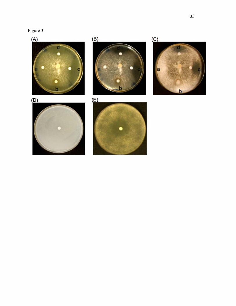
Sp-AMP3 strongly inhibited both hyphal growth and spore germination of H. annosum (Fig. 3).
Addition of 10 µg·ml–1 Sp-AMP3 caused nearly complete inhibition during a 3-day incubation
period. Controls (samples taken before induction, in the same buffer as the protein sample, as
well as the buffer itself) did not inhibit mycelial growth or spore germination. Sp-AMP3 samples
with and without His-tags had equivalent effects on the fungal growth.
Binding of fungal cell-wall polysaccharides to Sp-AMP3
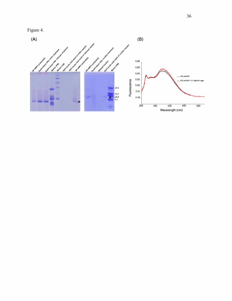
Sp-AMP3 did not bind to insoluble chitin and chitosan, with most of the protein remaining in the
supernatant when the protein was incubated with either of these polysaccharides (see Fig. 4a). By
contrast, treatment of the Sp-AMP3 solution with curdlan (an insoluble β-(1,3)-D-glucan)
resulted in strong binding; there was no protein evident in the initial supernatant or in the wash
with buffer, while the majority of the Sp-AMP3 was denatured/released with NaCl/acetic acid.
The binding of soluble sugars to Sp-AMP3 could be detected using the fluorescence change of at
least one tryptophan residue of the protein when sugar bound. The ligands tested included β-
(1,3)-glucan sugars ranging from laminaribiose up to laminariheptaose, glucan (laminarin),
glucose, cellobiose, cellotriose and cellopentaose. The sugars did not themselves fluoresce in the
absence of Sp-AMP3. Only sugars containing β-(1,3)-glucan (i.e. the laminarioligosaccharides
and laminarin) were found to bind significantly, as illustrated for laminaribiose in Fig. 4b;
additional results are shown in the Supplementary Materials. None of the other sugars tested
(including glucose) caused any change in the fluorescence signal.
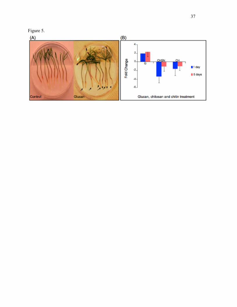
Effect of exogenous carboyhydrate treatment on Sp-AMP expression
17
To investigate the effect of fungal cell wall components on Sp-AMP expression and regulation,
we monitored the responses of Scots pine seedlings after treatment with chitin, chitosan or
glucan (laminarin). All three compounds provoked strong discoloration in the roots at 5 days
post-inoculation, although glucan treatment provoked the greatest response at both 1 and 5 days
(Fig. 5a). Analyses of Sp-AMP expression by qRT-PCR showed a 2-fold induction of Sp-AMPs
expression at a very early stage after glucan treatment, which persisted over time (Fig. 5b). The
qRT-PCR analysis also revealed that chitin and chitosan suppressed Sp-AMP expression.
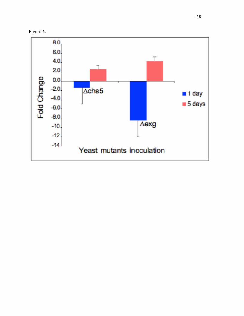
Effects of yeast mutants on Sp-AMP expression
The qRT-PCR analysis showed that inoculation with yeast mutants having ~4-fold reduced
levels of chitin (Δchs5 mutant; (Santos et al. 1997)) or increased levels of β-(1,6)-glucan (Δexg
mutant (Cappellaro et al. 1998)) both caused an increased transcription of Sp-AMP genes in
Scots pine roots relative to the wild type yeast control, at 5 days post-inoculation (Fig. 6).
Inoculation with Δexg yeast induced the expression of Sp-AMPs 4-fold, compared to the 2-fold
increase with Δchs5 yeast, at this time point. At a very early stage, there was no significant
differential expression with Δchs5 compared to the wild type yeast inoculation, but a significant
reduction in Sp-AMP transcript abundance was seen with the Δexg mutant (Fig. 6).
Sequence alignment and homology modeling
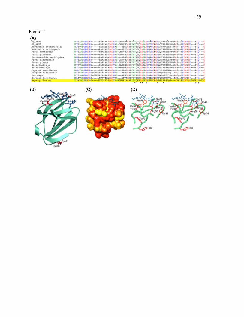
Differences among the four sequenced Sp-AMPs are very small. Sp-AMP1 differs from the other
three Sp-AMPs by virtue of only 5-6 amino acid changes in the 79-residue mature protein (i.e.
~93% identity). Sp-AMP2 and Sp-AMP4 are identical even at the level of the DNA sequence,
and differ from Sp-AMP3 at a single amino-acid position (residue 44 of the mature protein is
18
serine in Sp-AMP1 and Sp-AMP3, and alanine in Sp-AMP2 and Sp-AMP4). For these reasons,
only Sp-AMP1 and Sp-AMP2 sequences are shown in Fig. 7a.
Other sequences identified in a BLAST search represent mostly plant proteins with amino-acid
identities of at least 39% over the entire Sp-AMP sequence. A number of fungal proteins are also
found, with similar levels of sequence identity, although not covering the first 16 residues. One
example is highlighted in Fig. 7a, the protein from Aspergillus fumigatus, but others (with 67%
or greater identity to the A. fumigatus protein) have been reported in Uncinocarpus reesii,
Neosartorya fischeri and Aspergillus clavatus; a protein from Penicillium chrysogenum is
somewhat more distant. Residues that are strongly conserved among the plant proteins are also
well represented in these fungal sequences. The Heterobasidion genome (publically available at
http://genome.jgi-psf.org/Hetan2/Hetan2.home.html), however, does not appear to contain
equivalent proteins. The genomes of L. rufus and S. sanguinolentum are not yet available.
The only structure located by a BLAST search is that of MiAMP1 (solved by NMR, PDB entry
1C01; 68% amino acid identity to Sp-AMP3), an antimicrobial protein from macadamia with
unknown mode of action. A homology model of Sp-AMP3 based on MiAMP1 is illustrated in
Fig. 7b, allowing the conservation of various residues in the plant proteins to be placed in a
structural context. The largest cluster of conserved surface residues lies on a flat face of the
protein near Tyr46, as shown in Fig. 7c and d.
Structural similarity searches with DALI (Holm 1998) using the MiAMP1 structure identified a
number of additional proteins of interest (Z-scores of 4-7) belonging to the gamma-crystallin-like
19
superfamily of SCOP (Murzin et al. 1995); these have a fold similar to Sp-AMPs and MiAMP1,
but generally less than 15% sequence identity with them. Examples include the killer toxin
secreted by the yeast Williopsis mrakii (PDB entry 1WKT), as well as bacterial proteins such as
Streptomyces spp. killer toxin-like protein (PDB entry 1F53) and an anti-fungal protein from
Streptomyces tendae Tü901 (PDB entry 1G6E). The MiAMP1 comparison with the W. mrakii
protein, for instance, gives a DALI Z-score of 4.5, a root-mean-square difference of 2.6 Å over
72% of the α-carbons of the protein, and an amino-acid identity in the matched regions of the
sequence/structure of 13%.
Discussion
In this study, roots from Scots pine seedlings were used to study fungal-conifer interactions. The
validity of using seedling roots as an experimental model for H. annosum infection has been
addressed previously (Asiegbu et al. 1994). During development, the conifer root undergoes
several morphological and physiological transformations. The non-suberized seedling roots are
gradually suberized, as the cortex disappears and the secondary xylem in the vascular region
(stele) becomes dominant. Later, the epidermis, cortex and endodermis are replaced by the
rhytidome, phelloderm and phloem layers. These morphological changes occur in parallel with
constitutive and inducible defence responses. H. annosum is one of very few fungal pathogens
able to infect conifer roots of all ages (Asiegbu et al. 1993). In our experimental model, seedlings
are grown under controlled, sterile conditions, and used at an age when their genetic differences
are not strongly expressed, thus increasing the efficiency and reproducibility of the testing
protocol.
20
Scots pine roots responded differently when they were exposed to three Russulales fungi
belonging to distinct ecological functional groups. Sp-AMP expression was induced slightly upon
the first physical encounter with the saprotrophic or mutualistic fungi, but not with the pathogen.
By contrast, with prolonged incubation, the Sp-AMP gene was significantly expressed only in
response to the pathogen (Fig. 1). The differential expression shows that the host is able to
distinguish among the diverse lifestyles of the inoculated fungi, and suggests a role for Sp-AMPs
in defence. These results confirm and extend earlier work with more distantly related fungi
(Adomas et al. 2008), which included both saprotrophs, which derive their nutrients from the
surrounding dead organic matter, and symbiotic fungi, which depend on the living plant host for
carbon. In contrast to these two types of fungi, H. annosum is capable of invasive growth into
host cells. AMP induction is in fact occurring on a timescale similar to that observed for the
pathogen’s invasion into the plant tissues (Asiegbu et al. 1994; Li and Asiegbu 2004). The delay
in Sp-AMP expression during challenge with pathogenic fungi suggests the possibility of some
form of masking by the invading fungus as a means to evade host defences (Jones and Dangl
2006). Protoplasts from both pathogenic and mutualistic/beneficial, but not saprotrophic, fungi
induced strong Sp-AMP expression (Fig. 1c), indicating that factors other than cell-wall
components can also trigger the process, possibly including molecules secreted by the fungal
cells. However, we are at present unable to explain the different responses observed when pine
roots were inoculated with protoplasts versus mycelial homogenate from the mutualistic fungus.
It is possible that the protective cell wall of the mutualist fungus includes components that
regulate the course of symbiosis as well as the nature/pattern of associated host responses.
21
The crucial roles plant hormones play in regulating developmental processes, as well as
responses to a wide range of biotic and abiotic stresses, have been reviewed elsewhere (Bari and
Jones 2009). Sp-AMPs were shown here to be up-regulated after a 1-day treatment with SA or
the ethylene precursor, ACC, but not with MeJA or H2O2 (Fig. 2). The accumulation of SA in
response to elicitors and pathogen challenge was earlier shown in pine (Davis et al. 2002). SA is
furthermore known to induce specific sets of PR genes (Pieterse and van Loon 1999) prior to the
establishment of systemic acquired resistance (Grant and Lamb 2006). In contrast to the results
with Sp-AMP, expression of Pm-AMP1, a homolog in western white pine (Pinus monitcola) that
is involved in defence against the blister rust fungus Cronartium ribicola, is induced in healthy
foliage 4 days after MeJA treatment (Ekramoddoullah et al. 2006). The longer incubation time
before sampling may explain the different outcomes; it is also possible that the defence
signalling network activated by the plant is dependent on the nature of the pathogen and its mode
of pathogenicity (reviewed by (Bari and Jones 2009)) . The induction of Sp-AMP by ACC
suggests the involvement of ethylene in Sp-AMP regulation during biotic and abiotic stress.
Ethylene signalling has been implicated in induced cellular and chemical defences in conifers,
and is vital in the activation of cells specialized for the formation of defence-related terpenoids
and phenolics in the outermost bark and phloem tissues (Hudgins et al. 2006).
The similarity of the four known Sp-AMP sequences (93-100% amino acid sequence identity,
Fig. 7a) strongly suggests that all have similar structure and biological function. Homology
modeling based on the NMR structure of the macadamia antimicrobial protein, MiAMP1 (68%
identity), indicated that proteins of this type possess a Greek-key β-barrel fold, as illustrated in
Fig. 7b. The absence of a pocket or cleft indicates that enzymatic activity is unlikely, and so a
22
binding function was expected. The content of negatively-charged residues in the AMPs is
strikingly low (~2%, compared to a more usual value of ~12%, e.g.
http://expasy.org/sprot/relnotes/relstat.html), which leads to pIs of 8.5 or more. However, no
clustering of the positively charged residues is seen on the surface that would suggest binding of
negatively-charged ligands (such as many membrane lipids). Obtaining Sp-AMP for direct
biological and biochemical tests was viewed as a crucial next step. However, numerous
strategies for E. coli expression produced only large quantities of insoluble protein, which we
could not refold (unpublished data). Experiments with Pichia pastoris were more successful.
Although the yield of soluble Sp-AMP3 was extremely variable and very low at best, sufficient
material was obtained to demonstrate that the protein did possess antifungal activity (Fig. 3), and
had marked effects on fungal hyphae and spores (Sooriyaarachchi et al., unpublished
microscopic studies). To test the hypothesis that the biological function involves some
component of the fungal cell wall, our assays of binding activity included the main compounds
in that structure, as well as those in plant cell walls. The results showed that β-glucan sugars, in
both soluble and insoluble forms, bind to Sp-AMP3, while sugars from the chitin, chitosan and
cellulose classes did not (Fig. 4). Residues strongly conserved in plant sequences are clustered on
a flat face on the AMP surface, forming a patch that is of sufficient size to bind at least four, but
no more than six, sugar units in a β-(1,3)-glucan molecule (Fig. 7c and d); two of the three
conserved disulfide bonds (Cys21-Cys79 and Cys23-Cys51) help maintain its shape. This
surface includes polar residues, as well as two aromatics (Tyr46 and Phe76). Consideration of
the homology model also suggests that the change in fluorescence seen when soluble sugars are
bound reflects a (probably quite small) change in the overall protein structure, as neither of the
23
tryptophans of the sequence (Trp6 and Trp72) is expected to be exposed on the putative binding
surface, and so they will not make direct contact with a bound ligand (Fig. 7d).
Our experiments have thus revealed that the Sp-AMPs represent a novel family of β-glucan
binding proteins. Because of their antimicrobial properties, increased expression in response to
pathogen challenge, and presence in a wide variety of plants (as reviewed elsewhere (Manners
2009)), we believe they define a new PR family, PR-19. We note that PR-2 comprises β-(1,3)-
endoglucanases, but to date no PR family has been linked to the non-catalytic role of glucan
binding. Earlier anti-glucanase immunogold labeling experiments demonstrated that glucans are
accessible in Heterobasidion cell walls (Asiegbu et al. 1995), and so are a biologically
reasonable target for PR-19 proteins. The discovery that glucans are ligands of interest also
suggests that problems in heterologous expression were due to counterproductive interactions
between Sp-AMPs and the expression hosts tested. Alternate expression systems will be
explored in future work. The availability of additional protein will allow us to investigate many
interesting aspects of the Sp-AMP structure and function that are presently unclear.
In agreement with studies of the protein, addition of exogenous β-glucan induces Sp-AMP
expression, but addition of chitosan and chitin does not (Fig. 5). The fact that mutants with
increased β-(1,6)-glucan (Jiang et al. 1995), but normal levels of β-(1,3)-glucan and chitin,
induced Sp-AMP expression suggests a role for other structural forms of glucan (Fig. 6).
Structural similarities of the PR-19 proteins to fungal and bacterial ones like the killer protein
from the yeast W. mrakii are also intriguing. Although the relationships are very distant, the
24
killer protein is known to inhibit the action of β-(1,3)-glucan synthase in sensitive yeast strains,
apparently by competing for binding of β-glucan (Peng et al. 2010). Taken together with our
identification of AMP-like sequences in some fungal species (Fig. 7a), this suggests that such
proteins are not restricted to plants, but are a fairly widespread way for organisms to protect
themselves.. Differences in the fungal cell walls would allow the glucan-binding proteins to act
on some organisms, but leave others unscathed. In vulnerable fungi, Sp-AMP binding may, for
example, interfere with glucan assembly, which could alter cell wall structure, so causing
morphological distortion of hyphae. Effects on glucan could also lead to a weakening of the
membrane and compromise cell wall integrity, with the result of unusual spore and hyphal
swellings and consequently a burst.
Both our demonstration that recombinant Sp-AMP has a potent inhibitory effect on spore and
hyphae development of the conifer pathogen H. annosum, as well as evidence that this property
can be linked to the binding to major fungal cell wall components (i.e. glucans), provide vital
leads for future work. Exploring the practical applications of the Sp-AMPs themselves, further
investigation of their spectrum of antimicrobial action, and developing a functional synthetic
mimic, will be priorities.
Acknowledgements
This work was supported by grants from the Swedish Research Council for the Environment,
Agricultural Sciences and Spatial Planning (FORMAS) and the Swedish Research Council (VR)
to SLM, and from the Academy of Finland (AKA) and the University of Helsinki Research Fund
to FOA. We extend our sincere thanks to Christin Hansson, Johan Winquist and Kerstin Ahlgren
25
for their heroic attempts at E. coli expression. We also especially thank Prof. Ulf Hellman,
Ludwig Institute for Cancer Research, Uppsala, Sweden for mass spectroscopic analysis, and
Gulaim A. Seisenbaeva, Department of Chemistry, Swedish University of Agricultural Sciences
for help with the scanning electron microscopy.
References
Adomas A, Heller G, Li GS, Olson A, Chu TM, Osborne J, Craig D, Van Zyl L, Wolfinger R, Sederoff R, Dean RA, Stenlid J, Finlay R, Asiegbu FO (2007) Transcript profiling of a conifer pathosystem: response of Pinus sylvestris root tissues to pathogen (Heterobasidion annosum) invasion. Tree Physiology 27 (10):1441-1458
Adomas A, Heller G, Olson A, Osborne J, Karlsson M, Nahalkova J, Van Zyl L, Sederoff R, Stenlid J, Finlay R, Asiegbu FO (2008) Comparative analysis of transcript abundance in Pinus sylvestris after challenge with a saprotrophic, pathogenic or mutualistic fungus. Tree Physiol 28 (6):885-897
Altschul SF, Madden TL, Schaffer AA, Zhang J, Zhang Z, Miller W, Lipman DJ (1997) Gapped BLAST and PSI-BLAST: a new generation of protein database search programs. Nucleic Acids Res 25 (17):3389-3402
Asiegbu FO, Adomas A, Stenlid J (2005) Conifer root and butt rot caused by Heterobasidion annosum (Fr.) Bref. s.l. Molecular Plant Pathology 6 (4):395-409
Asiegbu FO, Choi W, Li G, Nahalkova J, Dean RA (2003) Isolation of a novel antimicrobial peptide gene (Sp-AMP) homologue from Pinus sylvestris (Scots pine) following infection with the root rot fungus Heterobasidion annosum. FEMS Microbiol Lett 228 (1):27-31
Asiegbu FO, Daniel G, Johansson M (1993) Studies on the infection of Norway spruce roots by Heterobasidion annosum. Canadian Journal of Botany 71:1552-1561
Asiegbu FO, Daniel G, Johansson M (1994) Defense-related reactions of seedling roots of norway spruce to infection by Heterobasidion annosum (Fr.) Bref. Physiological and Molecular Plant Pathology 45 (1):1-19
Asiegbu FO, Denekamp M, Daniel G, Johansson M (1995) Immune Cytochemical-Localization of Pathogenesis-Related Proteins in Roots of Norway Spruce Infected with Heterobasidion annosum. European Journal of Forest Pathology 25 (3):169-178
Bari R, Jones JD (2009) Role of plant hormones in plant defence responses. Plant Mol Biol 69 (4):473-488. doi:10.1007/s11103-008-9435-0
Benhamou N, Thériault G (1992) Treatment with chitosan enhances resistance of tomato plants to the crown and root pathogen Fusarium oxysporum f. sp. radicis-lycopersici. Physiol Mol Plant Pathol (41 ):34–52
Benson DA, Karsch-Mizrachi I, Lipman DJ, Ostell J, Sayers EW (2011) GenBank. Nucleic Acids Res 39 (Database issue):D32-37. doi:10.1093/nar/gkq1079
Berman HM, Westbrook J, Feng Z, Gilliland G, Bhat TN, Weissig H, Shindyalov IN, Bourne PE (2000) The Protein Data Bank. Nucleic Acids Res 28 (1):235-242
26
Broekaert WF, Cammue BPA, DeBolle MFC, Thevissen K, DeSamblanx GW, Osborn RW (1997) Antimicrobial peptides from plants. Critical Reviews in Plant Sciences 16 (3):297-323
Cappellaro C, Mrsa V, Tanner W (1998) New potential cell wall glucanases of Saccharomyces cerevisiae and their involvement in mating. J Bacteriol 180 (19):5030-5037
Chang S, Puryear J, Cairney J (1993) A simple and efficient method for isolating RNA from pine trees. Plant Molecular Biology Reporter 11:113-116
Davis JM, Wu H, Cooke JE, Reed JM, Luce KS, Michler CH (2002) Pathogen challenge, salicylic acid, and jasmonic acid regulate expression of chitinase gene homologs in pine. Mol Plant Microbe Interact 15 (4):380-387. doi:10.1094/MPMI.2002.15.4.380
Ekramoddoullah AK, Liu JJ, Zamani A (2006) Cloning and Characterization of a Putative Antifungal Peptide Gene (Pm-AMP1) in Pinus monticola. Phytopathology 96 (2):164-170. doi:10.1094/PHYTO-96-0164
Grant M, Lamb C (2006) Systemic immunity. Current Opinion in Plant Biology 9 (4):414-420. doi:Doi 10.1016/J.Pbi.2006.05.013
Harris M, Jones TA (2001) Molray - a web interface between O and the POV-Ray ray tracer. Acta Crystallographica Section D-Biological Crystallography 57:1201-1203
Hashimoto JG, Beadles-Bohling AS, Wiren KM (2004) Comparison of RiboGreen and 18S rRNA quantitation for normalizing real-time RT-PCR expression analysis. Biotechniques 36 (1):54-56, 58-60
Holm L (1998) Unification of protein families. Curr Opin Struct Biol 8 (3):372-379. doi:S0959-440X(98)80072-9 [pii]
Hudgins JW, Ralph SG, Franceschi VR, Bohlmann J (2006) Ethylene in induced conifer defense: cDNA cloning, protein expression, and cellular and subcellular localization of 1-aminocyclopropane-1-carboxylate oxidase in resin duct and phenolic parenchyma cells. Planta 224 (4):865-877. doi:Doi 10.1007/S00425-006-0274-4
Jiang B, Ram AF, Sheraton J, Klis FM, Bussey H (1995) Regulation of cell wall beta-glucan assembly: PTC1 negatively affects PBS2 action in a pathway that includes modulation of EXG1 transcription. Mol Gen Genet 248 (3):260-269
Jones JD, Dangl JL (2006) The plant immune system. Nature 444 (7117):323-329. doi:nature05286 [pii]
10.1038/nature05286 Jones TA, Zou JY, Cowan SW, Kjeldgaard M (1991) Improved methods for building protein
models in electron density maps and the location of errors in these models. Acta Crystallogr A 47 ( Pt 2):110-119
Kleywegt GJ, Zou JY, Kjeldgaard M, Jones TA (2001) Around O. Rossmann, M.G., et al. (Eds.). International Tables for Crystallography, Vol F Crystallography of Biological Macromolecules:pp. 353-356
Kraulis PJ (1991) Molscript - a program to produce both detailed and schematic plots of protein structures. Journal of Applied Crystallography 24:946-950
Li G, Asiegbu FO (2004) Use of Scots pine seedling roots as an experimental model to investigate gene expression during interaction with the conifer pathogen Heterobasidion annosum (P-type). J Plant Res 117 (2):155-162. doi:10.1007/s10265-003-0140-4
Livak KJ, Schmittgen TD (2001) Analysis of relative gene expression data using real-time quantitative PCR and the 2(-Delta Delta C(T)) Method. Methods 25 (4):402-408. doi:10.1006/meth.2001.1262
27
S1046-2023(01)91262-9 [pii] Manners JM (2009) Primitive Defence: The MiAMP1 Antimicrobial Peptide Family. Plant
Molecular Biology Reporter 27 (3):237-242. doi:Doi 10.1007/S11105-008-0083-Y McManus AM, Nielsen KJ, Marcus JP, Harrison SJ, Green JL, Manners JM, Craik DJ (1999)
MiAMP1, a novel protein from Macadamia integrifolia adopts a Greek key beta-barrel fold unique amongst plant antimicrobial proteins. J Mol Biol 293 (3):629-638
Montesinos E (2007) Antimicrobial peptides and plant disease control. FEMS Microbiol Lett 270 (1):1-11. doi:FML683 [pii]
10.1111/j.1574-6968.2007.00683.x Murzin AG, Brenner SE, Hubbard T, Chothia C (1995) SCOP: a structural classification of
proteins database for the investigation of sequences and structures. J Mol Biol 247 (4):536-540. doi:10.1006/jmbi.1995.0159
S0022283685701593 [pii] Peng Y, Chi Z, Wang X, Li J (2010) beta-1,3-glucanase inhibits activity of the killer toxin
produced by the marine-derived yeast Williopsis saturnus WC91-2. Mar Biotechnol (NY) 12 (4):479-485. doi:10.1007/s10126-009-9243-9
Pieterse CM, van Loon LC (1999) Salicylic acid-independent plant defence pathways. Trends Plant Sci 4 (2):52-58
Sambrook J, Russell DW (2001) Molecular Cloning: A Laboratory Manual. Cold Spring Harbor Laboratory Press. Cold Spring Harbor, New York
Santos B, Duran A, Valdivieso MH (1997) CHS5, a gene involved in chitin synthesis and mating in Saccharomyces cerevisiae. Mol Cell Biol 17 (5):2485-2496
Silberbach M, Schafer M, Huser AT, Kalinowski J, Puhler A, Kramer R, Burkovski A (2005) Adaptation of Corynebacterium glutamicum to ammonium limitation: a global analysis using transcriptome and proteome techniques. Appl Environ Microbiol 71 (5):2391-2402. doi:71/5/2391 [pii]
10.1128/AEM.71.5.2391-2402.2005 Stenlid J (1985) Population structure of Heterobasidion annosum as determined by somatic
incompatibility, sexual incompatibility and isoenzyme patterns. Can J Bot (63):187–198 Thomma BP, Eggermont K, Penninckx IA, Mauch-Mani B, Vogelsang R, Cammue BP,
Broekaert WF (1998) Separate jasmonate-dependent and salicylate-dependent defense-response pathways in Arabidopsis are essential for resistance to distinct microbial pathogens. Proc Natl Acad Sci U S A 95 (25):15107-15111
Thompson JD, Higgins DG, Gibson TJ (1994) Clustal-W - Improving the sensitivity of progressive multiple sequence alignment through sequence weighting, position-specific gap penalties and weight matrix choice. Nucleic Acids Res 22 (22):4673-4680
van Loon LC, Rep M, Pieterse CM (2006) Significance of inducible defense-related proteins in infected plants. Annu Rev Phytopathol 44:135-162. doi:10.1146/annurev.phyto.44.070505.143425
van Loon LC, Van Strien EA (1999) The families of pathogenesis-related proteins, their activities, and comparative analysis of PR-1 type proteins. Physiol Mol Plant Pathol 55:85-97
28
Figure legends
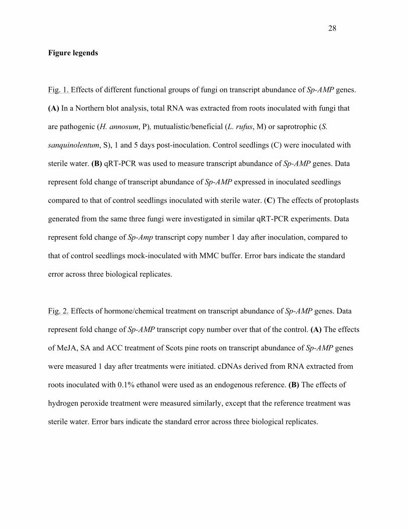
Fig. 1. Effects of different functional groups of fungi on transcript abundance of Sp-AMP genes.
(A) In a Northern blot analysis, total RNA was extracted from roots inoculated with fungi that
are pathogenic (H. annosum, P), mutualistic/beneficial (L. rufus, M) or saprotrophic (S.
sanquinolentum, S), 1 and 5 days post-inoculation. Control seedlings (C) were inoculated with
sterile water. (B) qRT-PCR was used to measure transcript abundance of Sp-AMP genes. Data
represent fold change of transcript abundance of Sp-AMP expressed in inoculated seedlings
compared to that of control seedlings inoculated with sterile water. (C) The effects of protoplasts
generated from the same three fungi were investigated in similar qRT-PCR experiments. Data
represent fold change of Sp-Amp transcript copy number 1 day after inoculation, compared to
that of control seedlings mock-inoculated with MMC buffer. Error bars indicate the standard
error across three biological replicates.
Fig. 2. Effects of hormone/chemical treatment on transcript abundance of Sp-AMP genes. Data
represent fold change of Sp-AMP transcript copy number over that of the control. (A) The effects
of MeJA, SA and ACC treatment of Scots pine roots on transcript abundance of Sp-AMP genes
were measured 1 day after treatments were initiated. cDNAs derived from RNA extracted from
roots inoculated with 0.1% ethanol were used as an endogenous reference. (B) The effects of
hydrogen peroxide treatment were measured similarly, except that the reference treatment was
sterile water. Error bars indicate the standard error across three biological replicates.
29
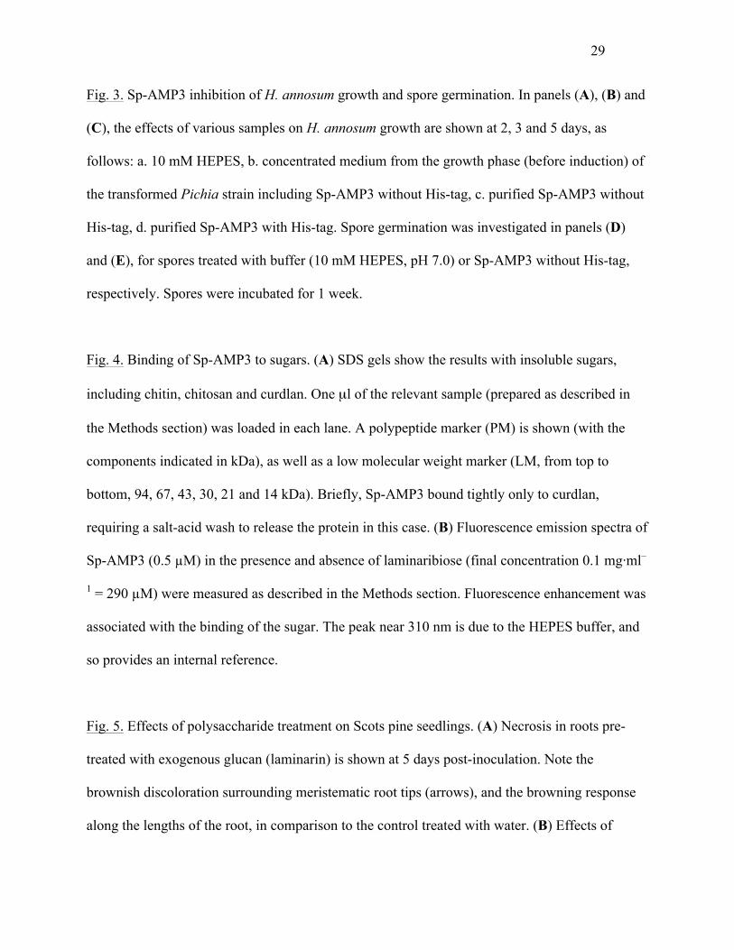
Fig. 3. Sp-AMP3 inhibition of H. annosum growth and spore germination. In panels (A), (B) and
(C), the effects of various samples on H. annosum growth are shown at 2, 3 and 5 days, as
follows: a. 10 mM HEPES, b. concentrated medium from the growth phase (before induction) of
the transformed Pichia strain including Sp-AMP3 without His-tag, c. purified Sp-AMP3 without
His-tag, d. purified Sp-AMP3 with His-tag. Spore germination was investigated in panels (D)
and (E), for spores treated with buffer (10 mM HEPES, pH 7.0) or Sp-AMP3 without His-tag,
respectively. Spores were incubated for 1 week.
Fig. 4. Binding of Sp-AMP3 to sugars. (A) SDS gels show the results with insoluble sugars,
including chitin, chitosan and curdlan. One µl of the relevant sample (prepared as described in
the Methods section) was loaded in each lane. A polypeptide marker (PM) is shown (with the
components indicated in kDa), as well as a low molecular weight marker (LM, from top to
bottom, 94, 67, 43, 30, 21 and 14 kDa). Briefly, Sp-AMP3 bound tightly only to curdlan,
requiring a salt-acid wash to release the protein in this case. (B) Fluorescence emission spectra of
Sp-AMP3 (0.5 µM) in the presence and absence of laminaribiose (final concentration 0.1 mg·ml–
1 = 290 µM) were measured as described in the Methods section. Fluorescence enhancement was
associated with the binding of the sugar. The peak near 310 nm is due to the HEPES buffer, and
so provides an internal reference.
Fig. 5. Effects of polysaccharide treatment on Scots pine seedlings. (A) Necrosis in roots pre-
treated with exogenous glucan (laminarin) is shown at 5 days post-inoculation. Note the
brownish discoloration surrounding meristematic root tips (arrows), and the browning response
along the lengths of the root, in comparison to the control treated with water. (B) Effects of
30
glucan (G), chitosan (CHSN) or chitin (CH) treatment of roots on transcript abundance of the Sp-
AMP gene at 1 and 5 days, measured using qRT-PCR, as described above. Data represent Sp-
AMP transcript copy number change compared to the control seedlings. Error bars indicate the
standard error across three biological replicates.
Fig. 6. Effects of yeast mutants on Sp-AMP transcription. Scots pine roots were inoculated with
yeast mutants with either 4-fold reduced levels of chitin (Δchs5 mutant (Santos et al., 1997)) or
increased levels of β-(1,6)-glucan (Δexg mutant (Cappellaro et al. 1998)). Transcript abundance
of Sp-AMP genes was measured using qRT-PCR at 1 and 5 days. Data represent Sp-Amp
transcript copy number change compared to that of the control inoculated with wild type S.
cerevisiae. Error bars indicate the standard error across three biological replicates.
Fig. 7. Sequence comparison and homology modeling. (A) Sequence alignment of Sp-AMP1,
Sp-AMP2, MiAMP1 and a number of similar plant proteins is shown, with conserved residues
colored red (identical), green (strongly similar) or blue (weakly similar), defined according to the
algorithms of CLUSTAL W. A related sequence that represents several fungal proteins is
highlighted in yellow at the bottom. Residues that differ among the Sp-AMPs are marked with –
above the sequences, while those on the proposed binding surface are marked with + below. (B)
Ribbon cartoon of the Sp-AMP3 homology model, showing the conserved disulfide bonds in
crimson. A manually modeled laminaritetraose in light steel blue is shown for purposes of
comparison, not to suggest a particular mode of binding. (C) Molecular surface calculated based
on the homology model, colored according to sequence conservation among the plant proteins
(ranging from yellow for the non-conserved residues, to red for those completely conserved).
31
The most conserved faces of the protein are shown; conserved residues not on the putative
binding surface represent buried residues critical to the protein’s fold. (D) Stereo representation
of the proposed binding surface with modeled laminaritetraose; portions of the protein structure
have been removed for clarity. The tryptophan residues (Trp6 and Trp72) that could explain the
fluorescence changes are also shown.

32
Figure 1.
33
34
Figure 2.
35
Figure 3.
36
Figure 4.
37
Figure 5.
38
Figure 6.
39
Figure 7.