Exploring the synthetic possibilities and siRNA delivery...
226
1 Exploring the synthetic possibilities and siRNA delivery potential of Small Molecule Carriers (SMoCs) by Matthew John Gooding Submitted in accordance with the requirements for the degree of Doctor of Philosophy AUGUST 2011 UCL I, Matthew John Gooding, confirm that the work presented in this thesis is my own. Where information has been derived from other sources, I confirm that this has been indicated in the thesis. ___________________________________________________
Transcript of Exploring the synthetic possibilities and siRNA delivery...
1
Exploring the synthetic possibilities
and siRNA delivery potential of
Small Molecule Carriers (SMoCs)
by
Matthew John Gooding
Submitted in accordance with the requirements
for the degree of
Doctor of Philosophy
AUGUST 2011
UCL
I, Matthew John Gooding, confirm that the work presented in this thesis is my own. Where
information has been derived from other sources, I confirm that this has been indicated in the
thesis.
___________________________________________________
2
Abstract
Delivery of proteins and nucleic acids into cells is a major challenge to the
development of biological therapeutics. Cell penetrating peptides (CPPs) and
cationic liposomes have been shown to internalise short interfering RNA (siRNA) to
achieve gene silencing, but no standard reagent exists which can safely deliver
macromolecules both in vitro and in vivo. Small Molecule Carriers (SMoCs) are
amphipathic α-helix mimetics displaying guanidine groups in order to mimic the
structure of the CPP penetratin. Previously, SMoCs were shown to effectively
deliver active proteins into cells. It is hypothesized that SMoCs may also be applied
to the delivery of siRNA into cells in order to knockdown target genes. In addition,
since cell surface binding is thought to be a crucial step in CPP internalisation, a new
SMoC which optimises binding to proteoglycans may be more efficiently taken up.
The aims of this thesis are to optimise the synthesis of the SMoCs in order to
increase the quantity of product; to demonstrate that SMoCs may be used as siRNA
delivery agents; and to design and synthesize a new SMoC which maximises siRNA
uptake. The synthesis of SMoCs has been significantly enhanced, with the
development of new reagents to improve the yields and cost of production. The
electrostatic interactions of SMoCs with siRNA have been characterised, using NMR
to examine a π-cation interaction which may contribute to anion binding, as well as
determination of binding affinities using ITC and gel shift assays. Initial
experiments using SMoC-siRNA complexes show significant mRNA knowdown
which demonstrates the potential of SMoCs as siRNA delivery vectors. Finally, a
new SMoC has been designed and synthesized which represents the first in a new
class of dendritic SMoCs which are designed to maximise binding to the cell surface.
This SMoC is also capable of delivering siRNA into cells, and may also be expanded
by the addition of targeting peptides.
3
Table of Contents
Abstract ........................................................................................................................ 2
Table of Contents ......................................................................................................... 3
List of Figures .............................................................................................................. 9
List of Schemes .......................................................................................................... 10
List of Tables.............................................................................................................. 11
List of Abbreviations.................................................................................................. 12
Acknowledgements .................................................................................................... 14
Chapter 1: Introduction .......................................................................................... 16
1.1. Macromolecular Therapeutics ......................................................................... 16
1.2. Cell Penetrating Peptides ................................................................................ 22
1.2.1. Uptake Mechanism .................................................................................. 23
1.2.1.1. Cell-surface binding .......................................................................... 24
1.2.1.2. Endosomal Escape ............................................................................ 27
1.3. RNA Interference ............................................................................................ 29
1.3.1. Mechanism ............................................................................................... 29
1.3.2. Delivery .................................................................................................... 32
1.3.2.1. Chemical Modifications .................................................................... 33
1.3.2.2. Liposomes and Lipoplexes ................................................................ 34
1.3.2.3. Cationic Polymers ............................................................................. 36
1.3.2.4. Cell Penetrating Peptides .................................................................. 38
1.3.2.5. Nanocarriers ...................................................................................... 48
1.4. SMoCs ............................................................................................................. 51
1.4.1. Synthesis .................................................................................................. 52
1.4.2. Translocation activity ............................................................................... 54
1.5. Thesis Overview.............................................................................................. 54

4
Chapter 2: Improving the synthetic route to the SMoCs ..................................... 58
2.1. Introduction ..................................................................................................... 58
2.2. Results ............................................................................................................. 60
2.2.1. Modifications to the synthetic route to 4G-SMoC ................................... 60
2.2.1.1. Use of PMC protecting group ........................................................... 60
2.2.1.2. Improving Suzuki-Miyaura coupling ................................................ 65
2.2.1.3. New Guanidinylation reagent ........................................................... 67
2.2.1.4. Linker nitrile reduction ..................................................................... 67
2.2.2. 4G-SMoC with meta linker ...................................................................... 69
2.2.2.1. Thioamide linker ............................................................................... 71
2.2.2.2. Acid Linker ....................................................................................... 75
2.2.3. 4G-SMoC disulfide-linked dimer ............................................................ 78
2.3. Discussion ....................................................................................................... 82
Chapter 3: Exploring the interactions of SMoCs with siRNA ............................. 87
3.1. Introduction ..................................................................................................... 87
3.1.1. The SMoC π-cation interaction ................................................................ 87
3.1.2. Characterisation of SMoC-siRNA complexes ......................................... 88
3.1.3. SMoC-mediated siRNA transfection ....................................................... 89
3.2. Results ............................................................................................................. 93
3.2.1. Molecular Dynamics ................................................................................ 93
3.2.2. NMR pKa study ........................................................................................ 95
3.2.3. 6G-SMoC Synthesis ............................................................................... 102
3.2.4. ITC ......................................................................................................... 105
3.2.5. Gel Shifts ................................................................................................ 106
3.2.6. siRNA transfection ................................................................................. 109
3.3. Discussion ..................................................................................................... 112

5
Chapter 4: Designing a new SMoC for siRNA delivery ..................................... 117
4.1. Introduction ................................................................................................... 117
4.2. Results ........................................................................................................... 119
4.2.1. Design of a new SMoC analogue ........................................................... 119
4.2.2. 4G-SMoC-Dr synthesis .......................................................................... 122
4.2.3. Conjugation to RGD .............................................................................. 125
4.2.4. Gel Shift ................................................................................................. 128
4.2.5. PCR ........................................................................................................ 130
4.3. Conclusions ................................................................................................... 131
Chapter 5: Experimental Methods ....................................................................... 134
5.1. General Methods ........................................................................................... 134
5.1.1. General chemistry methods .................................................................... 134
5.1.2. Gel shifts ................................................................................................ 134
5.1.3. Cell Culture ............................................................................................ 134
5.1.4. siRNA complexes .................................................................................. 135
5.1.5. siRNA Transfection ............................................................................... 135
5.1.6. RNA extraction and qRT-PCR............................................................... 135
5.1.7. Statistical Analysis ................................................................................. 136
5.2. Chapter 2 Methods ........................................................................................ 137
5.2.1. Syntheses ................................................................................................ 137
5.2.1.1. N-(2,2,5,7,8-pentamethylchroman-6-ylsulfonyl)-1H-pyrazole-1-carb-
oximidamide 12 ............................................................................................ 137
5.2.1.1. N-(N-(2-hydroxyethyl)carbamimidoyl)-2,2,5,7,8-pentamethylchrom-
an-6-sulfonamide 13..................................................................................... 138
5.2.1.2. N-(4,5-dihydro-1H-imidazol-2-yl)-2,2,5,7,8-pentamethylchroman-6-
sulfonamide 15 ............................................................................................. 139
5.2.1.3. Mitsunobu General Method ............................................................ 139
6
5.2.1.4. 2,3-Bis(2-benzyloxycarbonylamino)ethoxy)phenylboronic
acid 17 .......................................................................................................... 140
5.2.1.5. Potassium 2,3-Bis(2-benzyloxycarbonylamino)ethoxy)phenyltri-
fluoroborate 18 ............................................................................................. 141
5.2.1.6. Tetrabenzyl 2,2',2'',2'''-(4-cyanobiphenyl-2,2',3,3'-tetrayltetrakis-
(oxy))tetrakis(ethane-2,1-diyl)tetracarbamate 6 .......................................... 142
5.2.1.7. 2, 3, 2', 3'-Tetra(2-(N, N'-bis(tert-butoxycarbonyl)guanidino)ethyl-
oxy)-4-cyanobiphenyl 19 ............................................................................. 143
5.2.1.8. 2, 3, 2', 3'-Tetra(2-(N, N'-bis(tert-butoxycarbonyl)guanidino)ethyl-
oxy)-4-(aminomethyl)biphenyl 8 ................................................................. 144
5.2.1.9. tert-Butyl (2,2’,2’’,2’’’-(4-((3-(pyridine-2-yldsulfanyl)propanamido)-
methyl)biphenyl-2,2',3,3'-tetrayl)tetrakis(oxy)tetrakis(ethane-2,1-diyl)tetra-
kis(azanediyl)-tetrakis((tertbutoxycarbonylamino)methane-1-yl-1-ylidene)-
tetracarbamate 20 ......................................................................................... 145
5.2.1.10. 2,2’,2’’,2’’’-(4-((3-(Pyridine-2-yldsulfanyl)propanamido)methyl)-
biphenyl-2,2',3,3'-tetrayl)tetrakis(oxy)tetrakis(ethane-2,1-diyl)tetrakis(azane-
diyl)tetrakis(aminomethaniminium) 10 ....................................................... 146
5.2.1.11. 3-bromo-4,5-dihydroxybenzaldehyde 22 ...................................... 147
5.2.1.12. Benzyl 2,2'-(3-bromo-5-formyl-1,2-phenylene)bis(oxy)bis(ethane-
2,1-diyl)dicarbamate 23 ............................................................................... 148
5.2.1.13. Nitrile synthesis general method ................................................... 148
5.2.1.14. (E)-ethyl 3-(3-bromo-4,5-dihydroxyphenyl)acrylate 27 ............... 149
5.2.1.15. (E)-ethyl 3-(3,4-bis(2-(benzyloxycarbonylamino)ethoxy)-5-bromo-
phenyl)acrylate 28 ........................................................................................ 150
5.2.1.16. (E)-ethyl 3-(2',3',5,6-tetrakis(2-(benzyloxycarbonylamino)ethoxy)-
biphenyl-3-yl)acrylate 29 ............................................................................. 151
5.2.1.17. (2E)-ethyl 3-(2',3',5,6-tetrakis(2-(2,3-bis(tert-butoxycarbonyl)guan-
idino)ethoxy)biphenyl-3-yl)acrylate 30 ....................................................... 152
5.2.1.18. Ester hydrolysis ............................................................................. 153
7
5.2.1.19. 3,3'-disulfanediylbis(N-((2,2',3,3'-tetrakis(2-guanidinoethoxy)bi-
phenyl-4-yl)methyl)propanamide) 34 .......................................................... 154
5.3. Chapter 3 Methods ........................................................................................ 155
5.3.1. Molecular Dynamics .............................................................................. 155
5.3.2. NMR study ............................................................................................. 155
5.3.3. ITC ......................................................................................................... 155
5.3.4. Syntheses ................................................................................................ 156
5.3.4.1. Benzyl 2,2'-(biphenyl-2,2'-diylbis(oxy))bis(ethane-2,1-diyl)dicarba-
mate 35 ......................................................................................................... 156
5.3.4.2. Benzyl 2-(biphenyl-2-yloxy)ethylcarbamate 36 ............................. 157
5.3.4.3. Benzyl 2-(biphenyl-3-yloxy)ethylcarbamate 37 ............................. 158
5.3.4.4. Benzyl 2-phenoxyethylcarbamate 38 .............................................. 159
5.3.4.5. Tert-butyl (tert-butoxycarbonylamino)(2-phenoxyethylamino)methyl-
enecarbamate 39 ........................................................................................... 160
5.3.4.6. Tert-butyl (2,2'-(biphenyl-2,2'-diylbis(oxy))bis(ethane-2,1-diyl))bis-
(azanediyl) bis((tert-butoxycarbonylamino)methan-1-yl-1-ylidene)dicarbam-
ate 40 ............................................................................................................ 161
5.3.4.7. Tert-butyl (2-(biphenyl-2-yloxy)ethylamino)(tert-butoxycarbonyl-
amino)methylenecarbamate 41 .................................................................... 162
5.3.4.8. (Z)-tert-butyl (2-(biphenyl-3-yloxy)ethylamino)(tert-butoxycarbonyl-
amino)methylenecarbamate 42 .................................................................... 163
5.3.4.9. 1-(2-phenoxyethyl)guanidine 43 ..................................................... 164
5.3.4.10. 1,1'-(2,2'-(biphenyl-2,2'-diylbis(oxy))bis(ethane-2,1-diyl))di-
guanidine 44 ................................................................................................. 164
5.3.4.11. 1-(2-(biphenyl-3-yloxy)ethyl)guanidine 45 .................................. 165
5.3.4.12. 1-(2-(biphenyl-2-yloxy)ethyl)guanidine 46 .................................. 166
5.3.4.13. 6G-SMoC-NHCbz 48 ................................................................... 167
5.3.4.14. 6G-SMoC-Boc 50 ......................................................................... 169
8
5.3.4.15. 6G-SMoC 51 ................................................................................. 170
5.4. Chapter 4 Methods ........................................................................................ 170
5.4.1. Molecular dynamics ............................................................................... 170
5.4.2. Syntheses ................................................................................................ 171
5.4.2.1. 3,5-bis(2,3-bis(2-(((benzyloxy)carbonyl)amino)ethoxy)phenyl)ben-
zoic acid 53 .................................................................................................. 171
5.4.2.2. Tetra-Boc-4G-SMoC-dendrimer 54 ................................................ 172
5.4.2.3. 4G-SMoC-dendrimer 55 ................................................................. 173
5.4.2.4. Tetra-Boc-4G-SMoC-dendrimer-succinimidyl ester 56 ................. 174
5.4.2.5. 4G-SMoC-dendrimer-succinimidyl ester 57 ................................... 175
5.4.2.6. 4G-SMoC-dendrimer-[PEG2]2-cRGDfK 58 ................................... 176
Chapter 6: Discussion ............................................................................................ 178
6.1. Future Work .................................................................................................. 181
Bibliography ............................................................................................................. 185
Appendix I: Publication............................................................................................199
9
List of Figures
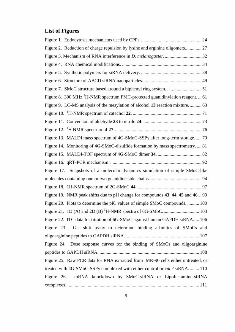
Figure 1. Endocytosis mechanisms used by CPPs. ................................................... 24
Figure 2. Reduction of charge repulsion by lysine and arginine oligomers.............. 27
Figure 3. Mechanism of RNA interference in D. melanogaster. ............................... 32
Figure 4. RNA chemical modifications. ................................................................... 34
Figure 5. Synthetic polymers for siRNA delivery. ................................................... 38
Figure 6. Structure of ABCD siRNA nanoparticles. ................................................. 49
Figure 7. SMoC structure based around a biphenyl ring system. ............................. 51
Figure 8. 300 MHz 1H-NMR spectrum PMC-protected guanidinylation reagent. ... 61
Figure 9. LC-MS analysis of the mesylation of alcohol 13 reaction mixture. .......... 63
Figure 10. 1H-NMR spectrum of catechol 22. .......................................................... 71
Figure 11. Conversion of aldehyde 23 to nitrile 24. ................................................. 73
Figure 12. 1H NMR spectrum of 27. ......................................................................... 76
Figure 13. MALDI mass spectrum of 4G-SMoC-SSPy after long-term storage. ..... 79
Figure 14. Monitoring of 4G-SMoC-disulfide formation by mass spectrometry. .... 81
Figure 15. MALDI-TOF spectrum of 4G-SMoC dimer 34. ..................................... 82
Figure 16. qRT-PCR mechanism. ............................................................................. 92
Figure 17. Snapshots of a molecular dynamics simulation of simple SMoC-like
molecules containing one or two guanidine side chains. ........................................... 94
Figure 18. 1H-NMR spectrum of 2G-SMoC 44. ...................................................... 97
Figure 19. NMR peak shifts due to pH change for compounds 43, 44, 45 and 46. .. 99
Figure 20. Plots to determine the pKa values of simple SMoC compounds. .......... 100
Figure 21. 1D (A) and 2D (B) 1H-NMR spectra of 6G-SMoC. .............................. 103
Figure 22. ITC data for titration of 6G-SMoC against human GAPDH siRNA. .... 106
Figure 23. Gel shift assay to determine binding affinities of SMoCs and
oligoarginine peptides to GAPDH siRNA. .............................................................. 107
Figure 24. Dose response curves for the binding of SMoCs and oligoarginine
peptides to GAPDH siRNA. .................................................................................... 108
Figure 25. Raw PCR data for RNA extracted from IMR-90 cells either untreated, or
treated with 4G-SMoC-SSPy complexed with either control or cdc7 siRNA. ........ 110
Figure 26. mRNA knockdown by SMoC-siRNA or Lipofectamine-siRNA
complexes. ................................................................................................................ 111
10
Figure 27. Illustration of the π-cation effect for a biphenyl SMoC molecule. ........ 112
Figure 28. cRGDfK peptide with a [PEG2]2 linker attached to the lysine residue.. 118
Figure 29. Examination of the crystal structure of heparin. .................................... 120
Figure 30. Chondroitin sulfate crystal structure obtained from the PDB. .............. 120
Figure 31. The energy minimised structure of 4G-SMoC-SSPy. ........................... 121
Figure 32. The energy minimised terphenyl structure of 4G-SMoC-Dr. ................ 122
Figure 33. 1H-NMR spectrum of 4G-SMoC-Dr. .................................................... 125
Figure 34. Monitoring of RGD coupling to 4G-SMoC-Dr by mass spectrometry. 128
Figure 35. siRNA gel shift assay for 4G-SMoC dendrimer. ................................... 129
Figure 36. mRNA expression levels in cells treated with 4G-SMoC-Dr:siRNA
complexes. ................................................................................................................ 130
Figure 37. mRNA knockdown mediated by 4G-SMoC-Dr:siRNA complexes. ..... 131
List of Schemes
Scheme 1. Current synthetic route to 4G-SMoC-SSPy. ........................................... 53
Scheme 2. Mechanism of 4G-SMoC Suzuki-Miyaura coupling using potassium
trifluoroborate salt. ..................................................................................................... 59
Scheme 3. Synthesis of a PMC-protected guanidine derivative. .............................. 62
Scheme 4. Cyclisation of alcohol 13 following mesylation. ..................................... 62
Scheme 5. Boronic acid synthesis. ............................................................................ 66
Scheme 6. Conversion of boronic ester to trifluoroborate salt. ................................ 66
Scheme 7. Guanidinylation using pyrazole reagent. ................................................. 67
Scheme 8. Reduction of nitrile to produce amine linker group. ............................... 68
Scheme 9. New synthetic route to 4G-SMoC-SSPy. ................................................ 69
Scheme 10. Demethylation of 5-bromovanillin. ....................................................... 70
Scheme 11. Proposed synthesis of thioamide linker. ................................................ 72
Scheme 12. Proposed aldol reaction to add ester group at linker position. .............. 75
Scheme 13. Proposed synthetic strategy for the preparation 4G-SMoC with a meta
carboxylic acid linker. ................................................................................................ 77
Scheme 14. Synthesis of 4G-SMoC-disulfide. ......................................................... 80
11
Scheme 15. Mechanism for coupling of the guanidine-PMC sidechain to phenol via
a Mitsunobu reaction using DIAD/TPP. .................................................................... 83
Scheme 16. Mechanism of the Wittig reaction. ........................................................ 85
Scheme 17. Synthesis of simple SMoC compounds for determination of guanidine
pKa. ............................................................................................................................. 96
Scheme 18. Synthesis of 6G-SMoC. ....................................................................... 104
Scheme 19. Retrosynthetic analysis of dendritic SMoC. ........................................ 123
Scheme 20. Synthesis of 4G-SMoC-dendrimer. ..................................................... 124
Scheme 21. Attempted direct coupling of cRGDfK to 4G-SMoC-Dr. ................... 126
Scheme 22. Conjugation of 4G-SMoC-Dr to cyclic RGD peptide. ........................ 127
Scheme 23. Proposed synthesis of a larger SMoC dendrimer from 1,3,5-
tris(bromophenyl)-benzene using the potassium trifluoroborate intermediate. ....... 182
List of Tables
Table 1. Current progress of clinical trials of siRNA-based therapeutics................. 19
Table 2. CPPs used for in vitro siRNA delivery. ....................................................... 46
Table 3. CPPs used for in vivo siRNA delivery ........................................................ 47
Table 4. Conditions tried for the mesylation of 13 ................................................... 64
Table 5. Reagents and conditions attempted for the coupling of 13 to phenols ....... 65
Table 6. Attempted nitrile 24 synthesis conditions. .................................................. 74
Table 7. Attempts to hydrolyse ester 30.................................................................... 78
Table 8. Measured lengths of the π-cation interactions observed in 1G- and 2G-
SMoC structures. ........................................................................................................ 95
Table 9. Calculated pKa values for the simple SMoC compounds. ........................ 101
12
List of Abbreviations
AIDS Acquired Immunodeficiency Syndrome
ATP Adenosine Triphosphate
BLAST Basic Local Alignment Search Tool
Boc Tert-butoxycarbonyl
bp Base Pairs
Cbz Carboxybenzyl
cDNA Complementary DNA
CI Chemical Ionisation
COSY Correlation Spectroscopy
CV Column Volumes
DCM Dichloromethane
DIEA N,N-Diisopropylethylamine
DMA Dimethylacetamide
DMEM Dulbecco's Modified Eagle's Medium
DMF Dimethylformamide
DNA Deoxyribonucleic Acid
dppf 1,1'-Bis(diphenylphosphino)ferrocene
EC50 Half maximal effective concentration
EGFP Enhanced Green Fluorescent Protein
EI Electron Ionisation
ES Electrospray Ionisation
FCS Fetal Calf Serum
FDA US Food and Drug Administration
GAPDH Glyceraldehyde 3-Phosphate Dehydrogenase
GFP Green Fluorescent Protein
HATU 2-(1H-7-azabenzotriazol-1-yl)-1,1,3,3-tetramethyl uronium
hexafluorophosphate methanaminium
HIV Human Immunodeficiency Virus
HRMS High Resolution Mass Spectroscopy
LC-MS Liquid Chromatography-Mass Spectrometer
LRMS Low Resolution Mass Spectroscopy
m/z Mass to charge ratio
MALDI Matrix-Assisted Laser Desorption/Ionisation
mRNA Messenger RNA
Ms- Mesyl
MW Microwave
NMP N-Methyl-2-pyrrolidone
NMR Nuclear Magnetic Resonance
PCR Polymerase Chain Reaction
PDB Protein Data Bank
PEG Polyethylene Glycol
ppm Parts per million
RISC RNA-Induced Silencing Complex

13
RNA Ribonucleic Acid
RNase Ribonuclease
rt Room temperature
TFA Trifluoroacetic Acid
THF Tetrahydrofuran
TIPS Triisopropylsilane
tlc Thin layer chromatography
TMEDA Tetramethylethylenediamine
TMSCl Trimethylsilyl chloride
TOF Time of Flight
UV Ultraviolet
14
Acknowledgements
I would like to thank my supervisor, David Selwood, for his excellent guidance,
support and constructive criticism over the last four years, and for allowing me to
work on this fascinating project. In addition, many thanks go to all members of the
group, both past and present, including Edith Chan, Henry Dube, Paul Gane, Cristina
Posada, Filipa Quinteiro, Michela Simone and Roberta Worthington, for their advice
and for their considerable wealth of scientific knowledge which has enabled me to
complete the experiments detailed here. I would also like to thank the many
members of the other labs who have helped me to complete this work, including the
groups of Kai Stoeber and Gareth Williams, for assistance with the molecular
biology experiments, and Tina Divita and Matt Webster of the ISMB Biophysics
Centre at Birkbeck University for assistance with the ITC work. Special thanks also
goes to Neale Foxwell at WIBR for his invaluable training and assistance in cell
culture, and for the excellent support that he provides to the whole institute. Finally,
I would like to thank my family for their support and understanding, and my friends,
especially Paul Amies, for their wisdom and unyielding encouragement.

15
CHAPTER 1
Introduction
16
1. Introduction
1.1. Macromolecular Therapeutics
Over the last decade, there has been increasing interest in the field of
macromolecular therapeutics, in which biological molecules such as proteins and
nucleic acids, rather than traditional small molecule inhibitors, are used to treat a
wide range of diseases. Macromolecular drugs are thought to hold the key to a new
era of treatments that show greater complexity and higher specificity than current
small molecule drugs. Proteins may be used to upregulate or restore function to
defective biochemical pathways, or to introduce a novel function not normally
present in the body1 and nucleic acids may be able to silence mutated genes with
high specificity, or replace non-functioning genes.2 Small-molecule drugs, on the
other hand, are limited in their ability to target many biological mechanisms such as
protein-protein interactions and genes. They are also often abandoned during the
early stages of development due to off-target side effects and toxicity problems.
The first protein therapeutic, human insulin, was approved by the FDA in 1982 to
treat type I and type II diabetes mellitus by replacing the defective protein produced
by individuals with the disease.1 Since then, other protein therapeutics which restore
function to a defective pathway, augment and existing pathway, or add a novel
function to the body have been approved, including most recently etanercept
(Enbrel®), a protein-based drug for treatment of rheumatoid arthritis, plaque
psoriasis and other autoimmune diseases. Etanercept is a fusion protein combining
human TNF receptor 2, which acts as a TNF inhibitor, with the Fc domain of human
immunoglobulin G1 (IgG1) which increases the drug's serum half-life by binding to
the salvage receptor FcRn.3 In addition, the fastest-growing class of protein
therapeutics are monoclonal antibodies (mAbs), which bind to defective cells and
label them for destruction by the immune system.3 The first mAb therapeutic to be
approved was muromonab-CD3 in 1986, and was a murine antibody which targeted
the T-cell CD3 receptor in order to suppress the immune response in organ transplant
patients. Recent advances have enabled the production of human mAbs using
transgenic mice possessing human IgG genes, or phage display libraries.4 As a
result, 24 mAb drugs have now been approved, with a further 30 in Phase 2/3 or
17
Phase 3 clinical trials at the end of 2010.5 In addition, mAbs may be used to target
drugs to their site of action by binding specific cell types, including conjugating
antibodies to radionuclides to destroy tumour cells, such as tositumomab, a murine
mAb linked to iodine-131 which is used to treat non-Hodgkin lymphoma. In total,
more than 130 different protein therapeutics have been approved by the FDA,1 with
more in development, showing that protein therapeutics are becoming an
increasingly important area of drug development, and with the growing wealth of
crystal structure data, will continue to be so in future years.
Despite lagging some years behind protein therapeutics, oligonucleotides have also
shown considerable potential for use in clinical treatments.2 The first
oligonucleotide drug candidates were based on antisense technology, whereby single
stranded nucleic acid molecules would bind sequence specifically to their
complementary mRNA target, thus triggering degradation of the duplex by the
RNase H system. The first and only antisense drug to be approved by the FDA was
fomiversin in 1998, which targets a main transcriptional protein of the
cytomegalovirus (CMV) in patients with CMV retinitis, a disease leading to
blindness common in AIDS sufferers.2 Despite several other antisense-based
therapies entering clinical trials, there have been no other drugs approved, mainly
due to their limited efficacy. However, in the same year that the first antisense drug
was approved, experiments by Fire et al.6 paved the way for a new approach to
oligonucleotide therapeutics which has since superseded the previous RNase H-based
antisense technology. This was the discovery of the RNA interference (RNAi)
pathway in which short interfering RNA (siRNA) or short hairpin RNA (shRNA) are
able to catalytically silence mRNA transcription by binding to their complementary
mRNA sequences and activating a previously unknown biochemical pathway leading
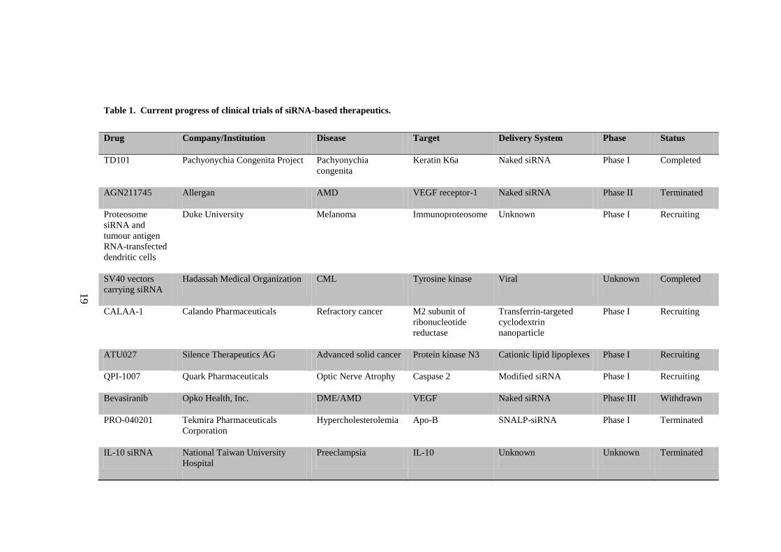
to degradation of the mRNA. A number of siRNA based drugs have now entered
clinical trials (Table 1), including ALN-RSV01 for treatment of the respiratory
syncytial virus (RSV), which causes upper and lower respiratory tract infections.7
The drug is now entering Phase IIb clinical trials, and so far has shown to be
effective at reducing infection rates in preliminary studies (www.alnylam.com).
Another siRNA-based drug undergoing Phase II clinical trials is PF-655 for
18
treatment of age-related macular degeneration (AMD) and diabetic macular edema
(DME) and targets the gene RTP801 which is upregulated as a result of ischemia,
hypoxia or oxidative stress.8 These trials show the potential of oligonucleotide based
therapeutics, and demonstrate their importance in the future of macromolecular drug
technologies.
19
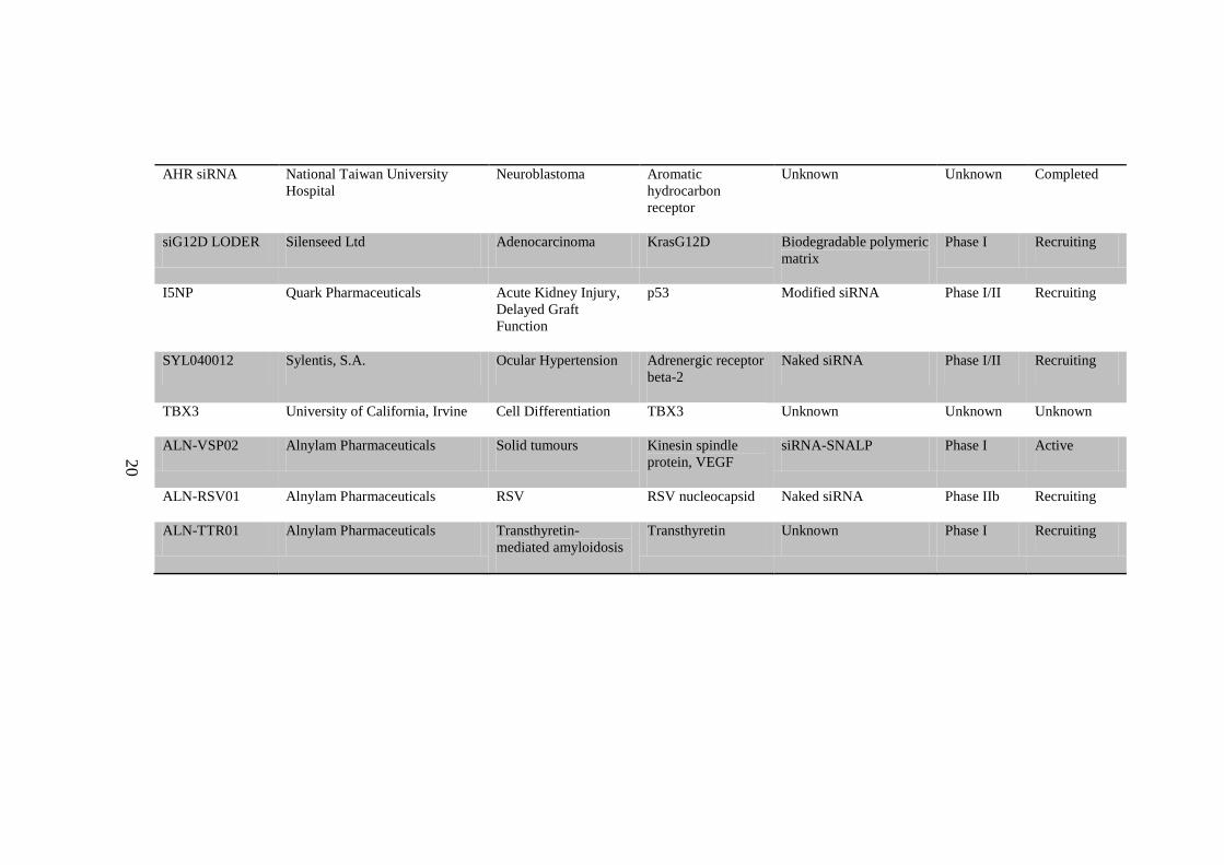
Table 1. Current progress of clinical trials of siRNA-based therapeutics.
Drug Company/Institution Disease Target Delivery System Phase Status
TD101 Pachyonychia Congenita Project Pachyonychia
congenita
Keratin K6a Naked siRNA Phase I Completed
AGN211745 Allergan AMD VEGF receptor-1 Naked siRNA Phase II Terminated
Proteosome
siRNA and
tumour antigen
RNA-transfected
dendritic cells
Duke University Melanoma Immunoproteosome Unknown Phase I Recruiting
SV40 vectors
carrying siRNA
Hadassah Medical Organization CML Tyrosine kinase Viral Unknown Completed
CALAA-1 Calando Pharmaceuticals Refractory cancer M2 subunit of
ribonucleotide
reductase
Transferrin-targeted
cyclodextrin
nanoparticle
Phase I Recruiting
ATU027 Silence Therapeutics AG Advanced solid cancer Protein kinase N3 Cationic lipid lipoplexes Phase I Recruiting
QPI-1007 Quark Pharmaceuticals Optic Nerve Atrophy Caspase 2 Modified siRNA Phase I Recruiting
Bevasiranib Opko Health, Inc. DME/AMD VEGF Naked siRNA Phase III Withdrawn
PRO-040201 Tekmira Pharmaceuticals
Corporation
Hypercholesterolemia Apo-B SNALP-siRNA Phase I Terminated
IL-10 siRNA National Taiwan University
Hospital
Preeclampsia IL-10 Unknown Unknown Terminated
20
AHR siRNA National Taiwan University
Hospital
Neuroblastoma Aromatic
hydrocarbon
receptor
Unknown Unknown Completed
siG12D LODER Silenseed Ltd Adenocarcinoma KrasG12D Biodegradable polymeric
matrix
Phase I Recruiting
I5NP Quark Pharmaceuticals Acute Kidney Injury,
Delayed Graft
Function
p53 Modified siRNA Phase I/II Recruiting
SYL040012 Sylentis, S.A. Ocular Hypertension Adrenergic receptor
beta-2
Naked siRNA Phase I/II Recruiting
TBX3 University of California, Irvine Cell Differentiation TBX3 Unknown Unknown Unknown
ALN-VSP02 Alnylam Pharmaceuticals Solid tumours Kinesin spindle
protein, VEGF
siRNA-SNALP Phase I Active
ALN-RSV01 Alnylam Pharmaceuticals RSV RSV nucleocapsid Naked siRNA Phase IIb Recruiting
ALN-TTR01 Alnylam Pharmaceuticals Transthyretin-
mediated amyloidosis
Transthyretin Unknown Phase I Recruiting
21
However, there are still many hurdles to be overcome before the macromolecular
drug revolution is finally upon us. A recent study into the safety of therapeutics
which are derived from a biological system, so-called biologics, including
recombinant proteins, antibodies and vaccines, indicated that a higher proportion of
these drugs have prompted safety alerts than for small molecule therapeutics.9 Of the
174 biologic drugs that were approved in either the US or Europe between January
1995 and June 2007, 23.6% prompted regulatory action as a result of safety concerns
compared with an average of 8% of small molecule drugs studied between 1975 and
1999. Other challenges that have continued to hinder the progress of
macromolecular therapeutics include the control of biodistribution and degradation
of macromolecules in the body, off-target effects and specificity, and delivery to
intracellular compartments.8 Many advances have been made that increase the
stability of proteins and oligonucleotides in vivo resulting in a longer half-life in the
body, including the chemical modification of oligonucleotide strands to prevent
degradation by endogenous nucleases. Similarly, modification of proteins by the
addition of polyethylene glycol (PEG) protects from proteases, as well as decreasing
renal clearance.1 Off-target effects are a particular issue for oligonucleotide drugs,
including both traditional antisense oligonucleotides and newer RNAi-based agents.
Despite these oligonucleotides being designed to target a single mRNA sequence,
binding to additional genes is often observed using DNA arrays, including to
sequences with little homology to the target.10
Improved design of siRNA sequences
using computational tools such as BLAST, as well as chemical modifications
(Section 1.3.2.1) have reduced off-target binding.
Finally, one of the largest challenges to the development of protein and
oligonucleotide-based drugs has been the lack of a suitable delivery agent to target
these macromolecules to the intracellular machinery. Proteins and oligonucleotides
are large and hydrophilic, thus preventing them from crossing the lipid membrane
surrounding cells. Traditional small molecule drugs usually conform to Lipinski's
rule of five,11
which stipulates that molecules should be small and lipophilic, making
them permeable to cell membranes. Over the last decade, much research has been
22
dedicated to finding an effective delivery mechanism for macromolecules, which
have included viral carriers, liposomes and cell penetrating peptides (CPPs).12
1.2. Cell Penetrating Peptides
CPPs, also called protein transduction domains (PTDs), are short peptides, usually
less than 30 amino acids long, which have the unusual property of being able to cross
the cell membrane. In 1988 it was found that a transcription factor from HIV called
the transactivator of transcription (TAT) was taken up by cells and migrated to the
nucleus to activate transcription of the viral genome.13
Shortly after, it was also
found that the Drosophila melanogaster Antennapedia homeodomain was also
internalised into cells.14
These observations led to the identification of the first
CPPs, the third helix of the Antennapedia homeodomain, named penetratin
(RQIKIYFQNRRMKWKK),15
and a short basic peptide derived from TAT
consisting of amino acids 47-57 (RKKRRQRRR).16
Many other CPPs have since
been described, including chimeric peptides such as MPG, a fusion peptide derived
from a hydrophobic domain from HIV gp41 and a hydrophilic domain from the
nuclear localisation sequence of Simian vacuolating virus 40 (SV40) T-antigen.17
CPPs can be divided into two main classes; the amphipathic CPPs, containing both a
hydrophobic and a hydrophilic domain, such as TAT, penetratin, and MPG, and the
polycationic CPPs containing mainly positively charged residues such as lysine and
arginine. Some CPPs are covalently linked to their cargo, such as TAT which was
incorporated into fusion proteins and successfully delivered to various mouse
tissues.18
Other CPPs are used to form stable non-covalent complexes with their
cargo, such as Pep-1 which was designed to form complexes with proteins and
peptides through hydrophobic interactions with a tryptophan-rich domain.19
The
formation of non-covalent complexes has also been particularly useful in the CPP-
mediated delivery of oligonucleotides, which are highly negatively charged and
therefore able to form electrostatic complexes with cationic peptides (Section
1.3.2.4). Over the last two decades, CPPs have been used to transport a wide range
of high molecular weight cargoes into cells, including biologically active proteins
23
both in vitro and in vivo,18, 20
quantum dots,21
and oligonucleotides,22-23
making them
attractive candidates for delivery agents for macromolecular drugs.
1.2.1. Uptake Mechanism
One of the most challenging questions surrounding CPPs is the mechanism by which
these peptides enter cells. Early studies using fluorescent dyes linked to CPPs
concluded that internalisation was energy-independent, occurring at both 4 °C and 37
°C.15, 24
However, this was later found to be an experimental artefact resulting from
CPPs adhering strongly to the cell membrane and not removed under the washing
conditions used in the early experiments.25
When the cells were fixed with methanol
for visualisation, the membranes were disrupted allowing the CPP-dye complex to
enter the cells, thus giving false-positive results for CPP internalisation. In addition,
the flow cytometry protocols did not distinguish between internalised fluorescent
CPPs and those adhered to the cell surface. Methods used to detect internalised
fluorescent CPPs now include a more thorough washing procedure, such as treatment
with trypsin or heparin, in order to remove extracellular peptide. In experiments by
Lundberg et al.,26
Chinese Hamster Ovary (CHO) cells were incubated with various
CPPs linked to GFP. These experiments used microscopy on live cells to determine
the extent of cell uptake and showed that for all the CPPs tested, the bulk of the
proteins were bound to the cell surface rather than internalised into the cells.
Subsequent studies on various CPPs such as TAT, penetratin and polyarginine
concluded that CPP internalisation is driven by endocytosis, and in particular lipid-
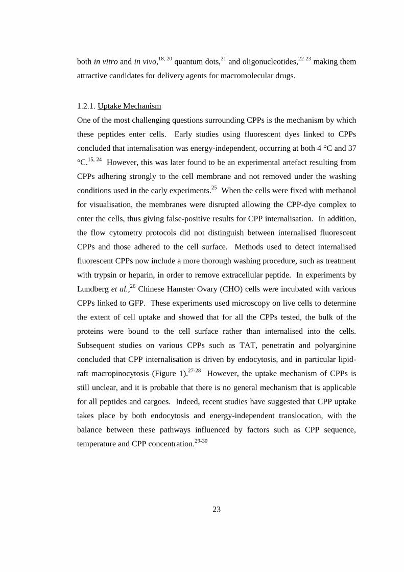
raft macropinocytosis (Figure 1).27-28
However, the uptake mechanism of CPPs is
still unclear, and it is probable that there is no general mechanism that is applicable
for all peptides and cargoes. Indeed, recent studies have suggested that CPP uptake
takes place by both endocytosis and energy-independent translocation, with the
balance between these pathways influenced by factors such as CPP sequence,
temperature and CPP concentration.29-30
24
Figure 1. Endocytosis mechanisms used by CPPs. Clathrin-mediated endocytosis occurs at
clathrin-coated pits which mature into clathrin-coated vesicles upon binding of a ligand to its cell-
surface receptor. Dynamin releases the vesicles from the membrane allowing them to progress into
the cell and fuse with early endosomes (EE). Some receptors, such as the low-density lipoprotein
(LDL) receptor are returned to the cell surface via recycling endosomes (RE). Alternatively, caveolin-
coated vesicles (caveolae) may form at lipid-rich regions of the membrane, so-called lipid rafts, which
are also released by dynamin into the cytoplasm and fuse with EEs. Finally, larger cargoes may be
internalised by macropinocytosis via interactions with proteoglycans in the extracellular matrix, which
results in rearrangement of the actin cytoskeleton to form lamellipodia. These structures engulf the
cargo, internalising it into macropinosomes (MP) which fuse to EEs. EEs mature into late endosomes
(LE) and finally lysosomes (Lys) where the contents is broken down by enzymes. Therefore, it is
important that the CPP-bound cargo escapes from early endosomes in order to exert its biological
effect.
1.2.1.1. Cell-surface binding
Many studies have shown that CPPs bind strongly to the glycosaminoglycans
(GAGs) of cell surface proteoglycans, such as heparan sulfate (HS) and chondroitin
sulfate (CS) as a precursor to internalisation.26, 28, 31-35
Proteoglycans such as
syndecans form part of the extracellular matrix, where they play a structural role in
cell adhesion, as well as a regulatory role in mediating lipid microdomains when
25
bound by heparin-binding ligands.36
It is thought that proteoglycan ligand binding
results in receptor clustering, leading to the reorganisation of the actin cytoskeleton
via activation of a signalling cascade involving the Rho-family of GTPases, therefore
leading to cellular uptake via macropinocytosis.37
HS a linear polysaccharide made up of highly sulfated NS domains and low sulfated
NA domains. Heparin is a closely related compound that has been used as a
substitute for HS in binding assays with a number of CPPs which have calculated
dissociation constants in the low micromolar to low nanomolar range.38
These strong
interactions are probably comprised of an ionic contribution as well as bi-dentate
hydrogen bonds between the guanidine groups of the peptide's arginine amino acids
and the sulfate anions of heparin. The internalisation of nonaarginine (R9), which
was found to bind heparin with a Kd of 109 nM, was found to be GAG-dependent by
comparing uptake in wild-type CHO cells with mutant cells deficient in HS and
CS.34
In GAG-deficient CHO cells, oligoarginine and TAT internalisation is
significantly reduced compared to wild-type cells and the presence of CPPs results in
activation of Rac1, a Rho-family GTPase which is involved with F-actin
reorganisation, suggesting that GAG binding may be a precursor for CPP uptake by
endocytosis.31
Similarly, it was found that the CPPs MPG-α and MPG-β, either
alone or in complexes with DNA, initiated remodelling of the actin network, leading
to the formation of lamellipodia, accompanied by upregulation of Rac1.35
In
addition, CPPs and other polycationic cell penetrating compounds (CPCs) are
capable of clustering GAGs, which is a precursor for endocytosis.39-40
A recent study, however, contradicts these findings claiming that whilst TAT does
bind to cell-surface HS, this interaction is independent of uptake, with enzymatic
removal of cell surface GAGs having little effect on the uptake of fluorescently-
tagged TAT.41
Contradictions such as these are commonplace in the literature
surrounding CPP internalisation, and demonstrates our limited understanding of the
mechanism of uptake of these peptides. It is possible that competing internalisation

26
mechanisms exist and that the pathway used is dependent on factors such as CPP
sequence, cargo and cell line.
Another possible route by which CPPs are internalised into cells involves binding to
anionic lipids on the cell surface. It is thought that the positively charged side chains
on the peptide interact with anionic membrane lipids, shielding the positive charge
and forming a neutral, lipophilic complex that is able to cross the membrane.40, 42-44
It has been shown that addition of anionic lipids such as 1,2-dioleoyl-sn-glycero-3-
phosphatidylglycerol (DOPG) are capable of shifting basic peptides from the
aqueous phase to the organic phase by the formation of lipophilic complexes.42, 45
This suggests that cell-surface phospholipids may act as counter-ions to the CPP’s
positively charged amino acids, allowing it to dissolve in the hydrophobic cell
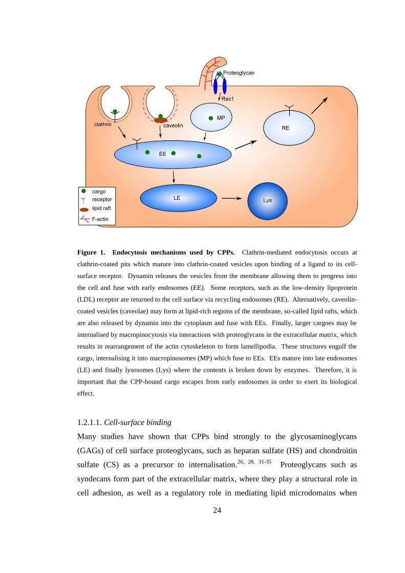
membrane. It is suggested that arginine favours this counterion-mediated membrane
translocation over lysine, due to the tendency of guanidinium groups to form
bidentate hydrogen bonds with anions in order to reduce charge repulsion with
adjacent arginine residues, whereas charge repulsion in lysines is generally countered
by a reduction in pKa (Figure 2).45
This may explain the high proportion of arginine
residues present in CPPs. In addition, the presence of a hydrophobic counteranion,
pyrenebutyrate, increases the translocation of arginine-containing CPPs.30
Polyarginine is the most affected, followed by TAT, and then by amphipathic CPPs,
showing that a high arginine content favours binding to counterions. The addition of
the counteranion increased membrane perturbation in large unilamellar vesicles
(LUVs), showing that increased hydrophobicity of the CPPs leads to greater
penetration of the membranes. In a splice-switching assay in which the same CPPs
were used to transport an antisense oligonucleotide complexed electrostatically to the
peptides into HeLa cells, pyrenebutyrate was found to increase the rate of
translocation, and therefore the biological effect, for arginine-rich peptides more than
amphipathic peptides. However, it was found that chloroquine, which delays the
maturation of endosomes into lysosomes, was required for a biological effect to be
observed, hence pointing to an endocytotic internalisation pathway, with
27
pyrenebutyrate enhancing translocation through the endosomal membrane, rather
than direct membrane penetration.
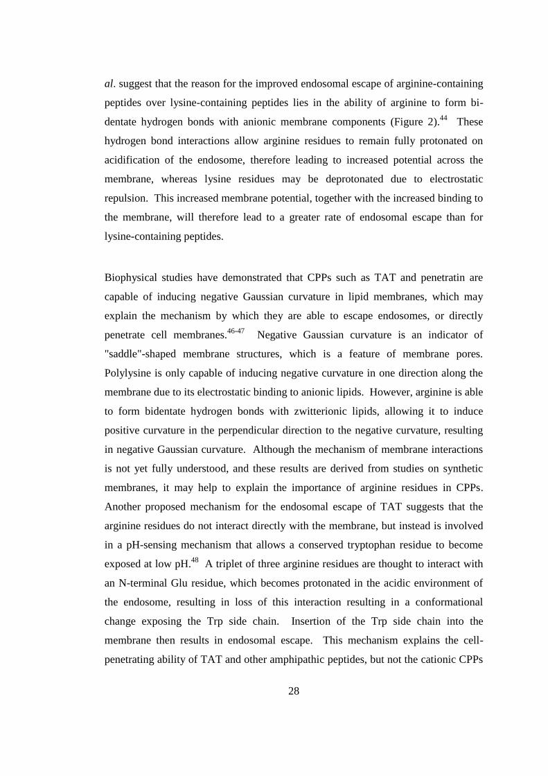
Figure 2. Reduction of charge repulsion by lysine and arginine oligomers. Amino groups in
polylysine (left) reduce charge repulsion by lowering their pKa, resulting in deprotonation. Guanidine
groups in polyarginine (right) form bidentate hydrogen bonds to anions such as phosphates. Arginine-
rich peptides may therefore more readily bind to counter-anions on the cell surface than lysine-
containing peptides.
1.2.1.2. Endosomal Escape
Overcoming entrapment by endosomes is one of the major challenges to the design
of efficient CPPs and other macromolecular transporters. By comparing the levels of
endosomal escape of polyarginine- and polylysine-coupled liposomes, El-Sayed et
28
al. suggest that the reason for the improved endosomal escape of arginine-containing
peptides over lysine-containing peptides lies in the ability of arginine to form bi-
dentate hydrogen bonds with anionic membrane components (Figure 2).44
These
hydrogen bond interactions allow arginine residues to remain fully protonated on
acidification of the endosome, therefore leading to increased potential across the
membrane, whereas lysine residues may be deprotonated due to electrostatic
repulsion. This increased membrane potential, together with the increased binding to
the membrane, will therefore lead to a greater rate of endosomal escape than for
lysine-containing peptides.
Biophysical studies have demonstrated that CPPs such as TAT and penetratin are
capable of inducing negative Gaussian curvature in lipid membranes, which may
explain the mechanism by which they are able to escape endosomes, or directly
penetrate cell membranes.46-47
Negative Gaussian curvature is an indicator of
"saddle"-shaped membrane structures, which is a feature of membrane pores.
Polylysine is only capable of inducing negative curvature in one direction along the
membrane due to its electrostatic binding to anionic lipids. However, arginine is able
to form bidentate hydrogen bonds with zwitterionic lipids, allowing it to induce
positive curvature in the perpendicular direction to the negative curvature, resulting
in negative Gaussian curvature. Although the mechanism of membrane interactions
is not yet fully understood, and these results are derived from studies on synthetic
membranes, it may help to explain the importance of arginine residues in CPPs.
Another proposed mechanism for the endosomal escape of TAT suggests that the
arginine residues do not interact directly with the membrane, but instead is involved
in a pH-sensing mechanism that allows a conserved tryptophan residue to become
exposed at low pH.48
A triplet of three arginine residues are thought to interact with
an N-terminal Glu residue, which becomes protonated in the acidic environment of
the endosome, resulting in loss of this interaction resulting in a conformational
change exposing the Trp side chain. Insertion of the Trp side chain into the
membrane then results in endosomal escape. This mechanism explains the cell-
penetrating ability of TAT and other amphipathic peptides, but not the cationic CPPs
29
which do not contain lipophilic amino acids. Therefore, endosomal escape may not
follow a general mechanism and further supports the hypothesis that different CPPs
are internalised via different pathways.
1.3. RNA Interference
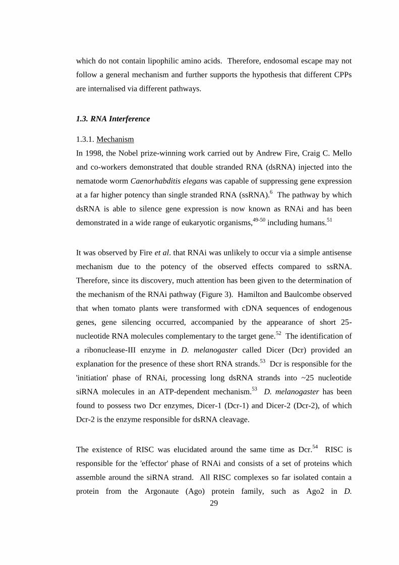
1.3.1. Mechanism
In 1998, the Nobel prize-winning work carried out by Andrew Fire, Craig C. Mello
and co-workers demonstrated that double stranded RNA (dsRNA) injected into the
nematode worm Caenorhabditis elegans was capable of suppressing gene expression
at a far higher potency than single stranded RNA (ssRNA).6 The pathway by which
dsRNA is able to silence gene expression is now known as RNAi and has been
demonstrated in a wide range of eukaryotic organisms,49-50
including humans.51
It was observed by Fire et al. that RNAi was unlikely to occur via a simple antisense
mechanism due to the potency of the observed effects compared to ssRNA.
Therefore, since its discovery, much attention has been given to the determination of
the mechanism of the RNAi pathway (Figure 3). Hamilton and Baulcombe observed
that when tomato plants were transformed with cDNA sequences of endogenous
genes, gene silencing occurred, accompanied by the appearance of short 25-
nucleotide RNA molecules complementary to the target gene.52
The identification of
a ribonuclease-III enzyme in D. melanogaster called Dicer (Dcr) provided an
explanation for the presence of these short RNA strands.53
Dcr is responsible for the
'initiation' phase of RNAi, processing long dsRNA strands into ~25 nucleotide
siRNA molecules in an ATP-dependent mechanism.53
D. melanogaster has been
found to possess two Dcr enzymes, Dicer-1 (Dcr-1) and Dicer-2 (Dcr-2), of which
Dcr-2 is the enzyme responsible for dsRNA cleavage.
The existence of RISC was elucidated around the same time as Dcr.54
RISC is
responsible for the 'effector' phase of RNAi and consists of a set of proteins which
assemble around the siRNA strand. All RISC complexes so far isolated contain a
protein from the Argonaute (Ago) protein family, such as Ago2 in D.
30
melanogaster,55
possessing PAZ domains capable of binding 3’ 2-nucleotide
overhangs.56
Following binding to RISC, one strand of the siRNA, the ‘passenger’
strand, is abandoned and the activated RISC-Ago2 complex together with the 'guide'
strand identifies and binds to the complementary mRNA sequence, which is finally
cleaved by endoribonuclease activity within the complex.57
Proteins that link the
initiation and effector phases, so-called dsRNA-binding proteins (dsRBPs), have
been identified and include the D. melanogaster protein R2D2, so named because of
its two RNA-binding domains (R2) and its ability to bind Dcr-2.58
R2D2 does not
affect the dsRNA-processing function of Dcr, however it has been shown to restore
RISC function in a system containing purified Dcr and RISC, suggesting that its
function is involved with loading siRNA onto RISC.58
Human variants of R2D2 are
yet to be elucidated, however two proteins, transactivating response RNA-binding
protein (TRBP) and protein activator of protein kinase R (PACT), have been shown
to bind to Dcr but their interaction with RISC is unclear.59
A mechanistic aspect of RNAi that is poorly understood is the separation of the two
siRNA strands, resulting in just the antisense strand which guides RISC to its target
mRNA sequence. Several proteins associated with RISC have been shown to
possess helicase domains, but no direct link with the unwinding of siRNA strands
has been observed, and the exact point along the RISC-assembly pathway at which
strand separation occurs is also unknown.49, 59
In addition, the mechanism by which
the guide strand is preferentially loaded onto RISC, whilst the passenger strand is
discarded, is also unknown. One theory suggests that Dcr and dsRBPs are able to
detect the thermodynamic stability of the binding of each strand to the other and
preferentially incorporate one strand over the other. Crosslinking experiments with
siRNA modified with 5-iodouracil residues showed that when one siRNA strand has
a greater thermodynamic stability at its 5' end than the other, Dcr-2 will bind to the
strand with the least stable 5' terminus. R2D2 will bind to the more stable 5'
terminus, resulting in the Dcr-bound strand being incorporated into RISC and the
R2D2 strand discarded as the passenger strand.60
This model states that siRNA
unwinding occurs prior to RISC incorporation, whereas others have suggested that
31
strand separation is carried out by Ago2 after the siRNA has been transferred from
the Dcr-2/R2D2 complex to RISC.61
Following this poorly understood RISC activation stage, the final step of the RNAi
pathway is cleavage of the target mRNA strand. This is accomplished by an ATP-
independent endonuclease reaction by the Ago proteins, which contain conserved
PIWI domains with a structure very similar to RNase H.62-63
The reaction does not
destroy the bound siRNA strand, so RISC is a multiple turnover enzyme, explaining
the catalytic nature of RNAi.49
Another form of RNAi is driven by microRNAs (miRNA) – hairpin RNA strands
that are naturally transcribed by the genome – and is thought to follow a similar
mechanism via processing by a Dcr enzyme, and activation of the RISC complex.49,
59 However, it is unclear how distinct these two pathways are, and how assembly of
the micro RISC (miRISC) differs from that of the siRISC. In D. melanogaster,
separate Dcr enzymes, Dcr-1 and Dcr-2, exist for processing of miRNA and dsRNA
respectively,64
as well as a separate dsRBP, R3D1,64
and a distinct RISC-associated
Ago protein, Ago1.65
However, in humans only one Dcr enzyme has been
indentified, suggesting more integration between the processing of miRNA and
siRNA. Sequence complementarity of microRNAs to their target mRNAs is less
exact, leading to blocking of translation by mRNA binding rather than silencing by
mRNA destruction.66
32
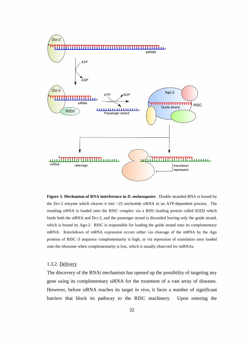
Figure 3. Mechanism of RNA interference in D. melanogaster. Double stranded RNA is bound by
the Dcr-2 enzyme which cleaves it into ~25 nucleotide siRNA in an ATP-dependent process. The
resulting siRNA is loaded onto the RISC complex via a RISC-loading protein called R2D2 which
binds both the siRNA and Dcr-2, and the passenger strand is discarded leaving only the guide strand,
which is bound by Ago-2. RISC is responsible for loading the guide strand onto its complementary
mRNA. Knockdown of mRNA expression occurs either via cleavage of the mRNA by the Ago
proteins of RISC if sequence complementarity is high, or via repression of translation once loaded
onto the ribosome when complementarity is low, which is usually observed for miRNAs.
1.3.2. Delivery
The discovery of the RNAi mechanism has opened up the possibility of targeting any
gene using its complementary siRNA for the treatment of a vast array of diseases.
However, before siRNA reaches its target in vivo, it faces a number of significant
barriers that block its pathway to the RISC machinery. Upon entering the
33
bloodstream, siRNA is vulnerable to degradation by endogenous nucleases, and renal
excretion due to its small size and highly anionic character. In addition, before
reaching its target cell, the siRNA must navigate the tight endothelial junctions of the
blood vessels and diffuse through the extracellular matrix. Due to its numerous
negative charges, siRNA does not readily bind to or cross the cell membrane, and
once inside the cells, it must escape from endosomes in order to interact with its
intracellular protein targets.67
Where siRNA can be applied directly to the target tissue, local delivery of naked,
unmodified siRNA has shown to be successful, and several treatments for age-related
macular degeneration involving the injection of siRNA directly into the eye have
reached clinical trials (Table 1).8 However, for many therapeutic targets, including
metastatic tumours, local delivery is not possible and systemic delivery of siRNA is
the only effective route. Therefore, in order to achieve successful delivery of siRNA
to these targets, a delivery system is needed which protects the siRNA from
degradation by endogenous enzymes and improves the bioavailability of the siRNA
to its target cells, enabling the siRNA to cross the cell membrane and escape the
endosomes to reach the intracellular machinery.8, 67-68
The first delivery vectors
developed for the delivery of siRNA were viral vectors and were shown to achieve
effective mRNA knockdown both in vivo and in vitro.69
However, due to safety
concerns, such as the observation of mutagenesis caused by insertions,69
more recent
siRNA delivery systems have focussed on non-viral approaches.
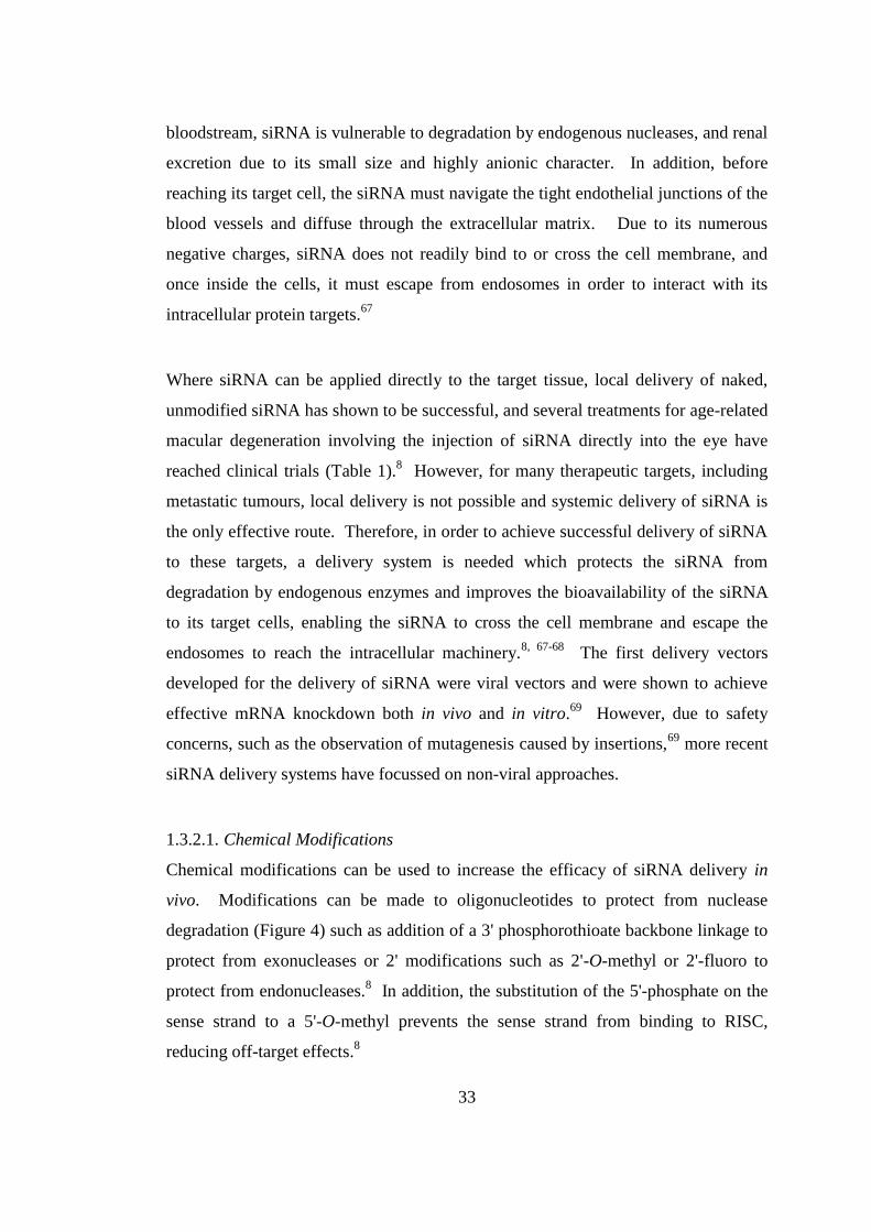
1.3.2.1. Chemical Modifications
Chemical modifications can be used to increase the efficacy of siRNA delivery in
vivo. Modifications can be made to oligonucleotides to protect from nuclease
degradation (Figure 4) such as addition of a 3' phosphorothioate backbone linkage to
protect from exonucleases or 2' modifications such as 2'-O-methyl or 2'-fluoro to
protect from endonucleases.8 In addition, the substitution of the 5'-phosphate on the
sense strand to a 5'-O-methyl prevents the sense strand from binding to RISC,
reducing off-target effects.8
34
Figure 4. RNA chemical modifications. A: 3'-phosphorothioate to prevent degradation by
endonucleases; B and C: 2'-O-methyl and 2'-fluoro to prevent degradation by exonucleases; D: 5'-O-
methyl on the sense strand to prevent binding to RISC.
One type of delivery method involves chemical conjugation of small molecules to
the sense strand in order to improve the biodistribution of siRNA. Cholesterol-
conjugated siRNA is able to knock down the ApoB gene in mice injected with the
conjugate, whereas naked siRNA was found to be ineffective.70
The cholesterol-
siRNA conjugates were also able to bind to human serum albumin, improving
biodistribution by restricting clearance by the renal system. The same effect is also
observed with various long chain fatty acids and bile acids, which bind albumin as
well as high and low density lipoproteins (HDL and LDL). Unconjugated siRNA
and siRNA linked to short and medium chain fatty acids do not knock down ApoB in
vivo and do not bind to albumin or HDL and LDL, which illustrates the importance
of lipophilicity in the biodistribution of siRNA.71
Another type of chemical
conjugation has been the attachment of RNA aptamers to siRNA in order to target
specific tissues or cell types. For example, an siRNA-RNA aptamer chimera
designed to target prostate cancer cells expressing prostate-specific membrane
antigen has been shown to knock down cancer survival gene Plk1 and inhibit tumour
growth in vivo.72
Therefore, conjugation of siRNA to small molecules may enhance
siRNA delivery by improving bioavailability and targeting, but do not offer any
assistance in crossing cell membranes.
1.3.2.2. Liposomes and Lipoplexes
One of the most commonly used types of delivery vector for siRNA are liposomes,
where the siRNA is encapsulated within a lipid bilayer. These liposomes are formed
from synthetic cationic lipids which are composed of a cationic head group, usually a
35
quaternary ammonium group, and a hydrophobic region consisting of one or two
alkyl chains or a steroid such as cholesterol. Delivery of nucleic acids using cationic
lipids, known as lipofection, was first demonstrated by Felgner et al. using the
synthetic lipid N-[1-(2,3-dioleoyloxy)propyl]-N,N,N-trimethylammonium chloride
(DOTMA).73-74
This was followed by studies with several other cationic lipids
which proved to be successful for in vitro delivery of nucleic acids to a wide variety
of cell types, including dioctadecylamidoglycylspermine (DOGS),75
3β[N-(N',N'-
dimethylaminoethane)-carbamoyl) cholesterol (DC-Chol),76
dimethyldioctadecyl-
ammonium bromide (DDAB),77
and dimyristyloxypropyl-3-dimethylhydroxyethyl
ammonium bromide (DMRIE),78
as well as commercially available products such as
Lipofectamine (Invitrogen), RNAifect (Qiagen) and Oligofectamine (Invitrogen).
However, many of the early cationic lipid delivery systems proved to be ineffective
in vivo and showed high levels of toxicity, with a microarray study of the widely
used lipofection agents Lipofectin and Oligofectamine showing alteration of the
expression of up to 16% of genes, including genes involved with apoptosis.79
Another study showed that siRNA complexed with several cationic lipids elicited an
inflammatory response in a mice by upregulating the expression of pro-inflammatory
cytokines such as IL-2, IFNγ and TNFα.80
Cationic lipids are often used in conjunction with other components in order to
improve their delivery efficiency. Neutral lipids such as dioleoyl phosphatidyl-
ethanolamine (DOPE)81
or cholesterol82
are often used as colipids to increase
endosomal escape by adopting the inversion hexagonal (HII) phase. Lipids that adopt
the HII phase are arranged in hexagonally packed tubes, with each tube containing a
narrow aqueous channel in the centre surrounded by the lipid chains on the outside.
The HII phase has been shown to be favoured by lipids that participate in membrane
fusion due to their decreased stability compared to the bilayer configuration.83
They
are therefore more prone to endosomal escape, making them advantageous for
siRNA delivery. However, the incorporation of DOPE into cationic lipid lipoplexes
with siRNA leads to instability of the complexes due to salt bridges forming between
the phosphate groups of DOPE and the positive charges of the cationic lipid. This
36
therefore decreases the strength of the interactions between the siRNA and the
liposomes and makes the complex more susceptible to destabilisation by acidic
serum proteins when used in vivo.84-85
Many recent studies have focussed on the development of stable lipid delivery
systems which show effective gene delivery in vivo. In particular, the stable nucleic
acid-lipid particles (SNALPs), consisting of small, monodisperse particles with low
surface charge have been successful in achieving siRNA-mediated gene silencing in
several in vivo models. SNALPs were first shown to be effective in targeting the
hepatitis B virus (HBV) in mice, where SNALP-formulated siRNA was given in
three daily injections of 3 mg/kg/day, resulting in a decrease in HBV levels by 1-2
orders of magnitude.86
SNALPs have also been shown to be effective in non-human
primates, with one study achieving a knockdown of ApoB in monkeys resulting in a
90% decrease in blood ApoB-100 protein two days after a 2.5 mg/kg injection.87
More recently a study on rhesus monkeys given a lethal dose of Zaire Ebola virus
(ZEBOV) showed that the animals could be rescued by seven daily 2 mg/kg
treatments of an anti-ZEBOV siRNA cocktail formulated with SNALPs.88
Other
groups are attempting to develop novel cationic peptides to produce more stable
lipoplexes by rational design.89-91
Semple et al. have taken a commonly used
SNALP cationic lipid, 1,2-dilinoleyloxy-3-dimethylaminopropane (DLinDMA), and
used a structure-activity relationship (SAR) approach to optimise the linker and polar
head group to produce a new cationic lipid which, when incorporated into SNALPs,
achieves a 50-fold increase in ED50 in mice and an ED50 of 0.3 mg/kg in monkeys.89
The first RNAi therapeutic using a SNALP has now entered Phase I clinical trials
(Table 1), demonstrating the potential of this type of delivery vector.
1.3.2.3. Cationic Polymers
Another type of delivery vehicle for siRNA is cationic polymers which form
electrostatic complexes, protecting the siRNA from degradation and aiding
internalisation by shielding the siRNA negative charges.68, 92
One of the most widely
studied polymers for nucleic acid delivery is polyethyleneimine (PEI, Figure 5a).
37
PEI is available as both linear and branched isomers, with linear PEI containing only
secondary amine groups (except for one terminal primary amine), whereas branched
PEI contains primary, secondary and tertiary amines.92
These ionisable amine
groups allow PEI to bind to siRNA, forming stable nanoparticles to protect the
siRNA from degradation by serum nucleases.93
The amine groups also provide PEI
with a mechanism for endosomal escape, known as the "proton sponge" hypothesis.94
The titratable amine groups of PEI are protonated in the endosomes, preventing
endosome acidification and causing an influx of chloride ions and water, with the
resulting osmotic pressure leading to swelling and lysis of the endosome, releasing
the nucleic acid cargo into the cytoplasm. The importance of the ionisable amine
groups of PEI was further highlighted by Thomas et al. who showed that
commercially-available PEI contained ~11% acylated nitrogens as a result of
inefficient removal of N-propionyl groups from the N-propionyl-PEI (PEOZ)
precursors.95
Following complete deacylation of PEI, an increase in its DNA-
condensing ability as well as PEI-DNA transfection efficiency was observed. The
fully deacylated PEI was used to deliver siRNA targeting the influenza nucleoprotein
to mice infected with the influenza virus, resulting in a 94% reduction of virus titre in
the lungs after 24 h. Another study used PEI to deliver siRNA targeted to the proto-
oncogene c-erbB2/neu (HER-2) both in vitro, where a 50% mRNA knockdown was
achieved in SKOV-3 cells, and in vivo, where significant reduction in tumour growth
was observed compared to a non-specific siRNA control using a subcutaneous mouse
tumour model.93
Therefore, PEI shows some potential as a siRNA transfection
agent, however its toxicity at higher concentrations resulting from membrane
disruption and apoptosis induction may limit its applications as a therapeutic delivery
agent.96
Other polymers used for siRNA transfection include cyclodextrin-based compounds
(Figure 5b). Cyclodextrins are widely used in the food and pharmaceutical industries
due to their ability to encapsulate small, hydrophobic molecules, thereby increasing
their stability and solubility, making them suitable for drug delivery and food
preservation. For use in nucleic acid delivery, cyclodextrins are modified by
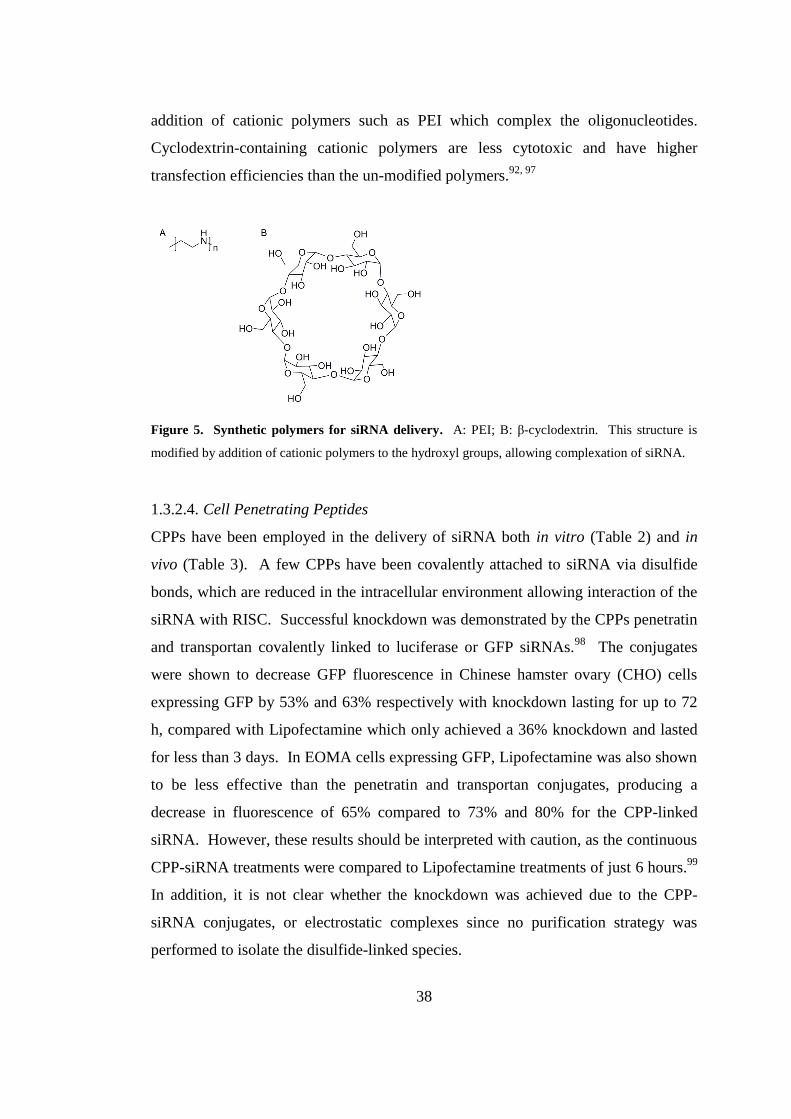
38
addition of cationic polymers such as PEI which complex the oligonucleotides.
Cyclodextrin-containing cationic polymers are less cytotoxic and have higher
transfection efficiencies than the un-modified polymers.92, 97
Figure 5. Synthetic polymers for siRNA delivery. A: PEI; B: β-cyclodextrin. This structure is
modified by addition of cationic polymers to the hydroxyl groups, allowing complexation of siRNA.
1.3.2.4. Cell Penetrating Peptides
CPPs have been employed in the delivery of siRNA both in vitro (Table 2) and in
vivo (Table 3). A few CPPs have been covalently attached to siRNA via disulfide
bonds, which are reduced in the intracellular environment allowing interaction of the
siRNA with RISC. Successful knockdown was demonstrated by the CPPs penetratin
and transportan covalently linked to luciferase or GFP siRNAs.98
The conjugates
were shown to decrease GFP fluorescence in Chinese hamster ovary (CHO) cells
expressing GFP by 53% and 63% respectively with knockdown lasting for up to 72
h, compared with Lipofectamine which only achieved a 36% knockdown and lasted
for less than 3 days. In EOMA cells expressing GFP, Lipofectamine was also shown
to be less effective than the penetratin and transportan conjugates, producing a
decrease in fluorescence of 65% compared to 73% and 80% for the CPP-linked
siRNA. However, these results should be interpreted with caution, as the continuous
CPP-siRNA treatments were compared to Lipofectamine treatments of just 6 hours.99
In addition, it is not clear whether the knockdown was achieved due to the CPP-
siRNA conjugates, or electrostatic complexes since no purification strategy was
performed to isolate the disulfide-linked species.
39
Chiu et al. demonstrated that 300 nM of disulfide-linked EGFP siRNA and TAT was
capable of delivering up to 70% gene silencing in HeLa cells after a 16 hour
incubation, compared to 87% for 20 μg of Lipofectamine.100
The same CPP was
also covalently linked to siRNA for the endogenous gene CDK9, and achieved an
82% knockdown after 42 h at a concentration of 400 nM. This study addressed the
issue of whether the knockdown effect was caused by the disulfide conjugate or an
electrostatic CPP-siRNA complex by including a control transfection consisting of
free siRNA and CPP. This control transfection produced no knockdown, suggesting
that the covalently linked species was responsible for the gene silencing. Another
study extended the principle of covalently linked CPPs and siRNA to primary cells,
using penetratin to deliver siRNA to hippocampal neuron cells.101
This system
achieved knockdown of the endogenous proteins SOD1 and Caspase-3, which were
decreased by 90% and 73% respectively at 80 nM according to Western blot
analysis. In addition, cell survival using penetratin-siRNA was compared to
Lipofectamine, and showed that the penetratin conjugate was much less harmful to
neurons, with a 92% survival rate compared to 58% for Lipofectamine. Once again,
this study does not purify the CPP-siRNA conjugate, and therefore the species
responsible for the observed siRNA delivery is unclear.99
More recently, CPP-siRNA conjugates containing penetratin and TAT, have been
tested in vivo for the first time.102
An siRNA sequence targeted to p38 MAP kinase,
a protein implicated in various inflammatory diseases, was linked to TAT(48-60),
penetratin, or cholesterol and the conjugates purified by anion-exchange HPLC and
characterised by electrospray mass spectrometry. When incubated with mouse
fibroblast cells, these conjugates show a modest 20-36% knockdown of p38 MAP
kinase, however when the three conjugates were administered to mouse lungs via
intratracheal injections, none of the constructs were able to produce a knockdown
significantly different to siRNA alone. In addition, penetratin-siRNA elicited high
levels of the immune markers TNFα and IL-12p40, suggesting that this CPP may be
responsible for triggering the innate immune system. These results suggest that the

40
use of covalently-conjugated CPPs might not be ideal delivery vectors for siRNA in
vivo.
Most of the published studies concerning the delivery of siRNA using CPPs have
used the cationic nature of the peptides to form electrostatic complexes, shielding the
siRNA negative charges to allow entry into cells. It is thought that these non-
covalent complexes may not be as effective as covalent conjugates, as the peptide
positive charges also play a role in internalisation by interacting with cell surface
proteoglycans (Section 1.2.1.1). Therefore, if these charges are tied up in
electrostatic interactions with siRNA molecules, it may not be possible to form
sufficient interactions with the cell membrane to allow internalisation.99
On the other
hand, using CPPs in electrostatic complexes may be beneficial as they provide
greater protection against nuclease degradation by forming a stable complex. In
addition, complex formation is less time consuming as the siRNA requires no
modification to allow CPP attachment. Electrostatic complexes remain the most
popular application for CPPs in siRNA delivery, and some significant progress has
been made both in vitro and in vivo by several groups using both natural and
synthetic CPPs.
One of the earliest such studies examined the siRNA delivery abilities of the
amphipathic CPP MPG, a chimera peptide of the hydrophobic fusion peptide domain
of HIV-1 gp41 protein and the hydrophilic nuclear localisation sequence (NLS) of
SV40 large T antigen.103
The MPG peptide was successful in internalising siRNA as
electrostatic complexes, achieving a 60% GAPDH knockdown at an siRNA
concentration of 100 nM after a 30 h transfection in human fibroblast HS-68 cells.
However, a mutant MPG peptide possessing a mutation in the NLS region,
MPGΔNLS
increased the knockdown to 80% at a siRNA concentration of just 25 nM,
supporting the general mechanism that the siRNA targets RISC in the cytoplasm.
This early study did not include any cytotoxicity data and also did not compare MPG
with any other available transfection reagent.

41
The use of MPG as an siRNA delivery vector was further explored by Veldhoen et
al. who formed electrostatic complexes between siRNA and MPGα, a derivative of
MPG differing by 6 amino acids in the hydrophobic region increasing its tendency to
form a helical conformation.104
MPGα was compared to Lipofectamine 2000 and
showed that whilst the CPP showed a comparable maximum knockdown ability to
Lipofectamine, achieving a 70-83% luciferase knockdown in HeLa cells compared to
88-97% for Lipofectamine, its IC50 was 30-fold higher than Lipofectamine (0.8 nM
compared to 0.02-0.04 nM). By measuring the total internalised siRNA at the half
maximal inhibition point of each compound using a highly sensitive liquid
hybridisation assay, it was found that around 10,000 molecules were required for half
maximal inhibition of MPGα compared to 300 for Lipofectamine. Therefore, the
amount of available siRNA was about 30-fold less using MPGα, suggesting that the
CPP was either inefficient at escaping from endosomes, or targeted to intracellular
compartments other than the cytoplasm, thus preventing the siRNA from binding to
RISC. These observations seem to contradict evidence that suggests that MPG-DNA
complexes enter cells via a non-endosomal route, and are unaffected by inhibitors of
endocytosis.103
Thus, it appears that very similar peptides may be internalised by
different pathways, perhaps influenced by differing sequences, cargoes or particle
size
The ability of siRNA complexes to escape the endosomes was studied using a
penetratin analogue called EB1 by replacing two amino acids with histidine which, in
theory, would promote endosomal escape by the formation of a helical structure upon
protonation in the endosome, therefore promoting membrane insertion.105
The
activity of EB1 was compared to the other well characterised CPPs penetratin,
MPGΔNLS
, bPrPp, another peptide known to promote endosomal escape,106
and
TP10. In the system tested, EB1, MPGΔNLS
and bPrPp were able to knockdown
luciferase expression to a similar extent to Lipofectamine, achieving a ~50%
knockdown with 100 nM siRNA. Under the conditions used, penetratin and TP10
did not show any significant knockdown at 100 nM siRNA. This highlights the
importance of localising the siRNA to the cytoplasm for interaction with RISC by
42
ensuring siRNA can escape from endosomes. Importantly, this study included a
toxicity study for all CPPs used, looking at both membrane stability and long term
toxicity. Membrane integrity was maintained for siRNA-peptide complexes, but a
higher membrane disturbance was observed for uncomplexed peptides, suggesting
that the use of electrostatic complexes may have toxicity advantages over covalent
conjugates. In a proliferation assay to assess long term toxicity over 48 h, no
significant reduction in proliferation was observed for the peptide-treated cells,
however Lipofectamine significantly reduced proliferation, showing that CPPs may
be more advantageous for in vivo siRNA delivery than cationic liposomes.
More recently, in a further attempt to design a CPP with built-in endosomolysis
functionality, a report by Andaloussi et al. described a modified version of the TP10
CPP called PepFect6, which is functionalised with an N-terminal stearyl group, as
well as four covalently attached chloroquine analogues.107
The N-terminal fatty acid
is incorporated to improve serum stability, whilst the chloroquine-like side chains are
included to allow the CPP-siRNA complex to escape from endosomes when
protonated. The results for PepFect6 show convincing siRNA internalisation across
a range of cell lines, including primary cells, which out-perform Lipofectamine with
lower toxicity. The peptide also promotes significant knockdown in vivo in two
mouse models, one using siRNA targeting the ubiquitous endogenous gene
hypoxanthine phosphoribosyltransferase 1 (HPRT1) and one in mice expressing
luciferase in the liver. The study showed that PepFect6 was capable of achieving
knockdown in several organs lasting 72 h after systemic injection of the CPP-siRNA
complexes at a concentration of 1 mg/kg. However the most significant knockdown
was in the liver, kidneys and lungs, probably due to the high blood supply to these
organs, suggesting that delivery to other areas requires more specific targeting
(Section 1.3.2.5).
The synthesis of a novel amphipathic peptide further highlighted that not all CPPs
are internalised using the same mechanism.108
CADY, a 20-amino acid peptide
containing arginine and tryptophan residues was found to form stable CPP-siRNA
43
complexes which are capable of >80% mRNA knockdown in 3T3C and HUVEC
primary cells at a siRNA concentration of 20 nM and a 40:1 CADY:siRNA ratio.
Biophysical analysis of CADY found that the peptide forms an amphipathic α-helix
structure upon hydrophobic binding to phospholipid mono- or bilayers, followed by
the formation of electrostatic interactions between the polar head groups of the
phospholipids with the cationic residues of the peptide. After interaction with the
membrane lipids, the helical peptide is then thought to disrupt the membrane,
allowing direct penetration of the CPP and cargo across the lipid bilayer and entry
into the cell without entrapment in endosomes.109
It was shown that, like MPG,
inhibitors of endocytosis do not reduce CADY-siRNA uptake or reduce mRNA
knockdown,108
suggesting that CADY-siRNA complexes may employ a more direct
internalisation mechanism.
One of the simplest cationic peptides, polyarginine, has also been used for siRNA
delivery. A 12-mer arginine peptide (R12) was the first homoarginine peptide shown
to internalise siRNA and knockdown a target gene in suspension tobacco cells.110
A
recent study using 15-mer arginine (R15) has further characterised the delivery of
siRNA both in vitro to mammalian cells and in vivo to mouse tumours.111
In Cos-7
cells, R15 achieved a >50% knockdown of luciferase after a 24h incubation with 150
nM siRNA and a R15:siRNA ratio of 3:1. In this case, confocal microscopy
suggested that the complexes were taken up via endocytosis into vesicles followed
by escape into the cytoplasm. This suggests that polyarginine peptides may have an
innate ability to escape endosomes, perhaps due their ability to form interactions with
phospholipids and GAGs (Section 1.2.1.1). In vivo, 4 μg of siRNA complexed with
R15 in a ratio of 3:1 R15:siRNA was administered directly to tumours every 3 days,
resulting in significant tumour reduction after 17 days with no observed toxicity.
This demonstrates that cationic peptides may have a role in the therapeutic delivery
of siRNA if successfully delivered to the cytoplasm.
As mentioned previously, one of the concerns about non-covalent attachment of
CPPs to siRNA is that the overall positive charge of the peptides may be neutralised
44
by the anionic RNA molecules, thus reducing the CPP's ability to bind to negatively
charged cell surface GAGs, and therefore the transfection ability of the CPP.99
Some
groups have attempted to modify existing CPPs by attaching RNA-binding
components which complex the siRNA leaving the CPP's cationic amino acids
available for cell surface interactions. Attaching additional cationic peptides such as
polylysine, to CPPs is one technique that has been shown to increase the uptake of
several CPPs including TAT, transportan and penetratin.112-113
It was found that
longer attached peptides encourage preferential binding of siRNA to the extended
polycationic amino acid chain rather than the original CPP, thus restoring the full
transfection capabilities of the CPP. However, significant cytotoxicity was observed
when using these fusion peptides, probably as a result of the large positive charges
present.
Another CPP modification involves coupling TAT to natural RNA-binding protein
domains which are not highly charged. This technique was first described by Endoh
et al. who linked TAT to the U1 small nuclear ribonucleoprotein A RNA-binding
domain (U1A RBD), which when complexed to a modified siRNA reduced
fluorescence in CHO cells expressing eGFP by ~65%.114
However, the approach
used in this study had many drawbacks, including the requirement to irradiate the
cells with light in order to induce endosomal escape, and the fact that the U1A RBD
is a sequence-specific binding domain, and therefore requires a specific RNA
sequence to be fused to the siRNA of interest. The concept of using RBDs was
further explored using an RNA binding domain from the human dsRNA-activated
protein kinase (DRBD), which is a non-sequence specific binding domain, fused to
TAT.115
In this study, significant knockdown of genes in a wide range of cell types
was achieved, including in cell lines in which transfection has previously been
difficult, such as Jurkat T-cells, HUVEC cells and human embryonic stem (hES)
cells. mRNA knockdown by this construct was shown to be significantly greater
than with Lipofectamine, and with very low toxicity, and local delivery in vivo was
also successful. This study shows that the use of RNA binding domains to complex
RNA, may be a useful technique for improving uptake of currently used CPPs,

45
perhaps by freeing up the peptides' basic amino acids to allow interaction with
anionic cell membrane GAGs. However, this technique depends upon the expression
and purification of recombinant proteins, which may be time consuming and
expensive.
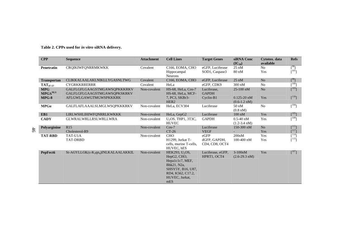
46
Table 2. CPPs used for in vitro siRNA delivery.
CPP Sequence Attachment Cell Lines Target Genes siRNA Conc
(IC50)
Cytotox. data
available
Refs
Penetratin CRQIKIWFQNRRMKWKK Covalent C166, EOMA, CHO
Hippocampal
Neurons
eGFP, Luciferase
SOD1, Caspase3
25 nM
80 nM
No
Yes
[98]
[101]
Transportan CLIKKALAALAKLNIKLLYGASNLTWG Covalent C166, EOMA, CHO eGFP, Luciferase 25 nM No [98]
TAT47-57 CYGRKKRRERRR Covalent HeLa eGFP, CDK9 300 nM No [100]
MPG
MPGΔNLS
MPG-8
GALFLGFLGAAGSTMGAWSQPKKKRKV
GALFLGFLGAAGSTMGAWSQPKSKRKV
AFLGWLGAWGTMGWSPKKKRK
Non-covalent HS-68, HeLa, Cos-7
HS-68, HeLa, MCF-
7, PC3, SKBr3-
HER2
Luciferase,
GAPDH
Cyclin-B1
25-100 nM
0.125-20 nM
(0.6-1.2 nM)
No
Yes
[103]
[116]
MPGα GALFLAFLAAALSLMGLWSQPKKKRKV Non-covalent HeLa, ECV304 Luciferase 50 nM
(0.8 nM)
No [104]
EB1 LIRLWSHLIHIWFQNRRLKWKKK Non-covalent HeLa, GepG2 Luciferase 100 nM Yes [105]
CADY GLWRALWRLLRSLWRLLWRA Non-covalent U2OS, THP1, 3T3C,
HUVEC
GAPDH 0.5-40 nM
(1.2-3.4 nM)
Yes [108]
Polyarginine R15
Cholesterol-R9
Non-covalent Cos-7
CT-26
Luciferase
VEGF
150-300 nM
-
No
Yes
[111]
[117]
TAT-RBD TAT-U1A
TAT-DRBD
Non-covalent CHO
H1299, Jurkat T-
cells, murine T-cells,
HUVEC, hES
eGFP
dGFP, GAPDH,
CD4, CD8, OCT4
200nM
100-400 nM
Yes
Yes
[114]
[115]
PepFect6 St-AGYLLGK(ε-K3qn4)INLKALAALAKKIL Non-covalent HEK293, U2OS,
HepG2, CHO,
Hepa1c1c7, MEF,
Bhk21, N2a,
SHSY5Y, B16, U87,
RD4, K562, C17.2,
HUVEC, Jurkat,
mES
Luciferase, eGFP,
HPRT1, OCT4
3-100nM
(2.6-29.3 nM)
Yes [107]
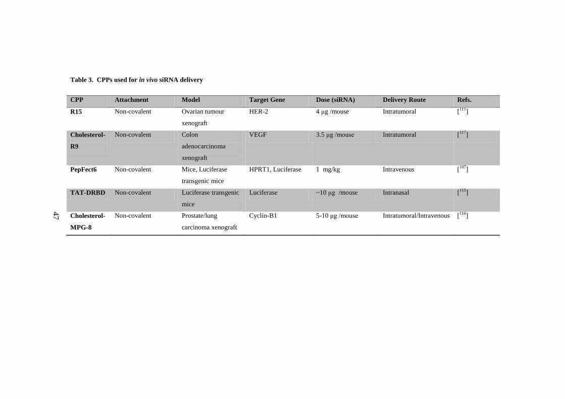
47
Table 3. CPPs used for in vivo siRNA delivery
CPP Attachment Model Target Gene Dose (siRNA) Delivery Route Refs.
R15 Non-covalent Ovarian tumour
xenograft
HER-2 4 μg /mouse Intratumoral [111
]
Cholesterol-
R9
Non-covalent Colon
adenocarcinoma
xenograft
VEGF 3.5 μg /mouse Intratumoral [117
]
PepFect6 Non-covalent Mice, Luciferase
transgenic mice
HPRT1, Luciferase 1 mg/kg Intravenous [107
]
TAT-DRBD Non-covalent Luciferase transgenic
mice
Luciferase ~10 μg /mouse Intranasal [115
]
Cholesterol-
MPG-8
Non-covalent Prostate/lung
carcinoma xenograft
Cyclin-B1 5-10 μg /mouse Intratumoral/Intravenous [116
]
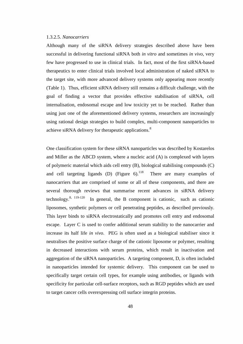
48
1.3.2.5. Nanocarriers
Although many of the siRNA delivery strategies described above have been
successful in delivering functional siRNA both in vitro and sometimes in vivo, very
few have progressed to use in clinical trials. In fact, most of the first siRNA-based
therapeutics to enter clinical trials involved local administration of naked siRNA to
the target site, with more advanced delivery systems only appearing more recently
(Table 1). Thus, efficient siRNA delivery still remains a difficult challenge, with the
goal of finding a vector that provides effective stabilisation of siRNA, cell
internalisation, endosomal escape and low toxicity yet to be reached. Rather than
using just one of the aforementioned delivery systems, researchers are increasingly
using rational design strategies to build complex, multi-component nanoparticles to
achieve siRNA delivery for therapeutic applications.8
One classification system for these siRNA nanoparticles was described by Kostarelos
and Miller as the ABCD system, where a nucleic acid (A) is complexed with layers
of polymeric material which aids cell entry (B), biological stabilising compounds (C)
and cell targeting ligands (D) (Figure 6).118
There are many examples of
nanocarriers that are comprised of some or all of these components, and there are
several thorough reviews that summarise recent advances in siRNA delivery
technology.8, 119-120
In general, the B component is cationic, such as cationic
liposomes, synthetic polymers or cell penetrating peptides, as described previously.
This layer binds to siRNA electrostatically and promotes cell entry and endosomal
escape. Layer C is used to confer additional serum stability to the nanocarrier and
increase its half life in vivo. PEG is often used as a biological stabiliser since it
neutralises the positive surface charge of the cationic liposome or polymer, resulting
in decreased interactions with serum proteins, which result in inactivation and
aggregation of the siRNA nanoparticles. A targeting component, D, is often included
in nanoparticles intended for systemic delivery. This component can be used to
specifically target certain cell types, for example using antibodies, or ligands with
specificity for particular cell-surface receptors, such as RGD peptides which are used
to target cancer cells overexpressing cell surface integrin proteins.
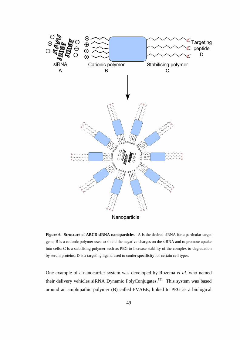
49
Figure 6. Structure of ABCD siRNA nanoparticles. A is the desired siRNA for a particular target
gene; B is a cationic polymer used to shield the negative charges on the siRNA and to promote uptake
into cells; C is a stabilising polymer such as PEG to increase stability of the complex to degradation
by serum proteins; D is a targeting ligand used to confer specificity for certain cell types.
One example of a nanocarrier system was developed by Rozema et al. who named
their delivery vehicles siRNA Dynamic PolyConjugates.121
This system was based
around an amphipathic polymer (B) called PVABE, linked to PEG as a biological
50
stabiliser (C) and a hepatocyte targeting ligand (D), with the siRNA covalently
conjugated to the polymer by a disulfide bond. The targeting and PEG moieties are
attached to PVABE via maleamate linkages to the amine groups of the polymer,
forming negatively charged nanoparticles which do not bind to serum proteins.
Following internalisation by endocytosis, the maleamate bonds are readily
hydrolysed on acidification of the endosome, exposing the cationic amine groups of
the polymer and inducing endosomal escape via the proton sponge effect. When
PEG was attached via a non-hydrolysable amide linkage, knockdown ability of the
polyconjugates was abolished, showing that exposure of the cationic residues of the
polymer are necessary for successful transfection. The polyconjugates were tested
both in vitro, knocking down apoB in mouse hepatocytes, and in vivo, targeting apoB
and ppara-1 in mouse livers. In cell cultures, an 80% mRNA knockdown was
achieved using ~50 nM siRNA conjugated to polyconjugates, which is comparable to
the CPPs listed in Table 2. In vivo, an 80-90% knockdown of apoB was achieved
using a 2.5 mg/kg dose, again comparable to both CPPs and SNALPs, with very little
toxicity. It was found that the presence of the hepatocyte targeting ligand N-
acetylgalactosamine (NAG) resulted in higher uptake in hepatocytes, whilst
replacement of NAG with mannose resulted in targeting of nonparenchymal cells in
the liver, confirming the ability of targeting ligands to direct the polyconjugates to
the required cell type.
Effective delivery of siRNA remains one of the great challenges currently facing
biological therapeutics. The exploitation of the RNAi pathway holds huge potential
for the treatment of a wide array of diseases, including cancers, viral infections and
genetic disorders. The key components of a successful siRNA delivery agent are the
ability to stabilise siRNA in the body, target it to the tissue of interest and deliver it
efficiently to the cytoplasm with minimal side effects and toxicity. A huge number
of potential delivery vehicles for siRNA have been described in the literature, and the
research interest in this area continues to grow year on year. As we discover more
efficient ways to achieve siRNA transfection, the prospect of a new era of genetic
medicines draws closer.
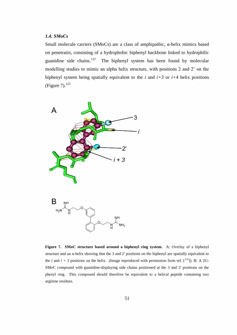
51
1.4. SMoCs
Small molecule carriers (SMoCs) are a class of amphipathic, α-helix mimics based
on penetratin, consisting of a hydrophobic biphenyl backbone linked to hydrophilic
guanidine side chains.122
The biphenyl system has been found by molecular
modelling studies to mimic an alpha helix structure, with positions 2 and 2’ on the
biphenyl system being spatially equivalent to the i and i+3 or i+4 helix positions
(Figure 7).123
Figure 7. SMoC structure based around a biphenyl ring system. A: Overlay of a biphenyl
structure and an α-helix showing that the 3 and 2' positions on the biphenyl are spatially equivalent to
the i and i + 3 positions on the helix. (Image reproduced with permission from ref. [122
]). B: A 2G-
SMoC compound with guanidine-displaying side chains positioned at the 3 and 2' positions on the
phenyl ring. This compound should therefore be equivalent to a helical peptide containing two
arginine residues.
52
Guanidine groups attached to the biphenyl structure mimic the arginine side chains
displayed on the surface of natural CPPs which are important for cell-surface
binding. SMoCs also contain a linker group consisting of a free amine which can be
used to couple dyes for visualisation by confocal microscopy. In addition, a
thiopyridyl derivative has been produced which may be used for covalent
conjugation of proteins via a disulfide linkage to a cysteine residue.
Various analogues of SMoC have been produced, with varying numbers and
positions of guanidine groups, and varying cargo linker positions.124
The SMoC
analogues are named according to the number of guanidine (G) groups attached to
the biphenyl backbone, for example 4G-SMoC denotes a SMoC compound with four
guanidines. Increasing the number of guanidine groups has been found to increase
the efficiency of transport across cell membranes, with 4G-SMoC possessing the
highest activity, possibly due to increased interactions with negatively charged
membrane GAGs. The position of the linker is also important, with steric effects
decreasing transport efficiency when placed para to the biphenyl bond, rather than
meta, where the macromolecular cargo is furthest from the guanidine groups. This
further demonstrates the significance of the interactions of the guanidine groups with
the cell membrane.
1.4.1. Synthesis
The current synthetic route to SMoC compounds (Scheme 1) centres around the
coupling of the respective mono-phenyl components substituted at the required
positions with Cbz-protected amines via Suzuki-Miyaura Pd(0) coupling chemistry.
A key intermediate is the iodobromo-derivative 2 of veratrole 1, which was
synthesized by sequential ortho metallations. The methoxy groups are cleaved with
boron tribromide, and a bis-alkylation is performed with 2-
(benzyloxycarbonylamino)ethyl methanesulfonate 3 in order to introduce two Cbz-
protected amine groups. The palladium-catalysed cyanation of the iodine with
Zn(CN)2 is carried out to produce the nitrile compound 4. This intermediate is then
coupled to the boronic ester 5, also substituted with two Cbz-protected amines,
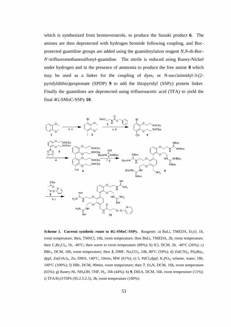
53
which is synthesized from bromoveratrole, to produce the Suzuki product 6. The
amines are then deprotected with hydrogen bromide following coupling, and Boc-
protected guanidine groups are added using the guanidinylation reagent N,N-di-Boc-
N'-trifluoromethanesulfonyl-guanidine. The nitrile is reduced using Raney-Nickel
under hydrogen and in the presence of ammonia to produce the free amine 8 which
may be used as a linker for the coupling of dyes, or N-succinimidyl-3-(2-
pyridyldithio)propionate (SPDP) 9 to add the thiopyridyl (SSPy) protein linker.
Finally the guanidines are deprotected using trifluoroacetic acid (TFA) to yield the
final 4G-SMoC-SSPy 10.
Scheme 1. Current synthetic route to 4G-SMoC-SSPy. Reagents: a) BuLi, TMEDA, Et2O, 1h,
room temperature, then, TMSCl, 16h, room temperature; then BuLi, TMEDA, 2h, room temperature;
then C2Br2Cl4, 1h, -40°C; then warm to room temperature (89%); b) ICl, DCM, 2h, -40°C (26%); c)
BBr3, DCM, 16h, room temperature; then 3, DMF, Na2CO3, 24h, 80°C (59%); d) Zn(CN)2, Pd2dba3,
dppf, Zn(OAc)2, Zn, DMA, 140°C, 10min, MW (61%); e) 5, PdCl2dppf, K3PO4, toluene, water, 18h,
100°C (100%); f) HBr, DCM, 90min, room temperature; then 7, Et3N, DCM, 16h, room temperature
(65%); g) Raney-Ni, NH4OH, THF, H2, 16h (44%); h) 9, DIEA, DCM, 16h, room temperature (11%);
i) TFA/H2O/TIPS (95:2.5:2.5), 3h, room temperature (100%).
54
1.4.2. Translocation activity
SMoCs have been shown to internalise both dyes and proteins into a variety of cell
types.122
2G- and 4G- SMoCs were coupled to fluorescein, and were found to be
internalised into U2OS cells and were observed in cytoplasmic vesicles, suggesting
an endocytotic mechanism. 4G-SMoC coupled to Alexa Fluor 488-tagged geminin
was also successfully internalised as measured by fluorescence-activated cell sorting
(FACS) analysis. When cells were treated with chlorpromazine, an inhibitor of
clathrin-mediated endocytosis, uptake of 4G-SMoC-geminin-Alexa Fluor 488 was
reduced by 70% suggesting an endocytotic mechanism for SMoC-coupled proteins.
Geminin is a cell cycle regulator, binding to Cdt1 to prevent loading onto the MCM
complex, thus blocking licensing of the DNA replication origin.125
Untagged
geminin was linked to 4G-SMoC via a disulfide linkage and its ability to suppress
DNA replication was analysed by measuring BrdU incorporation. Successful
delivery of active geminin was observed in a variety of cell types including NIH-3T3
fibroblasts, WI-38 HDF cells, HeLa cells and MOLT-4 leukemic lymphoblasts.122
1.5. Thesis Overview
Biological therapeutics has become an area of intense research interest over the last
two decades, and the discovery of the RNAi pathway has opened up the possibility of
effective gene therapy for a wide range of dieseases. The development of a cellular
transporter is one of the major challenges to the progress of macromolecular
therapeutics, and an efficient, safe method of delivering proteins and nucleic acids to
the intracellular environment would open up many possibilities for new drugs and
treatment strategies. While progress has been made in the search for delivery vectors
for macromolecular cargoes, significant challenges still remain. Large polymeric
carriers such as CPPs have a high cost of manufacture and are also susceptible to
degradation by serum proteases, requiring PEG modification in order to achieve
sufficient bioavailability. Cationic liposomes and cationic polymers such as PEI are
unsuitable for in vivo usage, due to their high toxicity. A small molecule transporter
such as SMoCs would be desirable due to their low cost of production and serum
stability.
55
The synthesis of the SMoC compounds has been optimised since the first published
method,124
however some steps are still low yielding, in particular the formation of
the boronic ester 5 for the Suzuki coupling step which has a yield of 30%, or
expensive, such as the guanidinylation step using N,N-di-Boc-N'-trifluoromethane-
sulfonylguanidine. It is hypothesized that more effective reagents exist which will
increase the yield of these steps, such as Molander-type reagents for Suzuki
coupling,126
and therefore the first aim of this thesis is to find more effective Suzuki
and guanidinylation reagents in order to produce higher quanitities of SMoCs. In
addition, it was shown that SMoCs with the linker in the meta position to the
biphenyl bond were more efficient protein transporters than those with the linker at
the para position, however a meta derivative of 4G-SMoC was not produced. Since
4G-SMoC was found to be the most efficient compound for cell internalisation, it
would be interesting to synthesize an analogue with a meta linker to determine
whether uptake could be increased.
Based on studies of CPPs, it is likely that the SMoC compounds bind to the GAGs of
cell surface proteoglycans as a precursor to internalisation. Guanidine groups are
able to form both bidentate hydrogen bonds and electrostatic interactions to GAGs
and phospholipids. In addition, guanidinium cations may also be used to form
electrostatic complexes with siRNA molecules. It is hypothesized that the biphenyl
SMoC backbone may give SMoCs an advantage over CPPs by stabilising the
guanidine positive charges via interactions between the π-electron cloud of the
biphenyl ring system and the cationic guanidine groups, so-called π-cation
interactions. The second aim of this thesis is to further characterise the SMoC
compounds in terms of their internal structure in order to understand how SMoCs
bind to anionic molecules such as GAGs and siRNA. The interactions between
SMoCs and siRNA will be studied in detail in order to determine the most efficient
SMoC compound for complexing siRNA.
56
Previous studies have shown that SMoCs function as molecular transporters, crossing
the cell membrane and delivering proteins which are able to exert a biological effect.
Due to their similarity to the CPP penetratin, it is hypothesized that SMoCs may be
used to transport additional macromolecular cargoes such as siRNA into cells in
order to achieve gene-specific knockdown. The third aim of this thesis is to use
SMoC-siRNA complexes to demonstrate that SMoCs may act as siRNA delivery
agents in vitro by performing transfection experiments in cultured cells.
Finally, it is hypothesized that GAG clustering by SMoCs may lead to internalisation
via macropinocytosis as suggested for CPPs, and therefore by increasing the ability
of SMoCs to bind and cluster common GAG chains, a greater level of cellular uptake
may be achieved. Therefore the final aim of this thesis is to design and synthesize a
new SMoC compound in order to more effectively bind cell surface proteoglycans,
and to compare the siRNA delivery properties of this SMoC against previous
compounds.

57
CHAPTER 2
Improving the synthetic route to the
SMoCs
58
2. Improving the synthetic route to the SMoCs
2.1. Introduction
The previously described method for synthesizing 4G-SMoC (Scheme 1) has so far
only yielded small quantities of the final product, with some steps producing
unsatisfactory yields, such as the synthesis of the boronic ester for the Suzuki-
Miyaura coupling which gives a 30% conversion, and the nitrile reduction step,
yielding 44% of product.124
The guanidinylation step uses an expensive triflate
reagent which also limits the total amount of SMoC that can be produced due to the
cost of materials. In order to produce larger quantities of SMoC to enable more
extensive biological testing, these problematic steps should be optimised. In
addition, in order to increase the speed of production, stable intermediates should be
produced on a large scale which can be easily isolated and stored for future use.
In order to increase the yield of the guanidinylation step, two routes will be explored.
Firstly, it is hypothesized that it may be possible to reduce the overall number of
steps in the SMoC synthetic route by producing an alkylation reagent bearing a
protected guanidine group. This reagent will use the pentamethylchromansulfonyl
(PMC) protecting group which is commonly used in peptide synthesis,127
and may be
used to alkylate the catechol precusor of 4G-SMoC. This will eliminate the Cbz
deprotection and subsequent gunaidinylation steps. An alternative method for
increasing the guanidinylation yield is to use an alternative reagent to the triflate
reagent 7, which if successful will decrease the cost of SMoC production.
The low yielding synthesis of the boronic ester may be overcome by using an
alternative reagent for the Suzuki coupling reaction. One such example is the
equivalent boronic acid, which is more reactive and therefore may also increase the
yield of the coupling. In addition, it is possible to use the trifluoroborate salt.
Aryltrifluoroborate salts, developed by Molander et al.,126, 128
are extremely stable to
air and moisture, allowing large scale synthesis of this key intermediate for long-term
storage. These salts are converted to the boronic acids in situ during the reaction,
and it is this species that is coupled to the halide, due to the lower energy barrier
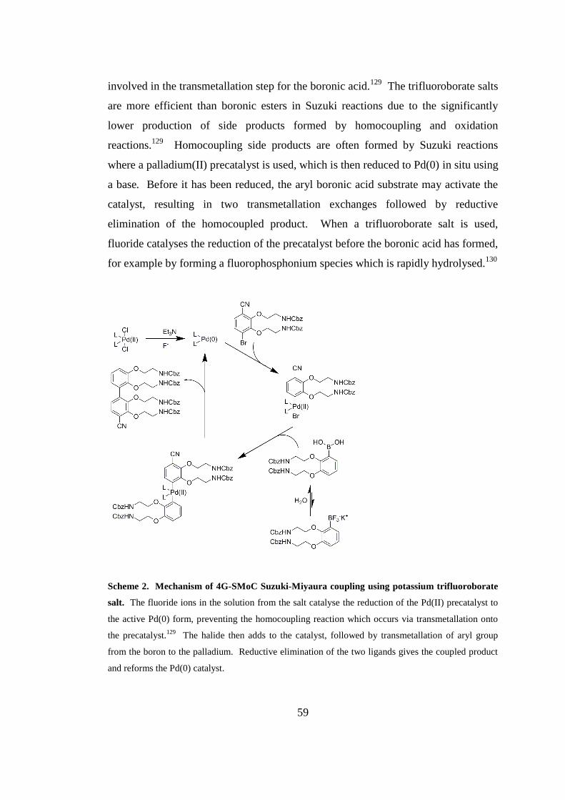
59
involved in the transmetallation step for the boronic acid.129
The trifluoroborate salts
are more efficient than boronic esters in Suzuki reactions due to the significantly
lower production of side products formed by homocoupling and oxidation
reactions.129
Homocoupling side products are often formed by Suzuki reactions
where a palladium(II) precatalyst is used, which is then reduced to Pd(0) in situ using
a base. Before it has been reduced, the aryl boronic acid substrate may activate the
catalyst, resulting in two transmetallation exchanges followed by reductive
elimination of the homocoupled product. When a trifluoroborate salt is used,
fluoride catalyses the reduction of the precatalyst before the boronic acid has formed,
for example by forming a fluorophosphonium species which is rapidly hydrolysed.130
Scheme 2. Mechanism of 4G-SMoC Suzuki-Miyaura coupling using potassium trifluoroborate
salt. The fluoride ions in the solution from the salt catalyse the reduction of the Pd(II) precatalyst to
the active Pd(0) form, preventing the homocoupling reaction which occurs via transmetallation onto
the precatalyst.129
The halide then adds to the catalyst, followed by transmetallation of aryl group
from the boron to the palladium. Reductive elimination of the two ligands gives the coupled product
and reforms the Pd(0) catalyst.
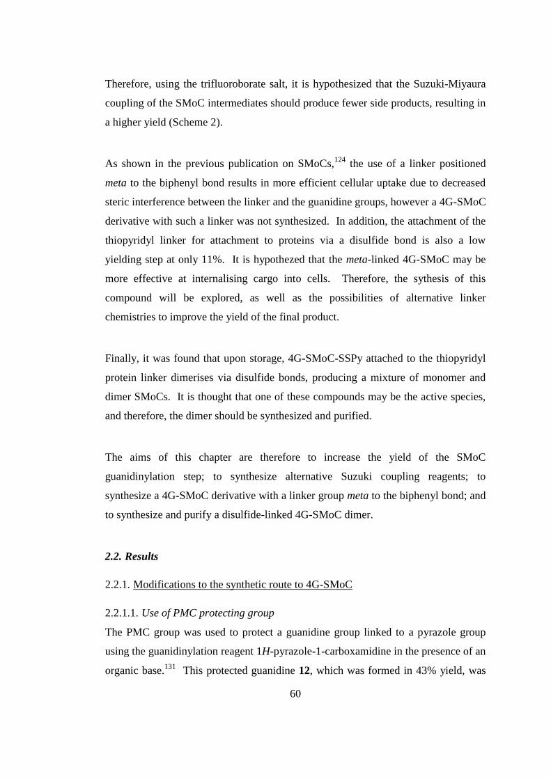
60
Therefore, using the trifluoroborate salt, it is hypothesized that the Suzuki-Miyaura
coupling of the SMoC intermediates should produce fewer side products, resulting in
a higher yield (Scheme 2).
As shown in the previous publication on SMoCs,124
the use of a linker positioned
meta to the biphenyl bond results in more efficient cellular uptake due to decreased
steric interference between the linker and the guanidine groups, however a 4G-SMoC
derivative with such a linker was not synthesized. In addition, the attachment of the
thiopyridyl linker for attachment to proteins via a disulfide bond is also a low
yielding step at only 11%. It is hypothezed that the meta-linked 4G-SMoC may be
more effective at internalising cargo into cells. Therefore, the sythesis of this
compound will be explored, as well as the possibilities of alternative linker
chemistries to improve the yield of the final product.
Finally, it was found that upon storage, 4G-SMoC-SSPy attached to the thiopyridyl
protein linker dimerises via disulfide bonds, producing a mixture of monomer and
dimer SMoCs. It is thought that one of these compounds may be the active species,
and therefore, the dimer should be synthesized and purified.
The aims of this chapter are therefore to increase the yield of the SMoC
guanidinylation step; to synthesize alternative Suzuki coupling reagents; to
synthesize a 4G-SMoC derivative with a linker group meta to the biphenyl bond; and
to synthesize and purify a disulfide-linked 4G-SMoC dimer.
2.2. Results
2.2.1. Modifications to the synthetic route to 4G-SMoC
2.2.1.1. Use of PMC protecting group
The PMC group was used to protect a guanidine group linked to a pyrazole group
using the guanidinylation reagent 1H-pyrazole-1-carboxamidine in the presence of an
organic base.131
This protected guanidine 12, which was formed in 43% yield, was
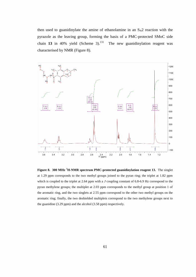
61
then used to guanidinylate the amine of ethanolamine in an SN2 reaction with the
pyrazole as the leaving group, forming the basis of a PMC-protected SMoC side
chain 13 in 40% yield (Scheme 3).131
The new guanidinylation reagent was
characterised by NMR (Figure 8).
Figure 8. 300 MHz 1H-NMR spectrum PMC-protected guanidinylation reagent 13. The singlet
at 1.29 ppm corresponds to the two methyl groups joined to the pyran ring; the triplet at 1.82 ppm
which is coupled to the triplet at 2.64 ppm with a J coupling constant of 6.8-6.9 Hz correspond to the
pyran methylene groups; the multiplet at 2.03 ppm corresponds to the methyl group at position 1 of
the aromatic ring, and the two singlets at 2.55 ppm correspond to the other two methyl groups on the
aromatic ring; finally, the two deshielded multiplets correspond to the two methylene groups next to
the guanidine (3.29 ppm) and the alcohol (3.58 ppm) respectively.
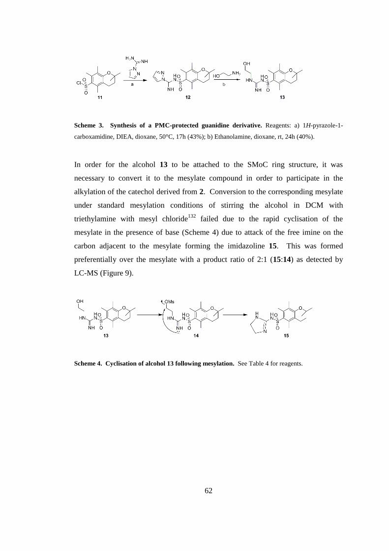
62
Scheme 3. Synthesis of a PMC-protected guanidine derivative. Reagents: a) 1H-pyrazole-1-
carboxamidine, DIEA, dioxane, 50°C, 17h (43%); b) Ethanolamine, dioxane, rt, 24h (40%).
In order for the alcohol 13 to be attached to the SMoC ring structure, it was
necessary to convert it to the mesylate compound in order to participate in the
alkylation of the catechol derived from 2. Conversion to the corresponding mesylate
under standard mesylation conditions of stirring the alcohol in DCM with
triethylamine with mesyl chloride132
failed due to the rapid cyclisation of the
mesylate in the presence of base (Scheme 4) due to attack of the free imine on the
carbon adjacent to the mesylate forming the imidazoline 15. This was formed
preferentially over the mesylate with a product ratio of 2:1 (15:14) as detected by
LC-MS (Figure 9).
Scheme 4. Cyclisation of alcohol 13 following mesylation. See Table 4 for reagents.
63
Figure 9. LC-MS analysis of the mesylation of alcohol 13 reaction mixture. A: UV trace of the
reaction mixture (blue line), peak 4 is the cyclised product 15, peak 6 is the mesylate 14, and peak 8 is
the starting material 13; B: Mass spectrum of the reaction mixture showing the [M + H]+ ions of 13
(m/z 370.4), 14 (m/z 448.4) and 15 (m/z 352.4).
The mesylation reaction was attempted under several different conditions, including
using two equivalents of MsCl in order to drive the mesylation reaction to
completion before the cyclisation could occur, and using a weaker base (Na2CO3), in
order to prevent the cyclisation (Table 4). None of the conditions were successful,
and so this line of enquiry was abandoned.

64
Table 4. Conditions tried for the mesylation of 13
Base Temp. °C Eq. MsCl Solvent
Et3N Rt 1 DCM
Et3N Rt 2 DCM
Na2CO3 Rt 1 DCM
As an alternative to alkylation using the mesylate, it was decided to investigate the
use of Mitsunobu chemistry which allows alcohols to be coupled to nucleophiles, to
attach the alcohol, 13, to phenol derivatives. A series of test reactions were carried
out using 3-methoxyphenol as the nucleophilic species (Table 5). The first reaction
used the standard Mitsunobu reagent diisopropylazodicarboxyl-ate (DIAD) and
triphenylphosphine (TPP) in THF, which is a widely reported method for this
reaction,133
however no reaction was observed. The more forcing reagents of
N,N,N',N'-tetraisopropylazodicarboxamide (TMAD) and tributylphosphine, which
are often used for less acidic nucleophiles with higher pKa values due to the
increased basicity of the azo nitrogen atoms,133
were then tried, using one, and then
two equivalents of the reagents. When still no reaction was seen, a biphenyl starting
material was tested with the same reagents, with the possibility that the guanidines
could be added following the Suzuki-Miyaura coupling step. In this reaction, a small
amount of the desired product was observed in the reaction mixture, but the low
conversion was not sufficient for further progress.

65
Table 5. Reagents and conditions attempted for the coupling of 13 to phenols
Phenol Coupling
reagents
Solvent Conditions Result
DIAD, PPh3 THF Rt, 24 h No reaction
TMAD, PBu3 THF Reflux, 48 h, N2 No reaction
TMAD, PBu3 (2
eq each)
THF Rt, 24 h No reaction
TMAD, PBu3 (2
eq each)
THF Rt, 24 h Small LC/MS
peak for mono-
product
Although several other Mitsunobu reagents are available,133
the fact that no reaction
was observed for the harsh TMAD regent, as well as time constraints meant that this
line of enquiry was abandoned in favour of the existing method of using Cbz-
protected amines.
2.2.1.2. Improving Suzuki-Miyaura coupling
NB. The work in this section was on going in the lab at the same time as the other experiments
described in this chapter and was carried out by Roberta Worthington. It is included here for
context as part of the overall development of the SMoC synthesis, and will be used later in
developing new SMoC reagents.
As an alternative to the boronic ester, the equivalent boronic acid 17 was synthesized
from the aryl bromide using a Grignard reagent with an organic ligand followed by
quenching with trimethyl borate, forming the boronic acid in 40% yield (Scheme 5).
The organic ligand moderates the reactivity of the arylmagnesium chloride in this
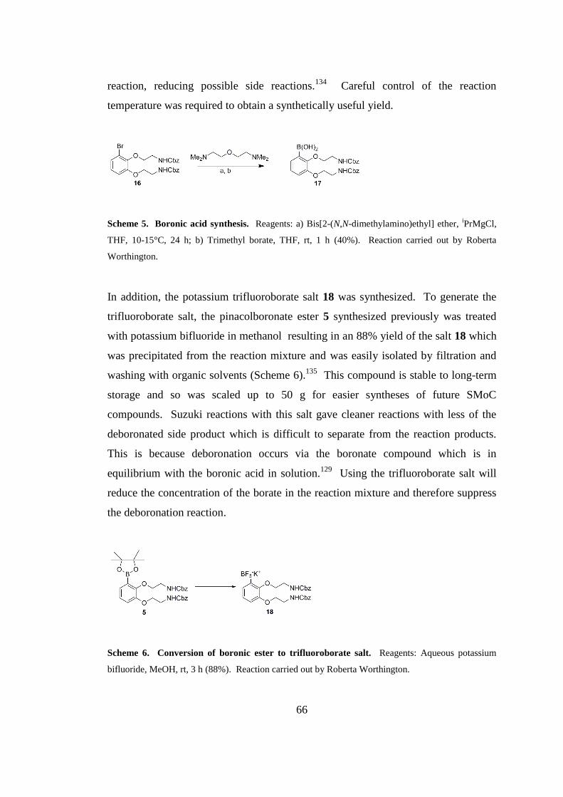
66
reaction, reducing possible side reactions.134
Careful control of the reaction
temperature was required to obtain a synthetically useful yield.
Scheme 5. Boronic acid synthesis. Reagents: a) Bis[2-(N,N-dimethylamino)ethyl] ether, iPrMgCl,
THF, 10-15°C, 24 h; b) Trimethyl borate, THF, rt, 1 h (40%). Reaction carried out by Roberta
Worthington.
In addition, the potassium trifluoroborate salt 18 was synthesized. To generate the
trifluoroborate salt, the pinacolboronate ester 5 synthesized previously was treated
with potassium bifluoride in methanol resulting in an 88% yield of the salt 18 which
was precipitated from the reaction mixture and was easily isolated by filtration and
washing with organic solvents (Scheme 6).135
This compound is stable to long-term
storage and so was scaled up to 50 g for easier syntheses of future SMoC
compounds. Suzuki reactions with this salt gave cleaner reactions with less of the
deboronated side product which is difficult to separate from the reaction products.
This is because deboronation occurs via the boronate compound which is in
equilibrium with the boronic acid in solution.129
Using the trifluoroborate salt will
reduce the concentration of the borate in the reaction mixture and therefore suppress
the deboronation reaction.
Scheme 6. Conversion of boronic ester to trifluoroborate salt. Reagents: Aqueous potassium
bifluoride, MeOH, rt, 3 h (88%). Reaction carried out by Roberta Worthington.

67
2.2.1.3. New Guanidinylation reagent
A problematic step in the original SMoC synthesis was the addition of Boc-protected
guanidine groups using the expensive N,N-di-Boc-N'-trifluoromethanesulfonyl-
guanidine reagent, which resulted in average yields. To improve this step, a different
guanidinylation reagent was used, N,N'-Di-Boc-1H-pyrazole-1-carboxamidine
(Scheme 7). The amines of compound 6 were deprotected as described using a 33%
solution of HBr in acetic acid and dried extensively under vacuum.124
The dried
amine was then suspended in a solution of DIEA in DCM and the guanidinylation
reagent was subsequently added. On completion, the tetra-guanidinylated product
was isolated by flash column chromatography. Using this reagent slightly increased
the yield of guanidinylation from ~65% to over 70% at around 10% of the cost of the
previous reagent. Therefore, the cost of SMoC production was significantly
decreased, allowing for production of larger quantities for future studies.
Scheme 7. Guanidinylation using pyrazole reagent. Reagents: a) 30% HBr in acetic acid, DCM,
rt, 1h; b) N,N'-Di-Boc-1H-pyrazole-1-carboxamidine, DIEA, DCM, rt, 18 h (71%).
2.2.1.4. Linker nitrile reduction
NB. The work in this section was on going in the lab at the same time as the other experiments
described in this chapter and was carried out by Roberta Worthington. It is included here for
context as part of the overall development of the SMoC synthesis.
Finally, the reduction of the nitrile in order to produce a primary amine for
attachment of a disulfide linker was another low-yielding step from the previous
route. The previous synthesis used Raney Nickel/H2 which resulted in 40-50%
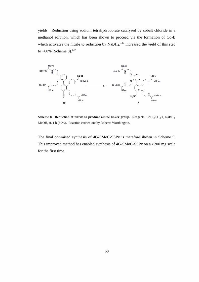
68
yields. Reduction using sodium tetrahydroborate catalysed by cobalt chloride in a
methanol solution, which has been shown to proceed via the formation of Co2B
which activates the nitrile to reduction by NaBH4,136
increased the yield of this step
to ~60% (Scheme 8).137
Scheme 8. Reduction of nitrile to produce amine linker group. Reagents: CoCl2.6H2O, NaBH4,
MeOH, rt, 1 h (60%). Reaction carried out by Roberta Worthington.
The final optimised synthesis of 4G-SMoC-SSPy is therefore shown in Scheme 9.
This improved method has enabled synthesis of 4G-SMoC-SSPy on a >200 mg scale
for the first time.
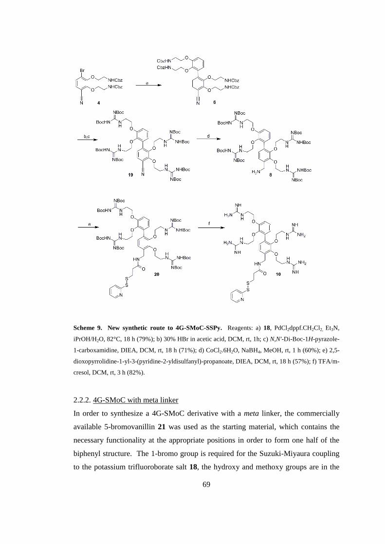
69
Scheme 9. New synthetic route to 4G-SMoC-SSPy. Reagents: a) 18, PdCl2dppf.CH2Cl2, Et3N,
iPrOH/H2O, 82°C, 18 h (79%); b) 30% HBr in acetic acid, DCM, rt, 1h; c) N,N'-Di-Boc-1H-pyrazole-
1-carboxamidine, DIEA, DCM, rt, 18 h (71%); d) CoCl2.6H2O, NaBH4, MeOH, rt, 1 h (60%); e) 2,5-
dioxopyrrolidine-1-yl-3-(pyridine-2-yldisulfanyl)-propanoate, DIEA, DCM, rt, 18 h (57%); f) TFA/m-
cresol, DCM, rt, 3 h (82%).
2.2.2. 4G-SMoC with meta linker
In order to synthesize a 4G-SMoC derivative with a meta linker, the commercially
available 5-bromovanillin 21 was used as the starting material, which contains the
necessary functionality at the appropriate positions in order to form one half of the
biphenyl structure. The 1-bromo group is required for the Suzuki-Miyaura coupling
to the potassium trifluoroborate salt 18, the hydroxy and methoxy groups are in the
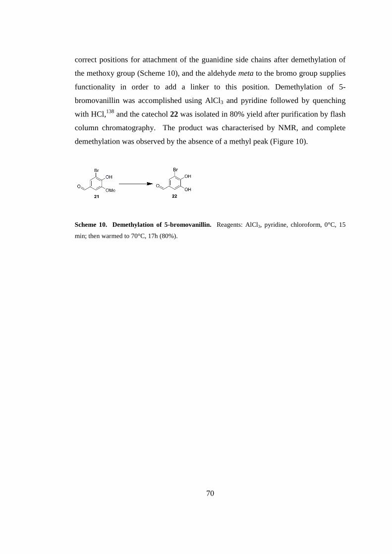
70
correct positions for attachment of the guanidine side chains after demethylation of
the methoxy group (Scheme 10), and the aldehyde meta to the bromo group supplies
functionality in order to add a linker to this position. Demethylation of 5-
bromovanillin was accomplished using AlCl3 and pyridine followed by quenching
with HCl,138
and the catechol 22 was isolated in 80% yield after purification by flash
column chromatography. The product was characterised by NMR, and complete
demethylation was observed by the absence of a methyl peak (Figure 10).
Scheme 10. Demethylation of 5-bromovanillin. Reagents: AlCl3, pyridine, chloroform, 0°C, 15
min; then warmed to 70°C, 17h (80%).
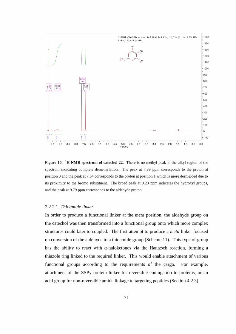
71
Figure 10. 1H-NMR spectrum of catechol 22. There is no methyl peak in the alkyl region of the
spectrum indicating complete demethylation. The peak at 7.39 ppm corresponds to the proton at
position 3 and the peak at 7.64 corresponds to the proton at position 1 which is more deshielded due to
its proximity to the bromo substituent. The broad peak at 9.23 ppm indicates the hydroxyl groups,
and the peak at 9.79 ppm corresponds to the aldehyde proton.
2.2.2.1. Thioamide linker
In order to produce a functional linker at the meta position, the aldehyde group on
the catechol was then transformed into a functional group onto which more complex
structures could later to coupled. The first attempt to produce a meta linker focused
on conversion of the aldehyde to a thioamide group (Scheme 11). This type of group
has the ability to react with α-haloketones via the Hantzsch reaction, forming a
thiazole ring linked to the required linker. This would enable attachment of various
functional groups according to the requirements of the cargo. For example,
attachment of the SSPy protein linker for reversible conjugation to proteins, or an
acid group for non-reversible amide linkage to targeting peptides (Section 4.2.3).
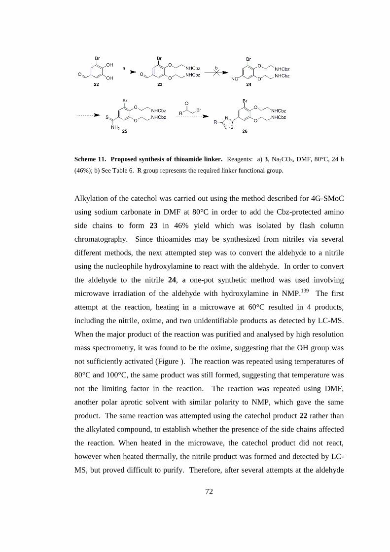
72
Scheme 11. Proposed synthesis of thioamide linker. Reagents: a) 3, Na2CO3, DMF, 80°C, 24 h
(46%); b) See Table 6. R group represents the required linker functional group.
Alkylation of the catechol was carried out using the method described for 4G-SMoC
using sodium carbonate in DMF at 80°C in order to add the Cbz-protected amino
side chains to form 23 in 46% yield which was isolated by flash column
chromatography. Since thioamides may be synthesized from nitriles via several
different methods, the next attempted step was to convert the aldehyde to a nitrile
using the nucleophile hydroxylamine to react with the aldehyde. In order to convert
the aldehyde to the nitrile 24, a one-pot synthetic method was used involving
microwave irradiation of the aldehyde with hydroxylamine in NMP.139
The first
attempt at the reaction, heating in a microwave at 60°C resulted in 4 products,
including the nitrile, oxime, and two unidentifiable products as detected by LC-MS.
When the major product of the reaction was purified and analysed by high resolution
mass spectrometry, it was found to be the oxime, suggesting that the OH group was
not sufficiently activated (Figure ). The reaction was repeated using temperatures of
80°C and 100°C, the same product was still formed, suggesting that temperature was
not the limiting factor in the reaction. The reaction was repeated using DMF,
another polar aprotic solvent with similar polarity to NMP, which gave the same
product. The same reaction was attempted using the catechol product 22 rather than
the alkylated compound, to establish whether the presence of the side chains affected
the reaction. When heated in the microwave, the catechol product did not react,
however when heated thermally, the nitrile product was formed and detected by LC-
MS, but proved difficult to purify. Therefore, after several attempts at the aldehyde
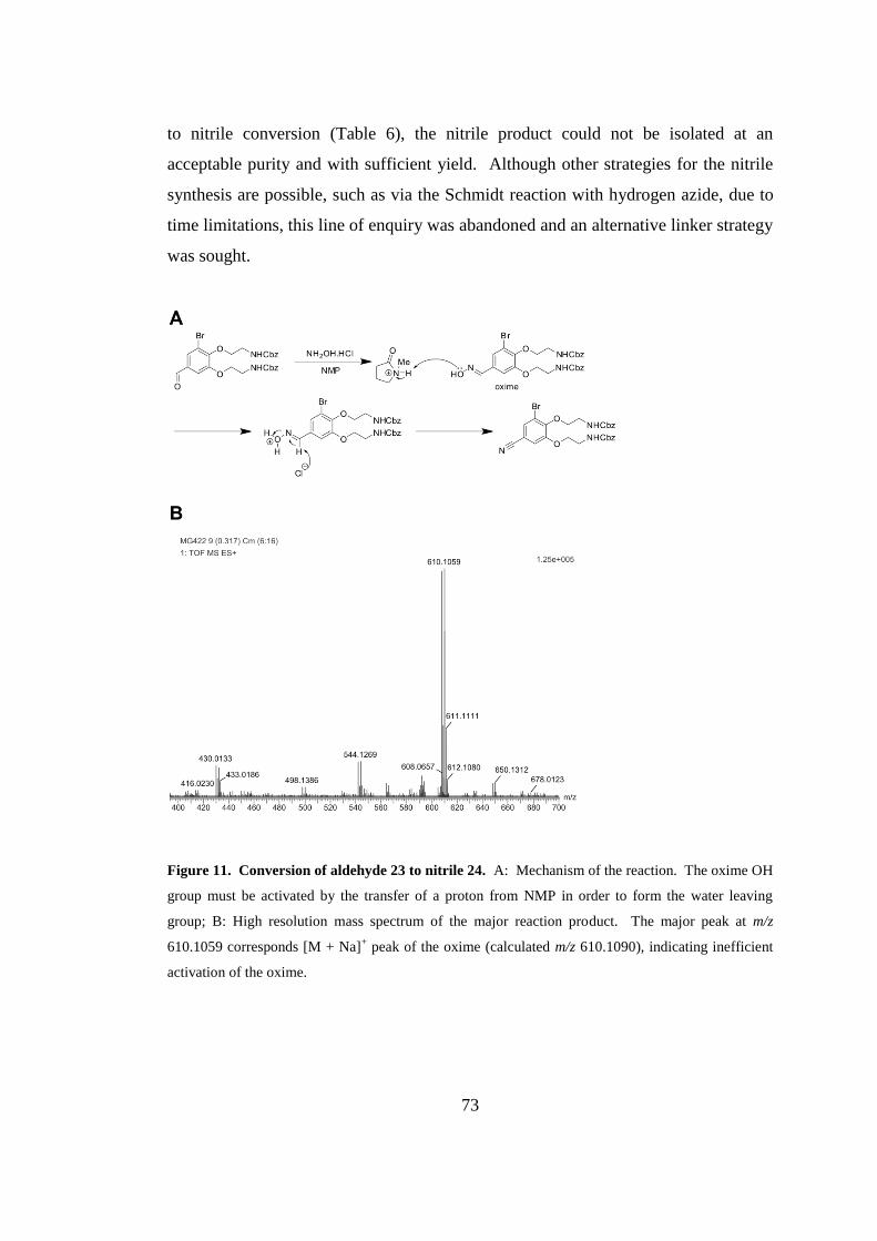
73
to nitrile conversion (Table 6), the nitrile product could not be isolated at an
acceptable purity and with sufficient yield. Although other strategies for the nitrile
synthesis are possible, such as via the Schmidt reaction with hydrogen azide, due to
time limitations, this line of enquiry was abandoned and an alternative linker strategy
was sought.
Figure 11. Conversion of aldehyde 23 to nitrile 24. A: Mechanism of the reaction. The oxime OH
group must be activated by the transfer of a proton from NMP in order to form the water leaving
group; B: High resolution mass spectrum of the major reaction product. The major peak at m/z
610.1059 corresponds [M + Na]+ peak of the oxime (calculated m/z 610.1090), indicating inefficient
activation of the oxime.
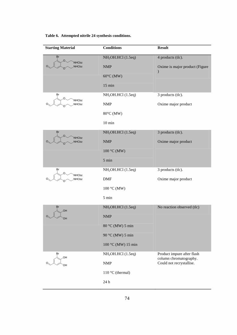
74
Table 6. Attempted nitrile 24 synthesis conditions.
Starting Material Conditions Result
NH2OH.HCl (1.5eq)
NMP
60°C (MW)
15 min
4 products (tlc).
Oxime is major product (Figure
)
NH2OH.HCl (1.5eq)
NMP
80°C (MW)
10 min
3 products (tlc).
Oxime major product
NH2OH.HCl (1.5eq)
NMP
100 °C (MW)
5 min
3 products (tlc).
Oxime major product
NH2OH.HCl (1.5eq)
DMF
100 °C (MW)
5 min
3 products (tlc).
Oxime major product
NH2OH.HCl (1.5eq)
NMP
80 °C (MW) 5 min
90 °C (MW) 5 min
100 °C (MW) 15 min
No reaction observed (tlc)
NH2OH.HCl (1.5eq)
NMP
110 °C (thermal)
24 h
Product impure after flash
column chromatography.
Could not recrystallise.
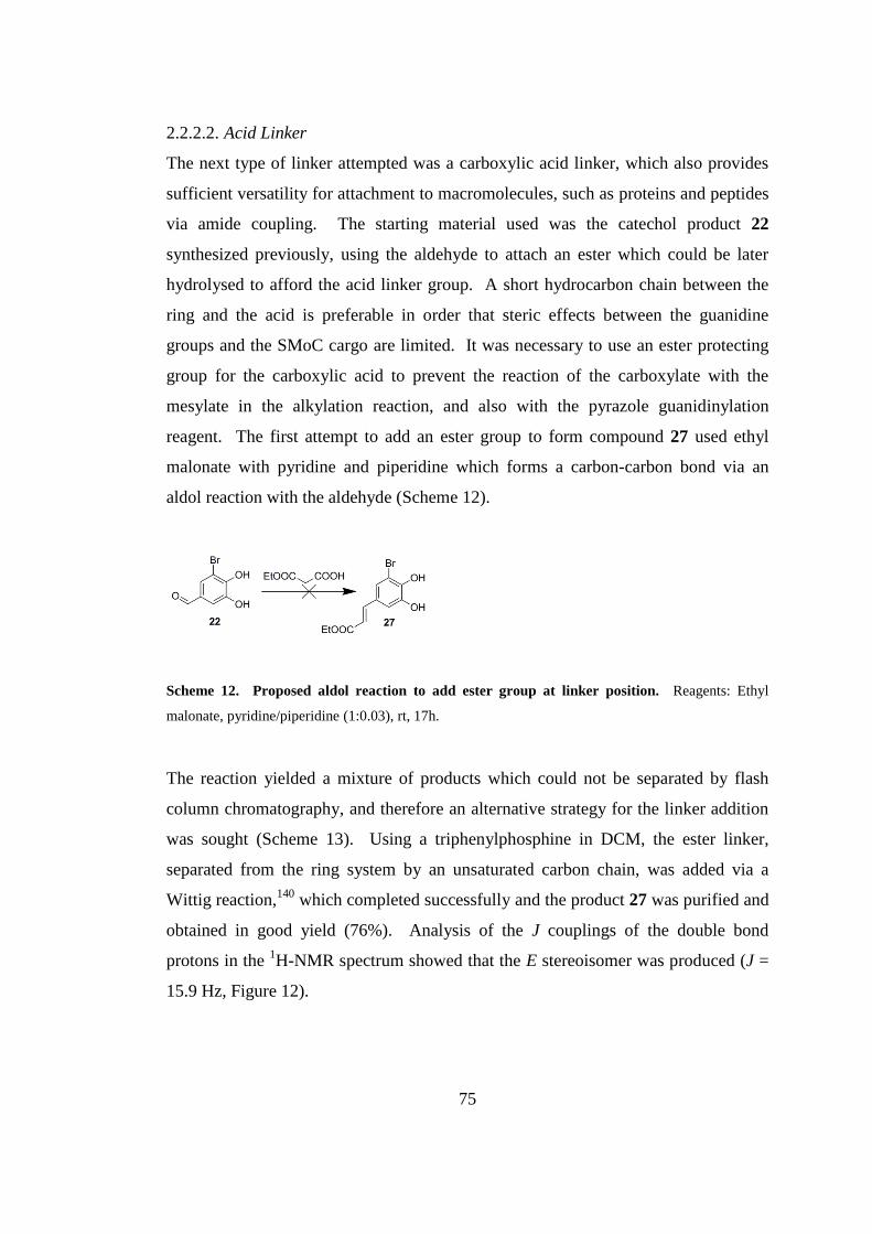
75
2.2.2.2. Acid Linker
The next type of linker attempted was a carboxylic acid linker, which also provides
sufficient versatility for attachment to macromolecules, such as proteins and peptides
via amide coupling. The starting material used was the catechol product 22
synthesized previously, using the aldehyde to attach an ester which could be later
hydrolysed to afford the acid linker group. A short hydrocarbon chain between the
ring and the acid is preferable in order that steric effects between the guanidine
groups and the SMoC cargo are limited. It was necessary to use an ester protecting
group for the carboxylic acid to prevent the reaction of the carboxylate with the
mesylate in the alkylation reaction, and also with the pyrazole guanidinylation
reagent. The first attempt to add an ester group to form compound 27 used ethyl
malonate with pyridine and piperidine which forms a carbon-carbon bond via an
aldol reaction with the aldehyde (Scheme 12).
Scheme 12. Proposed aldol reaction to add ester group at linker position. Reagents: Ethyl
malonate, pyridine/piperidine (1:0.03), rt, 17h.
The reaction yielded a mixture of products which could not be separated by flash
column chromatography, and therefore an alternative strategy for the linker addition
was sought (Scheme 13). Using a triphenylphosphine in DCM, the ester linker,
separated from the ring system by an unsaturated carbon chain, was added via a
Wittig reaction,140
which completed successfully and the product 27 was purified and
obtained in good yield (76%). Analysis of the J couplings of the double bond
protons in the 1H-NMR spectrum showed that the E stereoisomer was produced (J =
15.9 Hz, Figure 12).
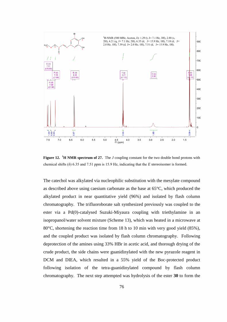
76
Figure 12. 1H NMR spectrum of 27. The J coupling constant for the two double bond protons with
chemical shifts (δ) 6.35 and 7.51 ppm is 15.9 Hz, indicating that the E stereoisomer is formed.
The catechol was alkylated via nucleophilic substitution with the mesylate compound
as described above using caesium carbonate as the base at 65°C, which produced the
alkylated product in near quantitative yield (96%) and isolated by flash column
chromatography. The trifluoroborate salt synthesized previously was coupled to the
ester via a Pd(0)-catalysed Suzuki-Miyaura coupling with triethylamine in an
isopropanol/water solvent mixture (Scheme 13), which was heated in a microwave at
80°C, shortening the reaction time from 18 h to 10 min with very good yield (85%),
and the coupled product was isolated by flash column chromatography. Following
deprotection of the amines using 33% HBr in acetic acid, and thorough drying of the
crude product, the side chains were guanidinylated with the new pyrazole reagent in
DCM and DIEA, which resulted in a 55% yield of the Boc-protected product
following isolation of the tetra-guanidinylated compound by flash column
chromatography. The next step attempted was hydrolysis of the ester 30 to form the
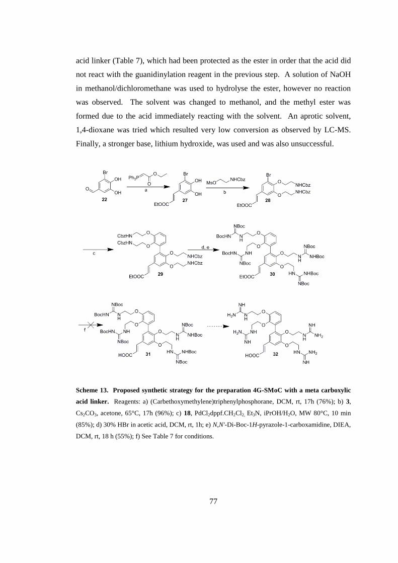
77
acid linker (Table 7), which had been protected as the ester in order that the acid did
not react with the guanidinylation reagent in the previous step. A solution of NaOH
in methanol/dichloromethane was used to hydrolyse the ester, however no reaction
was observed. The solvent was changed to methanol, and the methyl ester was
formed due to the acid immediately reacting with the solvent. An aprotic solvent,
1,4-dioxane was tried which resulted very low conversion as observed by LC-MS.
Finally, a stronger base, lithium hydroxide, was used and was also unsuccessful.
Scheme 13. Proposed synthetic strategy for the preparation 4G-SMoC with a meta carboxylic
acid linker. Reagents: a) (Carbethoxymethylene)triphenylphosphorane, DCM, rt, 17h (76%); b) 3,
Cs2CO3, acetone, 65°C, 17h (96%); c) 18, PdCl2dppf.CH2Cl2, Et3N, iPrOH/H2O, MW 80°C, 10 min
(85%); d) 30% HBr in acetic acid, DCM, rt, 1h; e) N,N'-Di-Boc-1H-pyrazole-1-carboxamidine, DIEA,
DCM, rt, 18 h (55%); f) See Table 7 for conditions.
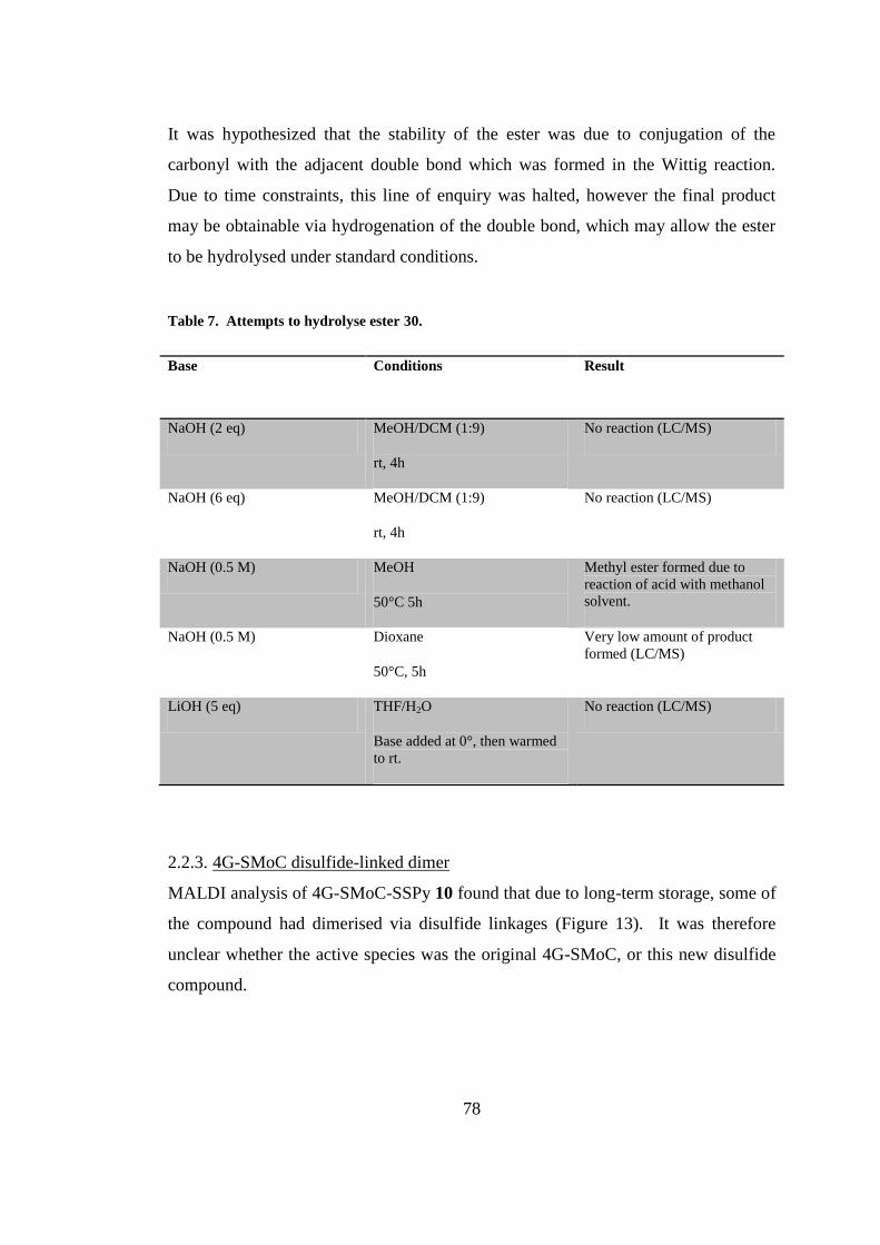
78
It was hypothesized that the stability of the ester was due to conjugation of the
carbonyl with the adjacent double bond which was formed in the Wittig reaction.
Due to time constraints, this line of enquiry was halted, however the final product
may be obtainable via hydrogenation of the double bond, which may allow the ester
to be hydrolysed under standard conditions.
Table 7. Attempts to hydrolyse ester 30.
Base Conditions Result
NaOH (2 eq) MeOH/DCM (1:9)
rt, 4h
No reaction (LC/MS)
NaOH (6 eq) MeOH/DCM (1:9)
rt, 4h
No reaction (LC/MS)
NaOH (0.5 M) MeOH
50°C 5h
Methyl ester formed due to
reaction of acid with methanol
solvent.
NaOH (0.5 M) Dioxane
50°C, 5h
Very low amount of product
formed (LC/MS)
LiOH (5 eq) THF/H2O
Base added at 0°, then warmed
to rt.
No reaction (LC/MS)
2.2.3. 4G-SMoC disulfide-linked dimer
MALDI analysis of 4G-SMoC-SSPy 10 found that due to long-term storage, some of
the compound had dimerised via disulfide linkages (Figure 13). It was therefore
unclear whether the active species was the original 4G-SMoC, or this new disulfide
compound.
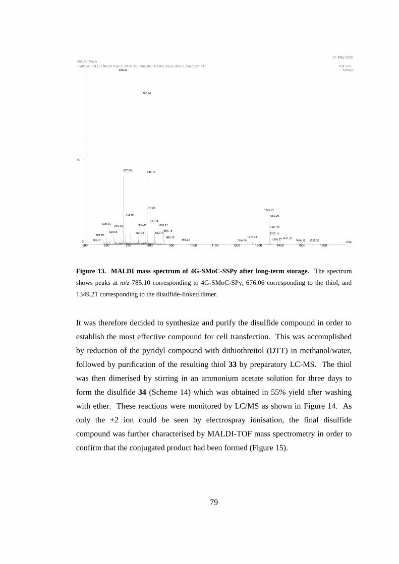
79
Figure 13. MALDI mass spectrum of 4G-SMoC-SSPy after long-term storage. The spectrum
shows peaks at m/z 785.10 corresponding to 4G-SMoC-SPy, 676.06 corresponding to the thiol, and
1349.21 corresponding to the disulfide-linked dimer.
It was therefore decided to synthesize and purify the disulfide compound in order to
establish the most effective compound for cell transfection. This was accomplished
by reduction of the pyridyl compound with dithiothreitol (DTT) in methanol/water,
followed by purification of the resulting thiol 33 by preparatory LC-MS. The thiol
was then dimerised by stirring in an ammonium acetate solution for three days to
form the disulfide 34 (Scheme 14) which was obtained in 55% yield after washing
with ether. These reactions were monitored by LC/MS as shown in Figure 14. As
only the +2 ion could be seen by electrospray ionisation, the final disulfide
compound was further characterised by MALDI-TOF mass spectrometry in order to
confirm that the conjugated product had been formed (Figure 15).
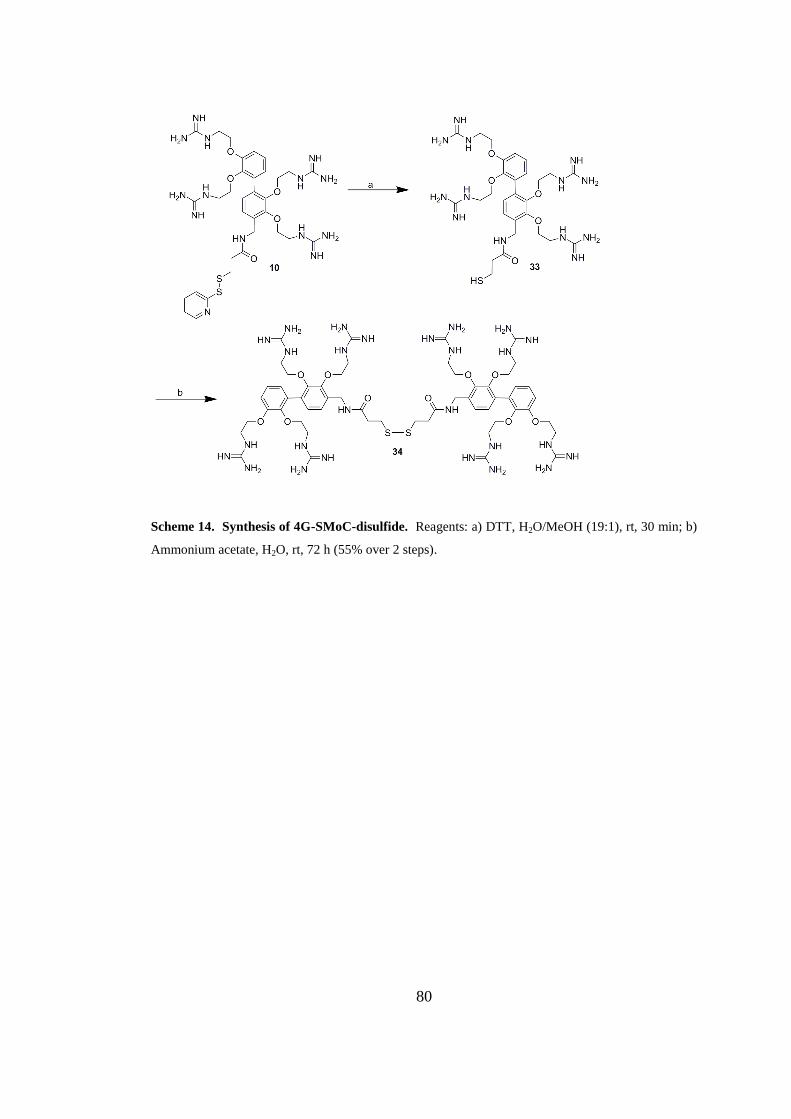
80
Scheme 14. Synthesis of 4G-SMoC-disulfide. Reagents: a) DTT, H2O/MeOH (19:1), rt, 30 min; b)
Ammonium acetate, H2O, rt, 72 h (55% over 2 steps).
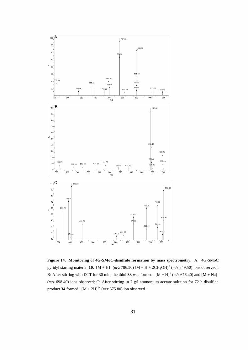
81
Figure 14. Monitoring of 4G-SMoC-disulfide formation by mass spectrometry. A: 4G-SMoC
pyridyl starting material 10. [M + H]+ (m/z 786.50) [M + H + 2CH3OH)
+ (m/z 849.50) ions observed ;
B: After stirring with DTT for 30 min, the thiol 33 was formed. [M + H]+ (m/z 676.40) and [M + Na]
+
(m/z 698.40) ions observed; C: After stirring in 7 g/l ammonium acetate solution for 72 h disulfide
product 34 formed. [M + 2H]2+
(m/z 675.80) ion observed.

82
Figure 15. MALDI-TOF spectrum of 4G-SMoC dimer 34. [M + H]+ (m/z 1350.35) ion observed.
2.3. Discussion
In this chapter, the synthesis of 4G-SMoC-SSPy has been examined and some steps
were successfully optimised, allowing for a greater yield of product. However, it
was not possible to reduce the number of steps by introducing PMC-protected
guanidine groups to the catechol precursor using alkyation reagent 13. The initial
attempt to synthesize the mesylate derivative failed due to an intramolecular
cyclisation resulting from the attack of the nucleophilic imine on the product. The
Mitsunobu reaction was then attempted in order to couple 13 directly to phenols
(Scheme 15). The mechanism of this reaction involves initial attack by a phosphine
on the azo-containing Mitsunobu reagent to form a basic zwitterionic intermediate
which is able to deprotonate the alcohol, forming an alkoxide. This then attacks the
positively charged phosphorus forming an oxophosphonium species and a second
basic nitrogen on the Mitsunobu reagent which can deprotonate the nucleophilic
starting material, in this case a phenol. The deprotonated phenol then attacks the
carbon of the oxophosphonium derivative, with phosphine oxide as the leaving group
to form the coupled product.
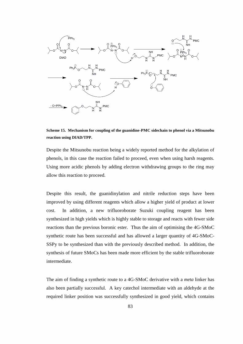
83
Scheme 15. Mechanism for coupling of the guanidine-PMC sidechain to phenol via a Mitsunobu
reaction using DIAD/TPP.
Despite the Mitsunobu reaction being a widely reported method for the alkylation of
phenols, in this case the reaction failed to proceed, even when using harsh reagents.
Using more acidic phenols by adding electron withdrawing groups to the ring may
allow this reaction to proceed.
Despite this result, the guanidinylation and nitrile reduction steps have been
improved by using different reagents which allow a higher yield of product at lower
cost. In addition, a new trifluoroborate Suzuki coupling reagent has been
synthesized in high yields which is highly stable to storage and reacts with fewer side
reactions than the previous boronic ester. Thus the aim of optimising the 4G-SMoC
synthetic route has been successful and has allowed a larger quantity of 4G-SMoC-
SSPy to be synthesized than with the previously described method. In addition, the
synthesis of future SMoCs has been made more efficient by the stable trifluoroborate
intermediate.
The aim of finding a synthetic route to a 4G-SMoC derivative with a meta linker has
also been partially successful. A key catechol intermediate with an aldehyde at the
required linker position was successfully synthesized in good yield, which contains

84
the necessary functionality at the appropriate positions to form one half of the SMoC
biphenyl structure. This intermediate may be successfully alkylated to add the amino
side chains, and the bromine successfully participates in a Suzuki-Miyaura coupling
reaction with the trifluoroborate intermediate to form the biphenyl backbone. The
attempt to produce a thioamide linker was unsuccessful, due to the difficulty in
forming a nitrile at the linker position, however an ester derivative was successfully
synthesized using a Wittig reaction. The resulting product was the E-stereoisomer,
as expected when using a phosphorane compound containing a carbonyl which is
able to form a stabilised ylide intermediate. The stabilised ylide means that
subsequent formation of the oxaphosphetene is reversible, and therefore the reaction
is thermodynamically controlled, proceeding via the more stable anti diastereomer to
form the E-alkene.141
This is in contrast to using an unstabilised ylide, where the
reaction is kinetically controlled. The oxaphosphetane formation is irreversible, and
therefore the syn diastereomer is produced via the most stable transition state, which
leads to Z-alkene product (Scheme 16). The Boc-protected SMoC intermediate was
successfully synthesized using the optimised reactions described previously.
Ester hydrolysis was unsuccessful under the attempted conditions, which may be due
to conjugation with the adjacent double bond. It has been observed previously that
the hydrolysis of unsaturated esters is considerably slower than expected from the
predicted steric effects of the substituents.142
When the unsaturation is moved further
from the ester group, this effect decreases suggesting that unsaturation is responsible
for the lower rate of reaction. With further experimentation, such as hydrogenation
of the double bond, it is possible that this intermediate may be hydrolysed, and
converted to the required SMoC compound by cleavage of the Boc protecting groups
using the previously described method.
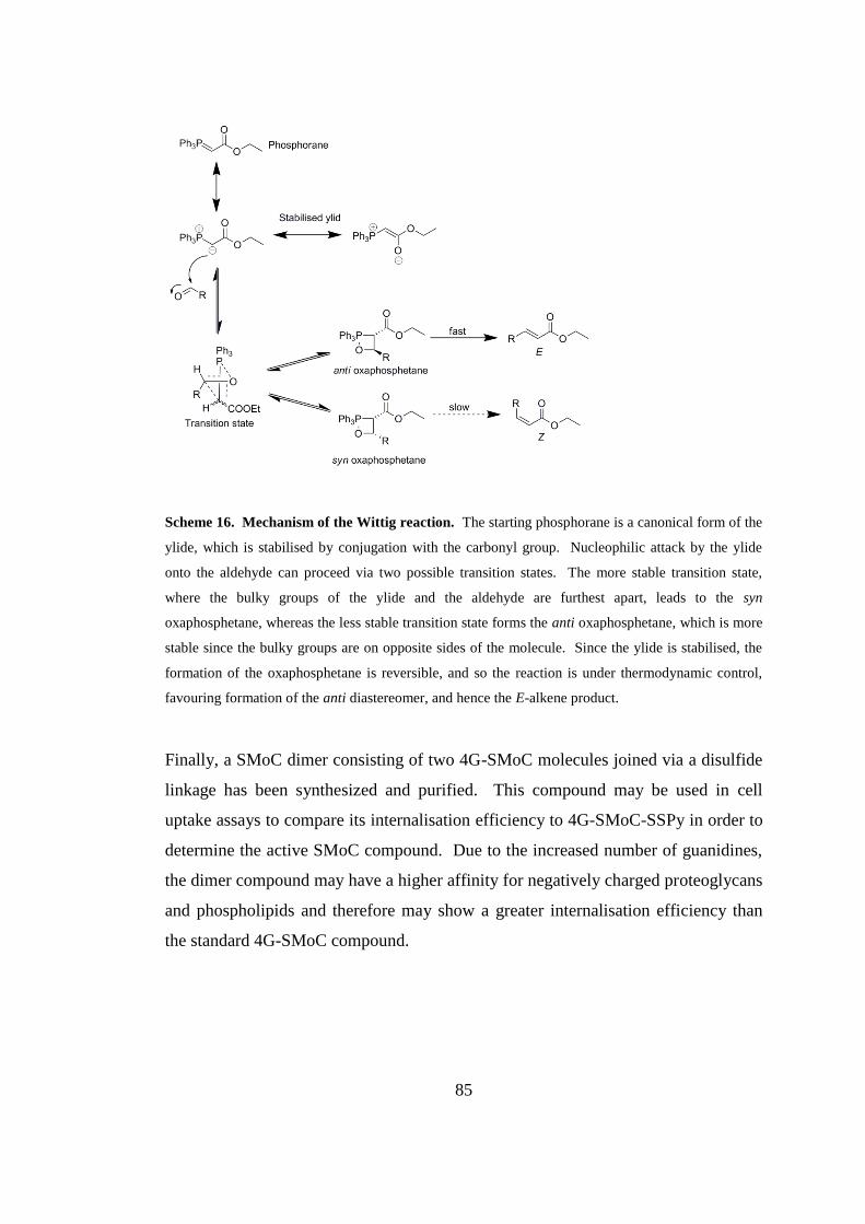
85
Scheme 16. Mechanism of the Wittig reaction. The starting phosphorane is a canonical form of the
ylide, which is stabilised by conjugation with the carbonyl group. Nucleophilic attack by the ylide
onto the aldehyde can proceed via two possible transition states. The more stable transition state,
where the bulky groups of the ylide and the aldehyde are furthest apart, leads to the syn
oxaphosphetane, whereas the less stable transition state forms the anti oxaphosphetane, which is more
stable since the bulky groups are on opposite sides of the molecule. Since the ylide is stabilised, the
formation of the oxaphosphetane is reversible, and so the reaction is under thermodynamic control,
favouring formation of the anti diastereomer, and hence the E-alkene product.
Finally, a SMoC dimer consisting of two 4G-SMoC molecules joined via a disulfide
linkage has been synthesized and purified. This compound may be used in cell
uptake assays to compare its internalisation efficiency to 4G-SMoC-SSPy in order to
determine the active SMoC compound. Due to the increased number of guanidines,
the dimer compound may have a higher affinity for negatively charged proteoglycans
and phospholipids and therefore may show a greater internalisation efficiency than
the standard 4G-SMoC compound.
86
CHAPTER 3
Exploring the interactions of SMoCs
with siRNA
87
3. Exploring the interactions of SMoCs with siRNA
3.1. Introduction
Given that SMoCs have been shown to be successful in delivering active proteins
into cells, another useful application would be the delivery of siRNA, a class of
macromolecule with great therapeutic potential. CPPs that have been used for
siRNA delivery are either covalently attached to siRNA or packaged as electrostatic
complexes, making use of their cationic arginine and lysine residues. Since SMoCs
are highly positively charged due to the presence of guanidine groups, it is
hypothesized that they may form non-covalent complexes with siRNA which are
capable of crossing the cell membrane resulting in gene-specific knockdown.
3.1.1. The SMoC π-cation interaction
Molecular dynamics studies on penetratin have suggested that the aromatic
tryptophan and phenylalanine amino acids form π-cation interactions with arginine
residues in the same molecule during attachment to the cell membrane.143
It was
suggested that these interactions help to shield the positive charges from the water
phase and therefore enhance insertion into lipid bilayers.
π-Cation interactions were first described by Burley and Petsko in 1986 when they
observed that the side chains of cationic amino acids such as lysine and arginine are
preferentially located near the π-electron cloud of aromatic amino acids such as
phenylalanine, tyrosine and tryptophan.144
It has been found that π-cation
interactions are very common in proteins, with one interaction per 77 residues on
average. Arginine residues are more favoured for π-cation interactions than lysine,
probably due to the guanidine group on arginine being larger and less solvated,
resulting in greater van der Waals interactions with aromatic rings than that of lysine.
In addition, guanidines have the ability to form hydrogen bonds and π-cation
interactions simultaneously, whereas the lysine sidechain can only participate in one
type of binding at a time.145
Guanidines can bind in two possible geometries, "T-
shaped" and "parallel", with T-shaped being favoured in the gas phase and parallel
88
favoured in solution, although T-shaped interactions are stronger on average (-10.6
kcal/mol for T-shaped compared to -4.1 kcal/mol for parallel).145
Since SMoCs are amphipathic, containing a hydrophobic biphenyl backbone
furnished with charged guanidine side chains, it is hypothesised that the guanidine
groups may interact with the biphenyl structure via π-cation interactions. Such
interactions may therefore enhance membrane insertion in the same way as in
penetratin. In addition, it is hypothesised that π-cation interactions in SMoCs may
also stabilise the guanidine positive charges, raising the guanidine pKa, therefore
allowing SMoCs to interact more readily with anions than free guanidine groups.
This may have implications for the binding of SMoCs to siRNA as well as to cell
surface proteoglycans, which are thought to bind CPP arginine side chains, leading to
internalisation by endocytosis (Section 1.2.1.1).
In order to test this hypothesis, the presence of π-cation interactions in SMoCs may
be predicted using molecular dynamics computational simulations and the magnitude
of any interaction may be measured by comparing the pKa values of different SMoC
structures determined using NMR spectroscopy.
3.1.2. Characterisation of SMoC-siRNA complexes
It is hypothesized that siRNA may be delivered to the cytoplasm using SMoC-siRNA
electrostatic complexes, as observed for several CPPs. To test this, such complexes
should be characterised, including determining the binding affinities of different
SMoCs for siRNA. Electrostatic interactions with oligonucleotides are often
characterised using a gel shift assay, in which the oligonucleotide is mixed with
increasing concentrations of the counter-ion species and the resultant complexes run
on an agarose gel.108, 111, 117
Agarose gel electrophoresis involves the migration of
negatively charged oligonucleotides through a gel towards a positive electrode. The
oligonucleotides on the gel are detected using the dye ethidium bromide (EtBr)
which fluoresces when bound between the base pairs of oligonucleotide molecules.
When the oligonucleotide is bound to a cation, the overall charge is decreased, and
89
hence the speed of migration through the gel is retarded. Therefore, addition of
increasing levels of SMoC to siRNA is predicted to cause the siRNA band to shift up
the gel, until the point at which the siRNA charge is saturated when the band will
remain static. Since the siRNA and SMoC components are at equilibrium, it may
therefore be possible to calculate a binding affinity based on the amount of free
siRNA that is unbound to SMoCs. The binding of various SMoCs may be compared
against oligoarginine peptides which have been shown to complex siRNA for
delivery into cells. If the π-cation stabilisation of the SMoC guanidine groups is
sufficient to affect binding to siRNA, it is predicted that 4G-SMoC will bind siRNA
with a greater affinity than tetraarginine (R4).
In order to gain a more accurate measure of binding affinity of siRNA to SMoCs, a
more quantitative technique should be used, such as isothermal titration calorimetry
(ITC). The isothermal titration calorimeter measures the temperature change when
small aliquots of a ligand are added to a macromolecule contained in a sample cell.
It does this by measuring the power difference required to maintain a constant
temperature between the sample cell and a reference cell containing only buffer. By
integrating the resulting spikes corresponding to the temperature evolved upon each
addition of ligand, a binding curve may be produced, and hence the binding affinity
of the ligand to the macromolecule may be calculated. This method can produce
very accurate values for binding affinity, stoichiometry and enthalpy changes. A
new SMoC compound, 6G-SMoC, containing six guanidine groups attached to a
terphenyl backbone with no linker will be synthesized and is hypothesised to have a
greater binding affinity than the existing 4G-SMoC-SSPy. The omission of a linker
group is predicted to reduce steric hindrance to siRNA binding, as no linker is
required in the formation of SMoC-siRNA complexes.
3.1.3. SMoC-mediated siRNA transfection
Following formation of electrostatic SMoC-siRNA complexes, it is necassary to test
whether these structures are capable of crossing the cell membrane. siRNA linked to
a fluorescent dye such as Cy3 may be used to determine uptake efficiency by FACS
90
analysis, or for visualisation of transfected cells using confocal microscopy. These
methods are useful for determining whether a specific delivery vector is effective at
promoting cellular uptake, however they do not assess whether the siRNA is active
once inside the cell and able to knockdown its target mRNA. Experiments resulting
in entrapment in endosomes, or degradation of the siRNA may give positive results
in these uptake assays, but unless the siRNA is able to successfully mediate RNAi,
the transfection cannot be considered successful. Therefore, it is important that
siRNA uptake results are accompanied by functional data measuring the knockdown
of the target gene. This may be achieved by such methods as quantification of the
target mRNA in transfected and untreated cells, Western blot analysis of the target
protein, or measurement of the activity of the target protein. In order to assess the
potential for the use of SMoCs as siRNA transfection agents, uptake data as well as
functional data should be acquired in order to determine the efficacy of both
internalisation and knockdown.
In this study, quantitative real-time PCR (qRT-PCR) will be used to measure the
extent of mRNA knockdown caused by siRNA transfection. A one-step qRT-PCR
kit will be used which includes a reverse transcriptase, which synthesizes the
corresponding cDNA strands from the mRNA contained in each sample prior to
amplification, followed by ~40 cycles of heating and cooling to amplify the required
DNA sequence. During each PCR cycle, the temperature of the reactions is raised to
95°C to denature the DNA strands, followed by cooling to 60°C which enables
annealing of the PCR primers to the target sequences of the separated strands and the
Taq polymerase enzyme extends the strand using the complementary DNA as a
template. In qRT-PCR, a dye such as SYBR Green is used which emits fluorescence
when bound to double stranded DNA. The amount of dsDNA corresponding to the
required gene will double on each cycle, and therefore the fluorescence of the
solution will increase exponentially (Figure 16A). By comparing the cycle number
at which the fluorescence of each sample raises above background noise, crossing the
so-called cycle threshold (Ct), the relative amounts of template can therefore be
calculated (Figure 16B). A housekeeping gene, such as GAPDH, is used to
91
normalise the mRNA content in each sample. The percentage knockdown of mRNA
can be calculated by comparing cells transfected with the target siRNA to those
treated with a non-targeting siRNA in order to take into account any off-target
effects, or side effects caused by the transfection reagent. It is hypothesized that
treatment of cultured cells with SMoC-siRNA complexes will result in knockdown
of the target gene as a result of cell internalisation. Lipofectamine, a standard
cationic liposome-based transfection reagent, will be used as a control to determine
whether successful knockdown has been acheived.
The overall aims of this chapter are therefore to determine the existance of an
intramolecular π-cation interaction in SMoC molecules using molecular dynamics
and NMR spectroscopy; to characterise the formation of SMoC-siRNA complexes
using gel shift assays and ITC; and to test whether siRNA may be delivered to the
intracellular environment of cultured cells by measuring mRNA levels using qRT-
PCR.
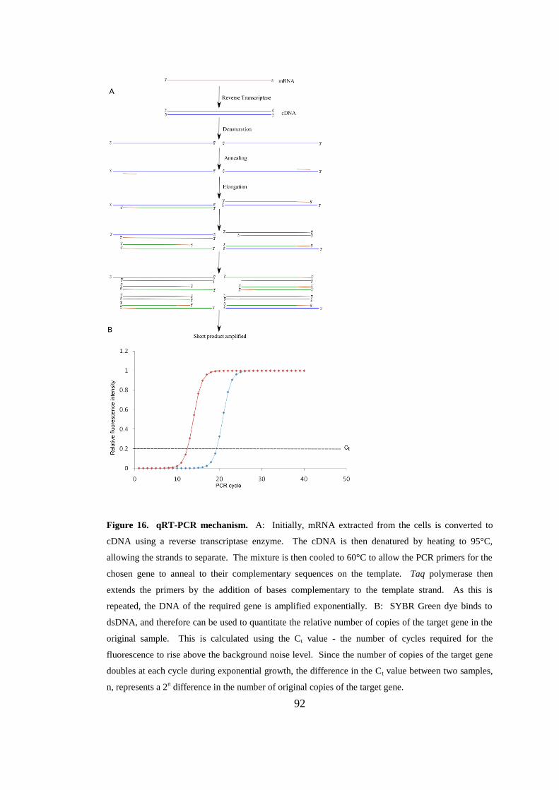
92
Figure 16. qRT-PCR mechanism. A: Initially, mRNA extracted from the cells is converted to
cDNA using a reverse transcriptase enzyme. The cDNA is then denatured by heating to 95°C,
allowing the strands to separate. The mixture is then cooled to 60°C to allow the PCR primers for the
chosen gene to anneal to their complementary sequences on the template. Taq polymerase then
extends the primers by the addition of bases complementary to the template strand. As this is
repeated, the DNA of the required gene is amplified exponentially. B: SYBR Green dye binds to
dsDNA, and therefore can be used to quantitate the relative number of copies of the target gene in the
original sample. This is calculated using the Ct value - the number of cycles required for the
fluorescence to rise above the background noise level. Since the number of copies of the target gene
doubles at each cycle during exponential growth, the difference in the Ct value between two samples,
n, represents a 2n difference in the number of original copies of the target gene.
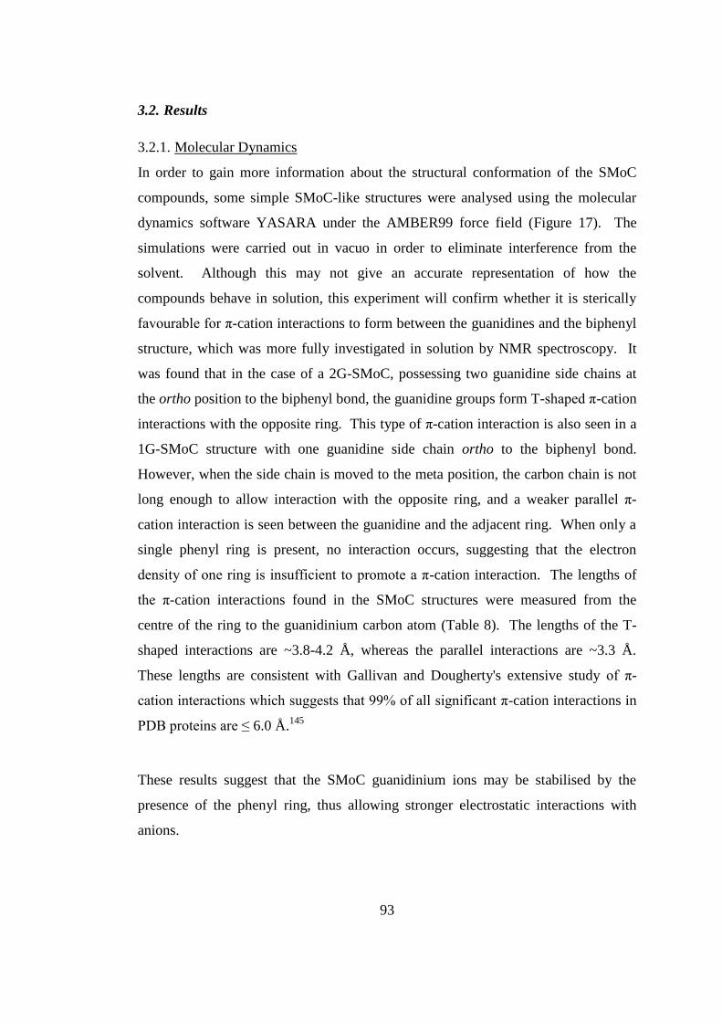
93
3.2. Results
3.2.1. Molecular Dynamics
In order to gain more information about the structural conformation of the SMoC
compounds, some simple SMoC-like structures were analysed using the molecular
dynamics software YASARA under the AMBER99 force field (Figure 17). The
simulations were carried out in vacuo in order to eliminate interference from the
solvent. Although this may not give an accurate representation of how the
compounds behave in solution, this experiment will confirm whether it is sterically
favourable for π-cation interactions to form between the guanidines and the biphenyl
structure, which was more fully investigated in solution by NMR spectroscopy. It
was found that in the case of a 2G-SMoC, possessing two guanidine side chains at
the ortho position to the biphenyl bond, the guanidine groups form T-shaped π-cation
interactions with the opposite ring. This type of π-cation interaction is also seen in a
1G-SMoC structure with one guanidine side chain ortho to the biphenyl bond.
However, when the side chain is moved to the meta position, the carbon chain is not
long enough to allow interaction with the opposite ring, and a weaker parallel π-
cation interaction is seen between the guanidine and the adjacent ring. When only a
single phenyl ring is present, no interaction occurs, suggesting that the electron
density of one ring is insufficient to promote a π-cation interaction. The lengths of
the π-cation interactions found in the SMoC structures were measured from the
centre of the ring to the guanidinium carbon atom (Table 8). The lengths of the T-
shaped interactions are ~3.8-4.2 Å, whereas the parallel interactions are ~3.3 Å.
These lengths are consistent with Gallivan and Dougherty's extensive study of π-
cation interactions which suggests that 99% of all significant π-cation interactions in
PDB proteins are ≤ 6.0 Å.145
These results suggest that the SMoC guanidinium ions may be stabilised by the
presence of the phenyl ring, thus allowing stronger electrostatic interactions with
anions.
B

94
Figure 17. Snapshots of a molecular dynamics simulation of simple SMoC-like molecules
containing one or two guanidine side chains. In a simple 2G-SMoC molecule (A), T-shaped π-
cation interactions form between the side chains and their opposite rings. This is also the case when
one side chain is removed (B). For a mono-phenyl structure with one side chain (C), no interaction is
present, whereas the addition of a second ring meta to the side chain (D) promotes a parallel π-cation
interaction onto the adjacent ring.
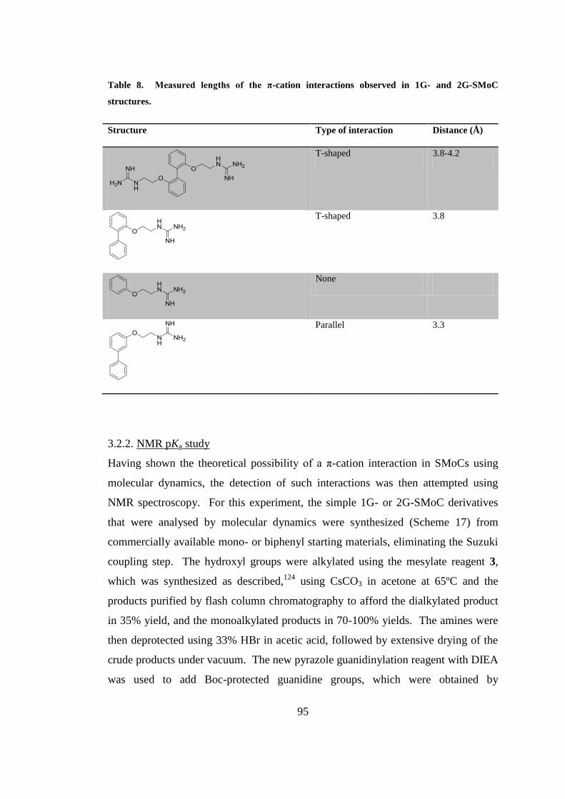
95
Table 8. Measured lengths of the π-cation interactions observed in 1G- and 2G-SMoC
structures.
Structure Type of interaction Distance (Å)
T-shaped 3.8-4.2
T-shaped 3.8
None
Parallel 3.3
3.2.2. NMR pKa study
Having shown the theoretical possibility of a π-cation interaction in SMoCs using
molecular dynamics, the detection of such interactions was then attempted using
NMR spectroscopy. For this experiment, the simple 1G- or 2G-SMoC derivatives
that were analysed by molecular dynamics were synthesized (Scheme 17) from
commercially available mono- or biphenyl starting materials, eliminating the Suzuki
coupling step. The hydroxyl groups were alkylated using the mesylate reagent 3,
which was synthesized as described,124
using CsCO3 in acetone at 65ºC and the
products purified by flash column chromatography to afford the dialkylated product
in 35% yield, and the monoalkylated products in 70-100% yields. The amines were
then deprotected using 33% HBr in acetic acid, followed by extensive drying of the
crude products under vacuum. The new pyrazole guanidinylation reagent with DIEA
was used to add Boc-protected guanidine groups, which were obtained by
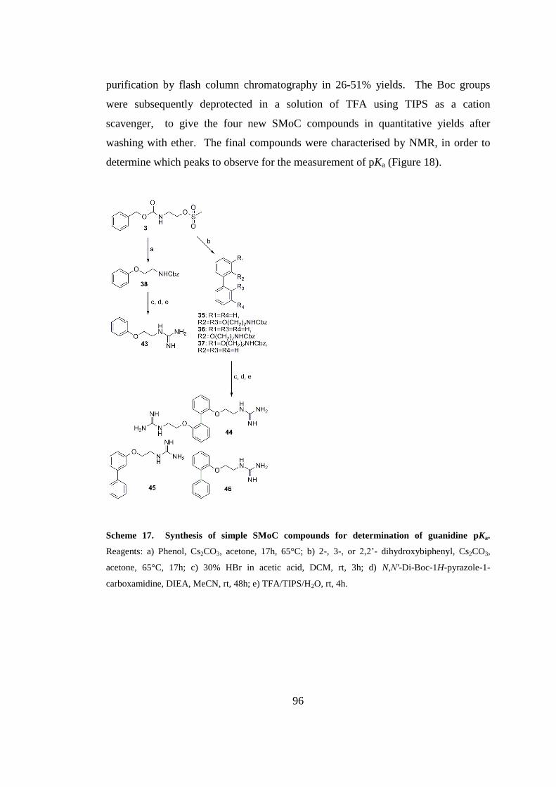
96
purification by flash column chromatography in 26-51% yields. The Boc groups
were subsequently deprotected in a solution of TFA using TIPS as a cation
scavenger, to give the four new SMoC compounds in quantitative yields after
washing with ether. The final compounds were characterised by NMR, in order to
determine which peaks to observe for the measurement of pKa (Figure 18).
Scheme 17. Synthesis of simple SMoC compounds for determination of guanidine pKa.
Reagents: a) Phenol, Cs2CO3, acetone, 17h, 65°C; b) 2-, 3-, or 2,2’- dihydroxybiphenyl, Cs2CO3,
acetone, 65°C, 17h; c) 30% HBr in acetic acid, DCM, rt, 3h; d) N,N′-Di-Boc-1H-pyrazole-1-
carboxamidine, DIEA, MeCN, rt, 48h; e) TFA/TIPS/H2O, rt, 4h.
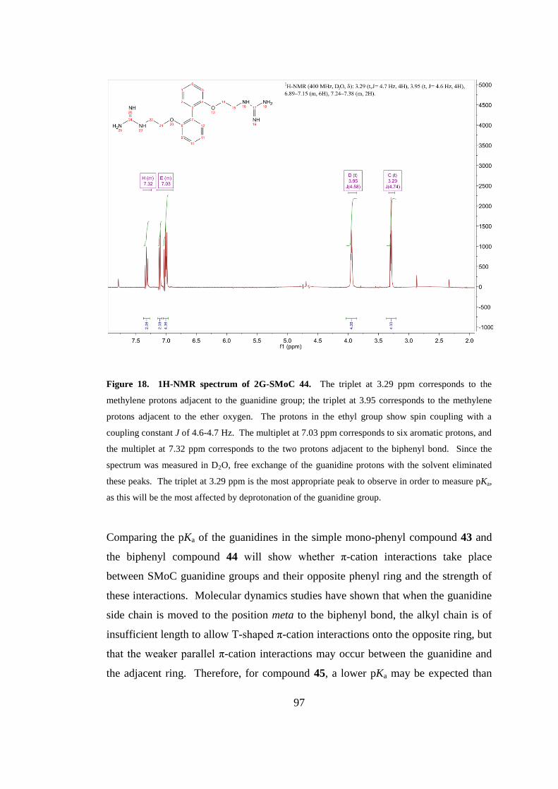
97
Figure 18. 1H-NMR spectrum of 2G-SMoC 44. The triplet at 3.29 ppm corresponds to the
methylene protons adjacent to the guanidine group; the triplet at 3.95 corresponds to the methylene
protons adjacent to the ether oxygen. The protons in the ethyl group show spin coupling with a
coupling constant J of 4.6-4.7 Hz. The multiplet at 7.03 ppm corresponds to six aromatic protons, and
the multiplet at 7.32 ppm corresponds to the two protons adjacent to the biphenyl bond. Since the
spectrum was measured in D2O, free exchange of the guanidine protons with the solvent eliminated
these peaks. The triplet at 3.29 ppm is the most appropriate peak to observe in order to measure pKa,
as this will be the most affected by deprotonation of the guanidine group.
Comparing the pKa of the guanidines in the simple mono-phenyl compound 43 and
the biphenyl compound 44 will show whether π-cation interactions take place
between SMoC guanidine groups and their opposite phenyl ring and the strength of
these interactions. Molecular dynamics studies have shown that when the guanidine
side chain is moved to the position meta to the biphenyl bond, the alkyl chain is of
insufficient length to allow T-shaped π-cation interactions onto the opposite ring, but
that the weaker parallel π-cation interactions may occur between the guanidine and
the adjacent ring. Therefore, for compound 45, a lower pKa may be expected than
98
for 46. The strength of this weaker interaction may also be measured by comparing
45 with 43.
For each compound, the change in ionisation state of the guanidine groups was
measured using NMR by titrating each compound with sodium hydroxide and
recording the 1H chemical shifts of the CH2 protons adjacent to the guanidine group
against the pH of the solution (Figure 19). The pKa of the guanidine group(s) is
given by the Henderson-Hasselbalch equation
][
][logpHp
B
BHKa
Deprotonation of the guanidine(s) will have a significant effect on the chemical shift
of the protons on the adjacent methylene group(s) due to the change in electron
density around the nitrogen atom, resulting in increased shielding of the CH2 protons.
The same effect could have been observed by measuring the 13
C shift of the
guanidine carbon atom, however this was rejected in favour of the proton NMR
approach for practical reasons, including the more time-consuming nature of
gathering 13
C-NMR data, and the higher concentration of compound needed to
ensure a high quality spectrum. The chemical shift of the charged form (δB+) may be
determined by measuring the chemical shift at a low pH. For these compounds, the
chemical shift was found to be at its maximum below pH 10. Likewise, at very high
pH (pH 14), the chemical shift of the deprotonated guanidine groups may be
measured (δB). Thus, the chemical shift at a given pH (δA) is representative of the
proportion of groups that are protonated at a given time. Therefore, the equation
may be expressed in terms of change in chemical shift:
AB
BAp a
logpHK
Hence, by plotting the log of the ratio of the charged and uncharged forms expressed
as chemical shifts against the pH of the solution, the pKa may be calculated from the
y-intercept of the linear trend line (Figure 20). The calculated pKa values are given
in Table 9.
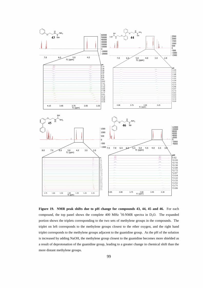
99
Figure 19. NMR peak shifts due to pH change for compounds 43, 44, 45 and 46. For each
compound, the top panel shows the complete 400 MHz 1H-NMR spectra in D2O. The expanded
portion shows the triplets corresponding to the two sets of methylene groups in the compounds. The
triplet on left corresponds to the methylene groups closest to the ether oxygen, and the right hand
triplet corresponds to the methylene groups adjacent to the guanidine group. As the pH of the solution
is increased by adding NaOH, the methylene group closest to the guanidine becomes more shielded as
a result of deprotonation of the guanidine group, leading to a greater change in chemical shift than the
more distant methylene groups.
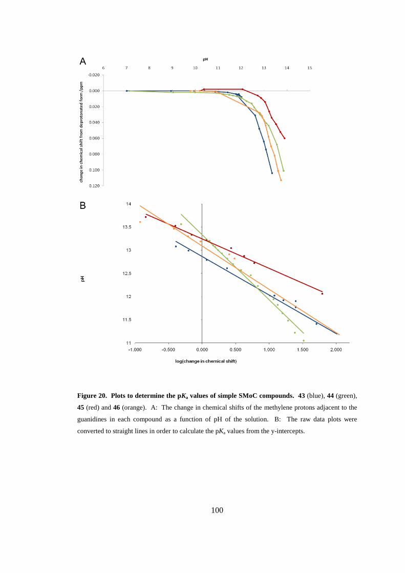
100
Figure 20. Plots to determine the pKa values of simple SMoC compounds. 43 (blue), 44 (green),
45 (red) and 46 (orange). A: The change in chemical shifts of the methylene protons adjacent to the
guanidines in each compound as a function of pH of the solution. B: The raw data plots were
converted to straight lines in order to calculate the pKa values from the y-intercepts.
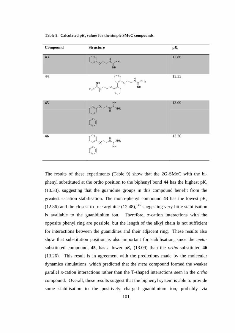
101
Table 9. Calculated pKa values for the simple SMoC compounds.
Compound Structure pKa
43
12.86
44
13.33
45
13.09
46
13.26
The results of these experiments (Table 9) show that the 2G-SMoC with the bi-
phenyl substituted at the ortho position to the biphenyl bond 44 has the highest pKa
(13.33), suggesting that the guanidine groups in this compound benefit from the
greatest π-cation stabilisation. The mono-phenyl compound 43 has the lowest pKa
(12.86) and the closest to free arginine (12.48),146
suggesting very little stabilisation
is available to the guanidinium ion. Therefore, π-cation interactions with the
opposite phenyl ring are possible, but the length of the alkyl chain is not sufficient
for interactions between the guanidines and their adjacent ring. These results also
show that substitution position is also important for stabilisation, since the meta-
substituted compound, 45, has a lower pKa (13.09) than the ortho-substituted 46
(13.26). This result is in agreement with the predictions made by the molecular
dynamics simulations, which predicted that the meta compound formed the weaker
parallel π-cation interactions rather than the T-shaped interactions seen in the ortho
compound. Overall, these results suggest that the biphenyl system is able to provide
some stabilisation to the positively charged guanidinium ion, probably via
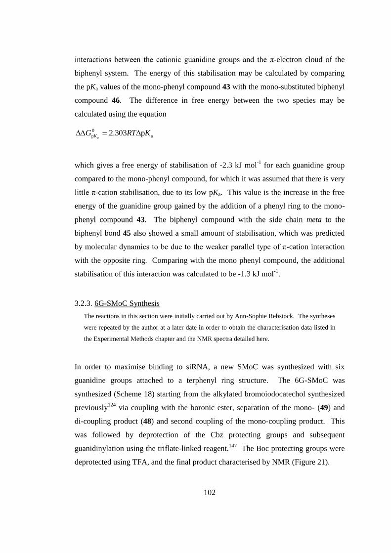
102
interactions between the cationic guanidine groups and the π-electron cloud of the
biphenyl system. The energy of this stabilisation may be calculated by comparing
the pKa values of the mono-phenyl compound 43 with the mono-substituted biphenyl
compound 46. The difference in free energy between the two species may be
calculated using the equation
aK KRTGa
p303.20
p
which gives a free energy of stabilisation of -2.3 kJ mol-1
for each guanidine group
compared to the mono-phenyl compound, for which it was assumed that there is very
little π-cation stabilisation, due to its low pKa. This value is the increase in the free
energy of the guanidine group gained by the addition of a phenyl ring to the mono-
phenyl compound 43. The biphenyl compound with the side chain meta to the
biphenyl bond 45 also showed a small amount of stabilisation, which was predicted
by molecular dynamics to be due to the weaker parallel type of π-cation interaction
with the opposite ring. Comparing with the mono phenyl compound, the additional
stabilisation of this interaction was calculated to be -1.3 kJ mol-1
.
3.2.3. 6G-SMoC Synthesis
The reactions in this section were initially carried out by Ann-Sophie Rebstock. The syntheses
were repeated by the author at a later date in order to obtain the characterisation data listed in
the Experimental Methods chapter and the NMR spectra detailed here.
In order to maximise binding to siRNA, a new SMoC was synthesized with six
guanidine groups attached to a terphenyl ring structure. The 6G-SMoC was
synthesized (Scheme 18) starting from the alkylated bromoiodocatechol synthesized
previously124
via coupling with the boronic ester, separation of the mono- (49) and
di-coupling product (48) and second coupling of the mono-coupling product. This
was followed by deprotection of the Cbz protecting groups and subsequent
guanidinylation using the triflate-linked reagent.147
The Boc protecting groups were
deprotected using TFA, and the final product characterised by NMR (Figure 21).
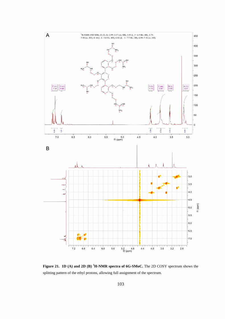
103
Figure 21. 1D (A) and 2D (B) 1H-NMR spectra of 6G-SMoC. The 2D COSY spectrum shows the
splitting pattern of the ethyl protons, allowing full assignment of the spectrum.
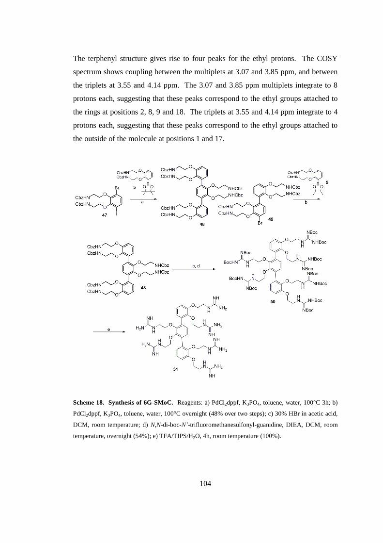
104
The terphenyl structure gives rise to four peaks for the ethyl protons. The COSY
spectrum shows coupling between the multiplets at 3.07 and 3.85 ppm, and between
the triplets at 3.55 and 4.14 ppm. The 3.07 and 3.85 ppm multiplets integrate to 8
protons each, suggesting that these peaks correspond to the ethyl groups attached to
the rings at positions 2, 8, 9 and 18. The triplets at 3.55 and 4.14 ppm integrate to 4
protons each, suggesting that these peaks correspond to the ethyl groups attached to
the outside of the molecule at positions 1 and 17.
Scheme 18. Synthesis of 6G-SMoC. Reagents: a) PdCl2dppf, K3PO4, toluene, water, 100°C 3h; b)
PdCl2dppf, K3PO4, toluene, water, 100°C overnight (48% over two steps); c) 30% HBr in acetic acid,
DCM, room temperature; d) N,N-di-boc-N’-trifluoromethanesulfonyl-guanidine, DIEA, DCM, room
temperature, overnight (54%); e) TFA/TIPS/H2O, 4h, room temperature (100%).
105
3.2.4. ITC
In order to calculate the strength of the electrostatic interactions between SMoC and
siRNA, isothermal titration calorimetry (ITC) was used to measure the heat evolved
when small aliquots of 6G-SMoC were injected into a solution of a standard 21-bp
siRNA for the human housekeeping gene glyceraldehyde 3-phosphate
dehydrogenase (GAPDH). The first experiment carried out involved injecting
aliquots of a 10 μM siRNA solution into a 75 μM SMoC solution contained in the
calorimeter cell. The resulting data showed that the SMoC was very quickly
saturated by the siRNA and therefore the data produced was insufficient to calculate
the binding parameters. As an siRNA molecule contains many more charges than a
SMoC molecule, it was decided to reverse the experimental setup and place the
SMoC in the injector and the siRNA in the calorimeter cell. Injections of 1 mM
SMoC into 2 μM siRNA resulted in more data points, however the saturation point
was still reached too quickly, meaning that a binding curve could not be fitted
(Figure 22A). Finally, by increasing the siRNA concentration to 3.5 μM and
decreasing the SMoC concentration to 250 μM, the experiment produced sufficient
data to fit a binding curve to calculate the binding parameters (Figure 22B). A
binding constant (K) of 87.3 μM was calculated with a binding stoichiometry of
4.4:1 (SMoC:siRNA). Binding was dominated by enthalpic contributions (-1646
Kcal/mol) with a minor or negligible entropic contribution. This is consistent with
electrostatic (charge) interactions between the guanidines and the phosphate groups
on the siRNA dominating binding.
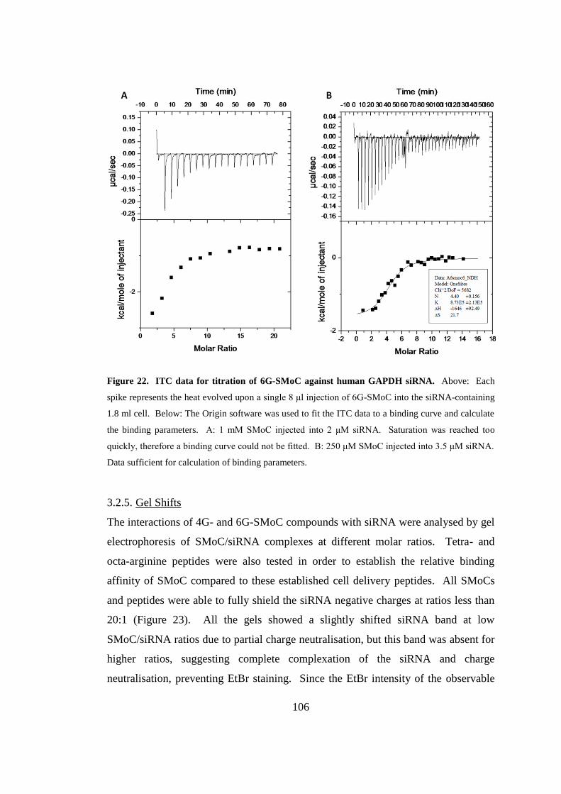
106
Figure 22. ITC data for titration of 6G-SMoC against human GAPDH siRNA. Above: Each
spike represents the heat evolved upon a single 8 μl injection of 6G-SMoC into the siRNA-containing
1.8 ml cell. Below: The Origin software was used to fit the ITC data to a binding curve and calculate
the binding parameters. A: 1 mM SMoC injected into 2 μM siRNA. Saturation was reached too
quickly, therefore a binding curve could not be fitted. B: 250 μM SMoC injected into 3.5 μM siRNA.
Data sufficient for calculation of binding parameters.
3.2.5. Gel Shifts
The interactions of 4G- and 6G-SMoC compounds with siRNA were analysed by gel
electrophoresis of SMoC/siRNA complexes at different molar ratios. Tetra- and
octa-arginine peptides were also tested in order to establish the relative binding
affinity of SMoC compared to these established cell delivery peptides. All SMoCs
and peptides were able to fully shield the siRNA negative charges at ratios less than
20:1 (Figure 23). All the gels showed a slightly shifted siRNA band at low
SMoC/siRNA ratios due to partial charge neutralisation, but this band was absent for
higher ratios, suggesting complete complexation of the siRNA and charge
neutralisation, preventing EtBr staining. Since the EtBr intensity of the observable
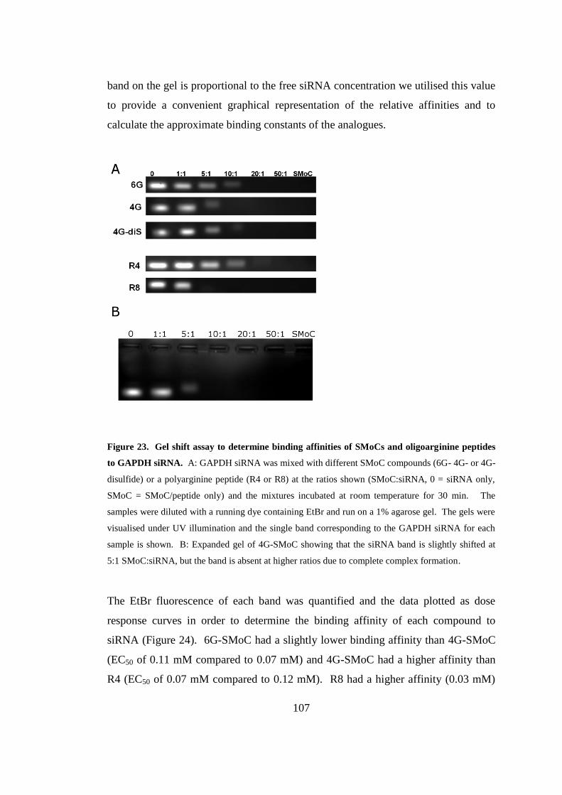
107
band on the gel is proportional to the free siRNA concentration we utilised this value
to provide a convenient graphical representation of the relative affinities and to
calculate the approximate binding constants of the analogues.
Figure 23. Gel shift assay to determine binding affinities of SMoCs and oligoarginine peptides
to GAPDH siRNA. A: GAPDH siRNA was mixed with different SMoC compounds (6G- 4G- or 4G-
disulfide) or a polyarginine peptide (R4 or R8) at the ratios shown (SMoC:siRNA, 0 = siRNA only,
SMoC = SMoC/peptide only) and the mixtures incubated at room temperature for 30 min. The
samples were diluted with a running dye containing EtBr and run on a 1% agarose gel. The gels were
visualised under UV illumination and the single band corresponding to the GAPDH siRNA for each
sample is shown. B: Expanded gel of 4G-SMoC showing that the siRNA band is slightly shifted at
5:1 SMoC:siRNA, but the band is absent at higher ratios due to complete complex formation.
The EtBr fluorescence of each band was quantified and the data plotted as dose
response curves in order to determine the binding affinity of each compound to
siRNA (Figure 24). 6G-SMoC had a slightly lower binding affinity than 4G-SMoC
(EC50 of 0.11 mM compared to 0.07 mM) and 4G-SMoC had a higher affinity than
R4 (EC50 of 0.07 mM compared to 0.12 mM). R8 had a higher affinity (0.03 mM)
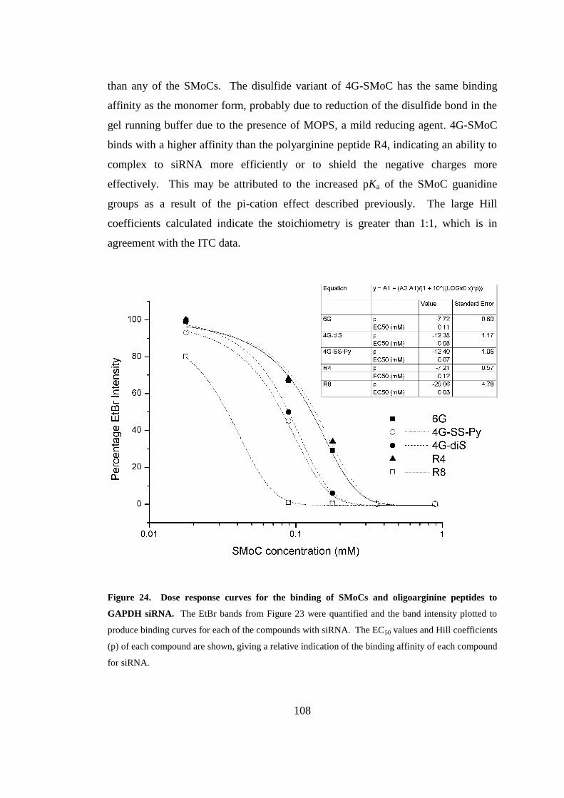
108
than any of the SMoCs. The disulfide variant of 4G-SMoC has the same binding
affinity as the monomer form, probably due to reduction of the disulfide bond in the
gel running buffer due to the presence of MOPS, a mild reducing agent. 4G-SMoC
binds with a higher affinity than the polyarginine peptide R4, indicating an ability to
complex to siRNA more efficiently or to shield the negative charges more
effectively. This may be attributed to the increased pKa of the SMoC guanidine
groups as a result of the pi-cation effect described previously. The large Hill
coefficients calculated indicate the stoichiometry is greater than 1:1, which is in
agreement with the ITC data.
Figure 24. Dose response curves for the binding of SMoCs and oligoarginine peptides to
GAPDH siRNA. The EtBr bands from Figure 23 were quantified and the band intensity plotted to
produce binding curves for each of the compounds with siRNA. The EC50 values and Hill coefficients
(p) of each compound are shown, giving a relative indication of the binding affinity of each compound
for siRNA.
109
3.2.6. siRNA transfection
In order to determine if SMoCs are capable of transporting siRNA into cells resulting
in a knockdown of the target gene, IMR-90 primary human fibroblasts and Human
Embryonic Kidney (HEK)-293 cells were transfected with SMoC or Lipofectamine
complexed with either a non-targeting control siRNA, or an siRNA targeted at cdc7.
This gene is a cell cycle kinase which promotes entry into the S phase of the cell
cycle in which DNA synthesis takes place and has been identified as a possible
therapeutic target in epithelial ovarian carcinoma.148
Lipofectamine was used
according to the manufacturer's instructions, and SMoC was used in a large molar
excess (1:5000) in order to ensure complete saturation of the siRNA by SMoC. After
72h, the cells were harvested, the RNA purified and the cdc7 mRNA measured using
qRT-PCR (Figure 25A). Both the siRNA sequences, and the PCR primers for cdc7
and GAPDH have previously been validated in human cancer cells.149
In order to
confirm the quality of the PCR amplification, a melting curve analysis was carried
out. This involves slow heating of the PCR samples following the amplification
cycles and measurement of the changes in fluorescence. When the DNA in the
sample reaches its melting point, the strands separate and SYBR is released, resulting
in a drop in fluorescence. Each DNA sequence has a unique melting point depending
on its constituent bases, resulting in a characteristic peak on the melting curve. By
plotting the change in fluorescence against temperature, it may be established how
many amplification products are present. In this case, two peaks were observed
across all the samples corresponding to cdc7 and GAPDH DNA which were
amplified in the PCR reaction (Figure 25B). For each compound tested, the cdc7
mRNA level was calibrated against a set of untreated cells (UT). The cells
transfected with non-targeting siRNA (CO) were used to take into account any effect
that the compound itself has on mRNA expression. The total knockdown was
calculated as the difference between the CO cells and the cells transfected with cdc7
siRNA. In both HEK-293 and IMR-90 cells, the difference between CO and cdc7
transfected cells using 6G-SMoC was insignificant (p=0.22 for HEK-293, p=0.41 for
IMR-90). 4G-SMoC did result in a significant difference between CO and cdc7
transfected cells, and produced a knockdown of 19.2% in HEK-293 and 36.75% in
IMR-90 cells. These results were similar to the knockdown achieved using the
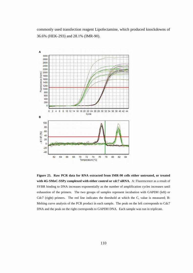
110
commonly used transfection reagent Lipofectamine, which produced knockdowns of
36.6% (HEK-293) and 28.1% (IMR-90).
Figure 25. Raw PCR data for RNA extracted from IMR-90 cells either untreated, or treated
with 4G-SMoC-SSPy complexed with either control or cdc7 siRNA. A: Fluorescence as a result of
SYBR binding to DNA increases exponentially as the number of amplification cycles increases until
exhaustion of the primers. The two groups of samples represent incubation with GAPDH (left) or
Cdc7 (right) primers. The red line indicates the threshold at which the Ct value is measured; B:
Melting curve analysis of the PCR product in each sample. The peak on the left corresponds to Cdc7
DNA and the peak on the right corresponds to GAPDH DNA. Each sample was run in triplicate.
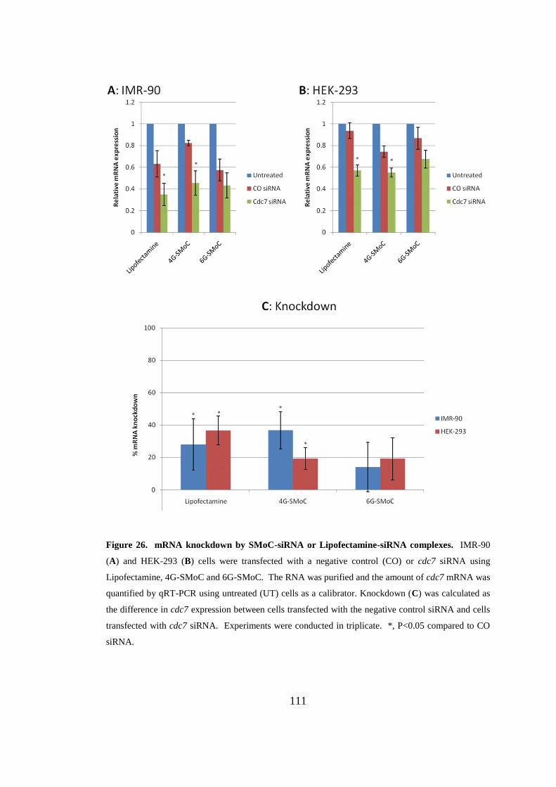
111
Figure 26. mRNA knockdown by SMoC-siRNA or Lipofectamine-siRNA complexes. IMR-90
(A) and HEK-293 (B) cells were transfected with a negative control (CO) or cdc7 siRNA using
Lipofectamine, 4G-SMoC and 6G-SMoC. The RNA was purified and the amount of cdc7 mRNA was
quantified by qRT-PCR using untreated (UT) cells as a calibrator. Knockdown (C) was calculated as
the difference in cdc7 expression between cells transfected with the negative control siRNA and cells
transfected with cdc7 siRNA. Experiments were conducted in triplicate. *, P<0.05 compared to CO
siRNA.
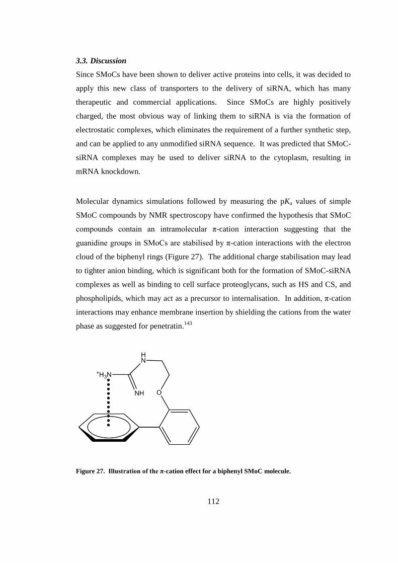
112
3.3. Discussion
Since SMoCs have been shown to deliver active proteins into cells, it was decided to
apply this new class of transporters to the delivery of siRNA, which has many
therapeutic and commercial applications. Since SMoCs are highly positively
charged, the most obvious way of linking them to siRNA is via the formation of
electrostatic complexes, which eliminates the requirement of a further synthetic step,
and can be applied to any unmodified siRNA sequence. It was predicted that SMoC-
siRNA complexes may be used to deliver siRNA to the cytoplasm, resulting in
mRNA knockdown.
Molecular dynamics simulations followed by measuring the pKa values of simple
SMoC compounds by NMR spectroscopy have confirmed the hypothesis that SMoC
compounds contain an intramolecular π-cation interaction suggesting that the
guanidine groups in SMoCs are stabilised by π-cation interactions with the electron
cloud of the biphenyl rings (Figure 27). The additional charge stabilisation may lead
to tighter anion binding, which is significant both for the formation of SMoC-siRNA
complexes as well as binding to cell surface proteoglycans, such as HS and CS, and
phospholipids, which may act as a precursor to internalisation. In addition, π-cation
interactions may enhance membrane insertion by shielding the cations from the water
phase as suggested for penetratin.143
Figure 27. Illustration of the π-cation effect for a biphenyl SMoC molecule.
113
The new guanidinylation reagent identified previously was particularly useful in the
synthesis of the simple SMoCs used in this study. The proton NMR spectrum of
Boc-protected biphenyl compound 40 was observed to have a unexpectedly high
chemical shift values for all peaks which were higher than for all other SMoC
compounds. This is thought to be due to incorrect calibration to the internal TMS
standard when measuring the spectrum, since the spectrum of the deprotected
compound 44 is as expected.
As hypothesized, various SMoC compounds were capable of forming electrostatic
interactions with siRNA, forming netural complexes. Using ITC, the binding affinity
of 6G-SMoC to siRNA was accurately measured as 87.3 μM, and the relative
affinities of other SMoC compounds, as well as tetra- and octaarginine peptides,
were measured using a gel shift assay. The binding affinities of the SMoCs and
peptides to siRNA does not seem to be related to the number of guanidine groups,
since 6G-SMoC has a lower affinity than 4G-SMoC-SSPy. The higher binding
affinity of 4G-SMoC-SSPy than R4 supports the π-cation hypothesis that the
guanidine groups of the SMoC compounds are stabilised, and hence have a higher
anion affinity. The weaker binding of 6G-SMoC than 4G-SMoC-SSPy may be
explained by the presence of the linker group on 4G-SMoC-SSPy which may act as
an electron donating group via induction from the methylene group. Ideally, this
experiment should be carried out using SMoCs without linker groups in order to
further establish the connection between number of guanidine groups and binding
affinity to siRNA. However, this experiment confirms that SMoC-siRNA complexes
may be formed in solution in a similar way to CPPs and that that 4G-SMoC-SSPy
has the highest affinity for siRNA, and therefore shows the greatest potential for
siRNA transfection.
Transfection using SMoCs was carried out in two cell lines; IMR-90 cells, a human
primary cell line, and HEK-293 cells, an immortalised cell line. As predicted,
SMoC-siRNA non-covalent complexes were capable of delivering functional siRNA
to the cytoplasm, resulting in signifcant levels of mRNA knockdown. This is a
114
significant new development for the SMoC compounds, as previously they have only
been shown to deliver covalently conjugated proteins and dyes. 4G-SMoC-SSPy
achieved a comparable level of knockdown to Lipofectamine, suggesting that this
compound has potential as an alternative in vitro reagent. However, having
established this potential, further uptake studies should be carried out in order to
compare 4G-SMoC-SSPy against other amphipathic peptide vectors such as
penetratin. If the observed π-cation interaction is involved in the internalisation
mechanism as described for penetratin, it may be expected that SMoCs will show a
similar uptake efficiency to the peptide. 6G-SMoC did not achieve a significant gene
knockdown, suggesting that affinity for siRNA is related to transfection ability. The
knockdowns recorded for Lipofectamine and 4G-SMoC-SSPy are significant
compared to untreated cells, however the knockdown achieved for Lipofectamine is
lower than in similar studies reported in the literature,105, 111-112
suggesting that the
experimental procedure used is not fully optimised. Further optimisation of the
methods may therefore result in more significant silencing using the SMoC
compounds. Time restrictions prevented a full optimisation study of the
experimental conditions, including the effect of cell culture media, siRNA and SMoC
concentrations, and the ratio of siRNA to SMoC. In particular, it should be tested
whether using SMoCs at the stoichiometric ratio indicated by the gel shift assays,
rather than in large excess, may improve uptake. This may be significant, since a
large excess of free SMoC in the transfection mixture may compete for binding to
cell surface GAGs with the siRNA-SMoC complexes. Since the guanidine groups of
the complexed SMoC molecules are engaged in electrostatic interactions with
siRNA, the uncomplexed SMoC may have a higher affinity for GAGs, and therefore
may block binding of the siRNA complexes, limiting siRNA internalisation.
An interesting result from this study was that 4G-SMoC-SSPy, originally developed
as a protein internalisation reagent due to its ability to form disulfide linkages with
cysteine residues was more successful at promoting siRNA transfection than 6G-
SMoC, which possesses more guanidine groups. It has been shown that a
nonaarginine (R9) peptide with terminal cysteine residues forms high molecular
115
weight (~96 kD) complexes which are able to condense DNA and promote
significantly higher gene uptake than momomeric R9.150
The formation of disulfide
linked dimers could explain the higher mRNA knockdown of 4G-SMoC-SSPy than
6G-SMoC in complex with siRNA. The purified disulfide form of 4G-SMoC 34 has
not yet been tested in transfection assays, but it is hypothesized that it will show
similar or higher uptake than 4G-SMoC-SSPy. Furthermore, it has been suggested
that CPPs containing intra- or intermolecular disulfide bonds may be cross-linked by
cell surface thiol proteins, resulting in a higher uptake efficiency.151
This hypothesis
may also apply to disulfide-containing SMoCs, which has implications for the design
of future compounds.

116
CHAPTER 4
Designing a new SMoC for siRNA
delivery
117
4. Designing a new SMoC for siRNA delivery
4.1. Introduction
In the previous chapter, it was shown that 4G-SMoC is capable of delivering siRNA
across cell membranes to achieve a significant gene silencing effect via the RNAi
mechanism. The small mRNA knockdown achieved by SMoC-siRNA complexes
implies that the siRNA is not available in the cytoplasm in high enough levels to
completely silence the gene, suggesting that the complexes are not sufficiently taken
up by the cells, or are trapped in endosomes preventing the siRNA from reaching its
target. Studies on CPPs have shown that binding to cell surface GAGs leads to
clustering of the attached proteoglycans resulting in reorganisation of the actin
skeleton which is an important precursor for internalisation by endocytosis.31
It is
therefore hypothesized that in order to achieve a high level of uptake, SMoCs must
have the ability to optimally bind these cell surface anions as well as siRNA. Two of
the most prevalent cell surface GAGs are HS, which is a component of proteoglycans
at the cell surface including transmembrane syndecans and GPI-anchored glypicans,
and CS which is the most prevalent GAG and has both structural and regulatory roles
in the extracellular matrix.152
Therefore, it is predicted that by analysing the crystal
structures of these GAGs, a new SMoC compound may be designed which retains
the siRNA-complexing properties but has improved uptake efficiency than the
previosuly described SMoCs.
As discussed previously (1.3.2.5), one of the features of a multi-component siRNA
delivery system is a targeting sequence in order to target the siRNA to a particular
location in vivo,118
for example a particular cell type via a cell surface receptor. It is
hypothesised that in addition to providing selectivity, binding to cell surface
receptors may also increase uptake efficiency by increasing the concentration of the
siRNA delivery vehicle at the cell surface as observed by nanaoparticles bearing cell-
specific targeting ligands.153
RGD peptides containing an arginine-glycine-aspartate
motif have been found to be potent ligands for cell surface integrins, which are
present on all cells and mediate binding to the extracellular matrix. In addition,
integrins carry out intracellular signalling roles, with overexpression of certain
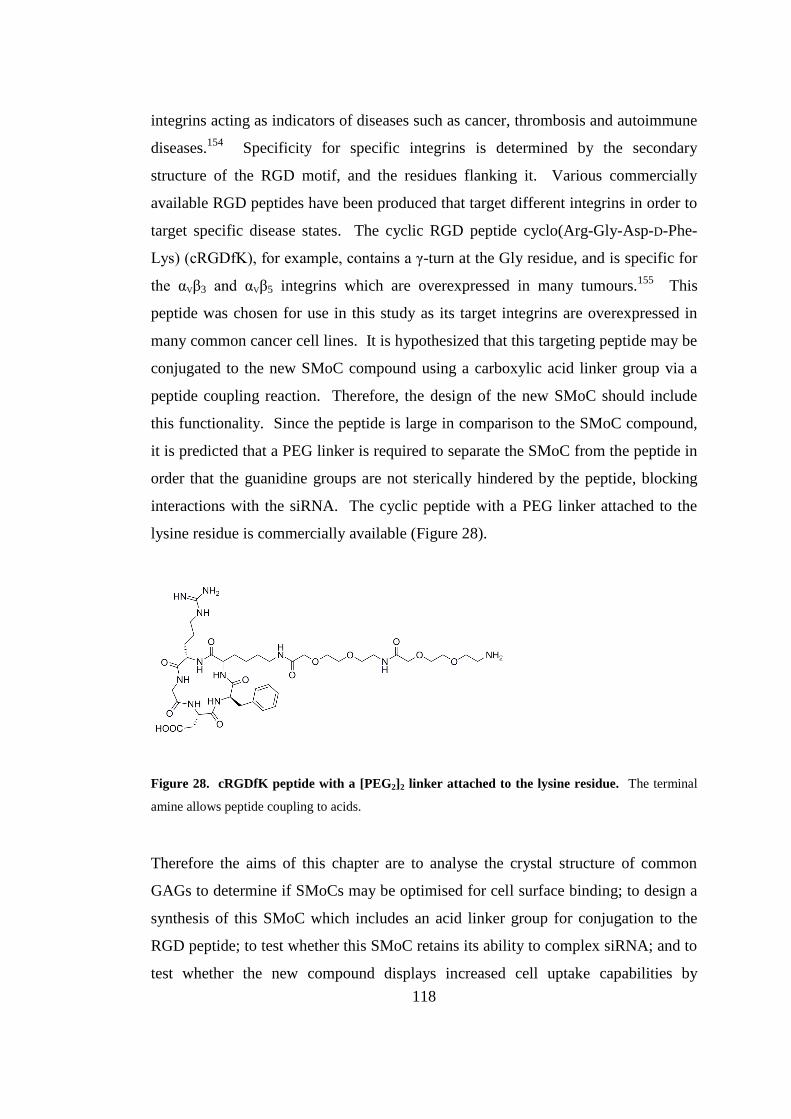
118
integrins acting as indicators of diseases such as cancer, thrombosis and autoimmune
diseases.154
Specificity for specific integrins is determined by the secondary
structure of the RGD motif, and the residues flanking it. Various commercially
available RGD peptides have been produced that target different integrins in order to
target specific disease states. The cyclic RGD peptide cyclo(Arg-Gly-Asp-D-Phe-
Lys) (cRGDfK), for example, contains a γ-turn at the Gly residue, and is specific for
the αVβ3 and αVβ5 integrins which are overexpressed in many tumours.155
This
peptide was chosen for use in this study as its target integrins are overexpressed in
many common cancer cell lines. It is hypothesized that this targeting peptide may be
conjugated to the new SMoC compound using a carboxylic acid linker group via a
peptide coupling reaction. Therefore, the design of the new SMoC should include
this functionality. Since the peptide is large in comparison to the SMoC compound,
it is predicted that a PEG linker is required to separate the SMoC from the peptide in
order that the guanidine groups are not sterically hindered by the peptide, blocking
interactions with the siRNA. The cyclic peptide with a PEG linker attached to the
lysine residue is commercially available (Figure 28).
Figure 28. cRGDfK peptide with a [PEG2]2 linker attached to the lysine residue. The terminal
amine allows peptide coupling to acids.
Therefore the aims of this chapter are to analyse the crystal structure of common
GAGs to determine if SMoCs may be optimised for cell surface binding; to design a
synthesis of this SMoC which includes an acid linker group for conjugation to the
RGD peptide; to test whether this SMoC retains its ability to complex siRNA; and to
test whether the new compound displays increased cell uptake capabilities by
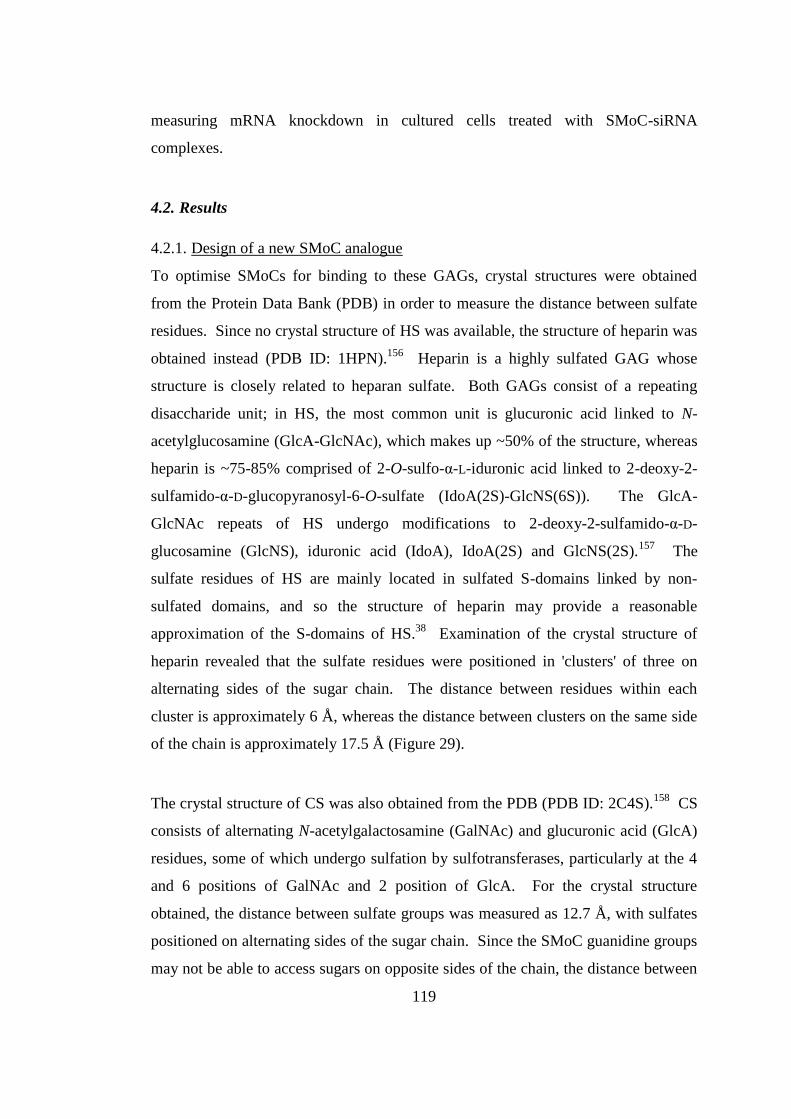
119
measuring mRNA knockdown in cultured cells treated with SMoC-siRNA
complexes.
4.2. Results
4.2.1. Design of a new SMoC analogue
To optimise SMoCs for binding to these GAGs, crystal structures were obtained
from the Protein Data Bank (PDB) in order to measure the distance between sulfate
residues. Since no crystal structure of HS was available, the structure of heparin was
obtained instead (PDB ID: 1HPN).156
Heparin is a highly sulfated GAG whose
structure is closely related to heparan sulfate. Both GAGs consist of a repeating
disaccharide unit; in HS, the most common unit is glucuronic acid linked to N-
acetylglucosamine (GlcA-GlcNAc), which makes up ~50% of the structure, whereas
heparin is ~75-85% comprised of 2-O-sulfo-α-L-iduronic acid linked to 2-deoxy-2-
sulfamido-α-D-glucopyranosyl-6-O-sulfate (IdoA(2S)-GlcNS(6S)). The GlcA-
GlcNAc repeats of HS undergo modifications to 2-deoxy-2-sulfamido-α-D-
glucosamine (GlcNS), iduronic acid (IdoA), IdoA(2S) and GlcNS(2S).157
The
sulfate residues of HS are mainly located in sulfated S-domains linked by non-
sulfated domains, and so the structure of heparin may provide a reasonable
approximation of the S-domains of HS.38
Examination of the crystal structure of
heparin revealed that the sulfate residues were positioned in 'clusters' of three on
alternating sides of the sugar chain. The distance between residues within each
cluster is approximately 6 Å, whereas the distance between clusters on the same side
of the chain is approximately 17.5 Å (Figure 29).
The crystal structure of CS was also obtained from the PDB (PDB ID: 2C4S).158
CS
consists of alternating N-acetylgalactosamine (GalNAc) and glucuronic acid (GlcA)
residues, some of which undergo sulfation by sulfotransferases, particularly at the 4
and 6 positions of GalNAc and 2 position of GlcA. For the crystal structure
obtained, the distance between sulfate groups was measured as 12.7 Å, with sulfates
positioned on alternating sides of the sugar chain. Since the SMoC guanidine groups
may not be able to access sugars on opposite sides of the chain, the distance between
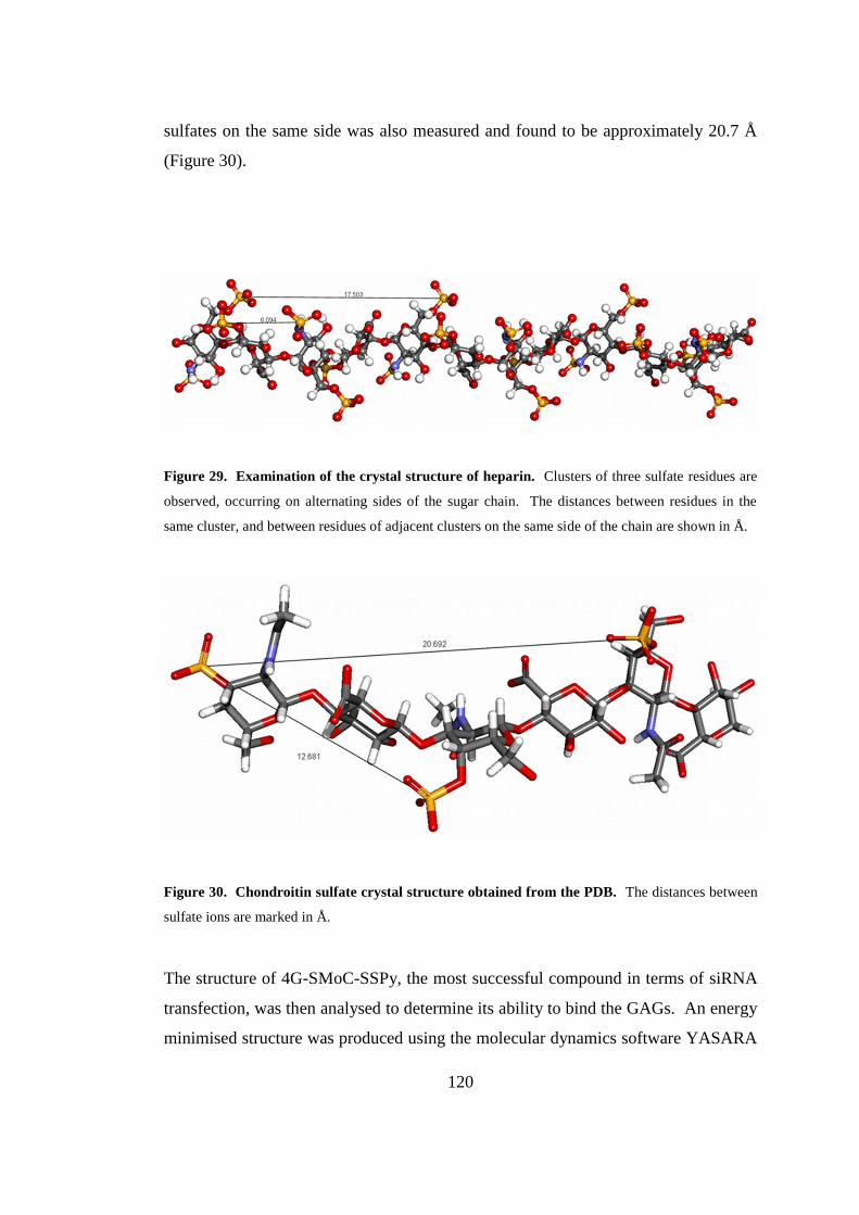
120
sulfates on the same side was also measured and found to be approximately 20.7 Å
(Figure 30).
Figure 29. Examination of the crystal structure of heparin. Clusters of three sulfate residues are
observed, occurring on alternating sides of the sugar chain. The distances between residues in the
same cluster, and between residues of adjacent clusters on the same side of the chain are shown in Å.
Figure 30. Chondroitin sulfate crystal structure obtained from the PDB. The distances between
sulfate ions are marked in Å.
The structure of 4G-SMoC-SSPy, the most successful compound in terms of siRNA
transfection, was then analysed to determine its ability to bind the GAGs. An energy
minimised structure was produced using the molecular dynamics software YASARA
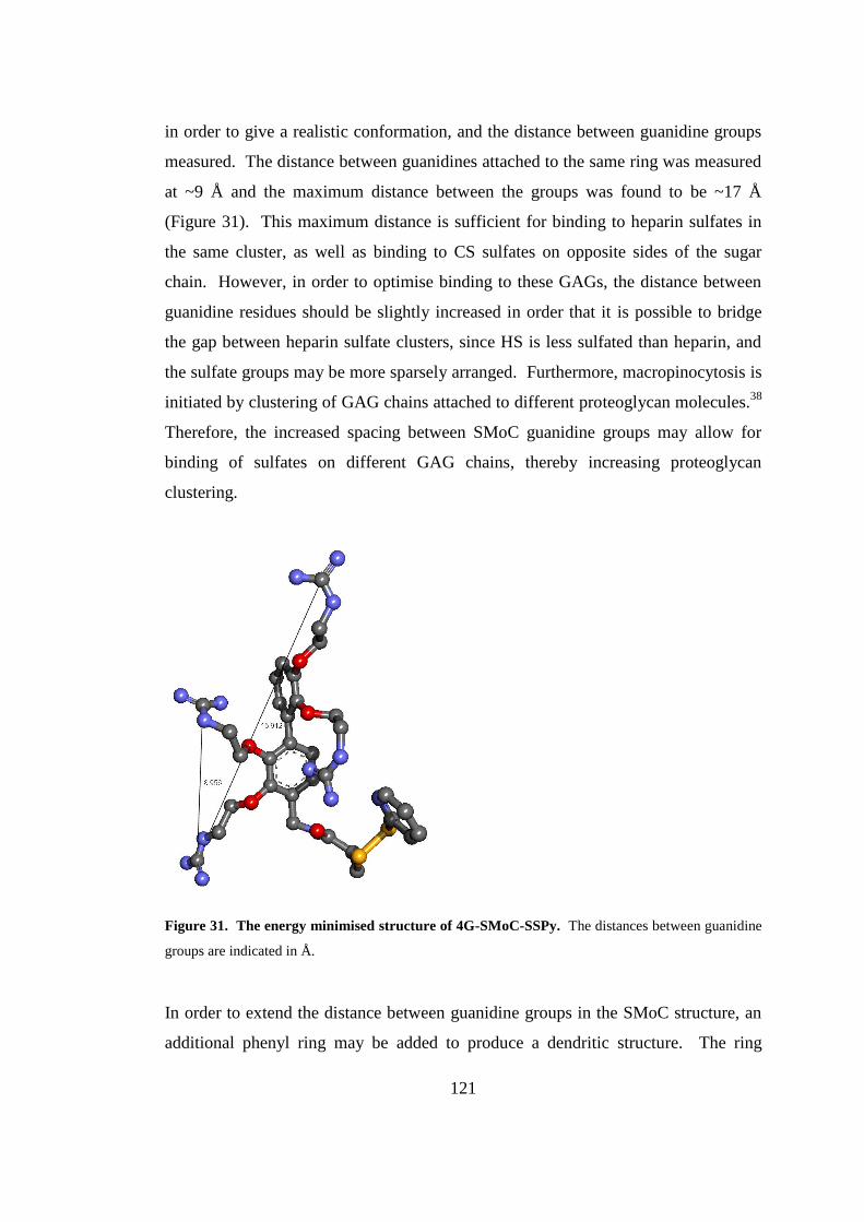
121
in order to give a realistic conformation, and the distance between guanidine groups
measured. The distance between guanidines attached to the same ring was measured
at ~9 Å and the maximum distance between the groups was found to be ~17 Å
(Figure 31). This maximum distance is sufficient for binding to heparin sulfates in
the same cluster, as well as binding to CS sulfates on opposite sides of the sugar
chain. However, in order to optimise binding to these GAGs, the distance between
guanidine residues should be slightly increased in order that it is possible to bridge
the gap between heparin sulfate clusters, since HS is less sulfated than heparin, and
the sulfate groups may be more sparsely arranged. Furthermore, macropinocytosis is
initiated by clustering of GAG chains attached to different proteoglycan molecules.38
Therefore, the increased spacing between SMoC guanidine groups may allow for
binding of sulfates on different GAG chains, thereby increasing proteoglycan
clustering.
Figure 31. The energy minimised structure of 4G-SMoC-SSPy. The distances between guanidine
groups are indicated in Å.
In order to extend the distance between guanidine groups in the SMoC structure, an
additional phenyl ring may be added to produce a dendritic structure. The ring
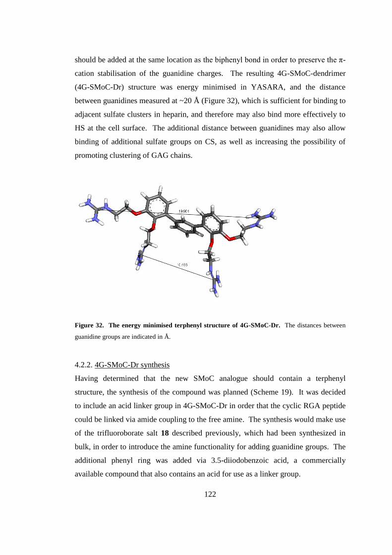
122
should be added at the same location as the biphenyl bond in order to preserve the π-
cation stabilisation of the guanidine charges. The resulting 4G-SMoC-dendrimer
(4G-SMoC-Dr) structure was energy minimised in YASARA, and the distance
between guanidines measured at ~20 Å (Figure 32), which is sufficient for binding to
adjacent sulfate clusters in heparin, and therefore may also bind more effectively to
HS at the cell surface. The additional distance between guanidines may also allow
binding of additional sulfate groups on CS, as well as increasing the possibility of
promoting clustering of GAG chains.
Figure 32. The energy minimised terphenyl structure of 4G-SMoC-Dr. The distances between
guanidine groups are indicated in Å.
4.2.2. 4G-SMoC-Dr synthesis
Having determined that the new SMoC analogue should contain a terphenyl
structure, the synthesis of the compound was planned (Scheme 19). It was decided
to include an acid linker group in 4G-SMoC-Dr in order that the cyclic RGA peptide
could be linked via amide coupling to the free amine. The synthesis would make use
of the trifluoroborate salt 18 described previously, which had been synthesized in
bulk, in order to introduce the amine functionality for adding guanidine groups. The
additional phenyl ring was added via 3.5-diiodobenzoic acid, a commercially
available compound that also contains an acid for use as a linker group.
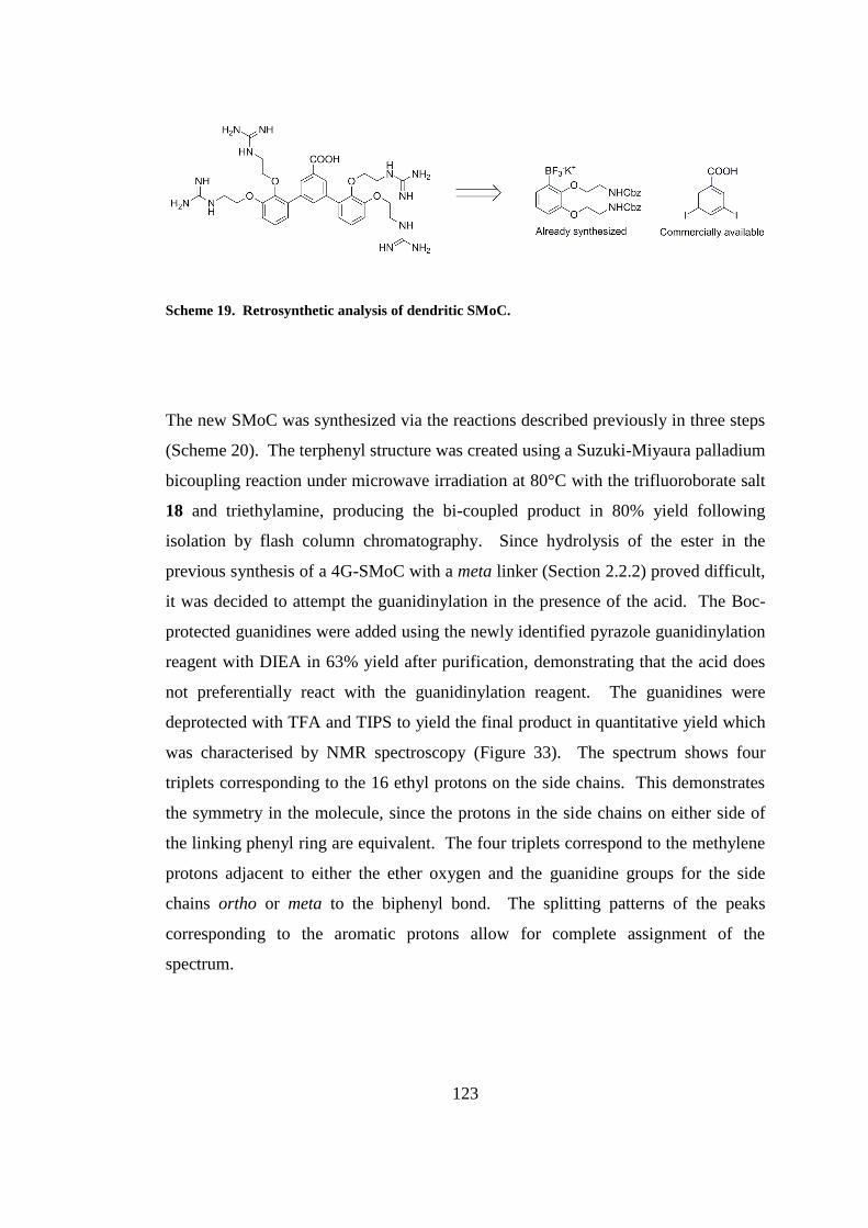
123
Scheme 19. Retrosynthetic analysis of dendritic SMoC.
The new SMoC was synthesized via the reactions described previously in three steps
(Scheme 20). The terphenyl structure was created using a Suzuki-Miyaura palladium
bicoupling reaction under microwave irradiation at 80°C with the trifluoroborate salt
18 and triethylamine, producing the bi-coupled product in 80% yield following
isolation by flash column chromatography. Since hydrolysis of the ester in the
previous synthesis of a 4G-SMoC with a meta linker (Section 2.2.2) proved difficult,
it was decided to attempt the guanidinylation in the presence of the acid. The Boc-
protected guanidines were added using the newly identified pyrazole guanidinylation
reagent with DIEA in 63% yield after purification, demonstrating that the acid does
not preferentially react with the guanidinylation reagent. The guanidines were
deprotected with TFA and TIPS to yield the final product in quantitative yield which
was characterised by NMR spectroscopy (Figure 33). The spectrum shows four
triplets corresponding to the 16 ethyl protons on the side chains. This demonstrates
the symmetry in the molecule, since the protons in the side chains on either side of
the linking phenyl ring are equivalent. The four triplets correspond to the methylene
protons adjacent to either the ether oxygen and the guanidine groups for the side
chains ortho or meta to the biphenyl bond. The splitting patterns of the peaks
corresponding to the aromatic protons allow for complete assignment of the
spectrum.
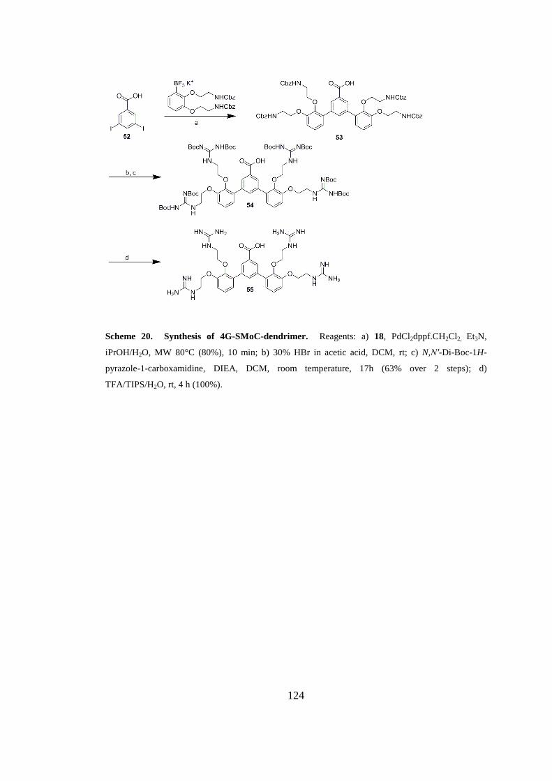
124
Scheme 20. Synthesis of 4G-SMoC-dendrimer. Reagents: a) 18, PdCl2dppf.CH2Cl2, Et3N,
iPrOH/H2O, MW 80°C (80%), 10 min; b) 30% HBr in acetic acid, DCM, rt; c) N,N′-Di-Boc-1H-
pyrazole-1-carboxamidine, DIEA, DCM, room temperature, 17h (63% over 2 steps); d)
TFA/TIPS/H2O, rt, 4 h (100%).
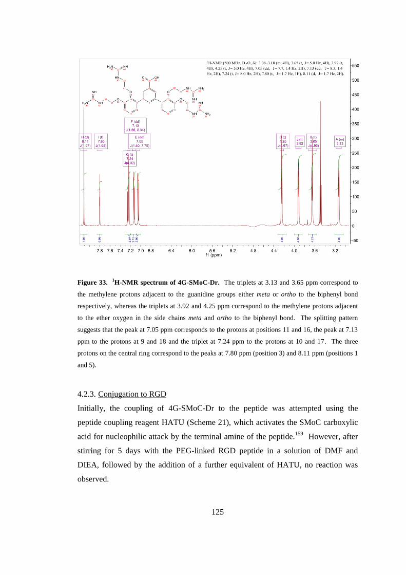
125
Figure 33. 1H-NMR spectrum of 4G-SMoC-Dr. The triplets at 3.13 and 3.65 ppm correspond to
the methylene protons adjacent to the guanidine groups either meta or ortho to the biphenyl bond
respectively, whereas the triplets at 3.92 and 4.25 ppm correspond to the methylene protons adjacent
to the ether oxygen in the side chains meta and ortho to the biphenyl bond. The splitting pattern
suggests that the peak at 7.05 ppm corresponds to the protons at positions 11 and 16, the peak at 7.13
ppm to the protons at 9 and 18 and the triplet at 7.24 ppm to the protons at 10 and 17. The three
protons on the central ring correspond to the peaks at 7.80 ppm (position 3) and 8.11 ppm (positions 1
and 5).
4.2.3. Conjugation to RGD
Initially, the coupling of 4G-SMoC-Dr to the peptide was attempted using the
peptide coupling reagent HATU (Scheme 21), which activates the SMoC carboxylic
acid for nucleophilic attack by the terminal amine of the peptide.159
However, after
stirring for 5 days with the PEG-linked RGD peptide in a solution of DMF and
DIEA, followed by the addition of a further equivalent of HATU, no reaction was
observed.
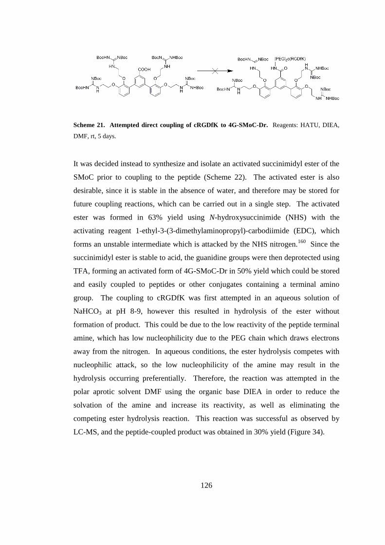
126
Scheme 21. Attempted direct coupling of cRGDfK to 4G-SMoC-Dr. Reagents: HATU, DIEA,
DMF, rt, 5 days.
It was decided instead to synthesize and isolate an activated succinimidyl ester of the
SMoC prior to coupling to the peptide (Scheme 22). The activated ester is also
desirable, since it is stable in the absence of water, and therefore may be stored for
future coupling reactions, which can be carried out in a single step. The activated
ester was formed in 63% yield using N-hydroxysuccinimide (NHS) with the
activating reagent 1-ethyl-3-(3-dimethylaminopropyl)-carbodiimide (EDC), which
forms an unstable intermediate which is attacked by the NHS nitrogen.160
Since the
succinimidyl ester is stable to acid, the guanidine groups were then deprotected using
TFA, forming an activated form of 4G-SMoC-Dr in 50% yield which could be stored
and easily coupled to peptides or other conjugates containing a terminal amino
group. The coupling to cRGDfK was first attempted in an aqueous solution of
NaHCO3 at pH 8-9, however this resulted in hydrolysis of the ester without
formation of product. This could be due to the low reactivity of the peptide terminal
amine, which has low nucleophilicity due to the PEG chain which draws electrons
away from the nitrogen. In aqueous conditions, the ester hydrolysis competes with
nucleophilic attack, so the low nucleophilicity of the amine may result in the
hydrolysis occurring preferentially. Therefore, the reaction was attempted in the
polar aprotic solvent DMF using the organic base DIEA in order to reduce the
solvation of the amine and increase its reactivity, as well as eliminating the
competing ester hydrolysis reaction. This reaction was successful as observed by
LC-MS, and the peptide-coupled product was obtained in 30% yield (Figure 34).
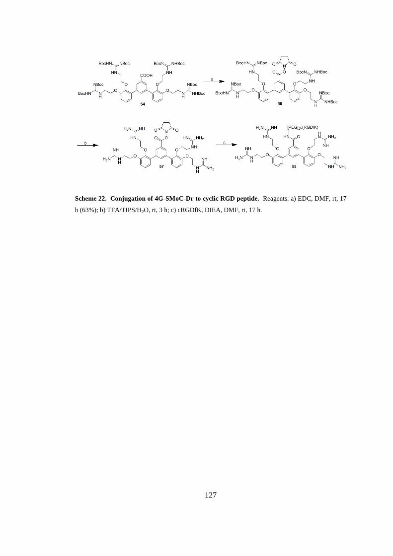
127
Scheme 22. Conjugation of 4G-SMoC-Dr to cyclic RGD peptide. Reagents: a) EDC, DMF, rt, 17
h (63%); b) TFA/TIPS/H2O, rt, 3 h; c) cRGDfK, DIEA, DMF, rt, 17 h.
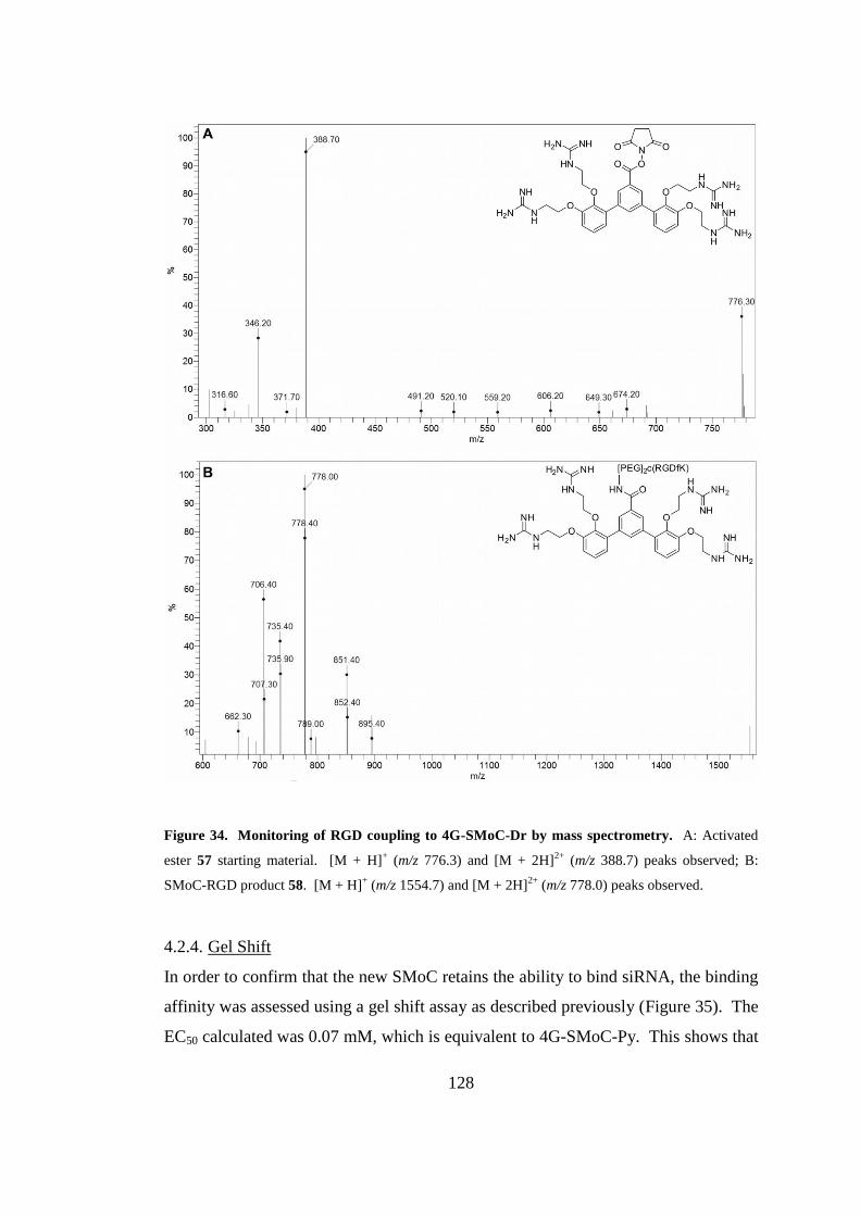
128
Figure 34. Monitoring of RGD coupling to 4G-SMoC-Dr by mass spectrometry. A: Activated
ester 57 starting material. [M + H]+ (m/z 776.3) and [M + 2H]
2+ (m/z 388.7) peaks observed; B:
SMoC-RGD product 58. [M + H]+ (m/z 1554.7) and [M + 2H]
2+ (m/z 778.0) peaks observed.
4.2.4. Gel Shift
In order to confirm that the new SMoC retains the ability to bind siRNA, the binding
affinity was assessed using a gel shift assay as described previously (Figure 35). The
EC50 calculated was 0.07 mM, which is equivalent to 4G-SMoC-Py. This shows that
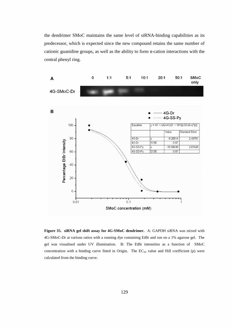
129
the dendrimer SMoC maintains the same level of siRNA-binding capabilities as its
predecessor, which is expected since the new compound retains the same number of
cationic guanidine groups, as well as the ability to form π-cation interactions with the
central phenyl ring.
Figure 35. siRNA gel shift assay for 4G-SMoC dendrimer. A: GAPDH siRNA was mixed with
4G-SMoC-Dr at various ratios with a running dye containing EtBr and run on a 1% agarose gel. The
gel was visualised under UV illumination. B: The EtBr intensities as a function of SMoC
concentration with a binding curve fitted in Origin. The EC50 value and Hill coefficient (p) were
calculated from the binding curve.
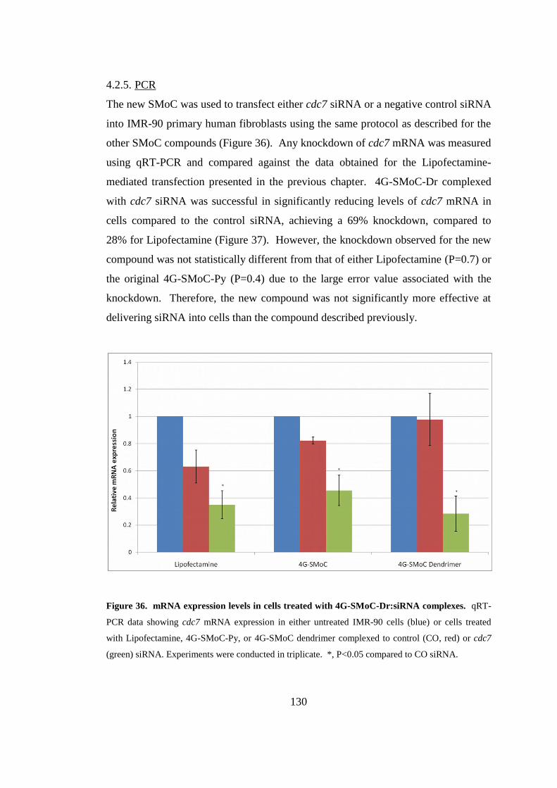
130
4.2.5. PCR
The new SMoC was used to transfect either cdc7 siRNA or a negative control siRNA
into IMR-90 primary human fibroblasts using the same protocol as described for the
other SMoC compounds (Figure 36). Any knockdown of cdc7 mRNA was measured
using qRT-PCR and compared against the data obtained for the Lipofectamine-
mediated transfection presented in the previous chapter. 4G-SMoC-Dr complexed
with cdc7 siRNA was successful in significantly reducing levels of cdc7 mRNA in
cells compared to the control siRNA, achieving a 69% knockdown, compared to
28% for Lipofectamine (Figure 37). However, the knockdown observed for the new
compound was not statistically different from that of either Lipofectamine (P=0.7) or
the original 4G-SMoC-Py (P=0.4) due to the large error value associated with the
knockdown. Therefore, the new compound was not significantly more effective at
delivering siRNA into cells than the compound described previously.
Figure 36. mRNA expression levels in cells treated with 4G-SMoC-Dr:siRNA complexes. qRT-
PCR data showing cdc7 mRNA expression in either untreated IMR-90 cells (blue) or cells treated
with Lipofectamine, 4G-SMoC-Py, or 4G-SMoC dendrimer complexed to control (CO, red) or cdc7
(green) siRNA. Experiments were conducted in triplicate. *, P<0.05 compared to CO siRNA.
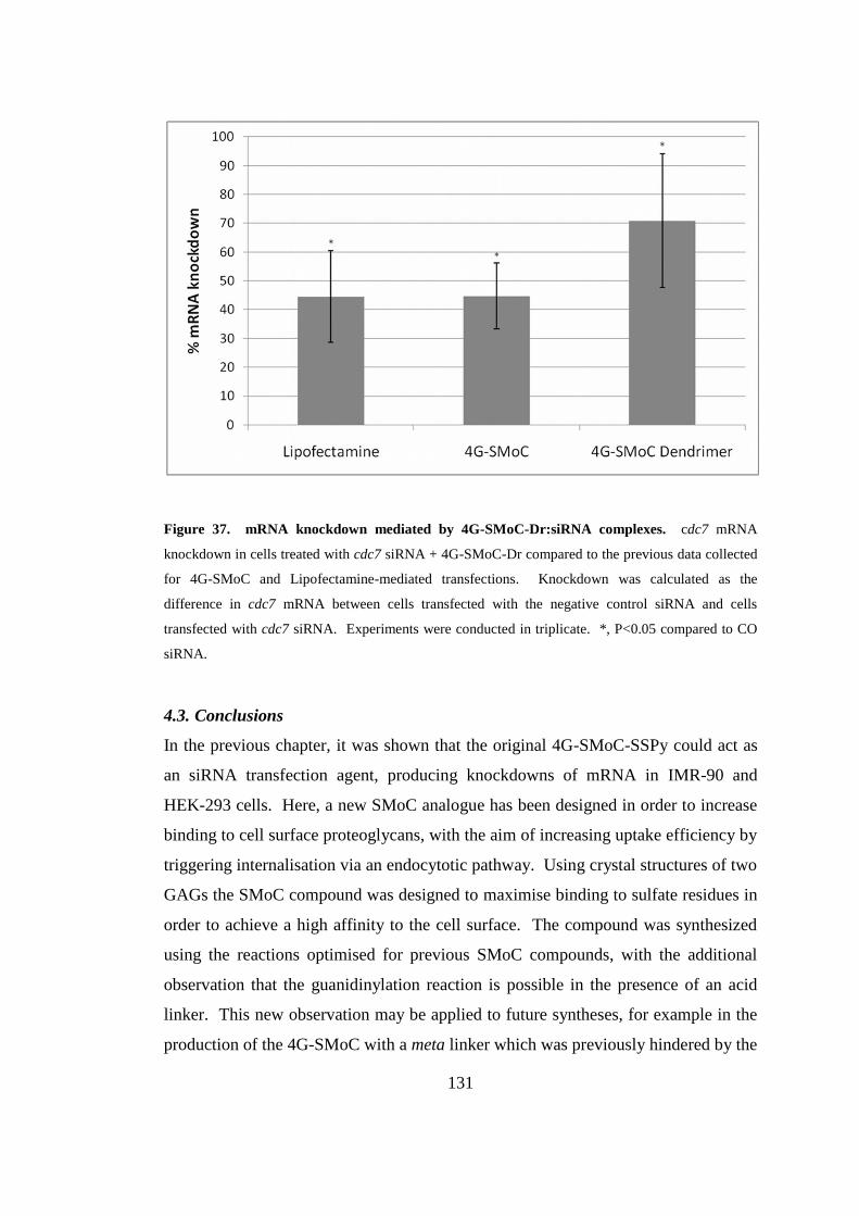
131
Figure 37. mRNA knockdown mediated by 4G-SMoC-Dr:siRNA complexes. cdc7 mRNA
knockdown in cells treated with cdc7 siRNA + 4G-SMoC-Dr compared to the previous data collected
for 4G-SMoC and Lipofectamine-mediated transfections. Knockdown was calculated as the
difference in cdc7 mRNA between cells transfected with the negative control siRNA and cells
transfected with cdc7 siRNA. Experiments were conducted in triplicate. *, P<0.05 compared to CO
siRNA.
4.3. Conclusions
In the previous chapter, it was shown that the original 4G-SMoC-SSPy could act as
an siRNA transfection agent, producing knockdowns of mRNA in IMR-90 and
HEK-293 cells. Here, a new SMoC analogue has been designed in order to increase
binding to cell surface proteoglycans, with the aim of increasing uptake efficiency by
triggering internalisation via an endocytotic pathway. Using crystal structures of two
GAGs the SMoC compound was designed to maximise binding to sulfate residues in
order to achieve a high affinity to the cell surface. The compound was synthesized
using the reactions optimised for previous SMoC compounds, with the additional
observation that the guanidinylation reaction is possible in the presence of an acid
linker. This new observation may be applied to future syntheses, for example in the
production of the 4G-SMoC with a meta linker which was previously hindered by the
132
inability to hydrolyse the ester to form the acid linker. This synthesis has
demonstrated that using the trifluoroborate salt developed earlier, a variety of new
SMoC-related compounds may be easily synthesized via Suzuki couplings with aryl
halides.
The interactions of the new compound, 4G-SMoC-Dr, with siRNA were
characterised using the gel shift assay used for previous compounds, and a binding
affinity calculated in order to compare it to the other SMoCs. As hypothesized, the
new compound retained the ability to complex siRNA displaying a similar binding
affinity to the original 4G-SMoC-SSPy, showing that the addition of a phenyl ring as
well as the presence of an acid does not affect its ability to complex siRNA. Finally,
IMR-90 cells were successfully transfected with 4G-SMoC-Dr, producing a
significant RNAi-induced knockdown of the target mRNA. The knockdown
achieved was not significantly different from the previous 4G-SMoC compound, or
from Lipofectamine. Therefore, the hypothesis that additional branching of the
SMoC side chains will increase internalisation via GAG clustering is not supported
by this data.
Using an activated ester of the 4G-SMoC-Dr compound, it was possible to attach the
RGD targeting peptide as predicted. The activated ester of 4G-SMoC-Dr is a
significant development in SMoC chemistry as it allows the simple attachment of
targeting ligands, such as peptides or antibodies, in order to target specific cell types
or cell receptors which may improve uptake. In addition, as seen in the previous
chapter, the inclusion of a disulfide-containing linker group may increase uptake due
to either dimerisation or by interaction with cell surface thiols. Therefore inclusion
of this type of functionality may improve the efficiency of siRNA internalisation for
4G-SMoC-Dr.

133
CHAPTER 5
Experimental Methods
134
5. Experimental Methods
5.1. General Methods
5.1.1. General chemistry methods
Starting materials were either commercially available or synthesized according to
methods reported in the literature. 1H and
13C NMR spectra were recorded on either
a Bruker AMX-300 or a Bruker AMX-500 spectrometer. Chemical shifts are
reported as ppm relative to TMS internal standard. Mass spectra were recorded on
either a Fisons VG70-SE spectrometer (EI, FAB) or an Agilent 6100 Series LC-mass
spectrometer using either a C-18 or C-4 column. Microwave reactions were carried
out using a CEM Discover microwave. All compounds were purified to ≥95% as
measured by LC-MS.
5.1.2. Gel shifts
Polyarginine peptides were obtained from Peptide Protein Research, Hampshire, UK.
1.7 nmol GAPDH siRNA (Dharmacon RNAi Technologies; sequences used were
sense 5' UGG UUU ACA UGU UCC AAU AUU 3', antisense 5' Phosphate U AUU
GGA ACA UGU AAA ACC UU 3') was mixed in RNAse-free water with varying
concentrations of either SMoC, tetraarginine or octaarginine in the molar ratios of
1/1, 1/5, 1/10, 1/20 and 1/50. The complexes were incubated for 30 min at room
temperature. The samples were diluted with 2x RNA Loading Dye (Fermentas UK)
containing ethidium bromide and analysed by gel electrophoresis on a 1% agarose
gel. The gels were analysed by UV illumination and the ethidium bromide bands
quantified using the ImageJ software. The EtBr intensities were plotted, a dose
response curve fitted and EC50 values calculated using the Origin software.
5.1.3. Cell Culture
IMR-90 (ATCC# CCL-186), a diploid primary human fibroblast adherent cell line
derived from fetal lung tissue, was obtained from LGC Standards (Middlesex, UK) at
population doubling (PD) 12. HEK-293 (ATCC# CRL-1573) cells were kindly
donated by Neale Foxwell (Wolfson Institute of Biomedical Research, UK). Cells
135
were cultured in DMEM (Invitrogen, UK) supplemented with 10% Fetal Bovine
Serum (Invitrogen, UK) and 2 mM L-glutamine (Invitrogen, UK) at 37 °C and 5%
CO2.
5.1.4. siRNA complexes
Solutions were made up in Opti-MEM® (Invitrogen, UK); 100 nM cdc7 siRNA
(sense 5'-GCU CAG CAG GAA AGG UGU UUU-3', antisense 5'-AAC ACC UUU
CCU GCU GAG CUU-3'; Applied Biosystems, UK), 100 nM Accell Non-targeting
siRNA 1 negative control siRNA (Dharmacon RNAi Technologies), 8 μl/ml
Lipofectamine LTX reagent (Invitrogen, UK), 0.5 mM SMoC. SMoC/siRNA and
Lipofectamine/siRNA complexes were made by mixing equal volumes of the
required solutions and incubating for 30 min at room temperature.
5.1.5. siRNA Transfection
Cells were plated in 6-well plates at a density of 5 x 10⁵ cells/well (IMR-90 cells) or
1 x 10⁵ cells/well (HEK-293 cells) in 1 ml DMEM containing 10% FCS and 2 mM
glutamine and incubated at 37 °C under 5% CO2 for 24 h. The media was removed
and replaced with 1.6 mL Opti-MEM® (Invitrogen, UK). 400 μl of SMoC/siRNA or
Lipofectamine/siRNA complexes were added and the cells incubated for a further 72
h. The cells were washed with PBS and trypsinised and the pellet stored at -80°C.
5.1.6. RNA extraction and qRT-PCR
RNA was extracted from the cell pellets using the PureLink RNA Mini Kit
(Invitrogen, UK) using the protocol provided by the manufacturer. RNA was
quantified using a Nanodrop spectrophotometer and 40 ng of RNA was mixed with
Superscript III RT / Platinum Taq Mix and SYBR Green Reaction Mix from the
Supersript III Platinum SYBR Green One-Step qRT-PCR Kit (Invitrogen, UK)
according with the manufacturer’s protocol. PCR conditions were 50 °C for 3 min,
95 °C for 10 min (1 cycle), 95 °C for 15 s, 47 °C for 30 s, 60 °C for 30 s (45 cycles),
95 °C for 15 s, 60 °C for 15 s, ramp to 95 °C over 20 min (1 cycle). Relative
quantitation data were obtained using the comparative Ct method with Realplex
136
software according
to the manufacturer's protocol (Eppendorf, Heidelberg,
Germany). cdc7 RNA was measured using the following primers: forward primer:
5’-AAC TTG CAG GTG TTA AAA AAG-3’: reverse primer: 5’-TGA AAG TGC
CTT CTC CAA T-3’. The following primers were used to measure GAPDH:
forward primer: 5’-TCA ACT ACA TGG TTT ACA TGT TC-3’: reverse primer: 5’-
GAT CTC GCT CCT GGA AGA T-3’. The signal obtained from the cdc7 primers
was normalised using the signal from the GAPDH primers.
5.1.7. Statistical Analysis
Groups were compared using an unpaired two-sample Student's t-test. A value of
P<0.05 was considered significant.
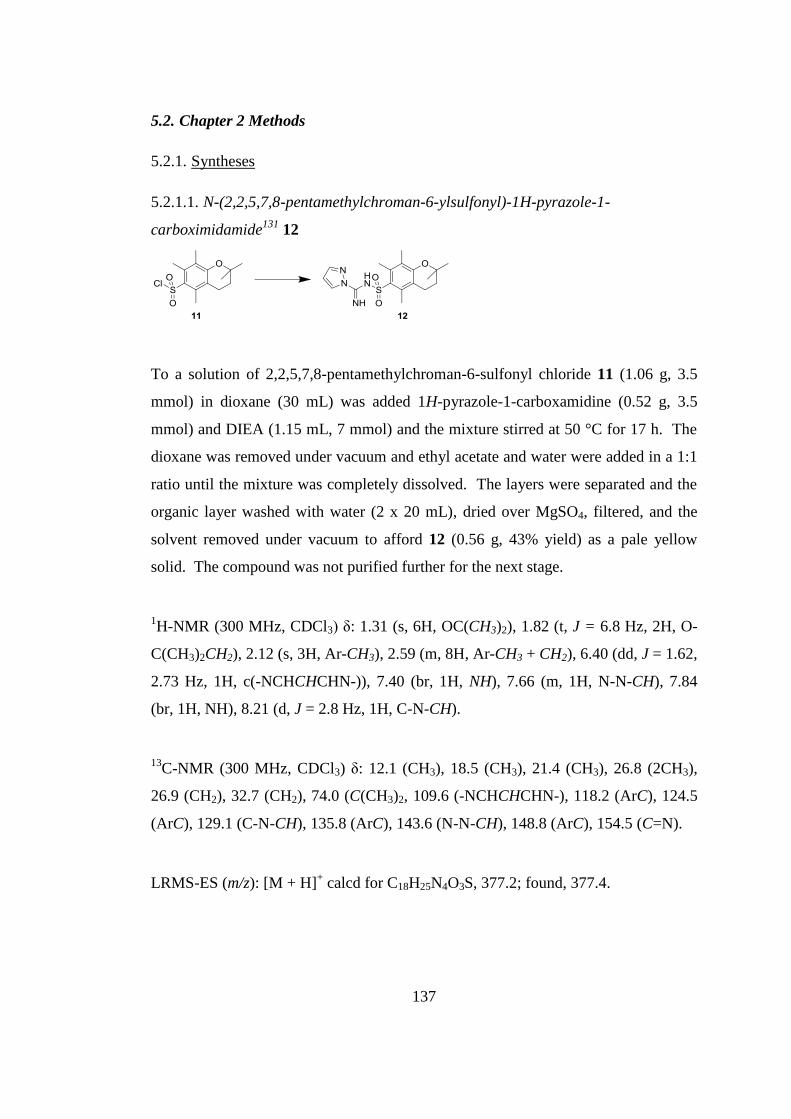
137
5.2. Chapter 2 Methods
5.2.1. Syntheses
5.2.1.1. N-(2,2,5,7,8-pentamethylchroman-6-ylsulfonyl)-1H-pyrazole-1-
carboximidamide131
12
To a solution of 2,2,5,7,8-pentamethylchroman-6-sulfonyl chloride 11 (1.06 g, 3.5
mmol) in dioxane (30 mL) was added 1H-pyrazole-1-carboxamidine (0.52 g, 3.5
mmol) and DIEA (1.15 mL, 7 mmol) and the mixture stirred at 50 °C for 17 h. The
dioxane was removed under vacuum and ethyl acetate and water were added in a 1:1
ratio until the mixture was completely dissolved. The layers were separated and the
organic layer washed with water (2 x 20 mL), dried over MgSO4, filtered, and the
solvent removed under vacuum to afford 12 (0.56 g, 43% yield) as a pale yellow
solid. The compound was not purified further for the next stage.
1H-NMR (300 MHz, CDCl3) δ: 1.31 (s, 6H, OC(CH3)2), 1.82 (t, J = 6.8 Hz, 2H, O-
C(CH3)2CH2), 2.12 (s, 3H, Ar-CH3), 2.59 (m, 8H, Ar-CH3 + CH2), 6.40 (dd, J = 1.62,
2.73 Hz, 1H, c(-NCHCHCHN-)), 7.40 (br, 1H, NH), 7.66 (m, 1H, N-N-CH), 7.84
(br, 1H, NH), 8.21 (d, J = 2.8 Hz, 1H, C-N-CH).
13C-NMR (300 MHz, CDCl3) δ: 12.1 (CH3), 18.5 (CH3), 21.4 (CH3), 26.8 (2CH3),
26.9 (CH2), 32.7 (CH2), 74.0 (C(CH3)2, 109.6 (-NCHCHCHN-), 118.2 (ArC), 124.5
(ArC), 129.1 (C-N-CH), 135.8 (ArC), 143.6 (N-N-CH), 148.8 (ArC), 154.5 (C=N).
LRMS-ES (m/z): [M + H]+ calcd for C18H25N4O3S, 377.2; found, 377.4.
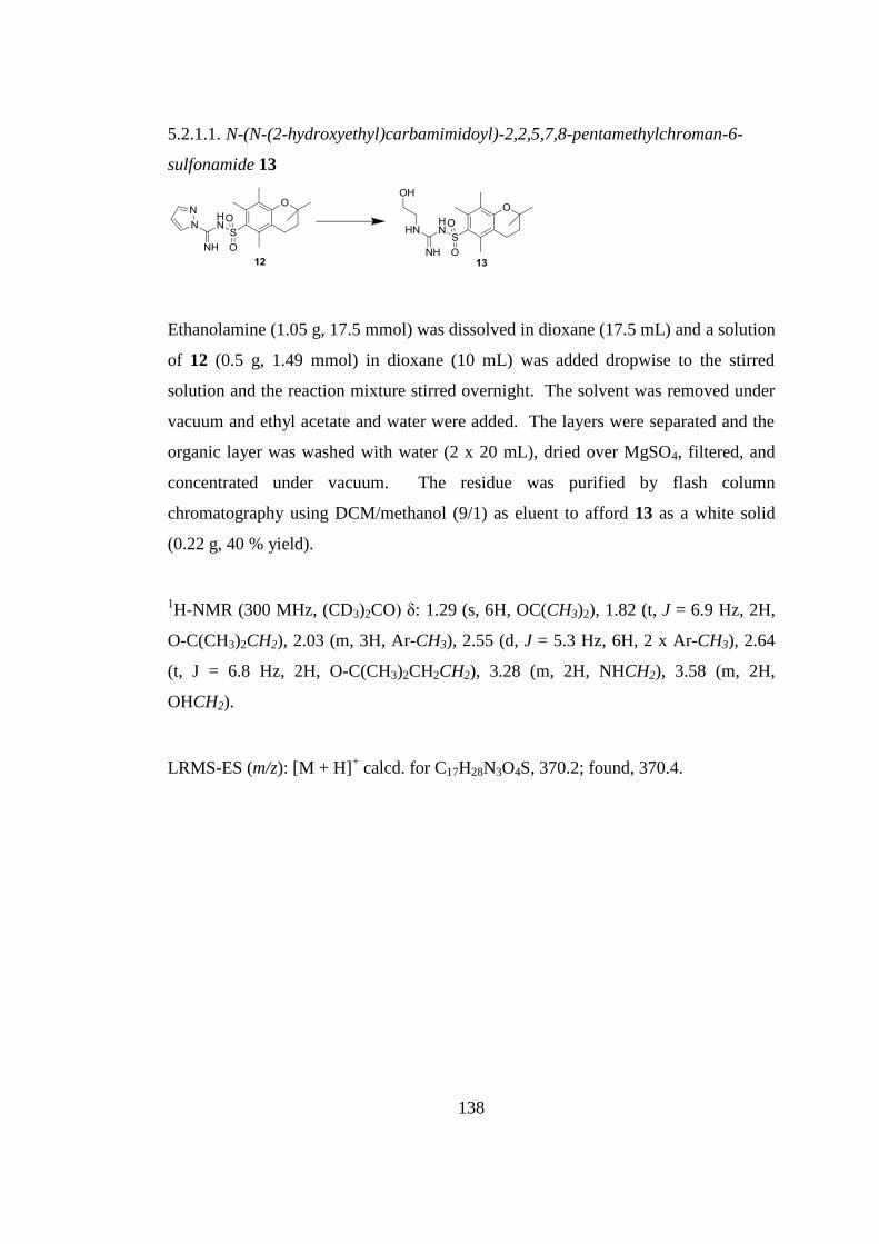
138
5.2.1.1. N-(N-(2-hydroxyethyl)carbamimidoyl)-2,2,5,7,8-pentamethylchroman-6-
sulfonamide 13
Ethanolamine (1.05 g, 17.5 mmol) was dissolved in dioxane (17.5 mL) and a solution
of 12 (0.5 g, 1.49 mmol) in dioxane (10 mL) was added dropwise to the stirred
solution and the reaction mixture stirred overnight. The solvent was removed under
vacuum and ethyl acetate and water were added. The layers were separated and the
organic layer was washed with water (2 x 20 mL), dried over MgSO4, filtered, and
concentrated under vacuum. The residue was purified by flash column
chromatography using DCM/methanol (9/1) as eluent to afford 13 as a white solid
(0.22 g, 40 % yield).
1H-NMR (300 MHz, (CD3)2CO) δ: 1.29 (s, 6H, OC(CH3)2), 1.82 (t, J = 6.9 Hz, 2H,
O-C(CH3)2CH2), 2.03 (m, 3H, Ar-CH3), 2.55 (d, J = 5.3 Hz, 6H, 2 x Ar-CH3), 2.64
(t, J = 6.8 Hz, 2H, O-C(CH3)2CH2CH2), 3.28 (m, 2H, NHCH2), 3.58 (m, 2H,
OHCH2).
LRMS-ES (m/z): [M + H]+ calcd. for C17H28N3O4S, 370.2; found, 370.4.
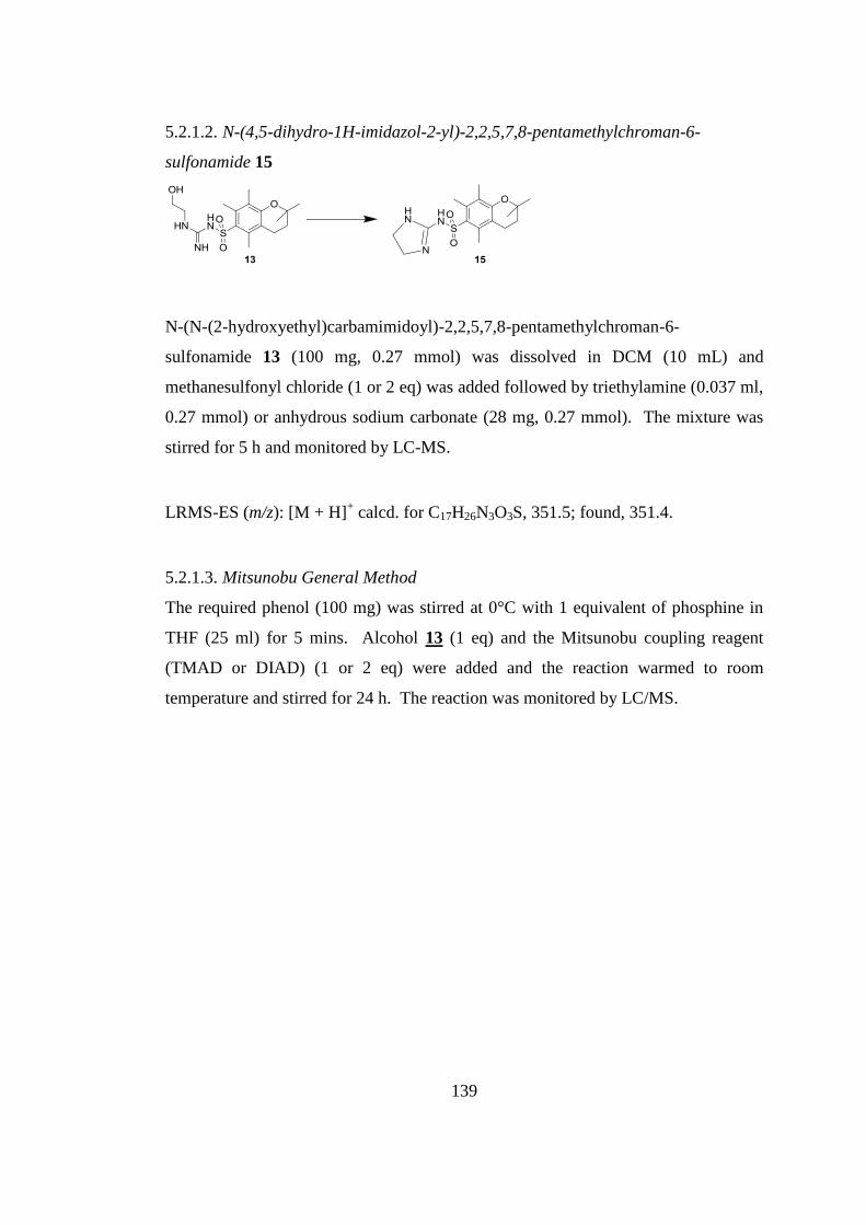
139
5.2.1.2. N-(4,5-dihydro-1H-imidazol-2-yl)-2,2,5,7,8-pentamethylchroman-6-
sulfonamide 15
N-(N-(2-hydroxyethyl)carbamimidoyl)-2,2,5,7,8-pentamethylchroman-6-
sulfonamide 13 (100 mg, 0.27 mmol) was dissolved in DCM (10 mL) and
methanesulfonyl chloride (1 or 2 eq) was added followed by triethylamine (0.037 ml,
0.27 mmol) or anhydrous sodium carbonate (28 mg, 0.27 mmol). The mixture was
stirred for 5 h and monitored by LC-MS.
LRMS-ES (m/z): [M + H]+ calcd. for C17H26N3O3S, 351.5; found, 351.4.
5.2.1.3. Mitsunobu General Method
The required phenol (100 mg) was stirred at 0°C with 1 equivalent of phosphine in
THF (25 ml) for 5 mins. Alcohol 13 (1 eq) and the Mitsunobu coupling reagent
(TMAD or DIAD) (1 or 2 eq) were added and the reaction warmed to room
temperature and stirred for 24 h. The reaction was monitored by LC/MS.
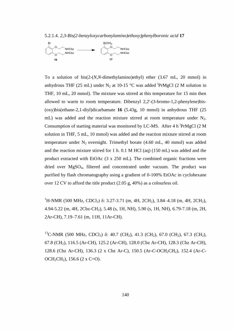
140
5.2.1.4. 2,3-Bis(2-benzyloxycarbonylamino)ethoxy)phenylboronic acid 17
To a solution of bis(2-(N,N-dimethylamino)ethyl) ether (3.67 mL, 20 mmol) in
anhydrous THF (25 mL) under N2 at 10-15 °C was added iPrMgCl (2 M solution in
THF, 10 mL, 20 mmol). The mixture was stirred at this temperature for 15 min then
allowed to warm to room temperature. Dibenzyl 2,2'-(3-bromo-1,2-phenylene)bis-
(oxy)bis(ethane-2,1-diyl)dicarbamate 16 (5.43g, 10 mmol) in anhydrous THF (25
mL) was added and the reaction mixture stirred at room temperature under N2.
Consumption of starting material was monitored by LC-MS. After 4 h iPrMgCl (2 M
solution in THF, 5 mL, 10 mmol) was added and the reaction mixture stirred at room
temperature under N2 overnight. Trimethyl borate (4.60 mL, 40 mmol) was added
and the reaction mixture stirred for 1 h. 0.1 M HCl (aq) (150 mL) was added and the
product extracted with EtOAc (3 x 250 mL). The combined organic fractions were
dried over MgSO4, filtered and concentrated under vacuum. The product was
purified by flash chromatography using a gradient of 0-100% EtOAc in cyclohexane
over 12 CV to afford the title product (2.05 g, 40%) as a colourless oil.
1H-NMR (500 MHz, CDCl3) δ: 3.27-3.71 (m, 4H, 2CH2), 3.84–4.18 (m, 4H, 2CH2),
4.94-5.22 (m, 4H, 2Cbz-CH2), 5.48 (s, 1H, NH), 5.90 (s, 1H, NH), 6.79-7.18 (m, 2H,
2Ar-CH), 7.19–7.61 (m, 11H, 11Ar-CH).
13C-NMR (500 MHz, CDCl3) δ: 40.7 (CH2), 41.3 (CH2), 67.0 (CH2), 67.3 (CH2),
67.8 (CH2), 116.5 (Ar-CH), 125.2 (Ar-CH), 128.0 (Cbz Ar-CH), 128.3 (Cbz Ar-CH),
128.6 (Cbz Ar-CH), 136.3 (2 x Cbz Ar-C), 150.5 (Ar-C-OCH2CH2), 152.4 (Ar-C-
OCH2CH2), 156.6 (2 x C=O).
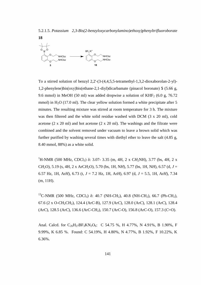
141
5.2.1.5. Potassium 2,3-Bis(2-benzyloxycarbonylamino)ethoxy)phenyltrifluoroborate
18
To a stirred solution of benzyl 2,2'-(3-(4,4,5,5-tetramethyl-1,3,2-dioxaborolan-2-yl)-
1,2-phenylene)bis(oxy)bis(ethane-2,1-diyl)dicarbamate (pinacol boronate) 5 (5.66 g,
9.6 mmol) in MeOH (50 ml) was added dropwise a solution of KHF2 (6.0 g, 76.72
mmol) in H2O (17.0 ml). The clear yellow solution formed a white precipitate after 5
minutes. The resulting mixture was stirred at room temperature for 3 h. The mixture
was then filtered and the white solid residue washed with DCM (3 x 20 ml), cold
acetone (2 x 20 ml) and hot acetone (2 x 20 ml). The washings and the filtrate were
combined and the solvent removed under vacuum to leave a brown solid which was
further purified by washing several times with diethyl ether to leave the salt (4.85 g,
8.40 mmol, 88%) as a white solid.
1H-NMR (500 MHz, CDCl3) δ: 3.07- 3.35 (m, 4H, 2 x CH2NH), 3.77 (bs, 4H, 2 x
CH2O), 5.19 (s, 4H, 2 x ArCH2O), 5.70 (bs, 1H, NH), 5.77 (bs, 1H, NH), 6.57 (d, J =
6.57 Hz, 1H, ArH), 6.73 (t, J = 7.2 Hz, 1H, ArH), 6.97 (d, J = 5.5, 1H, ArH), 7.34
(m, 11H).
13C-NMR (500 MHz, CDCl3) δ: 40.7 (NH-CH2), 40.8 (NH-CH2), 66.7 (Ph-CH2),
67.6 (2 x O-CH2CH2), 124.4 (ArC-B), 127.9 (ArC), 128.0 (ArC), 128.1 (ArC), 128.4
(ArC), 128.5 (ArC), 136.6 (ArC-CH2), 150.7 (ArC-O), 156.8 (ArC-O), 157.3 (C=O).
Anal. Calcd. for C26H27BF3KN2O6: C 54.75 %, H 4.77%, N 4.91%, B 1.90%, F
9.99%, K 6.85 %. Found: C 54.19%, H 4.80%, N 4.77%, B 1.92%, F 10.22%, K
6.36%.
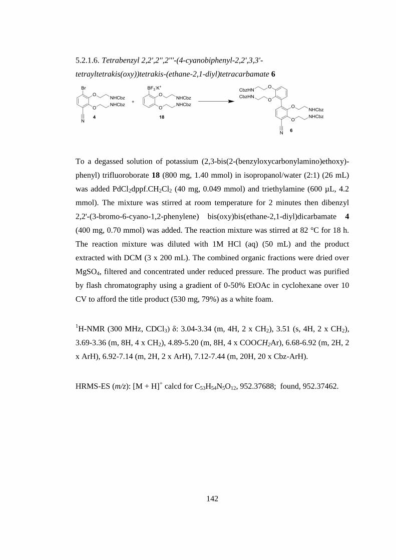
142
5.2.1.6. Tetrabenzyl 2,2',2'',2'''-(4-cyanobiphenyl-2,2',3,3'-
tetrayltetrakis(oxy))tetrakis-(ethane-2,1-diyl)tetracarbamate 6
To a degassed solution of potassium (2,3-bis(2-(benzyloxycarbonylamino)ethoxy)-
phenyl) trifluoroborate 18 (800 mg, 1.40 mmol) in isopropanol/water (2:1) (26 mL)
was added PdCl2dppf.CH2Cl2 (40 mg, 0.049 mmol) and triethylamine (600 µL, 4.2
mmol). The mixture was stirred at room temperature for 2 minutes then dibenzyl
2,2'-(3-bromo-6-cyano-1,2-phenylene) bis(oxy)bis(ethane-2,1-diyl)dicarbamate 4
(400 mg, 0.70 mmol) was added. The reaction mixture was stirred at 82 °C for 18 h.
The reaction mixture was diluted with 1M HCl (aq) (50 mL) and the product
extracted with DCM (3 x 200 mL). The combined organic fractions were dried over
MgSO4, filtered and concentrated under reduced pressure. The product was purified
by flash chromatography using a gradient of 0-50% EtOAc in cyclohexane over 10
CV to afford the title product (530 mg, 79%) as a white foam.
1H-NMR (300 MHz, CDCl3) δ: 3.04-3.34 (m, 4H, 2 x CH2), 3.51 (s, 4H, 2 x CH2),
3.69-3.36 (m, 8H, 4 x CH2), 4.89-5.20 (m, 8H, 4 x COOCH2Ar), 6.68-6.92 (m, 2H, 2
x ArH), 6.92-7.14 (m, 2H, 2 x ArH), 7.12-7.44 (m, 20H, 20 x Cbz-ArH).
HRMS-ES (m/z): [M + H]+ calcd for C53H54N5O12, 952.37688; found, 952.37462.
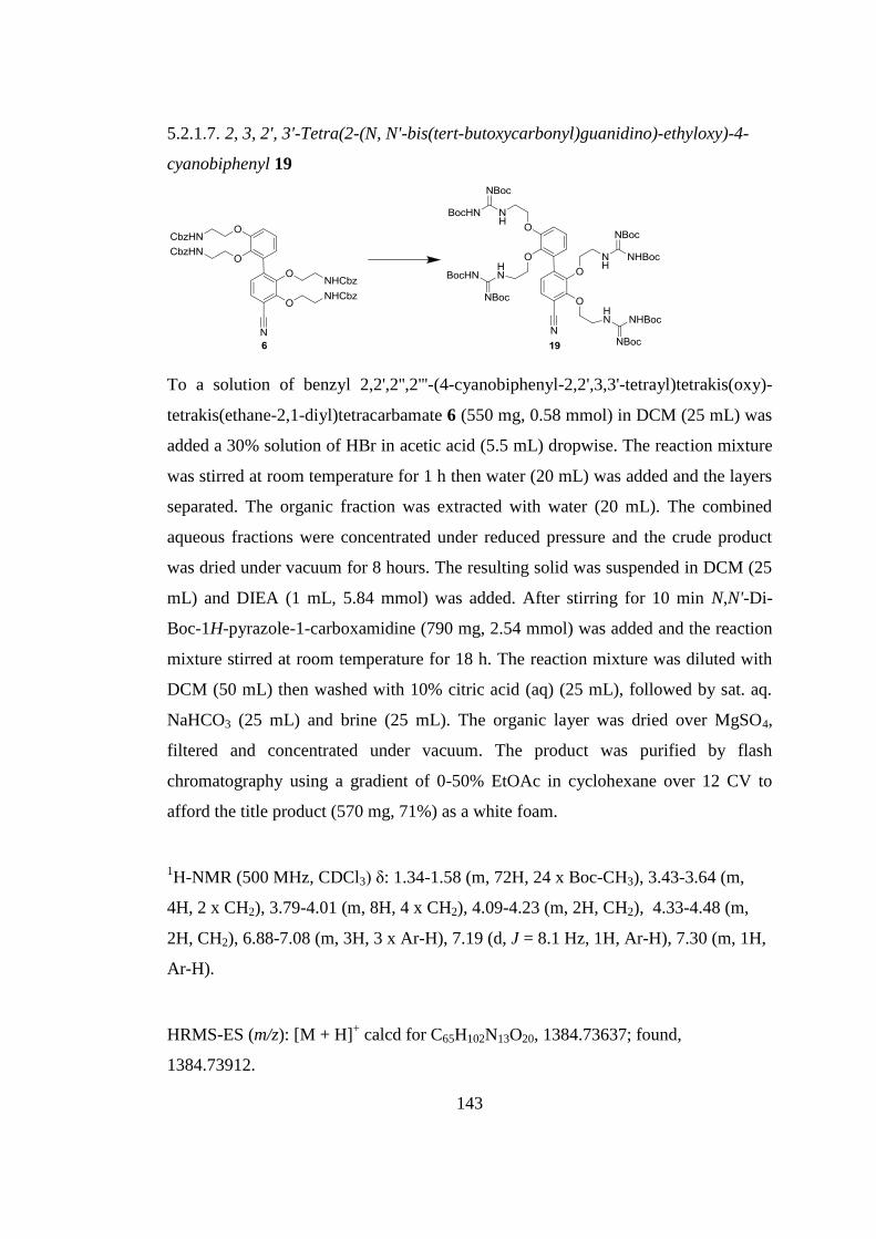
143
5.2.1.7. 2, 3, 2', 3'-Tetra(2-(N, N'-bis(tert-butoxycarbonyl)guanidino)-ethyloxy)-4-
cyanobiphenyl 19
To a solution of benzyl 2,2',2'',2'''-(4-cyanobiphenyl-2,2',3,3'-tetrayl)tetrakis(oxy)-
tetrakis(ethane-2,1-diyl)tetracarbamate 6 (550 mg, 0.58 mmol) in DCM (25 mL) was
added a 30% solution of HBr in acetic acid (5.5 mL) dropwise. The reaction mixture
was stirred at room temperature for 1 h then water (20 mL) was added and the layers
separated. The organic fraction was extracted with water (20 mL). The combined
aqueous fractions were concentrated under reduced pressure and the crude product
was dried under vacuum for 8 hours. The resulting solid was suspended in DCM (25
mL) and DIEA (1 mL, 5.84 mmol) was added. After stirring for 10 min N,N'-Di-
Boc-1H-pyrazole-1-carboxamidine (790 mg, 2.54 mmol) was added and the reaction
mixture stirred at room temperature for 18 h. The reaction mixture was diluted with
DCM (50 mL) then washed with 10% citric acid (aq) (25 mL), followed by sat. aq.
NaHCO3 (25 mL) and brine (25 mL). The organic layer was dried over MgSO4,
filtered and concentrated under vacuum. The product was purified by flash
chromatography using a gradient of 0-50% EtOAc in cyclohexane over 12 CV to
afford the title product (570 mg, 71%) as a white foam.
1H-NMR (500 MHz, CDCl3) δ: 1.34-1.58 (m, 72H, 24 x Boc-CH3), 3.43-3.64 (m,
4H, 2 x CH2), 3.79-4.01 (m, 8H, 4 x CH2), 4.09-4.23 (m, 2H, CH2), 4.33-4.48 (m,
2H, CH2), 6.88-7.08 (m, 3H, 3 x Ar-H), 7.19 (d, J = 8.1 Hz, 1H, Ar-H), 7.30 (m, 1H,
Ar-H).
HRMS-ES (m/z): [M + H]+ calcd for C65H102N13O20, 1384.73637; found,
1384.73912.
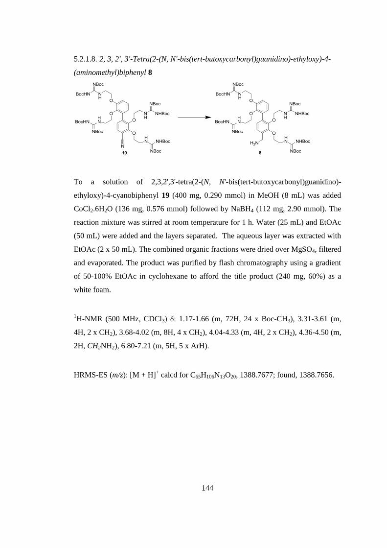
144
5.2.1.8. 2, 3, 2', 3'-Tetra(2-(N, N'-bis(tert-butoxycarbonyl)guanidino)-ethyloxy)-4-
(aminomethyl)biphenyl 8
To a solution of 2,3,2',3'-tetra(2-(N, N'-bis(tert-butoxycarbonyl)guanidino)-
ethyloxy)-4-cyanobiphenyl 19 (400 mg, 0.290 mmol) in MeOH (8 mL) was added
CoCl2.6H2O (136 mg, 0.576 mmol) followed by NaBH4 (112 mg, 2.90 mmol). The
reaction mixture was stirred at room temperature for 1 h. Water (25 mL) and EtOAc
(50 mL) were added and the layers separated. The aqueous layer was extracted with
EtOAc (2 x 50 mL). The combined organic fractions were dried over MgSO4, filtered
and evaporated. The product was purified by flash chromatography using a gradient
of 50-100% EtOAc in cyclohexane to afford the title product (240 mg, 60%) as a
white foam.
1H-NMR (500 MHz, CDCl3) δ: 1.17-1.66 (m, 72H, 24 x Boc-CH3), 3.31-3.61 (m,
4H, 2 x CH2), 3.68-4.02 (m, 8H, 4 x CH2), 4.04-4.33 (m, 4H, 2 x CH2), 4.36-4.50 (m,
2H, CH2NH2), 6.80-7.21 (m, 5H, 5 x ArH).
HRMS-ES (m/z): [M + H]+ calcd for C65H106N13O20, 1388.7677; found, 1388.7656.
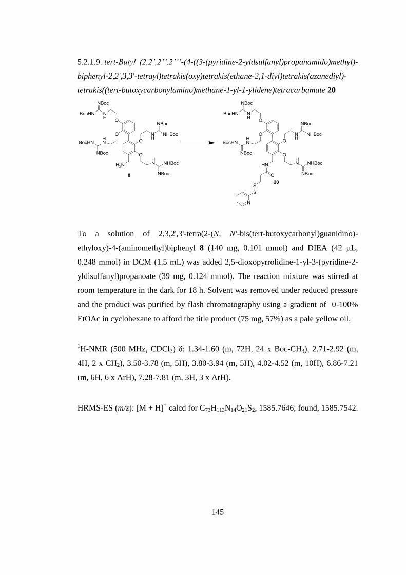
145
5.2.1.9. tert-Butyl (2,2’,2’’,2’’’-(4-((3-(pyridine-2-yldsulfanyl)propanamido)methyl)-
biphenyl-2,2',3,3'-tetrayl)tetrakis(oxy)tetrakis(ethane-2,1-diyl)tetrakis(azanediyl)-
tetrakis((tert-butoxycarbonylamino)methane-1-yl-1-ylidene)tetracarbamate 20
To a solution of 2,3,2',3'-tetra(2-(N, N'-bis(tert-butoxycarbonyl)guanidino)-
ethyloxy)-4-(aminomethyl)biphenyl 8 (140 mg, 0.101 mmol) and DIEA (42 µL,
0.248 mmol) in DCM (1.5 mL) was added 2,5-dioxopyrrolidine-1-yl-3-(pyridine-2-
yldisulfanyl)propanoate (39 mg, 0.124 mmol). The reaction mixture was stirred at
room temperature in the dark for 18 h. Solvent was removed under reduced pressure
and the product was purified by flash chromatography using a gradient of 0-100%
EtOAc in cyclohexane to afford the title product (75 mg, 57%) as a pale yellow oil.
1H-NMR (500 MHz, CDCl3) δ: 1.34-1.60 (m, 72H, 24 x Boc-CH3), 2.71-2.92 (m,
4H, 2 x CH2), 3.50-3.78 (m, 5H), 3.80-3.94 (m, 5H), 4.02-4.52 (m, 10H), 6.86-7.21
(m, 6H, 6 x ArH), 7.28-7.81 (m, 3H, 3 x ArH).
HRMS-ES (m/z): [M + H]+ calcd for C73H113N14O21S2, 1585.7646; found, 1585.7542.
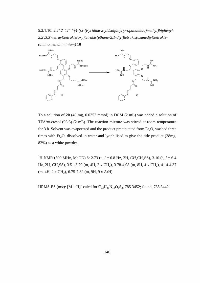
146
5.2.1.10. 2,2’,2’’,2’’’-(4-((3-(Pyridine-2-yldsulfanyl)propanamido)methyl)biphenyl-
2,2',3,3'-tetrayl)tetrakis(oxy)tetrakis(ethane-2,1-diyl)tetrakis(azanediyl)tetrakis-
(aminomethaniminium) 10
To a solution of 20 (40 mg, 0.0252 mmol) in DCM (2 mL) was added a solution of
TFA/m-cresol (95:5) (2 mL). The reaction mixture was stirred at room temperature
for 3 h. Solvent was evaporated and the product precipitated from Et2O, washed three
times with Et2O, dissolved in water and lyophilised to give the title product (28mg,
82%) as a white powder.
1H-NMR (500 MHz, MeOD) δ: 2.73 (t, J = 6.8 Hz, 2H, CH2CH2SS), 3.10 (t, J = 6.4
Hz, 2H, CH2SS), 3.51-3.79 (m, 4H, 2 x CH2), 3.78-4.08 (m, 8H, 4 x CH2), 4.14-4.37
(m, 4H, 2 x CH2), 6.75-7.32 (m, 9H, 9 x ArH).
HRMS-ES (m/z): [M + H]+ calcd for C33H49N14O5S2, 785.3452; found, 785.3442.
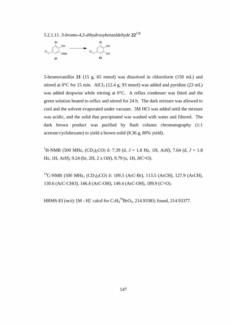
147
5.2.1.11. 3-bromo-4,5-dihydroxybenzaldehyde 22138
5-bromovanillin 21 (15 g, 65 mmol) was dissolved in chloroform (150 mL) and
stirred at 0°C for 15 min. AlCl3 (12.4 g, 93 mmol) was added and pyridine (23 mL)
was added dropwise while stirring at 0°C. A reflux condenser was fitted and the
green solution heated to reflux and stirred for 24 h. The dark mixture was allowed to
cool and the solvent evaporated under vacuum. 3M HCl was added until the mixture
was acidic, and the solid that precipitated was washed with water and filtered. The
dark brown product was purified by flash column chromatography (1:1
acetone:cyclohexane) to yield a brown solid (8.36 g, 80% yield).
1H-NMR (500 MHz, (CD3)2CO) δ: 7.39 (d, J = 1.8 Hz, 1H, ArH), 7.64 (d, J = 1.8
Hz, 1H, ArH), 9.24 (br, 2H, 2 x OH), 9.79 (s, 1H, HC=O).
13C-NMR (500 MHz, (CD3)2CO) δ: 109.5 (ArC-Br), 113.5 (ArCH), 127.9 (ArCH),
130.6 (ArC-CHO), 146.4 (ArC-OH), 149.4 (ArC-OH), 189.9 (C=O).
HRMS-EI (m/z): [M - H]- calcd for C7H4
79BrO3, 214.93383; found, 214.93377.
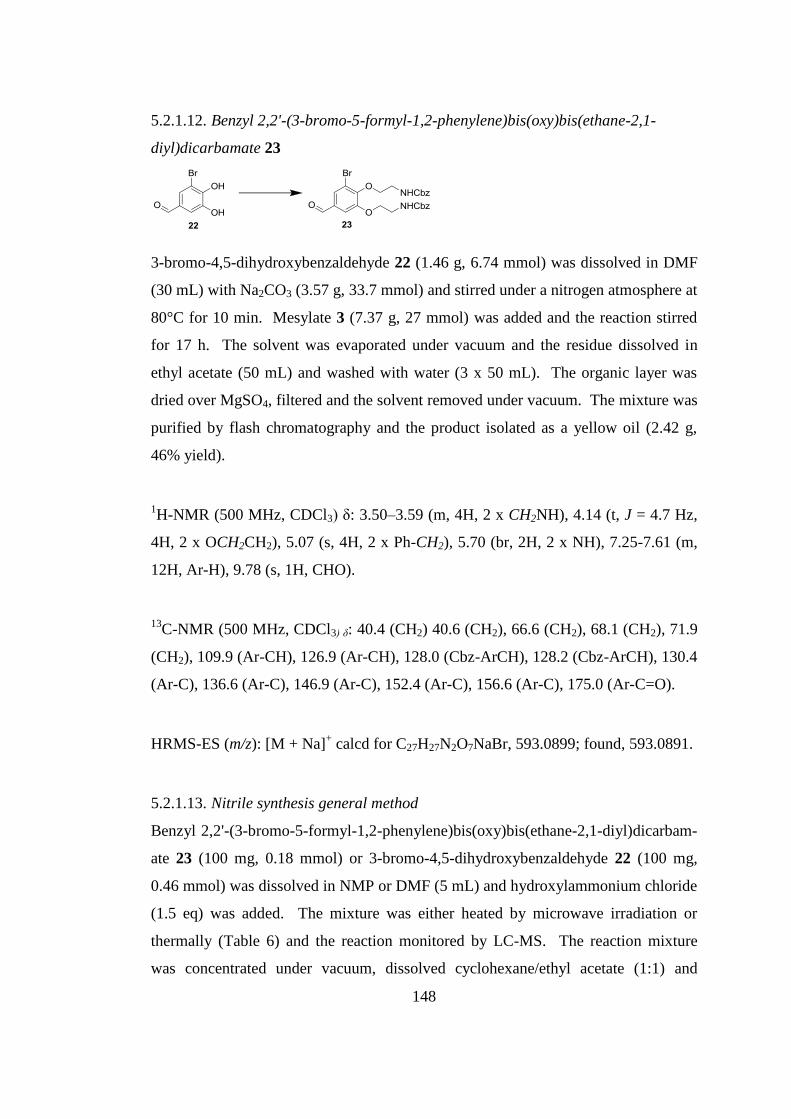
148
5.2.1.12. Benzyl 2,2'-(3-bromo-5-formyl-1,2-phenylene)bis(oxy)bis(ethane-2,1-
diyl)dicarbamate 23
3-bromo-4,5-dihydroxybenzaldehyde 22 (1.46 g, 6.74 mmol) was dissolved in DMF
(30 mL) with Na2CO3 (3.57 g, 33.7 mmol) and stirred under a nitrogen atmosphere at
80°C for 10 min. Mesylate 3 (7.37 g, 27 mmol) was added and the reaction stirred
for 17 h. The solvent was evaporated under vacuum and the residue dissolved in
ethyl acetate (50 mL) and washed with water (3 x 50 mL). The organic layer was
dried over MgSO4, filtered and the solvent removed under vacuum. The mixture was
purified by flash chromatography and the product isolated as a yellow oil (2.42 g,
46% yield).
1H-NMR (500 MHz, CDCl3) δ: 3.50–3.59 (m, 4H, 2 x CH2NH), 4.14 (t, J = 4.7 Hz,
4H, 2 x OCH2CH2), 5.07 (s, 4H, 2 x Ph-CH2), 5.70 (br, 2H, 2 x NH), 7.25-7.61 (m,
12H, Ar-H), 9.78 (s, 1H, CHO).
13C-NMR (500 MHz, CDCl3) δ: 40.4 (CH2) 40.6 (CH2), 66.6 (CH2), 68.1 (CH2), 71.9
(CH2), 109.9 (Ar-CH), 126.9 (Ar-CH), 128.0 (Cbz-ArCH), 128.2 (Cbz-ArCH), 130.4
(Ar-C), 136.6 (Ar-C), 146.9 (Ar-C), 152.4 (Ar-C), 156.6 (Ar-C), 175.0 (Ar-C=O).
HRMS-ES (m/z): [M + Na]+ calcd for C27H27N2O7NaBr, 593.0899; found, 593.0891.
5.2.1.13. Nitrile synthesis general method
Benzyl 2,2'-(3-bromo-5-formyl-1,2-phenylene)bis(oxy)bis(ethane-2,1-diyl)dicarbam-
ate 23 (100 mg, 0.18 mmol) or 3-bromo-4,5-dihydroxybenzaldehyde 22 (100 mg,
0.46 mmol) was dissolved in NMP or DMF (5 mL) and hydroxylammonium chloride
(1.5 eq) was added. The mixture was either heated by microwave irradiation or
thermally (Table 6) and the reaction monitored by LC-MS. The reaction mixture
was concentrated under vacuum, dissolved cyclohexane/ethyl acetate (1:1) and
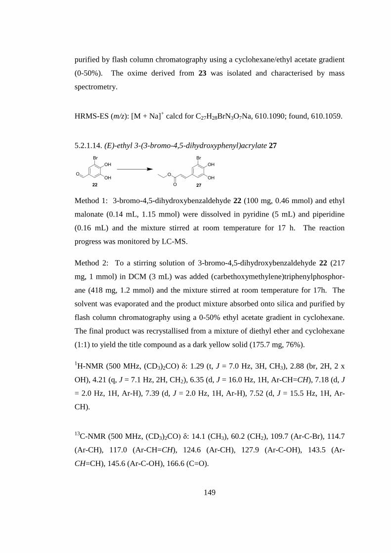
149
purified by flash column chromatography using a cyclohexane/ethyl acetate gradient
(0-50%). The oxime derived from 23 was isolated and characterised by mass
spectrometry.
HRMS-ES (m/z): [M + Na]+ calcd for C27H28BrN3O7Na, 610.1090; found, 610.1059.
5.2.1.14. (E)-ethyl 3-(3-bromo-4,5-dihydroxyphenyl)acrylate 27
Method 1: 3-bromo-4,5-dihydroxybenzaldehyde 22 (100 mg, 0.46 mmol) and ethyl
malonate (0.14 mL, 1.15 mmol) were dissolved in pyridine (5 mL) and piperidine
(0.16 mL) and the mixture stirred at room temperature for 17 h. The reaction
progress was monitored by LC-MS.
Method 2: To a stirring solution of 3-bromo-4,5-dihydroxybenzaldehyde 22 (217
mg, 1 mmol) in DCM (3 mL) was added (carbethoxymethylene)triphenylphosphor-
ane (418 mg, 1.2 mmol) and the mixture stirred at room temperature for 17h. The
solvent was evaporated and the product mixture absorbed onto silica and purified by
flash column chromatography using a 0-50% ethyl acetate gradient in cyclohexane.
The final product was recrystallised from a mixture of diethyl ether and cyclohexane
(1:1) to yield the title compound as a dark yellow solid (175.7 mg, 76%).
1H-NMR (500 MHz, (CD3)2CO) δ: 1.29 (t, J = 7.0 Hz, 3H, CH3), 2.88 (br, 2H, 2 x
OH), 4.21 (q, J = 7.1 Hz, 2H, CH2), 6.35 (d, J = 16.0 Hz, 1H, Ar-CH=CH), 7.18 (d, J
= 2.0 Hz, 1H, Ar-H), 7.39 (d, J = 2.0 Hz, 1H, Ar-H), 7.52 (d, J = 15.5 Hz, 1H, Ar-
CH).
13C-NMR (500 MHz, (CD3)2CO) δ: 14.1 (CH3), 60.2 (CH2), 109.7 (Ar-C-Br), 114.7
(Ar-CH), 117.0 (Ar-CH=CH), 124.6 (Ar-CH), 127.9 (Ar-C-OH), 143.5 (Ar-
CH=CH), 145.6 (Ar-C-OH), 166.6 (C=O).
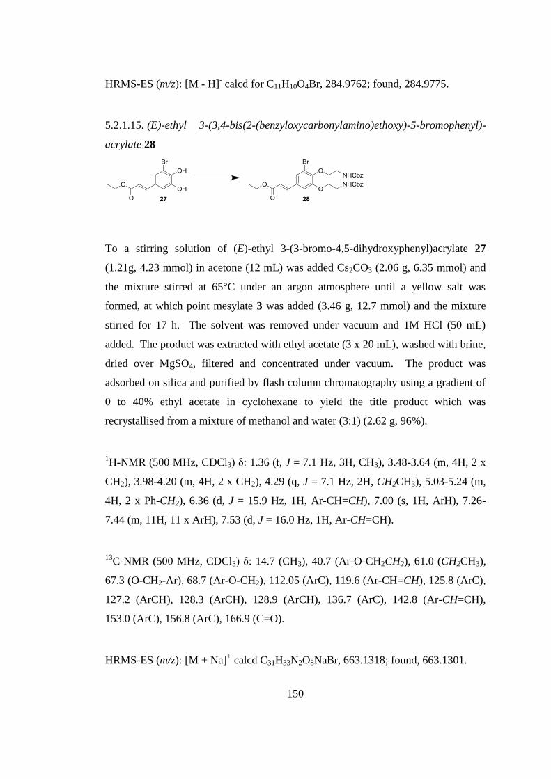
150
HRMS-ES (m/z): [M - H]- calcd for C11H10O4Br, 284.9762; found, 284.9775.
5.2.1.15. (E)-ethyl 3-(3,4-bis(2-(benzyloxycarbonylamino)ethoxy)-5-bromophenyl)-
acrylate 28
To a stirring solution of (E)-ethyl 3-(3-bromo-4,5-dihydroxyphenyl)acrylate 27
(1.21g, 4.23 mmol) in acetone (12 mL) was added Cs2CO3 (2.06 g, 6.35 mmol) and
the mixture stirred at 65°C under an argon atmosphere until a yellow salt was
formed, at which point mesylate 3 was added (3.46 g, 12.7 mmol) and the mixture
stirred for 17 h. The solvent was removed under vacuum and 1M HCl (50 mL)
added. The product was extracted with ethyl acetate (3 x 20 mL), washed with brine,
dried over MgSO4, filtered and concentrated under vacuum. The product was
adsorbed on silica and purified by flash column chromatography using a gradient of
0 to 40% ethyl acetate in cyclohexane to yield the title product which was
recrystallised from a mixture of methanol and water (3:1) (2.62 g, 96%).
1H-NMR (500 MHz, CDCl3) δ: 1.36 (t, J = 7.1 Hz, 3H, CH3), 3.48-3.64 (m, 4H, 2 x
CH2), 3.98-4.20 (m, 4H, 2 x CH2), 4.29 (q, J = 7.1 Hz, 2H, CH2CH3), 5.03-5.24 (m,
4H, 2 x Ph-CH2), 6.36 (d, J = 15.9 Hz, 1H, Ar-CH=CH), 7.00 (s, 1H, ArH), 7.26-
7.44 (m, 11H, 11 x ArH), 7.53 (d, J = 16.0 Hz, 1H, Ar-CH=CH).
13C-NMR (500 MHz, CDCl3) δ: 14.7 (CH3), 40.7 (Ar-O-CH2CH2), 61.0 (CH2CH3),
67.3 (O-CH2-Ar), 68.7 (Ar-O-CH2), 112.05 (ArC), 119.6 (Ar-CH=CH), 125.8 (ArC),
127.2 (ArCH), 128.3 (ArCH), 128.9 (ArCH), 136.7 (ArC), 142.8 (Ar-CH=CH),
153.0 (ArC), 156.8 (ArC), 166.9 (C=O).
HRMS-ES (m/z): [M + Na]+ calcd C31H33N2O8NaBr, 663.1318; found, 663.1301.
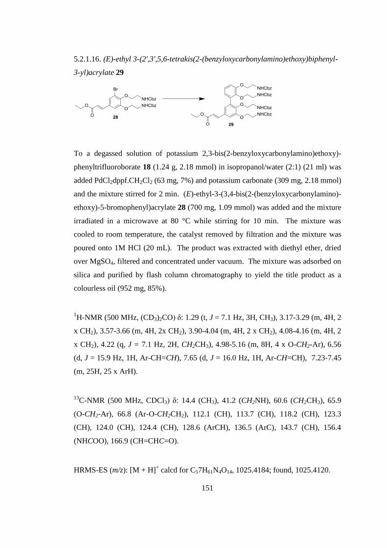
151
5.2.1.16. (E)-ethyl 3-(2',3',5,6-tetrakis(2-(benzyloxycarbonylamino)ethoxy)biphenyl-
3-yl)acrylate 29
To a degassed solution of potassium 2,3-bis(2-benzyloxycarbonylamino)ethoxy)-
phenyltrifluoroborate 18 (1.24 g, 2.18 mmol) in isopropanol/water (2:1) (21 ml) was
added PdCl2dppf.CH2Cl2 (63 mg, 7%) and potassium carbonate (309 mg, 2.18 mmol)
and the mixture stirred for 2 min. (E)-ethyl-3-(3,4-bis(2-(benzyloxycarbonylamino)-
ethoxy)-5-bromophenyl)acrylate 28 (700 mg, 1.09 mmol) was added and the mixture
irradiated in a microwave at 80 °C while stirring for 10 min. The mixture was
cooled to room temperature, the catalyst removed by filtration and the mixture was
poured onto 1M HCl (20 mL). The product was extracted with diethyl ether, dried
over MgSO4, filtered and concentrated under vacuum. The mixture was adsorbed on
silica and purified by flash column chromatography to yield the title product as a
colourless oil (952 mg, 85%).
1H-NMR (500 MHz, (CD3)2CO) δ: 1.29 (t, J = 7.1 Hz, 3H, CH3), 3.17-3.29 (m, 4H, 2
x CH2), 3.57-3.66 (m, 4H, 2x CH2), 3.90-4.04 (m, 4H, 2 x CH2), 4.08-4.16 (m, 4H, 2
x CH2), 4.22 (q, J = 7.1 Hz, 2H, CH2CH3), 4.98-5.16 (m, 8H, 4 x O-CH2-Ar), 6.56
(d, J = 15.9 Hz, 1H, Ar-CH=CH), 7.65 (d, J = 16.0 Hz, 1H, Ar-CH=CH), 7.23-7.45
(m, 25H, 25 x ArH).
13C-NMR (500 MHz, CDCl3) δ: 14.4 (CH3), 41.2 (CH2NH), 60.6 (CH2CH3), 65.9
(O-CH2-Ar), 66.8 (Ar-O-CH2CH2), 112.1 (CH), 113.7 (CH), 118.2 (CH), 123.3
(CH), 124.0 (CH), 124.4 (CH), 128.6 (ArCH), 136.5 (ArC), 143.7 (CH), 156.4
(NHCOO), 166.9 (CH=CHC=O).
HRMS-ES (m/z): [M + H]+ calcd for C57H61N4O14, 1025.4184; found, 1025.4120.
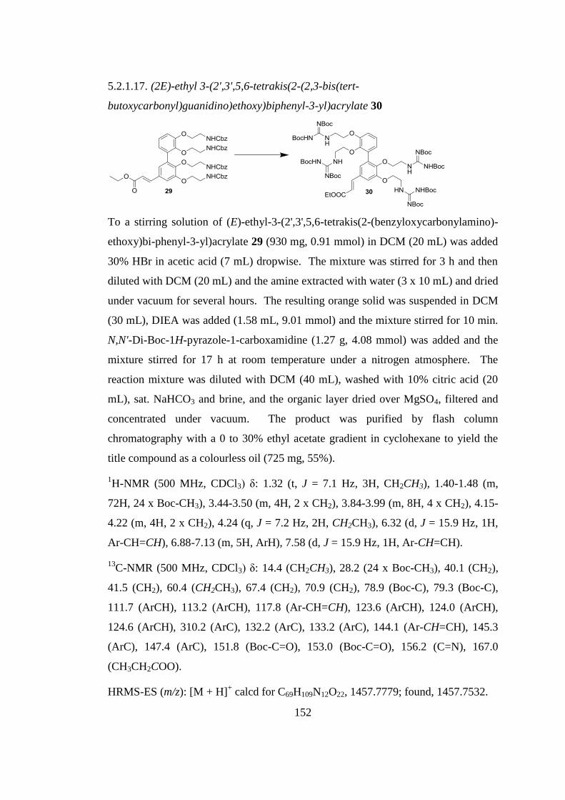
152
5.2.1.17. (2E)-ethyl 3-(2',3',5,6-tetrakis(2-(2,3-bis(tert-
butoxycarbonyl)guanidino)ethoxy)biphenyl-3-yl)acrylate 30
To a stirring solution of (E)-ethyl-3-(2',3',5,6-tetrakis(2-(benzyloxycarbonylamino)-
ethoxy)bi-phenyl-3-yl)acrylate 29 (930 mg, 0.91 mmol) in DCM (20 mL) was added
30% HBr in acetic acid (7 mL) dropwise. The mixture was stirred for 3 h and then
diluted with DCM (20 mL) and the amine extracted with water (3 x 10 mL) and dried
under vacuum for several hours. The resulting orange solid was suspended in DCM
(30 mL), DIEA was added (1.58 mL, 9.01 mmol) and the mixture stirred for 10 min.
N,N'-Di-Boc-1H-pyrazole-1-carboxamidine (1.27 g, 4.08 mmol) was added and the
mixture stirred for 17 h at room temperature under a nitrogen atmosphere. The
reaction mixture was diluted with DCM (40 mL), washed with 10% citric acid (20
mL), sat. NaHCO3 and brine, and the organic layer dried over MgSO4, filtered and
concentrated under vacuum. The product was purified by flash column
chromatography with a 0 to 30% ethyl acetate gradient in cyclohexane to yield the
title compound as a colourless oil (725 mg, 55%).
1H-NMR (500 MHz, CDCl3) δ: 1.32 (t, J = 7.1 Hz, 3H, CH2CH3), 1.40-1.48 (m,
72H, 24 x Boc-CH3), 3.44-3.50 (m, 4H, 2 x CH2), 3.84-3.99 (m, 8H, 4 x CH2), 4.15-
4.22 (m, 4H, 2 x CH2), 4.24 (q, J = 7.2 Hz, 2H, CH2CH3), 6.32 (d, J = 15.9 Hz, 1H,
Ar-CH=CH), 6.88-7.13 (m, 5H, ArH), 7.58 (d, J = 15.9 Hz, 1H, Ar-CH=CH).
13C-NMR (500 MHz, CDCl3) δ: 14.4 (CH2CH3), 28.2 (24 x Boc-CH3), 40.1 (CH2),
41.5 (CH2), 60.4 (CH2CH3), 67.4 (CH2), 70.9 (CH2), 78.9 (Boc-C), 79.3 (Boc-C),
111.7 (ArCH), 113.2 (ArCH), 117.8 (Ar-CH=CH), 123.6 (ArCH), 124.0 (ArCH),
124.6 (ArCH), 310.2 (ArC), 132.2 (ArC), 133.2 (ArC), 144.1 (Ar-CH=CH), 145.3
(ArC), 147.4 (ArC), 151.8 (Boc-C=O), 153.0 (Boc-C=O), 156.2 (C=N), 167.0
(CH3CH2COO).
HRMS-ES (m/z): [M + H]+ calcd for C69H109N12O22, 1457.7779; found, 1457.7532.
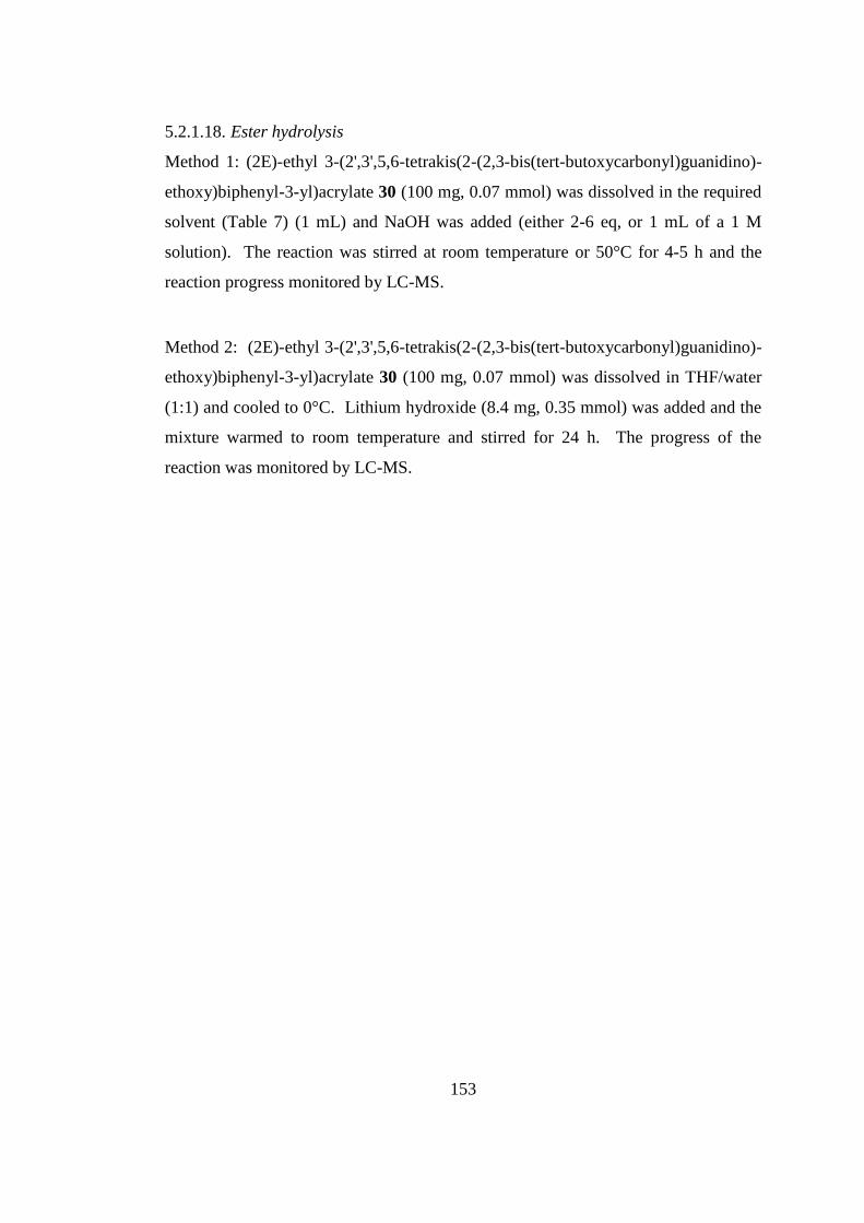
153
5.2.1.18. Ester hydrolysis
Method 1: (2E)-ethyl 3-(2',3',5,6-tetrakis(2-(2,3-bis(tert-butoxycarbonyl)guanidino)-
ethoxy)biphenyl-3-yl)acrylate 30 (100 mg, 0.07 mmol) was dissolved in the required
solvent (Table 7) (1 mL) and NaOH was added (either 2-6 eq, or 1 mL of a 1 M
solution). The reaction was stirred at room temperature or 50°C for 4-5 h and the
reaction progress monitored by LC-MS.
Method 2: (2E)-ethyl 3-(2',3',5,6-tetrakis(2-(2,3-bis(tert-butoxycarbonyl)guanidino)-
ethoxy)biphenyl-3-yl)acrylate 30 (100 mg, 0.07 mmol) was dissolved in THF/water
(1:1) and cooled to 0°C. Lithium hydroxide (8.4 mg, 0.35 mmol) was added and the
mixture warmed to room temperature and stirred for 24 h. The progress of the
reaction was monitored by LC-MS.
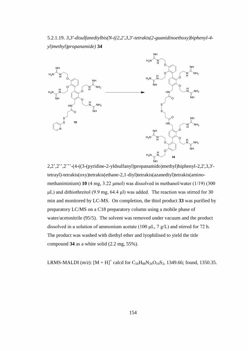
154
5.2.1.19. 3,3'-disulfanediylbis(N-((2,2',3,3'-tetrakis(2-guanidinoethoxy)biphenyl-4-
yl)methyl)propanamide) 34
2,2’,2’’,2’’’-(4-((3-(pyridine-2-yldsulfanyl)propanamido)methyl)biphenyl-2,2',3,3'-
tetrayl)-tetrakis(oxy)tetrakis(ethane-2,1-diyl)tetrakis(azanediyl)tetrakis(amino-
methaniminium) 10 (4 mg, 3.22 μmol) was dissolved in methanol/water (1/19) (300
μL) and dithiothreitol (9.9 mg, 64.4 μl) was added. The reaction was stirred for 30
min and monitored by LC-MS. On completion, the thiol product 33 was purified by
preparatory LC/MS on a C18 preparatory column using a mobile phase of
water/acetonitrile (95/5). The solvent was removed under vacuum and the product
dissolved in a solution of ammonium acetate (100 μL, 7 g/L) and stirred for 72 h.
The product was washed with diethyl ether and lyophilised to yield the title
compound 34 as a white solid (2.2 mg, 55%).
LRMS-MALDI (m/z): [M + H]+ calcd for C56H88N26O10S2, 1349.66; found, 1350.35.

155
5.3. Chapter 3 Methods
5.3.1. Molecular Dynamics
Molecular dynamics simulations were run for compounds 43, 44, 45 and 46 in the
YASARA161
software using the AMBER99 force field162
with default parameters.
The starting structures were contained in a simulation cell which extended 5 Å larger
than the SMoC structure along each axis in vacuo, the default pH set to 7.0 and the
structures were minimised under the AMBER99 force field starting at 298 K for 1 ps.
The simulation substep time was set to 1 fs and the intramolecular forces were
recalculated every 2 substeps and the temperature controlled by velocity rescaling. A
5 ns simulation was performed for each structure, after which snapshots were taken
and analysed in the DS Visualizer software. No atoms were kept frozen during the
simulations.
5.3.2. NMR study
The SMoC compounds were dissolved in 0.5 mL deuterium oxide to a final
concentration of around 10 mg/ml. 100 mM NaOH was added in small aliquots (5-30
μl) and the pH of the solution measured in the NMR tube using a micro electrode
attached to a pH meter. NMR spectra were recorded using a 400 MHz Bruker
spectrometer at 20 °C.
5.3.3. ITC
6G-SMoC at a concentration of 250 μM was loaded into the syringe of a Microcal
VP-ITC calorimeter (450 μl), and the cell (1.8 ml) filled with human GAPDH siRNA
(Dharmacon RNAi Technologies) at a concentration of 3.5 μM. A total of 37
injections of 8 μl each were made at 4 min intervals to ensure total saturation of the
siRNA. The integrated heats evolved for each injection were plotted against the
molar ratio of the mixture in the cell, and a binding curve fitted using non-linear least
squares analysis of the data. The thermodynamic parameters of the binding were
calculated using a one-site (or n identical sites) binding site model in the Origin 6.0
software (Microcal).
156
5.3.4. Syntheses
5.3.4.1. Benzyl 2,2'-(biphenyl-2,2'-diylbis(oxy))bis(ethane-2,1-diyl)dicarbamate 35
2,2’-dihydroxybiphenyl (100 mg, 0.54 mmol) was stirred in acetone (1.5 mL) with
caesium carbonate (228 mg, 0.70 mmol) at 65 °C under nitrogen. When a white salt
appeared, 2-(benzyloxycarbonylamino)ethyl methanesulfonate (381 mg, 1.40 mmol)
was added and the reaction stirred overnight. The mixture was concentrated under
vacuum and adsorbed onto silica gel for purification by flash column
chromatography using ethyl acetate/cyclohexane (1/2) as eluent to yield the title
compound (100 mg, 35% yield) as a viscous colourless oil.
1H-NMR (500 MHz, CD3OD) δ: 3.30 (t, J = 5.6 Hz, 4H, 2 x CH2NH), 3.93 (t, J = 5.6
Hz, 4H, 2 x CH2CH2NH), 5.00 (s, 4H, 2 x OCH2Ph), 6.92-7.31 (m, 18H, 18 x ArH).
13C-NMR (500 MHz, CD3OD) δ: 40.5 (2 x NHCH2) 67.6 (OCH2Ph), 68.7
(CH2CH2NH), 122.3 (ArCH), 128.2 (ArCH), 129.0 (ArCH), 129.5 (ArCH), 129.8
(ArC), 132.3 (ArC), 157.4 (C=O), 159.3 (ArC-O).
LRMS-ES (m/z): [M + Na]+ calcd for C32H32N2O6Na, 563.23; found, 564.20.
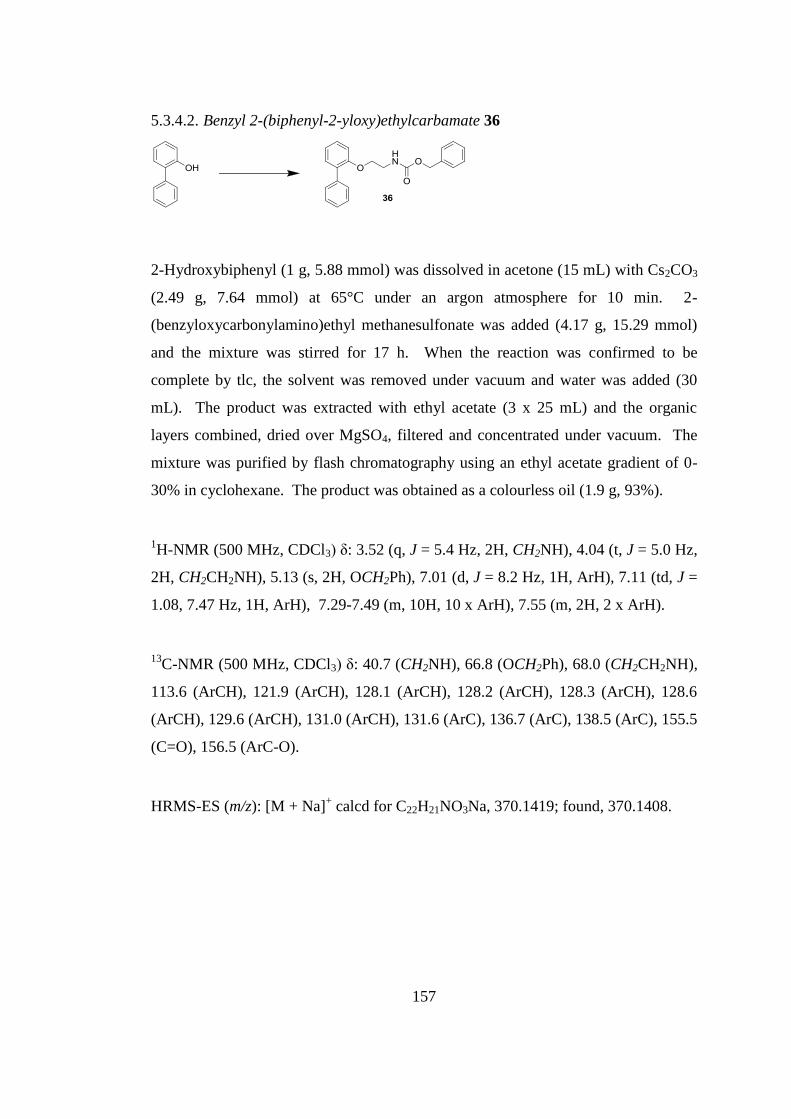
157
5.3.4.2. Benzyl 2-(biphenyl-2-yloxy)ethylcarbamate 36
2-Hydroxybiphenyl (1 g, 5.88 mmol) was dissolved in acetone (15 mL) with Cs2CO3
(2.49 g, 7.64 mmol) at 65°C under an argon atmosphere for 10 min. 2-
(benzyloxycarbonylamino)ethyl methanesulfonate was added (4.17 g, 15.29 mmol)
and the mixture was stirred for 17 h. When the reaction was confirmed to be
complete by tlc, the solvent was removed under vacuum and water was added (30
mL). The product was extracted with ethyl acetate (3 x 25 mL) and the organic
layers combined, dried over MgSO4, filtered and concentrated under vacuum. The
mixture was purified by flash chromatography using an ethyl acetate gradient of 0-
30% in cyclohexane. The product was obtained as a colourless oil (1.9 g, 93%).
1H-NMR (500 MHz, CDCl3) δ: 3.52 (q, J = 5.4 Hz, 2H, CH2NH), 4.04 (t, J = 5.0 Hz,
2H, CH2CH2NH), 5.13 (s, 2H, OCH2Ph), 7.01 (d, J = 8.2 Hz, 1H, ArH), 7.11 (td, J =
1.08, 7.47 Hz, 1H, ArH), 7.29-7.49 (m, 10H, 10 x ArH), 7.55 (m, 2H, 2 x ArH).
13C-NMR (500 MHz, CDCl3) δ: 40.7 (CH2NH), 66.8 (OCH2Ph), 68.0 (CH2CH2NH),
113.6 (ArCH), 121.9 (ArCH), 128.1 (ArCH), 128.2 (ArCH), 128.3 (ArCH), 128.6
(ArCH), 129.6 (ArCH), 131.0 (ArCH), 131.6 (ArC), 136.7 (ArC), 138.5 (ArC), 155.5
(C=O), 156.5 (ArC-O).
HRMS-ES (m/z): [M + Na]+ calcd for C22H21NO3Na, 370.1419; found, 370.1408.
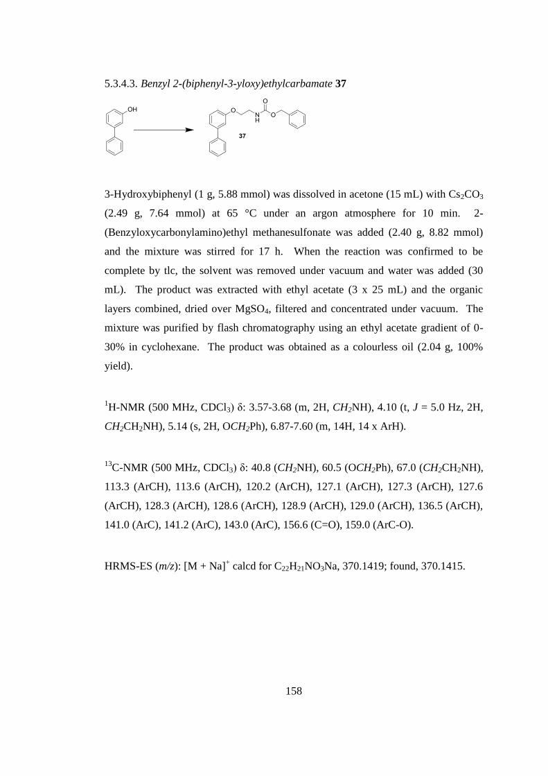
158
5.3.4.3. Benzyl 2-(biphenyl-3-yloxy)ethylcarbamate 37
3-Hydroxybiphenyl (1 g, 5.88 mmol) was dissolved in acetone (15 mL) with Cs2CO3
(2.49 g, 7.64 mmol) at 65 °C under an argon atmosphere for 10 min. 2-
(Benzyloxycarbonylamino)ethyl methanesulfonate was added (2.40 g, 8.82 mmol)
and the mixture was stirred for 17 h. When the reaction was confirmed to be
complete by tlc, the solvent was removed under vacuum and water was added (30
mL). The product was extracted with ethyl acetate (3 x 25 mL) and the organic
layers combined, dried over MgSO4, filtered and concentrated under vacuum. The
mixture was purified by flash chromatography using an ethyl acetate gradient of 0-
30% in cyclohexane. The product was obtained as a colourless oil (2.04 g, 100%
yield).
1H-NMR (500 MHz, CDCl3) δ: 3.57-3.68 (m, 2H, CH2NH), 4.10 (t, J = 5.0 Hz, 2H,
CH2CH2NH), 5.14 (s, 2H, OCH2Ph), 6.87-7.60 (m, 14H, 14 x ArH).
13C-NMR (500 MHz, CDCl3) δ: 40.8 (CH2NH), 60.5 (OCH2Ph), 67.0 (CH2CH2NH),
113.3 (ArCH), 113.6 (ArCH), 120.2 (ArCH), 127.1 (ArCH), 127.3 (ArCH), 127.6
(ArCH), 128.3 (ArCH), 128.6 (ArCH), 128.9 (ArCH), 129.0 (ArCH), 136.5 (ArCH),
141.0 (ArC), 141.2 (ArC), 143.0 (ArC), 156.6 (C=O), 159.0 (ArC-O).
HRMS-ES (m/z): [M + Na]+ calcd for C22H21NO3Na, 370.1419; found, 370.1415.
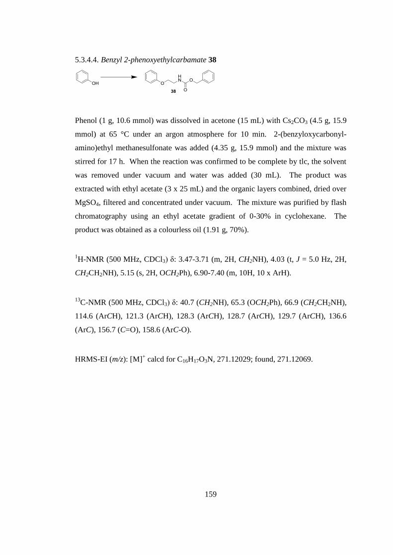
159
5.3.4.4. Benzyl 2-phenoxyethylcarbamate 38
Phenol (1 g, 10.6 mmol) was dissolved in acetone (15 mL) with Cs2CO3 (4.5 g, 15.9
mmol) at 65 °C under an argon atmosphere for 10 min. 2-(benzyloxycarbonyl-
amino)ethyl methanesulfonate was added (4.35 g, 15.9 mmol) and the mixture was
stirred for 17 h. When the reaction was confirmed to be complete by tlc, the solvent
was removed under vacuum and water was added (30 mL). The product was
extracted with ethyl acetate (3 x 25 mL) and the organic layers combined, dried over
MgSO4, filtered and concentrated under vacuum. The mixture was purified by flash
chromatography using an ethyl acetate gradient of 0-30% in cyclohexane. The
product was obtained as a colourless oil (1.91 g, 70%).
1H-NMR (500 MHz, CDCl3) δ: 3.47-3.71 (m, 2H, CH2NH), 4.03 (t, J = 5.0 Hz, 2H,
CH2CH2NH), 5.15 (s, 2H, OCH2Ph), 6.90-7.40 (m, 10H, 10 x ArH).
13C-NMR (500 MHz, CDCl3) δ: 40.7 (CH2NH), 65.3 (OCH2Ph), 66.9 (CH2CH2NH),
114.6 (ArCH), 121.3 (ArCH), 128.3 (ArCH), 128.7 (ArCH), 129.7 (ArCH), 136.6
(ArC), 156.7 (C=O), 158.6 (ArC-O).
HRMS-EI (m/z): [M]+ calcd for C16H17O3N, 271.12029; found, 271.12069.
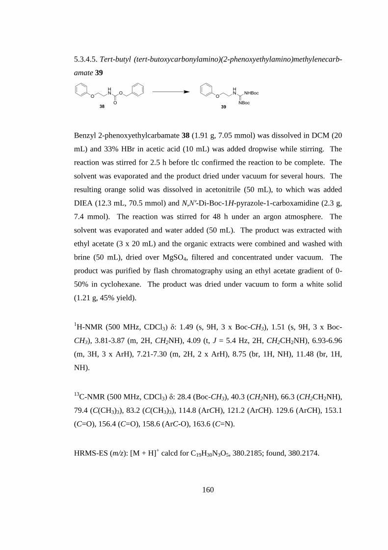
160
5.3.4.5. Tert-butyl (tert-butoxycarbonylamino)(2-phenoxyethylamino)methylenecarb-
amate 39
Benzyl 2-phenoxyethylcarbamate 38 (1.91 g, 7.05 mmol) was dissolved in DCM (20
mL) and 33% HBr in acetic acid (10 mL) was added dropwise while stirring. The
reaction was stirred for 2.5 h before tlc confirmed the reaction to be complete. The
solvent was evaporated and the product dried under vacuum for several hours. The
resulting orange solid was dissolved in acetonitrile (50 mL), to which was added
DIEA (12.3 mL, 70.5 mmol) and N,N′-Di-Boc-1H-pyrazole-1-carboxamidine (2.3 g,
7.4 mmol). The reaction was stirred for 48 h under an argon atmosphere. The
solvent was evaporated and water added (50 mL). The product was extracted with
ethyl acetate (3 x 20 mL) and the organic extracts were combined and washed with
brine (50 mL), dried over MgSO4, filtered and concentrated under vacuum. The
product was purified by flash chromatography using an ethyl acetate gradient of 0-
50% in cyclohexane. The product was dried under vacuum to form a white solid
(1.21 g, 45% yield).
1H-NMR (500 MHz, CDCl3) δ: 1.49 (s, 9H, 3 x Boc-CH3), 1.51 (s, 9H, 3 x Boc-
CH3), 3.81-3.87 (m, 2H, CH2NH), 4.09 (t, J = 5.4 Hz, 2H, CH2CH2NH), 6.93-6.96
(m, 3H, 3 x ArH), 7.21-7.30 (m, 2H, 2 x ArH), 8.75 (br, 1H, NH), 11.48 (br, 1H,
NH).
13C-NMR (500 MHz, CDCl3) δ: 28.4 (Boc-CH3), 40.3 (CH2NH), 66.3 (CH2CH2NH),
79.4 (C(CH3)3), 83.2 (C(CH3)3), 114.8 (ArCH), 121.2 (ArCH). 129.6 (ArCH), 153.1
(C=O), 156.4 (C=O), 158.6 (ArC-O), 163.6 (C=N).
HRMS-ES (m/z): [M + H]+ calcd for C19H30N3O5, 380.2185; found, 380.2174.
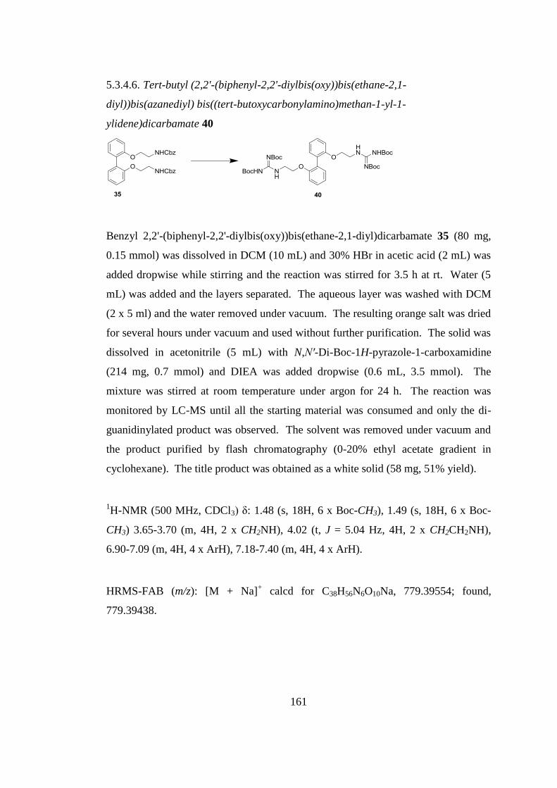
161
5.3.4.6. Tert-butyl (2,2'-(biphenyl-2,2'-diylbis(oxy))bis(ethane-2,1-
diyl))bis(azanediyl) bis((tert-butoxycarbonylamino)methan-1-yl-1-
ylidene)dicarbamate 40
Benzyl 2,2'-(biphenyl-2,2'-diylbis(oxy))bis(ethane-2,1-diyl)dicarbamate 35 (80 mg,
0.15 mmol) was dissolved in DCM (10 mL) and 30% HBr in acetic acid (2 mL) was
added dropwise while stirring and the reaction was stirred for 3.5 h at rt. Water (5
mL) was added and the layers separated. The aqueous layer was washed with DCM
(2 x 5 ml) and the water removed under vacuum. The resulting orange salt was dried
for several hours under vacuum and used without further purification. The solid was
dissolved in acetonitrile (5 mL) with N,N′-Di-Boc-1H-pyrazole-1-carboxamidine
(214 mg, 0.7 mmol) and DIEA was added dropwise (0.6 mL, 3.5 mmol). The
mixture was stirred at room temperature under argon for 24 h. The reaction was
monitored by LC-MS until all the starting material was consumed and only the di-
guanidinylated product was observed. The solvent was removed under vacuum and
the product purified by flash chromatography (0-20% ethyl acetate gradient in
cyclohexane). The title product was obtained as a white solid (58 mg, 51% yield).
1H-NMR (500 MHz, CDCl3) δ: 1.48 (s, 18H, 6 x Boc-CH3), 1.49 (s, 18H, 6 x Boc-
CH3) 3.65-3.70 (m, 4H, 2 x CH2NH), 4.02 (t, J = 5.04 Hz, 4H, 2 x CH2CH2NH),
6.90-7.09 (m, 4H, 4 x ArH), 7.18-7.40 (m, 4H, 4 x ArH).
HRMS-FAB (m/z): [M + Na]+ calcd for C38H56N6O10Na, 779.39554; found,
779.39438.
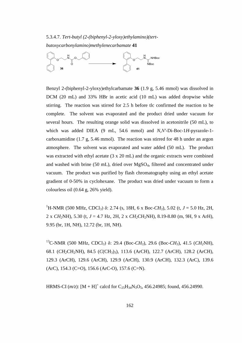
162
5.3.4.7. Tert-butyl (2-(biphenyl-2-yloxy)ethylamino)(tert-
butoxycarbonylamino)methylenecarbamate 41
Benzyl 2-(biphenyl-2-yloxy)ethylcarbamate 36 (1.9 g, 5.46 mmol) was dissolved in
DCM (20 mL) and 33% HBr in acetic acid (10 mL) was added dropwise while
stirring. The reaction was stirred for 2.5 h before tlc confirmed the reaction to be
complete. The solvent was evaporated and the product dried under vacuum for
several hours. The resulting orange solid was dissolved in acetonitrile (50 mL), to
which was added DIEA (9 mL, 54.6 mmol) and N,N′-Di-Boc-1H-pyrazole-1-
carboxamidine (1.7 g, 5.46 mmol). The reaction was stirred for 48 h under an argon
atmosphere. The solvent was evaporated and water added (50 mL). The product
was extracted with ethyl acetate (3 x 20 mL) and the organic extracts were combined
and washed with brine (50 mL), dried over MgSO4, filtered and concentrated under
vacuum. The product was purified by flash chromatography using an ethyl acetate
gradient of 0-50% in cyclohexane. The product was dried under vacuum to form a
colourless oil (0.64 g, 26% yield).
1H-NMR (500 MHz, CDCl3) δ: 2.74 (s, 18H, 6 x Boc-CH3), 5.02 (t, J = 5.0 Hz, 2H,
2 x CH2NH), 5.30 (t, J = 4.7 Hz, 2H, 2 x CH2CH2NH), 8.19-8.80 (m, 9H, 9 x ArH),
9.95 (br, 1H, NH), 12.72 (br, 1H, NH).
13C-NMR (500 MHz, CDCl3) δ: 29.4 (Boc-CH3), 29.6 (Boc-CH3), 41.5 (CH2NH),
68.1 (CH2CH2NH), 84.5 (C(CH3)3), 113.6 (ArCH), 122.7 (ArCH), 128.2 (ArCH),
129.3 (ArCH), 129.6 (ArCH), 129.9 (ArCH), 130.9 (ArCH), 132.3 (ArC), 139.6
(ArC), 154.3 (C=O), 156.6 (ArC-O), 157.6 (C=N).
HRMS-CI (m/z): [M + H]+ calcd for C25H34N3O5, 456.24985; found, 456.24990.
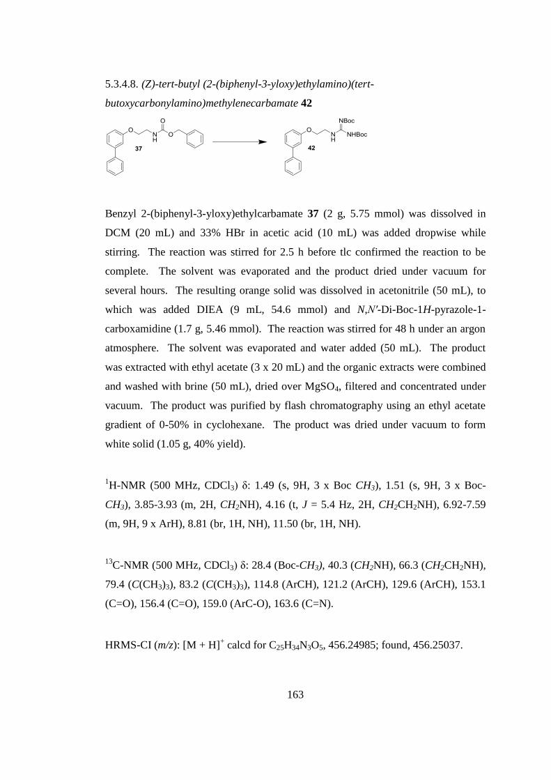
163
5.3.4.8. (Z)-tert-butyl (2-(biphenyl-3-yloxy)ethylamino)(tert-
butoxycarbonylamino)methylenecarbamate 42
Benzyl 2-(biphenyl-3-yloxy)ethylcarbamate 37 (2 g, 5.75 mmol) was dissolved in
DCM (20 mL) and 33% HBr in acetic acid (10 mL) was added dropwise while
stirring. The reaction was stirred for 2.5 h before tlc confirmed the reaction to be
complete. The solvent was evaporated and the product dried under vacuum for
several hours. The resulting orange solid was dissolved in acetonitrile (50 mL), to
which was added DIEA (9 mL, 54.6 mmol) and N,N′-Di-Boc-1H-pyrazole-1-
carboxamidine (1.7 g, 5.46 mmol). The reaction was stirred for 48 h under an argon
atmosphere. The solvent was evaporated and water added (50 mL). The product
was extracted with ethyl acetate (3 x 20 mL) and the organic extracts were combined
and washed with brine (50 mL), dried over MgSO4, filtered and concentrated under
vacuum. The product was purified by flash chromatography using an ethyl acetate
gradient of 0-50% in cyclohexane. The product was dried under vacuum to form
white solid (1.05 g, 40% yield).
1H-NMR (500 MHz, CDCl3) δ: 1.49 (s, 9H, 3 x Boc CH3), 1.51 (s, 9H, 3 x Boc-
CH3), 3.85-3.93 (m, 2H, CH2NH), 4.16 (t, J = 5.4 Hz, 2H, CH2CH2NH), 6.92-7.59
(m, 9H, 9 x ArH), 8.81 (br, 1H, NH), 11.50 (br, 1H, NH).
13C-NMR (500 MHz, CDCl3) δ: 28.4 (Boc-CH3), 40.3 (CH2NH), 66.3 (CH2CH2NH),
79.4 (C(CH3)3), 83.2 (C(CH3)3), 114.8 (ArCH), 121.2 (ArCH), 129.6 (ArCH), 153.1
(C=O), 156.4 (C=O), 159.0 (ArC-O), 163.6 (C=N).
HRMS-CI (m/z): [M + H]+ calcd for C25H34N3O5, 456.24985; found, 456.25037.
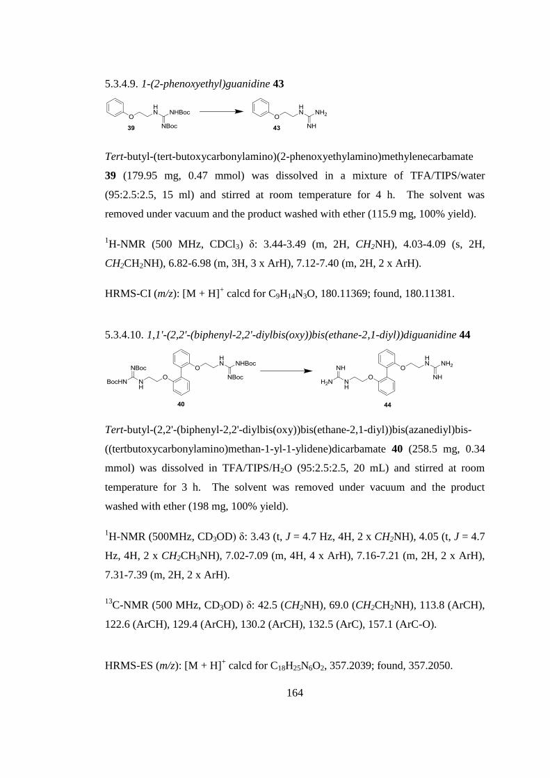
164
5.3.4.9. 1-(2-phenoxyethyl)guanidine 43
Tert-butyl-(tert-butoxycarbonylamino)(2-phenoxyethylamino)methylenecarbamate
39 (179.95 mg, 0.47 mmol) was dissolved in a mixture of TFA/TIPS/water
(95:2.5:2.5, 15 ml) and stirred at room temperature for 4 h. The solvent was
removed under vacuum and the product washed with ether (115.9 mg, 100% yield).
1H-NMR (500 MHz, CDCl3) δ: 3.44-3.49 (m, 2H, CH2NH), 4.03-4.09 (s, 2H,
CH2CH2NH), 6.82-6.98 (m, 3H, 3 x ArH), 7.12-7.40 (m, 2H, 2 x ArH).
HRMS-CI (m/z): [M + H]+ calcd for C9H14N3O, 180.11369; found, 180.11381.
5.3.4.10. 1,1'-(2,2'-(biphenyl-2,2'-diylbis(oxy))bis(ethane-2,1-diyl))diguanidine 44
Tert-butyl-(2,2'-(biphenyl-2,2'-diylbis(oxy))bis(ethane-2,1-diyl))bis(azanediyl)bis-
((tertbutoxycarbonylamino)methan-1-yl-1-ylidene)dicarbamate 40 (258.5 mg, 0.34
mmol) was dissolved in TFA/TIPS/H2O (95:2.5:2.5, 20 mL) and stirred at room
temperature for 3 h. The solvent was removed under vacuum and the product
washed with ether (198 mg, 100% yield).
1H-NMR (500MHz, CD3OD) δ: 3.43 (t, J = 4.7 Hz, 4H, 2 x CH2NH), 4.05 (t, J = 4.7
Hz, 4H, 2 x CH2CH3NH), 7.02-7.09 (m, 4H, 4 x ArH), 7.16-7.21 (m, 2H, 2 x ArH),
7.31-7.39 (m, 2H, 2 x ArH).
13C-NMR (500 MHz, CD3OD) δ: 42.5 (CH2NH), 69.0 (CH2CH2NH), 113.8 (ArCH),
122.6 (ArCH), 129.4 (ArCH), 130.2 (ArCH), 132.5 (ArC), 157.1 (ArC-O).
HRMS-ES (m/z): [M + H]+ calcd for C18H25N6O2, 357.2039; found, 357.2050.
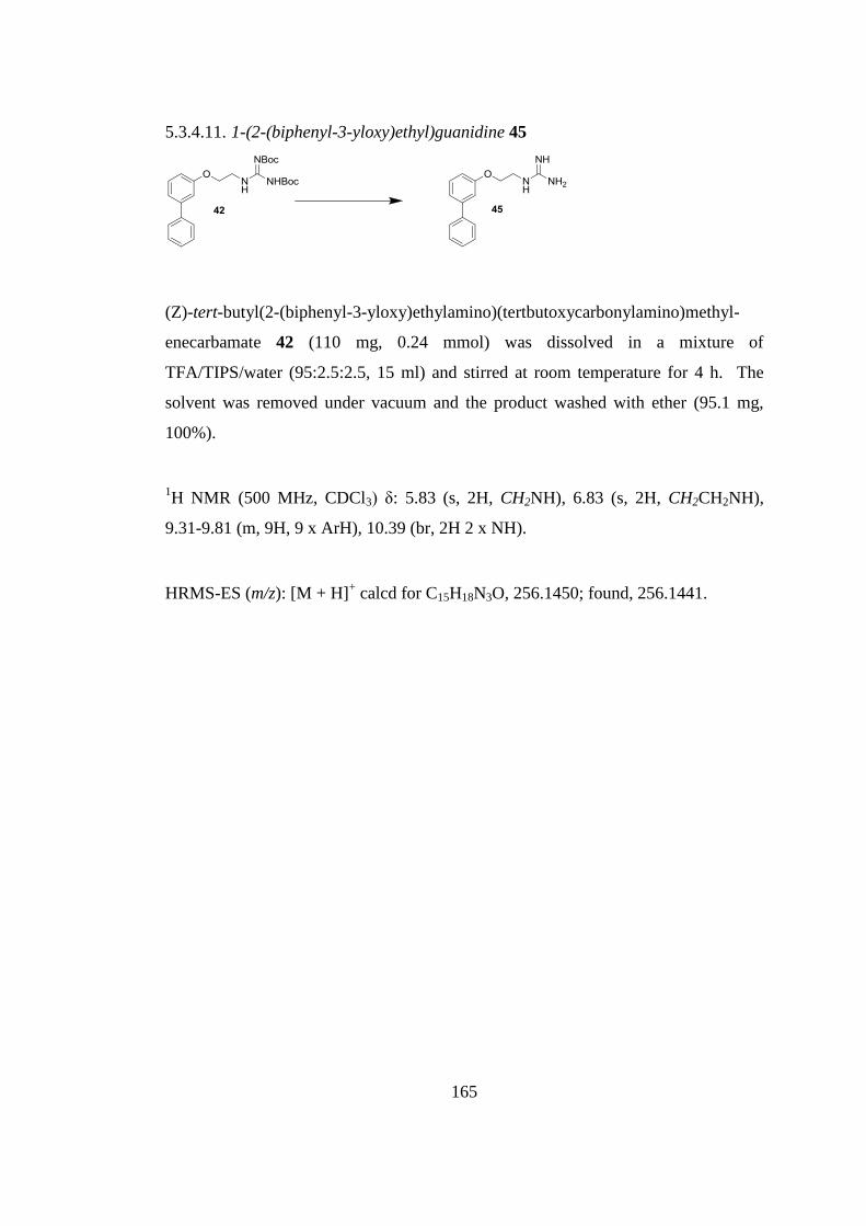
165
5.3.4.11. 1-(2-(biphenyl-3-yloxy)ethyl)guanidine 45
(Z)-tert-butyl(2-(biphenyl-3-yloxy)ethylamino)(tertbutoxycarbonylamino)methyl-
enecarbamate 42 (110 mg, 0.24 mmol) was dissolved in a mixture of
TFA/TIPS/water (95:2.5:2.5, 15 ml) and stirred at room temperature for 4 h. The
solvent was removed under vacuum and the product washed with ether (95.1 mg,
100%).
1H NMR (500 MHz, CDCl3) δ: 5.83 (s, 2H, CH2NH), 6.83 (s, 2H, CH2CH2NH),
9.31-9.81 (m, 9H, 9 x ArH), 10.39 (br, 2H 2 x NH).
HRMS-ES (m/z): [M + H]+ calcd for C15H18N3O, 256.1450; found, 256.1441.
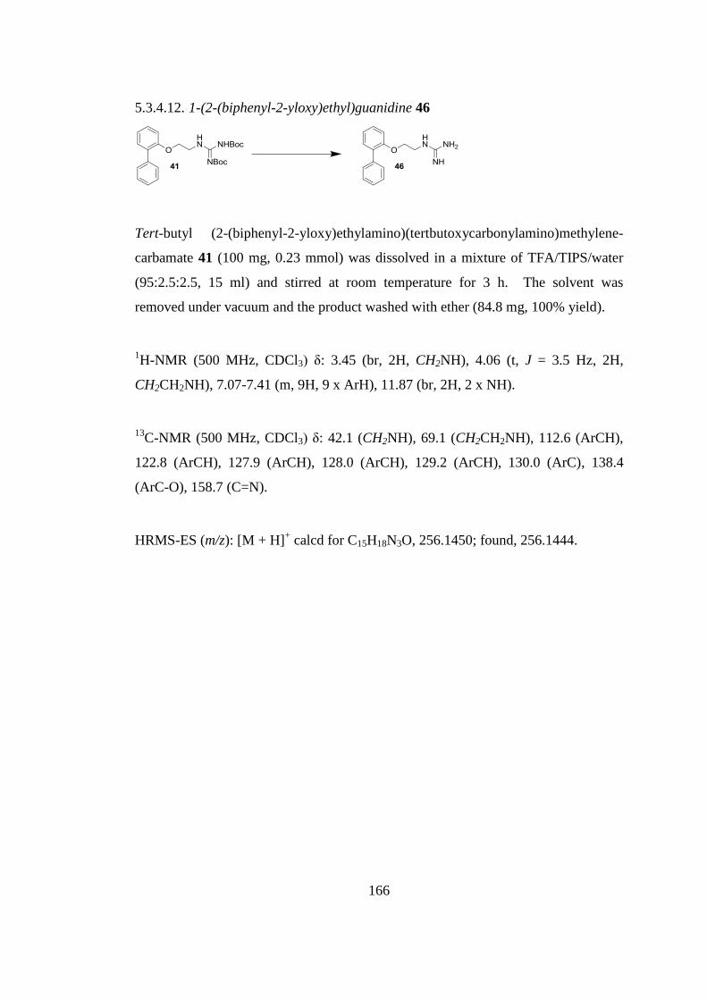
166
5.3.4.12. 1-(2-(biphenyl-2-yloxy)ethyl)guanidine 46
Tert-butyl (2-(biphenyl-2-yloxy)ethylamino)(tertbutoxycarbonylamino)methylene-
carbamate 41 (100 mg, 0.23 mmol) was dissolved in a mixture of TFA/TIPS/water
(95:2.5:2.5, 15 ml) and stirred at room temperature for 3 h. The solvent was
removed under vacuum and the product washed with ether (84.8 mg, 100% yield).
1H-NMR (500 MHz, CDCl3) δ: 3.45 (br, 2H, CH2NH), 4.06 (t, J = 3.5 Hz, 2H,
CH2CH2NH), 7.07-7.41 (m, 9H, 9 x ArH), 11.87 (br, 2H, 2 x NH).
13C-NMR (500 MHz, CDCl3) δ: 42.1 (CH2NH), 69.1 (CH2CH2NH), 112.6 (ArCH),
122.8 (ArCH), 127.9 (ArCH), 128.0 (ArCH), 129.2 (ArCH), 130.0 (ArC), 138.4
(ArC-O), 158.7 (C=N).
HRMS-ES (m/z): [M + H]+ calcd for C15H18N3O, 256.1450; found, 256.1444.
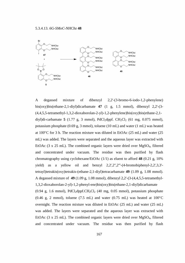
167
5.3.4.13. 6G-SMoC-NHCbz 48
A degassed mixture of dibenzyl 2,2'-(3-bromo-6-iodo-1,2-phenylene)
bis(oxy)bis(ethane-2,1-diyl)dicarbamate 47 (1 g, 1.5 mmol), dibenzyl 2,2'-(3-
(4,4,5,5-tetramethyl-1,3,2-dioxaborolan-2-yl)-1,2-phenylene)bis(oxy)bis(ethane-2,1-
diyl)di-carbamate 5 (1.77 g, 3 mmol), PdCl2dppf. CH2Cl2 (61 mg, 0.075 mmol),
potassium phosphate (0.69 g, 3 mmol), toluene (10 mL) and water (1 mL) was heated
at 100C for 3 h. The reaction mixture was diluted in EtOAc (25 mL) and water (25
mL) was added. The layers were separated and the aqueous layer was extracted with
EtOAc (3 x 25 mL). The combined organic layers were dried over MgSO4, filtered
and concentrated under vacuum. The residue was then purified by flash
chromatography using cyclohexane/EtOAc (1/1) as eluent to afford 48 (0.21 g, 10%
yield) as a yellow oil and benzyl 2,2',2'',2'''-(4-bromobiphenyl-2,2',3,3'-
tetrayl)tetrakis(oxy)tetrakis-(ethane-2,1-diyl)tetracarbamate 49 (1.09 g, 1.08 mmol).
A degassed mixture of 49 (1.09 g, 1.08 mmol), dibenzyl 2,2'-(3-(4,4,5,5-tetramethyl-
1,3,2-dioxaborolan-2-yl)-1,2-phenyl-ene)bis(oxy)bis(ethane-2,1-diyl)dicarbamate
(0.94 g, 1.6 mmol), PdCl2dppf.CH2Cl2 (40 mg, 0.05 mmol), potassium phosphate
(0.46 g, 2 mmol), toluene (7.5 mL) and water (0.75 mL) was heated at 100C
overnight. The reaction mixture was diluted in EtOAc (25 mL) and water (25 mL)
was added. The layers were separated and the aqueous layer was extracted with
EtOAc (3 x 25 mL). The combined organic layers were dried over MgSO4, filtered
and concentrated under vacuum. The residue was then purified by flash
168
chromatography using cyclohexane/EtOAc (1/1) as eluent to afford 48 (0.88 g, 59%
yield) as a yellow oil. The global yield was 48%.
1H-NMR (500 MHz, CDCl3) δ: 3.02-3.33 (m, 8H, 4 x CH2), 3.38-3.64 (m, 4H, 2 x
CH2), 3.67-4.05 (m, 12H, 6 x CH2), 4.92-5.21 (m, 12H, 6 x OCH2Ph), 6.66-7.63 (m,
38H, 38 x ArH).
13C-NMR (500 MHz, CDCl3) δ: 40.7 (CH2NH), 41.0 (CH2NH), 41.3 (CH2NH), 66.7
(OCH2Ph), 68.0 (CH2CH2NH), 72.4 (CH2CH2NH), 113.4 (ArCH), 123.4 (ArCH),
124.3 (ArCH), 126.1 (ArCH), 128.2 (ArCH), 128.5 (ArCH), 128.7 (ArCH), 132.4
(ArC), 136.4 (ArC), 136.7 (ArC), 151.6 (ArC-O), 156.3 (ArC-O), 156.5 (ArC-O),
171.2 (C=O).
HRMS-ES (m/z): (M + Na)+ calcd for C78H80N6O18Na, 1411.5427; found,
1411.5497.
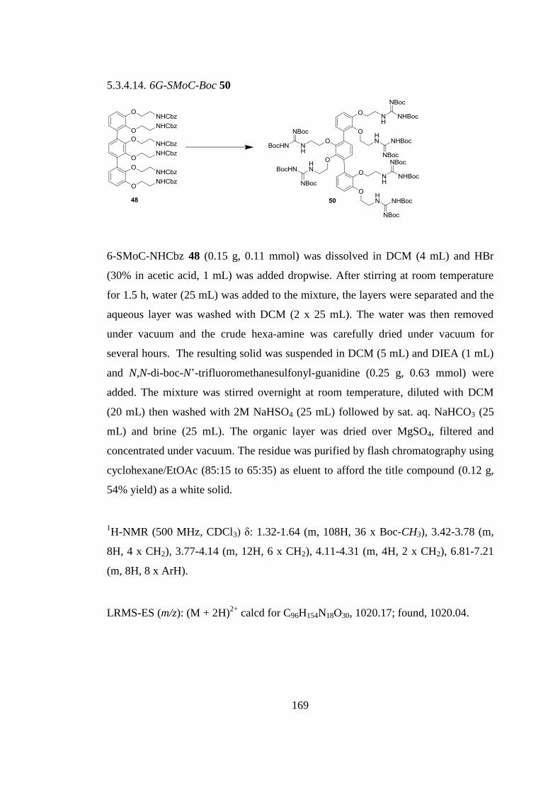
169
5.3.4.14. 6G-SMoC-Boc 50
6-SMoC-NHCbz 48 (0.15 g, 0.11 mmol) was dissolved in DCM (4 mL) and HBr
(30% in acetic acid, 1 mL) was added dropwise. After stirring at room temperature
for 1.5 h, water (25 mL) was added to the mixture, the layers were separated and the
aqueous layer was washed with DCM (2 x 25 mL). The water was then removed
under vacuum and the crude hexa-amine was carefully dried under vacuum for
several hours. The resulting solid was suspended in DCM (5 mL) and DIEA (1 mL)
and N,N-di-boc-N’-trifluoromethanesulfonyl-guanidine (0.25 g, 0.63 mmol) were
added. The mixture was stirred overnight at room temperature, diluted with DCM
(20 mL) then washed with 2M NaHSO4 (25 mL) followed by sat. aq. NaHCO3 (25
mL) and brine (25 mL). The organic layer was dried over MgSO4, filtered and
concentrated under vacuum. The residue was purified by flash chromatography using
cyclohexane/EtOAc (85:15 to 65:35) as eluent to afford the title compound (0.12 g,
54% yield) as a white solid.
1H-NMR (500 MHz, CDCl3) δ: 1.32-1.64 (m, 108H, 36 x Boc-CH3), 3.42-3.78 (m,
8H, 4 x CH2), 3.77-4.14 (m, 12H, 6 x CH2), 4.11-4.31 (m, 4H, 2 x CH2), 6.81-7.21
(m, 8H, 8 x ArH).
LRMS-ES (m/z): (M + 2H)2+
calcd for C96H154N18O30, 1020.17; found, 1020.04.
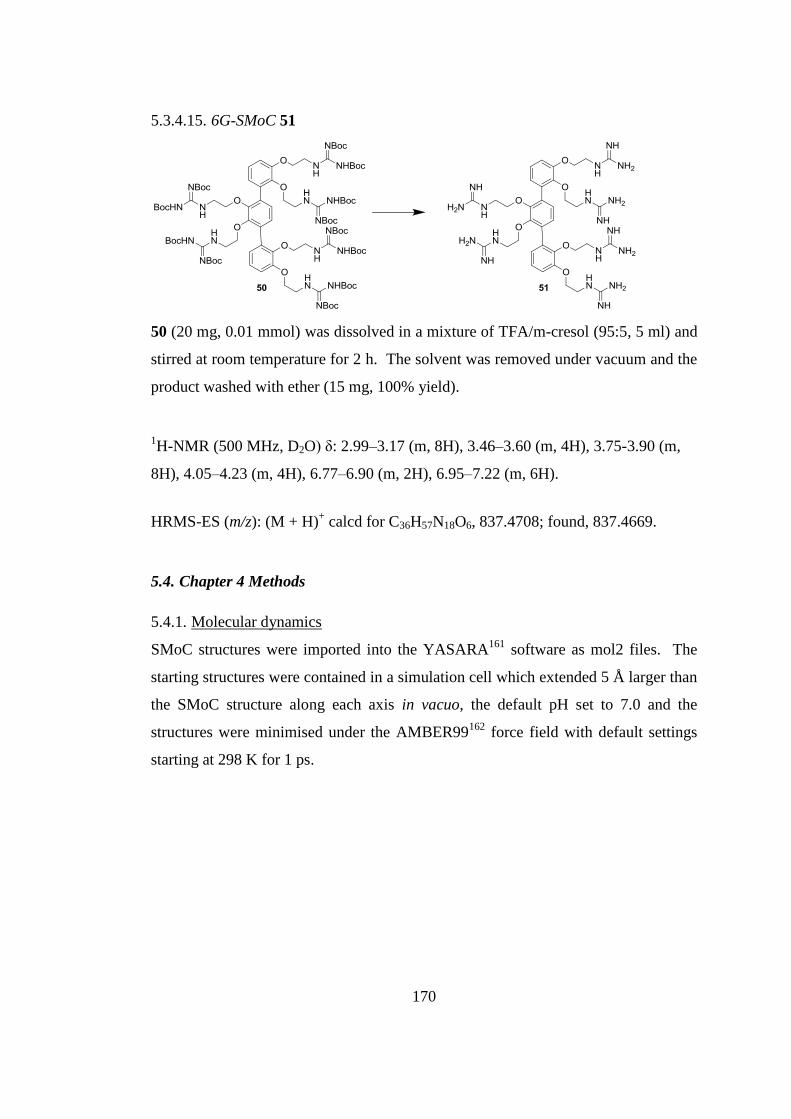
170
5.3.4.15. 6G-SMoC 51
50 (20 mg, 0.01 mmol) was dissolved in a mixture of TFA/m-cresol (95:5, 5 ml) and
stirred at room temperature for 2 h. The solvent was removed under vacuum and the
product washed with ether (15 mg, 100% yield).
1H-NMR (500 MHz, D2O) δ: 2.99–3.17 (m, 8H), 3.46–3.60 (m, 4H), 3.75-3.90 (m,
8H), 4.05–4.23 (m, 4H), 6.77–6.90 (m, 2H), 6.95–7.22 (m, 6H).
HRMS-ES (m/z): (M + H)+ calcd for C36H57N18O6, 837.4708; found, 837.4669.
5.4. Chapter 4 Methods
5.4.1. Molecular dynamics
SMoC structures were imported into the YASARA161
software as mol2 files. The
starting structures were contained in a simulation cell which extended 5 Å larger than
the SMoC structure along each axis in vacuo, the default pH set to 7.0 and the
structures were minimised under the AMBER99162
force field with default settings
starting at 298 K for 1 ps.
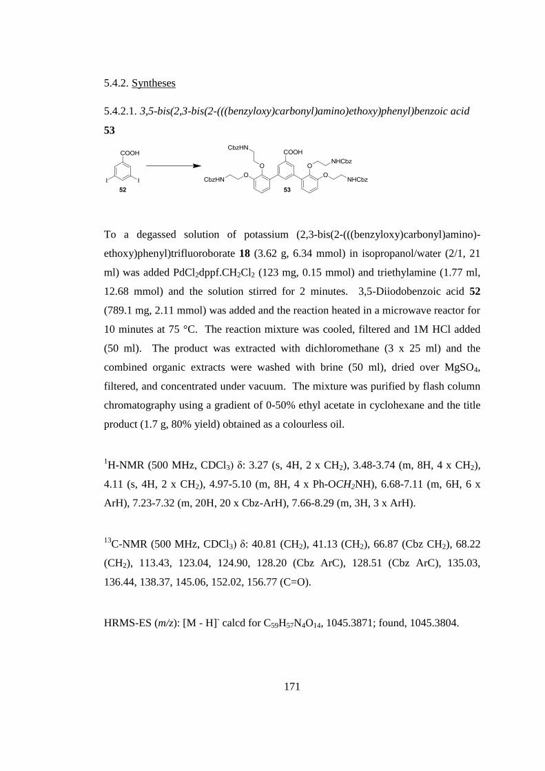
171
5.4.2. Syntheses
5.4.2.1. 3,5-bis(2,3-bis(2-(((benzyloxy)carbonyl)amino)ethoxy)phenyl)benzoic acid
53
To a degassed solution of potassium (2,3-bis(2-(((benzyloxy)carbonyl)amino)-
ethoxy)phenyl)trifluoroborate 18 (3.62 g, 6.34 mmol) in isopropanol/water (2/1, 21
ml) was added PdCl2dppf.CH2Cl2 (123 mg, 0.15 mmol) and triethylamine (1.77 ml,
12.68 mmol) and the solution stirred for 2 minutes. 3,5-Diiodobenzoic acid 52
(789.1 mg, 2.11 mmol) was added and the reaction heated in a microwave reactor for
10 minutes at 75 °C. The reaction mixture was cooled, filtered and 1M HCl added
(50 ml). The product was extracted with dichloromethane (3 x 25 ml) and the
combined organic extracts were washed with brine (50 ml), dried over MgSO4,
filtered, and concentrated under vacuum. The mixture was purified by flash column
chromatography using a gradient of 0-50% ethyl acetate in cyclohexane and the title
product (1.7 g, 80% yield) obtained as a colourless oil.
1H-NMR (500 MHz, CDCl3) δ: 3.27 (s, 4H, 2 x CH2), 3.48-3.74 (m, 8H, 4 x CH2),
4.11 (s, 4H, 2 x CH2), 4.97-5.10 (m, 8H, 4 x Ph-OCH2NH), 6.68-7.11 (m, 6H, 6 x
ArH), 7.23-7.32 (m, 20H, 20 x Cbz-ArH), 7.66-8.29 (m, 3H, 3 x ArH).
13C-NMR (500 MHz, CDCl3) δ: 40.81 (CH2), 41.13 (CH2), 66.87 (Cbz CH2), 68.22
(CH2), 113.43, 123.04, 124.90, 128.20 (Cbz ArC), 128.51 (Cbz ArC), 135.03,
136.44, 138.37, 145.06, 152.02, 156.77 (C=O).
HRMS-ES (m/z): [M - H]- calcd for C59H57N4O14, 1045.3871; found, 1045.3804.
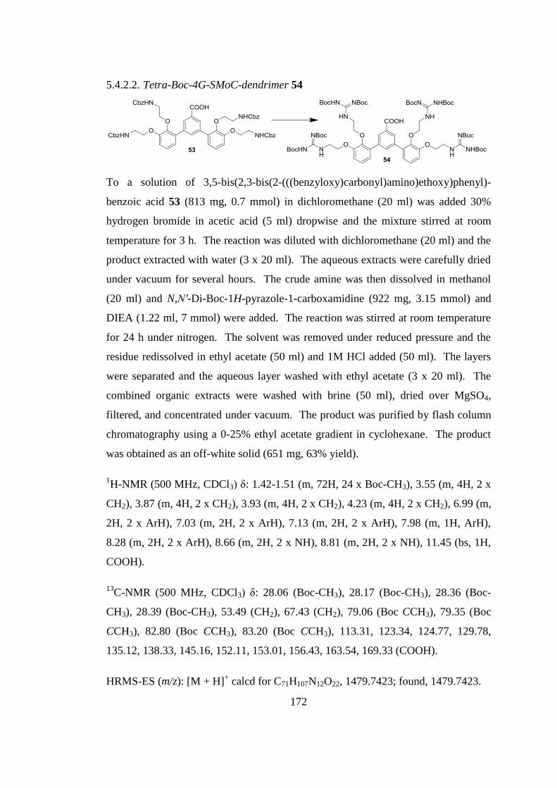
172
5.4.2.2. Tetra-Boc-4G-SMoC-dendrimer 54
To a solution of 3,5-bis(2,3-bis(2-(((benzyloxy)carbonyl)amino)ethoxy)phenyl)-
benzoic acid 53 (813 mg, 0.7 mmol) in dichloromethane (20 ml) was added 30%
hydrogen bromide in acetic acid (5 ml) dropwise and the mixture stirred at room
temperature for 3 h. The reaction was diluted with dichloromethane (20 ml) and the
product extracted with water (3 x 20 ml). The aqueous extracts were carefully dried
under vacuum for several hours. The crude amine was then dissolved in methanol
(20 ml) and N,N′-Di-Boc-1H-pyrazole-1-carboxamidine (922 mg, 3.15 mmol) and
DIEA (1.22 ml, 7 mmol) were added. The reaction was stirred at room temperature
for 24 h under nitrogen. The solvent was removed under reduced pressure and the
residue redissolved in ethyl acetate (50 ml) and 1M HCl added (50 ml). The layers
were separated and the aqueous layer washed with ethyl acetate (3 x 20 ml). The
combined organic extracts were washed with brine (50 ml), dried over MgSO4,
filtered, and concentrated under vacuum. The product was purified by flash column
chromatography using a 0-25% ethyl acetate gradient in cyclohexane. The product
was obtained as an off-white solid (651 mg, 63% yield).
1H-NMR (500 MHz, CDCl3) δ: 1.42-1.51 (m, 72H, 24 x Boc-CH3), 3.55 (m, 4H, 2 x
CH2), 3.87 (m, 4H, 2 x CH2), 3.93 (m, 4H, 2 x CH2), 4.23 (m, 4H, 2 x CH2), 6.99 (m,
2H, 2 x ArH), 7.03 (m, 2H, 2 x ArH), 7.13 (m, 2H, 2 x ArH), 7.98 (m, 1H, ArH),
8.28 (m, 2H, 2 x ArH), 8.66 (m, 2H, 2 x NH), 8.81 (m, 2H, 2 x NH), 11.45 (bs, 1H,
COOH).
13C-NMR (500 MHz, CDCl3) δ: 28.06 (Boc-CH3), 28.17 (Boc-CH3), 28.36 (Boc-
CH3), 28.39 (Boc-CH3), 53.49 (CH2), 67.43 (CH2), 79.06 (Boc CCH3), 79.35 (Boc
CCH3), 82.80 (Boc CCH3), 83.20 (Boc CCH3), 113.31, 123.34, 124.77, 129.78,
135.12, 138.33, 145.16, 152.11, 153.01, 156.43, 163.54, 169.33 (COOH).
HRMS-ES (m/z): [M + H]+ calcd for C71H107N12O22, 1479.7423; found, 1479.7423.
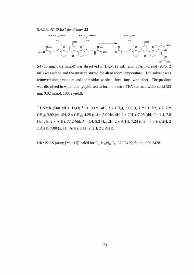
173
5.4.2.3. 4G-SMoC-dendrimer 55
54 (30 mg, 0.02 mmol) was dissolved in DCM (2 mL) and TFA/m-cresol (95/5, 2
mL) was added and the mixture stirred for 4h at room temperature. The solvent was
removed under vacuum and the residue washed three times with ether. The product
was dissolved in water and lyophilised to form the tetra-TFA salt as a white solid (23
mg, 0.02 mmol, 100% yield).
1H-NMR (500 MHz, D2O) δ: 3.13 (m, 4H, 2 x CH2), 3.65 (t, J = 5.0 Hz, 4H, 2 x
CH2), 3.92 (m, 4H, 2 x CH2), 4.25 (t, J = 5.0 Hz, 4H, 2 x CH2), 7.05 (dd, J = 1.4, 7.8
Hz, 2H, 2 x ArH), 7.12 (dd, J = 1.4, 8.3 Hz, 2H, 2 x ArH), 7.24 (t, J = 8.0 Hz, 2H, 2
x ArH), 7.80 (s, 1H, ArH), 8.11 (s, 2H, 2 x ArH).
HRMS-ES (m/z): [M + H]+ calcd for C31H43N12O6, 679.3429; found, 679.3428.
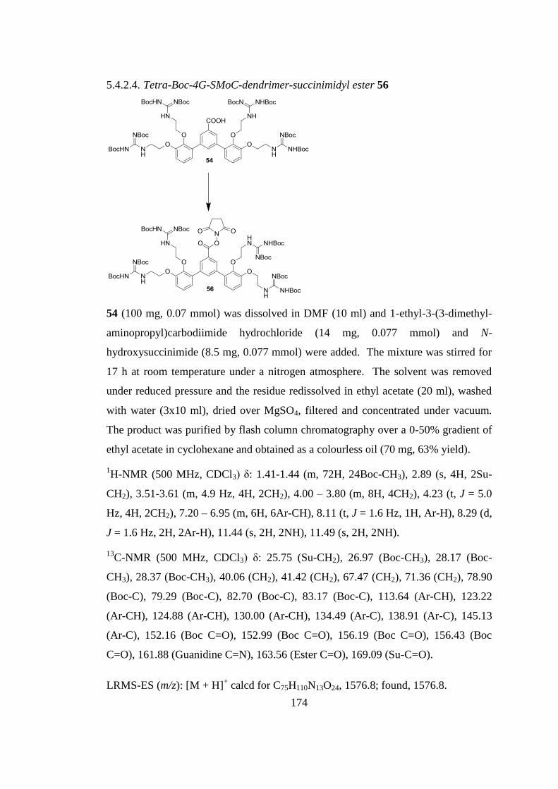
174
5.4.2.4. Tetra-Boc-4G-SMoC-dendrimer-succinimidyl ester 56
54 (100 mg, 0.07 mmol) was dissolved in DMF (10 ml) and 1-ethyl-3-(3-dimethyl-
aminopropyl)carbodiimide hydrochloride (14 mg, 0.077 mmol) and N-
hydroxysuccinimide (8.5 mg, 0.077 mmol) were added. The mixture was stirred for
17 h at room temperature under a nitrogen atmosphere. The solvent was removed
under reduced pressure and the residue redissolved in ethyl acetate (20 ml), washed
with water (3x10 ml), dried over MgSO4, filtered and concentrated under vacuum.
The product was purified by flash column chromatography over a 0-50% gradient of
ethyl acetate in cyclohexane and obtained as a colourless oil (70 mg, 63% yield).
1H-NMR (500 MHz, CDCl3) δ: 1.41-1.44 (m, 72H, 24Boc-CH3), 2.89 (s, 4H, 2Su-
CH2), 3.51-3.61 (m, 4.9 Hz, 4H, 2CH2), 4.00 – 3.80 (m, 8H, 4CH2), 4.23 (t, J = 5.0
Hz, 4H, 2CH2), 7.20 – 6.95 (m, 6H, 6Ar-CH), 8.11 (t, J = 1.6 Hz, 1H, Ar-H), 8.29 (d,
J = 1.6 Hz, 2H, 2Ar-H), 11.44 (s, 2H, 2NH), 11.49 (s, 2H, 2NH).
13C-NMR (500 MHz, CDCl3) δ: 25.75 (Su-CH2), 26.97 (Boc-CH3), 28.17 (Boc-
CH3), 28.37 (Boc-CH3), 40.06 (CH2), 41.42 (CH2), 67.47 (CH2), 71.36 (CH2), 78.90
(Boc-C), 79.29 (Boc-C), 82.70 (Boc-C), 83.17 (Boc-C), 113.64 (Ar-CH), 123.22
(Ar-CH), 124.88 (Ar-CH), 130.00 (Ar-CH), 134.49 (Ar-C), 138.91 (Ar-C), 145.13
(Ar-C), 152.16 (Boc C=O), 152.99 (Boc C=O), 156.19 (Boc C=O), 156.43 (Boc
C=O), 161.88 (Guanidine C=N), 163.56 (Ester C=O), 169.09 (Su-C=O).
LRMS-ES (m/z): [M + H]+ calcd for C75H110N13O24, 1576.8; found, 1576.8.
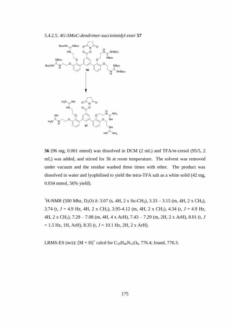
175
5.4.2.5. 4G-SMoC-dendrimer-succinimidyl ester 57
56 (96 mg, 0.061 mmol) was dissolved in DCM (2 mL) and TFA/m-cresol (95/5, 2
mL) was added, and stirred for 3h at room temperature. The solvent was removed
under vacuum and the residue washed three times with ether. The product was
dissolved in water and lyophilised to yield the tetra-TFA salt as a white solid (42 mg,
0.034 mmol, 56% yield).
1H-NMR (500 Mhz, D2O) δ: 3.07 (s, 4H, 2 x Su-CH2), 3.33 – 3.15 (m, 4H, 2 x CH2),
3.74 (t, J = 4.9 Hz, 4H, 2 x CH2), 3.95-4.12 (m, 4H, 2 x CH2), 4.34 (t, J = 4.9 Hz,
4H, 2 x CH2), 7.29 – 7.08 (m, 4H, 4 x ArH), 7.43 – 7.29 (m, 2H, 2 x ArH), 8.01 (t, J
= 1.5 Hz, 1H, ArH), 8.35 (t, J = 10.1 Hz, 2H, 2 x ArH).
LRMS-ES (m/z): [M + H]+ calcd for C35H46N13O8, 776.4; found, 776.3.
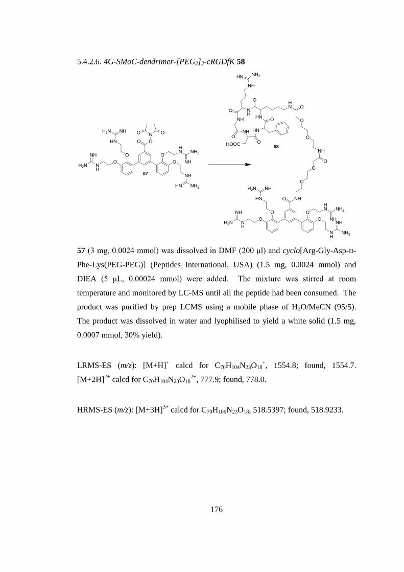
176
5.4.2.6. 4G-SMoC-dendrimer-[PEG2]2-cRGDfK 58
57 (3 mg, 0.0024 mmol) was dissolved in DMF (200 μl) and cyclo[Arg-Gly-Asp-D-
Phe-Lys(PEG-PEG)] (Peptides International, USA) (1.5 mg, 0.0024 mmol) and
DIEA (5 μL, 0.00024 mmol) were added. The mixture was stirred at room
temperature and monitored by LC-MS until all the peptide had been consumed. The
product was purified by prep LCMS using a mobile phase of H2O/MeCN (95/5).
The product was dissolved in water and lyophilised to yield a white solid (1.5 mg,
0.0007 mmol, 30% yield).
LRMS-ES (m/z): [M+H]+ calcd for C70H104N23O18
+, 1554.8; found, 1554.7.
[M+2H]2+
calcd for C70H104N23O182+
, 777.9; found, 778.0.
HRMS-ES (m/z): [M+3H]3+
calcd for C70H106N23O18, 518.5397; found, 518.9233.

177
CHAPTER 6
Discussion
178
6. Discussion
The extensive literature studies on the delivery of macromolecules such as proteins
and oligonucleotides to the intracellular environment demonstrate that this is a
prominent research question, with implications in the future of therapeutics. The
discovery of the RNAi mechanism has opened up the possibility of effective gene
therapy driven by siRNA-based drugs which act specifically and catalytically.
However the high surface charge and molecular weight of siRNA has so far
prohibited its delivery to its intracellular target. Viral delivery of gene therapies was
previously thought to be the answer to this challenge, however safety concerns have
led many researchers to seek alternative solutions. The discovery of CPPs showed
that macromolecules may be transported across cell membranes via an unknown non-
viral mechanism that appears to exist in a wide variety of cell types. However, CPPs
alone are insufficient for high efficiency, targeted delivery of biological drugs in vivo
due to their susceptibility to degradation, low bioavailability and lack of cell type
specificity. Therefore, much attention has been given to the development of
nanoparticles, built up from layers of different materials including lipids, peptides
and targeting ligands, acting in concert to navigate their nucleic acid cargoes to the
cytoplasms of their target cells.118
This approach has been successful, with the
application of nanoparticles demonstrated in vivo as demonstrated with SNALPs,87
and also in clinical trials.163
However, safety concerns still remain over
nanoparticles, with variation in the particle size and charge having large impacts on
their toxicity, as well as activation of the immune system.164
Therefore, the search
for a universal, safe and cost-effective delivery system for siRNA and other
macromolecular drugs continues.
The work presented in this thesis represents significant progress in the development
of the SMoC class of molecular transporters, which were first presented as protein
delivery agents in 2007.122
Tbe SMoCs may have significant advantages over
nanoparticle methods for the delivery of macromolecules, due to their low cost of
production and the possibility of overcoming the safety problems of more complex
vectors. The previously published synthetic route to the SMoCs has been
179
optimised,124
allowing higher yielding production of large quantities of compound at
lower cost. The global yield of 4G-SMoC using the new reagents (Scheme 9)
compared to the previous method (Scheme 1) has been increased 5-fold, and the
trifluoroborate salt has allowed long-term storage of a convenient intermediate.
Significant progress has also been made towards a new SMoC with a linker at the
meta position to the biphenyl bond, which has previously been shown to increase
3G-SMoC uptake.124
Previously, SMoCs had only been shown to internalise proteins and dyes into cells.
Since RNAi technology is becoming increasingly prevalent both as a research tool,
and also in clinical trials, it was decided to test the ability of SMoCs to transfect
siRNA into cells. Non-covalent complexes containing siRNA have been shown to be
successfully internalised using guanidine-containing CPPs,103-105, 107-108, 111, 114-117
and
are preferable over covalently linked siRNA conjugates since they may be formed
from non-modified siRNA and are easy to form, requiring no further synthetic steps.
Non-covelent complexes of CPPs and siRNA are thought to result in protection of
the siRNA from serum nucleases, as well as shielding the negative charges on the
siRNA, therefore decreasing renal clearance and allowing passage through the
lipophilic cell membrane. Stable SMoC-siRNA electrostatic complexes with
micromolar binding affinities have been formed, which show for the first time that
SMoCs may function as non-covalent as well as covalent macromolecular carriers in
the same way as CPPs such as TAT and penetratin. 4G-SMoC has a significantly
higher binding affinity than tetraarginine, perhaps due to the contribution from π-
cation interactions. It would be interesting to compare SMoCs to penetratin to
determine whether the amphipathic nature of the molecules account for this increased
nucleic acid binding.
Human cell lines were transfected with SMoC-siRNA complexes or Lipofectamine,
and 4G-SMoC resulted in a mRNA knockdown comparable to Lipofectamine. This
result shows that SMoCs are able to transport active siRNA into cells in order to
initiate RNAi-induced gene knockdown comparable to a commonly used laboratory

180
transfection reagent. This is particularly significant, as it expands the use of SMoCs
beyond proteins, demonstrating that they may have potential as universal
macromolecule delivery vectors for use in the laboratory and, with further
development, biological therapeutics. The fact that no such commercial reagent
which is able to transport both proteins and siRNA into cells currently exists makes
these results particularly interesting, and it is proposed that SMoCs may form a new
class of macromolecular transporter alongside viruses, CPPs and cationic liposomes.
In an attempt to enhance the uptake of SMoCs and increase the potency of the RNAi
knockdown, a new SMoC was designed using computational techniques to maximise
binding with cell surface GAGs, since it is proposed that GAG binding and
clustering is a precursor to macropinocytosis.38
This new compound represents the
first in a new class of modular SMoCs which are designed to maximise anion
complexation and synthesized using the new reagents and methods. It is also the first
SMoC that deviates significantly from the standard biphenyl ring system that was
modelled on a alpha-helical arginine-containing peptide. The high-yielding three-
step synthetic route demonstrates how new compounds can easily be assembled
using the new chemistry. In addition, the ability to form an activated ester of the
final compound enables the new SMoC reagent to be stored and coupled to an
appropriate cargo with ease at a later date. The mRNA knockdown produced using
the new compound was not significantly larger than using the original 4G-SMoC, but
was still comparable to Lipofectamine, confirming that these compounds may be
used as an alternative to the currently available transfection reagents, particularly if
they are able to overcome the difficulties of the current reagents, such as the high
cytotoxicity of cationic liposome-based compounds.80
The new compound has also
been shown to be successfully coupled to a targeting peptide. The targeting of
siRNA to specific cells is a key feature of successful delivery in vivo, where to
achieve effective gene silencing at low dosage, specificity is vital. Targeting ligands
are therefore considered to be an important component of siRNA delivery systems118
and several such ligands have been identified for a variety of disease states, including
tumour cells.165
The new SMoC compound fulfills this criteria of siRNA delivery
181
systems, therby increasing the potential of SMoCs for use as macromolecular drug
transporters.
6.1. Future Work
The work presented in this thesis has opened up further avenues of investigation for
the development of SMoCs, both in the short term and long term. The synthesis of
the dendrimer compound has demonstrated that modular SMoC compounds can
easily be synthesized by coupling the trifluoroborate salt to aryl halides. This
technique can be used to produce even larger macromolecular SMoC compounds,
which may be used to encapsulate hydrophilic macromolecules for cell
internalisation, in the same way that nanoparticles are formed using polymeric
compounds such as cyclodextrin and PEI for siRNA delivery. For example, using
the cheap, commercially available 1,3,5-tris(bromophenyl)benzene starting material,
a large dendrimeric SMoC compound displaying six guanidine groups may be
synthesized (Scheme 23). Such a molecule would have an increased number of
positive charges as well as increased hydrophobicity, which may result in a stronger
π-cation stabilising effect. In addition, by increasing the size of the SMoC molecule
and the number charges, the compound may have a higher binding affinity for
siRNA and cell surface GAGs, as well as being able to bind to the cell surface over a
greater area, therefore possibly increasing uptake due to the lower entropic barrier.
Analysis of the binding affinity of various SMoC compounds to heparin, including
the new 4G-SMoC-Dr, is currently in progress using a surface plasmon resonance
assay. The results from these experiments will indicate if the increased distance
between guanidine groups is related to its affinity for anionic GAGs, as
hypothesised.
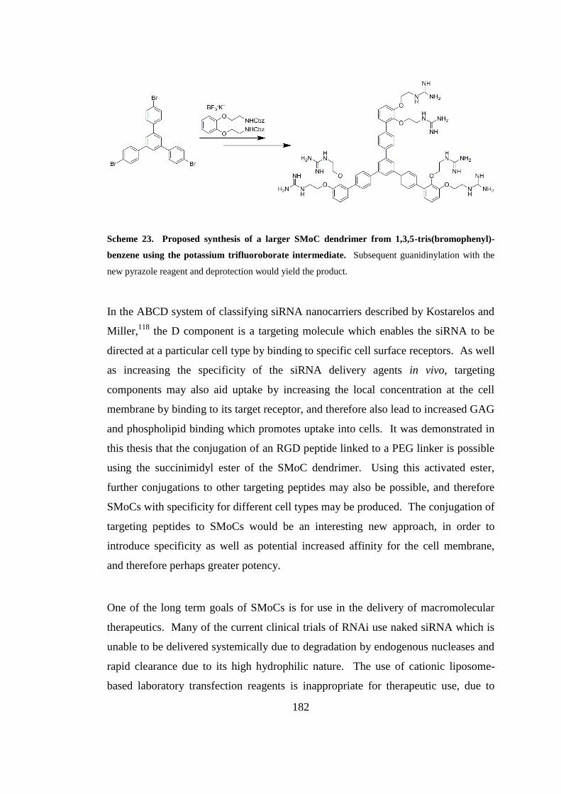
182
Scheme 23. Proposed synthesis of a larger SMoC dendrimer from 1,3,5-tris(bromophenyl)-
benzene using the potassium trifluoroborate intermediate. Subsequent guanidinylation with the
new pyrazole reagent and deprotection would yield the product.
In the ABCD system of classifying siRNA nanocarriers described by Kostarelos and
Miller,118
the D component is a targeting molecule which enables the siRNA to be
directed at a particular cell type by binding to specific cell surface receptors. As well
as increasing the specificity of the siRNA delivery agents in vivo, targeting
components may also aid uptake by increasing the local concentration at the cell
membrane by binding to its target receptor, and therefore also lead to increased GAG
and phospholipid binding which promotes uptake into cells. It was demonstrated in
this thesis that the conjugation of an RGD peptide linked to a PEG linker is possible
using the succinimidyl ester of the SMoC dendrimer. Using this activated ester,
further conjugations to other targeting peptides may also be possible, and therefore
SMoCs with specificity for different cell types may be produced. The conjugation of
targeting peptides to SMoCs would be an interesting new approach, in order to
introduce specificity as well as potential increased affinity for the cell membrane,
and therefore perhaps greater potency.
One of the long term goals of SMoCs is for use in the delivery of macromolecular
therapeutics. Many of the current clinical trials of RNAi use naked siRNA which is
unable to be delivered systemically due to degradation by endogenous nucleases and
rapid clearance due to its high hydrophilic nature. The use of cationic liposome-
based laboratory transfection reagents is inappropriate for therapeutic use, due to
183
their high toxicity, and so there no widely used standard delivery vector for use in
vivo. The development of a small molecule delivery vector based on SMoCs for
siRNA and other macromolecules would be a significant achievement, and would
offer many advantages over other compounds such as CPPs. For example, the ease
and low cost of production resulting from the new SMoC synthetic modifications, as
well as their ability to be coupled to a wide range of macromolecules either via the
original disulfide linker, or peptide coupling via the new activated ester. The use of
targeting peptides with SMoCs will allow for increased potency in vivo and the
targeting of specific cell types and disease states, such as tumour cells
overexpressing certain integrins. Therefore, in vivo testing of SMoCs is an important
aim for the future of this work, and ultimately may allow SMoCs to be used for a
wide range of applications.

184
Bibliography
185
Bibliography
1. Leader, B., Baca, Q. J., Golan, D. E. Protein therapeutics: A summary and
pharmacological classification. Nat. Rev. Drug Discov. 2008, 7 (1), 21-39.
2. Leaman, D. W. Recent progress in oligonucleotide therapeutics: antisense to
aptamers. Expert Opin. Drug Disc. 2008, 3 (9), 997-1009.
3. Carter, P. J. Introduction to current and future protein therapeutics: A protein
engineering perspective. Exp. Cell Res. 2011, 317 (9), 1261-1269.
4. Waldmann, T. A. Immunotherapy: past, present and future. Nat. Med. 2003, 9
(3), 269-277.
5. Reichert, J. M. Antibody-based therapeutics to watch in 2011. mAbs 2011, 3
(1), 76-99.
6. Fire, A., Xu, S. Q., Montgomery, M. K., Kostas, S. A., Driver, S. E., Mello,
C. C. Potent and specific genetic interference by double-stranded RNA in
Caenorhabditis elegans. Nature 1998, 391 (6669), 806-811.
7. de Fougerolles, A., Vornlocher, H. P., Maraganore, J., Lieberman, J.
Interfering with disease: a progress report on siRNA-based therapeutics. Nat. Rev.
Drug Discov. 2007, 6 (6), 443-453.
8. Guo, J., Fisher, K. A., Darcy, R., Cryan, J. F., O'Driscoll, C. Therapeutic
targeting in the silent era: advances in non-viral siRNA delivery. Mol. Biosyst. 2010,
6 (7), 1143-61.
9. Giezen, T. J., Mantel-Teeuwisse, A. K., Straus, S. M. J. M., Schellekens, H.,
Leufkens, H. G. M., Egberts, A. C. G. Safety-Related Regulatory Actions for
Biologicals Approved in the United States and the European Union. J. Am. Med.
Assoc. 2008, 300 (16), 1887-1896.
10. Juliano, R. Challenges to macromolecular drug delivery. Biochem. Soc.
Trans. 2007, 35, 41-43.
11. Lipinski, C. A., Lombardo, F., Dominy, B. W., Feeney, P. J. Experimental
and computational approaches to estimate solubility and permeability in drug
discovery and development settings. Adv. Drug Del. Rev. 2001, 46 (1-3), 3-26.
12. Belting, M., Wittrup, A. Macromolecular drug delivery: basic principles and
therapeutic applications. Mol. Biotechnol. 2009, 43 (1), 89-94.
13. Frankel, A. D., Pabo, C. O. Cellular uptake of the tat protein from human
immunodeficiency virus. Cell 1988, 55 (6), 1189-93.
186
14. Joliot, A., Pernelle, C., Deagostini-Bazin, H., Prochiantz, A. Antennapedia
homeobox peptide regulates neural morphogenesis. Proc. Natl. Acad. Sci. USA 1991,
88 (5), 1864-8.
15. Derossi, D., Joliot, A. H., Chassaing, G., Prochiantz, A. The third helix of the
Antennapedia homeodomain translocates through biological membranes. J. Biol.
Chem. 1994, 269 (14), 10444-10450.
16. Vivès, E., Brodin, P., Lebleu, B. A truncated HIV-1 Tat protein basic domain
rapidly translocates through the plasma membrane and accumulates in the cell
nucleus. J. Biol. Chem. 1997, 272 (25), 16010-7.
17. Morris, M. C., Vidal, P., Chaloin, L., Heitz, F., Divita, G. A new peptide
vector for efficient delivery of oligonucleotides into mammalian cells. Nucleic Acids
Res. 1997, 25 (14), 2730-2736.
18. Schwarze, S. R., Ho, A., Vocero-Akbani, A., Dowdy, S. F. In vivo Protein
Transduction: Delivery of a Biologically Active Protein into the Mouse. Science
1999, 285 (5433), 1569-1572.
19. Morris, M. C., Depollier, J., Mery, J., Heitz, F., Divita, G. A peptide carrier
for the delivery of biologically active proteins into mammalian cells. Nat.
Biotechnol. 2001, 19 (12), 1173-1176.
20. Fawell, S., Seery, J., Daikh, Y., Moore, C., Chen, L. L., Pepinsky, B.,
Barsoum, J. Tat-Mediated Delivery of Heterologous Proteins into Cells. Proc. Natl.
Acad. Sci. USA 1994, 91 (2), 664-668.
21. Ruan, G., Agrawal, A., Marcus, A. I., Nie, S. Imaging and tracking of tat
peptide-conjugated quantum dots in living cells: new insights into nanoparticle
uptake, intracellular transport, and vesicle shedding. J. Am. Chem. Soc. 2007, 129
(47), 14759-14766.
22. Abes, R., Arzumanov, A. A., Moulton, H. M., Abes, S., Lvanciva, G. D.,
Lversen, P. L., Gait, M. J., Lebleu, B. Cell-penetrating-peptide-based delivery of
oligonucleotides: an overview. Biochem. Soc. Trans. 2007, 35, 775-779.
23. Juliano, R., Alam, M. R., Dixit, V., Kang, H. Mechanisms and strategies for
effective delivery of antisense and siRNA oligonucleotides. Nucleic Acids Res. 2008,
36 (12), 4158-4171.
24. Derossi, D., Calvet, S., Trembleau, A., Brunissen, A., Chassaing, G.,
Prochiantz, A. Cell Internalization of the Third Helix of the Antennapedia
Homeodomain Is Receptor-independent. J. Biol. Chem. 1996, 271 (30), 18188-
18193.
25. Richard, J. P., Melikov, K., Vives, E., Ramos, C., Verbeure, B., Gait, M. J.,
Chernomordik, L. V., Lebleu, B. Cell-penetrating peptides - A reevaluation of the
mechanism of cellular uptake. J. Biol. Chem. 2003, 278 (1), 585-590.
187
26. Lundberg, M., Wikstrom, S., Johansson, M. Cell Surface Adherence and
Endocytosis of Protein Transduction Domains. Mol. Ther. 2003, 8 (1), 143-150.
27. Wadia, J. S., Stan, R. V., Dowdy, S. F. Transducible TAT-HA fusogenic
peptide enhances escape of TAT-fusion proteins after lipid raft macropinocytosis.
Nat. Med. 2004, 10 (3), 310-315.
28. Nakase, I., Niwa, M., Takeuchi, T., Sonomura, K., Kawabata, N., Koike, Y.,
Takehashi, M., Tanaka, S., Ueda, K., Simpson, J. C., Jones, A. T., Sugiura, Y.,
Futaki, S. Cellular Uptake of Arginine-Rich Peptides: Roles for Macropinocytosis
and Actin Rearrangement. Mol. Ther. 2004, 10 (6), 1011-1022.
29. Jiao, C. Y., Delaroche, D., Burlina, F., Alves, I. D., Chassaing, G., Sagan, S.
Translocation and Endocytosis for Cell-penetrating Peptide Internalization. J. Biol.
Chem. 2009, 284 (49), 33957-33965.
30. Guterstam, P., Madani, F., Hirose, H., Takeuchi, T., Futaki, S., El
Andaloussi, S., Gräslund, A., Langel, U. Elucidating cell-penetrating peptide
mechanisms of action for membrane interaction, cellular uptake, and translocation
utilizing the hydrophobic counter-anion pyrenebutyrate. Biochim. Biophys. Acta
2009, 1788 (12), 2509-17.
31. Nakase, I., Tadokoro, A., Kawabata, N., Takeuchi, T., Katoh, H., Hiramoto,
K., Negishi, M., Nomizu, M., Sugiura, Y., Futaki, S. Interaction of Arginine-Rich
Peptides with Membrane-Associated Proteoglycans Is Crucial for Induction of Actin
Organization and Macropinocytosis†. Biochemistry 2006, 46 (2), 492-501.
32. Simon, M. J., Gao, S., Kang, W. H., Banta, S., Morrison, B. TAT-mediated
intracellular protein delivery to primary brain cells is dependent on
glycosaminoglycan expression. Biotechnol. Bioeng. 2009, 104 (1), 10-9.
33. Patel, L. N., Zaro, J. L., Shen, W. C. Cell penetrating peptides: Intracellular
pathways and pharmaceutical perspectives. Pharm. Res. 2007, 24 (11), 1977-1992.
34. Fuchs, S. M., Raines, R. T. Pathway for Polyarginine Entry into Mammalian
Cells†. Biochemistry 2004, 43 (9), 2438-2444.
35. Gerbal-chaloin, S., Gondeau, C., Aldrian-herrada, G., Heitz, F., Gauthier-
rouvière, C., Divita, G. First step of the cell-penetrating peptide mechanism involves
Rac1 GTPase-dependent actin-network remodelling. Biol. Cell 2007, 099 (4), 223-
238.
36. Couchman, J. R. Syndecans: proteoglycan regulators of cell-surface
microdomains? Nat. Rev. Mol. Cell Biol. 2003, 4 (12), 926-938.
37. Conner, S. D., Schmid, S. L. Regulated portals of entry into the cell. Nature
2003, 422 (6927), 37-44.
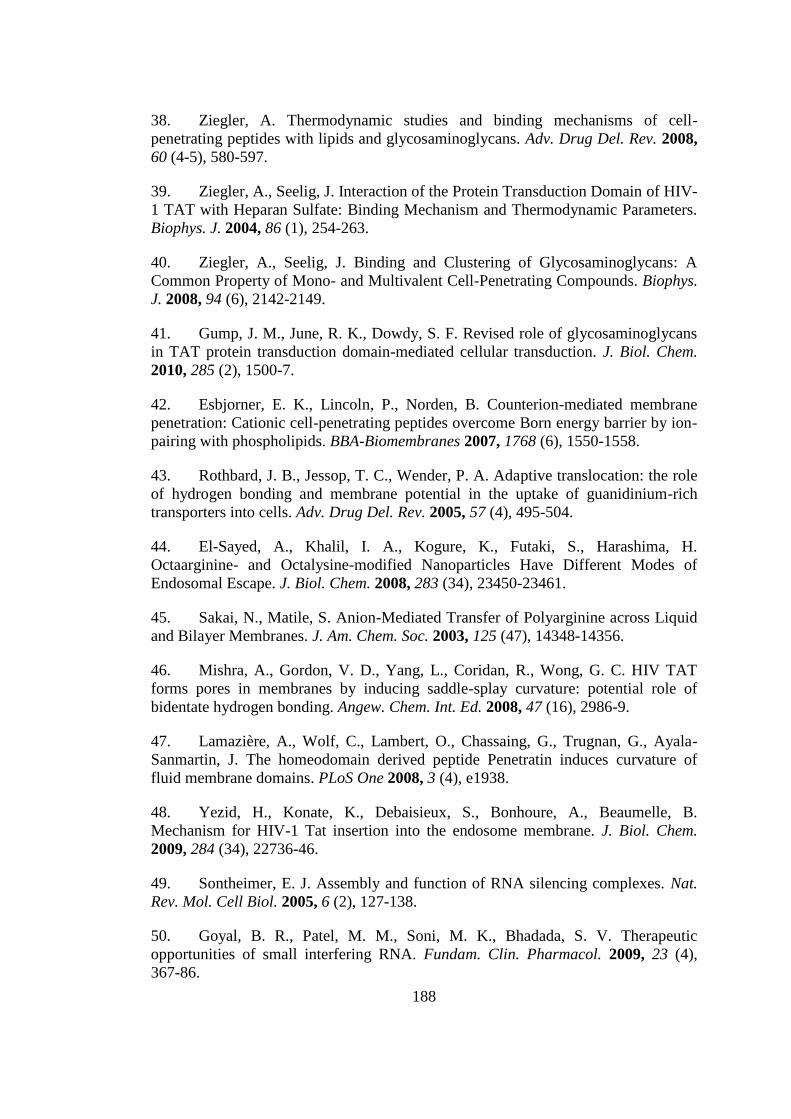
188
38. Ziegler, A. Thermodynamic studies and binding mechanisms of cell-
penetrating peptides with lipids and glycosaminoglycans. Adv. Drug Del. Rev. 2008,
60 (4-5), 580-597.
39. Ziegler, A., Seelig, J. Interaction of the Protein Transduction Domain of HIV-
1 TAT with Heparan Sulfate: Binding Mechanism and Thermodynamic Parameters.
Biophys. J. 2004, 86 (1), 254-263.
40. Ziegler, A., Seelig, J. Binding and Clustering of Glycosaminoglycans: A
Common Property of Mono- and Multivalent Cell-Penetrating Compounds. Biophys.
J. 2008, 94 (6), 2142-2149.
41. Gump, J. M., June, R. K., Dowdy, S. F. Revised role of glycosaminoglycans
in TAT protein transduction domain-mediated cellular transduction. J. Biol. Chem.
2010, 285 (2), 1500-7.
42. Esbjorner, E. K., Lincoln, P., Norden, B. Counterion-mediated membrane
penetration: Cationic cell-penetrating peptides overcome Born energy barrier by ion-
pairing with phospholipids. BBA-Biomembranes 2007, 1768 (6), 1550-1558.
43. Rothbard, J. B., Jessop, T. C., Wender, P. A. Adaptive translocation: the role
of hydrogen bonding and membrane potential in the uptake of guanidinium-rich
transporters into cells. Adv. Drug Del. Rev. 2005, 57 (4), 495-504.
44. El-Sayed, A., Khalil, I. A., Kogure, K., Futaki, S., Harashima, H.
Octaarginine- and Octalysine-modified Nanoparticles Have Different Modes of
Endosomal Escape. J. Biol. Chem. 2008, 283 (34), 23450-23461.
45. Sakai, N., Matile, S. Anion-Mediated Transfer of Polyarginine across Liquid
and Bilayer Membranes. J. Am. Chem. Soc. 2003, 125 (47), 14348-14356.
46. Mishra, A., Gordon, V. D., Yang, L., Coridan, R., Wong, G. C. HIV TAT
forms pores in membranes by inducing saddle-splay curvature: potential role of
bidentate hydrogen bonding. Angew. Chem. Int. Ed. 2008, 47 (16), 2986-9.
47. Lamazière, A., Wolf, C., Lambert, O., Chassaing, G., Trugnan, G., Ayala-
Sanmartin, J. The homeodomain derived peptide Penetratin induces curvature of
fluid membrane domains. PLoS One 2008, 3 (4), e1938.
48. Yezid, H., Konate, K., Debaisieux, S., Bonhoure, A., Beaumelle, B.
Mechanism for HIV-1 Tat insertion into the endosome membrane. J. Biol. Chem.
2009, 284 (34), 22736-46.
49. Sontheimer, E. J. Assembly and function of RNA silencing complexes. Nat.
Rev. Mol. Cell Biol. 2005, 6 (2), 127-138.
50. Goyal, B. R., Patel, M. M., Soni, M. K., Bhadada, S. V. Therapeutic
opportunities of small interfering RNA. Fundam. Clin. Pharmacol. 2009, 23 (4),
367-86.
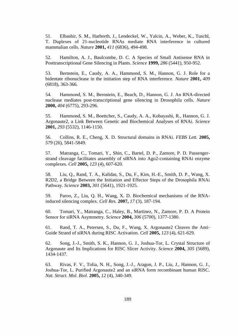
189
51. Elbashir, S. M., Harborth, J., Lendeckel, W., Yalcin, A., Weber, K., Tuschl,
T. Duplexes of 21-nucleotide RNAs mediate RNA interference in cultured
mammalian cells. Nature 2001, 411 (6836), 494-498.
52. Hamilton, A. J., Baulcombe, D. C. A Species of Small Antisense RNA in
Posttranscriptional Gene Silencing in Plants. Science 1999, 286 (5441), 950-952.
53. Bernstein, E., Caudy, A. A., Hammond, S. M., Hannon, G. J. Role for a
bidentate ribonuclease in the initiation step of RNA interference. Nature 2001, 409
(6818), 363-366.
54. Hammond, S. M., Bernstein, E., Beach, D., Hannon, G. J. An RNA-directed
nuclease mediates post-transcriptional gene silencing in Drosophila cells. Nature
2000, 404 (6775), 293-296.
55. Hammond, S. M., Boettcher, S., Caudy, A. A., Kobayashi, R., Hannon, G. J.
Argonaute2, a Link Between Genetic and Biochemical Analyses of RNAi. Science
2001, 293 (5532), 1146-1150.
56. Collins, R. E., Cheng, X. D. Structural domains in RNAi. FEBS Lett. 2005,
579 (26), 5841-5849.
57. Matranga, C., Tomari, Y., Shin, C., Bartel, D. P., Zamore, P. D. Passenger-
strand cleavage facilitates assembly of siRNA into Ago2-containing RNAi enzyme
complexes. Cell 2005, 123 (4), 607-620.
58. Liu, Q., Rand, T. A., Kalidas, S., Du, F., Kim, H.-E., Smith, D. P., Wang, X.
R2D2, a Bridge Between the Initiation and Effector Steps of the Drosophila RNAi
Pathway. Science 2003, 301 (5641), 1921-1925.
59. Paroo, Z., Liu, Q. H., Wang, X. D. Biochemical mechanisms of the RNA-
induced silencing complex. Cell Res. 2007, 17 (3), 187-194.
60. Tomari, Y., Matranga, C., Haley, B., Martinez, N., Zamore, P. D. A Protein
Sensor for siRNA Asymmetry. Science 2004, 306 (5700), 1377-1380.
61. Rand, T. A., Petersen, S., Du, F., Wang, X. Argonaute2 Cleaves the Anti-
Guide Strand of siRNA during RISC Activation. Cell 2005, 123 (4), 621-629.
62. Song, J.-J., Smith, S. K., Hannon, G. J., Joshua-Tor, L. Crystal Structure of
Argonaute and Its Implications for RISC Slicer Activity. Science 2004, 305 (5689),
1434-1437.
63. Rivas, F. V., Tolia, N. H., Song, J.-J., Aragon, J. P., Liu, J., Hannon, G. J.,
Joshua-Tor, L. Purified Argonaute2 and an siRNA form recombinant human RISC.
Nat. Struct. Mol. Biol. 2005, 12 (4), 340-349.
190
64. Jiang, F., Ye, X., Liu, X., Fincher, L., McKearin, D., Liu, Q. Dicer-1 and
R3D1-L catalyze microRNA maturation in Drosophila. Genes Dev. 2005, 19 (14),
1674-1679.
65. Okamura, K., Ishizuka, A., Siomi, H., Siomi, M. C. Distinct roles for
Argonaute proteins in small RNA-directed RNA cleavage pathways. Genes Dev.
2004, 18 (14), 1655-1666.
66. Meister, G., Tuschl, T. Mechanisms of gene silencing by double-stranded
RNA. Nature 2004, 431 (7006), 343-349.
67. Shim, M. S., Kwon, Y. J. Efficient and targeted delivery of siRNA in vivo.
FEBS J. 2010, 277 (23), 4814-27.
68. Whitehead, K., Langer, R., Anderson, D. Knocking down barriers: advances
in siRNA delivery. Nat. Rev. Drug Discov. 2009, 8 (2), 129-138.
69. Barquinero, J., Eixarch, H., Perez-Melgosa, M. Retroviral vectors: new
applications for an old tool. Gene Ther. 2004, 11 (S1), S3-S9.
70. Soutschek, J., Akinc, A., Bramlage, B., Charisse, K., Constien, R.,
Donoghue, M., Elbashir, S., Geick, A., Hadwiger, P., Harborth, J., John, M.,
Kesavan, V., Lavine, G., Pandey, R. K., Racie, T., Rajeev, K. G., Rohl, I.,
Toudjarska, I., Wang, G., Wuschko, S., Bumcrot, D., Koteliansky, V., Limmer, S.,
Manoharan, M., Vornlocher, H.-P. Therapeutic silencing of an endogenous gene by
systemic administration of modified siRNAs. Nature 2004, 432 (7014), 173-178.
71. Wolfrum, C., Shi, S., Jayaprakash, K. N., Jayaraman, M., Wang, G., Pandey,
R. K., Rajeev, K. G., Nakayama, T., Charrise, K., Ndungo, E. M., Zimmermann, T.,
Koteliansky, V., Manoharan, M., Stoffel, M. Mechanisms and optimization of in vivo
delivery of lipophilic siRNAs. Nat. Biotechnol. 2007, 25 (10), 1149-1157.
72. Dassie, J. P., Liu, X.-y., Thomas, G. S., Whitaker, R. M., Thiel, K. W.,
Stockdale, K. R., Meyerholz, D. K., McCaffrey, A. P., McNamara, J. O.,
Giangrande, P. H. Systemic administration of optimized aptamer-siRNA chimeras
promotes regression of PSMA-expressing tumors. Nat. Biotechnol. 2009, 27 (9),
839-846.
73. Felgner, P. L., Gadek, T. R., Holm, M., Roman, R., Chan, H. W., Wenz, M.,
Northrop, J. P., Ringold, G. M., Danielsen, M. Lipofection: a highly efficient, lipid-
mediated DNA-transfection procedure. Proc. Natl. Acad. Sci. USA 1987, 84 (21),
7413-7.
74. Malone, R. W., Felgner, P. L., Verma, I. M. Cationic liposome-mediated
RNA transfection. Proc. Natl. Acad. Sci. USA 1989, 86 (16), 6077-81.
75. Behr, J. P., Demeneix, B., Loeffler, J. P., Perez-Mutul, J. Efficient gene
transfer into mammalian primary endocrine cells with lipopolyamine-coated DNA.
Proc. Natl. Acad. Sci. USA 1989, 86 (18), 6982-6.
191
76. Gao, X., Huang, L. A novel cationic liposome reagent for efficient
transfection of mammalian cells. Biochem. Biophys. Res. Commun. 1991, 179 (1),
280-5.
77. Rose, J. K., Buonocore, L., Whitt, M. A. A new cationic liposome reagent
mediating nearly quantitative transfection of animal cells. BioTechniques 1991, 10
(4), 520-5.
78. San, H., Yang, Z. Y., Pompili, V. J., Jaffe, M. L., Plautz, G. E., Xu, L.,
Felgner, J. H., Wheeler, C. J., Felgner, P. L., Gao, X. Safety and short-term toxicity
of a novel cationic lipid formulation for human gene therapy. Hum. Gene Ther. 1993,
4 (6), 781-8.
79. Omidi, Y., Hollins, A. J., Benboubetra, M., Drayton, R., Benter, I. F., Akhtar,
S. Toxicogenomics of Non-viral Vectors for Gene Therapy: A Microarray Study of
Lipofectin- and Oligofectamine-induced Gene Expression Changes in Human
Epithelial Cells. J. Drug Targeting 2003, 11 (6), 311-323.
80. Kedmi, R., Ben-Arie, N., Peer, D. The systemic toxicity of positively charged
lipid nanoparticles and the role of Toll-like receptor 4 in immune activation.
Biomaterials 2010, 31 (26), 6867-6875.
81. Farhood, H., Serbina, N., Huang, L. The role of dioleoyl
phosphatidylethanolamine in cationic liposome mediated gene transfer. BBA-
Biomembranes 1995, 1235 (2), 289-295.
82. Xu, L., Anchordoquy, T. J. Cholesterol domains in cationic lipid/DNA
complexes improve transfection. BBA-Biomembranes 2008, 1778 (10), 2177-2181.
83. Siegel, D. P., Epand, R. M. The mechanism of lamellar-to-inverted hexagonal
phase transitions in phosphatidylethanolamine: implications for membrane fusion
mechanisms. Biophys. J. 1997, 73 (6), 3089-3111.
84. Simberg, D., Weisman, S., Talmon, Y., Faerman, A., Shoshani, T.,
Barenholz, Y. The Role of Organ Vascularization and Lipoplex-Serum Initial
Contact in Intravenous Murine Lipofection. J. Biol. Chem. 2003, 278 (41), 39858-
39865.
85. Hirsch-Lerner, D., Zhang, M., Eliyahu, H., Ferrari, M. E., Wheeler, C. J.,
Barenholz, Y. Effect of "helper lipid" on lipoplex electrostatics. BBA-Biomembranes
2005, 1714 (2), 71-84.
86. Morrissey, D. V., Lockridge, J. A., Shaw, L., Blanchard, K., Jensen, K.,
Breen, W., Hartsough, K., Machemer, L., Radka, S., Jadhav, V., Vaish, N., Zinnen,
S., Vargeese, C., Bowman, K., Shaffer, C. S., Jeffs, L. B., Judge, A., MacLachlan, I.,
Polisky, B. Potent and persistent in vivo anti-HBV activity of chemically modified
siRNAs. Nat. Biotechnol. 2005, 23 (8), 1002-1007.
192
87. Zimmermann, T. S., Lee, A. C. H., Akinc, A., Bramlage, B., Bumcrot, D.,
Fedoruk, M. N., Harborth, J., Heyes, J. A., Jeffs, L. B., John, M., Judge, A. D., Lam,
K., McClintock, K., Nechev, L. V., Palmer, L. R., Racie, T., Röhl, I., Seiffert, S.,
Shanmugam, S., Sood, V., Soutschek, J., Toudjarska, I., Wheat, A. J., Yaworski, E.,
Zedalis, W., Koteliansky, V., Manoharan, M., Vornlocher, H.-P., MacLachlan, I.
RNAi-mediated gene silencing in non-human primates. Nature 2006, 441 (7089),
111-114.
88. Geisbert, T. W., Lee, A. C. H., Robbins, M., Geisbert, J. B., Honko, A. N.,
Sood, V., Johnson, J. C., de Jong, S., Tavakoli, I., Judge, A., Hensley, L. E.,
MacLachlan, I. Postexposure protection of non-human primates against a lethal
Ebola virus challenge with RNA interference: a proof-of-concept study. Lancet 2010,
375 (9729), 1896-1905.
89. Semple, S. C., Akinc, A., Chen, J., Sandhu, A. P., Mui, B. L., Cho, C. K.,
Sah, D. W. Y., Stebbing, D., Crosley, E. J., Yaworski, E., Hafez, I. M., Dorkin, J. R.,
Qin, J., Lam, K., Rajeev, K. G., Wong, K. F., Jeffs, L. B., Nechev, L., Eisenhardt, M.
L., Jayaraman, M., Kazem, M., Maier, M. A., Srinivasulu, M., Weinstein, M. J.,
Chen, Q., Alvarez, R., Barros, S. A., De, S., Klimuk, S. K., Borland, T., Kosovrasti,
V., Cantley, W. L., Tam, Y. K., Manoharan, M., Ciufolini, M. A., Tracy, M. A., de
Fougerolles, A., MacLachlan, I., Cullis, P. R., Madden, T. D., Hope, M. J. Rational
design of cationic lipids for siRNA delivery. Nat. Biotechnol. 2010, 28 (2), 172-176.
90. Mével, M., Kamaly, N., Carmona, S., Oliver, M. H., Jorgensen, M. R.,
Crowther, C., Salazar, F. H., Marion, P. L., Fujino, M., Natori, Y., Thanou, M.,
Arbuthnot, P., Yaouanc, J.-J., Jaffrès, P. A., Miller, A. D. DODAG; a versatile new
cationic lipid that mediates efficient delivery of pDNA and siRNA. J. Controlled
Release 2010, 143 (2), 222-232.
91. Klein, E., Leborgne, C., Ciobanu, M., Klein, J., Frisch, B., Pons, F., Zuber,
G., Scherman, D., Kichler, A., Lebeau, L. Nucleic acid transfer with hemifluorinated
polycationic lipids. Biomaterials 2010, 31 (17), 4781-4788.
92. Zhu, L., Mahato, R. I. Lipid and polymeric carrier-mediated nucleic acid
delivery. Expert Opin. Drug Del. 2010, 7 (10), 1209-26.
93. Urban-Klein, B., Werth, S., Abuharbeid, S., Czubayko, F., Aigner, A. RNAi-
mediated gene-targeting through systemic application of polyethylenimine (PEI)-
complexed siRNA in vivo. Gene Ther. 2004, 12 (5), 461-466.
94. Sonawane, N. D., Szoka, F. C., Verkman, A. S. Chloride Accumulation and
Swelling in Endosomes Enhances DNA Transfer by Polyamine-DNA Polyplexes. J.
Biol. Chem. 2003, 278 (45), 44826-44831.
95. Thomas, M., Lu, J. J., Ge, Q., Zhang, C., Chen, J., Klibanov, A. M. Full
deacylation of polyethylenimine dramatically boosts its gene delivery efficiency and
specificity to mouse lung. Proc. Natl. Acad. Sci. USA 2005, 102 (16), 5679-5684.

193
96. Fischer, D., Li, Y., Ahlemeyer, B., Krieglstein, J., Kissel, T. In vitro
cytotoxicity testing of polycations: influence of polymer structure on cell viability
and hemolysis. Biomaterials 2003, 24 (7), 1121-1131.
97. Li, J., Loh, X. J. Cyclodextrin-based supramolecular architectures: Syntheses,
structures, and applications for drug and gene delivery. Adv. Drug Del. Rev. 2008, 60
(9), 1000-1017.
98. Muratovska, A., Eccles, M. R. Conjugate for efficient delivery of short
interfering RNA (siRNA) into mammalian cells. FEBS Lett. 2004, 558 (1-3), 63-68.
99. Meade, B. R., Dowdy, S. F. Exogenous siRNA delivery using peptide
transduction domains/cell penetrating peptides. Adv. Drug Del. Rev. 2007, 59 (2-3),
134-140.
100. Chiu, Y.-L., Ali, A., Chu, C.-y., Cao, H., Rana, T. M. Visualizing a
Correlation between siRNA Localization, Cellular Uptake, and RNAi in Living
Cells. Chem. Biol. 2004, 11 (8), 1165-1175.
101. Davidson, T. J., Harel, S., Arboleda, V. A., Prunell, G. F., Shelanski, M. L.,
Greene, L. A., Troy, C. M. Highly Efficient Small Interfering RNA Delivery to
Primary Mammalian Neurons Induces MicroRNA-Like Effects before mRNA
Degradation. J. Neurosci. 2004, 24 (45), 10040-10046.
102. Moschos, S. A., Jones, S. W., Perry, M. M., Williams, A. E., Erjefalt, J. S.,
Turner, J. J., Barnes, P. J., Sproat, B. S., Gait, M. J., Lindsay, M. A. Lung Delivery
Studies Using siRNA Conjugated to TAT(48−60) and Penetratin Reveal Peptide
Induced Reduction in Gene Expression and Induction of Innate Immunity. Bioconj.
Chem. 2007, 18 (5), 1450-1459.
103. Simeoni, F., Morris, M. C., Heitz, F., Divita, G. Insight into the mechanism
of the peptide‐based gene delivery system MPG: implications for delivery of siRNA
into mammalian cells. Nucleic Acids Res. 2003, 31 (11), 2717-2724.
104. Veldhoen, S., Laufer, S. D., Trampe, A., Restle, T. Cellular delivery of small
interfering RNA by a non-covalently attached cell-penetrating peptide: quantitative
analysis of uptake and biological effect. Nucleic Acids Res. 2006, 34 (22), 6561-
6573.
105. Lundberg, P., El Andaloussi, S., Sutlu, T., Johansson, H., Langel, U. Delivery
of short interfering RNA using endosomolytic cell-penetrating peptides. FASEB J.
2007, 21 (11), 2664-2671.
106. Magzoub, M., Pramanik, A., Graslund, A. Modeling the endosomal escape of
cell-penetrating peptides: Transmembrane pH gradient driven translocation across
phospholipid bilayers. Biochemistry 2005, 44 (45), 14890-14897.
107. El Andaloussi, S., Lehto, T., Mager, I., Rosenthal-Aizman, K., Oprea, II,
Simonson, O. E., Sork, H., Ezzat, K., Copolovici, D. M., Kurrikoff, K., Viola, J. R.,
194
Zaghloul, E. M., Sillard, R., Johansson, H. J., Said Hassane, F., Guterstam, P.,
Suhorutsenko, J., Moreno, P. M., Oskolkov, N., Halldin, J., Tedebark, U., Metspalu,
A., Lebleu, B., Lehtio, J., Smith, C. I., Langel, U. Design of a peptide-based vector,
PepFect6, for efficient delivery of siRNA in cell culture and systemically in vivo.
Nucleic Acids Res. 2011, 39 (9), 3972-3987.
108. Crombez, L., Aldrian-Herrada, G., Konate, K., Nguyen, Q. N., McMaster, G.
K., Brasseur, R., Heitz, F., Divita, G. A new potent secondary amphipathic cell-
penetrating peptide for siRNA delivery into mammalian cells. Mol. Ther. 2009, 17
(1), 95-103.
109. Konate, K., Crombez, L., Deshayes, S., Decaffmeyer, M., Thomas, A.,
Brasseur, R., Aldrian, G., Heitz, F., Divita, G. Insight into the cellular uptake
mechanism of a secondary amphipathic cell-penetrating peptide for siRNA delivery.
Biochemistry 2010, 49 (16), 3393-402.
110. Unnamalai, N., Kang, B. G., Lee, W. S. Cationic oligopeptide-mediated
delivery of dsRNA for post-transcriptional gene silencing in plant cells. FEBS Lett.
2004, 566 (1-3), 307-310.
111. Kim, S. W., Kim, N. Y., Choi, Y. B., Park, S. H., Yang, J. M., Shin, S. RNA
interference in vitro and in vivo using an arginine peptide/siRNA complex system. J.
Controlled Release 2010, 143 (3), 335-43.
112. Arthanari, Y., Pluen, A., Rajendran, R., Aojula, H., Demonacos, C. Delivery
of therapeutic shRNA and siRNA by Tat fusion peptide targeting BCR-ABL fusion
gene in Chronic Myeloid Leukemia cells. J. Controlled Release 2010, 145 (3), 272-
80.
113. Ishihara, T., Goto, M., Kodera, K., Kanazawa, H., Murakami, Y., Mizushima,
Y., Higaki, M. Intracellular delivery of siRNA by cell-penetrating peptides modified
with cationic oligopeptides. Drug Deliv. 2009, 16 (3), 153-159.
114. Endoh, T., Sisido, M., Ohtsuki, T. Cellular siRNA Delivery Mediated by a
Cell-Permeant RNA-Binding Protein and Photoinduced RNA Interference. Bioconj.
Chem. 2008, 19 (5), 1017-1024.
115. Eguchi, A., Meade, B. R., Chang, Y. C., Fredrickson, C. T., Willert, K., Puri,
N., Dowdy, S. F. Efficient siRNA delivery into primary cells by a peptide
transduction domain-dsRNA binding domain fusion protein. Nat. Biotechnol. 2009,
27 (6), 567-U110.
116. Crombez, L., Morris, M. C., Dufort, S., Aldrian-Herrada, G., Nguyen, Q., Mc
Master, G., Coll, J.-L., Heitz, F., Divita, G. Targeting cyclin B1 through peptide-
based delivery of siRNA prevents tumour growth. Nucleic Acids Res. 2009, 37 (14),
4559-4569.
117. Kim, W. J., Christensen, L. V., Jo, S., Yockman, J. W., Jeong, J. H., Kim, Y.-
H., Kim, S. W. Cholesteryl Oligoarginine Delivering Vascular Endothelial Growth
195
Factor siRNA Effectively Inhibits Tumor Growth in Colon Adenocarcinoma. Mol.
Ther. 2006, 14 (3), 343-350.
118. Kostarelos, K., Miller, A. D. Synthetic, self-assembly ABCD nanoparticles; a
structural paradigm for viable synthetic non-viral vectors. Chem. Soc. Rev. 2005, 34
(11), 970-994.
119. Rao, N. M. Cationic lipid-mediated nucleic acid delivery: beyond being
cationic. Chem. Phys. Lipids 2010, 163 (3), 245-52.
120. Li, W., Szoka, F. C. Lipid-based nanoparticles for nucleic acid delivery.
Pharm. Res. 2007, 24 (3), 438-49.
121. Rozema, D. B., Lewis, D. L., Wakefield, D. H., Wong, S. C., Klein, J. J.,
Roesch, P. L., Bertin, S. L., Reppen, T. W., Chu, Q., Blokhin, A. V., Hagstrom, J. E.,
Wolff, J. A. Dynamic PolyConjugates for targeted in vivo delivery of siRNA to
hepatocytes. Proc. Natl. Acad. Sci. USA 2007, 104 (32), 12982-12987.
122. Okuyama, M., Laman, H., Kingsbury, S. R., Visintin, C., Leo, E., Eward, K.
L., Stoeber, K., Boshoff, C., Williams, G. H., Selwood, D. L. Small-molecule mimics
of an [alpha]-helix for efficient transport of proteins into cells. Nat. Methods 2007, 4
(2), 153-159.
123. Jacoby, E. Biphenyls as potential mimetics of protein [alpha]-helix. Bioorg.
Med. Chem. Lett. 2002, 12 (6), 891-893.
124. Rebstock, A. S., Visintin, C., Leo, E., Posada, C. G., Kingsbury, S. R.,
Williams, G. H., Stoeber, K., Selwood, D. L. Modular assembly using sequential
palladium coupling gives easy access to the SMoC class of cellular transporters.
ChemBioChem 2008, 9 (11), 1787-1796.
125. Wohlschlegel, J. A., Dwyer, B. T., Dhar, S. K., Cvetic, C., Walter, J. C.,
Dutta, A. Inhibition of Eukaryotic DNA Replication by Geminin Binding to Cdt1.
Science 2000, 290 (5500), 2309-2312.
126. Molander, G. A., Figueroa, R. Organotrifluoroborates: Expanding
organoboron chemistry. Aldrichimica Acta 2005, 38 (2), 49-56.
127. Ramage, R., Green, J. NG-2,2,5,7,8-pentamethylchroman-6-sulphonyl-L-
arginine: A new acid labile derivative for peptide synthesis. Tetrahedron Lett. 1987,
28 (20), 2287-2290.
128. Molander, G. A., Ellis, N. Organotrifluoroborates: Protected boronic acids
that expand the versatility of the Suzuki coupling reaction. Acc. Chem. Res. 2007, 40
(4), 275-286.
129. Butters, M., Harvey, J. N., Jover, J., Lennox, A. J. J., Lloyd-Jones, G. C.,
Murray, P. M. Aryl Trifluoroborates in Suzuki–Miyaura Coupling: The Roles of
196
Endogenous Aryl Boronic Acid and Fluoride. Angew. Chem. Int. Ed. 2010, 49 (30),
5156-5160.
130. McLaughlin, P. A., Verkade, J. G. Fluoride-Catalyzed Reduction of
Palladium(II) to Palladium(0)−Phosphine Complexes. Organometallics 1998, 17
(26), 5937-5940.
131. Uno, T., Beausoleil, E., Goldsmith, R. A., Levine, B. H., Zuckermann, R. N.
New submonomers for poly N-substituted glycines (peptoids). Tetrahedron Lett.
1999, 40 (8), 1475-1478.
132. Tipson, R. S. On esters of p-toluenesulfonic acid. J. Org. Chem. 1944, 09 (3),
235-241.
133. Swamy, K. C. K., Kumar, N. N. B., Balaraman, E., Kumar, K. Mitsunobu and
Related Reactions: Advances and Applications. Chem. Rev. 2009, 109 (6), 2551-
2651.
134. Wang, X. J., Sun, X. F., Zhang, L., Xu, Y. B., Krishnamurthy, D.,
Senanayake, C. H. Noncryogenic I/Br-Mg exchange of aromatic halides bearing
sensitive functional groups using i-PrMgCl-bis 2-(N,N-dimethylamino)ethyl ether
complexes. Org. Lett. 2006, 8 (2), 305-307.
135. Skaff, O., Jolliffe, K. A., Hutton, C. A. Synthesis of the Side Chain Cross-
Linked Tyrosine Oligomers Dityrosine, Trityrosine, and Pulcherosine. J. Org. Chem.
2005, 70 (18), 7353-7363.
136. Osby, J. O., Heinzman, S. W., Ganem, B. Studies on the mechanism of
transition-metal-assisted sodium borohydride and lithium aluminum hydride
reductions. J. Am. Chem. Soc. 1986, 108 (1), 67-72.
137. Satoh, T., Suzuki, S., Suzuki, Y., Miyaji, Y., Imai, Z. Reduction of organic
compounds with sodium borohydride-transition metal salt systems : Reduction of
organic nitrile, nitro and amide compounds to primary amines. Tetrahedron Lett.
1969, 10 (52), 4555-4558.
138. Lange, R. G. Cleavage of Alkyl o-Hydroxyphenyl Ethers. J. Org. Chem.
1962, 27 (6), 2037-2039.
139. Chakraborti, A., Kaur, G. One-pot synthesis of nitriles from aldehydes under
microwave irradiation: Influence of the medium and mode of microwave irradiation
on product formation. Tetrahedron 1999, 55 (46), 13265-13268.
140. Mali, R. S., Papalkar, A. S. Synthesis of naturally occurring cinnamyl
cinnamates. J. Chem. Res., Synop . 2001, (10), 433-435.
141. Clayden, J., Greeves, N., Warren, S., Wothers, P., Organic Chemistry.
Oxford University Press: Oxford, UK, 2001.
197
142. Evans, C. G., Thomas, J. D. R. Effect of unsaturated substituents on the
hydrolysis of esters. Journal of the Chemical Society B: Physical Organic 1971,
1502-1504.
143. Lensink, M. F., Christiaens, B., Vandekerckhove, J., Prochiantz, A.,
Rosseneu, M. Penetratin-membrane association: W48/R52/W56 shield the peptide
from the aqueous phase. Biophys. J. 2005, 88 (2), 939-52.
144. Burley, S. K., Petsko, G. A. Amino-aromatic interactions in proteins. FEBS
Lett. 1986, 203 (2), 139-143.
145. Gallivan, J. P., Dougherty, D. A. Cation-pi interactions in structural biology.
Proc. Natl. Acad. Sci. USA 1999, 96 (17), 9459-9464.
146. Kumaki, Y., Nitta, K., Hikichi, K., Matsumoto, T., Matsushima, N. Side
Chain-Side Chain Interactions of Arginine with Tyrosine and Aspartic Acid in
Arg/Gly/Tyr-Rich Domains within Plant Glycine-Rich RNA Binding Proteins. J.
Biochem. 2004, 136 (1), 29-37.
147. Feichtinger, K., Sings, H. L., Baker, T. J., Matthews, K., Goodman, M.
Triurethane-Protected Guanidines and Triflyldiurethane-Protected Guanidines: New
Reagents for Guanidinylation Reactions. J. Org. Chem. 1998, 63 (23), 8432-8439.
148. Kulkarni, A. A., Kingsbury, S. R., Tudzarova, S., Hong, H. K., Loddo, M.,
Rashid, M., Rodriguez-Acebes, S., Prevost, A. T., Ledermann, J. A., Stoeber, K.,
Williams, G. H. Cdc7 kinase is a predictor of survival and a novel therapeutic target
in epithelial ovarian carcinoma. Clin. Cancer. Res. 2009, 15 (7), 2417-25.
149. Rodriguez-Acebes, S., Proctor, I., Loddo, M., Wollenschlaeger, A., Rashid,
M., Falzon, M., Prevost, A. T., Sainsbury, R., Stoeber, K., Williams, G. H. Targeting
DNA Replication before it Starts: Cdc7 as a Therapeutic Target in p53-Mutant
Breast Cancers. Am. J. Pathol. 2010, 177 (4), 2034-2045.
150. Won, Y.-W., Kim, H. A., Lee, M., Kim, Y.-H. Reducible Poly(oligo-D-
arginine) for Enhanced Gene Expression in Mouse Lung by Intratracheal Injection.
Mol. Ther. 2010, 18 (4), 734-742.
151. Aubry, S., Burlina, F., Dupont, E., Delaroche, D., Joliot, A., Lavielle, S.,
Chassaing, G., Sagan, S. Cell-surface thiols affect cell entry of disulfide-conjugated
peptides. FASEB J. 2009, 23 (9), 2956-67.
152. Alberts, B., Molecular Biology of the Cell. 4th ed.; Garland Science: New
York, 2002.
153. Cheng, C. J., Saltzman, W. M. Enhanced siRNA delivery into cells by
exploiting the synergy between targeting ligands and cell-penetrating peptides.
Biomaterials 2011, 32 (26), 6194-6203.
198
154. Dunehoo, A. L., Anderson, M., Majumdar, S., Kobayashi, N., Berkland, C.,
Siahaan, T. J. Cell adhesion molecules for targeted drug delivery. J. Pharm. Sci.
2006, 95 (9), 1856-1872.
155. Schottelius, M., Laufer, B., Kessler, H., Wester, H. J. Ligands for mapping
alphavbeta3-integrin expression in vivo. Acc. Chem. Res. 2009, 42 (7), 969-80.
156. Mulloy, B., Forster, M. J., Jones, C., Davies, D. B. NMR and molecular-
modeling studies of the solution conformation of heparin. Biochem. J. 1993, 293,
849-858.
157. Lodish, H. F., Molecular Cell Biology. 5th ed.; W. H. Freeman: New York,
2003.
158. Cael, J. J., Winter, W. T., Arnott, S. Calcium chondroitin 4-sulfate -
molecular-conformation and organization of polysaccharide chains in a proteoglycan.
J. Mol. Biol. 1978, 125 (1), 21-42.
159. Carpino, L. A. 1-Hydroxy-7-azabenzotriazole. An efficient peptide coupling
additive. J. Am. Chem. Soc. 1993, 115 (10), 4397-4398.
160. Bernardi, D., Ba, L. A., Kirsch, G. First synthesis of N-acylated
photoactivatable analogues of glutathione bearing an aryl azide moiety. Synthesis-
Stuttgart 2007, (1), 140-144.
161. Krieger, E., Koraimann, G., Vriend, G. Increasing the precision of
comparative models with YASARA NOVA--a self-parameterizing force field.
Proteins 2002, 47 (3), 393-402.
162. Wang, J. M., Cieplak, P., Kollman, P. A. How well does a restrained
electrostatic potential (RESP) model perform in calculating conformational energies
of organic and biological molecules? J. Comput. Chem. 2000, 21 (12), 1049-1074.
163. Davis, M. E., Zuckerman, J. E., Choi, C. H. J., Seligson, D., Tolcher, A.,
Alabi, C. A., Yen, Y., Heidel, J. D., Ribas, A. Evidence of RNAi in humans from
systemically administered siRNA via targeted nanoparticles. Nature 2010, 464
(7291), 1067-1070.
164. Wang, Y., Li, Z., Han, Y., Liang, L. H., Ji, A. Nanoparticle-based delivery
system for application of siRNA in vivo. Curr Drug Metab 2010, 11 (2), 182-96.
165. Vives, E., Schmidt, J., Pelegrin, A. Cell-penetrating and cell-targeting
peptides in drug delivery. Biochim. Biophys. Acta 2008, 1786 (2), 126-38.

199
Appendix I
Publication
200
Appendix I - Publication
The following paper based on the work in this thesis was accepted for publication in
the journal Chemical Biology and Drug Design on 24th September 2011.
Exploring the interaction between siRNA and the SMoC biomolecule
transporters: implications for small molecule mediated delivery of siRNA.
Matt Gooding1, Slavica Tudzarova
1, Roberta J. Worthington
2, Michela I. Simone
3,
Dimitris Lagos, Anne-Sophie Rebstock1, Cristina Visintin
1, Sarah R. Kingsbury
1,
Heike Laman4, Chris Boshoff
5, Gareth H. Williams
1, Kai Stoeber
1, and David L.
Selwood1.
1 The Wolfson Institute for Biomedical Research, UCL, Gower Street, London,
WC1E 6BT, UK.
2 Department of Chemistry, North Carolina State University, Raleigh, North Carolina
27695, USA.
3 School of Chemistry, University of Sydney, Sydney, NSW 2006, Australia.
4 University of Cambridge, Department of Pathology, Division of Cellular and
Genetic Pathology, Tennis Court Road, Cambridge CB2 1QP, UK.
5 UCL Cancer Institute, UCL, London, WC1E 6BT, UK.
*Corresponding author: [email protected]
Abstract
The Small Molecule Carrier (SMoC) class of biomolecule transporters, modelled on
the third helix of the Antennapedia homeodomain, has previously been shown to
transport active proteins into cells. Here, we show an improved synthetic route to
SMoCs, including Molander chemistry using trifluoroborate salts to improve the
yield of the Suzuki-Miyaura coupling step for formation of the biphenyl backbone.
The required boronic acids could be formed by reaction of a 2-(dimethylamino)ethyl
ether-modified aryl Grignard reagent with triisopropyl borate. The potential for the
use of SMoCs as oligonucleotide transporting agents was also explored by
characterising the interactions between SMoCs and siRNA. Molecular dynamics and
NMR analysis indicated that the SMoC guanidines are stabilised by π-cation
201
interactions with the biphenyl system, thus not only increasing the basicity or pKa
but also shielding the charge. The binding affinities of various SMoCs for siRNA
were investigated using ITC and gel shift assays. SMoC-mediated siRNA delivery to
cultured fibroblasts is demonstrated, showing that SMoCs possess the ability to
transport functional siRNA into cells. Knockdown of Cdc7 kinase, a target for
cancer, is achieved.
Introduction
The use of short interfering RNA (siRNA) to knockdown target genes via the
mechanism of RNA interference (RNAi) has received much attention over the last
decade due to the wide-ranging therapeutic implications of this technology. Indeed,
the first clinical trials of siRNA-based drugs have started to emerge in recent years,
with many more potential targets having been identified both in vitro and in vivo (1,
2). There are, however, major challenges to the development of RNAi therapeutics,
including degradation, specificity and delivery. In order to knockdown its target
gene, the siRNA must enter the cell and bind to a protein complex called the RNA-
induced silencing complex (RISC) (3). siRNA molecules are large and highly
negatively charged, rendering them unable to cross the lipid bilayer of the cell
membrane. Therefore, the development of a safe, effective delivery agent for siRNA
remains one of the biggest challenges in biological therapeutics.
Several types of delivery vectors have been successfully used to transport RNA into
cells, including liposomes (4), cationic polymers (5), and cell penetrating peptides
(CPPs) (6-9). Among the CPPs that have been shown to be successful in delivering
siRNA are TAT, penetratin, transportan and poly-arginine (8). Amphipathic CPPs,
which display both cationic and lipophilic residues in the same molecule, have been
shown to be particularly effective delivery agents, and several successful synthetic
amphipathic CPPs have been designed such as CADY (7, 10) and MPG (9).
Although the mechanism for CPP transduction is yet to be fully revealed, it is
thought that crucial steps include binding to negatively charged cell-surface
proteoglycans (7, 11, 12), followed by endocytotic internalisation, translocation (12)
and endosomal escape (13). The mechanism for internalization (14) has been widely
debated, recent studies point to multiple mechanisms operating (12) (15) with
peptide structure playing a key role.
π-Cation interactions were first described by Burley and Petsko in 1986 when they
observed that the side chains of cationic amino acids such as lysine and arginine are
preferentially located near the π-electron cloud of aromatic amino acids such as
phenylalanine, tyrosine and tryptophan (16). It has been found that π-cation
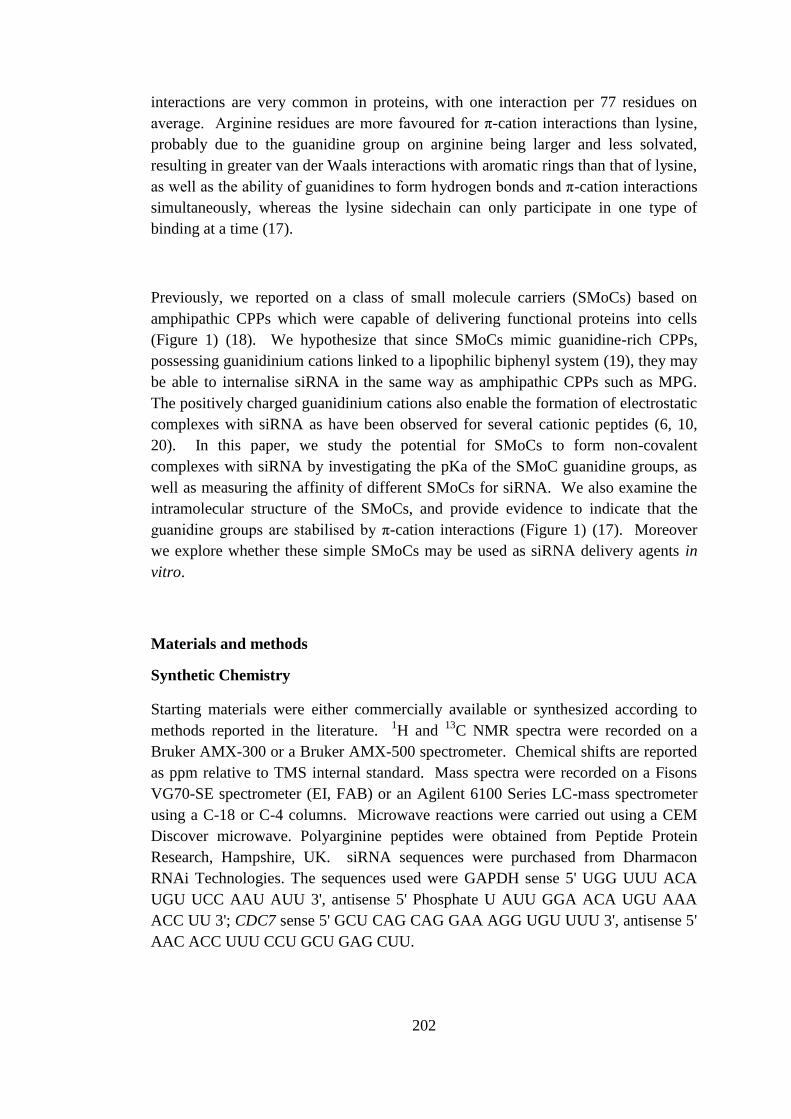
202
interactions are very common in proteins, with one interaction per 77 residues on
average. Arginine residues are more favoured for π-cation interactions than lysine,
probably due to the guanidine group on arginine being larger and less solvated,
resulting in greater van der Waals interactions with aromatic rings than that of lysine,
as well as the ability of guanidines to form hydrogen bonds and π-cation interactions
simultaneously, whereas the lysine sidechain can only participate in one type of
binding at a time (17).
Previously, we reported on a class of small molecule carriers (SMoCs) based on
amphipathic CPPs which were capable of delivering functional proteins into cells
(Figure 1) (18). We hypothesize that since SMoCs mimic guanidine-rich CPPs,
possessing guanidinium cations linked to a lipophilic biphenyl system (19), they may
be able to internalise siRNA in the same way as amphipathic CPPs such as MPG.
The positively charged guanidinium cations also enable the formation of electrostatic
complexes with siRNA as have been observed for several cationic peptides (6, 10,
20). In this paper, we study the potential for SMoCs to form non-covalent
complexes with siRNA by investigating the pKa of the SMoC guanidine groups, as
well as measuring the affinity of different SMoCs for siRNA. We also examine the
intramolecular structure of the SMoCs, and provide evidence to indicate that the
guanidine groups are stabilised by π-cation interactions (Figure 1) (17). Moreover
we explore whether these simple SMoCs may be used as siRNA delivery agents in
vitro.
Materials and methods
Synthetic Chemistry
Starting materials were either commercially available or synthesized according to
methods reported in the literature. 1H and
13C NMR spectra were recorded on a
Bruker AMX-300 or a Bruker AMX-500 spectrometer. Chemical shifts are reported
as ppm relative to TMS internal standard. Mass spectra were recorded on a Fisons
VG70-SE spectrometer (EI, FAB) or an Agilent 6100 Series LC-mass spectrometer
using a C-18 or C-4 columns. Microwave reactions were carried out using a CEM
Discover microwave. Polyarginine peptides were obtained from Peptide Protein
Research, Hampshire, UK. siRNA sequences were purchased from Dharmacon
RNAi Technologies. The sequences used were GAPDH sense 5' UGG UUU ACA
UGU UCC AAU AUU 3', antisense 5' Phosphate U AUU GGA ACA UGU AAA
ACC UU 3'; CDC7 sense 5' GCU CAG CAG GAA AGG UGU UUU 3', antisense 5'
AAC ACC UUU CCU GCU GAG CUU.
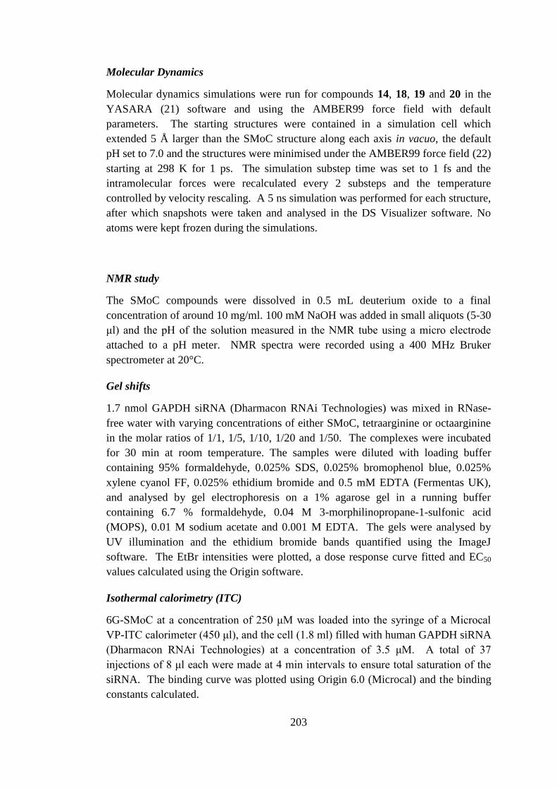
203
Molecular Dynamics
Molecular dynamics simulations were run for compounds 14, 18, 19 and 20 in the
YASARA (21) software and using the AMBER99 force field with default
parameters. The starting structures were contained in a simulation cell which
extended 5 Å larger than the SMoC structure along each axis in vacuo, the default
pH set to 7.0 and the structures were minimised under the AMBER99 force field (22)
starting at 298 K for 1 ps. The simulation substep time was set to 1 fs and the
intramolecular forces were recalculated every 2 substeps and the temperature
controlled by velocity rescaling. A 5 ns simulation was performed for each structure,
after which snapshots were taken and analysed in the DS Visualizer software. No
atoms were kept frozen during the simulations.
NMR study
The SMoC compounds were dissolved in 0.5 mL deuterium oxide to a final
concentration of around 10 mg/ml. 100 mM NaOH was added in small aliquots (5-30
μl) and the pH of the solution measured in the NMR tube using a micro electrode
attached to a pH meter. NMR spectra were recorded using a 400 MHz Bruker
spectrometer at 20°C.
Gel shifts
1.7 nmol GAPDH siRNA (Dharmacon RNAi Technologies) was mixed in RNase-
free water with varying concentrations of either SMoC, tetraarginine or octaarginine
in the molar ratios of 1/1, 1/5, 1/10, 1/20 and 1/50. The complexes were incubated
for 30 min at room temperature. The samples were diluted with loading buffer
containing 95% formaldehyde, 0.025% SDS, 0.025% bromophenol blue, 0.025%
xylene cyanol FF, 0.025% ethidium bromide and 0.5 mM EDTA (Fermentas UK),
and analysed by gel electrophoresis on a 1% agarose gel in a running buffer
containing 6.7 % formaldehyde, 0.04 M 3-morphilinopropane-1-sulfonic acid
(MOPS), 0.01 M sodium acetate and 0.001 M EDTA. The gels were analysed by
UV illumination and the ethidium bromide bands quantified using the ImageJ
software. The EtBr intensities were plotted, a dose response curve fitted and EC50
values calculated using the Origin software.
Isothermal calorimetry (ITC)
6G-SMoC at a concentration of 250 μM was loaded into the syringe of a Microcal
VP-ITC calorimeter (450 μl), and the cell (1.8 ml) filled with human GAPDH siRNA
(Dharmacon RNAi Technologies) at a concentration of 3.5 μM. A total of 37
injections of 8 μl each were made at 4 min intervals to ensure total saturation of the
siRNA. The binding curve was plotted using Origin 6.0 (Microcal) and the binding
constants calculated.
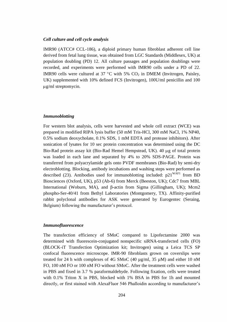
204
Cell culture and cell cycle analysis
IMR90 (ATCC# CCL-186), a diploid primary human fibroblast adherent cell line
derived from fetal lung tissue, was obtained from LGC Standards (Middlesex, UK) at
population doubling (PD) 12. All culture passages and population doublings were
recorded, and experiments were performed with IMR90 cells under a PD of 22.
IMR90 cells were cultured at 37 C with 5% CO2 in DMEM (Invitrogen, Paisley,
UK) supplemented with 10% defined FCS (Invitrogen), 100U/ml penicillin and 100
g/ml streptomycin.
Immunoblotting
For western blot analysis, cells were harvested and whole cell extract (WCE) was
prepared in modified RIPA lysis buffer (50 mM Tris-HCl, 300 mM NaCl, 1% NP40,
0.5% sodium deoxycholate, 0.1% SDS, 1 mM EDTA and protease inhibitors). After
sonication of lysates for 10 sec protein concentration was determined using the DC
Bio-Rad protein assay kit (Bio-Rad Hemel Hempstead, UK). 40 g of total protein
was loaded in each lane and separated by 4% to 20% SDS-PAGE. Protein was
transferred from polyacrylamide gels onto PVDF membranes (Bio-Rad) by semi-dry
electroblotting. Blocking, antibody incubations and washing steps were performed as
described (23). Antibodies used for immunoblotting included: p21WAF1
from BD
Biosciences (Oxford, UK), p53 (Ab-6) from Merck (Beeston, UK); Cdc7 from MBL
International (Woburn, MA), and β-actin from Sigma (Gillingham, UK); Mcm2
phospho-Ser-40/41 from Bethyl Laboratories (Montgomery, TX). Affinity-purified
rabbit polyclonal antibodies for ASK were generated by Eurogentec (Seraing,
Belgium) following the manufacturer’s protocol.
Immunofluorescence
The transfection efficiency of SMoC compared to Lipofectamine 2000 was
determined with fluorescein-conjugated nonspecific siRNA-transfected cells (FO)
(BLOCK-iT Transfection Optimization kit; Invitrogen) using a Leica TCS SP
confocal fluorescence microscope. IMR-90 fibroblasts grown on coverslips were
treated for 24 h with complexes of 4G SMoC (40 µg/ml, 35 µM) and either 10 nM
FO, 100 nM FO or 100 nM FO without SMoC. After the treatment cells were washed
in PBS and fixed in 3.7 % paraformaldehyde. Following fixation, cells were treated
with 0.1% Triton X in PBS, blocked with 1% BSA in PBS for 1h and mounted
directly, or first stained with AlexaFluor 546 Phalloidin according to manufacturer’s
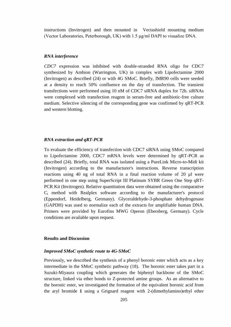
205
instructions (Invitrogen) and then mounted in Vectashield mounting medium
(Vector Laboratories, Peterborough, UK) with 1.5 μg/ml DAPI to visualize DNA.
RNA interference
CDC7 expression was inhibited with double-stranded RNA oligo for CDC7
synthesized by Ambion (Warrington, UK) in complex with Lipofectamine 2000
(Invitrogen) as described (24) or with 4G SMoC. Briefly, IMR90 cells were seeded
at a density to reach 50% confluence on the day of transfection. The transient
transfections were performed using 10 nM of CDC7 siRNA duplex for 72h. siRNAs
were complexed with transfection reagent in serum-free and antibiotic-free culture
medium. Selective silencing of the corresponding gene was confirmed by qRT-PCR
and western blotting.
RNA extraction and qRT-PCR
To evaluate the efficiency of transfection with CDC7 siRNA using SMoC compared
to Lipofectamine 2000, CDC7 mRNA levels were determined by qRT-PCR as
described (24). Briefly, total RNA was isolated using a PureLink Micro-to-Midi kit
(Invitrogen) according
to the manufacturer's instructions. Reverse transcription
reactions using 40 ng of total RNA in a final reaction volume of 20 µl were
performed in one step using SuperScript III Platinum SYBR Green One Step qRT-
PCR Kit (Invitrogen). Relative quantitation data were obtained using the comparative
Ct method with Realplex software according
to the manufacturer's protocol
(Eppendorf, Heidelberg, Germany). Glyceraldehyde-3-phosphate dehydrogenase
(GAPDH) was used to normalize each of the extracts for amplifiable human DNA.
Primers were provided by Eurofins MWG Operon (Ebersberg, Germany). Cycle
conditions are available upon request.
Results and Discussion
Improved SMoC synthetic route to 4G-SMoC
Previously, we described the synthesis of a phenyl boronic ester which acts as a key
intermediate in the SMoC synthetic pathway (18). The boronic ester takes part in a
Suzuki-Miyaura coupling which generates the biphenyl backbone of the SMoC
structure, linked via ether bonds to Z-protected amine groups. As an alternative to
the boronic ester, we investigated the formation of the equivalent boronic acid from
the aryl bromide 1 using a Grignard reagent with 2-(dimethylamino)ethyl ether

206
followed by quenching with trimethyl borate to yield the boronic acid 2 (Figure 2).
As described by Wang et al. (25), the organic ligand moderates the reactivity of the
aryl Grignard reagent in this reaction, reducing possible side reactions. Careful
control of the reaction temperature was required to obtain a synthetically useful yield
of 40%.
In addition, the potassium trifluoroborate salt was synthesized (Figure 3).
Trifluoroborate salts, developed by Molander et al (26, 27), are extremely stable to
air and moisture, allowing large scale synthesis of this key intermediate for long-term
storage. The pinacolboronate ester 3 synthesized previously was treated with
potassium bifluoride in methanol, resulting in an 88% yield of the salt 4 which is
precipitated from the reaction mixture and is easily isolated by filtration and washing
with organic solvents. Suzuki reactions with this salt and the alkylated nitrile 5 gave
cleaner conversion to the biphenyl 6 with less of the deboronated side product that
was difficult to separate from the desired product.
A problematic step in the original SMoC synthesis was the addition of Boc-protected
guanidine groups using the expensive N,N-di-Boc-N'-trifluoromethanesulfonyl-
guanidine reagent, which resulted in modest yields. To improve this step, a less
expensive and more stable guanidinylation reagent was used, N,N'-Di-Boc-1H-
pyrazole-1-carboxamidine. Thus following removal of the CBz groups using HBr in
acetic acid, the use of this reagent increased the yield of guanidinylation from ~65%
to over 70% of 7 at a much reduced cost of materials.
Finally, the reduction of the nitrile 7 in order to produce a primary amine for
attachment of diverse linkers was another low-yielding step from our previous route.
The previous synthesis which used Raney Nickel/H2 resulted in 40-50% yields.
Reduction using sodium borohydride catalysed by cobalt chloride increased the yield
of this step to ~60% of 8 (28, 29). Amide coupling with the thiopyridylpropionic
acid O-succinimide ester gave the desired thiopyridyl intermediate 9 and
deprotection with TFA provided 10. Synthesis of the disulfide 11 was easily
accomplished in a two step procedure with DTT and then aerial oxidation.
The final optimised synthesis of 4G-SMoC is therefore shown in Figure 4. This
improved method has enabled us for the first time to synthesize 4G-SMoC on a >200
mg scale.
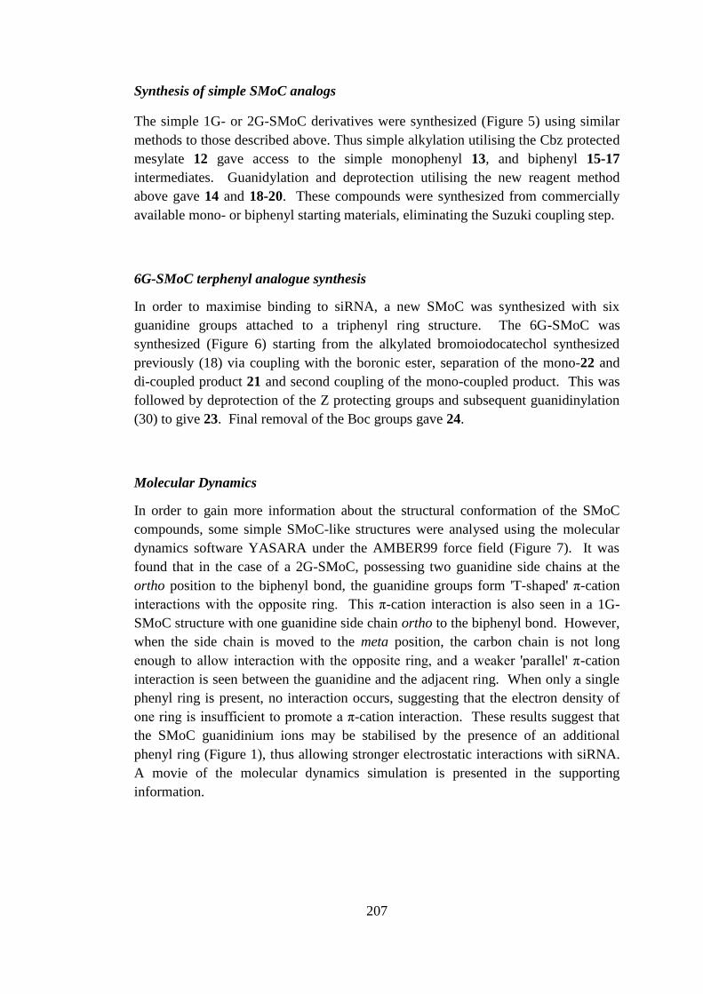
207
Synthesis of simple SMoC analogs
The simple 1G- or 2G-SMoC derivatives were synthesized (Figure 5) using similar
methods to those described above. Thus simple alkylation utilising the Cbz protected
mesylate 12 gave access to the simple monophenyl 13, and biphenyl 15-17
intermediates. Guanidylation and deprotection utilising the new reagent method
above gave 14 and 18-20. These compounds were synthesized from commercially
available mono- or biphenyl starting materials, eliminating the Suzuki coupling step.
6G-SMoC terphenyl analogue synthesis
In order to maximise binding to siRNA, a new SMoC was synthesized with six
guanidine groups attached to a triphenyl ring structure. The 6G-SMoC was
synthesized (Figure 6) starting from the alkylated bromoiodocatechol synthesized
previously (18) via coupling with the boronic ester, separation of the mono-22 and
di-coupled product 21 and second coupling of the mono-coupled product. This was
followed by deprotection of the Z protecting groups and subsequent guanidinylation
(30) to give 23. Final removal of the Boc groups gave 24.
Molecular Dynamics
In order to gain more information about the structural conformation of the SMoC
compounds, some simple SMoC-like structures were analysed using the molecular
dynamics software YASARA under the AMBER99 force field (Figure 7). It was
found that in the case of a 2G-SMoC, possessing two guanidine side chains at the
ortho position to the biphenyl bond, the guanidine groups form 'T-shaped' π-cation
interactions with the opposite ring. This π-cation interaction is also seen in a 1G-
SMoC structure with one guanidine side chain ortho to the biphenyl bond. However,
when the side chain is moved to the meta position, the carbon chain is not long
enough to allow interaction with the opposite ring, and a weaker 'parallel' π-cation
interaction is seen between the guanidine and the adjacent ring. When only a single
phenyl ring is present, no interaction occurs, suggesting that the electron density of
one ring is insufficient to promote a π-cation interaction. These results suggest that
the SMoC guanidinium ions may be stabilised by the presence of an additional
phenyl ring (Figure 1), thus allowing stronger electrostatic interactions with siRNA.
A movie of the molecular dynamics simulation is presented in the supporting
information.
B
208
NMR pKa Study
In order to investigate the strength of the proposed π-cation interaction, NMR was
used to determine the pKa of SMoC guanidine groups by directly measuring the
change in chemical shift of the adjacent methylene hydrogens (31, 32). A change in
the pKa of the guanidine would be expected if a π-cation interaction was present
(31).
For each compound, the change in ionisation state of the guanidine groups was
measured using NMR by titrating each compound with sodium hydroxide and
recording the 1H chemical shifts of the CH2 protons adjacent to the guanidine group
against the pH of the solution (Figure 8). The pKa of the guanidine group is given
by the Henderson-Hasselbalch equation (33):
][
][log
B
BHpHpKa
Deprotonation of the guanidine is expected to have a significant effect on the
chemical shift of the protons on the adjacent methylene group due to the change in
electron density around the nitrogen atom, resulting in increased shielding of the CH2
protons. The chemical shift of the charged form (δB+) may be determined by
measuring the chemical shift at a low pH. For these compounds, the chemical shift
was found to be at its maximum below pH 10. Likewise, at very high pH (pH 14)
the chemical shift of the deprotonated guanidine groups may be measured (δB).
Thus, the chemical shift at a given pH (δA) is representative of the proportion of
groups that are protonated at a given time. Therefore, the equation may be expressed
in terms of change in chemical shift:
AB
BApHpKa
log
Hence, by plotting the log of the ratio of the charged and uncharged forms expressed
as chemical shifts against the pH of the solution (Figure 9), the pKa may be
calculated from the y-intercept of the linear trend line.
The results of these experiments (Table 1) show that the 2G-SMoC with the bi-
phenyl substituted at the ortho position to the biphenyl bond, 18, has the highest pKa
(13.33), suggesting that the guanidine groups in this compound benefit from the
greatest stabilisation. The mono-phenyl compound, 14 (12.86), has the lowest pKa
209
and the closest to free arginine (12.48) (34), suggesting very little stabilisation is
available to the guanidinium ion. These results also show that substitution position is
also important for stabilisation, since the meta-substituted compound, 19, has a lower
pKa (13.09) than the ortho-substituted 20 (13.26). These results suggest that the
biphenyl system is able to provide some stabilisation to the positively charged
guanidinium ion. The energy of this stabilisation may be calculated by comparing
the pKa values of the mono-phenyl compound, 20, and the mono-substituted
biphenyl compound 19 using the equation:
pKaRTGpKa 303.20
This gives a free energy of stabilisation of -2.3 kJ mol-1
for each guanidine group
compared to the mono-phenyl compound, for which it was assumed that there is very
little π-cation stabilisation, due to its low pKa. This value is the increase in the free
energy of the guanidine group gained by the addition of a phenyl ring to the mono-
phenyl compound 14.
ITC of 6G-SMoC indicates a high enthalpic contribution to binding.
In order to calculate the strength of the interactions between SMoC and siRNA,
isothermal titration calorimetry (ITC) was used to measure the heat evolved when
small aliquots of 6G-SMoC, 24 were injected into a solution of a standard 21-bp
siRNA (Figure 10). A binding constant (K) of 87.3 μM was calculated with a
binding stoichiometry of 4.4:1 (SMoC:siRNA). Binding was dominated by enthalpic
contributions (-1646 Kcal/mol) with a minor or negligible entropic contribution. This
is consistent with electrostatic (charge) interactions between the guanidines and the
phosphate groups on the siRNA dominating binding.
Gel shift studies on siRNA show that SMoCs can effectively complex to siRNA
duplexes.
The interactions of 4G- and 6G-SMoC compounds with siRNA were analysed by gel
electrophoresis of SMoC/siRNA complexes at different molar ratios. Tetra- and octa-
arginine peptides were also tested in order to establish the relative binding affinity of
SMoCs compared to these established cell delivery peptides. As shown in Figure 11,
all SMoCs and peptides were able to fully shield the siRNA negative charges at
ratios less than 20:1. Since the observable band on the gel is proportional to the free
siRNA concentration we utilised this value to provide a convenient graphical
representation of the relative affinities and to calculate the approximate binding
210
constants of the analogues. The ethidium bromide (EtBr) fluorescence of each band
was quantified and the data plotted as dose response curves (Figure 12). 6G-SMoC,
24 had a slightly lower binding affinity than 4G SMoC, 10 (EC50 of 110 μM
compared to 70 μM) and 4G-SMoC had a higher affinity than R4 (EC50 of 70 μM
compared to 120 μM). However R8 had a higher affinity (3 μM) than any of the
SMoCs. The disulfide variant (4G-SMoCS)2, 11 has the same binding affinity as the
monomer form, probably due to reduction of the disulfide bond in the gel running
buffer. 4G-SMoC, 10 binds with a higher affinity than the polyarginine peptide R4,
indicating an ability to complex to siRNA more efficiently or to shield the negative
charges more effectively. This may be attributed to the increased pKa of the SMoC
guanidine groups as a result of the π-cation effect described previously.
Confocal analysis of SMoC mediated siRNA uptake
To determine if siRNA-SMoC complexes are taken up by cells, the simple SMoCs
described above were complexed with a fluorescently-tagged siRNA oligo at
concentrations of either 10 nM or 100 nM and incubated with IMR-90 cells, a diploid
human fibroblast adherent cell strain derived from foetal lung tissue, for 24 h. Upon
examination under confocal microscopy, only the 4G-SMoC compound 10 showed
significant uptake (others not shown). For compound 10, the fluorescent oligo was
observed in the cytoplasm in a punctate distribution for both concentrations, showing
that uptake occurs into vesicles, possibly endosomes formed via an endocytotic
pathway (Figure 13).
siRNA-SMoC gene and protein knockdown
To determine if SMoCs are capable of transporting siRNA into cells, IMR-90 cells
were transfected with SMoC or Lipofectamine complexed with a double-stranded
RNA oligo targeted at Cdc7, a cell cycle kinase which has been identified as a new
cancer therapeutic target (35). Cancer cells establish only a limited number of DNA
replication forks under Cdc7 rate-limiting conditions, causing fork stalling and
collapse during an abortive S phase and triggering apoptotic cell death. Tumour cell
specificity comes from normal cells avoiding lethal S phase progression in the
presence of low Cdc7 kinase levels by eliciting a p53-dependent replication origin
activation checkpoint response that arrests the cells at the G1/S boundary (24).
Seventy-two hours after the treatment, IMR90 cells were harvested, and CDC7
mRNA knockdown was monitored by qRT-PCR. For each compound tested, the
CDC7 mRNA level was determined relative to control cells treated only with
Lipofectamine or SMoC. Treatment with complexes of 4G-SMoC, 10 and CDC7-
siRNA reduced CDC7 mRNA levels by 61%. These results were similar to the
CDC7 knockdown achieved with Lipofectamine plus CDC7-siRNA, which reduced
211
mRNA levels by 80% (Figure 14A). RNAi against CDC7, mediated through 4G-
SMoC or Lipofectamine, caused an accumulation of IMR90 cells with G1 DNA
content and a concomitant reduction in the S phase fraction, while the fraction of
cells with less than 2C DNA content was negligible, indicating that the Cdc7-
depleted cells remained viable (Figure 14B). We recently demonstrated the triggering
of an origin activation checkpoint in Cdc7-depleted IMR90 cells (24). Consistent
with this study, western blotting of extracts prepared from IMR90 cells treated with
complexes of 4G-SMoC, 10 and CDC7-siRNA showed that phosphorylation of the
replication initiation factor Mcm2 at the Cdc7-dependent phosphor-site Ser-40/41
was markedly reduced when Cdc7 kinase and its regulatory subunit ASK were
downregulated. In keeping with activation of the origin activation checkpoint in the
Cdc7-depleted cells, we also noted p53 stabilization and increased levels of the CDK
inhibitor protein p21 (Figure 14C). Taken together, these data demonstrate that
SMoCs are capable of mediating the uptake of siRNAs into cultured human cells
without altering their biological activity. Note that 6G-SMoC, 24 was also tested for
siRNA transfection but did not result in significant knockdown (data not shown).
Conclusions
The SMoC class of compounds, modelled on amphipathic CPPs, have previously
been shown to transport active proteins as well as conjugated dyes into a variety of
cell types (18). Here we have improved the synthetic route to the SMoC compounds,
including the use of Molander chemistry in the form of a stable trifluoroborate
intermediate which can be used to easily build new, varied SMoC-like structures, as
well as optimisation of some of the previously difficult steps in order to increase the
scale of the synthesis.
We have also shown in this study that SMoCs show potential as siRNA transfection
agents. One surprise from this study is that the most effective delivery and
knockdown was achieved with the 4G-SMoC, compound 10 where the thiopyridyl
group is commonly regarded as a reactive protein thiol reagent. An improvement in
CPP delivery has been noted for some cysteine containing peptides (36) where
dimerization or oligimerization is indicated. In addition enhancement of
internalization has been noted for CPPs containing cysteines conjugated with the 2-
nitropyridyl group (37). An interaction with cell surface thiols was suggested for this
observation and we speculate that compound 10 may also interact with cell surface
thiols to promote internalization.
siRNA delivery remains a significant challenge in biological therapeutics, and an
effective transfection agent will open up a vast array of possibilities in the treatment
of many diseases by specifically targeting the underlying genetic malfunctions. The
212
standard in vitro transfection agent, lipofectamine is demonstrably toxic, by several
measures (38) indicating the need for further research in this area. Cell penetrating
peptides show a great deal of promise as transfection agents, and many studies have
successfully introduced siRNA into cells using both covalently- and non-covalently
linked CPPs (8). As CPP mimics, it is therefore likely that SMoCs will also possess
this ability to act as siRNA delivery vectors, possessing positively charged guanidine
groups which allow the formation of electrostatic complexes with siRNA, and are
also essential for successful passage across the cell membrane. Our MD and NMR
studies indicate that the guanidinium ions on the SMoC side chains are stabilised by
π-cation. The potential impact of π-cation effects has also been advanced for the
Arg-Trp interaction in the penetratin CPP (39). We probed the affinity of the SMoC-
siRNA interaction using two methods ITC (the gold standard for evaluating binding
interactions) and simple gel shift analysis. These were in good agreement indicating
binding affinities in the range 70 to 110 µM. The poly arginine peptide R8 had an
affinity of 30 µM. The optimal binding affinity for SMoC-siRNA delivery remains to
be determined. As we show here, SMoCs can deliver biologically active siRNA into
cultured human cells, and achieved RNAi knockdown with a similar magnitude to
the widely used Lipofectamine reagent. Further optimisation of SMoCs to increase
the uptake of SMoC-siRNA complexes will be reported in due course.
Acknowledgements
We thank the Association for International Cancer Research (AICR) for funding this
project (Grant Ref: 06-0076). This work was also partly funded by Cancer Research
UK Scientific Programme Grant C428/A6263 (KS and GHW). We would also like
to thank Tina Divita and Matt Webster of the ISMB Biophysics Centre at Birkbeck
College for their assistance with the ITC work.
Supporting information
Full experimental details of chemistry syntheses and a movie of the molecular
dynamics simulation are presented.
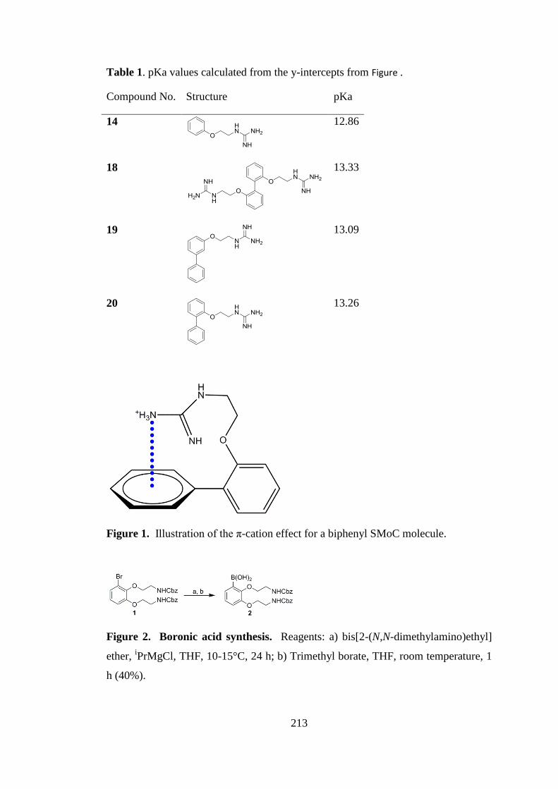
213
Table 1. pKa values calculated from the y-intercepts from Figure .
Compound No. Structure pKa
14
12.86
18
13.33
19
13.09
20
13.26
Figure 1. Illustration of the π-cation effect for a biphenyl SMoC molecule.
Figure 2. Boronic acid synthesis. Reagents: a) bis[2-(N,N-dimethylamino)ethyl]
ether, iPrMgCl, THF, 10-15°C, 24 h; b) Trimethyl borate, THF, room temperature, 1
h (40%).
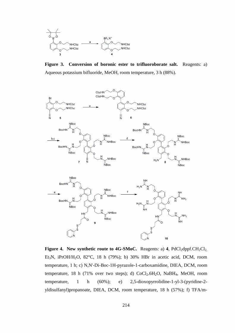
214
Figure 3. Conversion of boronic ester to trifluoroborate salt. Reagents: a)
Aqueous potassium bifluoride, MeOH, room temperature, 3 h (88%).
Figure 4. New synthetic route to 4G-SMoC. Reagents: a) 4, PdCl2dppf.CH2Cl2,
Et3N, iPrOH/H2O, 82°C, 18 h (79%); b) 30% HBr in acetic acid, DCM, room
temperature, 1 h; c) N,N'-Di-Boc-1H-pyrazole-1-carboxamidine, DIEA, DCM, room
temperature, 18 h (71% over two steps); d) CoCl2.6H2O, NaBH4, MeOH, room
temperature, 1 h (60%); e) 2,5-dioxopyrrolidine-1-yl-3-(pyridine-2-
yldisulfanyl)propanoate, DIEA, DCM, room temperature, 18 h (57%); f) TFA/m-
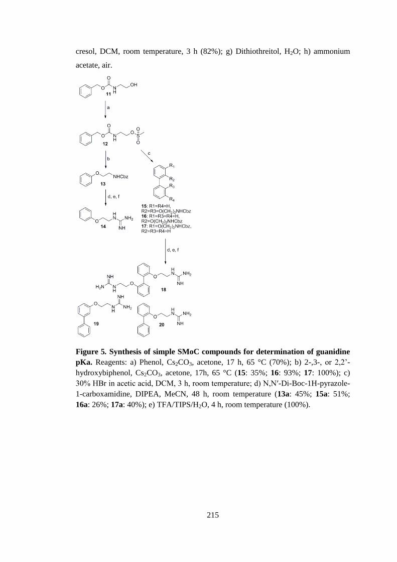
215
cresol, DCM, room temperature, 3 h (82%); g) Dithiothreitol, H2O; h) ammonium
acetate, air.
Figure 5. Synthesis of simple SMoC compounds for determination of guanidine
pKa. Reagents: a) Phenol, Cs2CO3, acetone, 17 h, 65 °C (70%); b) 2-,3-, or 2,2’-
hydroxybiphenol, Cs2CO3, acetone, 17h, 65 °C (15: 35%; 16: 93%; 17: 100%); c)
30% HBr in acetic acid, DCM, 3 h, room temperature; d) N,N′-Di-Boc-1H-pyrazole-
1-carboxamidine, DIPEA, MeCN, 48 h, room temperature (13a: 45%; 15a: 51%;
16a: 26%; 17a: 40%); e) TFA/TIPS/H2O, 4 h, room temperature (100%).
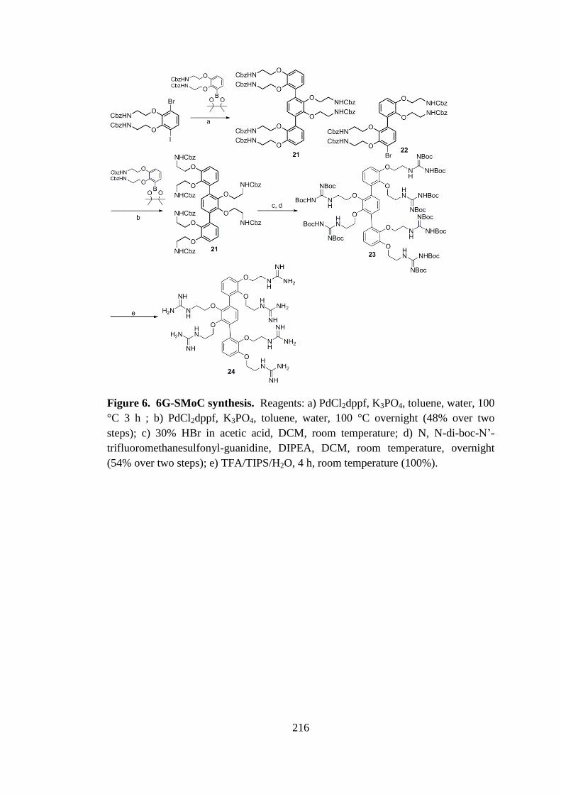
216
Figure 6. 6G-SMoC synthesis. Reagents: a) PdCl2dppf, K3PO4, toluene, water, 100
°C 3 h ; b) PdCl2dppf, K3PO4, toluene, water, 100 °C overnight (48% over two
steps); c) 30% HBr in acetic acid, DCM, room temperature; d) N, N-di-boc-N’-
trifluoromethanesulfonyl-guanidine, DIPEA, DCM, room temperature, overnight
(54% over two steps); e) TFA/TIPS/H2O, 4 h, room temperature (100%).
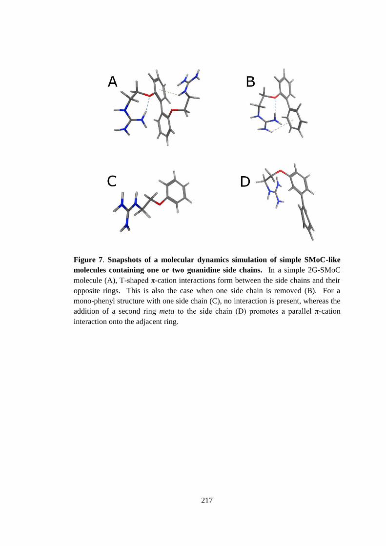
217
Figure 7. Snapshots of a molecular dynamics simulation of simple SMoC-like
molecules containing one or two guanidine side chains. In a simple 2G-SMoC
molecule (A), T-shaped π-cation interactions form between the side chains and their
opposite rings. This is also the case when one side chain is removed (B). For a
mono-phenyl structure with one side chain (C), no interaction is present, whereas the
addition of a second ring meta to the side chain (D) promotes a parallel π-cation
interaction onto the adjacent ring.
A
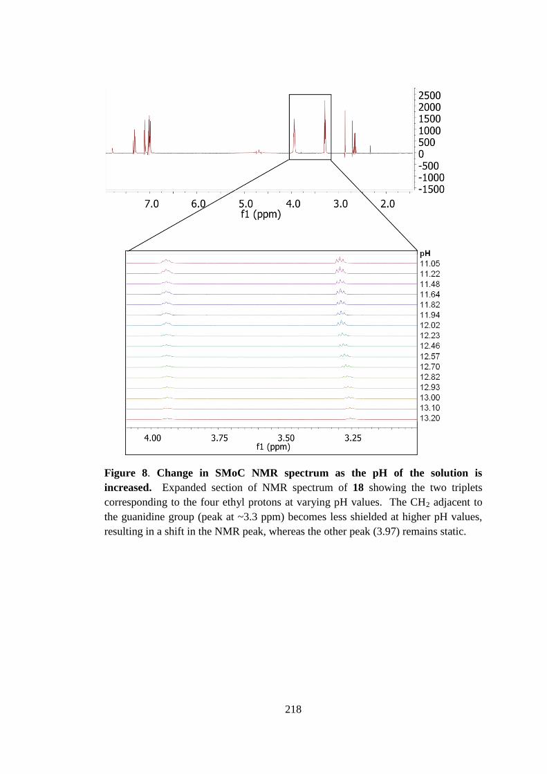
218
Figure 8. Change in SMoC NMR spectrum as the pH of the solution is
increased. Expanded section of NMR spectrum of 18 showing the two triplets
corresponding to the four ethyl protons at varying pH values. The CH2 adjacent to
the guanidine group (peak at ~3.3 ppm) becomes less shielded at higher pH values,
resulting in a shift in the NMR peak, whereas the other peak (3.97) remains static.
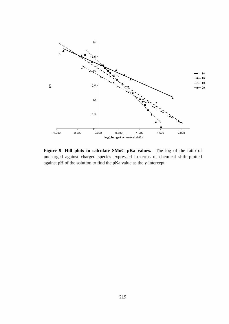
219
Figure 9. Hill plots to calculate SMoC pKa values. The log of the ratio of
uncharged against charged species expressed in terms of chemical shift plotted
against pH of the solution to find the pKa value as the y-intercept.
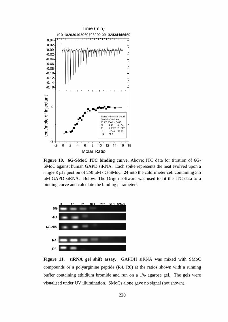
220
Figure 10. 6G-SMoC ITC binding curve. Above: ITC data for titration of 6G-
SMoC against human GAPD siRNA. Each spike represents the heat evolved upon a
single 8 μl injection of 250 μM 6G-SMoC, 24 into the calorimeter cell containing 3.5
μM GAPD siRNA. Below: The Origin software was used to fit the ITC data to a
binding curve and calculate the binding parameters.
Figure 11. siRNA gel shift assay. GAPDH siRNA was mixed with SMoC
compounds or a polyarginine peptide (R4, R8) at the ratios shown with a running
buffer containing ethidium bromide and run on a 1% agarose gel. The gels were
visualised under UV illumination. SMoCs alone gave no signal (not shown).

221
Figure 12. Binding curves derived from gel shift assay data. The EtBr bands
were quantified and the band intensity plotted to produce binding curves for each of
the compounds with siRNA. The EC50 and Hill coefficient (p) values of each
compound are shown, giving a relative indication of the binding affinity of each
compound to siRNA.
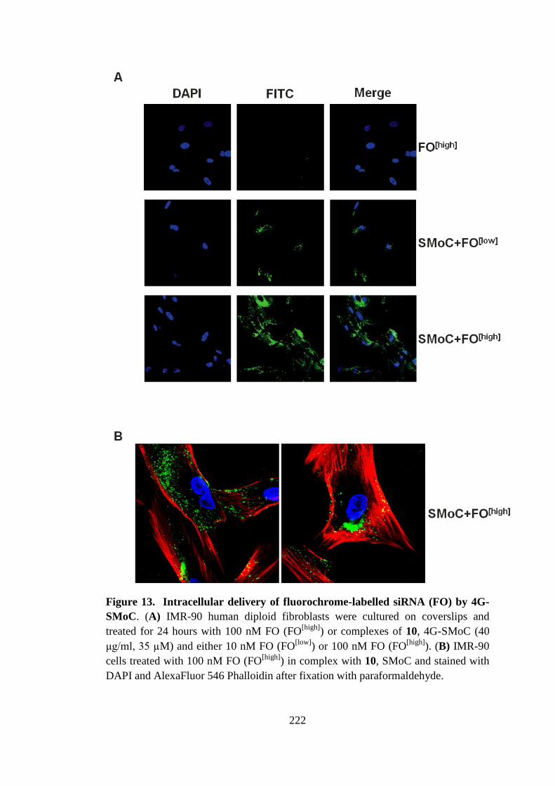
222
Figure 13. Intracellular delivery of fluorochrome-labelled siRNA (FO) by 4G-
SMoC. (A) IMR-90 human diploid fibroblasts were cultured on coverslips and
treated for 24 hours with 100 nM FO (FO[high]
) or complexes of 10, 4G-SMoC (40
μg/ml, 35 µM) and either 10 nM FO (FO[low]
) or 100 nM FO (FO[high]
). (B) IMR-90
cells treated with 100 nM FO (FO[high]
) in complex with 10, SMoC and stained with
DAPI and AlexaFluor 546 Phalloidin after fixation with paraformaldehyde.
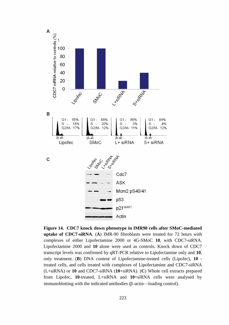
223
Figure 14. CDC7 knock down phenotype in IMR90 cells after SMoC-mediated
uptake of CDC7-siRNA. (A) IMR-90 fibroblasts were treated for 72 hours with
complexes of either Lipofectamine 2000 or 4G-SMoC 10, with CDC7-siRNA.
Lipofectamine 2000 and 10 alone were used as controls. Knock down of CDC7
transcript levels was confirmed by qRT-PCR relative to Lipofectamine only and 10,
only treatment. (B) DNA content of Lipofectamine-treated cells (Lipofec), 10 -
treated cells, and cells treated with complexes of Lipofectamine and CDC7-siRNA
(L+siRNA) or 10 and CDC7-siRNA (10+siRNA). (C) Whole cell extracts prepared
from Lipofec, 10-treated, L+siRNA and 10+siRNA cells were analysed by
immunoblotting with the indicated antibodies (-actin—loading control).
224
Bibliography
1. Guo J, Fisher KA, Darcy R, Cryan JF, O'Driscoll C (2010) Therapeutic
targeting in the silent era: advances in non-viral siRNA delivery. Molecular
Biosystems;6: 1143-61.
2. Whitehead K, Langer R, Anderson D (2009) Knocking down barriers:
advances in siRNA delivery. Nature Reviews Drug Discovery;8: 129-38.
3. Sontheimer EJ (2005) Assembly and function of RNA silencing complexes.
Nature Reviews Molecular Cell Biology;6: 127-38.
4. Li W, Szoka FC (2007) Lipid-based nanoparticles for nucleic acid delivery.
Pharm Res;24: 438-49.
5. Rozema DB, Lewis DL, Wakefield DH, Wong SC, Klein JJ, Roesch PL, et
al. (2007) Dynamic PolyConjugates for targeted in vivo delivery of siRNA to
hepatocytes. Proceedings of the National Academy of Sciences of the United States of
America;104: 12982-7.
6. Kim SW, Kim NY, Choi YB, Park SH, Yang JM, Shin S (2010) RNA
interference in vitro and in vivo using an arginine peptide/siRNA complex system.
Journal of Controlled Release;143: 335-43.
7. Konate K, Crombez L, Deshayes S, Decaffmeyer M, Thomas A, Brasseur R,
et al. (2010) Insight into the cellular uptake mechanism of a secondary amphipathic
cell-penetrating peptide for siRNA delivery. Biochemistry;49: 3393-402.
8. Eguchi A, Dowdy SF (2009) siRNA delivery using peptide transduction
domains. Trends Pharmacol Sci;30: 341-5.
9. Veldhoen S, Laufer SD, Trampe A, Restle T (2006) Cellular delivery of small
interfering RNA by a non-covalently attached cell-penetrating peptide: quantitative
analysis of uptake and biological effect. Nucleic Acids Res;34: 6561-73.
10. Crombez L, Aldrian-Herrada G, Konate K, Nguyen QN, McMaster GK,
Brasseur R, et al. (2009) A new potent secondary amphipathic cell-penetrating
peptide for siRNA delivery into mammalian cells. Molecular Therapy;17: 95-103.
11. Gump JM, June RK, Dowdy SF (2010) Revised role of glycosaminoglycans
in TAT protein transduction domain-mediated cellular transduction. Journal of
Biological Chemistry;285: 1500-7.
12. Jiao CY, Delaroche D, Burlina F, Alves ID, Chassaing G, Sagan S (2009)
Translocation and Endocytosis for Cell-penetrating Peptide Internalization. J Biol
Chem;284: 33957-65.
13. El-Sayed A, Khalil IA, Kogure K, Futaki S, Harashima H (2008)
Octaarginine- and Octalysine-modified Nanoparticles Have Different Modes of
Endosomal Escape. J Biol Chem;283: 23450-61.
225
14. Chao T-Y, Raines RT (2011) Mechanism of Ribonuclease A Endocytosis:
Analogies to Cell-Penetrating Peptides. Biochemistry;null-null.
15. Duchardt F, Fotin-Mleczek M, Schwarz H, Fischer R, Brock R (2007) A
Comprehensive Model for the Cellular Uptake of Cationic Cell-penetrating Peptides.
Traffic;8: 848-66.
16. Burley SK, Petsko GA (1986) Amino-aromatic interactions in proteins. FEBS
Lett;203: 139-43.
17. Gallivan JP, Dougherty DA (1999) Cation-pi interactions in structural
biology. Proceedings of the National Academy of Sciences of the United States of
America;96: 9459-64.
18. Rebstock AS, Visintin C, Leo E, Posada CG, Kingsbury SR, Williams GH, et
al. (2008) Modular assembly using sequential palladium coupling gives easy access
to the SMoC class of cellular transporters. ChemBioChem;9: 1787-96.
19. Okuyama M, Laman H, Kingsbury SR, Visintin C, Leo E, Eward KL, et al.
(2007) Small-molecule mimics of an [alpha]-helix for efficient transport of proteins
into cells. Nat Methods;4: 153-9.
20. Lundberg P, El Andaloussi S, Sutlu T, Johansson H, Langel U (2007)
Delivery of short interfering RNA using endosomolytic cell-penetrating peptides.
FASEB J;21: 2664-71.
21. Krieger E, Koraimann G, Vriend G (2002) Increasing the precision of
comparative models with YASARA NOVA--a self-parameterizing force field.
Proteins;47: 393-402.
22. Wang JM, Cieplak P, Kollman PA (2000) How well does a restrained
electrostatic potential (RESP) model perform in calculating conformational energies
of organic and biological molecules? J Comput Chem;21: 1049-74.
23. Kingsbury SR, Loddo M, Fanshawe T, Obermann EC, Prevost AT, Stoeber
K, et al. (2005) Repression of DNA replication licensing in quiescence is
independent of geminin and may define the cell cycle state of progenitor cells. Exp
Cell Res;309: 56-67.
24. Tudzarova S, Trotter MWB, Wollenschlaeger A, Mulvey C, Godovac-
Zimmermann J, Williams GH, et al. (2010) Molecular architecture of the DNA
replication origin activation checkpoint. EMBO J;29: 3381-94.
25. Wang XJ, Sun XF, Zhang L, Xu YB, Krishnamurthy D, Senanayake CH
(2006) Noncryogenic I/Br-Mg exchange of aromatic halides bearing sensitive
functional groups using i-PrMgCl-bis 2-(N,N-dimethylamino)ethyl ether complexes.
Org Lett;8: 305-7.
26. Molander GA, Figueroa R (2005) Organotrifluoroborates: Expanding
organoboron chemistry. Aldrichimica Acta;38: 49-56.
226
27. Molander GA, Ellis N (2007) Organotrifluoroborates: Protected boronic acids
that expand the versatility of the Suzuki coupling reaction. Acc Chem Res;40: 275-
86.
28. Ducatel H, Van Nhien AN, Postel D (2008) Novel glycine like amino acids
from glyco-[alpha]-aminonitriles as building blocks for peptide synthesis.
Tetrahedron: Asymmetry;19: 67-81.
29. Feldman KS, Ngernmeesri P (2005) Dragmacidin E synthesis studies.
Preparation of a model cycloheptannelated indole fragment. Org Lett;7: 5449-52.
30. Feichtinger K, Sings HL, Baker TJ, Matthews K, Goodman M (1998)
Triurethane-Protected Guanidines and Triflyldiurethane-Protected Guanidines: New
Reagents for Guanidinylation Reactions. The Journal of Organic Chemistry;63:
8432-9.
31. Acharya P, Plashkevych O, Morita C, Yamada S, Chattopadhyaya J (2003) A
repertoire of pyridinium-phenyl-methyl cross-talk through a cascade of
intramolecular electrostatic interactions. J Org Chem;68: 1529-38.
32. Oganesyan A, Cruz IA, Amador RB, Sorto NA, Lozano J, Godinez CE, et al.
(2007) High yield selective acylation of polyamines: proton as protecting group.
Organic Letters;9: 4967-70.
33. Atkins P, de Paula J (2002) Atkins' Physical Chemistry. 7 edn: Oxford
University Press.
34. Kumaki Y, Nitta K, Hikichi K, Matsumoto T, Matsushima N (2004) Side
Chain-Side Chain Interactions of Arginine with Tyrosine and Aspartic Acid in
Arg/Gly/Tyr-Rich Domains within Plant Glycine-Rich RNA Binding Proteins. J
Biochem;136: 29-37.
35. Swords R, Mahalingam D, O'Dwyer M, Santocanale C, Kelly K, Carew J, et
al. (2010) Cdc7 kinase - A new target for drug development. Eur J Cancer;46: 33-
40.
36. Won Y-W, Kim HA, Lee M, Kim Y-H (2010) Reducible Poly(oligo-D-
arginine) for Enhanced Gene Expression in Mouse Lung by Intratracheal Injection.
Mol Ther;18: 734-42.
37. Aubry S, Aussedat B, Delaroche D, Jiao CY, Bolbach G, Lavielle S, et al.
(2010) MALDI-TOF mass spectrometry: A powerful tool to study the internalization
of cell-penetrating peptides. Biochim Biophys Acta-Biomembr;1798: 2182-9.
38. Eguchi A, Meade BR, Chang YC, Fredrickson CT, Willert K, Puri N, et al.
(2009) Efficient siRNA delivery into primary cells by a peptide transduction domain-
dsRNA binding domain fusion protein. Nat Biotechnol;27: 567-U110.
39. Lensink MF, Christiaens B, Vandekerckhove J, Prochiantz A, Rosseneu M
(2005) Penetratin-membrane association: W48/R52/W56 shield the peptide from the
aqueous phase. Biophys J;88: 939-52.