EXPERT REVIEW Molecular psychiatry of zebra sh
16
EXPERT REVIEW Molecular psychiatry of zebrafish AM Stewart 1,2,8 , JFP Ullmann 2,3,8 , WHJ Norton 2,4,8 , CH Brennan 5 , MO Parker 5 , R Gerlai 6 and AV Kalueff 1,2,7 Due to their well-characterized neural development and high genetic homology to mammals, zebrafish (Danio rerio) have emerged as a powerful model organism in the field of biological psychiatry. Here, we discuss the molecular psychiatry of zebrafish, and its implications for translational neuroscience research and modeling central nervous system (CNS) disorders. In particular, we outline recent genetic and technological developments allowing for in vivo examinations, high-throughput screening and whole-brain analyses in larval and adult zebrafish. We also summarize the application of these molecular techniques to the understanding of neuropsychiatric disease, outlining the potential of zebrafish for modeling complex brain disorders, including attention-deficit/ hyperactivity disorder (ADHD), aggression, post-traumatic stress and substance abuse. Critically evaluating the advantages and limitations of larval and adult fish tests, we suggest that zebrafish models become a rapidly emerging new field in modern biological psychiatry research. Molecular Psychiatry (2014) 00, 000–000. doi:10.1038/mp.2014.128 INTRODUCTION Psychiatric disorders affect many brain and behavioral processes, including thoughts, feeling, sociability and mood. These complex human diseases also cause a significant burden to society, especially as the available drug therapies have not sufficiently progressed. 1,2 The etiology of psychiatric disorders is multifaceted, and involves the interaction of multiple genetic and environ- mental factors. Genetic mutations trigger psychiatric disorders by altering gene expression and disrupting normal physiological functions of the brain. Mutations linked to psychiatric disorders are often found in genes expressed during embryogenesis, and affect early brain development, cell division, migration, differentiation or survival, as well as axon path finding, dendritic architecture and wiring of neural circuits. For example, Rett syndrome is caused by mutations in the X-linked MECP2 gene, encoding methyl CpG binding protein-2 that binds to methylated DNA and controls gene expression. 3 While MECP2 is particularly critical during brain development, its mutations are also associated with other ‘early- onset’ developmental brain disorders, such as X-linked mental retardation and autism 3 (also see the substantial contribution of mutations in genes with higher expression in early fetal life to schizophrenia 4 ). Environmental factors also have an important role in psychiatric disorders, leading to well-established gene x environment (G x E) interactions. 5 The notion that psychiatric disorders have fundamental under- lying molecular mechanisms is critical for understanding complex brain illnesses, such as anxiety, depression, schizophrenia or addiction. The promise of such an approach is that once these mechanisms are identified and understood, the brain disorder can be treated, reversed or prevented. 6,7 Clearly ‘mainstream’ today, molecular psychiatry had a long journey before being accepted as a new, dynamic field of trans- lational neuroscience. 7 Utilizing various in vivo tests and applying a wide range of model organisms (Figure 1) is critical for uncover- ing novel mechanisms of brain disorders. 8 Zebrafish (Danio rerio) have long been underrepresented in experimental biological psychiatry, and only recently emerged as a model organism in this field. 9–11 It is therefore timely to discuss the molecular psychiatry of zebrafish and its implications for clinical as well as translational neuroscience research. THE UTILITY OF ZEBRAFISH FOR TRANSLATIONAL NEUROSCIENCE Understanding ‘zebrafish psychiatry’ While zebrafish represent a powerful model for developmental neuroscience (Figure 1), a combination of well-characterized neural development and several cutting-edge genetic tools also makes them an ideal species to study complex psychiatric disorders. Although similar manipulations are often available in other vertebrates (for example, mice), the ease of generating large numbers of fish, their external fertilization and transparent embryonic/larval stages are particularly useful, also facilitating high-throughput screening and central nervous system (CNS) imaging. 10,11 Zebrafish also have an excellent potential to link genes to CNS disorders, because the nucleotide sequence of zebrafish genes is homologous (usually exceeding 70%) to the corresponding human genes, and their function in these distantly related species is often similar. 12 As zebrafish genes can be mutated with high efficiency, the strains (lines) carrying interest- ing mutations may be detected using phenotype screens, and the mutated genes can be efficiently identified genetically (see 1 ZENEREI Institute, Slidell, LA, USA; 2 International Zebrafish Neuroscience Research Consortium (ZNRC), Slidell, LA, USA; 3 Centre for Advanced Imaging, University of Queensland, Brisbane, QLD, Australia; 4 Department of Biology, College of Medicine, Biological Sciences and Psychiatry, University of Leicester, Leicester, UK; 5 School of Biological and Chemical Sciences, Queen Mary University of London, London, UK; 6 Department of Psychology, University of Toronto at Mississauga, Mississauga, Ontario, Canada and 7 Research Institute for Marine Drugs and Nutrition, Guangdong Ocean University, Zhanjiang, Guangdong, Q1 China. Correspondence: Professor AV Kalueff, ZENEREI Institute and ZNRC, 309 Palmer Court, Slidell, LA 70458, USA. E-mail: [email protected] 8 These authors contributed equally to this work. Received 6 July 2014; revised 27 August 2014; accepted 28 August 2014 Molecular Psychiatry (2014), 1 – 16 © 2014 Macmillan Publishers Limited All rights reserved 1359-4184/14 www.nature.com/mp
Transcript of EXPERT REVIEW Molecular psychiatry of zebra sh
EXPERT REVIEW
Molecular psychiatry of zebrafishAM Stewart1,2,8, JFP Ullmann2,3,8, WHJ Norton2,4,8, CH Brennan5, MO Parker5, R Gerlai6 and AV Kalueff1,2,7
Due to their well-characterized neural development and high genetic homology to mammals, zebrafish (Danio rerio) have emergedas a powerful model organism in the field of biological psychiatry. Here, we discuss the molecular psychiatry of zebrafish, and itsimplications for translational neuroscience research and modeling central nervous system (CNS) disorders. In particular, we outlinerecent genetic and technological developments allowing for in vivo examinations, high-throughput screening and whole-brainanalyses in larval and adult zebrafish. We also summarize the application of these molecular techniques to the understanding ofneuropsychiatric disease, outlining the potential of zebrafish for modeling complex brain disorders, including attention-deficit/hyperactivity disorder (ADHD), aggression, post-traumatic stress and substance abuse. Critically evaluating the advantages andlimitations of larval and adult fish tests, we suggest that zebrafish models become a rapidly emerging new field in modernbiological psychiatry research.
Molecular Psychiatry (2014) 00, 000–000. doi:10.1038/mp.2014.128
INTRODUCTIONPsychiatric disorders affect many brain and behavioral processes,including thoughts, feeling, sociability and mood. These complexhuman diseases also cause a significant burden to society,especially as the available drug therapies have not sufficientlyprogressed.1,2 The etiology of psychiatric disorders is multifaceted,and involves the interaction of multiple genetic and environ-mental factors.Genetic mutations trigger psychiatric disorders by altering gene
expression and disrupting normal physiological functions of thebrain. Mutations linked to psychiatric disorders are often found ingenes expressed during embryogenesis, and affect early braindevelopment, cell division, migration, differentiation or survival, aswell as axon path finding, dendritic architecture and wiring ofneural circuits. For example, Rett syndrome is caused by mutationsin the X-linked MECP2 gene, encoding methyl CpG bindingprotein-2 that binds to methylated DNA and controls geneexpression.3 While MECP2 is particularly critical during braindevelopment, its mutations are also associated with other ‘early-onset’ developmental brain disorders, such as X-linked mentalretardation and autism3 (also see the substantial contribution ofmutations in genes with higher expression in early fetal life toschizophrenia4). Environmental factors also have an important rolein psychiatric disorders, leading to well-established gene xenvironment (G x E) interactions.5
The notion that psychiatric disorders have fundamental under-lying molecular mechanisms is critical for understanding complexbrain illnesses, such as anxiety, depression, schizophrenia oraddiction. The promise of such an approach is that once thesemechanisms are identified and understood, the brain disorder canbe treated, reversed or prevented.6,7
Clearly ‘mainstream’ today, molecular psychiatry had a longjourney before being accepted as a new, dynamic field of trans-lational neuroscience.7 Utilizing various in vivo tests and applyinga wide range of model organisms (Figure 1) is critical for uncover-ing novel mechanisms of brain disorders.8 Zebrafish (Danio rerio)have long been underrepresented in experimental biologicalpsychiatry, and only recently emerged as a model organism in thisfield.9–11 It is therefore timely to discuss the molecular psychiatryof zebrafish and its implications for clinical as well as translationalneuroscience research.
THE UTILITY OF ZEBRAFISH FOR TRANSLATIONALNEUROSCIENCEUnderstanding ‘zebrafish psychiatry’While zebrafish represent a powerful model for developmentalneuroscience (Figure 1), a combination of well-characterizedneural development and several cutting-edge genetic tools alsomakes them an ideal species to study complex psychiatricdisorders. Although similar manipulations are often available inother vertebrates (for example, mice), the ease of generating largenumbers of fish, their external fertilization and transparentembryonic/larval stages are particularly useful, also facilitatinghigh-throughput screening and central nervous system (CNS)imaging.10,11 Zebrafish also have an excellent potential to linkgenes to CNS disorders, because the nucleotide sequence ofzebrafish genes is homologous (usually exceeding 70%) to thecorresponding human genes, and their function in these distantlyrelated species is often similar.12 As zebrafish genes can bemutated with high efficiency, the strains (lines) carrying interest-ing mutations may be detected using phenotype screens, andthe mutated genes can be efficiently identified genetically (see
1ZENEREI Institute, Slidell, LA, USA; 2International Zebrafish Neuroscience Research Consortium (ZNRC), Slidell, LA, USA; 3Centre for Advanced Imaging, University of Queensland,Brisbane, QLD, Australia; 4Department of Biology, College of Medicine, Biological Sciences and Psychiatry, University of Leicester, Leicester, UK; 5School of Biological and ChemicalSciences, Queen Mary University of London, London, UK; 6Department of Psychology, University of Toronto at Mississauga, Mississauga, Ontario, Canada and 7Research Institutefor Marine Drugs and Nutrition, Guangdong Ocean University, Zhanjiang, Guangdong,Q1 China. Correspondence: Professor AV Kalueff, ZENEREI Institute and ZNRC, 309 PalmerCourt, Slidell, LA 70458, USA.E-mail: [email protected] authors contributed equally to this work.Received 6 July 2014; revised 27 August 2014; accepted 28 August 2014
Molecular Psychiatry (2014), 1–16© 2014 Macmillan Publishers Limited All rights reserved 1359-4184/14
www.nature.com/mp

Genetic and develop-mental neurobiology
Complex psychiatricdisorders
High-throughputscreens (HTS)
Neuro-toxicology
‘Extensive’ high-throughputresearch (e.g., drug screening)
‘Intensive’ mechanistic research(e.g., into disordered pathways)
Novel therapies
Candidatemolecular pathways
Candidate ‘hits’
Imaging candidateCNS circuits
Neurologicaldisorders
2010s1970s Zebrafish strains
Wild type AB strain
Casper strain
Spiegeldanio strain
Nacre strain
Adult zebrafish
Larval zebrafish
a
b
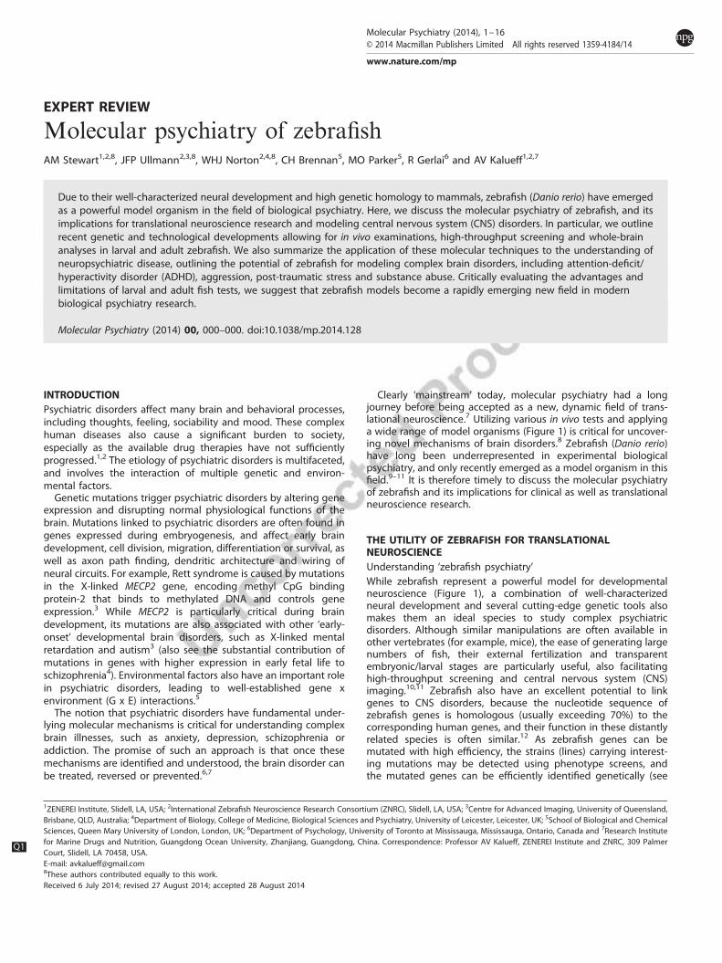
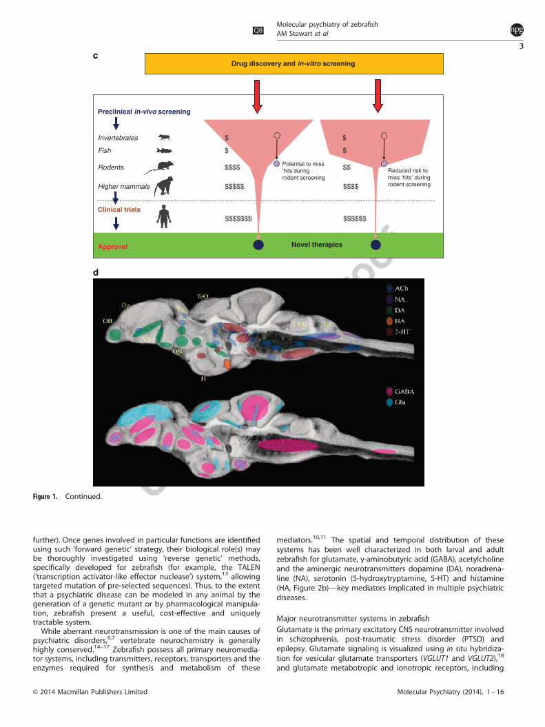
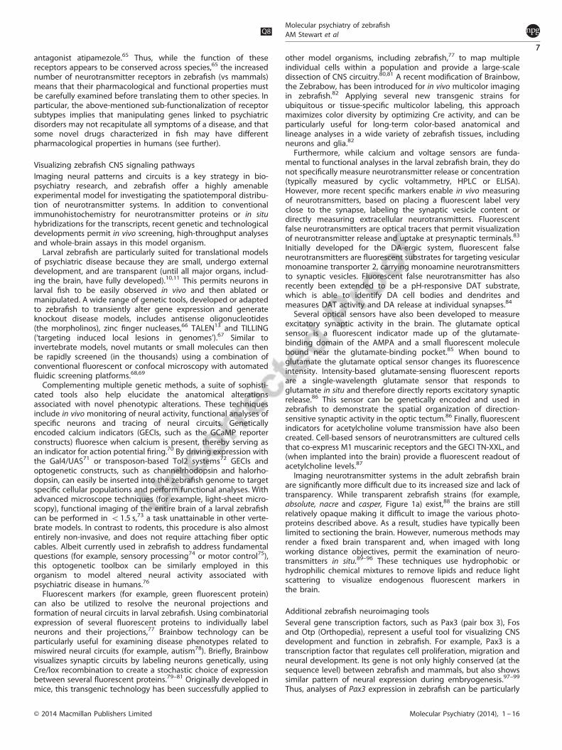
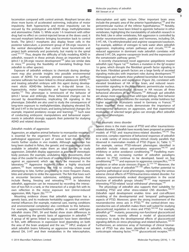
Figure 1. A brief summary of zebrafish experimental models in neuroscience research. (a) The evolving nature of zebrafish models in the last50 years, initially used mainly for basic genetic and neurodevelopmental studies, but more recently applied to developing in vivo models forcomplex brain disorders, such as autism, depression and psychoses, is shown. Inset: selected zebrafish strains useful in biological psychiatryresearch (top to bottom: adult wild-type zebrafish, casper, spiegeldanio and nacre mutants); photos courtesy of the Kalueff (ZENEREI Institute,USA), the Norton (University of Leicester, UK), the Parichy (University of Washington, USA) laboratories and Carolina Biological SupplyCompany (Burlington, USA). (b) Two research strategies which can both be applied to zebrafish models are illustrated. As a vertebrate speciesamenable to in vivo analyses and with high genetic/physiological homology to humans, zebrafish are ideal for ‘intensive’, mechanisticallydriven neuroscience research into conserved, core molecular pathways or neural circuits (photo). Due to their small size, ease of maintenanceand short generation time, zebrafish also represent an excellent model for ‘extensive’ biomedical research, including low-cost high-throughputscreening for small molecules or genetic mutations. Both strategies can lead to the development of novel therapies for major psychiatricdisorders. Photo: visualizing zebrafish signaling as an example of intensive, pathway-oriented mechanistic research (the left image showsexpression patterns of genetically encoded calcium indicator (GECI); zebrafish at 5 dpf show pan-neuronal expression of HuC:Gal4; UAS:GCaMP5,a zoomed-in image on the right shows a single-cell resolution: note that some cells are activated, as indicated by the increased fluorescence;scale bar 100 μm). (c) The benefits of including inexpensive zebrafish models into preclinical screening batteries are illustrated. A substantialfiscal saving (shown as $) can be achieved by narrowing screening to potentially active compounds using fish (rather than mice) as the firstvertebrate model organism in the screening pipeline. In addition, the risks of missing promising ‘hits’ are lower because the subsequentscreening in mammals will be ‘confirmatory’, and based on more valid data generated from zebrafish (rather than invertebrate or in vitro) tests.(d) Major neurotransmitter systems in the adult zebrafish brain are summarized: ACh—acetylcholine (dark blue), NA—noradrenergic (purple),DA—dopamine (green), HA—histamine (orange), 5-HT—serotonin (red); GABA—γ-aminobutyric acid (pink) and Glu—glutamate (light blue).The highlighted brain regions include olfactory bulb (OB), dorsal pallium (Dp), ventral pallium (Vp), habenula (Ha), optic nerve (ON), facial lobe(LVII) and vagal lobe (LX). Highlighted areas have been overlaid over a minimum deformation model of the adult zebrafish brain.
Molecular psychiatryQ8 of zebrafishAM Stewart et al
2
Molecular Psychiatry (2014), 1 – 16 © 2014 Macmillan Publishers Limited
further). Once genes involved in particular functions are identifiedusing such ‘forward genetic’ strategy, their biological role(s) maybe thoroughly investigated using ‘reverse genetic’ methods,specifically developed for zebrafish (for example, the TALEN(‘transcription activator-like effector nuclease’) system,13 allowingtargeted mutation of pre-selected sequences). Thus, to the extentthat a psychiatric disease can be modeled in any animal by thegeneration of a genetic mutant or by pharmacological manipula-tion, zebrafish present a useful, cost-effective and uniquelytractable system.While aberrant neurotransmission is one of the main causes of
psychiatric disorders,6,7 vertebrate neurochemistry is generallyhighly conserved.14–17 Zebrafish possess all primary neuromedia-tor systems, including transmitters, receptors, transporters and theenzymes required for synthesis and metabolism of these
mediators.10,11 The spatial and temporal distribution of thesesystems has been well characterized in both larval and adultzebrafish for glutamate, γ-aminobutyric acid (GABA), acetylcholineand the aminergic neurotransmitters dopamine (DA), noradrena-line (NA), serotonin (5-hydroxytryptamine, 5-HT) and histamine(HA, Figure 2b)—key mediators implicated in multiple psychiatricdiseases.
Major neurotransmitter systems in zebrafishGlutamate is the primary excitatory CNS neurotransmitter involvedin schizophrenia, post-traumatic stress disorder (PTSD) andepilepsy. Glutamate signaling is visualized using in situ hybridiza-tion for vesicular glutamate transporters (VGLUT1 and VGLUT2),18
and glutamate metabotropic and ionotropic receptors, including
Drug discovery and in-vitro screening
$$$$$$$ $$$$$$
$$$$$ $$$$
$ $
Reduced risk tomiss ‘hits’ duringrodent screening
$ $
Novel therapies
Potential to miss‘hits’duringrodent screening
$$$$ $$
Preclinical in-vivo screening
Invertebrates
Fish
Rodents
Higher mammals
Clinical trials
Approval
Figure 1. Continued.
Molecular psychiatryQ8
of zebrafishAM Stewart et al
3
© 2014 Macmillan Publishers Limited Molecular Psychiatry (2014), 1 – 16
Activity
Activity
Activity
Cognition
Cognition
Affect
Affect
Affect
Aggression
Aggression
Aggression
Reward
Reward
Reward
Aggression
ADHD
PTSD
Control tank(no marble)
Acute predator exposure test
Leaf fish Predator avoidance
Predator side Opposite side
% Timein zone
Neophobia (novel objectexposure test)
*
Marble
Marble avoidance
Mirror exposure test
Cognition
Mirror reflection
50
Distance swum, mm/5 min
% Time spent inaggressive display
Locomotor traces
Test battery
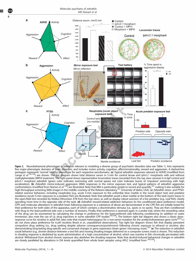
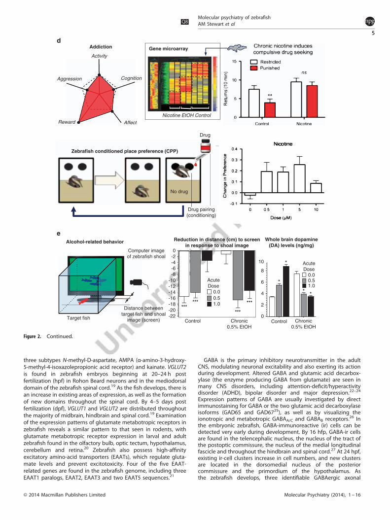
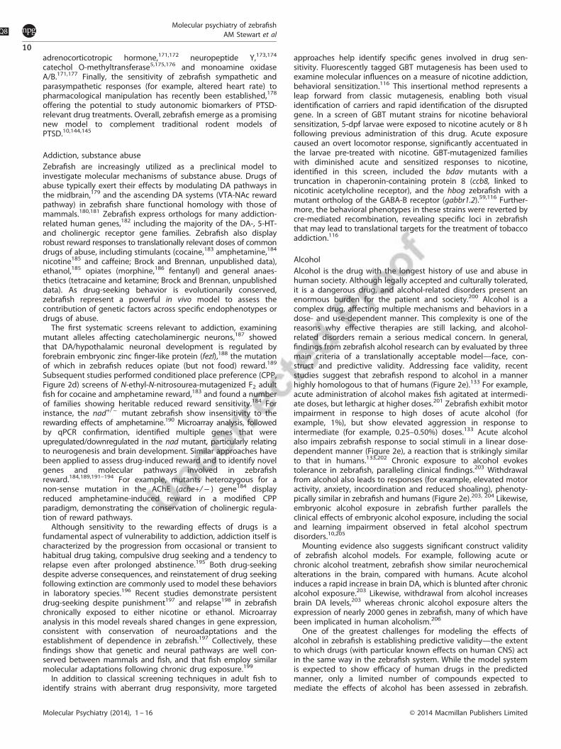
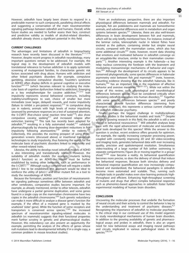
Figure 2. Neurobehavioral phenotypes in zebrafish relevant to modeling a diverse group of psychiatric disorders (also see Table 1). Axis representsfive major phenotypic domains of brain disorders, and includes motor activity, cognition, affect/emotionality, reward and aggression. A dashed-linepentagon represents ‘normal’ healthy phenotype for each respective axis/domains. (a) Typical zebrafish responses relevant to ADHD (modified fromLange et al.125,126) are shown. The bar diagram shows total distance swum in 5min for control larvae and lphn3.1 morphants with and withoutmethylphenidate (MPH) treatment. The right panel shows representative locomotion traces (recorded from the top view camera) in 6-dpf control andlphn3.1 morphant zebrafish (green color indicates swimming with normal speed, red color indicates bursts of ‘impulsive’ swimming with highacceleration). (b) Zebrafish mirror-induced aggression (MIA) responses in the mirror exposure test, and typical patterns of zebrafish aggressiveconfrontations (modified from Norton et al.139) are illustrated. Note that MIA is particularly simple to record and quantify,161 making it also suitable forhigh-throughput screening (MIA images in the middle: courtesy of the Robison laboratory,161 University of Idaho, USA). (c) Zebrafish stress- and PTSD-related aversive behaviors, including neophobia (e.g., acute 5-min exposure to the unfamiliar blue marble in the novel object test) and predatoravoidance (acute 5-min exposure to a predator fish) are illustrated. Note that zebrafish avoid a blue marble at the bottom of the tank (swim traces inthe open-field test recorded by Noldus Ethovision XT8 from the top view), as well as display robust aversion of a live predator (e.g., Leaf fish), clearlyspending more time in the opposite side of the tank. (d) Zebrafish reward-related addiction behaviors (in the conditioned place preference model,CPP) and molecular alterations in zebrafish brain following exposure to a substance of abuse are demonstrated. In the CPP, fish are initially tested fortheir preference for both sides of the apparatus, each of which contains a discriminative stimulus (i.e., spots vs no spots). They are then conditionedwith drug to the least-preferred side over a number of sessions. Finally, their preference is assessed again in a probe trial, where the rewarding valueof the drug can be ascertained by calculating the change in preference for the least-preferred side following conditioning (in addition to waterimmersion, also note the use of i.p. drug injections in some zebrafish CPP studies184,190). The bottom right bar diagram also shows a classic dose–response curve for nicotine in adult fish (also note that mutant heterozygous for a non-sense mutation for the acetlycholinesterase gene (achesb55/+)do not show place preference for 6 μM nicotine; Brock et al., unpublished observations). Top right bar diagram shows fish that display persistentcompulsive drug seeking (i.e., drug seeking despite adverse consequences/punishment) following chronic exposure to ethanol or nicotine, alsodemonstrating long-lasting drug-specific and conserved changes in gene expression (brain genes’microarray, inset).197 (e) The reduction in zebrafishsocial behavior (e.g., shorter distance between a test fish and moving shoaling images delivered on a computer screen; inset) is shown. This reductionin shoaling response is abolished by acute administration of a high concentration of alcohol, which was inactive in fish chronically pre-treated withalcohol. Withdrawal from alcohol chronic exposure also abolishes the shoaling response. Note that alcohol-induced behavioral changes in zebrafishare closely paralleled by alterations in DA levels quantified from whole brain samples using HPLC (modified from 203,230).
Molecular psychiatryQ8
of zebrafishAM Stewart et al
4
Molecular Psychiatry (2014), 1 – 16 © 2014 Macmillan Publishers Limited
three subtypes N-methyl-D-aspartate, AMPA (α-amino-3-hydroxy-5-methyl-4-isoxazolepropionic acid receptor) and kainate. VGLUT2is found in zebrafish embryos beginning at 20–24 h postfertilization (hpf) in Rohon Beard neurons and in the mediodorsaldomain of the zebrafish spinal cord.19 As the fish develops, there isan increase in existing areas of expression, as well as the formationof new domains throughout the spinal cord. By 4–5 days postfertilization (dpf), VGLUT1 and VGLUT2 are distributed throughoutthe majority of midbrain, hindbrain and spinal cord.19 Examinationof the expression patterns of glutamate metabotropic receptors inzebrafish reveals a similar pattern to that seen in rodents, withglutamate metabotropic receptor expression in larval and adultzebrafish found in the olfactory bulb, optic tectum, hypothalamus,cerebellum and retina.20 Zebrafish also possess high-affinityexcitatory amino-acid transporters (EAATs), which regulate gluta-mate levels and prevent excitotoxicity. Four of the five EAAT-related genes are found in the zebrafish genome, including threeEAAT1 paralogs, EAAT2, EAAT3 and two EAAT5 sequences.21
GABA is the primary inhibitory neurotransmitter in the adultCNS, modulating neuronal excitability and also exerting its actionduring development. Altered GABA and glutamic acid decarbox-ylase (the enzyme producing GABA from glutamate) are seen inmany CNS disorders, including attention-deficit/hyperactivitydisorder (ADHD), bipolar disorder and major depression.22–24
Expression patterns of GABA are usually investigated by directimmunostaining for GABA or the two glutamic acid decarboxylaseisoforms (GAD65 and GAD6725), as well as by visualizing theionotropic and metabotropic GABAA/C and GABAB receptors.26 Inthe embryonic zebrafish, GABA-immunoreactive (ir) cells can bedetected very early during development. By 16 hfp, GABA-ir cellsare found in the telencephalic nucleus, the nucleus of the tract ofthe postoptic commissure, the nucleus of the medial longitudinalfascicle and throughout the hindbrain and spinal cord.27 At 24 hpf,existing ir-cell clusters increase in cell numbers, and new clustersare located in the dorsomedial nucleus of the posteriorcommissure and the primordium of the hypothalamus. Asthe zebrafish develops, three identifiable GABAergic axonal
Activity
Cognition
Affect
Aggression
RewardNicotine EtOH Control
Zebrafish conditioned place preference (CPP)
Gene microarray
Drug
No drug
Alcohol-related behavior
-22-20-18-16-14-12-10
-8-6-4-20
0.00.51.0
AcuteDose
0
2
4
6
80.00.51.0
AcuteDose
10
Computer imageof zebrafish shoal
Target fish
Whole brain dopamine(DA) levels (ng/mg)
Control
******
***
***
*
*
**
Reduction in distance (cm) to screenin response to shoal image
Control Chronic0.5% EtOH
Chronic0.5% EtOH
Addiction
Drug pairing(conditioning)
Distance betweentarget fish and shoal
image (screen)
Figure 2. Continued.
Molecular psychiatryQ8
of zebrafishAM Stewart et al
5
© 2014 Macmillan Publishers Limited Molecular Psychiatry (2014), 1 – 16

projections include the supraoptic tract, the tract of the postopticcommissure and the medial longitudinal fascicle. By 3 dpf, thedistribution of GABA-ergic neurons in the zebrafish brain is verysimilar to its mammalian counterparts at a corresponding deve-lopmental stage.28 GABA-ir cells are widely distributed, locating inthe olfactory bulb, subpallium, posterior preoptic area, the dience-phalic basal plate, the central optic tectum, torus semicircularis,ventral mesencephalic tegmentum, valvula of the cerebellum andmedulla oblongata.19,28 In adult zebrafish, GABA is broadlydistributed. GABA-ergic expression in the telencephalon is verysimilar to embryonic zebrafish, with GABA-ir cells in the internalcellular layer of the olfactory bulb29 and all regions of the sub-pallium, as well as the preoptic, pretectal, ventral, thalamic, hypo-thalamic, preglomerular and posterior tubercular nuclei, cerebellarcorpus, valvus and vestibulolateral lobe.25,26
DA and NA are the primary catecholamine neurotransmitters inzebrafish. DA has a key role in modulating learning, memory,motor control, food intake and motivation. Deficits of DA neuro-transmission have been implicated in bipolar disorder, schizo-phrenia, ADHD and drug addiction. NA-ergic neurons serve animportant function in the autonomic nervous system, but alsomodulate attention, arousal, reward, depression and schizophre-nia. In zebrafish, the catecholaminergic signaling pathways havebeen characterized by visualizing the temporal and spatialexpression of tyrosine hydroxylase (TH)30,31 or the th transcript32
in all catecholaminergic neurons, the DA transporter (dat) for DAneurons and DA beta hydroxylase (dbh) for NA neurons,33,34 andthe three zebrafish DA receptors D1, D2 and D3.35,36 Zebrafish DA-ergic neurons are first formed in the telencephalon (olfactory bulband subpallium) and diencephalon (preoptic area, pretectum,ventral thalamus, posterior tuberculum and hypothalamus) withprojections that terminate locally.30,33 In adult zebrafish, th1 andth2 are differentially expressed, with th1 expressed in the tele-ncephalon, ventral thalamus and pretectum, th2 more broadlydistributed in the hypothalamus, and both genes stronglyexpressed in the posterior tuberculum and preoptic region.32
There are some notable differences between zebrafish andmammals: for example, zebrafish lack DA-ergic cell groups inthe mesencephalon14,33 and ventral midbrain,37 but possess anadditional pretectal group.18 The zebrafish NA system is verysimilar to mammals, with NA cell groups located in the locuscoeruleus, medulla oblongata and area postrema (AP).38,39
Zebrafish NA neurons can already be identified by 16 hpf, withfew changes between larval and adult fish.40 Projections from NAneurons are also conserved between zebrafish and mammals,37
with neurons in the locus coeruleus projecting to telencephalon,diencephalon and mesencephalon, in the medulla projecting tohindbrain and spinal cord, and in the AP projecting to the sameareas as the locus coeruleus.40
HA also has a key role in regulating diverse brain functions (forexample, higher cognitive functions, circadian rhythms andlocomotor activity) and modulating other aminergic systems.The zebrafish HA-ergic system is very similar to other vertebrates,with L-histidine decarboxylase and three of the four HA receptors(H1, H2 and H3) found in the brain.41 The first HA-ir neuronsdetected in zebrafish embryos are located in the ventralhypothalamus at ~ 85 hpf, and by 90 hpf the first ir-fibers canbe detected in the dorsal telencephalon.42 In adult zebrafish, HA-irneurons are found in the ventrocaudal region of the hypothala-mus surrounding the posterior recess16 and innervate every majorregion in the brain (except the cerebellum).14
5-HT mediates excitatory and inhibitory neurotransmission,serving a critical role in aggression, anxiety, cognition, learning,memory, mood and sleep. Perturbation of 5-HT levels duringdevelopment have been implicated in abnormal psychiatricbehaviors, such as anxiety spectrum disorders, schizophreniaand depression.43 Zebrafish express four 5-HT receptors (htr1aa,htr1ab, htr1bd and htr2c),44,45 the spatial expression of which in
the brain is primarily visualized by immuno-histochemistry andin situ hybridization.46 More recently, transgenic strains expressingenhanced green fluorescent protein under control of the zebrafishraphe-specific gene pet1 have also been developed.47 5-HTneurons are expressed in the spinal cord of embryonic zebrafishby 1 dpf, with additional clusters distributed throughout the brain(including the telencephalon and the hindbrain) by 5 dpf.30,48 Inadult zebrafish, 5-HT neurons are primarily located in the raphenucleus with ascending projections terminating in telencephalon,diencephalon, tegmentum, hindbrain and spinal cord.47 Addi-tional groups of anatomically distinct clusters of 5-HT-ir cells arealso found in the pineal, pretectal area, posterior tuberculum andhypothalamus, reticular formation, AP and spinal cord.14,47,49 5-HTneurons have also been identified by the expression of othermarkers, such as three tryptophan hydroxylase genes (tph1a,tph1b and tph2),50,51 two 5-HT transporters genes (serta andsertb)44,52 and two vesicular monoamine transporter genes (vmat1and vmat2).53 Examination of the transporter genes permits morespecific analysis of temporal and spatial expression pattern of5-HT, since it can be unique, with serta expressed in the pineal,pretectal area and raphe, and sertb in the retina, posteriortuberculum/hypothalamus and AP.54
Acetylcholine has a fundamental role in CNS functions (forexample, sleep, learning/memory and attention) and dysfunctions(for example, schizophrenia, ADHD and depression). The zebrafishcholinergic system is generally similar to other vertebrates, withboth muscarinic55 and the full set of nicotinic receptors.56,57 Thedistribution of cholinergic neurons in the zebrafish brain has beenprimarily described by mapping the enzymes choline acetyltrans-ferase and acetylcholinesterase (AChE). Cholinergic-ir neurons firstappear immediately after hatching in the zebrafish embryo optictectum. By 60 hpf, additional cells are seen in the preoptic areaand tegmentum, and by the completion of the embryonic stage,additional reactivity is seen in the isthmic region and medullaoblongata.58 Adult zebrafish display widespread distribution ofboth choline acetyltransferase and AChE. AChE-ir neurons arefound in the preoptic region, periventricular layer of the optictectum, rostral tegmental, oculomotor, trochlear nuclei of themesencephalic tegmentum, isthmic region, octaval nuclei andmotor nuclei of the cranial nerves.59 In contrast, choline acetyl-transferase-ir neurons are located in the pretectum and thetectum, except the marginal layer, the mesencephalic tegmentum,isthmic region, in Purkinje and granule cells in the cerebellum, andthroughout the medulla.Finally, it is important to consider the fish-specific genome
duplication that occurred around 450 million years ago.60 Inzebrafish, this duplication resulted in expressing more neuro-transmitter receptors than other species. For example, zebrafishhave an estimated 122 G-protein coupled receptors for biogenicamines (such as 5-HT, NA, DA, HA, adrenaline and trace amines),compared with 57 in mouse and 44 in humans.61 Importantly, notall zebrafish receptor paralogs have been identified and char-acterized. For example, to date only three 5-HT receptors havebeen cloned, despite the existence of seven distinct 5-HT receptorfamilies in other animals.44 Furthermore, the functional andpharmacological properties of zebrafish receptors have not beenwell studied,62 making it more difficult to translate informationacross species. One reasonably well-characterized group is thealpha-2 adrenoceptors (adra2). The zebrafish genome containsfive adra2 receptors, three of which (adra2a, adra2b and adra2c)are conserved with mammals.63 However, the other two subtypes,adra2da and adra2d, are zebrafish specific, whereas mammalianadra2a-c paralogs are not found in fish. Strikingly, despite fairlylow sequence homology (~55% overall, rising to ~ 75% in thetrans-membrane domains), human and zebrafish receptors showsimilar ligand-binding profiles.64 For example, the adra2 agonistdexmedetomidine acts as a sedative in zebrafish and otherspecies, an effect that can be inhibited by the adra2-specific
Molecular psychiatryQ8
of zebrafishAM Stewart et al
6
Molecular Psychiatry (2014), 1 – 16 © 2014 Macmillan Publishers Limited
antagonist atipamezole.65 Thus, while the function of thesereceptors appears to be conserved across species,65 the increasednumber of neurotransmitter receptors in zebrafish (vs mammals)means that their pharmacological and functional properties mustbe carefully examined before translating them to other species. Inparticular, the above-mentioned sub-functionalization of receptorsubtypes implies that manipulating genes linked to psychiatricdisorders may not recapitulate all symptoms of a disease, and thatsome novel drugs characterized in fish may have differentpharmacological properties in humans (see further).
Visualizing zebrafish CNS signaling pathwaysImaging neural patterns and circuits is a key strategy in bio-psychiatry research, and zebrafish offer a highly amenableexperimental model for investigating the spatiotemporal distribu-tion of neurotransmitter systems. In addition to conventionalimmunohistochemistry for neurotransmitter proteins or in situhybridizations for the transcripts, recent genetic and technologicaldevelopments permit in vivo screening, high-throughput analysesand whole-brain assays in this model organism.Larval zebrafish are particularly suited for translational models
of psychiatric disease because they are small, undergo externaldevelopment, and are transparent (until all major organs, includ-ing the brain, have fully developed).10,11 This permits neurons inlarval fish to be easily observed in vivo and then ablated ormanipulated. A wide range of genetic tools, developed or adaptedto zebrafish to transiently alter gene expression and generateknockout disease models, includes antisense oligonucleotides(the morpholinos), zinc finger nucleases,66 TALEN13 and TILLING(‘targeting induced local lesions in genomes’).67 Similar toinvertebrate models, novel mutants or small molecules can thenbe rapidly screened (in the thousands) using a combination ofconventional fluorescent or confocal microscopy with automatedfluidic screening platforms.68,69
Complementing multiple genetic methods, a suite of sophisti-cated tools also help elucidate the anatomical alterationsassociated with novel phenotypic alterations. These techniquesinclude in vivo monitoring of neural activity, functional analyses ofspecific neurons and tracing of neural circuits. Geneticallyencoded calcium indicators (GECIs, such as the GCaMP reporterconstructs) fluoresce when calcium is present, thereby serving asan indicator for action potential firing.70 By driving expression withthe Gal4/UAS71 or transposon-based Tol2 systems72 GECIs andoptogenetic constructs, such as channelrhodopsin and halorho-dopsin, can easily be inserted into the zebrafish genome to targetspecific cellular populations and perform functional analyses. Withadvanced microscope techniques (for example, light-sheet micro-scopy), functional imaging of the entire brain of a larval zebrafishcan be performed in o1.5 s,73 a task unattainable in other verte-brate models. In contrast to rodents, this procedure is also almostentirely non-invasive, and does not require attaching fiber opticcables. Albeit currently used in zebrafish to address fundamentalquestions (for example, sensory processing74 or motor control75),this optogenetic toolbox can be similarly employed in thisorganism to model altered neural activity associated withpsychiatric disease in humans.76
Fluorescent markers (for example, green fluorescent protein)can also be utilized to resolve the neuronal projections andformation of neural circuits in larval zebrafish. Using combinatorialexpression of several fluorescent proteins to individually labelneurons and their projections,77 Brainbow technology can beparticularly useful for examining disease phenotypes related tomiswired neural circuits (for example, autism78). Briefly, Brainbowvisualizes synaptic circuits by labeling neurons genetically, usingCre/lox recombination to create a stochastic choice of expressionbetween several fluorescent proteins.79–81 Originally developed inmice, this transgenic technology has been successfully applied to
other model organisms, including zebrafish,77 to map multipleindividual cells within a population and provide a large-scaledissection of CNS circuitry.80,81 A recent modification of Brainbow,the Zebrabow, has been introduced for in vivo multicolor imagingin zebrafish.82 Applying several new transgenic strains forubiquitous or tissue-specific multicolor labeling, this approachmaximizes color diversity by optimizing Cre activity, and can beparticularly useful for long-term color-based anatomical andlineage analyses in a wide variety of zebrafish tissues, includingneurons and glia.82
Furthermore, while calcium and voltage sensors are funda-mental to functional analyses in the larval zebrafish brain, they donot specifically measure neurotransmitter release or concentration(typically measured by cyclic voltammetry, HPLC or ELISA).However, more recent specific markers enable in vivo measuringof neurotransmitters, based on placing a fluorescent label veryclose to the synapse, labeling the synaptic vesicle content ordirectly measuring extracellular neurotransmitters. Fluorescentfalse neurotransmitters are optical tracers that permit visualizationof neurotransmitter release and uptake at presynaptic terminals.83
Initially developed for the DA-ergic system, fluorescent falseneurotransmitters are fluorescent substrates for targeting vesicularmonoamine transporter 2, carrying monoamine neurotransmittersto synaptic vesicles. Fluorescent false neurotransmitter has alsorecently been extended to be a pH-responsive DAT substrate,which is able to identify DA cell bodies and dendrites andmeasures DAT activity and DA release at individual synapses.84
Several optical sensors have also been developed to measureexcitatory synaptic activity in the brain. The glutamate opticalsensor is a fluorescent indicator made up of the glutamate-binding domain of the AMPA and a small fluorescent moleculebound near the glutamate-binding pocket.85 When bound toglutamate the glutamate optical sensor changes its fluorescenceintensity. Intensity-based glutamate-sensing fluorescent reportsare a single-wavelength glutamate sensor that responds toglutamate in situ and therefore directly reports excitatory synapticrelease.86 This sensor can be genetically encoded and used inzebrafish to demonstrate the spatial organization of direction-sensitive synaptic activity in the optic tectum.86 Finally, fluorescentindicators for acetylcholine volume transmission have also beencreated. Cell-based sensors of neurotransmitters are cultured cellsthat co-express M1 muscarinic receptors and the GECI TN-XXL, and(when implanted into the brain) provide a fluorescent readout ofacetylcholine levels.87
Imaging neurotransmitter systems in the adult zebrafish brainare significantly more difficult due to its increased size and lack oftransparency. While transparent zebrafish strains (for example,absolute, nacre and casper, Figure 1a) exist,88 the brains are stillrelatively opaque making it difficult to image the various photo-proteins described above. As a result, studies have typically beenlimited to sectioning the brain. However, numerous methods mayrender a fixed brain transparent and, when imaged with longworking distance objectives, permit the examination of neuro-transmitters in situ.89–96 These techniques use hydrophobic orhydrophilic chemical mixtures to remove lipids and reduce lightscattering to visualize endogenous fluorescent markers inthe brain.
Additional zebrafish neuroimaging toolsSeveral gene transcription factors, such as Pax3 (pair box 3), Fosand Otp (Orthopedia), represent a useful tool for visualizing CNSdevelopment and function in zebrafish. For example, Pax3 is atranscription factor that regulates cell proliferation, migration andneural development. Its gene is not only highly conserved (at thesequence level) between zebrafish and mammals, but also showssimilar pattern of neural expression during embryogenesis.97–99
Thus, analyses of Pax3 expression in zebrafish can be particularly
Molecular psychiatryQ8
of zebrafishAM Stewart et al
7
© 2014 Macmillan Publishers Limited Molecular Psychiatry (2014), 1 – 16

useful for imaging early CNS development,97,98 which is relevantto modeling many neurodevelopmental disorders.The homeodomain transcription factor Otp is an important
regulator of neuroendocrine and dopaminergic cells in vertebratespecies.100,101 Otp has two paralogs in zebrafish (otpa and otpb),101
and genetic manipulations suppressing Otp deplete certain dopa-minergic neurons, enabling a functional dissection of dopaminer-gic contribution to CNS circuitry in early larval zebrafish.102
Likewise, an early proto-oncogene c-fos serves as a well-established marker of neuronal activation in various species,including humans,103 rodents104,105 and zebrafish.106 For example,c-fos expression analyses of whole-brain zebrafish samples can beused to assess neuronal activation globally (for example, reflectingCNS excitation during epilepsy106,107 or following anxiogenic/anxiolytic or hallucinogenic treatments108,109). Region-specificanalyses of c-fos expression in zebrafish brain offer a moredetailed functional mapping of neural circuits during variousbehavioral tests.110 For instance, in the light-dark test, c-fosneuronal activity mapping implicated several brain regions,including several telencephalic areas (the teleost homologs ofthe mammalian amygdala and striatum), during zebrafish‘anxiogenic’ light avoidance behavior.110 Characterizing neuronalactivity induced in zebrafish by acute amphetamine, drug-seekingor the light avoidance behavior, quantitative analyses of c-fosexpression can also be combined with neuronal subtype-specificmarkers at single-cell resolution.111 Showing the recruitment ofthe medial and the lateral pallium (structures homologous to themammalian amygdala and hippocampus), these data suggestevolutionarily conserved function of amygdala-like structures inpositive emotions and motivated behavior in zebrafish andmammals.111 Collectively, this evidence strongly supports theutility of c-fos and other biomarkers in visualizing normal andaberrant CNS development and functioning.Finally, with the increased use of zebrafish in disease genetics
research, a quantitative voxel-based technique may help comparedisease models with wild-type zebrafish. Magnetic resonanceimaging is an inherent 3D tool that facilitates investigations inmorphology, connectivity and function of the brain. Due to its 3Dnature, multiple data sets can be mapped to a standardcoordinate space and registered to produce a model representa-tive of a population (thus, limiting individual morphologicalvariance and instead demonstrating the average morphology ofthe entire population). Data collected from magnetic resonanceimaging can include mean volumes of individual brain regions,the stereotaxic locations of individual structures and major fiberbundles, and functional activity. Importantly, magnetic resonanceimaging models can also serve as a canonical space forregistration of other imaging data sets. For example, recentwhole-brain immunostaining and light microscope imaging (forexample, SeeDB or Clarity), optical projection tomography and
microCT can all be registered to the same stereotaxic space,collectively enabling quantitative analysis of a wide range ofmarkers in zebrafish brain.
ZEBRAFISH MODELS OF BRAIN DISORDERSBridging molecular events with behaviorMany psychiatric disorders are moderately or highly heritable, andhave developmental trajectories.112,113 As behavior is controlledby activity within neural circuits, a major challenge is to map thegenotype–phenotype relationship to understand how geneticvariations affect neural circuits that control behavior. Variousmolecular tools have been effectively used for uncovering brainmechanisms in zebrafish. For example, in addition to traditionalN-ethyl-N-nitrosourea-induced114 and viral-vector-mediated115
mutagenesis, new sophisticated ‘gene-breaking transposon’(GBT)116 screens and ‘clustered regularly interspaced shortpalindromic repeats’ approaches for generating targeted zebrafishmutants have been developed.117 Combined with TILLING andTALEN tools, this makes the process of exploring genotype–phenotype relationships in zebrafish better targeted and lesslabor-intensive. With the establishment of translationally relevantzebrafish behavioral assays9–11 comes the ability to apply thesemolecular techniques to modeling psychiatric diseases. Here, wediscuss how various zebrafish models can be utilized to targetselected psychiatric disorders and their neurodevelopmentaltrajectories. ADHD, aggression, post-traumatic stress and drugabuse (Figure 2) were selected as examples, reflecting a wide anddiverse spectrum of psychiatric disorders that can be modeledusing the zebrafish.
Modeling ADHDZebrafish have recently been utilized to investigate some aspectsof ADHD (Table 1; Figure 2a), a common psychiatric disorderaffecting ~ 3–5% of children worldwide. ADHD symptoms(inattention, hyperactivity and impulsivity) persist into adulthoodin ~ 50% of cases, reducing the patients’ quality of life, academicperformance and sociality.118,119 Altered DA, NA, 5-HT andglutamate neurotransmission in the prefrontal cortex, striatum,parietal cortex, vermis and the inferior lobes of the cerebellum areimplicated in ADHD.120–123
An interesting example of using larval zebrafish to model thisdisorder involves a human gene LATROPHILIN3 (LPHN3), identifiedas candidate ADHD gene by linkage analysis.124 In zebrafish, itshomolog latrophilin3.1 (lphn3.1) has a developmental role, and isexpressed in differentiated neurons throughout the brain up to 6dpf. The lphn3.1 morphants display increased distance swum at 6dpf (Figure 2a)125,126—the hyperactivity phenotype also main-tained during the night, demonstrating a permanent increase in
Table 1. Examples of a diverse group of psychiatric disorders (Figure 2) which can be modeled in zebrafish (see Kalueff et al.135 for a comprehensivecatalog of zebrafish normal and pathological behaviors)
Selected disorders Zebrafish phenotypes References
Attention deficit hyperactivitydisorder (ADHD)
Impulsive swimming with hyperactive locomotion and impulsivity-like burstsof accelerated swimming, which can be reduced by anti-ADHD drugs (Figure 2a)
125,126,217,218,231
Aggression Social deficits (disrupted shoaling behavior), increased aggressive behaviors(follows, chases and biting), mirror-induced aggression (MIA) (Figure 2b)
135,139,161
Post-traumatic stressdisorder (PTSD)
Reduced exploration following stress, increased avoidance (e.g., neophobia;Figure 2c), erratic behavior and freezing, elevated cortisol and brain c-fos(all highly sensitive to a wide range of anxiolytic and anxiogenic agents),social deficits (e.g., disrupted shoaling behavior)
144,146,147,232
Substance abuse, addiction(reward-related behavior)
Robust conditioned preference for commonly abused substances, compulsivedrug seeking (i.e., despite adverse consequences; Figure 2d), withdrawal syndrome,stimulus-induced relapse following extinction, altered social behavior (Figure 2e)
197,198,201,202,205,233–235
Molecular psychiatryQ8
of zebrafishAM Stewart et al
8
Molecular Psychiatry (2014), 1 – 16 © 2014 Macmillan Publishers Limited
locomotion compared with control animals. Morphant larvae alsoshow more bursts of accelerated swimming, indicative of motorimpulsivity. Both hyperactivity and motor impulsivity can berescued by the prototypical anti-ADHD drugs methylphenidateand atomoxetine (Table 1). While acute 1-h treatment with eitherdrug had no effect on control-injected larvae at the doses used, itrescued morphant behavior bringing locomotion back to controllevels. The lphn3.1 morphants also display fewer cells in theposterior tuberculum, a prominent group of DA-ergic neurons inthe ventral diencephalon that control larval locomotion andproject both anteriorly and posteriorly, but mainly to the spinalcord.101,127 Thus, despite the difficulty of fully modeling ADHD inzebrafish, research in these animals shows an important role forlphn3.1 in DA-ergic neuron development128 (also see similar datain mice,129 proving the feasibility of translating findings fromzebrafish to other species).Exposure of zebrafish embryos to chemicals during develop-
ment may also provide insights into possible environmentalcauses of ADHD. For example, prenatal exposure to perfluor-ooctane sulfonate has been linked to clinical adolescent ADHD,130
and treating zebrafish embryos with this agent during develop-ment elicits ADHD-like behaviors in 6-day-old larvae (i.e.,hyperactivity, motor impulsivity and hyper-responsiveness tostartle).131 This phenotype is reminiscent of the behavior oflphn3.1 larvae and strikingly, these behavioral changes can berescued by dexamfetamine, implicating DA signaling in thisphenotype. Zebrafish are also used to study the consequences oflong-term exposure to methylphenidate, displaying elevated DA,NE and 5-HT in the larval brain, and lasting behavioral changes (forexample, impaired predatory escape and learning).132 Such easeof conducting embryonic manipulations and behavioral experi-ments in zebrafish strongly supports their potential for studyingADHD and related disorders.
Zebrafish models of aggressionAggression, an adaptive animal behavior to monopolize resources,is essential for the organism’s fitness and survival. However,pathological aggression is part of various brain disorders, andmerits further studies in vivo models. Although aggression haslong been studied in fishes, the genetic and neurobiological toolsavailable in zebrafish make them an ideal species to studyaggression. Zebrafish display aggression as a series of stereotypicbody postures (for example, undulating body movements, shortslaps of the caudal fin and bouts of swimming and biting directedagainst an opponent) which can easily be measured in thelaboratory.133 Aggressive incidents follow a highly structuredpattern,134 from extending the fins to circling, chasing andattempting to bite, further progressing to more frequent chases,bites and attempts to strike the opponent. The fish that loses suchas encounter becomes submissive, exhibiting postures thatinclude fleeing, freezing and retreating.134,135 In laboratoryzebrafish, aggression can be measured by recording the interac-tion of two fish in a tank, or the interaction of a single fish with itsown reflection in the mirror exposure test (mirror-inducedaggression, MIA; Figure 2b).133,134
Mounting evidence shows that zebrafish aggression has agenetic basis, and its moderate heritability suggests that environ-mental influences (for example, maternal care, rearing conditionsand environmental complexity) also contribute to this behavior.For example, aggression/boldness profiles in several wild-typezebrafish strains (TM1, SH and Nadia) show strain differences inMIA, supporting the genetic basis of aggression in zebrafish.136
A group of 40 genes linked to aggression have been identifiedin fish, with differences in expression level depending on boththe area of the brain analyzed and the sex.137 HPLC analyses ofadult zebrafish brains following an aggressive interaction revealaltered DA, 5-HT and their metabolites in the telencephalon,
diencephalon and optic tectum. Other important brain areasinclude the preoptic area of the anterior hypothalamus138 and theperiventricular nucleus of the inferior hypothalamus.139 Notably,similar brain areas have been linked to agonistic behavior in othervertebrates, highlighting the translatability of zebrafish research inthis field. Like in other vertebrates, fish aggression is controlled bysimilar neurotransmitters, peptides and hormones, including DA,5-HT, HA, 17α-ethinylestradiol and arginine vasopressin/vasotocin.For example, addition of estrogen to tank water alters zebrafishaggression, implicating certain pathways and circuits,133,140 asreduced aggression in dominant male zebrafish correlates withaltered 5-HT, DA, arginine vasopressin levels and the expression ofthe somatostatin pathway genes.141
A recently characterized novel aggressive spiegeldanio mutantzebrafish (spd, Figure 1a)142 harbors a mutation in the fgf receptor1a gene, which disrupts the fibroblast growth factor signaling inthe brain. Fibroblast growth factors are a large family of secretedsignaling molecules with important roles during development.143
Homozygous spd mutants show unaltered locomotion but increasedaggression, boldness and exploration (Figure 2b), correlated withelevated breakdown of HA in the brain, and reduced neuro-transmitter signaling in the hypothalamic periventricular nucleus.Importantly, pharmacological increase in HA rescues all threebehavioral alterations in spd mutants.139 Although spd zebrafishare always more aggressive than wild-type siblings, their behaviordiffers between the laboratories (for example, similar boldness buthigher aggression in mutants raised in Germany vs France).139
Taken together, these results demonstrate the importance ofenvironmental influences on aggression, and that experimentalmodulation of selected target genes can strongly affect zebrafishaggressive phenotypes.
Post-traumatic stress disorderSevere stress is the main cause of PTSD and other trauma/stress-related disorders. Zebrafish have recently been proposed as potentialmodels of PTSD and trauma/stress-related disorders.144,145 Theextensive, complex repertoire of behavioral responses of zebrafishto a wide variety of stress-evoking stimuli closely parallels thatobserved in mammalian models of PTSD (Figure 2c; Table 1).145
For example, various PTSD-relevant phenotypes identified inzebrafish include robust anti-predatory responses146,147 andinhibitory or active avoidance conditioning.148 Paralleling mam-malian tests, an increasing number of zebrafish paradigmsrelevant to PTSD continue to be developed, based on fearconditioning148–152 and exposure to aggressive conspecifics,134,138
predators or other acute stressors (Figure 2).153–155 Moreover, thehighly social nature of zebrafish156–158 offers further potential toexamine pathological social phenotypes, representing the seriousadverse clinical effects of PTSD/trauma/stress-related disorder. Forinstance, experimentally evoked disorganization in the socialstructure of zebrafish groups (shoals)158–161 may represent aninteresting PTSD-related ‘social withdrawal’ phenotype.145
The physiology of zebrafish also supports their suitability formodeling PTSD and other stress-related CNS disorders.120,121
Zebrafish rapid development enables time- and cost-efficientmodeling of the developmental and ‘temporal’ pathogeneticaspects of PTSD. Moreover, given the strong involvement of theneuroendocrine stress axis in PTSD,162 the cortisol-driven neu-roendocrine axis of zebrafish makes them well suited for studyingglucocorticoid regulation in stress and PTSD.163,164 For example,zebrafish grs357 mutants, possessing non-functional glucocorticoidreceptors, have recently offered a model of glucocorticoidresistance to study the developmental effects of glucocorticoidsignaling deficits on stress physiology and related behaviors.165,166
Furthermore, an extensive array of homologs of clinical biomar-kers of PTSD has also been identified in zebrafish, includingcorticotropin releasing factor,167,168 glucocorticoid receptor,169,170
Molecular psychiatryQ8
of zebrafishAM Stewart et al
9
© 2014 Macmillan Publishers Limited Molecular Psychiatry (2014), 1 – 16
adrenocorticotropic hormone,171,172 neuropeptide Y,173,174
catechol O-methyltransferase5,175,176 and monoamine oxidaseA/B.171,177 Finally, the sensitivity of zebrafish sympathetic andparasympathetic responses (for example, altered heart rate) topharmacological manipulation has recently been established,178
offering the potential to study autonomic biomarkers of PTSD-relevant drug treatments. Overall, zebrafish emerge as a promisingnew model to complement traditional rodent models ofPTSD.10,144,145
Addiction, substance abuseZebrafish are increasingly utilized as a preclinical model toinvestigate molecular mechanisms of substance abuse. Drugs ofabuse typically exert their effects by modulating DA pathways inthe midbrain,179 and the ascending DA systems (VTA-NAc rewardpathway) in zebrafish share functional homology with those ofmammals.180,181 Zebrafish express orthologs for many addiction-related human genes,182 including the majority of the DA-, 5-HT-and cholinergic receptor gene families. Zebrafish also displayrobust reward responses to translationally relevant doses of commondrugs of abuse, including stimulants (cocaine,183 amphetamine,184
nicotine185 and caffeine; Brock and Brennan, unpublished data),ethanol,185 opiates (morphine,186 fentanyl) and general anaes-thetics (tetracaine and ketamine; Brock and Brennan, unpublisheddata). As drug-seeking behavior is evolutionarily conserved,zebrafish represent a powerful in vivo model to assess thecontribution of genetic factors across specific endophenotypes ordrugs of abuse.The first systematic screens relevant to addiction, examining
mutant alleles affecting catecholaminergic neurons,187 showedthat DA/hypothalamic neuronal development is regulated byforebrain embryonic zinc finger-like protein (fezl),188 the mutationof which in zebrafish reduces opiate (but not food) reward.189
Subsequent studies performed conditioned place preference (CPP,Figure 2d) screens of N-ethyl-N-nitrosourea-mutagenized F2 adultfish for cocaine and amphetamine reward,183 and found a numberof families showing heritable reduced reward sensitivity.184 Forinstance, the nad+/− mutant zebrafish show insensitivity to therewarding effects of amphetamine.190 Microarray analysis, followedby qPCR confirmation, identified multiple genes that wereupregulated/downregulated in the nad mutant, particularly relatingto neurogenesis and brain development. Similar approaches havebeen applied to assess drug-induced reward and to identify novelgenes and molecular pathways involved in zebrafishreward.184,189,191–194 For example, mutants heterozygous for anon-sense mutation in the AChE (ache+/− ) gene184 displayreduced amphetamine-induced reward in a modified CPPparadigm, demonstrating the conservation of cholinergic regula-tion of reward pathways.Although sensitivity to the rewarding effects of drugs is a
fundamental aspect of vulnerability to addiction, addiction itself ischaracterized by the progression from occasional or transient tohabitual drug taking, compulsive drug seeking and a tendency torelapse even after prolonged abstinence.195 Both drug-seekingdespite adverse consequences, and reinstatement of drug seekingfollowing extinction are commonly used to model these behaviorsin laboratory species.196 Recent studies demonstrate persistentdrug-seeking despite punishment197 and relapse198 in zebrafishchronically exposed to either nicotine or ethanol. Microarrayanalysis in this model reveals shared changes in gene expression,consistent with conservation of neuroadaptations and theestablishment of dependence in zebrafish.197 Collectively, thesefindings show that genetic and neural pathways are well con-served between mammals and fish, and that fish employ similarmolecular adaptations following chronic drug exposure.199
In addition to classical screening techniques in adult fish toidentify strains with aberrant drug responsivity, more targeted
approaches help identify specific genes involved in drug sen-sitivity. Fluorescently tagged GBT mutagenesis has been used toexamine molecular influences on a measure of nicotine addiction,behavioral sensitization.116 This insertional method represents aleap forward from classic mutagenesis, enabling both visualidentification of carriers and rapid identification of the disruptedgene. In a screen of GBT mutant strains for nicotine behavioralsensitization, 5-dpf larvae were exposed to nicotine acutely or 8 hfollowing previous administration of this drug. Acute exposurecaused an overt locomotor response, significantly accentuated inthe larvae pre-treated with nicotine. GBT-mutagenized familieswith diminished acute and sensitized responses to nicotine,identified in this screen, included the bdav mutants with atruncation in chaperonin-containing protein 8 (ccb8, linked tonicotinic acetylcholine receptor), and the hbog zebrafish with amutant ortholog of the GABA-B receptor (gabbr1.2).59,116 Further-more, the behavioral phenotypes in these strains were reverted bycre-mediated recombination, revealing specific loci in zebrafishthat may lead to translational targets for the treatment of tobaccoaddiction.116
AlcoholAlcohol is the drug with the longest history of use and abuse inhuman society. Although legally accepted and culturally tolerated,it is a dangerous drug, and alcohol-related disorders present anenormous burden for the patient and society.200 Alcohol is acomplex drug, affecting multiple mechanisms and behaviors in adose- and use-dependent manner. This complexity is one of thereasons why effective therapies are still lacking, and alcohol-related disorders remain a serious medical concern. In general,findings from zebrafish alcohol research can by evaluated by threemain criteria of a translationally acceptable model—face, con-struct and predictive validity. Addressing face validity, recentstudies suggest that zebrafish respond to alcohol in a mannerhighly homologous to that of humans (Figure 2e).133 For example,acute administration of alcohol makes fish agitated at intermedi-ate doses, but lethargic at higher doses.201 Zebrafish exhibit motorimpairment in response to high doses of acute alcohol (forexample, 1%), but show elevated aggression in response tointermediate (for example, 0.25–0.50%) doses.133 Acute alcoholalso impairs zebrafish response to social stimuli in a linear dose-dependent manner (Figure 2e), a reaction that is strikingly similarto that in humans.133,202 Chronic exposure to alcohol evokestolerance in zebrafish, paralleling clinical findings.203 Withdrawalfrom alcohol also leads to responses (for example, elevated motoractivity, anxiety, incoordination and reduced shoaling), phenoty-pically similar in zebrafish and humans (Figure 2e).203, 204 Likewise,embryonic alcohol exposure in zebrafish further parallels theclinical effects of embryonic alcohol exposure, including the socialand learning impairment observed in fetal alcohol spectrumdisorders.10,205
Mounting evidence also suggests significant construct validityof zebrafish alcohol models. For example, following acute orchronic alcohol treatment, zebrafish show similar neurochemicalalterations in the brain, compared with humans. Acute alcoholinduces a rapid increase in brain DA, which is blunted after chronicalcohol exposure.203 Likewise, withdrawal from alcohol increasesbrain DA levels,203 whereas chronic alcohol exposure alters theexpression of nearly 2000 genes in zebrafish, many of which havebeen implicated in human alcoholism.206
One of the greatest challenges for modeling the effects ofalcohol in zebrafish is establishing predictive validity—the extentto which drugs (with particular known effects on human CNS) actin the same way in the zebrafish system. While the model systemis expected to show efficacy of human drugs in the predictedmanner, only a limited number of compounds expected tomediate the effects of alcohol has been assessed in zebrafish.
Molecular psychiatryQ8
of zebrafishAM Stewart et al
10
Molecular Psychiatry (2014), 1 – 16 © 2014 Macmillan Publishers Limited
However, zebrafish have largely been shown to respond in apredictable manner to such compounds, paralleling clinical effectsand suggesting a conservation of the main neurotransmittersignaling pathways between zebrafish and humans. Overall, whilefuture studies are needed to further assess their face, constructand predictive validity as models of alcohol-related disorders,zebrafish already shows a significant potential in this field.
CURRENT CHALLENGESThe advantages and limitations of zebrafish in biopsychiatryresearch have recently been discussed in the literature.9–11,174
However, despite recent progress in zebrafish neurobiology, manyimportant questions remain to be addressed. For example, thelogical step in the development of zebrafish models withtranslational relevance to human addiction is to apply systematicscreening of mutagenized families, to characterize genetic riskfactors associated with drug abuse. Humans with addiction andother linked psychiatric disorders (for example, compulsivegambling, obsessive compulsive disorder, Tourette’s syndromeor ADHD) often display deficits in general cognitive or executivefunction207–210 (which becomes relevant to studying the mole-cular basis of cognitive dysfunction linked to addiction). Emergingas a key endophenotype for cocaine addiction,211,212 traitimpulsivity represents several behavioral constructs, includinginability/unwillingness to wait, choice of smaller but moreimmediate (over larger, delayed) rewards, and motor impulsivity(failure to inhibit a pre-potent response).213 In compulsive druguse in rodents, animals with high levels of motor impulsivity(assessed by premature responses on a test of sustained attention,the 5-CSRTT (five-choice serial reaction time task)214) also showcompulsive cocaine seeking211 and increased relapse afterextinction training.215 This task has recently been adapted forzebrafish,216 resulting in a fully automated model,217 validatedpharmacologically by showing dose-dependent reductions in fishimpulsivity following atomoxetine,218 similar to rodents.219
Collectively, this provides the exciting prospect of using three-generation screens (discussed above) or insertional transposontechnologies to study impulsivity, thus fostering the search for themolecular basis of psychiatric disorders linked to impulsivity andother reward-related traits.Likewise, the ability to develop novel zebrafish models of ADHD
is also interesting and translationally relevant. However, thesuitability of zebrafish models (for example, mutants lackinglphn3.1 function) as an ADHD-like model must be furtherestablished by testing other behaviors, such as performance inthe 5-CSRTT.217 Although such a complex task will require a stablelphn3.1 line to be established, this approach would be ideal toreinforce the utility of lphn3.1 and other mutant fish as a tool tostudy the neurobiology of ADHD.Because the formation, position and function of neurotransmit-
ter signaling pathways sometimes differ between zebrafish andother vertebrates, comparative studies become important. Forexample, as already mentioned, similar to other teleosts, zebrafishhave undergone a partial genome duplication, and some genesfound in a single copy on the mammalian chromosome have twocopies in the zebrafish chromosome.220 Such genetic redundancycan make it more difficult to analyze a disease gene’s function (forexample, if the effect of a mutated gene is masked by theunaltered ‘sister’ gene). While the function of some receptors andtransporters is conserved across species,17,65 the increasedspectrum of neurotransmitter signaling-related molecules inzebrafish (vs mammals) suggests that their functional propertiesmerit further scrutiny in genetic and pharmacological studies.However, this redundancy can also present an opportunity (forexample, permitting the analysis of the effects of genes whosenull mutations lead to developmental lethality if in a single copy, acommon problem in mouse knockout studies).
From an evolutionary perspective, there are also importantphysiological differences between mammals and zebrafish. Forexample, fish are poikiloterms and mammals are homeothermicanimals, and substantial differences exist in metabolism and organsystems between species.221 Likewise, there are also well-knowndifferences in brain development between fish and mammals,which will be only briefly mentioned here. For instance, instead ofthe laminar cortex, telencephalic nuclei in teleost fishes haveevolved as the pallium, containing similar but simpler neuralcircuits, compared with the mammalian cortex, which also hassome additional circuits222 (note, however, recent data showingthat emotions and motivated behavior in zebrafish converge onan amygdala-like structure, similar to their mammalian counter-parts111). Another interesting example is the habenula—a keyrelay nucleus connecting the forebrain with the brainstem andmodulating monoaminergic mechanisms of behavior and cogni-tion.223 Although the development of its circuits appears to beconserved phylogenetically, some species differences in habenularasymmetry exist between fish and mammals223 (note, however,mounting evidence showing that the zebrafish habenula, like inhumans,224 has an important role in the regulation of affectivebehavior and aversive memories152,225–227). While not within thescope of this review, such physiological and neurobiologicaldifferences between species must be considered critically whentranslating zebrafish responses into human disorders.221 Togetherwith some genetic differences between species12 and poorlycharacterized protein function differences (stemming fromdivergent evolution), this represents a serious current challengefor zebrafish molecular psychiatry.Finally, perhaps the biggest challenge in multidisciplinary
zebrafish studies is the behavioral models and tools.9,10 Despitea rapidly growing research in this field, the zebrafish is still a newmodel in behavioral neuroscience.11 Will zebrafish analyses keeppace with the rapid advancement of molecular and neurobiolo-gical tools developed for this species? While the answer to thisquestion is unclear, recent evidence offers grounds for optimism.For example, the rapidly increasing computer processing power,coupled with constantly improving 2D and 3D video-recordingand imaging tools, generates behavioral data with ever increasingquality, precision and spatiotemporal resolution. Simultaneousvideo-tracking of a large number of fish (either swimming inseparate compartments; Figure 2b or moving together in a group/shoal)10,160 now became a reality. As behavioral quantificationbecomes more precise, so does the delivery of stimuli that inducethe behavioral responses. Because both stimulus delivery andbehavioral response quantification are now increasingly compu-terized and standardized, the behavioral paradigms in zebrafishbecome more automated and scalable. Thus, running suchmultiple tasks in parallel makes even slow learning protocols high-throughput and efficient. Enhancing high-throughput screeningof mutants and drugs that affect complex behavioral responsessuch as phenomics-based approaches in zebrafish foster furtherexperimental modeling of human brain disorders.
CONCLUSIONUncovering the molecular processes that underlie the formationof neural circuits and their activity to control the behavior is key tothe understanding and treatment of psychiatric diseases.6,7
Recognizing the importance of zebrafish to molecular psychiatryis the critical step in our continued use of this model organismto study neurobiological mechanisms of human brain disorders.In addition to the growing availability of genetic and pharmaco-logical assays and probes in zebrafish, there is also an urgentneed for new behavioral assays and imaging neural pathwaysand circuits implicated in various pathological states in thisspecies.228,229
Molecular psychiatryQ8
of zebrafishAM Stewart et al
11
© 2014 Macmillan Publishers Limited Molecular Psychiatry (2014), 1 – 16

The availability of genetic, electrophysiological and optogenetictools, as well as multiple mutant and transgenic strains, physio-logical biomarkers and robust protocols to measure behavior,collectively supports zebrafish potential for modeling psychiatricdisorders. As both larval and adult zebrafish are now rapidlybecoming key model organisms in neuroscience research, theyrepresent a useful novel species for translational molecularpsychiatry and CNS drug discovery.
CONFLICT OF INTERESTThe authors declare no conflict of interest.
ACKNOWLEDGMENTSThe authors’ research has been supported by grants from the National Institutes ofHealth (NIAAA AA015325-01A2 to RG, NIDA DA030900-02 to AVK and NINDSNS077295-01 to JFPU), the EU FP7 Framework Program (Grant 602805 to WHJN), aswell as the National Center for the Replacement, Reduction and Refinement ofAnimals in Research (NC3Rs, UK, G1000053 to CHB) and the Medical Research Council(MRC, UK to CHB). CHB is a Royal Society (UK) Industrial Research Fellow. The studywas coordinated and facilitated by the International Zebrafish Neuroscience ResearchConsortium (ZNRC). AVK is the Director of ZENEREI Institute and Chair of ZNRC.
REFERENCES1 WHO. The Global Burden of Disease: 2004 Update. WHO (World Health Organi-
zation: Geneva, Switzerland, 2008.2 Griebel G, Holmes A. 50 years of hurdles and hope in anxiolytic drug discovery.
Nat Rev Drug Discov 2013; 12: 667–687.3 Gonzales ML, LaSalle JM. The role of MeCP2 in brain development and neuro-
developmental disorders. Curr Psychiatry Rep 2010; 12: 127–134.4 Xu B, Ionita-Laza I, Roos JL, Boone B, Woodrick S, Sun Y et al. De novo gene
mutations highlight patterns of genetic and neural complexity in schizophrenia.Nat Genet 2012; 44: 1365–1369.
5 Mitchell K. The miswired brain: making connections from neurodevelopment topsychopathology. BMC Biol 2011; 9: 23.
6 Duman RS, Heninger GR, Nestler EJ. Molecular psychiatry. Adaptations ofreceptor-coupled signal transduction pathways underlying stress- and drug-induced neural plasticity. J Nerv Ment Dis 1994; 182: 692–700.
7 Nestler EJ. The origins of molecular psychiatry. J Mol Psychiatry 2013; 1: 3.8 Kalueff AV, Wheaton M, Murphy DL. What's wrong with my mouse model?
Advances and strategies in animal modeling of anxiety and depression. BehavBrain Res 2007; 179: 1–18.
9 Kalueff AV, Echevarria DJ, Stewart AM. Gaining translational momentum: Morezebrafish models for neuroscience research. Progr Neuropsychopharmacol BiolPsychiatry 2014 pii S0278-5846: 00037–2.
10 Kalueff AV, Stewart AM, Gerlai R. Zebrafish as an emerging model for studyingcomplex brain disorders. Trends Pharmacol Sci 2014; 35: 63–75.
11 Stewart AM, Braubach O, Spitsbergen J, Gerlai R, Kalueff AV. Zebrafish models fortranslational neuroscience research: from tank to bedside. Trends Neurosci 2014;37: 264–278.
12 Howe K, Clark MD, Torroja CF, Torrance J, Berthelot C, Muffato M et al. Thezebrafish reference genome sequence and its relationship to the humangenome. Nature 2013; 496: 498–503.
13 Zu Y, Tong X, Wang Z, Liu D, Pan R, Li Z et al. TALEN-mediated precise genomemodification by homologous recombination in zebrafish. Nat Methods 2013; 10:329–331.
14 Kaslin J, Panula P. Comparative anatomy of the histaminergic and other ami-nergic systems in the zebrafish (Danio rerio). J Comp Neurol 2001; 440: 342–377.
15 Sallinen V, Sundvik M, Reenila I, Peitsaro N, Khrustalyov D, Anichtchik O et al.Hyperserotonergic phenotype after monoamine oxidase inhibition in larvalzebrafish. J Neurochem 2009; 109: 403–415.
16 Sundvik M, Panula P. Organization of the histaminergic system in adult zebrafish(Danio rerio) brain: neuron number, location, and cotransmitters. J Comp Neurol2012; 520: 3827–3845.
17 Maximino C, Herculano AM. A review of monoaminergic neuropsychopharma-cology in zebrafish. Zebrafish 2010; 7: 359–378.
18 Filippi A, Mueller T, Driever W. Vglut2 and gad expression reveal distinct patternsof dual GABAergic versus glutamatergic cotransmitter phenotypes of dopami-nergic and noradrenergic neurons in the zebrafish brain. J Comp Neurol 2014;522: 2019–2037.
19 Higashijima S, Mandel G, Fetcho JR. Distribution of prospective glutamatergic,glycinergic, and GABAergic neurons in embryonic and larval zebrafish. J CompNeurol 2004; 480: 1–18.
20 Haug MF, Gesemann M, Mueller T, Neuhauss SC. Phylogeny and expressiondivergence of metabotropic glutamate receptor genes in the brain of zebrafish(Danio rerio). J Comp Neurol 2013; 521: 1533–1560.
21 Rico EP, de Oliveira DL, Rosemberg DB, Mussulini BH, Bonan CD, Dias RD et al.Expression and functional analysis of Na(+)-dependent glutamate transportersfrom zebrafish brain. Brain Res Bull 2010; 81: 517–523.
22 Brambilla P, Perez J, Barale F, Schettini G, Soares JC. GABAergic dysfunction inmood disorders. Mol Psychiatry 2003; 8: 721–737, 715.
23 Benes FM, Berretta S. GABAergic interneurons: implications for understandingschizophrenia and bipolar disorder. Neuropsychopharmacology 2001; 25: 1–27.
24 Edden RA, Crocetti D, Zhu H, Gilbert DL, Mostofsky SH. Reduced GABA con-centration in attention-deficit/hyperactivity disorder. Arch Gen Psychiatry 2012;69: 750–753.
25 Mueller T, Guo S. The distribution of GAD67-mRNA in the adult zebrafish (teleost)forebrain reveals a prosomeric pattern and suggests previously unidentifiedhomologies to tetrapods. J Comp Neurol 2009; 516: 553–568.
26 Delgado L, Schmachtenberg O. Immunohistochemical localization of GABA,GAD65, and the receptor subunits GABAAalpha1 and GABAB1 in the zebrafishcerebellum. Cerebellum 2008; 7: 444–450.
27 Doldan MJ, Prego B, Holmqvist BI, de Miguel E. Distribution of GABA-immunolabeling in the early zebrafish (Danio rerio) brain. Eur J Morphol 1999; 37:126–129.
28 Mueller T, Vernier P, Wullimann MF. A phylotypic stage in vertebrate braindevelopment: GABA cell patterns in zebrafish compared with mouse. J CompNeurol 2006; 494: 620–634.
29 Kim YJ, Nam RH, Yoo YM, Lee CJ. Identification and functional evidence ofGABAergic neurons in parts of the brain of adult zebrafish (Danio rerio). NeurosciLett 2004; 355: 29–32.
30 McLean DL, Fetcho JR. Ontogeny and innervation patterns of dopaminergic,noradrenergic, and serotonergic neurons in larval zebrafish. J Comp Neurol 2004;480: 38–56.
31 Rink E, Wullimann MF. The teleostean (zebrafish) dopaminergic systemascending to the subpallium (striatum) is located in the basal diencephalon(posterior tuberculum). Brain Res 2001; 889: 316–330.
32 Yamamoto K, Ruuskanen JO, Wullimann MF, Vernier P. Two tyrosine hydroxylasegenes in vertebrates New dopaminergic territories revealed in thezebrafish brain. Mol Cell Neurosci 2010; 43: 394–402.
33 Holzschuh J, Ryu S, Aberger F, Driever W. Dopamine transporter expressiondistinguishes dopaminergic neurons from other catecholaminergic neurons inthe developing zebrafish embryo. Mech Dev 2001; 101: 237–243.
34 Yamamoto K, Ruuskanen JO, Wullimann MF, Vernier P. Differential expression ofdopaminergic cell markers in the adult zebrafish forebrain. J Comp Neurol 2011;519: 576–598.
35 Boehmler W, Obrecht-Pflumio S, Canfield V, Thisse C, Thisse B, Levenson R.Evolution and expression of D2 and D3 dopamine receptor genes in zebrafish.Dev Dyn 2004; 230: 481–493.
36 Li P, Shah S, Huang L, Carr AL, Gao Y, Thisse C et al. Cloning and spatial andtemporal expression of the zebrafish dopamine D1 receptor. Dev Dyn 2007; 236:1339–1346.
37 Tay TL, Ronneberger O, Ryu S, Nitschke R, Driever W. Comprehensive catecho-laminergic projectome analysis reveals single-neuron integration of zebrafishascending and descending dopaminergic systems. Nat Commun 2011; 2: 171.
38 Ma PM. Catecholaminergic systems in the zebrafish. I. Number, morphology, andhistochemical characteristics of neurons in the locus coeruleus. J Comp Neurol1994; 344: 242–255.
39 Ma PM. Catecholaminergic systems in the zebrafish. II. Projection pathways andpattern of termination of the locus coeruleus. J Comp Neurol 1994; 344: 256–269.
40 Schweitzer J, Lohr H, Filippi A, Driever W. Dopaminergic and noradrenergiccircuit development in zebrafish. Dev Neurobiol 2012; 72: 256–268.
41 Peitsaro N, Sundvik M, Anichtchik OV, Kaslin J, Panula P. Identification of zeb-rafish histamine H1, H2 and H3 receptors and effects of histaminergic ligands onbehavior. Biochem Pharmacol 2007; 73: 1205–1214.
42 Eriksson KS, Peitsaro N, Karlstedt K, Kaslin J, Panula P. Development of thehistaminergic neurons and expression of histidine decarboxylase mRNA in thezebrafish brain in the absence of all peripheral histaminergic systems. Eur JNeurosci 1998; 10: 3799–3812.
43 Lucki I. The spectrum of behaviors influenced by serotonin. Biol Psychiatry 1998;44: 151–162.
44 Norton WH, Folchert A, Bally-Cuif L. Comparative analysis of serotonin receptor(HTR1A/HTR1B families) and transporter (slc6a4a/b) gene expression in thezebrafish brain. J Comp Neurol 2008; 511: 521–542.
Molecular psychiatryQ8
of zebrafishAM Stewart et al
12
Molecular Psychiatry (2014), 1 – 16 © 2014 Macmillan Publishers Limited

45 Schneider H, Fritzky L, Williams J, Heumann C, Yochum M, Pattar K et al. Cloningand expression of a zebrafish 5-HT(2C) receptor gene. Gene 2012; 502: 108–117.
46 Sallinen V, Sundvik M, Reenila I, Peitsaro N, Khrustalyov D, Anichtchik O et al.Hyperserotonergic phenotype after monoamine oxidase inhibition in larvalzebrafish. J Neurochem 2009; 109: 403–415.
47 Lillesaar C, Stigloher C, Tannhauser B, Wullimann MF, Bally-Cuif L. Axonal pro-jections originating from raphe serotonergic neurons in the developing andadult zebrafish, Danio rerio, using transgenics to visualize raphe-specific pet1expression. J Comp Neurol 2009; 512: 158–182.
48 Drapeau P, Saint-Amant L, Buss RR, Chong M, McDearmid JR, Brustein E. Develop-ment of the locomotor network in zebrafish. Prog Neurobiol 2002; 68: 85–111.
49 Rink E, Guo S. The too few mutant selectively affects subgroups of mono-aminergic neurons in the zebrafish forebrain. Neuroscience 2004; 127: 147–154.
50 Teraoka H, Russell C, Regan J, Chandrasekhar A, Concha ML, Yokoyama R et al.Hedgehog and Fgf signaling pathways regulate the development of tphR-expressing serotonergic raphe neurons in zebrafish embryos. J Neurobiol 2004;60: 275–288.
51 Bellipanni G, Rink E, Bally-Cuif L. Cloning of two tryptophan hydroxylase genesexpressed in the diencephalon of the developing zebrafish brain. Mech Dev2002; 119 Suppl 1: S215–S220.
52 Wang Y, Takai R, Yoshioka H, Shirabe K. Characterization and expression ofserotonin transporter genes in zebrafish. Tohoku J Exp Med 2006; 208: 267–274.
53 Yamamoto K, Vernier P. The evolution of dopamine systems in chordates. FrontNeuroanat 2011; 5: 21.
54 Lillesaar C. The serotonergic system in fish. J Chem Neuroanat 2011; 41: 294–308.55 Hsieh DJ, Liao CF. Zebrafish M2 muscarinic acetylcholine receptor: cloning,
pharmacological characterization, expression patterns and roles in embryonicbradycardia. Br J Pharmacol 2002; 137: 782–792.
56 Papke RL, Ono F, Stokes C, Urban JM, Boyd RT. The nicotinic acetylcholinereceptors of zebrafish and an evaluation of pharmacological tools used fortheir study. Biochem Pharmacol 2012; 84: 352–365.
57 Klee EW, Schneider H, Clark KJ, Cousin MA, Ebbert JO, Hooten WM et al.Zebrafish: a model for the study of addiction genetics. Hum Genet 2012; 131:977–1008.
58 Arenzana FJ, Clemente D, Sanchez-Gonzalez R, Porteros n, Aijon J, Arevalo R.Development of the cholinergic system in the brain and retina of the zebrafish.Brain Res Bull 2005; 66: 421–425.
59 Clemente D, Porteros A, Weruaga E, Alonso JR, Arenzana FJ, Aijun J et al. Cho-linergic elements in the zebrafish central nervous system: Histochemical andimmunohistochemical analysis. J Comp Neurol 2004; 474: 75–107.
60 Postlethwait JH, Yan YL, Gates MA, Horne S, Amores A, Brownlie A et al. Verte-brate genome evolution and the zebrafish gene map. Nat Genet 1998; 18:345–349.
61 Fredriksson R, Schioth HB. The repertoire of G-protein-coupled receptors in fullysequenced genomes. Mol Pharmacol 2005; 67: 1414–1425.
62 Panula P, Chen YC, Priyadarshini M, Kudo H, Semenova S, Sundvik M et al. Thecomparative neuroanatomy and neurochemistry of zebrafish CNS systems ofrelevance to human neuropsychiatric diseases. Neurobiol Dis 2010; 40: 46–57.
63 Ruuskanen JO, Xhaard H, Marjamaki A, Salaneck E, Salminen T, Yan YL et al.Identification of duplicated fourth alpha2-adrenergic receptor subtype bycloning and mapping of five receptor genes in zebrafish. Mol Biol Evol 2004; 21:14–28.
64 Ruuskanen JO, Laurila J, Xhaard H, Rantanen VV, Vuoriluoto K, Wurster S et al.Conserved structural, pharmacological and functional properties among thethree human and five zebrafish alpha 2-adrenoceptors. Br J Pharmacol 2005;144: 165–177.
65 Ruuskanen JO, Peitsaro N, Kaslin JV, Panula P, Scheinin M. Expression andfunction of alpha-adrenoceptors in zebrafish: drug effects, mRNA and receptordistributions. J Neurochem 2005; 94: 1559–1569.
66 Doyon Y, McCammon JM, Miller JC, Faraji F, Ngo C, Katibah GE et al. Heritabletargeted gene disruption in zebrafish using designed zinc-finger nucleases. NatBiotechnol 2008; 26: 702–708.
67 Moens CB, Donn TM, Wolf-Saxon ER, Ma TP. Reverse genetics in zebrafish byTILLING. Brief Funct Genomic Proteomic 2008; 7: 454–459.
68 Peravali R, Gehrig J, Giselbrecht S, Lutjohann DS, Hadzhiev Y, Muller F et al.Automated feature detection and imaging for high-resolution screening ofzebrafish embryos. Biotechniques 2011; 50: 319–324.
69 Pardo-Martin C, Chang TY, Koo BK, Gilleland CL, Wasserman SC, Yanik MF. High-throughput in vivo vertebrate screening. Nat Methods 2010; 7: 634–636.
70 Akerboom J, Chen TW, Wardill TJ, Tian L, Marvin JS, Mutlu S et al. Optimization ofa GCaMP calcium indicator for neural activity imaging. J Neurosci 2012; 32:13819–13840.
71 Scott EK, Mason L, Arrenberg AB, Ziv L, Gosse NJ, Xiao T et al. Targeting neuralcircuitry in zebrafish using GAL4 enhancer trapping. Nat Methods 2007; 4:323–326.
72 Kawakami K, Takeda H, Kawakami N, Kobayashi M, Matsuda N, Mishina M.A transposon-mediated gene trap approach identifies developmentally regu-lated genes in zebrafish. Dev Cell 2004; 7: 133–144.
73 Ahrens MB, Orger MB, Robson DN, Li JM, Keller PJ. Whole-brain functionalimaging at cellular resolution using light-sheet microscopy. Nat Methods 2013;10: 413–420.
74 Kubo F, Hablitzel B, Dal Maschio M, Driever W, Baier H, Arrenberg AB. Functionalarchitecture of an optic flow-responsive area that drives horizontal eye move-ments in zebrafish. Neuron 2014; 81: 1344–1359.
75 Huang KH, Ahrens MB, Dunn TW, Engert F. Spinal projection neurons controlturning behaviors in zebrafish. Curr Biol 2013; 23: 1566–1573.
76 Tye KM, Deisseroth K. Optogenetic investigation of neural circuits underlyingbrain disease in animal models. Nat Rev Neurosci 2012; 13: 251–266.
77 Pan YA, Livet J, Sanes JR, Lichtman JW, Schier AF. Multicolor Brainbow imaging inzebrafish. Cold Spring Harb Protoc 2011; 2011pdb prot5546.
78 Geschwind DH. Genetics of autism spectrum disorders. Trends Cogn Sci 2011; 15:409–416.
79 Weinberger DR. From neuropathology to neurodevelopment. Lancet 1995; 346:552–557.
80 Livet J, Weissman TA, Kang H, Draft RW, Lu J, Bennis RA et al. Transgenic stra-tegies for combinatorial expression of fluorescent proteins in thenervous system. Nature 2007; 450: 56–62.
81 Cai D, Cohen KB, Luo T, Lichtman JW, Sanes JR. Improved tools for the Brainbowtoolbox. Nat Methods 2013; 10: 540–547.
82 Pan YA, Freundlich T, Weissman TA, Schoppik D, Wang XC, Zimmerman S et al.Zebrabow: multispectral cell labeling for cell tracing and lineage analysis inzebrafish. Development 2013; 140: 2835–2846.
83 Gubernator NG, Zhang H, Staal RG, Mosharov EV, Pereira DB, Yue M et al.Fluorescent false neurotransmitters visualize dopamine release from individualpresynaptic terminals. Science 2009; 324: 1441–1444.
84 Rodriguez PC, Pereira DB, Borgkvist A, Wong MY, Barnard C, Sonders MS et al.Fluorescent dopamine tracer resolves individual dopaminergic synapses andtheir activity in the brain. Proc Natl Acad Sci USA 2013; 110: 870–875.
85 Okubo Y, Sekiya H, Namiki S, Sakamoto H, Iinuma S, Yamasaki M et al. Imagingextrasynaptic glutamate dynamics in the brain. Proc Natl Acad Sci USA 2010; 107:6526–6531.
86 Marvin JS, Borghuis BG, Tian L, Cichon J, Harnett MT, Akerboom J et al. Anoptimized fluorescent probe for visualizing glutamate neurotransmission. NatMethods 2013; 10: 162–170.
87 Nguyen QT, Schroeder LF, Mank M, Muller A, Taylor P, Griesbeck O et al. Anin vivo biosensor for neurotransmitter release and in situ receptor activity. NatNeurosci 2010; 13: 127–132.
88 White RM, Sessa A, Burke C, Bowman T, LeBlanc J, Ceol C et al. Transparent adultzebrafish as a tool for in vivo transplantation analysis. Cell Stem Cell 2008; 2:183–189.
89 Dodt H-U, Leischner U, Schierloh A, Jahrling N, Mauch CP, Deininger K et al.Ultramicroscopy: three-dimensional visualization of neuronal networks in thewhole mouse brain. Nat Methods 2007; 4: 331–336.
90 Erturk A, Mauch CP, Hellal F, Forstner F, Keck T, Becker K et al. Three-dimensionalimaging of the unsectioned adult spinal cord to assess axon regeneration andglial responses after injury. Nat Med 2012; 18: 166–171.
91 Hama H, Kurokawa H, Kawano H, Ando R, Shimogori T, Noda H et al. Scale: achemical approach for fluorescence imaging and reconstruction of transparentmouse brain. Nat Neurosci 2011; 14: 1481–1488.
92 Erturk A, Becker K, Jahrling N, Mauch CP, Hojer CD, Egen JG et al. Three-dimensional imaging of solvent-cleared organs using 3DISCO. Nat Protoc 2012;7: 1983–1995.
93 Chung K, Wallace J, Kim SY, Kalyanasundaram S, Andalman AS, Davidson TJ et al.Structural and molecular interrogation of intact biological systems. Nature 2013;497: 332–337.
94 Ke MT, Fujimoto S, Imai T. SeeDB: a simple and morphology-preserving opticalclearing agent for neuronal circuit reconstruction. Nat Neurosci 2013; 16:1154–1161.
95 Kuwajima T, Sitko AA, Bhansali P, Jurgens C, Guido W, Mason C. ClearT: adetergent- and solvent-free clearing method for neuronal and non-neuronal tissue. Development 2013; 140: 1364–1368.
96 Susaki EA, Tainaka K, Perrin D, Kishino F, Tawara T, Watanabe TM et al. Whole-brain imaging with single-cell resolution using chemical cocktails and compu-tational analysis. Cell 2014; 157: 726–739.
97 Seo HC, Saetre BO, Havik B, Ellingsen S, Fjose A. The zebrafish Pax3 and Pax7homologues are highly conserved, encode multiple isoforms and show dynamicsegment-like expression in the developing brain. Mech Dev 1998; 70: 49–63.
98 Moore S, Ribes V, Terriente J, Wilkinson D, Relaix F, Briscoe J. Distinct regulatorymechanisms act to establish and maintain Pax3 expression in the developingneural tube. PLoS Genet 2013; 9: e1003811.
Molecular psychiatryQ8
of zebrafishAM Stewart et al
13
© 2014 Macmillan Publishers Limited Molecular Psychiatry (2014), 1 – 16

99 Garnett AT, Square TA, Medeiros DM. BMP, Wnt and FGF signals are integratedthrough evolutionarily conserved enhancers to achieve robust expression ofPax3 and Zic genes at the zebrafish neural plate border. Development 2012; 139:4220–4231.
100 Fernandes AM, Beddows E, Filippi A, Driever W. Orthopedia transcription factorotpa and otpb paralogous genes function during dopaminergic and neuro-endocrine cell specification in larval zebrafish. PLoS ONE 2013; 8: e75002.
101 Ryu S, Mahler J, Acampora D, Holzschuh J, Erhardt S, Omodei D et al. Orthopediahomeodomain protein is essential for diencephalic dopaminergic neurondevelopment. Curr Biol: CB 2007; 17: 873–880.
102 Kastenhuber E, Kratochwil CF, Ryu S, Schweitzer J, Driever W. Genetic dissectionof dopaminergic and noradrenergic contributions to catecholaminergic tracts inearly larval zebrafish. J Comp Neurol 2010; 518: 439–458.
103 Zhang P, Hirsch EC, Damier P, Duyckaerts C, Javoy-Agid F. c-fos protein-likeimmunoreactivity: distribution in the human brain and over-expression in thehippocampus of patients with Alzheimer's disease. Neuroscience 1992; 46: 9–21.
104 Singewald N. Altered brain activity processing in high-anxiety rodents revealedby challenge paradigms and functional mapping. Neurosci Biobehav Rev 2007;31: 18–40.
105 Singewald N, Salchner P, Sharp T. Induction of c-Fos expression in specific areasof the fear circuitry in rat forebrain by anxiogenic drugs. Biol Psychiatry 2003; 53:275–283.
106 Baraban SC, Taylor MR, Castro PA, Baier H. Pentylenetetrazole induced changesin zebrafish behavior, neural activity and c-fos expression. Neuroscience 2005;131: 759–768.
107 Ellis LD, Seibert J, Soanes KH. Distinct models of induced hyperactivity inzebrafish larvae. Brain Res 2012; 1449: 46–59.
108 Cachat J, Kyzar EJ, Collins C, Gaikwad S, Green J, Roth A et al. Unique and potenteffects of acute ibogaine on zebrafish: the developing utility of novel aquaticmodels for hallucinogenic drug research. Behav Brain Res 2013; 236: 258–269.
109 Riehl R, Kyzar E, Allain A, Green J, Hook M, Monnig L et al. Behavioral andphysiological effects of acute ketamine exposure in adult zebrafish. NeurotoxicolTeratol 2011; 33: 658–667.
110 Lau BY, Mathur P, Gould GG, Guo S. Identification of a brain center whoseactivity discriminates a choice behavior in zebrafish. Proc Natl Acad Sci USA 2011;108: 2581–2586.
111 von Trotha JW, Vernier P, Bally-Cuif L. Emotions and motivated behavior con-verge on an amygdala-like structureQ2 in the zebrafish. Eur J Neurosci 2014.
112 Virdee K, Cumming P, Caprioli D, Jupp B, Rominger A, Aigbirhio FI et al. Appli-cations of positron emission tomography in animal models of neurological andneuropsychiatric disorders. Neurosci Biobehav Rev 2012; 36: 1188–1216.
113 Arnsten AF, Rubia K. Neurobiological circuits regulating attention, cognitivecontrol, motivation, and emotion: disruptions in neurodevelopmental psychiatricdisorders. J Am Acad Child Adolesc Psychiatry 2012; 51: 356–367.
114 Mullins MC, Hammerschmidt M, Haffter P, Nüsslein-Volhard C. Large-scalemutagenesis in the zebrafish: in search of genes controlling development in avertebrate. Curr Biol 1994; 4: 189–202.
115 Amsterdam A, Burgess S, Golling G, Chen W, Sun Z, Townsend K et al. A large-scale insertional mutagenesis screen in zebrafish. Genes Dev 1999; 13:2713–2724.
116 Petzold AM, Balciunas D, Sivasubbu S, Clark KJ, Bedell VM, Westcot SE et al.Nicotine response genetics in the zebrafish. Proc Natl Acad Sci 2009; 106:18662–18667.
117 Hwang WY, Fu Y, Reyon D, Maeder ML, Tsai SQ, Sander JD et al. Efficient genomeediting in zebrafish using a CRISPR-Cas system. Nat Biotechnol 2013; 31:227–229.
118 Barkley RA, Fischer M, Smallish L, Fletcher K. Young adult outcome of hyper-active children: adaptive functioning in major life activities. J Am Acad ChildAdolesc Psychiatry 2006; 45: 192–202.
119 Schmidt S, Petermann F. Developmental psychopathology: Attention DeficitHyperactivity Disorder (ADHD). BMC Psychiatry 2009; 9: 58.
120 Berquin PC, Giedd JN, Jacobsen LK, Hamburger SD, Krain AL, Rapoport JL et al.Cerebellum in attention-deficit hyperactivity disorder: a morphometricMRI study. Neurology 1998; 50: 1087–1093.
121 Arnsten AF. Catecholamine and second messenger influences on prefrontalcortical networks of "representational knowledge": a rational bridge betweengenetics and the symptoms of mental illness. Cereb Cortex 2007; 17 Suppl 1:i6–15.
122 Bush G. Attention-deficit/hyperactivity disorder and attention networks. Neuro-psychopharmacology 2010; 35: 278–300.
123 Rubia K. "Cool" inferior frontostriatal dysfunction in attention-deficit/hyper-activity disorder versus "hot" ventromedial orbitofrontal-limbic dysfunction inconduct disorder: a review. Biol Psychiatry 2011; 69: e69–e87.
124 Arcos-Burgos M, Jain M, Acosta MT, Shively S, Stanescu H, Wallis D et al.A common variant of the latrophilin 3 gene, LPHN3, confers susceptibility to
ADHD and predicts effectiveness of stimulant medication. Mol Psychiatry 2010;15: 1053–1066.
125 Lange M, Norton W, Coolen M, Chaminade M, Merker S, Proft F et al. The ADHD-linked gene Lphn3.1 controls locomotor activity and impulsivity in zebrafish. MolPsychiatry 2012; 17: 855.
126 Lange M, Norton W, Coolen M, Chaminade M, Merker S, Proft F et al. The ADHD-susceptibility gene lphn3.1 modulates dopaminergic neuron formation andlocomotor activity during zebrafish development. Mol Psychiatry 2012; 17:946–954.
127 Tay TL, Ronneberger O, Ryu S, Nitschke R, Driever W. Comprehensive catecho-laminergic projectome analysis reveals single-neuron integration of zebrafishascending and descending dopaminergic systems. Nat Commun 2011; 2: 171.
128 Lange M, Norton W, Coolen M, Chaminade M, Merker S, Proft F et al. The ADHD-susceptibility gene lphn3.1 modulates dopaminergic neuron formation andlocomotor activity during zebrafish development. Mol Psychiatry 2012; 17:946–954.
129 Wallis D, Hill DS, Mendez IA, Abbott LC, Finnell RH, Wellman PJ et al. Initialcharacterization of mice null for Lphn3, a gene implicated in ADHD andaddiction. Brain Res 2012; 1463: 85–92.
130 Hoffman K, Webster TF, Weisskopf MG, Weinberg J, Vieira VM. Exposure topolyfluoroalkyl chemicals and attention deficit/hyperactivity disorder in U.S.children 12-15 years of age. Environ Health Perspect 2010; 118: 1762–1767.
131 Spulber S, Kilian P, Wan Ibrahim WN, Onishchenko N, Ulhaq M, Norrgren L et al.PFOS induces behavioral alterations, including spontaneous hyperactivity that iscorrected by dexamfetamine in zebrafish larvae. PLoS ONE 2014; 9: e94227.
132 Levin ED, Sledge D, Roach S, Petro A, Donerly S, Linney E. Persistent behavioralimpairment caused by embryonic methylphenidate exposure in zebrafish.Neurotoxicol Teratol 2011; 33: 668–673.
133 Gerlai R, Lahav M, Guo S, Rosenthal A. Drinks like a fish: zebra fish (Danio rerio) asa behavior genetic model to study alcohol effects. Pharmacol Biochem Behav2000; 67: 773–782.
134 Oliveira RF, Silva JF, Simoes JM. Fighting zebrafish: characterization of aggressivebehavior and winner-loser effects. Zebrafish 2011; 8: 73–81.
135 Kalueff AV, Gebhardt M, Stewart AM, Cachat JM, Brimmer M, Chawla JS et al.Towards a comprehensive catalog of zebrafish behavior 1.0 and beyond. Zeb-rafish 2013; 10: 70–86.
136 Moretz JA, Martins EP, Robison BD. Behavioural syndromes and Q3the evolution ofcorrelated behavior in zebrafish. Behav Ecol 2007; 1–7.
137 Filby AL, Paull GC, Hickmore TF, Tyler CR. Unravelling the neurophysiologicalbasis of aggression in a fish model. BMC Genomics 2010; 11: 498.
138 Larson ET, O'Malley DM, Melloni RH Jr. Aggression and vasotocin are associatedwith dominant-subordinate relationships in zebrafish. Behav Brain Res 2006; 167:94–102.
139 Norton WH, Stumpenhorst K, Faus-Kessler T, Folchert A, Rohner N, Harris MPet al. Modulation of Fgfr1a signaling in zebrafish reveals a genetic basis for theaggression-boldness syndrome. J Neurosci 2011; 31: 13796–13807.
140 Sneddon LU, Schmidt R, Fang Y, Cossins AR. Molecular correlates of socialdominance: a novel role for ependymin in aggression. PLoS ONE 2011; 6: e18181.
141 Filby AL, Paull GC, Searle F, Ortiz-Zarragoitia M, Tyler CR. Environmentalestrogen-induced alterations of male aggression and dominance hierarchies infish: a mechanistic analysis. Environ Sci Technol 2012; 46: 3472–3479.
142 Rohner N, Bercsenyi M, Orban L, Kolanczyk ME, Linke D, Brand M et al. Dupli-cation of fgfr1 permits Fgf signaling to serve as a target for selection duringdomestication. Curr Biol 2009; 19: 1642–1647.
143 Thisse B, Thisse C. Functions and regulations of fibroblast growth factor sig-naling during embryonic development. Dev Biol 2005; 287: 390–402.
144 Caramillo EM, Khan KM, Collier AD, Echevarria DJ. Modeling PTSD in the zeb-rafish: Are we there yet? Behav Brain Res 2014; pii S0166-4328: 00296–4.
145 Stewart AM, Yang E, Nguyen M, Kalueff AV. Developing zebrafish models rele-vant to PTSD and other trauma- and stressor-related disorders. ProgNeuropsychopharmacol Biol Psychiatry 2014; pii S0278-5846: 00153–00155.
146 Bass SL, Gerlai R. Zebrafish (Danio rerio) responds differentially to stimulus fish:the effects of sympatric and allopatric predators and harmless fish. Behav BrainRes 2008; 186: 107–117.
147 Gerlai R. Zebrafish antipredatory responses: a future for translational research?Behav Brain Res 2010; 207: 223–231.
148 Blank M, Guerim LD, Cordeiro RF, Vianna MR. A one-trial inhibitory avoidancetask to zebrafish: rapid acquisition of an NMDA-dependent long-term memory.Neurobiol Learn Mem 2009; 92: 529–534.
149 Morin C, de Souza Silva MA, Muller CP, Hardigan P, Spieler RE. Active avoidancelearning in zebrafish (Danio rerio)--the role of sensory modality and inter-stimulus interval. Behav Brain Res 2013; 248: 141–143.
150 Xu X, Scott-Scheiern T, Kempker L, Simons K. Active avoidance conditioning inzebrafish (Danio rerio). Neurobiol Learn Mem 2007; 87: 72–77.
Molecular psychiatryQ8 of zebrafishAM Stewart et al
14
Molecular Psychiatry (2014), 1 – 16 © 2014 Macmillan Publishers Limited

151 Pradel G, Schmidt R, Schachner M. Involvement of L1.1 in memory consolida-tion after active avoidance conditioning in zebrafish. J Neurobiol 2000; 43:389–403.
152 Agetsuma M, Aoki T, Aoki R, Okamoto H. Cued fear conditioning in zebrafish(Danio rerio). In: Kalueff AV, Stewart AM (eds). Zebrafish Protocols for Neuro-behavioral Research vol. 66.Q4 Humana Press: New York, NY, pp 257–264, 2012.
153 Egan RJ, Bergner CL, Hart PC, Cachat JM, Canavello PR, Elegante MF et al.Understanding behavioral and physiological phenotypes of stress and anxiety inzebrafish. Behav Brain Res 2009; 205: 38–44.
154 Speedie N, Gerlai R. Alarm substance induced behavioral responses in zebrafish(Danio rerio). Behav Brain Res 2008; 188: 168–177.
155 Wong K, Elegante M, Bartels B, Elkhayat S, Tien D, Roy S et al. Analyzing habi-tuation responses to novelty in zebrafish (Danio rerio). Behav Brain Res 2010;208: 450–457.
156 Mahabir S, Chatterjee D, Buske C, Gerlai R. Maturation of shoaling in two zeb-rafish strains: a behavioral and neurochemical analysis. Behav Brain Res 2013;247: 1–8.
157 Miller N, Gerlai R. Quantification of shoaling behaviour in zebrafish (Danio rerio).Behav Brain Res 2007; 184: 157–166.
158 Miller NY, Gerlai R. Oscillations in shoal cohesion in zebrafish (Danio rerio). BehavBrain Res 2008; 193: 148–151.
159 Stewart AM, Nguyen M, Wong K, Poudel MK, Kalueff AV. Developing zebrafishmodels of autism spectrum disorder (ASD). Prog Neuropsychopharmacol BiolPsychiatry 2014; 50: 27–36.
160 Green J, Collins C, Kyzar EJ, Pham M, Roth A, Gaikwad S et al. Automated high-throughput neurophenotyping of zebrafish social behavior. J Neurosci Methods2012; 210: 266–271.
161 Pham M, Raymond J, Hester J, Kyzar E, Gaikwad S, Bruce I et al. Assessing socialbehavior phenotypes in adult zebrafish: shoaling, social preference and mirrorbiting tests. In: Kalueff AV, Stewart AM (eds). Zebrafish Protocols for Neuro-behavioral Research. Humana Press: New York, NY, 2012.
162 Hageman I, Andersen HS, Jorgensen MB. Post-traumatic stress disorder: a reviewof psychobiology and pharmacotherapy. Acta Psychiatr Scand 2001; 104:411–422.
163 Alderman SL, Bernier NJ. Ontogeny of the corticotropin-releasing factor systemin zebrafish. Gen Comp Endocrinol 2009; 164: 61–69.
164 Alsop D, Vijayan M. The zebrafish stress axis: molecular fallout from the teleost-specific genome duplication event. Gen Comp Endocrinol 2009; 161: 62–66.
165 Griffiths BB, Schoonheim PJ, Ziv L, Voelker L, Baier H, Gahtan E. A zebrafishmodel of glucocorticoid resistance shows serotonergic modulation of the stressresponse. Front Behav Neurosci 2012; 6: 68.
166 Ziv L, Muto A, Schoonheim PJ, Meijsing SH, Strasser D, Ingraham HA et al. Anaffective disorder in zebrafish with mutation of the glucocorticoid receptor. MolPsychiatry 2013; 18: 681–691.
167 Yehuda R. Biology of posttraumatic stress disorder. J Clin Psychiatry 2001; 62Suppl 17: 41–46.
168 Chakravarty S, Reddy BR, Sudhakar SR, Saxena S, Das T, Meghah V et al. Chronicunpredictable stress (CUS)-induced anxiety and related mood disorders in azebrafish model: altered brain proteome profile implicates mitochondrial dys-function. PLoS ONE 2013; 8: e63302.
169 Gotovac K, Sabioncello A, Rabatic S, Berki T, Dekaris D. Flow cytometric deter-mination of glucocorticoid receptor (GCR) expression in lymphocyte sub-populations: lower quantity of GCR in patients with post-traumatic stressdisorder (PTSD). Clin Exp Immunol 2003; 131: 335–339.
170 Schaaf MJ, Champagne D, van Laanen IH, van Wijk DC, Meijer AH, Meijer OC et al.Discovery of a functional glucocorticoid receptor β-isoform in zebrafish. Endo-crinology 2008; 149: 1591–1599.
171 Mehta D, Binder EB. Gene x environment vulnerability factors for PTSD: theHPA-axis. Neuropharmacology 2012; 62: 654–662.
172 Alsop D, Vijayan M. The zebrafish stress axis: molecular fallout from the teleost-specific genome duplication event. Gen Comp Endocrinol 2009; 161: 62–66.
173 Dutton MA, Lee EW, Zukowska Z. NPY and extreme stress: lessons learned fromposttraumatic stress disorder. EXS 2006; 95: 213–222.
174 Nguyen M, Yang E, Neelkantan N, Mikhaylova A, Arnold R, Poudel MK et al.Developing ‘integrative’ zebrafish models of behavioral and metabolic disorders.Behav Brain Res 2013; 256: 172–187.
175 Tyrka A Biomarkers of Risk for Post-Traumatic Stress Disorder (PTSD): DTQ5 ICDocument 2011.
176 Alazizi A, Liu MY, Williams FE, Kurogi K, Sakakibara Y, Suiko M et al. Identification,characterization, and ontogenic study of a catechol O-methyltransferase fromzebrafish. Aquat Toxicol (Amsterdam, Netherlands) 2011; 102: 18–23.
177 Aldeco M, Arslan BK, Edmondson DE. Catalytic and inhibitor binding propertiesof zebrafish monoamine oxidase (zMAO): comparisons with human MAO Aand MAO B. Comp Biochem Physiol B Biochem Biol 2011; 159: 78–83.
178 Mann KD, Hoyt C, Feldman S, Blunt L, Raymond A, Page-McCaw PS.Cardiac response to startle stimuli in larval zebrafish: sympathetic and para-sympathetic components. Am J Physiol Regul Integr Comp Physiol 2010; 298:R1288–R1297.
179 Robinson TE, Berridge KC. The neural basis of drug craving: an incentive-sensitization theory of addiction. Brain Res Rev 1993; 18: 247–291.
180 Rink E, Wullimann MF. The teleostean (zebrafish) dopaminergic systemascending to the subpallium (striatum) is located in the basal diencephalon(posterior tuberculum). Brain Res 2001; 889: 316–330.
181 Rink E, Wullimann MF. Development of the catecholaminergic system in theearly zebrafish brain: an immunohistochemical study. Dev Brain Res 2002; 137:89–100.
182 Klee EW, Ebbert JO, Schneider H, Hurt RD, Ekker SC. Zebrafish for the study of thebiological effects of nicotine. Nicotine Tob Res 2011; 13: 301–312.
183 Darland T, Dowling JE. Behavioral screening for cocaine sensitivity in muta-genized zebrafish. Proc Natl Acad Sci USA 2001; 98: 11691–11696.
184 Ninkovic J, Folchert A, Makhankov YV, Neuhauss SC, Sillaber I, Straehle U et al.Genetic identification of AChE as a positive modulator of addiction to the psy-chostimulant D-amphetamine in zebrafish. J Neurobiol 2006; 66: 463–475.
185 Kily LJ, Cowe YC, Hussain O, Patel S, McElwaine S, Cotter FE et al. Geneexpression changes in a zebrafish model of drug dependency suggest con-servation of neuro-adaptation pathways. J Exp Biol 2008; 211: 1623–1634.
186 Lau B, Bretaud S, Huang Y, Lin E, Guo S. Dissociation of food and opiate pre-ference by a genetic mutation in zebrafish. Genes Brain Behav 2006; 5: 497–505.
187 Guo S, Wilson SW, Cooke S, Chitnis AB, Driever W, Rosenthal A. Mutations in thezebrafish unmask shared regulatory pathways controlling the development ofcatecholaminergic neurons. Dev Biol 1999; 208: 473–487.
188 Levkowitz G, Zeller J, Sirotkin HI, French D, Schilbach S, Hashimoto H et al. Zincfinger protein too few controls the development of monoaminergic neurons.Nat Neurosci 2002; 6: 28–33.
189 Lau B, Bretaud S, Huang Y, Lin E, Guo S. Dissociation of food and opiate pre-ference by a genetic mutation in zebrafish. Genes Brain Behav 2006; 5: 497–505.
190 Webb KJ, Norton WH, Trumbach D, Meijer AH, Ninkovic J, Topp S et al. Zebrafishreward mutants reveal novel transcripts mediating the behavioral effects ofamphetamine. Genome Biol 2009; 10: R81.
191 Mathur P, Lau B, Guo S. Conditioned place preference behavior in zebrafish. NatProtoc 2011; 6: 338–345.
192 Mathur P, Berberoglu MA, Guo S. Preference for ethanol in zebrafish following asingle exposure. Behav Brain Res 2011; 217: 128–133.
193 Bretaud S, Li Q, Lockwood B, Kobayashi K, Lin E, Guo S. A choice behavior formorphine reveals experience-dependent drug preference and underlying neuralsubstrates in developing larval zebrafish. Neuroscience 2007; 146: 1109–1116.
194 Ponzoni L, Braida D, Pucci L, Andrea D, Fasoli F, Manfredi I et al. The cytisinederivatives, CC4 and CC26, reduce nicotine-induced conditioned place pre-ference in zebrafish by acting on heteromeric neuronal nicotinic acetylcholine Q6
receptors. Psychopharmacology 2014; 1–13.195 Everitt BJ, Robbins TW. Neural systems of reinforcement for drug addiction: from
actions to habits to compulsion. Nat Neurosci 2005; 8: 1481–1489.196 Pelloux Y, Everitt BJ, Dickinson A. Compulsive drug seeking by rats under pun-
ishment: effects of drug taking history. Psychopharmacology 2007; 194: 127–137.197 Kily LJM, Cowe YCM, Hussain O, Patel S, McElwaine S, Cotter FE et al. Gene
expression changes in a zebrafish model of drug dependency suggest con-servation of neuro-adaptation pathways. J Exp Biol 2008; 211: 1623–1634.
198 Brennan CH, Parmar A, Kily LKM, Ananthathevan A, Doshi A, Patel S et al. Con-ditioned place preference models of drug dependence and relapse to drugseeking: studies with nicotine and ethanol. Neuromethods 2011; 52: 163–180.
199 Shaham Y, Hope BT. The role of neuroadaptations in relapse to drug seeking. NatNeurosci 2005; 8: 1437–1439.
200 Navarro HJ, Doran CM, Shakeshaft AP. Measuring costs of alcohol harm toothers: a review of the literature. Drug Alcohol Depend 2011; 114: 87–99.
201 Tran S, Gerlai R. Recent advances with a novel model organism: alcohol toler-ance and sensitization in zebrafish (Danio rerio). Prog Neuropsychopharmacol BiolPsychiatry 2014; pii S0278-5846: 00038–4.
202 Miller N, Greene K, Dydinski A, Gerlai R. Effects of nicotine and alcohol onzebrafish (Danio rerio) shoaling. Behav Brain Res 2013; 240: 192–196.
203 Gerlai R, Chatterjee D, Pereira T, Sawashima T, Krishnannair R. Acute and chronicalcohol dose: population differences in behavior and neurochemistry of zebra-fish. Genes Brain Behav 2009; 8: 586–599.
204 Tran S, Chatterjee D, Gerlai R. An integrative analysis of ethanol tolerance andwithdrawal in zebrafish (Danio rerio). Behav Brain Res 2014; pii S0166-4328:00109–0.
205 Fernandes Y, Tran S, Abraham E, Gerlai R. Embryonic alcohol exposure impairsassociative learning performance in adult zebrafish. Behav Brain Res 2014; 265:181–187.
Molecular psychiatryQ8 of zebrafishAM Stewart et al
15
© 2014 Macmillan Publishers Limited Molecular Psychiatry (2014), 1 – 16
206 Pan Y, Kaiguo M, Razak Z, Westwood JT, Gerlai R. Chronic alcohol exposureinduced gene expression changes in the zebrafish brain. Behav Brain Res 2011;216: 66–76.
207 Barkley RA. Behavioral inhibition, sustained attention, and executive functions:constructing a unifying theory of ADHD. Psychol Bull 1997; 121: 65.
208 Dalley JW, Cardinal RN, Robbins TW. Prefrontal executive and cognitive functionsin rodents: neural and neurochemical substrates. Neurosci Biobehav Rev 2004; 28:771–784.
209 Urcelay GP, Dalley JW. Linking ADHD, impulsivity, and drug abuse: a neu-ropsychological perspective. Curr Top Behav Neurosci 2012; 9: 173–197.
210 Clayton IC, Richards JC, Edwards CJ. Selective attention in obsessive‚ compulsivedisorder. J Abnorm Psychol 1999; 108: 171.
211 Belin D, Mar AC, Dalley JW, Robbins TW, Everitt BJ. High impulsivity predicts theswitch to compulsive cocaine-taking. Science 2008; 320: 1352–1355.
212 Molander AC, Mar A, Norbury A, Steventon S, Moreno M, Caprioli D et al. Highimpulsivity predicting vulnerability to cocaine addiction in rats: some relation-ship with novelty preference but not novelty reactivity, anxiety or stress.Psychopharmacology 2011; 215: 721–731.
213 Dalley JW, Everitt BJ, Robbins TW. Impulsivity, compulsivity, and top-downcognitive control. Neuron 2011; 69: 680–694.
214 Robbins T. The 5-choice serial reaction time task: behavioural pharmacology andfunctional neurochemistry. Psychopharmacology 2002; 163: 362–380.
215 Economidou D, Pelloux Y, Robbins TW, Dalley JW, Everitt BJ. High impulsivitypredicts relapse to cocaine-seeking after punishment-induced abstinence. BiolPsychiatry 2009; 65: 851–856.
216 Parker MO, Millington ME, Combe FJ, Brennan CH. Development and imple-mentation of a three-choice serial reaction time task for zebrafish (Danio rerio).Behav Brain Res 2012; 227: 73–80.
217 Parker M, Ife D, Ma J, Pancholi M, Smeraldi F, Straw C et al. Development andautomation of a test of impulse control in zebrafish. Front Syst Neurosci 2013; 7: 65.
218 Parker MO, Brock AJ, Sudwarts A, Brennan CH. Atomoxetine reducesQ7 anticipatoryresponding in a 5-choice serial reaction time task for adult zebrafish. Psycho-pharmacology 2014; 1–9.
219 Robinson ES, Eagle DM, Mar AC, Bari A, Banerjee G, Jiang X et al. Similar effects ofthe selective noradrenaline reuptake inhibitor atomoxetine on three distinctforms of impulsivity in the rat. Neuropsychopharmacology 2008; 33: 1028–1037.
220 Force A, Lynch M, Pickett FB, Amores A, Yan YL, Postlethwait J. Preservation ofduplicate genes by complementary, degenerative mutations. Genetics 1999; 151:1531–1545.
221 Nguyen M, Yang E, Neelkantan N, Mikhaylova A, Arnold R, Poudel MK et al.Developing 'integrative' zebrafish models of behavioral and metabolic disorders.Behav Brain Res 2013; 256: 172–187.
222 Ito H, Yamamoto N. Non-laminar cerebral cortex in teleost fishes? Biol Lett 2009;5: 117–121.
223 Aizawa H. Habenula and the asymmetric development of the vertebrate brain.Anat Sci Int 2013; 88: 1–9.
224 Tomaiuolo M, Gonzalez C, Medina JH, Piriz J. Lateral Habenula determines long-term storage of aversive memories. Front Behav Neurosci 2014; 8: 170.
225 Agetsuma M, Aizawa H, Aoki T, Nakayama R, Takahoko M, Goto M et al. Thehabenula is crucial for experience-dependent modification of fear responses inzebrafish. Nat Neurosci 2010; 13: 1354–1356.
226 Amo R, Aizawa H, Takahoko M, Kobayashi M, Takahashi R, Aoki T et al. Identi-fication of the zebrafish ventral habenula as a homolog of the mammalianlateral habenula. J Neurosci 2010; 30: 1566–1574.
227 Okamoto H, Agetsuma M, Aizawa H. Genetic dissection of the zebrafish habe-nula, a possible switching board for selection of behavioral strategy to cope withfear and anxiety. Dev Neurobiol 2012; 72: 386–394.
228 Shimada Y, Hirano M, Nishimura Y, Tanaka T. A high-throughput fluorescence-based assay system for appetite-regulating gene and drug screening. PLoS ONE2012; 7: e52549.
229 Chen TW, Wardill TJ, Sun Y, Pulver SR, Renninger SL, Baohan A et al. Ultra-sensitive fluorescent proteins for imaging neuronal activity. Nature 2013; 499:295–300.
230 Chatterjee D, Shams S, Gerlai R. Chronic and acute alcohol administrationinduced neurochemical changes in the brain: comparison of distinct zebrafishpopulations. Amino acids 2014; 46: 921–930.
231 Hough LB. Histamine. Lippincott-Raven: Phidadelphia, 1999.232 Gerlai R. Antipredatory behavior of zebrafish: adaptive function and a tool for
translational research. Evol Psychol 2013; 11: 591–605.233 Cachat J, Canavello P, Elegante M, Bartels B, Hart P, Bergner C et al.
Modeling withdrawal syndrome in zebrafish. Behav Brain Res 2010; 208:371–376.
234 Chatterjee D, Gerlai R. High precision liquid chromatography analysis of dopa-minergic and serotoninergic responses to acute alcohol exposure in zebrafish.Behav Brain Res 2009; 200: 208–213.
235 Pan Y, Chatterjee D, Gerlai R. Strain dependent gene expression and neuro-chemical levels in the brain of zebrafish: focus on a few alcohol related targets.Physiol Behav 2012; 107: 773–780.
Molecular psychiatryQ8 of zebrafishAM Stewart et al
16
Molecular Psychiatry (2014), 1 – 16 © 2014 Macmillan Publishers Limited