Expert Forum - Research Review€¦ · the epidermis and dermis places the premature newborn at...
8
Making Education Easy Expert Forum 1 www.researchreview.co.nz a RESEARCH REVIEW publication NEONATAL SKIN HEALTH AND SKIN CARE SYMPOSIUM Welcome to this review of the Australian College of Neonatal Nurses (ACNN) Conference Symposium on Neonatal Skin Health and Skin Care, held in Sydney on 12th September 2015. The forum featured presentations from international and local experts including Professor Michael Cork from the University of Sheffield, UK and Neonatal Clinical Nurse Specialist Joanne Kuller, USA. This review is a summary of the presentations at the symposium. SKIN ASSESSMENT AND PRESSURE INJURY TOOLS AND TREATMENT Ms Joanne Kuller, Neonatal Clinical Nurse Specialist, Children’s Hospital and Research Center, Oakland, USA Protecting the newborn’s delicate skin and promoting an intact and healthy skin barrier is challenging but is important in the neonatal period, especially for infants in NICU. Routine assessment of skin is necessary; it allows for assessment of skin integrity breakdown risk and for early identification and treatment of skin problems. Skin breakdown can lead to systemic infection, increased morbidity, and increased cost of care. What is skin barrier function? The skin has the ability function as barrier against toxins and pathogenic organisms. Skin barrier function can be measured by the skin’s ability to hold on to water (i.e. reduce transepidermal water loss [TEWL]), stay hydrated, and regulate pH. Immaturity, alterations in pH, skin injury or disease can all result in impaired barrier function. Skin barrier function is also influenced by the microorganisms colonized on the surface. Figure 1 shows a cross section of the layers of the skin. The upper most layer of the skin is called the epidermis. The epidermis is comprised of two parts – the stratum corneum and the basal layer of the epidermis. The stratum corneum is a non-living layer of skin comprised of fat and protein, often described as arranged like the bricks and mortar of a wall. The stratum corneum provides the barrier functions of the skin, protecting against toxins and microorganisms and retaining heat and fluid. The basal layer of the epidermis contains cells called keratinocytes, which create the stratum corneum. Beneath the epidermis is the dermis. The dermis contains collagen and elastic fibers in a gel continuum and this provides the elasticity of skin over joints, as well as cushioning the body. Hair follicles, nerve cells, sweat glands and sebaceous glands originate in the dermis. Beneath the dermis is the subcutaneous layer, which is a layer of fat that is deposited largely in the third trimester of pregnancy. Many premature infants do not have a subcutaneous layer if they are born very early. Infants have a defective skin barrier Thinner stratum corneum The stratum corneum contains 10 to 20 layers in adult skin. It does not function as well as adult skin throughout the first year of life and is about 30% thinner than that of adult skin. 1 There are only about two or three layers of stratum corneum in a baby born at < 30 weeks gestation, 2 while babies born at 23–24 weeks gestation have virtually no stratum corneum and therefore a negligible barrier function, 3 and high TEWL and heat loss. 4 Directly beneath the stratum corneum is the basal layer of the epidermis, and this is about 20% thinner than that of the adult. 5 Keratinocyte cells in this layer have a higher turnover rate, which may account for the faster wound healing that has been observed in neonates. 5 Decreased cohesion between epidermis and dermis The dermis in the newborn is thinner and not as well developed as the adult dermis. Collagen fibers are shorter and less dense, and the reticular layer of the dermis is absent, which makes the skin feel soft. 5 Between the epidermis and dermis are fibrils that connect these two layers of the skin. In premature infants, the fibrils are fewer in number than in full-term or adult epidermis, with wide spaces between connecting points. 2 The decreased cohesion between the epidermis and dermis places the premature newborn at risk for skin injury when medical adhesives attached to the skin are removed. The bond between the adhesive and the epidermis may be stronger than that between the epidermis and dermis, resulting in stripping of the epidermal layer and decreased skin barrier function. 6 Skin pH Skin consists of an ‘acid mantle’ which inhibits the growth of pathogenic microorganisms and gives immunologic properties to the skin. 7 Full-term newborns are born with an alkaline skin surface (pH > 6.0) but within 4 days the pH typically falls to < 5.0. 8 The skin pH in premature infants has been reported to be more than 6 on the first day of life, decreasing to 5.5 by the end of the first week, and then to 5.1 by the end of the first month. 9 Bathing in normal tap water and other topical treatments transiently affect skin pH, 10 and skin covered by nappies has a higher pH because of the combined effects of urine and occlusion. 11 Neonatal Clinical Nurse Specialist Children’s Hospital and Research Center, Oakland, USA Ms. Kuller has a 30-year career in neonatal care and is currently a neonatal clinical nurse specialist at Children’s Hospital and Research Center in Oakland, California. She has written numerous articles and book chapters on neonatal skin care and has been involved in several clinical research projects assessing the barrier function of the neonate’s skin. Her special areas of research include the effects of adhesives, phototherapy, and the first bath on neonatal skin. Ms. Kuller has been a member of the AWHONN Neonatal Skin Care Evidence-Based Guideline development team for all three editions. Professor of Dermatology Academic Unit of Dermatology Research Department of Infection and Immunity The University of Sheffield Medical School, Sheffield, UK. Prof. Michael Cork is Head of Academic Dermatology at the University of Sheffield, School of Medicine and Biomedical Sciences, and an Honorary Consultant Dermatologist to Sheffield Children’s Hospital and Sheffield Teaching Hospitals NHS Foundation Trust. His research is aimed at identifying gene-environment interactions leading to skin barrier breakdown in the development of atopic dermatitis and understanding how topical agents interact with the skin barrier, using this information to enhance the treatment of atopic dermatitis. In addition, he is closely involved in developing new treatments for psoriasis, such as the vitamin A metabolic pathway inhibitors. Prof. Cork was a member of the guideline development group for treatment of atopic eczema in children for NICE. About the Reviewers Mrs Joanne Kuller, RN, MS Professor Michael J Cork, BSc MB PhD FRCP
Transcript of Expert Forum - Research Review€¦ · the epidermis and dermis places the premature newborn at...

Making Education Easy
Expert Forum
1
www.researchreview.co.nz a RESEARCH REVIEW publication
NEONATAL SKIN HEALTH AND SKIN CARE SYMPOSIUM
Welcometo this review of the Australian College of Neonatal Nurses (ACNN) Conference Symposium on Neonatal Skin Health and Skin Care, held in Sydney on 12th September 2015. The forum featured presentations from international and local experts including Professor Michael Cork from the University of Sheffield, UK and Neonatal Clinical Nurse Specialist Joanne Kuller, USA. This review is a summary of the presentations at the symposium.
SKIN ASSESSMENT AND PRESSURE INJURY TOOLS AND TREATMENT Ms Joanne Kuller, Neonatal Clinical Nurse Specialist,
Children’s Hospital and Research Center, Oakland, USA
Protecting the newborn’s delicate skin and promoting an intact and healthy skin barrier is challenging but is important in the neonatal period, especially for infants in NICU. Routine assessment of skin is necessary; it allows for assessment of skin integrity breakdown risk and for early identification and treatment of skin problems. Skin breakdown can lead to systemic infection, increased morbidity, and increased cost of care.
What is skin barrier function?The skin has the ability function as barrier against toxins and pathogenic organisms. Skin barrier function can be measured by the skin’s ability to hold on to water (i.e. reduce transepidermal water loss [TEWL]), stay hydrated, and regulate pH. Immaturity, alterations in pH, skin injury or disease can all result in impaired barrier function. Skin barrier function is also influenced by the microorganisms colonized on the surface.
Figure 1 shows a cross section of the layers of the skin. The upper most layer of the skin is called the epidermis. The epidermis is comprised of two parts – the stratum corneum and the basal layer of the epidermis.
The stratum corneum is a non-living layer of skin comprised of fat and protein, often described as arranged like the bricks and mortar of a wall. The stratum corneum provides the barrier functions of the skin, protecting against toxins and microorganisms and retaining heat and fluid. The basal layer of the epidermis contains cells called keratinocytes, which create the stratum corneum. Beneath the epidermis is the dermis.
The dermis contains collagen and elastic fibers in a gel continuum and this provides the elasticity of skin over joints, as well as cushioning the body. Hair follicles, nerve cells, sweat glands and sebaceous glands originate in the dermis. Beneath the dermis is the subcutaneous layer, which is a layer of fat that is deposited largely in the third trimester of pregnancy. Many premature infants do not have a subcutaneous layer if they are born very early.
Infants have a defective skin barrierThinner stratum corneumThe stratum corneum contains 10 to 20 layers in adult skin. It does not function as well as adult skin throughout the first year of life and is about 30% thinner than that of adult skin.1 There are only about two or three layers of stratum corneum in a baby born at < 30 weeks gestation,2 while babies born at 23–24 weeks gestation have virtually no stratum corneum and therefore a negligible barrier function,3 and high TEWL and heat loss.4 Directly beneath the stratum corneum is the basal layer of the epidermis, and this is about 20% thinner than that of the adult.5 Keratinocyte cells in this layer have a higher turnover rate, which may account for the faster wound healing that has been observed in neonates.5
Decreased cohesion between epidermis and dermisThe dermis in the newborn is thinner and not as well developed as the adult dermis. Collagen fibers are shorter and less dense, and the reticular layer of the dermis is absent, which makes the skin feel soft.5 Between the epidermis and dermis are fibrils that connect these two layers of the skin. In premature infants, the fibrils are fewer in number than in full-term or adult epidermis, with wide spaces between connecting points.2 The decreased cohesion between the epidermis and dermis places the premature newborn at risk for skin injury when medical adhesives attached to the skin are removed. The bond between the adhesive and the epidermis may be stronger than that between the epidermis and dermis, resulting in stripping of the epidermal layer and decreased skin barrier function.6
Skin pHSkin consists of an ‘acid mantle’ which inhibits the growth of pathogenic microorganisms and gives immunologic properties to the skin.7 Full-term newborns are born with an alkaline skin surface (pH > 6.0) but within 4 days the pH typically falls to < 5.0.8 The skin pH in premature infants has been reported to be more than 6 on the first day of life, decreasing to 5.5 by the end of the first week, and then to 5.1 by the end of the first month.9 Bathing in normal tap water and other topical treatments transiently affect skin pH,10 and skin covered by nappies has a higher pH because of the combined effects of urine and occlusion.11
Neonatal Clinical Nurse Specialist Children’s Hospital and Research Center, Oakland, USA Ms. Kuller has a 30-year career in neonatal care and is currently a neonatal clinical nurse specialist at Children’s Hospital and Research Center in Oakland, California. She has written numerous articles and book chapters on neonatal skin care and has been involved in several clinical research projects assessing the barrier function of the neonate’s skin. Her special areas of research include the effects of adhesives, phototherapy, and the first bath on neonatal skin. Ms. Kuller has been a member of the AWHONN Neonatal Skin Care Evidence-Based Guideline development team for all three editions.
Professor of Dermatology Academic Unit of Dermatology Research Department of Infection and Immunity The University of Sheffield Medical School, Sheffield, UK. Prof. Michael Cork is Head of Academic Dermatology at the University of Sheffield, School of Medicine and Biomedical Sciences, and an Honorary Consultant Dermatologist to Sheffield Children’s Hospital and Sheffield Teaching Hospitals NHS Foundation Trust. His research is aimed at identifying gene-environment interactions leading to skin barrier breakdown in the development of atopic dermatitis and understanding how topical agents interact with the skin barrier, using this information to enhance the treatment of atopic dermatitis. In addition, he is closely involved in developing new treatments for psoriasis, such as the vitamin A metabolic pathway inhibitors. Prof. Cork was a member of the guideline development group for treatment of atopic eczema in children for NICE.
About the Reviewers
Mrs Joanne Kuller, RN, MS
Professor Michael J Cork, BSc MB PhD FRCP

2
www.researchreview.co.nz a RESEARCH REVIEW publication
Expert Forum NEONATAL SKIN HEALTH AND SKIN CARE SYMPOSIUM
Skin and risk assessment toolsWhile there are a number of tools for assessing risk for skin injury, Ms Kuller cautioned that risk assessment is not the same as skin assessment and actually just looking at a baby’s skin to determine whether it has broken down. One of the challenges in skin risk assessment is that the term “pressure Injury” can encompass multiple categories of iatrogenic tissue damage to the skin.
Braden Q risk assessment toolThe Braden Q risk assessment tool is difficult to use in NICU infants because it measures factors such as mobility, activity and ability to respond to verbal commands (absent in all NICU infants) and moisture (present in all NICU infants). Furthermore, no studies to date suggest that risk assessment tools reduce the development of new pressure ulcers. Therefore, the Braden Q is not a worthwhile tool in Ms Kuller’s opinion.
Neonatal Infant Pressure Injury Risk and Assessment toolDeanne August and colleagues from Townsville Hospital reviewed 247 patients with mean gestational age of 28 weeks and published a cohort study on the prevalence of pressure injuries and identified contributing factors (Neonatal Infant Pressure Injury Risk and Assessment [NIPIRA] tool).12 Skin injury was identified in 31% of babies. Causes of injuries included indwelling vascular catheters (22.5%), noninvasive CPAP delivery devices (14.0%), and oxygen saturation and temperature probes (18%). Interestingly, they found that 32% of injuries could not be associated with a specific risk factor. The NIPIRA tool is being further developed and validated.
Neonatal Skin Condition ScoreBecause pressure ulcers from devices are the most common pressure-related injuries in the neonatal period, it is necessary to be aware of which devices are often involved (such as nasal CPAP), and to assess skin condition frequently. Consider using a valid and reliable assessment tool to provide an objective measurement of skin condition. One such tool is the Neonatal Skin Condition Score (NSCS). The NSCS is not a risk assessment tool but rather evaluates overall skin condition on a nine-point scale according to three categories: erythema, breakdown, and dryness. It was used in the original AWHONN Neonatal Skin Project (2,820 neonates) and has been validated for all gestational ages for detection of pathologic skin conditions – although is not specific for pressure ulcers.
Risk factors and causes of skin injuryRisk factors for skin injury in neonates include the following:
• Gestational age < 32 weeks
• Oedema• Poor nutritional status • Immobility• Use of vasopressors• Surgical wound• Ostomies• Nasal CPAP• Use of endotracheal tubes, nasogastric or orogastric tubes, vascular access devices, monitors,
electrodes, probes• High-frequency ventilators• Extracorporeal membrane oxygenation• Prolonged EEG monitoring
Potential causes of skin injuries in neonates include adhesive removal, burn/thermal injury, abrasion/friction, nappy dermatitis, pressure ulcers, infection, and use of a cooling blanket.
Pressure ulcersPressure ulcers are one of the most common causes of skin injury in premature infants. Pressure ulcers are defined as localized areas of tissue destruction that develop when soft tissue is compressed between a bony prominence and an external surface for an extended period of time. This compression causes tissue ischaemia and buildup of metabolic wastes at the site, leading to the development of a pressure ulcer.13
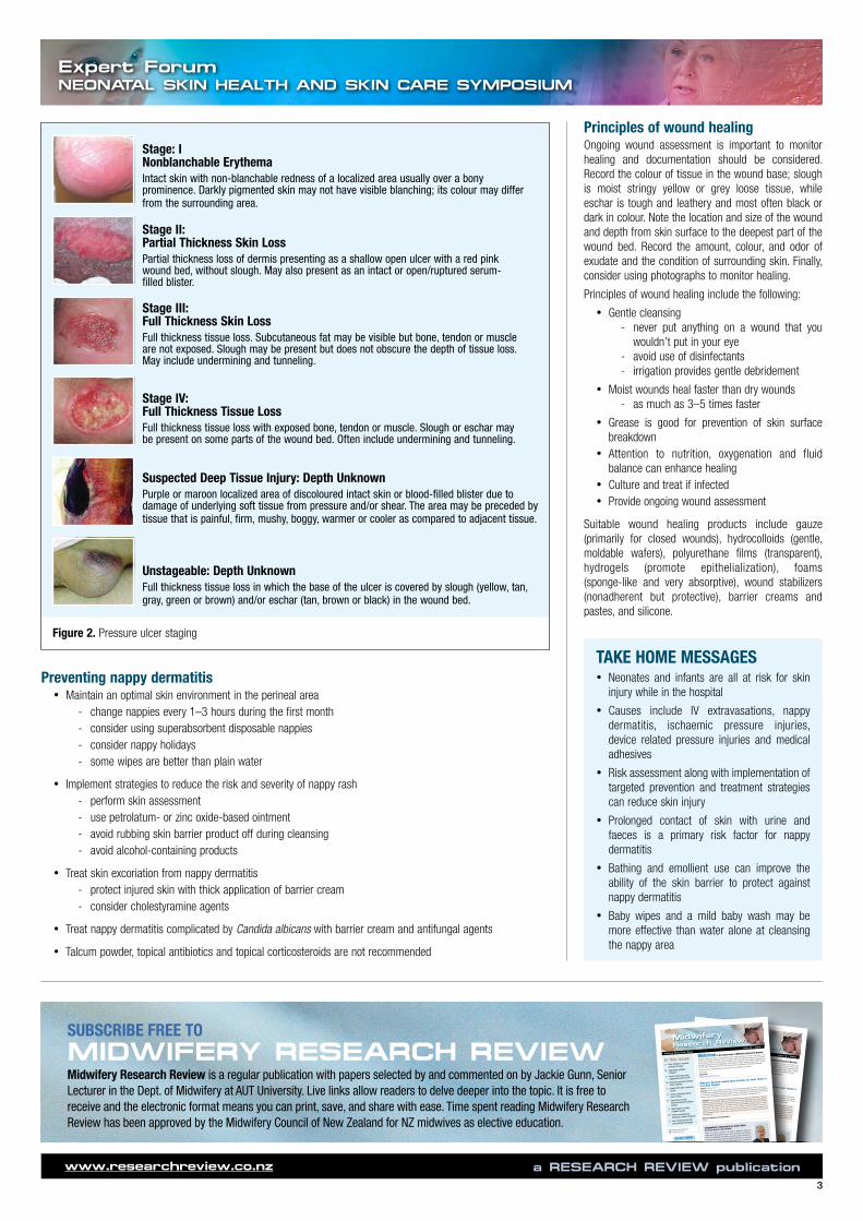
Pressure ulcer scoring is shown in Figure 2. It is important that stage I is identified early so that immediate intervention can occur before the ulcer progresses to stage II.
Pressure ulcers can be prevented by vigilant inspection, use of gel pillows, special beds and surfaces, frequent turning, good nutrition and oxygenation, maintaining a table skin temperature and use of a barrier ointment.
Nappy dermatitisProlonged contact of skin with urine and faeces is a primary cause of nappy dermatitis. This is because some of the bacteria in faeces contain enzymes that release ammonia from urine, contributing to raising skin pH which in turn activates proteases and disrupts the epidermis. Nappy use also increases skin surface pH and skin wetness, with wet skin known to increase the susceptibility of skin to damage from friction.14 Risk factors for nappy dermatitis include malabsorption, foecal incontinence, atopic dermatitis, oral antibiotics, and simply wearing nappies.
Preventing nappy dermatitisSkin care practices such as bathing and emollient use can greatly influence the ability of the skin to function as a barrier against environmental stresses such as those causing nappy dermatitis.15
Frequent nappy changes and use of absorbent nappies helps decrease skin wetness and contact with faecal enzymes, thereby maintaining skin pH. Super absorbent disposable nappies have been associated with a reduced incidence and decreased severity of irritant nappy dermatitis when compared with washable cloth nappies.14
Water alone is not an effective cleanserWater and cotton, mild soap and water, or baby wipes are adequate for cleansing the nappy area.16 Water alone may be insufficient to remove faeces and fats.17 Some studies show that water and mild cleansers have similar effects on skin pH and hydration, while others show that water alone may be more drying.18 A baby wash containing emollients may offer further protective effects.19 Excessive scrubbing and washing may promote irritation and further damage skin barrier properties, therefore gentle cleansing, rinsing and patting dry is recommended.20
Wipes may be better than waterA lot of parents do not want to use baby wipes at all. While wipes containing alcohol are not advisable, not all wipes are bad. Studies have shown that wipes may be a better cleanser than water because they have a lower pH and therefore do not disrupt the baby’s acid mantle.
Baby wipes with a pH buffering capacity have been shown to be well tolerated and more comfortable among infants aged 3–24 months with atopic dermatitis when compared to the use of water only and a wash cloth.21 A randomised, controlled trial of 280 full-term infants showed the use of wipes to be similar to the use of cotton wool and water when measuring TEWL, pH, redness, and skin colonization at 48 hours and 4 weeks.22 Mothers of infants in the water group reported more nappy rash. A randomised, controlled trial of 130 NICU infants comparing two types of wipes to cloth and water only found improved nappy area skin condition and barrier function when using wipes made from a soft, nonwoven material with water and emollient cleansers.23
Figure 1. Cross-section of the skin
Dermis
Epidermis
Stratum Corneum
Basal Layer
Hair follicle Subcutaneous gland Sweat gland
Subcutaneous tissue
3
www.researchreview.co.nz a RESEARCH REVIEW publication
Expert Forum NEONATAL SKIN HEALTH AND SKIN CARE SYMPOSIUM
Preventing nappy dermatitis• Maintain an optimal skin environment in the perineal area
- change nappies every 1–3 hours during the first month - consider using superabsorbent disposable nappies - consider nappy holidays - some wipes are better than plain water
• Implement strategies to reduce the risk and severity of nappy rash - perform skin assessment - use petrolatum- or zinc oxide-based ointment - avoid rubbing skin barrier product off during cleansing - avoid alcohol-containing products
• Treat skin excoriation from nappy dermatitis - protect injured skin with thick application of barrier cream - consider cholestyramine agents
• Treat nappy dermatitis complicated by Candida albicans with barrier cream and antifungal agents
• Talcum powder, topical antibiotics and topical corticosteroids are not recommended
Principles of wound healingOngoing wound assessment is important to monitor healing and documentation should be considered. Record the colour of tissue in the wound base; slough is moist stringy yellow or grey loose tissue, while eschar is tough and leathery and most often black or dark in colour. Note the location and size of the wound and depth from skin surface to the deepest part of the wound bed. Record the amount, colour, and odor of exudate and the condition of surrounding skin. Finally, consider using photographs to monitor healing.
Principles of wound healing include the following:
• Gentle cleansing - never put anything on a wound that you
wouldn’t put in your eye - avoid use of disinfectants - irrigation provides gentle debridement
• Moist wounds heal faster than dry wounds - as much as 3–5 times faster
• Grease is good for prevention of skin surface breakdown
• Attention to nutrition, oxygenation and fluid balance can enhance healing
• Culture and treat if infected• Provide ongoing wound assessment
Suitable wound healing products include gauze (primarily for closed wounds), hydrocolloids (gentle, moldable wafers), polyurethane films (transparent), hydrogels (promote epithelialization), foams (sponge-like and very absorptive), wound stabilizers (nonadherent but protective), barrier creams and pastes, and silicone.
TAKE HOME MESSAGES• Neonates and infants are all at risk for skin
injury while in the hospital
• Causes include IV extravasations, nappy dermatitis, ischaemic pressure injuries, device related pressure injuries and medical adhesives
• Risk assessment along with implementation of targeted prevention and treatment strategies can reduce skin injury
• Prolonged contact of skin with urine and faeces is a primary risk factor for nappy dermatitis
• Bathing and emollient use can improve the ability of the skin barrier to protect against nappy dermatitis
• Baby wipes and a mild baby wash may be more effective than water alone at cleansing the nappy area
Making Education Easy
MidwiferyResearch Review™
www.researchreview.co.nz
1
a RESEARCH REVIEW publication
Welcome to the latest issue of Midwifery Research Review.
Highlights include a NZ study of rural midwives’ decision-making processes for women in ‘slow labour’, and
a report of our Midwifery First Year of Practice programme and how it supports the retention of new midwives
in the maternity workforce. We also present an analysis of the Growing Up in New Zealand longitudinal study
that assessed timeliness of LMC engagement, and an initiative to reduce caesarean delivery rates in Canada.
We finish with an interesting report of the use of telemedicine by parents after early postnatal discharge.
I hope you enjoy the selected studies and look forward to any feedback you may have.
Kind regards,Jackie [email protected]
In this issue: Rural midwives’ decision-making processes
Supporting graduate midwives Factors influencing early engagement with an LMC
Reducing caesarean delivery rates in Quebec Vaginal birth after caesarean section
Factors affecting planned place of birth Experiences of fetal movements before stillborn delivery
Weight gain in healthy pregnant women Perineal management techniques among midwives Use of telemedicine after early postnatal discharge
Issue 9 – 2015
Abbreviations used in this issueLMC = Lead Maternity Carer
Midwives’ decision making about transfers for ‘slow’ labour in
rural New ZealandAuthors: Patterson J et al.Summary: This NZ study examined rural midwives’ decision-making processes for women in ‘slow labour’.
15 midwives who provided lead maternity care to women in rural areas shared their experiences. They described
the ‘mind shift’ needed when considering the transfer of women in slow labour to secondary care. Their decision
making process was influenced by colleague input, rural context and distance from specialist care.
Comment: This careful study explores midwives decision-making processes and relates them to decision-
making theory in the literature. While this is a relevant and very useful study about rural midwives decision-
making processes about transfer of women with ‘slow labour’, the findings are also useful for all midwives
to use the processes described here to consider their own decision-making processes during for example,
‘long labours’. The need to take a ‘step back’ to a more objective look at the situation or to call in a
colleague to provide ‘fresh eyes’ will not be unfamiliar to many midwives. This is a very accessible article
and I encourage midwives to access it and to read it in full as it provides food for reflection on practice for
all practitioners.
Reference: Midwifery 2015;31(6):606-12Abstract
FOR MORE INFORMATION ON HOW TO CLAIM POINTS PLEASE CLICK HERE
Time spent reading this publication has been approved by the Midwifery Council of New Zealand for NZ midwives as elective education.
Independent commentary by Jackie Gunn MA Massey BHSC Ng C.Sturt RGON RMJackie is a Senior Lecturer in the Dept of Midwifery, Faculty of
Health and Environmental Science at AUT University. She has been involved in leadership of midwifery education at AUT University for more than two decades and has practised midwifery in tertiary and primary maternity units and as an LMC midwife. She is a foundation member of the New Zealand College of Midwives. Jackie has a particular interest in midwifery practices that support physiological pregnancy, childbirth and transition to parenthood processes, midwifery education, and development of midwifery practice.
to follow us on twitter
CLICK HEREto join us on facebook
CLICK HERE
CLICK HEREto read previous issues of Midwifery Research Review
Making Education Easy
MidwiferyResearch Review™
www.researchreview.co.nz
1a RESEARCH REVIEW publication
Welcome to the latest issue of Midwifery Research Review.
Highlights include a NZ study of rural midwives’ decision-making processes for women in ‘slow labour’, and
a report of our Midwifery First Year of Practice programme and how it supports the retention of new midwives
in the maternity workforce. We also present an analysis of the Growing Up in New Zealand longitudinal study
that assessed timeliness of LMC engagement, and an initiative to reduce caesarean delivery rates in Canada.
We finish with an interesting report of the use of telemedicine by parents after early postnatal discharge.
I hope you enjoy the selected studies and look forward to any feedback you may have.
Kind regards,
Jackie [email protected]
In this issue: Rural midwives’ decision-
making processes
Supporting graduate
midwives
Factors influencing early
engagement with an LMC
Reducing caesarean delivery
rates in Quebec
Vaginal birth after caesarean
section
Factors affecting planned
place of birth
Experiences of fetal
movements before stillborn
delivery
Weight gain in healthy
pregnant women
Perineal management
techniques among midwives
Use of telemedicine after
early postnatal discharge
Issue 9 – 2015
Abbreviations used in this issue
LMC = Lead Maternity Carer
Midwives’ decision making about transfers for ‘slow’ labour in
rural New ZealandAuthors: Patterson J et al.
Summary: This NZ study examined rural midwives’ decision-making processes for women in ‘slow labour’.
15 midwives who provided lead maternity care to women in rural areas shared their experiences. They described
the ‘mind shift’ needed when considering the transfer of women in slow labour to secondary care. Their decision
making process was influenced by colleague input, rural context and distance from specialist care.
Comment: This careful study explores midwives decision-making processes and relates them to decision-
making theory in the literature. While this is a relevant and very useful study about rural midwives decision-
making processes about transfer of women with ‘slow labour’, the findings are also useful for all midwives
to use the processes described here to consider their own decision-making processes during for example,
‘long labours’. The need to take a ‘step back’ to a more objective look at the situation or to call in a
colleague to provide ‘fresh eyes’ will not be unfamiliar to many midwives. This is a very accessible article
and I encourage midwives to access it and to read it in full as it provides food for reflection on practice for
all practitioners.
Reference: Midwifery 2015;31(6):606-12
Abstract
FOR MORE INFORMATION
ON HOW TO CLAIM POINTS PLEASE
CLICK HERE
Time spent reading this publication
has been approved by the Midwifery
Council of New Zealand for NZ
midwives as elective education.
Independent commentary by Jackie Gunn
MA Massey BHSC Ng C.Sturt RGON RM
Jackie is a Senior Lecturer in the Dept of Midwifery, Faculty of
Health and Environmental Science at AUT University. She has been
involved in leadership of midwifery education at AUT University for
more than two decades and has practised midwifery in tertiary and
primary maternity units and as an LMC midwife. She is a foundation
member of the New Zealand College of Midwives. Jackie has a
particular interest in midwifery practices that support physiological
pregnancy, childbirth and transition to parenthood processes,
midwifery education, and development of midwifery practice.
to follow us on
CLICK HERE
to join us on facebook
CLICK HERE
CLICK HERE
to read previous issues of
Midwifery Research Review
SUBSCRIBE FREE TO
MIDWIFERY RESEARCH REVIEWMidwifery Research Review is a regular publication with papers selected by and commented on by Jackie Gunn, Senior Lecturer in the Dept. of Midwifery at AUT University. Live links allow readers to delve deeper into the topic. It is free to receive and the electronic format means you can print, save, and share with ease. Time spent reading Midwifery Research Review has been approved by the Midwifery Council of New Zealand for NZ midwives as elective education.
Stage: INonblanchable ErythemaIntact skin with non-blanchable redness of a localized area usually over a bony prominence. Darkly pigmented skin may not have visible blanching; its colour may differ from the surrounding area.
Stage II: Partial Thickness Skin Loss Partial thickness loss of dermis presenting as a shallow open ulcer with a red pink wound bed, without slough. May also present as an intact or open/ruptured serum-filled blister.
Stage III: Full Thickness Skin Loss Full thickness tissue loss. Subcutaneous fat may be visible but bone, tendon or muscle are not exposed. Slough may be present but does not obscure the depth of tissue loss. May include undermining and tunneling.
Stage IV: Full Thickness Tissue Loss Full thickness tissue loss with exposed bone, tendon or muscle. Slough or eschar may be present on some parts of the wound bed. Often include undermining and tunneling.
Unstageable: Depth UnknownFull thickness tissue loss in which the base of the ulcer is covered by slough (yellow, tan, gray, green or brown) and/or eschar (tan, brown or black) in the wound bed.
Suspected Deep Tissue Injury: Depth UnknownPurple or maroon localized area of discoloured intact skin or blood-filled blister due to damage of underlying soft tissue from pressure and/or shear. The area may be preceded by tissue that is painful, firm, mushy, boggy, warmer or cooler as compared to adjacent tissue.
Figure 2. Pressure ulcer staging
4
www.researchreview.co.nz a RESEARCH REVIEW publication
Expert Forum NEONATAL SKIN HEALTH AND SKIN CARE SYMPOSIUM
DEVELOPMENT OF THE NEONATAL SKIN MICROBIOMEMs Joanne Kuller, Neonatal Clinical Nurse Specialist, Children’s Hospital and Research Center, Oakland, USA
In addition to the growing body of evidence about the uniqueness of neonatal skin, recent advances have enabled clinicians to understand the processes involved in colonization of skin with microorganisms. The term microbiome describes the collective genomes and gene products of the microbes living within and on humans.
As a result of the NIH-sponsored Human Microbiome Project, bacteria are now identified through DNA analysis.24 Most of these bacteria are healthy or commensal bacteria and some are pathogens. New research suggests that a disease state may not simply be the presence of pathogens but the absence of commensal bacteria. Beneficial bacteria aid in development and stimulation of the gut mucosa, development of immunity, protection from diverse pathogens, and vitamin production.
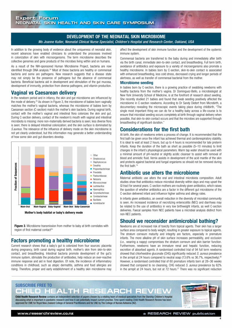
Vaginal vs Caesarean deliveryIn the newborn period and in infancy, the skin and gut microbiome are influenced by the mode of delivery.25 As shown in Figure 3, the microbiome of babies born vaginally matches the mother’s vaginal bacteria, whereas the microbiome of babies born by Caesarean section (C-section) match the mother’s skin bacteria. During vaginal birth, contact with the mother’s vaginal and intestinal flora colonizes the skin and gut. During C-section delivery, contact of the newborn’s mouth with vaginal and intestinal microbiota is missing: more non-maternally derived bacteria is seen; less diverse flora is seen; there is delayed intestinal colonization; and the skin surface is dominated by S.aureus. The relevance of the influence of delivery mode on the skin microbiome is not yet clearly understood, but this information may generate a better understanding of how some skin and gut disorders develop.
Factors promoting a healthy microbiomeCurrent research shows that a baby’s gut is colonized from four sources: placenta during pregnancy, birth canal during vaginal birth, mother’s skin from skin-to-skin contact, and breastfeeding. Intestinal bacteria promote development of the gut’s immune system, stimulate the production of antibodies, help reduce an over-reactive immune response and aid in food digestion. Of note, the incidence of inflammatory conditions in childhood, such as atopic dermatitis, asthma and food allergies are rising. Therefore, proper and early establishment of a healthy skin microbiome may
OtherStreptococcusStaphylococcusSneathiaPropionibacterineaePrevotellaPasteurellaceaeNeisseriaMicrococcineaeLactobacillusHaemophilusCorynebacterineaeCoriobacterineaeBacillalesAcinetobacter
Baby-Vaginal Mum-Skin Baby-Caeserean
Frac
tion
of S
eque
nces
Mum-Oral Mum-Vaginal
Mother’s body habitat or baby’s delivery mode
Frac
tion
of S
eque
nces
Mother’s body habitat or baby’s delivery mode
Lactobacillus Haemophilus Corynebacterineae Coriobacterineae Bacillales Acinetobacter
Propionibacterineae Prevotella Pasteurellaceae Neisseria Micrococcineae
Other Streptococcus Staphylococcus Sneathia
Mom-Oral Mom-Vaginal Baby-Vaginal Mom-Skin Baby-Cesarean
Figure 3. Microbiome transmission from mother to baby at birth correlates with region of first maternal contact25
affect the development of skin immune function and the development of the systemic immune system.
Commensal bacteria are transferred to the baby during and immediately after birth via the birth canal, immediate skin-to-skin contact, and breastfeeding. Full term birth, avoidance of antibiotics and exposure to a variety of microorganisms also promote a healthy microbiome. In babies born by C-section, skin-to-skin contact is associated with enhanced breastfeeding, less cold stress, decreased crying and longer periods of alertness, as well as transfer of commensal bacterial from the mother.
Microbiome seedingIn babies born by C-section, there is a growing practice of swabbing newborns with healthy bacteria from the mother’s vagina. Dr Dominguez-Bello, a microbiologist at New York University School of Medicine, is at the forefront of research about seeding. She recently studied 21 babies and found that swab seeding positively affected the microbiome in C-section newborns. According to Dr Sandy Dietert from Microbirth, a documentary revealing the microscopic events taking place during childbirth, “The single most important thing we can do for a healthy baby across a life-course is to ensure that microbial seeding occurs completely at birth through vaginal delivery when possible, that skin-to-skin contact occurs and that the microbes are supported through breastfeeding of significant duration.”
Considerations for the first bathAt birth, the skin of newborns enters a process of change. It is recommended that the first bath be given once the infant has achieved thermal and cardiorespiratory stability. It is ideal to wait at least 2 hours, but up to 4 hours is recommended for late preterm infants. Keep the duration of the bath as short as possible (5–10 minutes) to limit changes in the infant’s physiological parameters. Warm tap water should be used with a minimal amount of pH-neutral or slightly acidic cleanser to assist with removal of blood and amniotic fluid. Vernix assists in development of the acid mantle of the skin and protects against bacterial and fungal organisms so should not be removed during the first bath.14
Antibiotic use alters the microbiomeMaternal antibiotic use alters the oral and intestinal microbiota composition. Adult studies show that antibiotics reduce microbial diversity within days and may upset the GI tract for several years. C-section mothers are routinely given antibiotics, which raises the question of whether antibiotics are a factor in the different gut microbiome of the C-section delivered infant and influence higher asthma and allergy rates.
In infants given antibiotics, an overall reduction in the diversity of microbial community is seen. An increased incidence of necrotizing enterocolitis (NEC) and diarrhoea may be related to the use of antibiotics in very low birthweight infants, as well C-section deliveries. Faecal samples from NEC patients have a microbial analysis distinct from non-NEC patients.
Should we reconsider antimicrobial bathing?Newborns are at increased risk of toxicity from topical agents. Their skin has a larger surface area compared to body weight, resulting in greater exposure to topical agents. The stratum corneum maturity and integrity are factors, especially in premature infants. The more alkaline pH of skin surface increases permeability, and occlusion (i.e., wearing a nappy) compromises the stratum corneum and skin barrier function. Furthermore, newborns have an immature renal and hepatic function, reducing excretion of absorbed agents. A randomised controlled trial of 94 full term newborns showed that chlorhexidine gluconate (CHG) significantly reduced S. aureus prevalence in the armpit at 24 hours compared to neutral soap (13.6% vs 36.7%, respectively).26 However, a randomised controlled trial of 60 premature infants born at 28–36 weeks showed that compared to no cleansing, CHG reduced S. aureus prevalence by 62% in the armpit at 24 hours, but not at 72 hours.27 There was no significant reduction
Research Review TM
Child HealthMaking Education Easy
www.researchreview.co.nz
1
a RESEARCH REVIEW publication
Issue 8 – 2015Welcome to the latest issue of Child Health Research Review.
Selection and comments for this issue have been provided by the Paediatric Allergy Team at Starship Children’s
Hospital (led by Dr Jan Sinclair) and Paediatric Dermatologist Dr Diana Purvis. Highlights include evidence that eating
peanuts in infancy may actually prevent the development of peanut allergy in high-risk children, plus we report more
research on predicting food allergy in infants. Immunotherapies for grass pollen allergies are under the spotlight,
with subcutaneous and sublingual preparations having comparable efficacies. We also present promising findings
for topical rapamycin in children with tuberous sclerosis, and review the use of systemic cyclosporin in children with
atopic dermatitis.We hope you find these and the other selected studies interesting and useful in your clinical practice.
Kind regards,Dr Chris Tofield Medical Advisor, Research [email protected]
In this issue: Eating peanuts in infancy reduces allergy risk
Predicting food allergy in infants Baked milk and egg consumption in allergic children Immunotherapies for grass pollen allergies
Preventing AD in neonates Topical rapamycin for angiofibromas Port-wine stains and Sturge-Weber syndrome
Melanonychia striata in childhood Systemic cyclosporin in children with AD Henoch-Schönlein purpura in children
Randomized trial of peanut consumption in infants at risk for
peanut allergyAuthors: Du Toit G et al., for the LEAP Study TeamSummary: The LEAP study compared the effectiveness of 2 strategies (peanut consumption and peanut
avoidance) for preventing the development of peanut allergy in high-risk infants. 640 infants with severe eczema,
egg allergy, or both, were randomised to consume or avoid peanuts until age 5. Among the 530 infants in the
intention-to-treat population who initially had negative results on the skin-prick test, the prevalence of peanut
allergy at age 5 years was 13.7% in the avoidance group and 1.9% in the consumption group (p<0.001). In those
who initially had a positive skin-prick test (n=98), the prevalence of peanut allergy at age 5 years was 35.3% in
the avoidance group and 10.6% in the consumption group (p=0.004). Comment: Rising rates of food allergy initially led to a knee jerk reaction promoting food avoidance for
prevention. This was clearly ineffective, with lower rates of peanut allergy in Israel (where peanut is generally
eaten in the first year) compared with London (where peanut avoidance was promoted). This study has carefully
demonstrated that for high risk infants, starting peanut at under 10 months of age results in both primary
(where the initial skin test was negative) and secondary (with initial small positive skin test) prevention of peanut
allergy. The challenge is now to clarify whether this principal extends to other foods and to all infants, but has
the potential to change current infant feeding guidelines.Reference: N Engl J Med 2015;372:803-813Abstract
Skin prick test responses and allergen-specific IgE levels as
predictors of peanut, egg, and sesame allergy in infantsAuthors: Peters R et al., for the HealthNuts studySummary: The HealthNuts study developed skin prick test (SPT) and allergen-specific IgE (sIgE) thresholds with
95% positive predictive values (PPVs) for challenge-confirmed food allergy in infants. 5276 one-year-old infants
underwent SPTs to 4 allergens: egg, peanut, sesame, and cow’s milk. Those with a detectable SPT response were
invited to undergo oral food challenge and sIgE testing. Peanut SPT responses of ≥8mm, egg SPT responses of
≥4mm, and sesame SPT responses of ≥8mm had 95% PPVs for challenge-confirmed food allergy. Peanut sIgE
levels of ≥34 kUA/L and egg sIgE levels of ≥1.7 kUA/L had 95% PPVs for challenge-confirmed food allergy.
Egg SPT responses and sIgE levels were poor predictors of allergy to egg in baked goods.Comment: Diagnosis of food allergy can be complicated. Many infants who have positive tests to foods
(“sensitised”) are not clinically allergic. Diagnosis needs to balance unnecessary food avoidance against
unnecessary food challenge if true allergy is almost certain. HealthNuts assessed food allergy in a large
unselected infant group, using the gold standard of supervised food challenge to determine food allergy. The
data from this paper reinforce that the implication of skin test and sIgE results vary between foods. HealthNuts
provides clear guidance as to test results that indicate likelihood of allergy to common foods in young infants.
Reference: J Allergy Clin Immunol 2013;132(4):874-80Abstract
Abbreviations used in this issueAD = atopic dermatitisIgE = immunoglobulin E
Time spent reading this publication has been approved for CME for Royal New Zealand College of General Practitioners (RNZCGP) General Practice Educational Programme Stage 2 (GPEP2) and the Maintenance of Professional Standards (MOPS) purposes, provided that a Learning Reflection Form is completed. Please CLICK HERE to download your CPD MOPS Learning Reflection Form. One form per review read would be required.Time spent reading this publication has been approved
for CNE by The College of Nurses Aotearoa (NZ) for RNs and NPs. For more information on how to claim CNE hours please CLICK HERE.
origins and solutions
WELLINGTONCapital of arts and culture
Wednesday 25 to Friday 27 November 2015 Museum of New Zealand Te Papa Tongarewa, Wellington
www.psnz2015.co.nz
Paediatric Society of New Zealand67th Annual Scientific Meeting
Research ReviewTMChild Health
Making Education Easy
www.researchreview.co.nz
1a RESEARCH REVIEW publication
Issue 8 – 2015
Welcome to the latest issue of Child Health Research Review.
Selection and comments for this issue have been provided by the Paediatric Allergy Team at Starship Children’s
Hospital (led by Dr Jan Sinclair) and Paediatric Dermatologist Dr Diana Purvis. Highlights include evidence that eating
peanuts in infancy may actually prevent the development of peanut allergy in high-risk children, plus we report more
research on predicting food allergy in infants. Immunotherapies for grass pollen allergies are under the spotlight,
with subcutaneous and sublingual preparations having comparable efficacies. We also present promising findings
for topical rapamycin in children with tuberous sclerosis, and review the use of systemic cyclosporin in children with
atopic dermatitis.
We hope you find these and the other selected studies interesting and useful in your clinical practice.
Kind regards,
Dr Chris Tofield
Medical Advisor, Research Review
In this issue: Eating peanuts in infancy reduces
allergy risk
Predicting food allergy in infants
Baked milk and egg consumption
in allergic children
Immunotherapies for grass pollen
allergies
Preventing AD in neonates
Topical rapamycin for
angiofibromas
Port-wine stains and Sturge-
Weber syndrome
Melanonychia striata in childhood
Systemic cyclosporin in children
with AD
Henoch-Schönlein purpura in
children
Randomized trial of peanut consumption in infants at risk for
peanut allergyAuthors: Du Toit G et al., for the LEAP Study Team
Summary: The LEAP study compared the effectiveness of 2 strategies (peanut consumption and peanut
avoidance) for preventing the development of peanut allergy in high-risk infants. 640 infants with severe eczema,
egg allergy, or both, were randomised to consume or avoid peanuts until age 5. Among the 530 infants in the
intention-to-treat population who initially had negative results on the skin-prick test, the prevalence of peanut
allergy at age 5 years was 13.7% in the avoidance group and 1.9% in the consumption group (p<0.001). In those
who initially had a positive skin-prick test (n=98), the prevalence of peanut allergy at age 5 years was 35.3% in
the avoidance group and 10.6% in the consumption group (p=0.004).
Comment: Rising rates of food allergy initially led to a knee jerk reaction promoting food avoidance for
prevention. This was clearly ineffective, with lower rates of peanut allergy in Israel (where peanut is generally
eaten in the first year) compared with London (where peanut avoidance was promoted). This study has carefully
demonstrated that for high risk infants, starting peanut at under 10 months of age results in both primary
(where the initial skin test was negative) and secondary (with initial small positive skin test) prevention of peanut
allergy. The challenge is now to clarify whether this principal extends to other foods and to all infants, but has
the potential to change current infant feeding guidelines.
Reference: N Engl J Med 2015;372:803-813
Abstract
Skin prick test responses and allergen-specific IgE levels as
predictors of peanut, egg, and sesame allergy in infants
Authors: Peters R et al., for the HealthNuts study
Summary: The HealthNuts study developed skin prick test (SPT) and allergen-specific IgE (sIgE) thresholds with
95% positive predictive values (PPVs) for challenge-confirmed food allergy in infants. 5276 one-year-old infants
underwent SPTs to 4 allergens: egg, peanut, sesame, and cow’s milk. Those with a detectable SPT response were
invited to undergo oral food challenge and sIgE testing. Peanut SPT responses of ≥8mm, egg SPT responses of
≥4mm, and sesame SPT responses of ≥8mm had 95% PPVs for challenge-confirmed food allergy. Peanut sIgE
levels of ≥34 kUA/L and egg sIgE levels of ≥1.7 kUA/L had 95% PPVs for challenge-confirmed food allergy.
Egg SPT responses and sIgE levels were poor predictors of allergy to egg in baked goods.
Comment: Diagnosis of food allergy can be complicated. Many infants who have positive tests to foods
(“sensitised”) are not clinically allergic. Diagnosis needs to balance unnecessary food avoidance against
unnecessary food challenge if true allergy is almost certain. HealthNuts assessed food allergy in a large
unselected infant group, using the gold standard of supervised food challenge to determine food allergy. The
data from this paper reinforce that the implication of skin test and sIgE results vary between foods. HealthNuts
provides clear guidance as to test results that indicate likelihood of allergy to common foods in young infants.
Reference: J Allergy Clin Immunol 2013;132(4):874-80
Abstract
Abbreviations used in this issue
AD = atopic dermatitis
IgE = immunoglobulin E
Time spent reading this publication has been approved
for CME for Royal New Zealand College of General
Practitioners (RNZCGP) General Practice Educational
Programme Stage 2 (GPEP2) and the Maintenance of
Professional Standards (MOPS) purposes, provided that
a Learning Reflection Form is completed. Please CLICK
HERE to download your CPD MOPS Learning Reflection
Form. One form per review read would be required.
Time spent reading this publication has been approved
for CNE by The College of Nurses Aotearoa (NZ) for RNs
and NPs. For more information on how to claim CNE
hours please CLICK HERE.
origins and solutions WELLINGTON
Capital of arts and culture
Wednesday 25 to Friday 27 November 2015
Museum of New Zealand Te Papa Tongarewa, Wellington
www.psnz2015.co.nz
Paediatric Society of New Zealand
67th Annual Scientific Meeting
SUBSCRIBE FREE TO
CHILD HEALTH RESEARCH REVIEWChild Health Research Review contains an independent selection of papers chosen by a rotating team of medical specialists from the Starship Children’s Hospital discussing what is important in paediatric research and how it can potentially impact current practise. Time spent reading Child Health Research Review has been approved for CME for Royal New Zealand College of General Practitioners and for CNE by The College of Nurses Aotearoa (NZ).

5
www.researchreview.co.nz a RESEARCH REVIEW publication
Expert Forum NEONATAL SKIN HEALTH AND SKIN CARE SYMPOSIUM
compared to saline cleansing. In the groin, there was no significant difference among the three groups at 24 or 72 hours. All of these factors have led clinicians to question whether we should reconsider routine antimicrobial bathing of NICU neonates.
The US FDA has issued a labelling change for antiseptics containing CHG, warning that CHG-containing skin antiseptics should be used with caution in premature infants or infants less than 2 months of age, as they may cause chemical burns. At the same time, case reports of CHG/alcohol skin disinfectants and dressings causing skin injuries are becoming more frequent; therefore, the selection of skin disinfectants for extremely premature infants remains a dilemma for clinicians.
TAKE HOME MESSAGES• Goal is to protect neonatal skin and promote future skin health• Normal skin flora are helpful in protecting skin from infection• Care practices should promote presence of commensal bacteria• Bathing with water alone may not be better than using gentle baby wash• Skin disinfection vs maintenance and promotion of commensal bacteria is a
complicated issue
Figure 4. The future of atopic dermatitis treatment
1
15 0 5 10
Inci
denc
e
Atopic dermatitis
Food allergy
Asthma
Rhinitis
Time (years)
Figure 5. Atopic dermatitis: part of the atopic march
Ms Kuller: overall conclusions Protecting newborn’s skin and maintaining a normal skin barrier is crucial in the neonatal period, particularly for infants in NICU. Normal skin flora is helpful in protecting the skin from infection and as such, care practices should promote the presence of beneficial bacteria. Routine assessment of infant skin enables early identification and treatment of skin injury, of which nappy dermatitis is a common cause. Bathing with a mild baby wash, and use of wipes and emollients can improve the ability of the skin barrier to protect against nappy dermatitis, and may be more effective than water alone.
PREVENTING ATOPIC DERMATITIS BY CHANGING THE WAY WE TREAT A NEWBORN’S SKIN FROM BIRTHProfessor Michael Cork, Head of Academic Dermatology, University of Sheffield Medical School, UK
Maintaining the skin barrier is key to healthy skin. The normal skin barrier protects the body from the penetration of irritants and allergens. In an infant the skin barrier has not matured and as a result is much less protective against the environment than in an older child and an adult. This is why the infant is vulnerable to diseases which are caused by a defective skin barrier. The presence of a normal skin barrier is important in the maintenance of the normal microbiome.
What is at stake if baby skin is not cared for appropriately?A baby with the genetic predisposition to develop atopic dermatitis can develop it if we don’t care for their skin appropriately. Atopic dermatitis can be mild, or much more severe with devastating effects on the child and their parents at a crucial time in their development. The prevalence of atopic dermatitis among babies and children has risen from around 5% in the 1940s to up to 25% today. Clearly over this period genetics haven’t changed, but our environment has, particularly the way we treat babies’ skin.
In 1991, Duff made the point that current medicine only treats clinical disease. He identified the treatment of genetic susceptibility and preclinical disease as the future of medicine for all diseases. If we could treat a baby with a genetic predisposition for any disease we might be able to prevent it from developing. In Figure 4, a baby is shown with the genetic susceptibility to develop atopic dermatitis. The baby has a breakdown in the skin barrier, leading to subclinical inflammation; but the baby’s skin still looks totally normal. Eventually the baby develops atopic dermatitis and is treated. The future of atopic dermatitis treatment lies in proactively preventing it from developing in the first place.
Preventing the ‘atopic march’Atopic dermatitis is caused by skin barrier disruption, as a result of a complex interaction between genes and negative environmental factors such as harsh soaps and detergents that break down the skin barrier. Allergens can then enter the skin and atopic dermatitis develops. Atopic dermatitis may lead on to other diseases such as food allergies, asthma, and hay fever; this process is known as the ‘atopic march’. But it can be prevented by changing the environment a new born baby is exposed to from negative to positive. Importantly, a window of opportunity exists in the first few months after birth to change the environment to prevent the development of atopic dermatitis. By changing the way we treat a baby’s skin from birth, we can prevent the development of atopic dermatitis. Everything we put on a baby’s skin from birth, including emollients, wash products and wipes, should be designed to enhance the skin barrier rather than damage it.
The importance of low pH for maintaining normal skinFigure 6 shows a normal skin barrier compared with a defective skin barrier. Within the skin are the skin cells (corneocytes) which act like bricks in a brick wall. The lipid lamellae act like mortar in a brick wall. Corneodesmosomes are like iron rods, linking the skin cells together, giving a normal skin barrier tensile strength. Within the corneocytes is a substance termed natural moisturising factor (NMF), which is generated by the breakdown of filaggrin. NMF has two main roles: 1) it attracts water, making skin cells swell to close gaps between them; and 2) it contains acids which are absolutely pivotal to normal skin barrier function. These acids keep the pH of normal skin to around 5–5.5. Skin cells are shed from the surface of the skin by the
Genetic susceptibility
Future treatment“Prevention and proactive”
Current treatment“Reactive”
Sub-clinicalskin barrier
defect
Sub-clinical inflammation
IntrinsicAD
Low IgE
ExtrinsicAD
High IgE

6
www.researchreview.co.nz a RESEARCH REVIEW publication
Expert Forum NEONATAL SKIN HEALTH AND SKIN CARE SYMPOSIUM
NMF components, cytokines and pH (Figure 7). These assays are much more sensitive than clinical scoring. A protease assay by Cork and colleagues showed the clinical relevance of the effect of skin pH. Protease activity in normal skin (pH 5.0) was turned off, but was switched on in skin with current atopic dermatitis (pH 5.7) – without the addition of any topical products. With the addition of products such as soap or detergent, pH was raised even more (8.5), leading to acute atopic dermatitis.
Myths about baby skin care: water is goodIs water good? How safe is water? What is water? The pH of water alone is 7.2, but water hardness and harsh soap raises pH even more. Water pH can be lowered to an optimal 5.5 using appropriate cleansers. Water is irritant because of the calcium carbonate content, and hard water has been shown to increase the prevalence of atopic dermatitis.30 However, a randomised controlled trial of water softeners showed no benefit for atopic dermatitis,31 because water softeners substitute sodium for calcium, which has a slightly higher pH. So the challenge is to lower the pH of water by using a wash product that soaks up calcium, known as a ‘chelator’.
Myths about baby skin care: all wash products are badWater alone is not an effective cleanser because it is unable to remove protein, fats, urine and faeces. Detergents are able to remove these impurities without the need for excessive friction. But there are many types of detergent; some have a high pH and can disrupt the skin barrier, such those containing sodium lauryl sulfate (SLS), while at the other end of the enormous spectrum are very, very mild detergents with low pH (Figure 8).
Professor Cork and colleagues conducted a study comparing the effects of ivory soap versus a wash product formulated for newborns (Top To Toe) on skin surface pH and protease activity. [Danby and Cork, unpublished] A 2-minute wash in bath water containing ivory soap raised skin pH to 6.8 at 15 minutes and maintained it at around that level for 4 hours. In contrast, Top To Toe wash raised skin pH to about 5.7 at 15 minutes, and maintained it at slightly lower than that level for 4 hours. These results were reflected in measurements of
A healthy skin barrier
NMF H2O
NMF H2O
NMF H2O
Intact corneodesmosomes
Swelled corneocytes
Cornified envelope
Lipid lamellae
Degradatory proteases
Protease inhibitors Profilaggrin
Filaggrin
Lipid delivery
Lipid processing
pH 5.0
A defective skin barrier in atopic dermatitis
NMF H2O
Abnormal cornified envelope
Degraded corneodesmosomes
Defective lipid lamellae
NMF H2O
NMF H2O
Profilaggrin
Filaggrin Degradatory Proteases
Protease inhibitors Lipid delivery
Lipid processing
pH 7.0
1. Biological assays (protease activity, antimicrobial peptides, NMF components, cytokines) pH
2. Tape-strip/TEWL, Optical Coherence Tomography elastography [FTIR spectroscopy]
3. Morphological Optical coherence tomography/Histology
5. High frequency ultrasound
6. Profilometry
8. Clinical scoring
4. Basal TEWL [mexameter, hydration]
7. Dermatoscope
TIME:- Increasing skin barrier damage
breakdown of corneodesmosomes by protease enzymes. It is very important that the proteases are kept in check because otherwise corneodesmosomes would break down all the way through the skin barrier allowing allergens to enter. Proteases are held in check in normal skin by protease inhibitors, but perhaps even more important is low pH. Low pH switches off proteases, which is why low pH is important for maintaining normal skin. Low pH also switches on lipid lamellae (‘mortar’) production.
When an infant is genetically predisposed to atopic dermatitis, there is a defect in the filaggrin gene,28 leading to less NMF, and therefore less acids; the pH of the skin rises to 7.0. High pH switches on proteases which breakdown the corneodesmosomes. High pH also switches off the lipid processing enzyme so the lipid lamellae start to breakdown. The result is a defective skin barrier, leaving the infant more vulnerable to the effects of the environment. When the stratum corneum is impaired, e.g. by hard water, soap and detergents elevating skin pH from 5.5 to 7, 8, or even 9, the skin barrier can no longer prevent allergens and infections from reaching the dermis, leading to inflammation characteristic in atopic dermatitis and the development of allergies. The skin barrier may be restored by avoiding negative environmental influences, and protected through skincare regimes that involve products of optimal pH that repair/respect the skin barrier. By protecting the skin barrier, the atopic march of atopic dermatitis, asthma, food allergies and hay fever may be prevented in some babies.
Development of peanut allergy in childhoodIn 2003 Lack and colleagues showed that the main route of sensitization to peanuts was not by eating them but through the skin.29 At that time, peanut oil was used in multiple products from emollients to barrier creams. The group have since shown that feeding peanuts to non-allergic children can actually induce tolerance. This shows that the way food allergens enter the immune system and the time at which they enter during newborn development is very important.
Assessing the effect of topical products on the skin barrierWe cannot use clinical measures to assess the effects of topical products on the skin barrier, because babies do not have clinical atopic dermatitis at birth. What we can do is look inside the skin and determine whether the barrier is breaking down. Assays have been developed to measure the effect of wash products, wipes and emollients on the skin barrier by measuring protease activity, antimicrobial peptides,
Figure 6. A normal vs defective skin barrier
Figure 7. Measuring the effects of wash products on skin: sensitivity of assessments

7
www.researchreview.co.nz a RESEARCH REVIEW publication
Expert Forum NEONATAL SKIN HEALTH AND SKIN CARE SYMPOSIUM
protease activity over 4 hours; ivory soap increased protease activity substantially whereas Top To Toe wash did not increase protease activity.
A randomised controlled trial of 370 healthy full term infants showed that bathing with Top to Toe wash was no worse than bathing with water alone in terms of skin water loss, pH alterations, and clinical observations of dry skin.32
Professor Cork and colleagues then evaluated the effects of three different cleansers on newborn skin. [Danby and Cork, unpublished]. At weeks 2 and 4, an organic baby soap (Earth Mamma Angel Baby) increased pH, TEWL and protease activity substantially compared to two liquid cleansers (Top To Toe and Neutrogena). When adult women used these products on their skin for 4 weeks, the organic soap altered the microbiome but the two liquid cleansers did not. Professor Cork concluded
that not only did the soap product damage the skin barrier but it also damaged the microbiome. So it really matters which type of product we use on the skin because harsh products can have a profound effect on the development of atopic dermatitis.
TAKE HOME MESSAGES• Gene-environment interactions drive atopic dermatitis
progression• Negative environmental factors such as harsh soaps and
detergents can break down the skin barrier• Genetically-predisposed infants are at risk for atopic dermatitis
if their skin is not cared for appropriately• Atopic dermatitis and the atopic march can be prevented by
changing the environment skin is exposed to from birth• Low pH is important for maintaining normal skin• Water alone is not an effective cleanser:
- cannot dissolve impurities- high pH
• The high pH of water can be lowered using an optimal wash formulation
• Detergents containing SLS have a high pH and can disrupt the skin barrier
• Optimal wash products, wipes and emollients with very mild detergent maintain a normal skin barrier and normal skin microbiome and can prevent the development of atopic dermatitis
Harsh surfactant SLS
pH > 7.0
Mild complex surfactant Top To Toe pH = 5.5
High Protease + Skin Barrier Damage
Low Protease - No Skin Barrier
Damage
Figure 8. Spectrum of surfactant wash products: effects on protease activity in the skin barrier
50%
45%
40%
35%
30%
25%
20%
15%
10%
5%
0%
Prev
alen
ce o
f AD
at 6
mon
ths
Relative riskreduction of 50%Cl: 0.28-0.90
EmollientTreated,
22%
Control, 43%
EMOLLIENT THERAPY: TREATMENT AND PREVENTION OF ATOPIC DERMATITISProfessor Michael Cork, Head of Academic Dermatology, University of Sheffield Medical School, UK
Not all emollient formulations are the sameRepair of the skin barrier is the first step in both the treatment and prevention of atopic dermatitis. This involves removing all negative environmental factors and replacing them by positive interventions. Emollients formulated with ingredients that restore lipid levels, improve hydration, replenish depleted levels of NMF, and which offer significant buffering capacity, such that skin pH is normalised and the microbiome minimally affected, may offer clinically protective benefits. However emollients which are not correctly formulated can damage the skin barrier rather than repair it. When selecting an emollient it is essential to know its formulation and the effect of the formulation on the skin barrier.
In a pilot trial of infants genetically predisposed to develop atopic dermatitis, emollient use from birth reduced the prevalence of atopic dermatitis by 50% at 6 months compared to no treatment (Figure 9).33 Emollients used in the trial were sunflower seed oil with a high ratio of linoleic/oleic acid, Doublebase Gel, liquid paraffin 50% in white soft paraffin, Cetaphil Cream, or Aquaphor Healing Ointment. None of the emollients contained SLS.
Simple occlusive emollients containing oil such as paraffin or petrolatum are useful in infants with a very defective skin barrier, but are not very cosmetically acceptable and need to be applied very frequently (every 30–60 minutes to have optimal effect). Furthermore, such ointments only produce a partial repair of the skin barrier – the underlying defect is not repaired.
Repair of the skin barrier can be achieved by humectants, which lower pH and retain water in the skin barrier. Examples include urocanic acid, pyrrolidone carboxylic acid, lactate citrate, urea and glycerol. When humectants are combined with occlusive emollients, the skin barrier can be partially repaired at the surface as well as within.
Lipids can be returned to the skin barrier by incorporating physiological lipids such as ceramides, cholesterol, linoleic acid, and palmitic acid into emollients. Natural lipids may help to restore normal barrier function; however, this is an area of ongoing research.
Buffers can also be added to emollients to reduce pH. Importantly, pH buffered products need to keep skin pH lowered for several hours, not just a few minutes, in order to repair the skin barrier and microbiome.
Aqueous cream should never be used in atopic dermatitisWould you use a cream produced in 1958 on a baby’s skin? Aqueous cream first appeared in the British National Formulary in 1958. Its formulation containing 1% SLS remains largely unchanged today. SLS is one of the harshest surfactants and can cause significant damage to the skin barrier.
Figure 9. Emollient enhancement of the skin barrier from birth – prevention of atopic dermatitis33
Aqueous cream was shown in an audit of infants and children with atopic dermatitis to cause irritation (sometimes very severe) in 55% of them.34 Consequently, further studies of the effects of aqueous cream were conducted and it was shown to cause severe damage to the skin barrier in adults with a previous history of atopic dermatitis,35 and 20% thinning of the skin barrier in normal adult skin.36 In a study in healthy volunteers, aqueous cream raised skin TEWL, whereas yellow soft paraffin reduced TEWL.[Danby and Cork, unpublished] Importantly, NICE guidelines have recommended that aqueous cream should never be used as a leave-on emollient in infants and children with atopic dermatitis.37
Myths about baby skin care: olive oil is goodOlive oil contains oleic acid which can disrupt skin barrier function.38 Topical application of olive oil has been shown to compromise the integrity of the adult stratum corneum and induce mild skin irritation.39 In contrast, the same study showed that sunflower seed oil, which

8
www.researchreview.co.nz a RESEARCH REVIEW publication
Expert Forum NEONATAL SKIN HEALTH AND SKIN CARE SYMPOSIUM
© 2015 RESEARCH REVIEW
This publication has been created with an educational grant from Johnson & Johnson Pacific. The content is entirely independent and based on published studies and the writer and commentators’ opinions.
ABOUT RESEARCH REVIEW Research Review is an independent medical publishing organisation producing electronic publications in a wide variety of specialist areas. Research Review publications are intended for New Zealand medical professionals.
ABOUT EXPERT FORUMS Expert Forum publications are designed to encapsulate the essence of a local meeting of health professionals who have a keen interest in a condition or disease state. These meetings are typically a day in duration, and will include presentations of local research and discussion of guidelines and management strategies. Even for local events it is not always possible for everyone with a similar therapeutic interest to attend. Expert Forum publications capture what was said and allows it to be made available to a wider audience through the Research Review membership or through physical distribution.
contains linoleic acid, preserved stratum corneum integrity, did not cause irritation, and improved hydration. Of note, some sunflower oils are genetically modified in order to taste like olive oil and as such contain high levels of oleic acid and low levels of linoleic acid.
Colloidal oatmeal repairs the skin barrierColloidal oatmeal, a natural product derived from oat grains, induces the skin to repair itself by producing more fatty acids to make ceramides. A paper from 1953 demonstrated that colloidal oatmeal reduced the skin pH of elderly people with atopic dermatitis for three hours.40 A randomised controlled clinical trial in 30 adults with dry skin showed significant benefits of a moisturizing lotion containing colloidal oatmeal (Aveeno) versus its vehicle control for scaling, skin dryness and hydration at 21 and 28 days.41 Another study compared the effects of Aveeno, aqueous cream and Epaderm cream (0.5% SLS at the time of study; now contains no SLS) on skin pH.[Danby and Cork, unpublished] Aqueous cream and Epaderm cream raised pH for 6 hours but Aveeno lowered pH for 3 hours.
TAKE HOME MESSAGES• Not all emollient formulations are the same• When selecting an emollient it is essential to know its formulation and its
effect on the skin barrier• Emollients which are not correctly formulated can damage the skin barrier
rather than repair it• Aqueous cream contains 1% SLS which can cause significant damage to the
skin barrier• NICE guidelines recommend that aqueous cream should never be used as a
leave-on emollient in infants and children with atopic dermatitis• Olive oil can damage the skin barrier• Colloidal oatmeal can repair the skin barrier• The correct emollient formulations can treat existing atopic dermatitis, prevent
flares and prevent atopic dermatitis developing
Professor Cork: overall conclusionsIn conclusion, a normal infant has a defective skin barrier. If this skin is not cared for appropriately, an infant with the genetic predisposition to develop atopic dermatitis is at high risk of developing it, because gene-environment interactions drive the progression of the disease. The skin barrier may be restored by avoiding negative environmental influences, and protected through skincare regimes that involve products that repair/respect the skin barrier. Such regimes include washing with mild cleansers that maintain normal protease activity and washing with water that is not hard. Emollients formulated with ingredients that normalise skin pH and minimally affect the microbiome may offer clinically protective benefits. Enhancing skin barrier function, through the avoidance of negative environmental influences such as harsh detergents and harsh emollients, in babies at risk of atopic dermatitis could prevent the development of the disease in some of them.
References1. Nikolovski J, et al. Barrier function and water-holding and transport properties of infant stratum corneum are different from
adult and continue to develop through the first year of life. J Invest Dermatol. 2008;128:1728-36.2. Holbrook KA. A histological comparison of infant and adult skin. In: Maibach HI, Boisits EK, eds. Neonatal skin: Structure and
function (1st ed. p 3-31). New York: Marcel Dekker, 1982.3. Agren J, et al. Transepidermal water loss in infants born at 24 and 25 weeks of gestation. Acta Paediatrica.
1998;87(11):1185-90.4. Bhatia J. Fluid and electrolyte management in the very low birthweight neonate. J Perinatol. 2006;26(Suppl 1):S19-21.5. Stamatas GN, et al. Infant skin physiology and development during the first years of life: a review of recent findings based
on in vivo studies. Int J Cosmet Sci. 2011;33(1):17-24.6. Lund CH, Kuller JM. Integumentary system. In: Kenner C and Lott JW (eds). Comprehensive neonatal care: an
interdisciplinary approach (4th ed., p 65-91). St Louis, MO: Saunders Elsevier, 2007.7. Larson A, Dinulos J. Cutaneous bacterial infections in the newborn. Curr Opin Pediatr. 2005;17:481-5.8. Behrendt H, Green M. Patterns of skin pH from birth through to adolescence. Springfield, IL: Charles C. Thomas. 1971.9. Fox C, et al. The timing of skin acidification in very low birth weight infants. J Perinatol. 1998;18:272-5.10. Gfatter R, et al. Effects of soap and detergents on skin surface pH, stratum corneum hydration and fat content in infants.
Dermatol. 1997;195:258-62.11. Visscher M, et al. Biomedical assessment and instrumental evaluation of healthy infant skin. Pediatr Dermatol. 2002;19:473-82.12. August D, et al. Pressure injuries to the skin in a neonatal unit: Fact or fiction. J Neonatal Nurs. 20(3);129-137.13. McCord S, et al. Risk factors associated with pressure ulcers in the pediatric intensive care unit. J Wound Ostomy Continence
Nurs. 2004;31(4):179-83.14. Association of Women’s Health, Obstetric and Neonatal Nurses. Neonatal skin care. Evidence-based clinical practice
guideline. (3rd ed.). 201315. Atherton DJ. The aetiology and management of irritant diaper dermatitis. J Eur Acad Dermatol Venereol. 2001;15(Suppl 1):1-4.16. American Academy of Pediatrics & American College of Obstetricians and Gynecologists. Guidelines for perinatal care.
(7th ed.). Elk Grove Village, IL: American Academy of Pediatrics; Washington, DC: American College of Obstetricians and Gynecologists. 2012.
17. Gelmetti C. Skin cleansing in children. J Eur Acad Dermatol Venereol. 2001;15 Suppl 1:12-5.18. Tsai TF, Maibach HI. How irritant is water? An overview. Contact Dermatitis. 1999;41(6):311-4.19. Blume-Peytavi U, et al. Bathing and cleansing in newborns from day 1 to first year of life: Recommendations from a European
round table meeting. J Eur Acad Dermatol and Venereol. 2009;23:751-9.20. Jackson PD. Diaper dermatitis. Protecting the bottom line. Advance for Nurse Practitioners. 2010;18:35-36, 38-41.21. Adam R, et al. Clinical demonstration of skin mildness and suitability for sensitive infant skin of a new baby wipe. Pediatr
Dermatol. 2009;26:506-13.
22. Lavender T, et al. Effect on skin hydration of using baby wipes to clean the napkin area of newborn babies: Assessor-blinded randomised controlled equivalence trial. BMC Pediatr. 2012;12:59.
23. Visscher M, et al. Skin care in the NICU patient: Effects of wipes versus cloth and water on stratum corneum integrity. Neonatology. 2009;96:226-34.
24. Turnbaugh P, et al. The human microbiome project. Nature. 2007;449:804-10.25. Dominguez-Bello MG, et al. Delivery mode shapes the acquisition and structure of the initial microbiota across multiple body
habitats in newborns. Proc Natl Acad Sci U S A. 2010;107(26):11971-5.26. Da Cunha ML, et al. Effect of the first bath with chlorhexidine on skin colonization with Staphylococcus aureus in normal
healthy term newborns. Scand J Infect Dis. 2008;40(8):615-20.27. Sankar MJ, et al. Does skin cleansing with chlorhexidine affect skin condition, temperature and colonization in hospitalized
preterm low birth weight infants?: a randomized clinical trial. J Perinatol. 2009;29(12):795-801. 28. McLean WH, Irvine AD. Heritable filaggrin disorders: the paradigm of atopic dermatitis. J Invest Dermatol. 2012;132(E1):E20-1. 29. Lack G, et al. Factors associated with the development of peanut allergy in childhood. N Engl J Med. 2003;348(11):977-85.30. McNally NJ, et al. Atopic eczema and domestic water hardness. Lancet. 1998;352(9127):527-31.31. Thomas KS, et al. A multicentre randomised controlled trial and economic evaluation of ion-exchange water softeners for the
treatment of eczema in children: the Softened Water Eczema Trial (SWET). Health Technol Assess. 2011;15(8):v-vi, 1-156.32. Lavender T, et al. Randomized, controlled trial evaluating a baby wash product on skin barrier function in healthy, term
neonates. J Obstet Gynecol Neonatal Nurs. 2013;42(2):203-14. 33. Simpson EL, et al. Emollient enhancement of the skin barrier from birth offers effective atopic dermatitis prevention. J Allergy
Clin Immunol. 2014;134(4):818-23.34. Cork MJ, et al. An audit of adverse drug reactions to aqueous cream in children with atopic eczema. Pharmaceutical Journal.
2003;271(7277):747–8.35. Danby SG, et al. The effect of aqueous cream BP on the skin barrier in volunteers with a previous history of atopic dermatitis.
Br J Dermatol. 2011;165(2):329-34. 36. Tsang M, Guy RH. Effect of aqueous cream BP on human stratum corneum in vivo. Br J Dermatol. 2010;163(5):954-8. 37. NICE. Atopic eczema in children: Management of atopic eczema in children from birth up to the age of 12 years. NICE
Guidelines. December 2007.38. Mack Correa MC, et al. Molecular interactions of plant oil components with stratum corneum lipids correlate with clinical
measures of skin barrier function. Exp Dermatol. 2014;23(1):39-44. 39. Danby SG, et al. Effect of olive and sunflower seed oil on the adult skin barrier: implications for neonatal skin care. Pediatr
Dermaotol. 2013;30(1):42-50. 40. Grais ML. Role of colloidal oatmeal in dermatologic treatment of the aged. AMA Arch Derm Syphilol. 1953;68(4):402-7.41. Kalaaji AN, Wallo W. A randomized controlled clinical study to evaluate the effectiveness of an active moisturizing lotion
with colloidal oatmeal skin protectant versus its vehicle for the relief of xerosis. J Drugs Dermatol. 2014;13(10):1265-8.