Anastomosis of Vessels Less Than 2 Mm With the Vascular Clip System Clip Applier

© 2015 Vokrri et al. This work is published by Dove Medical Press Limited, and licensed under Creative Commons Attribution – Non Commercial (unported, v3.0) License. The full terms of the License are available at http://creativecommons.org/licenses/by-nc/3.0/. Non-commercial uses of the work are permitted without any further
permission from Dove Medical Press Limited, provided the work is properly attributed. Permissions beyond the scope of the License are administered by Dove Medical Press Limited. Information on how to request permission may be found at: http://www.dovepress.com/permissions.php
Vascular Health and Risk Management 2015:11 211–217
Vascular Health and Risk Management Dovepress
submit your manuscript | www.dovepress.com
Dovepress 211
O R I G I N A L R E S E A R C H
open access to scientific and medical research
Open Access Full Text Article
http://dx.doi.org/10.2147/VHRM.S73104
Experimental study of sutureless vascular anastomosis with use of glued prosthesis in rabbits
Lulzim Vokrri1–3,5
Arsim Qavdarbasha1–3
Hajriz Rudari1–3
Halil Ahmetaj1–3
Suzana Manxhuka-Kërliu1–3
Nexhmi Hyseni1–3
Paolo Porcu4
Philippe Cinquin4,5
Carmine Sessa4,6
1Department of Vascular Surgery, University Clinical Center of Kosovo, 2Medical Faculty, University of Pristina, 3Veterinary Institute, Pristina, Republic of Kosovo; 4Department of Vascular Surgery, University Clinical Center of Grenoble, 5University of Grenoble Alpes/CNRS/TIMC-IMAG UMR 5525 (GMCAO team), Grenoble, 38000, 6University of Grenoble Alpes, Grenoble 38000, France
Correspondence: Lulzim Vokrri Department of Vascular Surgery, University Clinical Center of Kosovo, Lagja e Spitalit Street NN, 10000 Pristina, Republic of Kosovo Tel +381 38 55 4863 Fax +381 38 60 3754 Email [email protected]
Objective: The objective of this study is to explore the feasibility and efficacy of a new tech-
nique for sutureless vascular anastomosis, using glued prosthesis, as a sole anastomosis fixation
method in rabbits.
Methods: Ten rabbits were randomly selected to conduct the experiment. Five rabbits underwent
direct anastomosis of infrarenal abdominal aorta, with glued prosthesis. In five other rabbits,
reconstruction was done by sutured anastomosis. All animals were immediately examined by
echo-Doppler for patency of anastomosis. The burst pressure of the glued anastomosis was
measured and compared with that of a sutured artery. The animals were euthanized, and tissue
samples were taken for histological examination immediately after the experiment.
Results: Compared to conventional anastomoses, sutureless vascular anastomoses required
shorter time of creation and significantly reduced blood loss (P,5%). There was no significant
difference on the average blood flow through the anastomosis between two groups at the end of
surgery. All anastomoses with glued prosthesis, examined by echo-Doppler, were patent at the
anastomotic site, except one, which was stenosed immediately after surgery. In the control group,
except one with stenosis, all conventional anastomoses were patent. Mean burst pressure at the
anastomotic site for sutureless anastomoses was lower than in control group. Macroscopically,
the BioGlue did not demonstrate any adhesion to the surrounding tissue as it was covered by
the vascular prosthesis. Histological examination showed low-grade inflammatory reaction in
glued anastomoses versus no inflammatory reaction at the sutured anastomoses.
Conclusion: This technique may provide a feasible and successful alternative in vascular
surgery. However, further long-term studies are necessary to elucidate the break pressure and
degree of inflammation at the anastomotic site.
Keywords: sutureless vascular anastomosis, polytetrafluoroethylene prosthesis, BioGlue,
rabbit aorta
IntroductionThe standard surgical technique to perform end-to-end vascular anastomosis is a hand
suture based on the principles described by Carrel in 1902.1
The design of this experiment of sutureless vascular anastomosis with BioGlue was
inspired from two experiments of Schiller et al, adapting it for the rabbit infrarenal
abdominal aorta.2,3
Most vascular anastomoses are performed with sutures, which are sometimes
technically challenging and can lead to failure from intimal hyperplasia and foreign
body reaction.
The introduction of medical adhesive provided another anastomotic technique.
Several types of medical adhesives have been successfully used as tissue strengtheners

Vascular Health and Risk Management 2015:11submit your manuscript | www.dovepress.com
Dovepress
Dovepress
212
Vokrri et al
and hemostatic agents. Tissue adhesives and bioadhesives act
through polymerization between themselves and adjacent
tissues. BioGlue surgical adhesive (CryoLife, Inc, Kennesaw,
GA, USA) is currently being used to provide hemostasis on
cardiovascular anastomoses in adults.4–7
Schiller et al, in their short-term study, have identified
a significant inflammatory response after application of the
glue in the glue control and glue anastomosis groups of the
carotid arteries of rabbit. Two cases of early calcification
were also detected.2
In their second experimental work on rabbits, Schiller
et al reported good results with in vivo use of BioGlue for
sutureless anastomosis in 34 carotid arteries of rabbits.
However, stenosis, thrombosis, and pseudo-aneurysms were
also registered.3
Wippermann et al have found severe inflammatory
reactions associated with the use of BioGlue in 12 porcine
coronary arteries, when they compared three different types
of adhesives: in group I gelatin–resorcinol–formaldehyde
glue, in group II n-butyl-2-cyanoacrylate glue, and in group
III albumin–glutaraldehyde glue.8
Gundry et al have successfully tested in vitro and in vivo
coronary artery bypass graft anastomoses, using BioGlue for-
mulated from bovine albumin and glutaraldehyde on 12 bovine
hearts.9 Belleghem et al described the procedure of sutureless
anastomosis in iliac arteries of eight mongrel dogs using Bio-
Glue, as moderately successful with good permeability (57%)
of anastomosis. In contrast to previous reported studies, they
have noticed a clear enzymatic breakdown of the glue before
total disappearance in the anastomotic site.10
Our study has evaluated the feasibility and efficacy of an
anastomotic technique for sutureless vascular anastomosis by
glued prosthesis in an experiment with rabbits. Conventional
sutured anastomosis was used as a control group.
Materials and methodsTechnologyBioGlue is a surgical adhesive that combines two agents
with distinct properties, concentrated, 45% bovine albumin,
and 10% glutaraldehyde. BioGlue is a transparent liquid.
The compound becomes active once the two components
are mixed within the applicator gun, by passing through a
specially designed delivery tip.
When coming into contact with blood, it polymerizes into
a solid substance with the shape of a glue membrane. The
glue gets around 60% of its holding power in 20 seconds,
increasing to 95% within 2 minutes, ensuring a rapid
procedure. The glue is approved for use in North America
and Europe for the treatment of acute aortic dissection, for
facilitating sutured arterial anastomoses or sealing off the
suture lines.4–6
The Ethical Committee for Animal Experiments of the
Medical Faculty of the Pristina University has approved
all procedures. The animal experiment was designed under
the Animal Protection Act and accepted by the Commission
for Animal Protection in Pristina, Kosovo. Twelve randomly
selected healthy gray rabbits, of different age, sex, and weight,
were used for the study. Two of them were used for testing the
feasibility of the experiment. The remaining ten animals were
randomly divided into a study group and a control group. The
average age was 22.2 weeks (range 20–25 weeks) with an aver-
age weight 3.01 kg (range 2.5–3.8 kg). Sutureless anastomoses
of infrarenal abdominal aorta, using glued prosthesis, were
performed in five animals. All animals were anesthetized with
a combination of ketamine hydrochloride (60–100 mg/kg)
(Ketavet; Pharmacia GmbH, Erlangen, Germany) and xylazine
hydrochloride (Rompun; Vital com, Backrest, Romania).
After the animal was positioned on operating table,
a peripheral intravenous line was placed in the rabbit’s ear
vein. Then 10 mg/kg cefazolin was injected intramuscularly
for antibiotic prophylaxis. Intravenous crystalloid fluids were
also given for maintenance during surgery. Heart rate and
rhythm were monitored continuously. Anesthesia was main-
tained by intramuscular xylazine hydrochloride 5–8 mg/kg/h,
for muscle relaxation. After the animals were prepared, a 7-cm
long incision in the abdomen was made. Infrarenal abdominal
aorta was dissected and mobilized for approximately 6–7 cm.
Heparin (1 mg/kg) was administered intravenously, and the
abdominal aorta was clamped before transection.
After total transection of the infrarenal abdominal aorta,
they underwent direct anastomosis (Figure 1A). In the
study group initially, at the distal part of aorta (4 cm distally
from the site of anastomosis), a percutaneous transluminal
angioplasty balloon catheter 3 mm in diameter and 20 mm
long (Boston Scientific, Marlborough, MA, USA) was
inserted, as a temporary internal stent to create and seal the
anastomosis during gluing. Then, both transected ends of
aorta were drawn over the angioplasty catheter and aligned
(Figure 1B). A longitudinal incision was made on 20-mm
long ePTFE prosthesis with a 4 mm diameter. A small
amount of BioGlue (0.5 mL) was applied to the inner surface
of prosthesis. Subsequently, the endovascular balloon was
inflated and glued prosthesis was applied from outside and
around the anastomosis (Figure 1C). Glued prosthesis was
essential to avoid inhomogeneous distribution of the glue.
After allowing the adhesive to polymerize for 2 minutes, the

Vascular Health and Risk Management 2015:11 submit your manuscript | www.dovepress.com
Dovepress
Dovepress
213
Study of sutureless vascular anastomosis with glued prosthesis
A
Proximal end Distal end
Balloon catheter
The suture
B
C
DFigure 1 Schematic representation of sutureless anastomosis with glued prosthesis.Notes: (A) Positioning of the ends of the aorta. (B) Insertion of balloon catheter. (C) Positioning of glued prosthesis around anastomosis. (D) Completion of anastomosis.
vascular clamps were removed to reestablish the circulation.
The surgical field was carefully kept dry. The site of catheter
introduction at the aorta was sutured with one suture, after
removal of the balloon catheter (Figure 1D).
In the control group, end-to-end anastomosis of the infra-
renal abdominal aorta was performed on five other animals,
using a 6/0 polypropylene (Ethicon) non-absorbable continuous
suture. The distal pulsation was routinely examined to verify
the patency of anastomosis. The surgical procedure was accom-
plished without any major technical difficulty in all animals.
During the experiment, the anastomosis creation time,
blood loss, blood flow, and burst pressure of each anasto-
mosis were measured. Data on intraoperative variables were
collected, and statistically analyzed.
In vivo measurementsBlood lossBlood oozing out of the anastomosis during a period of
5 minutes after de-clamping was aspirated and its volume
measured.
Doppler controlImmediately after surgery, the patency of the anastomoses
was assessed in all the animals with a perivascular flow probe
(Perivascular Flow probes; Siemens, Munich, Germany).
Moreover, in order to identify any thrombosis or stenosis of
the anastomoses, all the animals were examined using Doppler
(Sonoace, Medison Co., Ltd. Seoul, South Korea) while still
under anesthesia.
The echo-Doppler showed the laminar flow and the
absence of blood turbulence at the site of anastomosis, with
the exception of the first anastomosis, which was stenosed.
This was probably due to the bigger doses of BioGlue used
in the vascular prosthesis.
Burst pressure measurement in the anastomotic siteThe glued anastomosed infrarenal abdominal aorta was cut
off and rinsed with heparin and liquid saline. Then, with the
vascular anastomotic break pressure-measuring instrument, the
anastomotic break pressure was measured (Figure 2). One end
of the artery was connected to a plain needle and fixed with
thread; the other end was clipped with a hemostat. Then, the
needle was introduced to a distal part of anastomosis, and it was
connected to a pressure syringe, a blood transfusion catheter,
and blood pressure apparatus, through a three-way stopcock.
Subsequently, the anastomosed aorta was irrigated continuously
with liquid saline. When the liquid saline in the syringe was
simultaneously injected into the aorta and the catheter linked
to the blood pressure apparatus, the pressure number read from
the apparatus reflected the pressure bearing of the anastomosis.
The anastomotic burst pressure was measured.
Macroscopic assessmentBioGlue did not demonstrate adhesions to the surround-
ing tissue, because it was covered by vascular prosthesis.
On macroscopic examination, the surgery sites were assessed
Figure 2 Vascular anastomotic break pressure-measuring instrument.Notes: 1, blood pressure apparatus (product of Omron, Medical Instrument Factory, Mannheim Germany); 2, connecting tube (20 mm length and 2.0 mm in diameter, Groupe Didactic, Le Mirlibut, Etainhus, France); 3, three-way turn cock (a product of Instrument Co, Samora Correia, Portugal); 4, pressure syringe (20 mL, a product of Medtronic Company, Deggendorf, Germany); and 5, measuring tube and needle 18 G with plain tip (a product of Groupe Didactic).

Vascular Health and Risk Management 2015:11submit your manuscript | www.dovepress.com
Dovepress
Dovepress
214
Vokrri et al
Figure 3 Intraoperative view of sutureless vascular anastomosis by glued prosthesis in rabbit (black arrow).
4.00Sutured
anastomoses
Min
ute
s
Suturelessanastomoses
*
6.00
8.00
10.00
12.00
14.00
16.00
18.00
20.00
Figure 4 Time of anastomosis (n=10, *significance at 5%, P=1.32×10⁻¹¹).
Suturedanastomoses
Mill
ilite
rs
Suturelessanastomoses
0.00
1.00
2.00
3.00
4.00
5.00
6.00
7.00
8.00
9.00
10.00 *
Figure 5 Blood loss of anastomosis (n=10, *significance at 5%, P=4.19×10⁻¹²).
for homogeneity and firmness of the glued prosthesis. They
looked identical, and there were no signs of thrombosis on
the anastomotic site. While still anesthetized, the rabbits were
euthanized with an injection of T61 (Hoechst Veterinaer Co,
Frankfurt, Germany). The infrarenal abdominal aortas were
removed, longitudinally opened, macroscopically assessed,
and fixed in a 4% formaldehyde solution (Roti-Histofix; Carl
Roth GmbH-Co, Karlsruhe, Germany).
Statistical analysisData are expressed as means ± standard deviations. t-Tests
have been used to test the statistical significance of the
difference between the study and the control group means.
The means were deemed as significant only at 5% level of
significance (P,0.05). The difference between the study and
the control group was significant for all outcome variables,
with the exception of the blood flow of anastomosis.
ResultsThe technical procedure of end-to-end sutureless anastomosis
with glued prosthesis in rabbit’s infra-abdominal aorta was
successfully realized, without leakage or visible occlusion.
No hematoma was detected in either group (Figure 3).
In order to test the efficiency of glued prosthesis anastomosis
compared to conventional anastomosis, data on four variables
have been collected.
The anastomosis by glued prosthesis was completed
with a shorter completion time. The mean anastomosis
time was 7.20±0.29 minutes for the study group and
17.44±0.30 minutes for the control group. The high mag-
nitude of the t-statistic value shows that the difference
between the study group mean and the control group mean
is significantly different. Thus, the anastomosis time using
glued prosthesis is significantly lower (Figure 4).
The mean blood loss was 0.20±0.14 mL for sutureless
anastomoses, versus 8.58±0.26 mL for sutured anastomoses.
Our results have shown that there is statistically significant
difference between the two groups. The blood loss in anasto-
mosis by glued prosthesis is significantly lower, than sutured
anastomosis (Figure 5).
The average blood flow through anastomosis with glued
prosthesis at the end of operation was 39±2.55 mL/min and
40.4±2.41 mL/min in the sutureless and sutured anastomoses,
respectively, in the control group. The color-Doppler shows
the laminar flow and the absence of blood turbulences at the
anastomotic site of sutureless aortic anastomosis. One anasto-
mosis by the glued prosthesis was found to be stenosed imme-
diately, probably due to exceeded doses of applied BioGlue.
In three other anastomoses, a low degree of inflammation was
Vascular Health and Risk Management 2015:11 submit your manuscript | www.dovepress.com
Dovepress
Dovepress
215
Study of sutureless vascular anastomosis with glued prosthesis
Suturedanastomoses
Mill
ilite
rs/m
inu
te
25.00
27.00
29.00
31.00
33.00
35.00
37.00
39.00
41.00
43.00
45.00
Suturelessanastomoses
Figure 6 The average blood flow of anastomosis (n=10, P=0.398).
Suturedanastomoses
Suturelessanastomoses
0.00
50.00
100.00
150.00
mm
Hg
200.00
250.00
300.00*
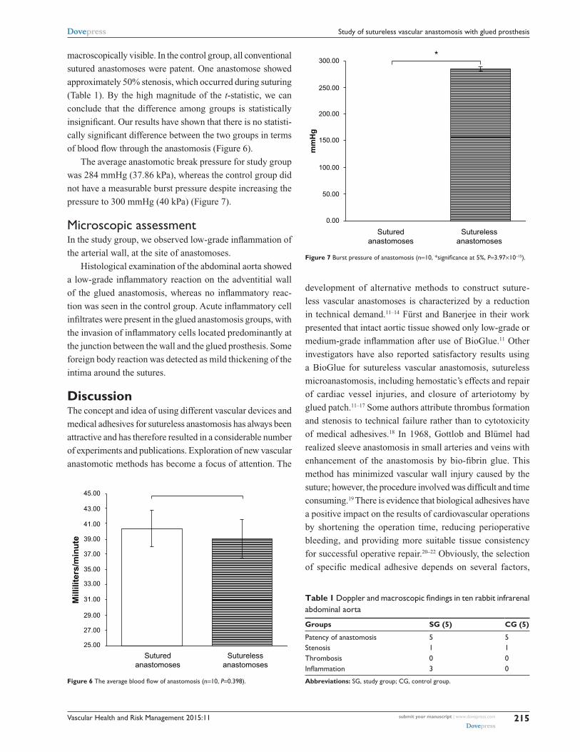
Figure 7 Burst pressure of anastomosis (n=10, *significance at 5%, P=3.97×10⁻¹⁵).
macroscopically visible. In the control group, all conventional
sutured anastomoses were patent. One anastomose showed
approximately 50% stenosis, which occurred during suturing
(Table 1). By the high magnitude of the t-statistic, we can
conclude that the difference among groups is statistically
insignificant. Our results have shown that there is no statisti-
cally significant difference between the two groups in terms
of blood flow through the anastomosis (Figure 6).
The average anastomotic break pressure for study group
was 284 mmHg (37.86 kPa), whereas the control group did
not have a measurable burst pressure despite increasing the
pressure to 300 mmHg (40 kPa) (Figure 7).
Microscopic assessmentIn the study group, we observed low-grade inflammation of
the arterial wall, at the site of anastomoses.
Histological examination of the abdominal aorta showed
a low-grade inflammatory reaction on the adventitial wall
of the glued anastomosis, whereas no inflammatory reac-
tion was seen in the control group. Acute inflammatory cell
infiltrates were present in the glued anastomosis groups, with
the invasion of inflammatory cells located predominantly at
the junction between the wall and the glued prosthesis. Some
foreign body reaction was detected as mild thickening of the
intima around the sutures.
DiscussionThe concept and idea of using different vascular devices and
medical adhesives for sutureless anastomosis has always been
attractive and has therefore resulted in a considerable number
of experiments and publications. Exploration of new vascular
anastomotic methods has become a focus of attention. The
development of alternative methods to construct suture-
less vascular anastomoses is characterized by a reduction
in technical demand.11–14 Fürst and Banerjee in their work
presented that intact aortic tissue showed only low-grade or
medium-grade inflammation after use of BioGlue.11 Other
investigators have also reported satisfactory results using
a BioGlue for sutureless vascular anastomosis, sutureless
microanastomosis, including hemostatic’s effects and repair
of cardiac vessel injuries, and closure of arteriotomy by
glued patch.11–17 Some authors attribute thrombus formation
and stenosis to technical failure rather than to cytotoxicity
of medical adhesives.18 In 1968, Gottlob and Blümel had
realized sleeve anastomosis in small arteries and veins with
enhancement of the anastomosis by bio-fibrin glue. This
method has minimized vascular wall injury caused by the
suture; however, the procedure involved was difficult and time
consuming.19 There is evidence that biological adhesives have
a positive impact on the results of cardiovascular operations
by shortening the operation time, reducing perioperative
bleeding, and providing more suitable tissue consistency
for successful operative repair.20–22 Obviously, the selection
of specific medical adhesive depends on several factors,
Table 1 Doppler and macroscopic findings in ten rabbit infrarenal abdominal aorta
Groups SG (5) CG (5)
Patency of anastomosis 5 5Stenosis 1 1Thrombosis 0 0Inflammation 3 0
Abbreviations: SG, study group; CG, control group.

Vascular Health and Risk Management 2015:11submit your manuscript | www.dovepress.com
Dovepress
Dovepress
216
Vokrri et al
including quantity of adhesive required, its mechanical
properties, hemostatic efficacy, effects on wound healing,
and the inflammatory response it stimulates. These factors are
especially important when performing microanastomoses, as
these are usually time consuming and challenging.23–25
Anastomosis techniques without suturing can eliminate
vascular injury and foreign body injury within the vessel wall.
In this study, we have designed a sutureless anastomosis tech-
nique, using a glued prosthesis, which combines the advan-
tages of the sutured and the glued anastomotic technique.
This technique may be used in small- and medium-sized
vessels. Our experiment suggests that the sutureless anas-
tomosis has moderate advantages over the sutured method.
The anastomosing time was shorter, and the blood loss was
not significant. As the two vascular ends are anastomosed
by glued prosthesis, anastomotic bleeding can safely be
prevented. The immediate patency rates and average blood
flow of anastomosis were similar.
Nevertheless, a question remained on whether the glued
anastomosis supports high intraluminal blood pressure. The
answer mainly depends on the intraluminal blood pressure and
the sustaining pressure of the anastomosis. Normal human
blood pressure is 80 mmHg (10.66 kPa) to 120 mmHg
(16.0 kPa), and may increase to 280 mmHg (37.33 kPa) or
more in some very rare pathological conditions. The mean
burst pressure of the sutureless anastomosis obtained in this
experiment was 284 mmHg (37.86 kPa), so we can conclude
that the glued anastomosis can sustain normal arterial blood
pressure. The anastomosis was able to maintain integrity
even at high pressures, which we may find in pathological
situations. This implies that the sutureless anastomosis
using glued prosthesis can effectively be used in patients
with well-controlled systolic blood pressure. However,
larger experimental studies with long-term follow-up are
needed to more accurately determine the burst pressures
of these anastomoses. Unfortunately, the excess doses of
medical adhesive applied in the inner surface of ePTFE
graft prosthesis probably led to anastomotic stenosis seen
on the first case of the study group. The macro- and micro-
histological results showed that inflammation in the study
group was higher than in the control group. Our in vivo
study in rabbits, although small in number, suggests that this
anastomosis using glued prosthesis can be effective. These
results demonstrate that biological glue made from the com-
bination of bovine albumin and glutaraldehyde is effective
in vivo in the creation of sutureless anastomosis. However,
the inflammatory effect of BioGlue needs to be taken into
account, when choosing to use such a method. Further
studies are needed to better evaluate the long-term effects
of such methods on the vessel wall.
Rabbits are the small animal of choice for conduits of
1–4 mm in diameter, enabling implantation of longer con-
duits and having a greater similarity than rats to humans in
coagulation, endothelialization, and patency.26
ConclusionOur first experience with glued prosthesis as a method of
sutureless vascular anastomosis has shown that vascular anas-
tomosis was feasible, simple, and fast and can be considered
reliable, without major signs of complication. Medical adhe-
sives deserve to be reconsidered as an alternative to suturing in
surgery. The hybrid sutureless vascular anastomosis technique
with medical adhesives in the future may provide a promising
alternative to manual suturing. However, further long-term
studies are necessary to elucidate the burst pressure and degree
of inflammation in the anastomotic site and to investigate the
applicability of this technique in human practice.
Acknowledgment This work was supported by French state funds managed by
the ANR within the Investissements d’Avenir programme
(Labex CAMI) under reference ANR-11-LABX-0004
DisclosureThe authors report no conflicts of interest in this work.
References1. Carrel A. La technique operatoire des anastomoses vasculaires et
la transplantation des visceres [The operative technique of vas-cular anastomoses and the transplantation of viscera]. Lyon Med. 1902;98:859–863.
2. Schiller W, Rudorf H, Kiderlen MJ, et al. Short-term tissue response of lapine carotid artery microanastomoses to BioGlue. J Thorac Cardiovasc Surg. 2007;55(5):298–303.
3. Schiller W, Rudorf H, Welzel CB, et al. Sutureless anastomoses of rab-bit carotid arteries with BioGlue. J Thorac Cardiovasc Surg. 2007;134: 1513–1518.
4. Wheat JC, Wolf JS Jr. Advances in bioadhesives, tissue sealants, and hemostatic agents. Urol Clin North Am. 2009;36(2):265–275.
5. Summary of Safety and Effectiveness CryoLife, Inc. BioGlue® Surgi-cal Adhesive; 2001. Available from: http://www.accessdata.fda.gov/./P010003b.pdf.
6. Perrin B, Brichon PY, Bracini M, et al. Une revue des colles utilisées en chirurgies cardiaque, thoracique et vasculaire [A review of adhe-sives used in cardiac, thoracic and vascular surgery]. Chir Cardiovasc. 2012;16(1):33–42.
7. Duarte AP, Coelho JF, Bordado JC, et al. Surgical adhesives: systematic review of the main types and development forecast. Prog Polym Sci. 2012;37(8):1031–1050.
8. Wippermann J, Konstas C, Breuer M, Kosmehl H, Wahlers T, Albes JM. Long term effects in distal coronary anastomoses using different adhesives in a porcine off-pump model. J Thorac Cardiovasc Surg. 2006;132:325–331.

Vascular Health and Risk Management
Publish your work in this journal
Submit your manuscript here: http://www.dovepress.com/vascular-health-and-risk-management-journal
Vascular Health and Risk Management is an international, peer-reviewed journal of therapeutics and risk management, focusing on concise rapid reporting of clinical studies on the processes involved in the maintenance of vascular health; the monitoring, prevention and treatment of vascular disease and its sequelae; and the involvement of
metabolic disorders, particularly diabetes. This journal is indexed on PubMed Central and MedLine. The manuscript management system is completely online and includes a very quick and fair peer-review system, which is all easy to use. Visit http://www.dovepress.com/ testimonials.php to read real quotes from published authors.
Vascular Health and Risk Management 2015:11 submit your manuscript | www.dovepress.com
Dovepress
Dovepress
Dovepress
217
Study of sutureless vascular anastomosis with glued prosthesis
9. Gundry SR, Black K, Izutani H. Sutureless coronary artery bypass with biologic glued anastomoses: preliminary in vivo and in vitro results. J Thorac Cardiovasc Surg. 2000;120:473–481.
10. Belleghem YV, Forsyth RG, Narine K, Moerman A, Taeymans Y, Van Nooten GJ. Bovine glue (Bio-Glue) is catabolized by enzymatic reaction in the vascular dog model. Ann Thorac Surg. 2004;77:2177–2182.
11. Fürst W, Banerjee A. Release of glutaraldehyde from an albumin glutaraldehyde tissue adhesive causes significant in vitro and in vivo toxicity. Ann Thorac Surg. 2005;79:1522–1528.
12. Qu L, Jing Z, Wang Y. Sutureless anastomoses of small and medium sized vessels by medical adhesive. Eur J Vasc Endovasc Surg. 2004;28: 526–533.
13. Van Nooten G, Van Belleghem Y, Foubert L, et al. An experimental model of coronary anastomosis without suturing. Cardiovasc Surg. 2003;11:80–84.
14. Van Nooten GJ, Somers P, Forsyth R, et al. Autologous glue: part of the sticky mystery unraveled. J Thorac Cardiovasc Surg. 2007;134: 415–423.
15. Galvão FHF, Bacchella T, Machado MC. Cuff-glue sutureless microanastomosis. Microsurgery. 2007;27(4):271–276.
16. Agrifoglio M, Barili F, Kassem S, et al. Sutureless patch-and-glue technique for the repair of coronary sinus injuries. J Thorac Cardiovasc Surg. 2007;134:522–523.
17. Bastiaanse J, Borst C, van der Helm YJM, Loo KHH, Gründeman PF. Arteriotomy closure by glued patch in the porcine carotid artery. Ann Thorac Surg. 2000;70:1384–1388.
18. Gummert JF, Demertzis S, Matschke K, et al. Six-month angiographic follow-up of the PAS-Port II clinical trial. Ann Thorac Surg. 2006;81: 90–96.
19. Gottlob R, Blümel G. Anastomoses of small arteries and veins by means of bushing and adhesive. J Cardiovasc Surg (Torino). 1968;9: 337–341.
20. von Segesser LK, Oechslin E, Jenni R, Turina MI. Use of glue to avoid formation of perfused recesses in aortic allograft implantation. Ann Thorac Surg. 1994;57:494–495.
21. Fundaro P, Velardi AR, Santoli C. Fibrin adhesive: clinical application in coronary artery bypass surgery. Tex Heart Inst J. 1985;12:275–278.
22. Hagberg RC, Safi HJ, Sabik J, Conte J, Block JE. Improved intraopera-tive management of anastomotic bleeding during aortic reconstruction: results of a randomized controlled trial. Am Surg. 2004;70:307–311.
23. Alfieri A, Reinert M. Glue-enhanced excimer laser-assisted nonocclu-sive anastomosis: a laboratory investigation. Eur Surg Res. 2011;46(1): 32–37.
24. Schwaiger N, Wu J, Wright B, Morrissey L, Harris M, Rohanizadeh R. BioWeld(®) tube and surgical glue for experimental sutureless venous microanastomosis. Br J Surg. 2010;97(12):1825–1830.
25. Oda S, Morita S, Tanoue Y, Eto M, Matsuda T, Tominaga R. Experimental use of an elastomeric surgical sealant for arterial hemo-stasis and its long-term tissue response. Interact Cardiovasc Thorac Surg. 2010;10(2):258–261.
26. Byrom MJ, Bannon PG, White GH, Ng MK. Animal models for the assessment of novel vascular conduits. J Vasc Surg. 2010;52:176–195.