
Experimental plasmacytomas in relation to human multiple myeloma
7
A nnals of C linical and L aboratory S cience , Vol. 4, No. 3 Copyright © 1974, Institute for Clinical Science Experimental Plasmacytomas in Relation to Human Multiple Myeloma HENRY A. AZAR, M.D. Laboratory Service, VA Hospital, Tampa, FL 33612 and Department of Pathology, University of South Florida College of Medicine, Tampa, FL 33620 ABSTRACT Among animal models of plasma cell tumors, that induced in BALB/c mice by means of intraperitoneal injections of oils remains the most reproducible and the most intensively studied. The BALB/c oil-induced and transplantable plasmacytomas resemble hu- man myeloma in their ability to produce a monoclonal immunoglobulin or Bence Jones protein. Bence Jones type nephrosis in BALB/c mice closely mimics its human counterpart. There are also similarities in background of immunodeficiency and in antigen-binding affinity of monoclonal immuno- globulins, as well as in interesting interrelationships with malignant lympho- mas. Unlike the BALB/c tumor model, human myeloma is, however, prin- cipally a skeletal disease, not a gut-oriented or peritoneal plasmacytoma. The intriguing presence of intracistemal type A virus-like particles in BALB/c plasmacytoma cells and their absence in human myeloma is another major difference between the two forms of growth. The pathogenesis of human myeloma remains obscure but the availability of experimental plasmacytoma models offers a means of systematically analyz- ing events leading to the neoplastic transformation of antibody-forming cells. Introduction In addition to man, several mammals were observed to develop plasma cell tu- mors spontaneously. For example, the mul- tiple myeloma of the dog resembles human myeloma and features osteolytic lesions, monoclonal gammapathy and Bence Jones type nephropathy.16 Multiple myeloma has been also observed in a cat.10 Two plasma- cytomas have been described in the Syrian hamster: one arose in the neck and the other in a mesenteric lymph node.4 These spontaneous tumors constitute uncommon though fascinating events. Plasma cell tu- mors can be induced in a predictable fashion in mice, particularly the BALB/c strain. The reader is referred to Potters comprehensive review of experimental plasma cell tumors of mice.18 The aim of this presentation is to outline the characteristics of induced plasma cell tumors of mice, particularly the BALB/c
Transcript of Experimental plasmacytomas in relation to human multiple myeloma

A n n a l s o f C l i n i c a l a n d L a b o r a t o r y S c i e n c e , Vol. 4, No. 3 Copyright © 1974, Institute for Clinical Science
Experimental Plasmacytomas in Relation to Human Multiple Myeloma
HENRY A. AZAR, M.D.
Laboratory Service, VA Hospital, Tampa, FL 33612and
Department of Pathology, University of South Florida College of Medicine, Tampa, FL 33620
ABSTRACT
Among animal models of plasma cell tumors, that induced in B A L B /c mice by means of intraperitoneal injections of oils remains the most reproducible and the most intensively studied.
The BA LB /c oil-induced and transplantable plasmacytomas resemble human myeloma in their ability to produce a monoclonal immunoglobulin or Bence Jones protein. Bence Jones type nephrosis in BA LB /c mice closely mimics its human counterpart. There are also similarities in background of immunodeficiency and in antigen-binding affinity of monoclonal immunoglobulins, as well as in interesting interrelationships with malignant lymphomas. Unlike the BA LB /c tumor model, human myeloma is, however, principally a skeletal disease, not a gut-oriented or peritoneal plasmacytoma. The intriguing presence of intracistemal type A virus-like particles in BA LB/c plasmacytoma cells and their absence in human myeloma is another major difference between the two forms of growth.
The pathogenesis of human myeloma remains obscure but the availability of experimental plasmacytoma models offers a means of systematically analyzing events leading to the neoplastic transformation of antibody-forming cells.
In tro d u ctio n
In addition to man, several mammals were observed to develop plasma cell tumors spontaneously. For example, the multiple myeloma of the dog resembles human myeloma and features osteolytic lesions, monoclonal gammapathy and Bence Jones type nephropathy.16 Multiple myeloma has been also observed in a cat.10 Two plasmacytomas have been described in the Syrian hamster: one arose in the neck and the
other in a mesenteric lymph node.4 These spontaneous tumors constitute uncommon though fascinating events. Plasma cell tumors can be induced in a predictable fashion in mice, particularly the BA LB /c strain. The reader is referred to Potters comprehensive review of experimental plasma cell tumors of mice.18
The aim of this presentation is to outline the characteristics of induced plasma cell tumors of mice, particularly the BA LB /c

1 5 8 AZAR
TA BLE I
P l a s m a c y t o m a s o f M i c e
"P lasm a C e ll Leukem ia" i n S t r e e t S t r a i n Mice 1951
R a s k - N ie l s e n , R . and G orm sen, H.
I l e o c e c a l P lasm acytom as o f C3H M ice 1954
Dunn, T .B .
In d u ced P e r i t o n e a l P lasm acytom as (BALB/c M ice) 1960
P o t t e r , M. and R o b e r t so n , C .L .
P lasm a C e ll Tumors i n NZB and NZBX BALB/c Mice 1966
W erner, N. e t a l
P leo m o rp h ic Lymphomas in S JL /J Mice 1969
M urphy, E .D .
strain, and relate these to the problem of human myeloma.
P la sm acy to m as o f M ice
The more important experimental plasma cell tumor models of mice are listed in table I. Although the reader’s attention will be drawn to the oil-induced peritoneal plasmacytomas of BA LB /c mice, a brief outline of other models of plasmacytic and related lymphoreticular growths is in order.
The “plasma cell leukemia” in Street strain mice described by Rask-Nielsen and and Gormsen20 in 1951 has the morphologic features of reticulum cell sarcoma with plasmacytic differentiation. In later studies, (C BA X D BA /2) F I hybrids, including mice injected with a variety of cell-free material, developed a high incidence of “plasma cell leukemia.”21 Several of the transplanted reticular and plasmacytic neoplasms transplanted by Ebbesen and Rask- Nielsen9 were associated with “paraproteinemia” and amyloidosis.
Thelma Dunn6 reported in 1954 that several mice had an inflammatory lesion of the cecum. In some of these lesions, plasmacytic neoplasms arise in the ileocecal region and in relation to the cecitis. A later survey7 indicated that ileocecal plasmacytomas were commoner in C3H mice than in other strains and that they occurred more frequently in old mice.
Murphy15 described in 1963 a new in- bred strain of mice (S JL / J ) with a high incidence of reticulum cell neoplasms. Fifty percent of these neoplasms were associated with “paraproteinemia.” Unfortunately, the production of monoclonal immunoglobulin has been unstable following transplantation of these tumors. Fujinaga et al11 transmitted “S JL /J disease” to BA LB/c mice with cell-free extracts. They succeeded in producing reticulum cell neoplasms but not plasmacytomas.
Plasmacytomas are successfully induced by intraperitoneal injection of oily substances in BA LB/c and NZB mice as well as in (B A LB /c X NZB) F I hybrids.18 The NZB strain is also genetically susceptible to the development of nephritis and autoimmune phenomena. The oil-induced plasmacytomas are by far the most widely studied model of immunoglobulin-producing experimental tumors. Primary and transplanted BA LB /c plasmacytomas are under active investigation in our laboratory. They are considered to be a dependable and readily available source of tumor that strikingly resembles human myeloma.
O il-Induced P la sm acy to m as o f B A L B /c M ice
In an experiment designed to test the survival of allogeneic grafts as well as the behavior of M.T.A. virus, Merwin and Al- gire13 implanted estrogen-treated BA LB/c mice with Millipore diffusion chambers containing C3H mammary tumor. Six months later, the BA LB/c recipients developed hemorrhagic ascites owing to per
E X P E R IM E N T A L PLA SM A CYTO M A S 1 5 9
itoneal plasma cell tumor or fibrosarcoma.8 Empty chambers, leucite discs or borings also caused the development of plasma cell tumors.14 Subsequently, Potter and Boyce19 reported that plasma cell tumors could be induced by means of intraperitoneal injections of mineral oil alone or adjuvant mixtures. A variety of oils have been tested since. More recently, pristane (2,6,10,14- tetramethylpentadecane) was found to be an extremely potent inducer of plasmacytomas in BA LB /c mice.1
The experimental model of oil-induced plasmacytomas has been described in detail elsewhere.18 This model has been extensively exploited to demonstrate the chain of events leading to the synthesis of specific classes and types of immunoglobulin. The same model has been used to study the effect of hereditary and hormonal influences on the induction of plasma cell tumors. Since the majority of oil-induced plasmacytomas are immunoglobulin-forming tumors, the role of intestinal bacteria and bacterial antigens on the differentiation of plasmacytomas has also been extensively investigated.
M o r p h o l o g ic F e a t u r e s
The sequence of events following the intraperitoneal injections of mineral oil or pristane starts with an acute inflammatory reaction which first covers the peritoneal surfaces and surrounds oil droplets. No plasma cells or lymphocytes are noted at this stage. At a later period, from one to three months after the initial injection of the oil, individual or small clusters of plasma cells begin to form within the oil granulomas. From five months to eight months following the injection of the oil, larger clusters of plasma cell precursors are observed. Such aggregates are particularly noted under the diaphragm and over the surface of the liver. Ultimately, these granules grow into small tumors which become autonomous plasmacytomas that can be
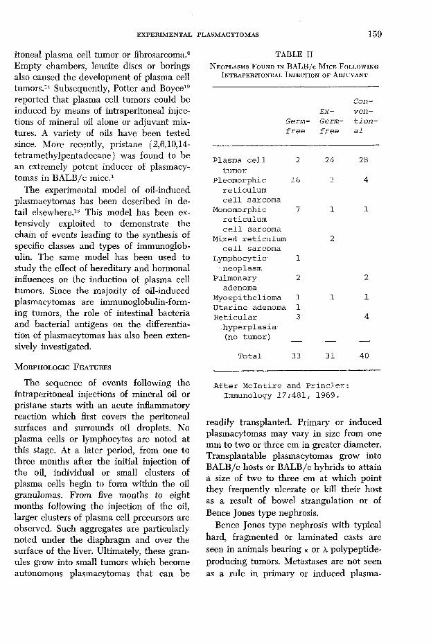
T A B L E II
N e o p l a s m s F o u n d i n B A L B / c M i c e F o l l o w i n g I n t r a p e r i t o n e a l I n j e c t i o n o f A d j u v a n t
Germ-f r e e
Ex—Germ-f r e e
Con-v en -t i o n -a l
P lasm a c e l l 2 24 28tum or
P leo m o rp h ic 16 3 4r e t ic u lu mc e l l sarcom a
Monomorphic 7 1 1re t ic u lu mc e l l sarcom a
Mixed r e t ic u lu m 2c e l l sarcom a
L ym phocytic 1■ neoplasm
Pulm onary 2 2adenoma
M y o ep ith e lio m a 1 1 1U te r in e adenoma 1R e t i c u l a r 3 4
h y p e r p la s ia(no tum or) — — —
T o ta l 33 31 40
A f te r M c ln t i r e and P r i n c l e r : Immunology 1 7 :4 8 1 , 1969.
readily transplanted. Primary or induced plasmacytomas may vary in size from one mm to two or three cm in greater diameter. Transplantable plasmacytomas grow into BA LB/c hosts or BA LB/c hybrids to attain a size of two to three cm at which point they frequently ulcerate or kill their host as a result of bowel strangulation or of Bence Jones type nephrosis.
Bence Jones type nephrosis with typical hard, fragmented or laminated casts are seen in animals bearing k or A polypeptide- producing tumors. Metastases are not seen as a rule in primary or induced plasma-

1 6 0 AZAR
cytomas but do develop in animals bearing ulum cells except for their basophilic andlarge transplanted plasmacytomas. pyroninophilic cytoplasm. Seldom do they
Plasmacytoma cells often resemble retie- appear to be frankly plasmacytic. Trans-
F i c u i i e 1. Electron micrograph of portion of Pristane-induced plasmacytoma cell illustrating a large number of intracisternal type A particles. Some of these appear to be budding from the endoplasmic reticulum lining the cisternae. Uranyl acetate and lead citrate, X 51,000.

E X PER IM EN T A L PLASM ACYTOM AS 1 6 1
planted plasmacytomas are even more anaplastic looking than primary plasma cell tumors. On electron microscopy, plasmacytoma cells generally display an abundance of rough endoplasmic reticulin as well as free polyribosomes.
T y p e A — I n t r a c i s t e r n a l P a r t i c l e s
Whereas the normal lymphoid tissue of the BA LB/c mouse may contain type B or C RNA viruses with well-formed nucleoids, one does not readily observe type A particles in either lymphocytes or plasma cells. In contrast to the lack of type A intracisternal particles within reactive plasma cells, these particles are found in abundance within the cisternae of plasmacytoma cells.2’5 In figure 1 is illustrated the intracisternal clustering of type A particles within a BA LB/c pristane-induced plasmacytoma cell. It appears that type A particles are markers for plasmacytoma cells although their significance is far from known. These particles are non-infective; they lack a nucleoid and are poor in RNA. They are known to be rich in DNA-poly- merase which is thought to represent a defective tumor virus enzyme.22 It should be emphasized that human myeloma cells are free of type A particles. Furthermore, there have been no consistent or well-documented observations of any type of virus within human myeloma cells.
M o n o c l o n a l I m m u n o g l o b u l i n s
As in human myeloma, the majority of oil-induced primary plasma cell tumors are associated with production of an immunoglobulin and/or of a light-chain polypeptide. The majority of immunoglobulins produced have been monoclonal IgA. There is also a marked predominance of k over A polypeptides.18 Some of the monoclonal immunoglobulins produced by B A LB/c plasmacytomas as well as in human myeloma cases were found to have antigen- binding properties.17’18
H0PC1 S
RPC5 S
TEPC183 S M0PC321 S RPC20 S
NP2 S M0PC315 S I
F i g u r e 2. Electrophoretic cellulose acetate patterns of sera from BALB/c mice bearing transplantable plasmacytomas. Note the M component in each of the three upper patterns.
The electrophoretic serum patterns of seven transplantable plasma cell tumors are shown in figure 2. A distinct M component is revealed in the upper three patterns: HOPC-1 and RPC-5 produce, respectively, an IgG A or k monoclonal protein; TEPC-138, an IgM k . MOPC-321 produces k polypeptide alone and its serum electrophoretic pattern shows no M component; its urine contains Bence Jones protein. RPC-20 produces A polypeptide alone. Its serum as well as its urine electrophoretic patterns revealed an M component. NP-2 is a “non-producer” and shows a normal pattern. MOPC-315 is an IgA producer; no M component is apparent on this electrophoretic pattern.
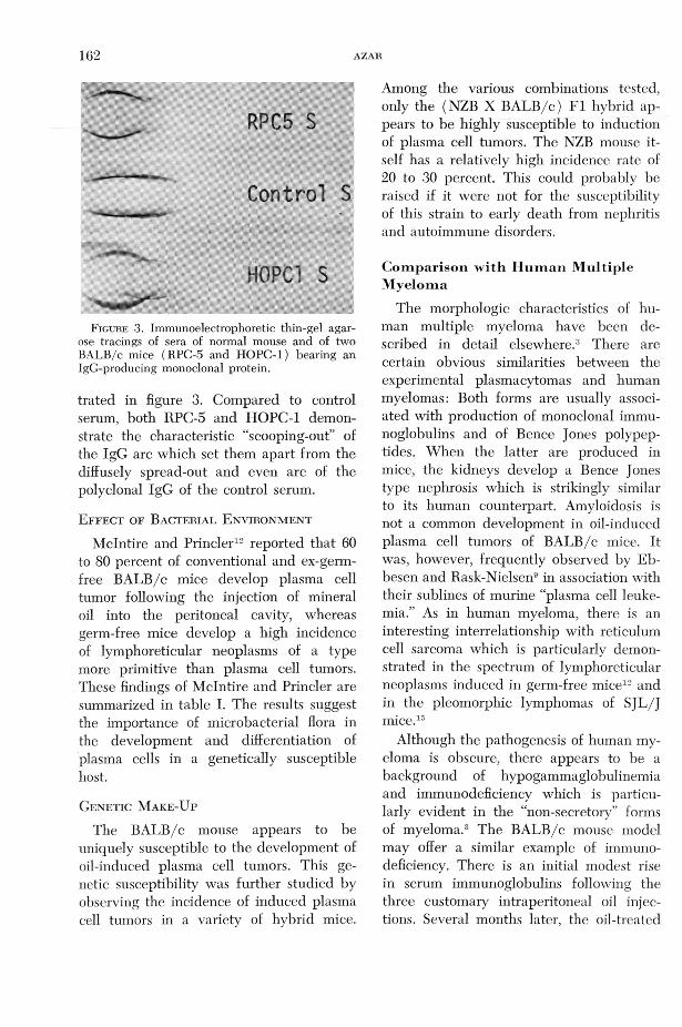
The monoclonicity of the class of immunoglobulin produced by these BA LB/c transplantable plasmacytomas is revealed in the immunoelectrophoretic studies illus-
162 AZAR
Control S
H0PC1 S
F ig u r e 3 . Immunoelectrophoretic thin-gel agarose tracings of sera of normal mouse and of two BALB/c mice (RPC-5 and HOPC-1) bearing an IgG-producing monoclonal protein.
trated in figure 3. Compared to control serum, both RPC-5 and HOPC-1 demonstrate the characteristic “scooping-out” of the IgG arc which set them apart from the diffusely spread-out and even arc of the polyclonal IgG of the control serum.
E f f e c t o f B a c t e r i a l E n v i r o n m e n t
Mclntire and Princler12 reported that 60 to 80 percent of conventional and ex-germ- free BA LB/c mice develop plasma cell tumor following the injection of mineraloil into the peritoneal cavity, whereas germ-free mice develop a high incidence of lymphoreticular neoplasms of a type more primitive than plasma cell tumors. These findings of Mclntire and Princler are summarized in table I. The results suggest the importance of microbacterial flora in the development and differentiation of plasma cells in a genetically susceptible host.
G e n e t i c M a k e - U p
The BA LB/c mouse appears to be uniquely susceptible to the development of oil-induced plasma cell tumors. This genetic susceptibility was further studied by observing the incidence of induced plasma cell tumors in a variety of hybrid mice.
Among the various combinations tested, only the (NZB X BA LB/c) F I hybrid appears to be highly susceptible to induction of plasma cell tumors. The NZB mouse itself has a relatively high incidence rate of 20 to 30 percent. This could probably be raised if it were not for the susceptibility of this strain to early death from nephritis and autoimmune disorders.
C o m p a r i so n w ith H u m a n M u lt ip le M y e lo m a
The morphologic characteristics of human multiple myeloma have been described in detail elsewhere.3 There are certain obvious similarities between the experimental plasmacytomas and human myelomas: Both forms are usually associated with production of monoclonal immunoglobulins and of Bence Jones polypeptides. When the latter are produced in mice, the kidneys develop a Bence Jones type nephrosis which is strikingly similar to its human counterpart. Amyloidosis is not a common development in oil-induced plasma cell tumors of BA LB/c mice. It was, however, frequently observed by Eb- besen and Rask-Nielsen9 in association with their sublines of murine “plasma cell leukemia.” As in human myeloma, there is an interesting interrelationship with reticulum cell sarcoma which is particularly demonstrated in the spectrum of lymphoreticular neoplasms induced in germ-free mice12 and in the pleomorphic lymphomas of S JL /J mice.15
Although the pathogenesis of human myeloma is obscure, there appears to be a background of hypogammaglobulinemia and immunodeficiency which is particularly evident in the “non-secretory” forms of myeloma.3 The BA LB/c mouse model may offer a similar example of immunodeficiency. There is an initial modest rise in serum immunoglobulins following the three customaiy intraperitoneal oil injections. Several months later, the oil-treated
E X P E R IM E N T A L PLA SM ACYTO M AS 1 6 3
mice develop a relative hypogammaglobulinemia that ushers the production of plasma cell tumors.
There are also some marked differences between the experimental mouse plasmacytomas and human myelomas. The type A intracistemal particles have been consistently observed in oil-induced as well as in transplanted plasmacytoma cells but not in myeloma cells.2’5 Granulomas and other states of chronic inflammation are generally absent in the clinical period preceding human myeloma. Hormonal and genetic influences appear to be unimportant or of obscure significance in man. Finally, human myeloma is principally a skeletal disease and, as such, it is more akin to the canine or feline myeloma than to the BA LB /c oil- induced peritoneal plasmacytomas.
R eferences1. A n d e r s o n , P . N. a n d P o t t e r , M.: Induction
of plasma cell tumours in BA LB/c mice with 2,6,10,14-tetramethylpentadecane ( Pristane). Nature 222:994-995, 1969.
2. A z a r , H. A.: Significance of adjuvant-induced plasmacytomas of mice in relation to the pathogenesis of human multiple myeloma. Ann. Allerg. 26:293-298, 1968.
3. A z a r , H. A . a n d P o t t e r , M., eds.: Multiple Myeloma and Related Disorders, Vol. 1. Harper & Row, Hagerstown, MD, 428 pp.,1973.
4. C o t r a n , R. S. a n d F o r t n e r , J. G.: Serum protein abnormality in a transplantable plasmacytoma of the Syrian golden hamster. J. Nat. Cancer Inst. 28:1193-1205, 1962.
5. D a l t o n , A. J., P o t t e r , M . , a n d M e r w i n ,
R. M . : Some ultrastructural characteristics of a series of primary and transplanted plasma cell tumors of the mouse. J. Nat. Cancer Inst. 26:1221-1268, 1961.
6. D u n n , T. B.: Normal and pathologic anatomy of the reticular tissue in laboratory mice with a classification and discussion of neoplasms. J. Nat. Cancer Inst. 14:1281—1433, 1954.
7. D u n n , T. B. a n d D e r i n g e r , M. K.: Reticulum cell neoplasms type B or the “Hodgkin’s like lesion” of the mouse. J. Nat. Cancer Inst. 40: 771-821, 1968.
8. D u n n , T. B., P o t t e r , M . , F a h e y , J. L., a n d
M e r w i n , R. M.: Morphology and serum pro
tein changes in plasma cell neoplasms in mice. Arch. De Vecchi Anat. Patol. 31:67-77, 1960.
9. E b b e s e n , P. a n d R a s k - N i e l s e n , R . : On amyloidosis and paraproteinemia in seven transplantation sublines of a murine plasma cell leukemia. J. Nat. Cancer Inst. 38:723-739,1967.
10. F a r r o w , B. R. H. a n d P e n n y , R.: Multiple myeloma in a cat. J . Amer. Vet. Med. Assoc. 158:606-609, 1971.
11. F u j i n a g a , S., P o e l , W . E., W i l l i a m s , W . C., a n d D m o c h o w s k i , L .: Biological and morphological studies of S JL /J strain reticulum cell neoplasms induced and transmitted serially in low leukemia strain mice. Cancer Res. 30:729- 742, 1970.
12. M c In t i r e , K. R. a n d P r i n c l e r , G . L .: Prolonged adjuvant stimulation in germ-free BA LB /c mice: development of plasma cell neoplasia. Immunology 17:481-487, 1969.
13. M e r w i n , R. M. a n d A l g i r e , G . H.: Induction of plasma-cell neoplasms and fibrosarcomas in BA LB /c mice carrying diffusion chambers. Proc. Soc. Exp. Biol. Med. 101:437-439, 1959.
14. M e r w i n , R . M . a n d R e d m o n , L. W.: Induction of plasma cell tumors and sarcomas in mice by diffusion chambers placed in the peritoneal cavity. J . Nat. Cancer Inst. 31:998- 1017, 1963.
15. M u r p h y , E. D.: S JL /J, a new inbred strain of mouse with a high early incidence of reticulum-cell neoplasms. Proc. Amer. Assoc. Cancer Res. 4:46, 1963.
16. O s b o r n e , C. A., P e r m a n , V., S a u t t e r , J. H . ,
S t e v e n s , J. B., a n d H a n l o n , G . F . : Multiple myeloma in the dog. J. Amer. Vet. Med. Assoc, i 53:10, 1300-1319, 1968.
17. P o t t e r , M.: Antigen-binding myeloma proteins in mice. Ann. NY Acad. Sci. 190:306- 321, 1971.
18. P o t t e r , M.: Experimental plasma cell tumors and other immunoglobulin-producing lympho- reticular neoplasms in mice. Multiple Myeloma and Related Disorders, Vol. I, Azar,H. A. and Potter, M., eds. Harper & Row, Hagerstown, MD, pp. 153-194, 1973.
19. P o tte r , M. a n d Boyce, C. R.: Induction of plasma-cell neoplasms in strain BA LB /c mice with mineral oil and mineral oil adjuvants. Nature ¿93:1086-1087, 1962.
20. R a s k - N i e l s e n , R . a n d G o r m s e n , H.: Spontaneous and induced plasma cell neoplasia in a strain of mice. Cancer 4 :387-397, 1951.
21. R a s k - N i e l s e n , R . a n d G o r m s e n , H.: On the occurence of plasma-cell leukemia in various strains of mice. J. Nat. Cancer Inst. i6 :1137- 1147, 1956.
22. W i l s o n , S. H. a n d K u f f , E. L .: A novel DNA polymerase activity found in association with intracistemal A-type particles. Proc. Nat. Acad. Sci. 69:1531-1536, 1972.


