EXPERIMENTAL AND NUMERICAL SIMULATION OF …
11
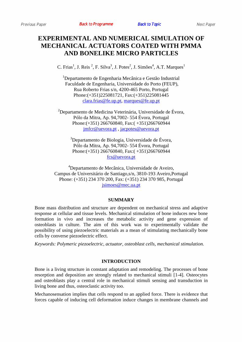
EXPERIMENTAL AND NUMERICAL SIMULATION OF MECHANICAL ACTUATORS COATED WITH PMMA AND BONELIKE MICRO PARTICLES C. Frias 1 , J. Reis 2 , F. Silva 3 , J. Potes 2 , J. Simões 4 , A.T. Marques 1 1 Departamento de Engenharia Mecânica e Gestão Industrial Faculdade de Engenharia, Universidade do Porto (FEUP), Rua Roberto Frias s/n, 4200-465 Porto, Portugal Phone:(+351)225081721, Fax:(+351)225081445 [email protected] , [email protected] 2 Departamento de Medicina Veterinária, Universidade de Évora, Pólo da Mitra, Ap. 94,7002- 554 Évora, Portugal Phone:(+351) 266760840, Fax:( +351)266760944 [email protected] , [email protected] 3 Departamento de Biologia, Universidade de Évora, Pólo da Mitra, Ap. 94,7002- 554 Évora, Portugal Phone:(+351) 266760840, Fax:( +351)266760944 [email protected] 4 Departamento de Mecânica, Universidade de Aveiro, Campus de Universitário de Santiago,s/n, 3810-193 Aveiro,Portugal Phone: (+351) 234 370 200, Fax: (+351) 234 370 985, Portugal [email protected] SUMMARY Bone mass distribution and structure are dependent on mechanical stress and adaptive response at cellular and tissue levels. Mechanical stimulation of bone induces new bone formation in vivo and increases the metabolic activity and gene expression of osteoblasts in culture. The aim of this work was to experimentally validate the possibility of using piezoelectric materials as a mean of stimulating mechanically bone cells by converse piezoelectric effect. Keywords: Polymeric piezoelectric, actuator, osteoblast cells, mechanical stimulation. INTRODUCTION Bone is a living structure in constant adaptation and remodeling. The processes of bone resorption and deposition are strongly related to mechanical stimuli [1-4]. Osteocytes and osteoblasts play a central role in mechanical stimuli sensing and transduction in living bone and thus, osteoclastic activity too. Mechanosensation implies that cells respond to an applied force. There is evidence that forces capable of inducing cell deformation induce changes in membrane channels and
Transcript of EXPERIMENTAL AND NUMERICAL SIMULATION OF …
EXPERIMENTAL AND NUMERICAL SIMULATION OF MECHANICAL ACTUATORS COATED WITH PMMA
AND BONELIKE MICRO PARTICLES
C. Frias1, J. Reis 2, F. Silva3, J. Potes2, J. Simões4, A.T. Marques1
1Departamento de Engenharia Mecânica e Gestão Industrial Faculdade de Engenharia, Universidade do Porto (FEUP),
Rua Roberto Frias s/n, 4200-465 Porto, Portugal Phone:(+351)225081721, Fax:(+351)225081445
[email protected], [email protected]
2Departamento de Medicina Veterinária, Universidade de Évora, Pólo da Mitra, Ap. 94,7002- 554 Évora, Portugal Phone:(+351) 266760840, Fax:( +351)266760944
[email protected] , [email protected]
3Departamento de Biologia, Universidade de Évora, Pólo da Mitra, Ap. 94,7002- 554 Évora, Portugal Phone:(+351) 266760840, Fax:( +351)266760944
4Departamento de Mecânica, Universidade de Aveiro, Campus de Universitário de Santiago,s/n, 3810-193 Aveiro,Portugal
Phone: (+351) 234 370 200, Fax: (+351) 234 370 985, Portugal [email protected]
SUMMARY
Bone mass distribution and structure are dependent on mechanical stress and adaptive response at cellular and tissue levels. Mechanical stimulation of bone induces new bone formation in vivo and increases the metabolic activity and gene expression of osteoblasts in culture. The aim of this work was to experimentally validate the possibility of using piezoelectric materials as a mean of stimulating mechanically bone cells by converse piezoelectric effect.
Keywords: Polymeric piezoelectric, actuator, osteoblast cells, mechanical stimulation.
INTRODUCTION
Bone is a living structure in constant adaptation and remodeling. The processes of bone resorption and deposition are strongly related to mechanical stimuli [1-4]. Osteocytes and osteoblasts play a central role in mechanical stimuli sensing and transduction in living bone and thus, osteoclastic activity too.
Mechanosensation implies that cells respond to an applied force. There is evidence that forces capable of inducing cell deformation induce changes in membrane channels and
on protein structure [5, 6] and that ultimately, cytoskeleton deformation may directly influence on cell nuclei [6-9]. In response to mechanical stimuli, osteoblasts produce a number of substances that function as messenger molecules, like prostaglandins (particularly PGE2) and nitric oxide [10-13]. A mechanical stimulus may be propagated to surrounding osteocytes by a single originating osteocyte, via extracellular soluble signalling factors like nitric oxide [14]. Many devices have been tested for mechanical stimulation of cells and tissues in vitro, namely of osteocytes and osteoblasts [15-19], although many of these systems are hardly adaptable to an in vivo device. Strain, load and frequency of the stimulus determine how cell responds; dynamic, short loading causes the strongest bone adaptation response, and bone cells tend to accommodate to a routine, so the stimulus must vary in order to be followed by a same level of response; a stochastic bone cells response in vitro and in vivo has been reported [8, 11, 20-26]. According to some authors, high frequency associated with a high enough number of cycles are needed to maximize osteoblast proliferation in vitro [27].
Takada described the use for in vitro assays of a piezoelectric actuator in which the cells were seeded on a collagen gel block; this block was then submitted to uniaxial tension and/or compression by the displacement originated by two piezoelectric ceramic layers by the loading of voltage; both strain and frequency applied could vary [19].
As Fukada and Yasuda described, the bone has piezoelectric properties, and mechanical stress applied to dried bone produces polarization and submission of bone to an electric field originates strain [28].
METHODS
Piezoelectric substract
Physical phenomena of the piezoelectric substract
The polymeric piezoelectric films used (Polyvinylidene Fluoride (PVDF)) were supplied by Measurement Specialties Inc Company (USA). These films consist of a 12×13mm active area, printed with silver ink electrodes on both surfaces in an 15×40mm die-cut piezoelectric polymer substrate. It's polarized along the thickness and admits as piezoelectric strain constants ��� � 23 � 10�� and ��� � �33 �10�� �
� ��
� �, see figure 1. Theoretically, based on the converse piezoelectricity effect,
when a voltage is applied along the polarized direction (3 � ����), the polymer strains in the direction 1 � ����, given the intrinsic properties of this specific material. The amount of free strain is given by the equation (1).
��� � ���� �� (1)
Where � is the polymer thickness, and �� the applied voltage.

Figure 1 – The scheme illustrated a piezoelectric polymer. The black region corresponds to the
Coating the polymeric piezoelectric substrate with PMMA and microBonelike.
To ensure osteoblasts to adhere the silver electrodes were covered with an electric insulator material.
The chosen material for covering was an acrylic, poly (methyl methacrylate) (PMMA), (PERFEX®, International Dental Products, USA). Micro particles of Bonelike kindly offered by INESCPorto were added. The PVDF was covered with a homogenous layer of PMMA and Bonelike by a Deepmm⁄s, see figure 2. Each PVDF had four layers, guarantying electrical insulation. The coating was performed in a clean room (Class 1) (INESCPorto).
Figure 2- Deep-coating machine develop to coated the piezoelectric actuators for bone cells.
Sterilizing process of the coated polymeric piezoelectricThe coated polymeric piezoelectric substrates were submitted to dosis of 25 kGy) for sterilization prior to cell culture (ITN, Lisbon).
Finite element method (FNM)
The scheme illustrated a piezoelectric polymer. The black region corresponds to the
active areas (silver electrode).
piezoelectric substrate with PMMA and micro
To ensure osteoblasts to adhere the silver electrodes were covered with an electric
The chosen material for covering was an acrylic, poly (methyl methacrylate) (PMMA), PERFEX®, International Dental Products, USA). Micro particles of Bonelike kindly
offered by INESCPorto were added. The PVDF was covered with a homogenous layer of PMMA and Bonelike by a Deep-Coating process, at constant velocity of 0,238
2. Each PVDF had four layers, guarantying electrical insulation. The coating was performed in a clean room (Class 1) (INESCPorto).
coating machine develop to coated the piezoelectric actuators for bone cells.
coated polymeric piezoelectric The coated polymeric piezoelectric substrates were submitted to γ-irradiationdosis of 25 kGy) for sterilization prior to cell culture (ITN, Lisbon).
Finite element method (FNM)
The scheme illustrated a piezoelectric polymer. The black region corresponds to the
piezoelectric substrate with PMMA and micro-particles of
To ensure osteoblasts to adhere the silver electrodes were covered with an electric
The chosen material for covering was an acrylic, poly (methyl methacrylate) (PMMA), PERFEX®, International Dental Products, USA). Micro particles of Bonelike kindly
offered by INESCPorto were added. The PVDF was covered with a homogenous layer Coating process, at constant velocity of 0,238
2. Each PVDF had four layers, guarantying electrical insulation. The
coating machine develop to coated the piezoelectric actuators for bone cells.
irradiation (normed

FNM estimated and quantified the amount of stress/strain distribution along the piezoelectric surface. The mesh was of quadratic piezoelectric solid elements with three degrees of freedom, through Finite Elements Analysis (FEA) using the solver Abaqus 6.7-1 in static conditions. The material properties used for the numerical simulation were provided by the supplier. The model was composed by 9109 nodes. Experimental measuring of the piezoelectric displacement To understand and quantified the really amount of the displacement and is distributed along the piezoelectric actuator surface were used an ESPI - electronic speckle pattern interferometry process.
Cells culture
The cell line used, MC3T3-E1 cells (11th passage, gently offered by Ineb, Porto) exhibit a developmental sequence typical for osteoblasts [29]. This cell line has been used in many studies on the effects of mechanical stimulation [33-35].
MCT3T3-E1 cells were cultured under standard conditions (37ºC, 5% carbon dioxide), using α-MEM medium (Cambrex), 2 mM L-Glutamine (Cambrex), 10% of bovine fetal serum (Gibco), 0,5% gentamicin and 1% amphotericin B (Gibco).
Piezoelectric substrates (standing on culture dishes, TPP) and controls (standard culture dishes, TPP) were seeded with 16x104 cells, with a total volume of 100 µL of cell suspension. Cells were allowed to adhere to the substrate, before adding the rest of culture medium, and then grown in both static and dynamic piezoelectric substrates (n=6); on the substrates submitted to dynamic conditions, stimulation was done with a alternating sinusoidal current (AC), of 5V, at 1Hz and 3Hz for 15 minutes at each frequency (24h and 48 hours post-seeding). All experiments were repeated three times.
Determination of viability and metabolic activity with resarzurin method The resarzurin-based method utilizes the redox dye resarzurin that upon reduction by metabolically active cells is converted into a highly fluorescent product (resorufin). Nonviable cells have no metabolic capacity and, thus, will not reduce the dye, so the fluorescence intensity observed is a measure of the viable cells [36-38]. After stimulation, the medium was aspirated and new medium with 10% resarzurin solution added. Cells cultures were then incubated for 3 hours before collection of samples and fluorescence readings using a fluorescence spectrophotometer (Shimadzu, Japan). Measurement of nitric oxide (NO) in culture medium NO is a messenger molecule produced in response to mechanical stimulation of osteoblasts and osteocytes, with a large variety of biological functions [12, 39]. NO is quickly oxidized to nitrate and nitrite in biological systems, and these are the two primary, stable and nonvolatile breakdown products of NO. In aqueous buffers and culture conditions nitrite is the principal oxidation product of NO [40]. In this study, culture medium samples were collected immediately after stimulation and NO measured, using NO Assay Kit (Biochain), based on the Griess reaction, after sample deproteinization, and according to the manufacturer’s instructions. Statistical analysis Normal distribution of the results was verified using the Shapiro-Wilk normality test for n>3, and differences between groups tested using one-way ANOVA and Significant differences were considered at a P value 0,05. The statistical analysis was done using

software OriginPro 7.5 (OriginLab Corporation, USA).
Coating the polymeric piezoelectric substrate with PMMA and microBonelike.
The Figure 3 shoes the active area alreadyof µm, and was guaranteed the electrical isolation of the surface.
Figure 3 - Optical microscopic (Palatine inverted optical microscope, Olympus PMG3) images of the coatings distribution in the polymeric piezoelectric surface.
Finite element method (FNM)
FNM gave an estimation of stress/strain and displacement diuncoated polymeric piezoelectric surfahigher displacement was observed in the Figure 5). It's possible to observe a region, but the strain values These values are near the theoeach cycle are present in the Figure
Figure 4 - Displacement variation in nanometers along the PVDF actuator surface o
software OriginPro 7.5 (OriginLab Corporation, USA).
RESULTS
Coating the polymeric piezoelectric substrate with PMMA and micro
shoes the active area already coated. The device has a total , and was guaranteed the electrical isolation of the surface.
Optical microscopic (Palatine inverted optical microscope, Olympus PMG3) images of the coatings distribution in the polymeric piezoelectric surface.
Finite element method (FNM)
FNM gave an estimation of stress/strain and displacement distribution along the polymeric piezoelectric surface. The values range is 6,4443<y<
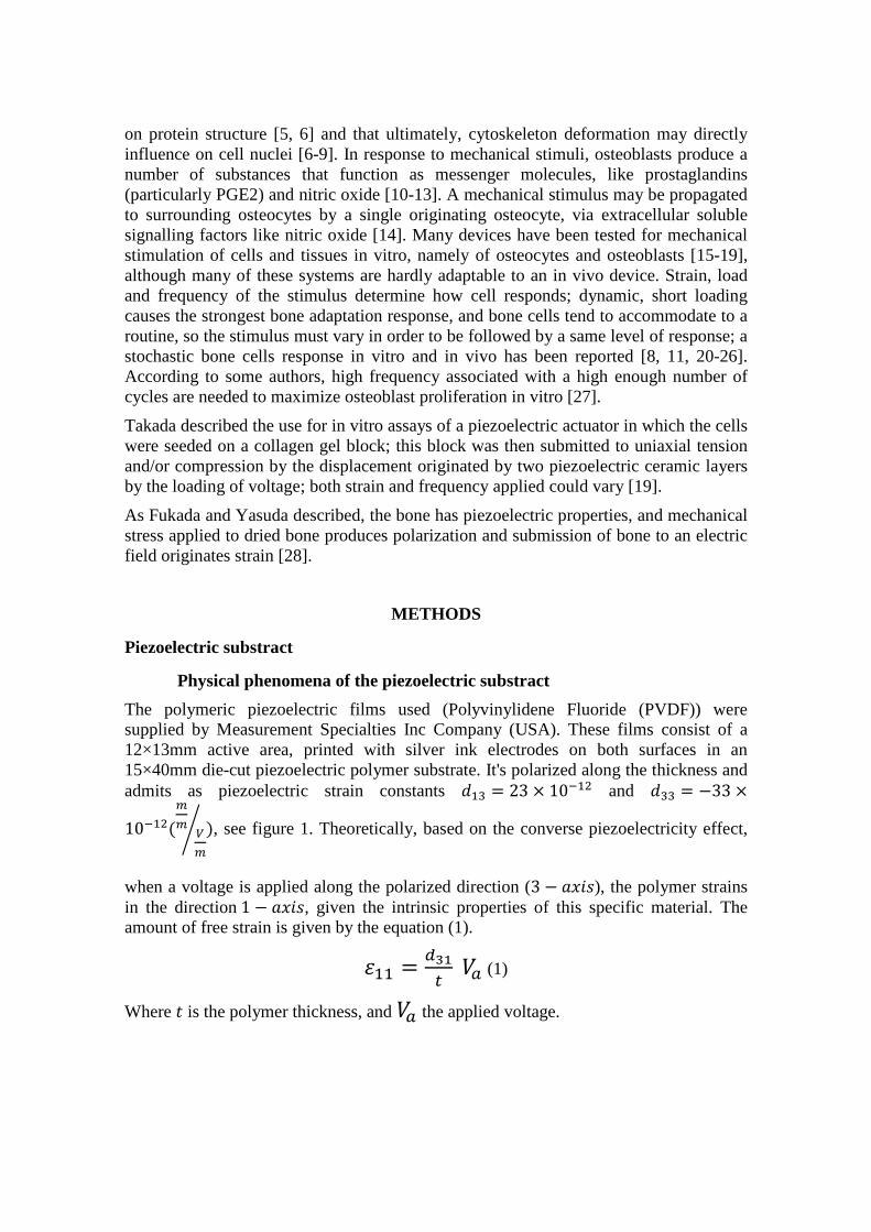
isplacement was observed in the piezoelectric free extremity (see, Figure 4). It's possible to observe a sinusoidal numerical perturbation in the encastr
region, but the strain values are around εyy = 2,2µε along the piezoelectricvalues are near the theoretical ones, see equation (1). The stress conditions for
each cycle are present in the Figure 5.
Displacement variation in nanometers along the PVDF actuator surface o
software OriginPro 7.5 (OriginLab Corporation, USA).
Coating the polymeric piezoelectric substrate with PMMA and micro -particles of
coated. The device has a total thickness
Optical microscopic (Palatine inverted optical microscope, Olympus PMG3) images of the
stribution along the y<77,3 (nm) . The
xtremity (see, Figure 4 and sinusoidal numerical perturbation in the encastre
along the piezoelectric surface. The stress conditions for
Displacement variation in nanometers along the PVDF actuator surface on each cycle.
0,00 0,01 0,02 0,031,5
2,0
2,5
3,0
0,01 0,02 0,032,0
2,5
Cells
Mic
roS
trai
n (µ
ε)
Mic
roS
trai
n (µ
ε)
PVDF length (m)
Sinusoidal numerical instability
Figure 5 - Figure 4a - Strain variation in the longitudinal direction of the PVDF actuator surface
(���� � �� ) along the PVDF length, for each cycle.
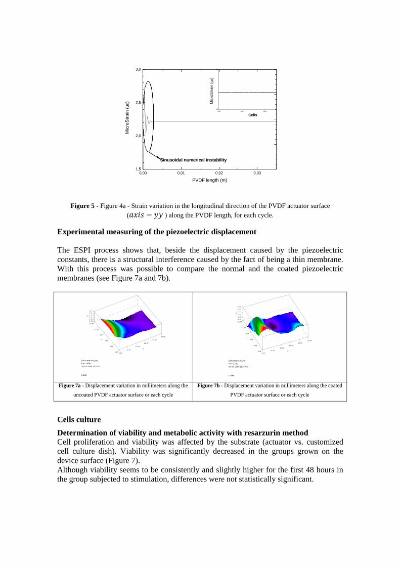
Experimental measuring of the piezoelectric displacement The ESPI process shows that, beside the displacement caused by the piezoelectric constants, there is a structural interference caused by the fact of being a thin membrane. With this process was possible to compare the normal and the coated piezoelectric membranes (see Figure 7a and 7b).
Figure 7a - Displacement variation in millimeters along the
uncoated PVDF actuator surface or each cycle
Figure 7b - Displacement variation in millimeters along the coated
PVDF actuator surface or each cycle
Cells culture
Determination of viability and metabolic activity with resarzurin method Cell proliferation and viability was affected by the substrate (actuator vs. customized cell culture dish). Viability was significantly decreased in the groups grown on the device surface (Figure 7). Although viability seems to be consistently and slightly higher for the first 48 hours in the group subjected to stimulation, differences were not statistically significant.
30-03-2009 11:11:07
Deformat ion [µm]
PV= 1.898
LOME
X
0.50
6. 37
12. 23
18. 10
23.97
29.83
Y
15.89
11.98
8.07
4.16
0.24
-3.67
Z
-1.53-1.15-0.77-0.39-0.010.37
30-03-2009 11:27:02
Deformat ion [ µm]
PV= 0.790
LOME
X0. 17
6. 10
12. 03
17. 97
23.90
29.83
Y
16. 08
12.07
8.06
4.05
0.05
-3.96
Z
-0. 74
-0. 58
-0. 42
-0. 27
-0. 11
0.05

Figure 7 - Cell viability 24 and 48 hours after seeding and daily stimulation of the dynamic group, results are expressed in percent related to controls (standard cell culture dish, TPP). Bars show Means and Error bars show Means ± Standard Error. Measurement of nitric oxide (NO) in culture medium Nitric oxide in culture medium after stimulation was significantly higher in dynamic conditions vs. static, both 24 and 48 hours after seeding (see Figure 8). When the population means of the controls were compared, no significant difference was found at 24 and 48 hours; the dynamic group at 24h and 48h behaved in similar way, when means of the NO measurements were compared within this group.
Static Dynamic0
1
2
3
4
5
6
7
NO
( µµ µµm
ol/m
L)
24h after seeding
Static Dynamic0
1
2
3
4
5
NO
(µm
ol/m
L)
48h after seeding
Figure 8 – Nitric oxide immediately after stimulation of the dynamic group, comparing to static group. Bars show Means and Error bars show Means ± Standard Error. Statistical analysis Normal distribution of the results was verified using the Shapiro-Wilk normality test for n>3, and differences between groups tested using one-way ANOVA and Significant differences were considered at a P value 0,05. The statistical analysis was done using software OriginPro 7.5 (OriginLab Corporation, USA).
CONCLUSION AND DISCUSSION
In this work the stress/strain was constant, because although the frequency varied, the applied voltage was constant. According to the definition of piezoelectricity every time
Static Dynamic Control0,00
25,00
50,00
75,00
100,00
% V
iab
ility
24
h
�
�
�
Static Dynamic Control
0
25
50
75
100
% V
iab
ility
48
h
�
�
�

a voltage was applied a maximum high of strain was obtained and then material recovered the initial shape, εyy = 2,2µε . The amount of stress/strain distribution along the piezoelectric material was assumed as an acceptable value. These results suggest that the devices affected negatively cell viability and proliferation. Although Braga et al. describe no evidence of negative effects of extracts obtained by immersing PVDF/HA composite membranes in medium used [41], few studies on PVDF cytocompatibility are available. Another study using human epithelial cell line L132, refers a proliferation of 37% three days after seeding on virgin PVDF, increasing to 45% at 6 days post-seeding, when comparing to to control [42]. Hung et al. described that PVDF had an inhibitory effect on neural stem cells differentiation and PVDF seemed to decrease consistently MTT reduction activity [43]. Apart from the impact of the PVDF itself, the coating may increase or diminish protein adsorption and cell adhesion. For adherent cell lines like osteoblasts, this is most important. Surface properties are also influenced by the sterilization method. In this study, γ-irradiation (normed dosis 25 kGy) was used to sterilize the devices prior to cell culture. The method used may increase protein adsorption on virgin PVDF foils and, although it may not strongly influence cell surface density, it may influence coating oxidation phenomena [44]. The slightly higher values of resorufin in the dynamic group in this assay is in agreement with the expected proliferation enhancement related to the mechanical stimulation to which osteoblasts were subjected, in accordance with the literature [15, 45]. The elevation in the NO values in the culture medium under dynamic conditions suggests that piezoelectric materials can be effective mechanical stimuli generators. By using piezoelectric material for bone cells stimulation, the control of mechanical ranges only requires the control of the amount of electrical energy applied; the fast answer to electric stimulus also allows working in physiological frequencies, as are the ones used 1Hz and 3Hz, in this study. Another advantage is the possibility by changing the piezoelectric constants of a biocompatible piezoelectric material to stimulate bone in different directions apart the one used in this work (dyy).
ACKNOWLEDGEMENTS
The authors would like to thank the Portuguese Foundation for Science and Technology (FCT) for financial support under the grant PTDC/EMEPME/70155/2006 grants SFRH / BD / 22856 / 2005 and SFRH / BD/31895/2006, to INEB (OPorto) and ITN (Lisbon), especially to Prof. Doutora Luísa Botelho.
References
1. Judex, S. and R.F. Zernicke, High-impact exercise and growing bone: relation between high strain rates and enhanced bone formation. J Appl Physiol, 2000. 88(6): p. 2183-2191.

2. Forwood, M.R. and C.H. Turner, Skeletal adaptations to mechanical usage: results from tibial loading studies in rats. Bone, 1995. 17(4, Supplement 1): p. S197-S205.
3. Bourrin, S., et al., Effect of physical training on bone adaptation in three zones of the rat tibia. J Bone Miner Res, 1995. 10: p. 1745 - 1752.
4. Hillam, R.A. and T.M. Skerry, Inhibition of bone resorption and stimulation of formation by mechanical loading of the modeling rat ulna in vivo. J Bone Miner Res, 1995. 10: p. 683 - 689.
5. Gudi, S.R.P., et al., Equibiaxial strain and strain rate stimulate early activation of G proteins in cardiac fibroblasts. Am J Physiol Cell Physiol, 1998. 274(5): p. C1424-1428.
6. Charras, G.T., et al., Estimating the Sensitivity of Mechanosensitive Ion Channels to Membrane Strain and Tension. Biophys. J., 2004. 87(4): p. 2870-2884.
7. Burger, E.H. and J. Klein-Nulend, Mechanotransduction in bone—role of the lacuno-canalicular network. FASEB J., 1999. 13(9001): p. 101-112.
8. Bacabac, R.G., et al., Bone cell responses to high-frequency vibration stress: does the nucleus oscillate within the cytoplasm? FASEB J., 2006. 20(7): p. 858-864.
9. Jessop, H.L., et al., Mechanical strain and fluid movement both activate extracellular regulated kinase (ERK) in osteoblast-like cells but via different signaling pathways. Bone, 2002. 31(1): p. 186-194.
10. Kanamaru, Y., et al., Effect of nitric oxide on mouse clonal osteogenic cell, MC3T3-E1, proliferation in vitro. Kobe J Med Sci, 2001. 47(1): p. 1- 11.
11. Bakker, A.D., et al., The production of nitric oxide and prostaglandin E2 by primary bone cells is shear stress dependent. Journal of Biomechanics, 2001. 34(5): p. 671-677.
12. Smalt, R., et al., Induction of NO and prostaglandin E2 in osteoblasts by wall-shear stress but not mechanical strain. Am J Physiol Endocrinol Metab, 1997. 273(4): p. E751-758.
13. Xian Fan, J.A.R.T.C.M.M.S.N.E.M.G.J.R., Response to mechanical strain in an immortalized pre-osteoblast cell is dependent on ERK1/2. Journal of Cellular Physiology, 2006. 207(2): p. 454-460.
14. Vatsa, A., T.H. Smit, and J. Klein-Nulend, Extracellular NO signalling from a mechanically stimulated osteocyte. Journal of Biomechanics, 2007. 40(Supplement 1): p. S89-S95.
15. Lewandowska-Szumiel, M., et al., Osteoblast response to the elastic strain of metallic support. Journal of Biomechanics, 2007. 40(3): p. 554- 560.
16. Brown, T.D., Techniques for mechanical stimulation of cells in vitro: a review. Journal of Biomechanics, 2000. 33(1): p. 3-14.
17. McGarry, J.G., et al., Stimulation of nitric oxide mechanotransduction in single osteoblasts using atomic force microscopy. J Orthop Res, 2008. 26(4): p. 513-21.
18. Appleford, M.R., et al., Ultrasound effect on osteoblast precursor cells in trabecular calcium phosphate scaffolds. Biomaterials, 2007. 28(32): p. 4788-4794.
19. Tanaka, S.M., A new mechanical stimulator for cultured bone cells using piezoelectric actuator. Journal of Biomechanics, 1999. 32(4): p. 427-430.

20. Tanaka, S.M., I.M. Alam, and C.H. Turner, Stochastic resonance in osteogenic response to mechanical loading. FASEB J., 2003. 17(2): p. 313-314.
21. Turner, C.H., I. Owan, and Y. Takano, Mechanotransduction in bone: role of strain rate. Am J Physiol Endocrinol Metab, 1995. 269(3): p. E438-442.
22. Burr, D.B., A.G. Robling, and C.H. Turner, Effects of biomechanical stress on bones in animals. Bone, 2002. 30(5): p. 781-786.
23. Cullen, D.M., R.T. Smith, and M.P. Akhter, Bone-loading response varies with strain magnitude and cycle number. J Appl Physiol, 2001. 91(5): p. 1971-1976.
24. Hsieh, Y.F. and C.H. Turner, Effects of loading frequency on mechanically induced bone formation. J Bone Miner Res, 2001. 16: p. 918 - 924.
25. Robling, A.G., D.B. Burr, and C.H. Turner, Recovery periods restore mechanosensitivity to dynamically loaded bone. J Exp Biol, 2001. 204(19): p. 3389 3399.
26. Tanaka, S.M., et al., Effects of broad frequency vibration on cultured osteoblasts. Journal of Biomechanics, 2003. 36(1): p. 73-80.
27. Kaspar, D., et al., Proliferation of human-derived osteoblast-like cells depends on the cycle number and frequency of uniaxial strain. Journal of Biomechanics, 2002. 35(7): p. 873-880.
28. Fukada E, Y.I., On the Piezoelectric effect of Bone. JOURNAL OF THE PHYSICAL SOCIETY OF JAPAN, 1957 12(10): p. 1158-1162
29. Sudo, H., et al., In vitro differentiation and calcification in a new clonal osteogenic cell line derived from newborn mouse calvaria. J. Cell Biol., 1983. 96(1): p. 191-198.
30. Wang, D., et al., Isolation and Characterization of MC3T3-E1 Preosteoblast Subclones with Distinct In Vitro and In Vivo Differentiation/Mineralization Potential. Journal of Bone and Mineral Research, 1999. 14(6): p. 893-903.
31. Chung, C.Y., et al., Serial Passage of MC3T3-E1 Cells Alters Osteoblastic Function and Responsiveness to Transforming Growth Factor-[beta]1 and Bone Morphogenetic Protein-2. biochemical and biophysical research communications, 1999. 265(1): p. 246 251.
32. Wenstrup, R.J., et al., Discordant Expression of Osteoblast Markers in MC3T3-E1 Cells that Synthesize a High Turnover Matrix. J. Biol. Chem., 1996. 271(17): p. 10271 10276.
33. Saunders, M.M., et al., Gap junctions and fluid flow response in MC3T3- E1 cells. Am J Physiol Cell Physiol, 2001. 281(6): p. C1917-1925.
34. Liu, D., et al., Activation of extracellular-signal regulated kinase (ERK1/2) by fluid shear is Ca2+- and ATP-dependent in MC3T3-E1 osteoblasts. Bone, 2008. 42(4): p. 644-652.
35. Jaasma, M.J. and F.J. O'Brien, Mechanical Stimulation of Osteoblasts Using Steady and Dynamic Fluid Flow. Tissue Engineering Part A. 0(0).

36. Ansar Ahmed, S., R.M. Gogal Jr, and J.E. Walsh, A new rapid and simple non-radioactive assay to monitor and determine the proliferation of lymphocytes: an alternative to [3H]thymidine incorporation assay. Journal of Immunological Methods, 1994. 170(2): p. 211-224.
37. Zhi-Jun, Y., et al., A dye-based lymphocyte proliferation assay that permits multiple immunological analyses: mRNA, cytogenetic, apoptosis, and immunophenotyping studies. Journal of Immunological Methods, 1997. 210(1): p. 25-39.
38. Slaughter, M.R., P.J. Bugelski, and P.J. O'Brien, Evaluation of Alamar Blue Reduction for the In Vitro Assay of Hepatocyte Toxicity. Toxicology in Vitro, 1999. 13(4-5): p. 567-569.
39. Rob J. van'T Hof, S.H.R., Nitric oxide and bone. Immunology, 2001. 103(3): p. 255-261.
40. Ignarro, L.J., et al., Oxidation of nitric oxide in aqueous solution to nitrite but not nitrate: comparison with enzymatically formed nitric oxide from Larginine. Proceedings of the National Academy of Sciences of the United States of America, 1993. 90(17): p. 8103-8107.
41. Braga, F.J.C., et al., Characterization of PVDF/HAP composites for medical applications. Materials Research, 2007. 10: p. 247-251.
42. Tabary, N., et al., Functionalization of PVDF membranes with carbohydrate derivates for the controlled delivery of chlorhexidin. Biomolecular Engineering, 2007. 24(5): p. 472-476.
43. Hung, C.-H., Y.-L. Lin, and T.-H. Young, The effect of chitosan and PVDF substrates on the behavior of embryonic rat cerebral cortical stem cells. Biomaterials, 2006. 27(25): p. 4461-4469.
44. Lleixà Calvet, J., et al., Sterilization effects on starPEG coated polymer surfaces: characterization and cell viability. Journal of Materials Science: Materials in Medicine, 2008. 19(4): p. 1631-1636.
45. Fermor, B., et al., Primary Human Osteoblast Proliferation and Prostaglandin E2 Release in Response to Mechanical Strain In Vitro. Bone, 1998. 22(6): p. 637-643.