
Exosomal lncRNA GAS5 regulates the apoptosis of ... filesel wall. Oxidized low density lipoprotein...
10
RESEARCH ARTICLE Exosomal lncRNA GAS5 regulates the apoptosis of macrophages and vascular endothelial cells in atherosclerosis Lei Chen, Wenjin Yang, Yijun Guo, Wei Chen, Ping Zheng, Jinsong Zeng, Wusong Tong* Department of Neurosurgery, The People’s Hospital of Pudong New Area, Shanghai, PR China * [email protected] Abstract Atherosclerosis is universally recognized as a chronic lipid-induced inflammation of the ves- sel wall. Oxidized low density lipoprotein (oxLDL) drives the onset of atherogenesis involving macrophages and endothelial cells (ECs). Our earlier work showed that expression of long noncoding RNA-growth arrest-specific 5 (lncRNA GAS5) was significantly increased in the plaque of atherosclerosis collected from patients and animal models. In this study, we found that knockdown of lncRNA GAS5 reduced the apoptosis of THP-1 cells treated with oxLDL. On the contrary, overexpression of lncRNA GAS5 significantly elevated the apoptosis of THP-1 cells after oxLDL stimulation. The expressions of apoptotic factors including Cas- pases were changed with lncRNA GAS5 levels. Moreover, lncRNA GAS5 was found in THP-1 derived-exosomes after oxLDL stimulation. Exosomes derived from lncRNA GAS5- overexpressing THP-1 cells enhanced the apoptosis of vascular endothelial cells after tak- ing up these exosomes. However, exosomes shed by lncRNA GAS5 knocked-down THP-1 cells inhibited the apoptosis of endothelial cells. These findings reveal the function of lncRNA GAS5 in atherogenesis which regulates the apoptosis of macrophages and endo- thelial cells via exosomes and suggest that suppressing the lncRNA GAS5 might be an effective way for the therapy of atherosclerosis. Introduction Atherosclerosis, which can cause cardiovascular disease, is a leading cause of morbidity and mortality in industrialized country [1]. The onset of atherosclerosis is partly mediated by the dysfunction of endothelial cells (ECs) and infiltration of leukocytes, such as monocytes, which may differentiate into macrophages and dendritic cells. Meanwhile, the deposition of modi- fied lipoproteins in the artery wall increases endothelial permeability and promotes the forma- tion of foam cells and necrotic core along with lipid uptake of macrophages [2]. It is widely recognized that apoptosis in lesional macrophages, along with their defective function in effer- ocytosis, promotes plaque necrosis, which leads to plaque instability and thrombosis [3, 4]. Understanding the multifactorial process consisting of the interactions of several key compo- nents, including lipoproteins, inflammatory cells and vascular cells is extremely crucial [5]. PLOS ONE | https://doi.org/10.1371/journal.pone.0185406 September 25, 2017 1 / 10 a1111111111 a1111111111 a1111111111 a1111111111 a1111111111 OPEN ACCESS Citation: Chen L, Yang W, Guo Y, Chen W, Zheng P, Zeng J, et al. (2017) Exosomal lncRNA GAS5 regulates the apoptosis of macrophages and vascular endothelial cells in atherosclerosis. PLoS ONE 12(9): e0185406. https://doi.org/10.1371/ journal.pone.0185406 Editor: Gianfranco Pintus, Qatar University College of Health Sciences, QATAR Received: May 15, 2017 Accepted: September 12, 2017 Published: September 25, 2017 Copyright: © 2017 Chen et al. This is an open access article distributed under the terms of the Creative Commons Attribution License, which permits unrestricted use, distribution, and reproduction in any medium, provided the original author and source are credited. Data Availability Statement: All relevant data are within the paper. Funding: This research was funded by Shanghai Pudong New Area health system excellent young medical talents training plan (No. PWRq2016-29) and Pudong New Area Science and Technology Development Fund (PKJ2015-Y29) to LC. The funders had no role in study design, data collection and analysis, decision to publish, or preparation of the manuscript.
Transcript of Exosomal lncRNA GAS5 regulates the apoptosis of ... filesel wall. Oxidized low density lipoprotein...
RESEARCH ARTICLE
Exosomal lncRNA GAS5 regulates the
apoptosis of macrophages and vascular
endothelial cells in atherosclerosis
Lei Chen, Wenjin Yang, Yijun Guo, Wei Chen, Ping Zheng, Jinsong Zeng, Wusong Tong*
Department of Neurosurgery, The People’s Hospital of Pudong New Area, Shanghai, PR China
Abstract
Atherosclerosis is universally recognized as a chronic lipid-induced inflammation of the ves-
sel wall. Oxidized low density lipoprotein (oxLDL) drives the onset of atherogenesis involving
macrophages and endothelial cells (ECs). Our earlier work showed that expression of long
noncoding RNA-growth arrest-specific 5 (lncRNA GAS5) was significantly increased in the
plaque of atherosclerosis collected from patients and animal models. In this study, we found
that knockdown of lncRNA GAS5 reduced the apoptosis of THP-1 cells treated with oxLDL.
On the contrary, overexpression of lncRNA GAS5 significantly elevated the apoptosis of
THP-1 cells after oxLDL stimulation. The expressions of apoptotic factors including Cas-
pases were changed with lncRNA GAS5 levels. Moreover, lncRNA GAS5 was found in
THP-1 derived-exosomes after oxLDL stimulation. Exosomes derived from lncRNA GAS5-
overexpressing THP-1 cells enhanced the apoptosis of vascular endothelial cells after tak-
ing up these exosomes. However, exosomes shed by lncRNA GAS5 knocked-down THP-1
cells inhibited the apoptosis of endothelial cells. These findings reveal the function of
lncRNA GAS5 in atherogenesis which regulates the apoptosis of macrophages and endo-
thelial cells via exosomes and suggest that suppressing the lncRNA GAS5 might be an
effective way for the therapy of atherosclerosis.
Introduction
Atherosclerosis, which can cause cardiovascular disease, is a leading cause of morbidity and
mortality in industrialized country [1]. The onset of atherosclerosis is partly mediated by the
dysfunction of endothelial cells (ECs) and infiltration of leukocytes, such as monocytes, which
may differentiate into macrophages and dendritic cells. Meanwhile, the deposition of modi-
fied lipoproteins in the artery wall increases endothelial permeability and promotes the forma-
tion of foam cells and necrotic core along with lipid uptake of macrophages [2]. It is widely
recognized that apoptosis in lesional macrophages, along with their defective function in effer-
ocytosis, promotes plaque necrosis, which leads to plaque instability and thrombosis [3, 4].
Understanding the multifactorial process consisting of the interactions of several key compo-
nents, including lipoproteins, inflammatory cells and vascular cells is extremely crucial [5].
PLOS ONE | https://doi.org/10.1371/journal.pone.0185406 September 25, 2017 1 / 10
a1111111111
a1111111111
a1111111111
a1111111111
a1111111111
OPENACCESS
Citation: Chen L, Yang W, Guo Y, Chen W, Zheng
P, Zeng J, et al. (2017) Exosomal lncRNA GAS5
regulates the apoptosis of macrophages and
vascular endothelial cells in atherosclerosis. PLoS
ONE 12(9): e0185406. https://doi.org/10.1371/
journal.pone.0185406
Editor: Gianfranco Pintus, Qatar University College
of Health Sciences, QATAR
Received: May 15, 2017
Accepted: September 12, 2017
Published: September 25, 2017
Copyright: © 2017 Chen et al. This is an open
access article distributed under the terms of the
Creative Commons Attribution License, which
permits unrestricted use, distribution, and
reproduction in any medium, provided the original
author and source are credited.
Data Availability Statement: All relevant data are
within the paper.
Funding: This research was funded by Shanghai
Pudong New Area health system excellent young
medical talents training plan (No. PWRq2016-29)
and Pudong New Area Science and Technology
Development Fund (PKJ2015-Y29) to LC. The
funders had no role in study design, data collection
and analysis, decision to publish, or preparation of
the manuscript.
Long noncoding (lnc) RNAs are generally defined as non-protein-coding RNAs that have
at least 200 bp to 100 kb in length with highly conserved sequences [6, 7, 8]. LncRNAs have
various functions including signaling transduction, molecular decoys, scaffolding and guiding
ribonucleoprotein complexes. Accumulating evidences relate regulatory lncRNAs to human
diseases [9, 10, 11]. A growing amount of studies have identified that lncRNAs regulate the
functions of endothelial cells, macrophages, vascular inflammation and metabolism, suggest-
ing the possibility of lncRNAs in influencing the progression of atherosclerosis [12]. The
lncRNA growth-arrest specific transcript 5 (GAS5) is a 50-terminal oligopyrimidine class of
genes which regulates cell growth, proliferation and survival [13, 14]. The biogenesis of
lncRNA GAS5 has been established. LncRNA GAS5 gene transcribes several snoRNAs as well
as four splice variants of lncRNA GAS5 mRNA. However, due to the presence of STOP codon,
none of the transcripts is transcribed into protein and degrade via the nonsense-mediated
decay (NMD) pathway when translation is initiated. The RNA level of lncRNA GAS5 is regu-
lated by its degradation instead of regulation at its transcriptional level [15]. One recent study
identified that low expression of lncRNA GAS5 facilitated human saphenous vein smooth
muscle cells proliferation and migration through Annexin A2 and thereby the pathogenesis of
great saphenous veins varicosities [16].
Exosomes, small membrane particles (40–100 nm in diameter) originating from multivesi-
cular bodies (MVBs), are generated from many cell types, and play key roles in the intercellular
communication via the horizontal transfer of proteins, and RNAs to target cells [17]. Studies
have discovered a list of statistically significant lncRNAs are differentially expressed in the exo-
somes in cancer [18]. Our earlier studies found that the molecular pathways underlying plaque
formation in atherosclerosis were related to immune response, angiogenesis, cell proliferation,
apoptosis and hypoxic microenvironments. And three lncRNAs, GAS5, SNHG6 and Zfas1,
were significantly increased in the plaque of atherosclerotic patients [19]. LncRNA GAS5 has
been found enriched in exosomes [20]. Here, the initial aim of this study was to investigate the
function of lncRNA GAS5 in atherosclerosis progression. Whether macrophage derived exo-
somal lncRNA GAS5 modulate the function of endothelial cells in atherosclerosis is also
investigated.
Materials and methods
Cell lines and cell culture
THP-1 cells and HUVEC were purchased from American Type Culture Collection (ATCC)
and cultured in DMEM supplemented with 10% fetal bovine serum (Gibco, mexico). All the
cells were cultured at 37˚C in a humidified atmosphere with 5% CO2.
Lentivirus mediated shRNA gene knockdown lncRNA GAS5
The stable knockdown lncRNA GAS5 cell lines were generated by transduction a lentiviral-
mediated expression siRNA specific target of lncRNA GAS5. The targeted knockdown
sequence was 5’-ggatgacttgcttgggtaa-3’. The virus transfected THP-1 cell lines with 8 μg/ml
polybrene. After 48 hours, the cells are harvested, and the knockdown efficiency was tested by
real-time-PCR.
Lentivirus mediated over-expression of lncRNA GAS5
The stable over-expression of lncRNA GAS5 cell lines were generated by transduction a lenti-
viral-mediated overexpression lncRNA GAS5. The virus transfected THP-1 cell lines with
Exosomal GAS5 regulates the apoptosis of macrophages and vascular endothelial cells
PLOS ONE | https://doi.org/10.1371/journal.pone.0185406 September 25, 2017 2 / 10
Competing interests: The authors have declared
that no competing interests exist.
8μg/ml polybrene. After 48 hours, the cells are harvested, and the over-expression efficiency
was tested by real-time- PCR.
Real-time-PCR (RT-PCR)
Cellular RNA was isolated by Trizol-Reagent according to the manufacturer’s instructions.
Briefly, the DNA was removed from the samples using DNase treatment (DNA-free kit;
Ambion Applied Biosystems) and cDNA was synthesized from the purified RNA using Molo-
ney murine leukemia virus reverse transcription kit (Promega). Gene-specific primer sets are
listed in Table 1 and Actin primer sets are used to produce a normalization control. Real-time
PCR was carried out in triplicate with the SYBR Green PCR Master Mix (Applied Biosystems)
and a 7900HT Fast Real-Time PCR machine (Applied Biosystems).
Cell apoptosis assay
Apoptosis was determined by translocation of phosphatidylserine to the cell surface using an
Annexin V-FITC/ Annexin V-PE and PI/7-ADD apoptosis detection kit (Nanjing KeyGen
Biotech. Co. Ltd., China). The stable knockdown and over-expression lncRNA GAS5 THP-1
cells and its negative control cells were harvested and washed twice in cold PBS, and re-sus-
pended in Annexin V-FITC/ Annexin V-PE and PI/7-ADD for 30 min in the dark. Cell apo-
ptosis was analyzed by using Cell Quest software on a FACSAria Flow Cytometer (BD Inc.,
USA). Fluorescence was detected with an excitation wavelength of 480 nm.
Western blot analysis
RIPA buffer in the presence of protease inhibitor cocktail and phosphorylation inhibitor cock-
tail were used to extract total protein. Appropriate mount protein was loaded into 10–15%
SDS-polyacrylamide gel and transferred onto a nitrocellulose membrane (Millipore, Billerica,
MA, USA). Primary antibodies were incubated overnight and secondary antibodies were incu-
bated for 1 h at the appropriate dilutions. The signal was observed and developed with Kodak
film by exposure to Enhanced Chemiluminescence (ECL) plus Western Blotting Detection
Reagents (Amersham Biosciences, Piscataway, NJ, USA). Western blots were used to analyze
the incorporation of each protein into cells. The antibodies against apoptosis associated Cas-
pase3, Caspase7, Caspase9, P53 and actin used as control, were obtained from Cell Signaling
Technology.
Isolation and identification of exosomes released from the stable lncRNA
GAS5 knocked-down or over-expressing THP-1 cells
80% confluent THP-1 cell lines cultured for an additional 48 hours in DMEM media deprived
of FBS. The conditioned media of THP-1 cell lines was obtained and centrifuged at 300 × g for
10 min and 2000 × g for 10 min at 4˚C to remove dead cells and cellular debris. Subsequently,
the supernatant was filtered using a 0.45 μm filter sterilize Steritop TM (Millipore) to remove
residual dead cells and cellular debris. Hereafter, the supernatant was centrifuged at 4000 × g
Table 1. Primer sequences for RT-PCR.
GAS5-F 5’-ACACAGGCATTAGACAGAA-3’
GAS5-R 5’-CCAGGAGCAGAACCATTA-3’
Actin-F 5’-CACCATTGGCAATGAGCGGTTC-3’
Actin-R 5’-AGGTCTTTGCGGATGTCCACGT-3’
https://doi.org/10.1371/journal.pone.0185406.t001
Exosomal GAS5 regulates the apoptosis of macrophages and vascular endothelial cells
PLOS ONE | https://doi.org/10.1371/journal.pone.0185406 September 25, 2017 3 / 10
at 4˚C to about 200 μL by ultra-filtration in a 15 mL Amicon Ultra-15 Centrifugal Filter Unit
(Millipore) and then centrifuged at 100,000 × g for one hour at 4˚C to pellet the small vesicles.
Exosomes were stored at − 80˚C or used for downstream experiments. The protein concentra-
tion of the exosomes was determined using the Micro Bicinchoninic Acid (BCA) Protein
Assay Kit (Thermo Fisher, Waltham, MA, USA).
Effects of THP-1-exosomes on human vascular endothelial cells
(HUVECs)
The exosomes (100 μg/mL) were labeled with PHK67 and incubated with HUVECs seeded
onto 24-well plates. After 24 hours (t = 24 hours), the HUVECs were then photographed
under a fluorescence microscope (Leica AF6000). The results were analyzed by observing the
fluorescence into the cells.
Influence of exosomes on the expression of lncRNAGAS5 genes of HUVECs was evaluated
with RT-PCR. And influence of exosomes on apoptosis of HUVECs was analyzed by using the
Flow Cytometer.
Statistical analysis
For quantitative data, all results were expressed as the mean SD. Statistical significance
between groups was determined using the Student’s t-test using SPSS 18.0 (SPSS, USA). Each
experiment was repeated at least three times. P< 0.05 was considered statistically significant.
Results and discussion
Knockdown of lncRNA GAS5 reduced the apoptosis of THP-1 cells
treated with oxLDL
Using lentivirus mediated shRNA gene knockdown system, we knocked down lncRNA
GAS5 expression in THP-1 cells. From the mRNA level, more than 70% of lncRNA GAS5
expression was decreased in gene knockdown system (Fig 1A). Then, we treated lncRNA
GAS5 knocked-down THP-1 cells with 75μg/mL oxLDL (Intracel Resources, Frederick,
MD, USA) for 24 h and detected the percentage of apoptotic cells using flow cytometry.
Compared with its negative control cells, we found that the percentage of apoptotic lncRNA
GAS5 knocked-down THP-1 cells was increased after treated with oxLDL (Fig 1B and 1C).
Western blot results demonstrated that the expressions of P53, Caspase 3, Caspase 7 and
Caspase 9 were reduced after inhibition of lncRNA GAS5 expression in response to oxLDL
stimulation (Fig 1D).
Over-expression of lncRNA GAS5 increased the apoptosis of THP-1
cells treated with oxLDL
Meanwhile we over expressed lncRNA GAS5 expression in THP-1 cells using lentivirus medi-
ated over-expression system. At the mRNA level, lncRNA GAS5 expression was increased
more than 4 folds in gene over-expression system (Fig 2A). Then, we treated lncRNA GAS5
over-expressing THP-1 cells with 75μg/mL oxLDL for 24 h and detected the percentage of apo-
ptotic cells using flow cytometry. Compared with its negative control cells, we found that after
treated with oxLDL, the percentage of apoptotic lncRNA GAS5 over-expressing THP-1 cells
was increased (Fig 2B and 2C). Western blot results showed that the expressions of P53, Cas-
pase 3, Caspase 7 and Caspase 9 were upregulated after lncRNA GAS5 over-expressing (Fig
2D). These results highlighted the importance of lncRNA GAS5 in the regulation of
Exosomal GAS5 regulates the apoptosis of macrophages and vascular endothelial cells
PLOS ONE | https://doi.org/10.1371/journal.pone.0185406 September 25, 2017 4 / 10
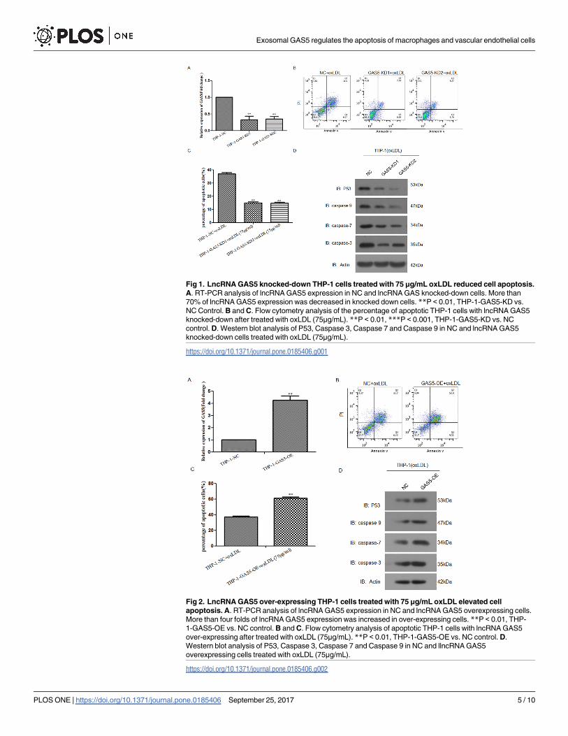
Fig 1. LncRNA GAS5 knocked-down THP-1 cells treated with 75 μg/mL oxLDL reduced cell apoptosis.
A. RT-PCR analysis of lncRNA GAS5 expression in NC and lncRNA GAS knocked-down cells. More than
70% of lncRNA GAS5 expression was decreased in knocked down cells. **P < 0.01, THP-1-GAS5-KD vs.
NC Control. B and C. Flow cytometry analysis of the percentage of apoptotic THP-1 cells with lncRNA GAS5
knocked-down after treated with oxLDL (75μg/mL). **P < 0.01, ***P < 0.001, THP-1-GAS5-KD vs. NC
control. D. Western blot analysis of P53, Caspase 3, Caspase 7 and Caspase 9 in NC and lncRNA GAS5
knocked-down cells treated with oxLDL (75μg/mL).
https://doi.org/10.1371/journal.pone.0185406.g001
Fig 2. LncRNA GAS5 over-expressing THP-1 cells treated with 75 μg/mL oxLDL elevated cell
apoptosis. A. RT-PCR analysis of lncRNA GAS5 expression in NC and lncRNA GAS5 overexpressing cells.
More than four folds of lncRNA GAS5 expression was increased in over-expressing cells. **P < 0.01, THP-
1-GAS5-OE vs. NC control. B and C. Flow cytometry analysis of apoptotic THP-1 cells with lncRNA GAS5
over-expressing after treated with oxLDL (75μg/mL). **P < 0.01, THP-1-GAS5-OE vs. NC control. D.
Western blot analysis of P53, Caspase 3, Caspase 7 and Caspase 9 in NC and llncRNA GAS5
overexpressing cells treated with oxLDL (75μg/mL).
https://doi.org/10.1371/journal.pone.0185406.g002
Exosomal GAS5 regulates the apoptosis of macrophages and vascular endothelial cells
PLOS ONE | https://doi.org/10.1371/journal.pone.0185406 September 25, 2017 5 / 10

macrophage apoptosis and suggested that lncRNA GAS5 may have implications for the ther-
apy of atherosclerosis.
Identification of lncRNA GAS5 in THP-1 derived exosomes treated with
oxLDL
Western blotting analysis showed that exosomes isolated from the THP-1 cultured medium
expressed characteristic exosomal surface marker proteins: CD63 and CD9 (Fig 3A). To exam-
ine whether lncRNA GAS5 exist in THP-1 cells derived exosomes and whether the levels of
lncRNA GAS5 in these exosomes altered by oxLDL treatment, RT-PCR analysis was per-
formed. To our surprise, lncRNA GAS5 was found in THP-1 derived exosomes. More, com-
pared to the control group, lncRNA GAS5 was profoundly up-regulated after oxLDL
treatment (Fig 3B). These results indicate that lncRNA GAS5 was markedly enriched in the
THP-1-exosomes after oxLDL stimulation.
Exosomes derived from lncRNA GAS5 knocked down THP-1 cells
reduced the apoptosis of vascular endothelial cells
Furthermore, fluorescence microscopy analysis showed the GFP-labeled THP-1-exosomes
were taken up and transferred to the perinuclear region of vascular endothelial cells (Fig 4A),
indicating that the THP-1-exosomes might influence the function of vascular endothelial cells.
The expression of lncRNA GAS5 in vascular endothelial cells was determined by RT-PCR
analysis, and the results showed that compared with control, endothelial cells incubated with
exosomes isolated from the cultured medium of lncRNA GAS5 knocked down THP-1 cells
contained lower lncRNA GAS5 (Fig 4B). Cell apoptotic assay was used to examine the effect of
THP-1-exosomes with different levels of lncRNA GAS5 on the apoptosis of endothelial cells.
The results showed that the HUVEC cells incubated with exosomes isolated from the lncRNA
GAS5 knocked down THP-1 cells cultured medium reduced the percentage of apoptotic
HUVECs compared with wild-type control (Fig 4C and 4D). To further analyze whether apo-
ptosis-related genes were altered, Western blot analysis was performed. The expressions of
P53, Caspase 3, Caspase 7 and Caspase 9 were profoundly down-regulated after treated with
the exosomes isolated from the lncRNA GAS5 knocked down THP-1 cells compared with neg-
ative control (Fig 4E).
Fig 3. Identification of THP-1-Exos and expression of lncRNA GAS5 in exosomes derived from oxLDL
treated THP-1 cells. A. Western blotting analysis of exosomal surface marker proteins CD63 and CD9 in
THP-1-Exos. B. RT-PCR analysis of lncRNA GAS5 gene expression profoundly up-regulated in the THP-
1-Exos after oxLDL (75μg/mL) stimulation 24h. **P < 0.001, THP-1+oxLDL-Exos vs. NC control.
https://doi.org/10.1371/journal.pone.0185406.g003
Exosomal GAS5 regulates the apoptosis of macrophages and vascular endothelial cells
PLOS ONE | https://doi.org/10.1371/journal.pone.0185406 September 25, 2017 6 / 10

Exosomes derived from lncRNA GAS5 overexpressing THP-1 cells
elevated the apoptosis of vascular endothelial cells
The expression of lncRNA GAS5 in vascular endothelial cells was determined by RT-PCR
analysis, the results showed that exosomes isolated from the over-expressing lncRNA GAS5
THP-1 cells markedly increased the lncRNA GAS5 expression in HUVECs compared to con-
trol (Fig 5A). Cell apoptotic assay was used to examine the effect of these exosomes on the apo-
ptosis of endothelial cells, the results found that the exosomes isolated from the lncRNA GAS5
over-expressing THP-1 cells enhanced the percentage of apoptotic HUVECs (Fig 5B and 5C).
To further analyze whether apoptosis-related genes expression was altered in HUVEC cells
after internalization of these exosomes, Western blot analysis was performed. The expressions
of P53, Caspase 3, Caspase 7 and Caspase 9 were up-regulated after treated with the exosomes
isolated from the lncRNA GAS5 over-expressing THP-1 cells (Fig 5D). These results indicated
that lncRNA GAS5 exist in THP-1 cells derived exosomes and exosomal lncRNA GAS5 modu-
lated the apoptosis of endothelial cells.
Atherosclerotic process is related to pro-atherogenic and pro-inflammatory mediators that
lead to formation of plaques and progressive stenosis [21, 22]. Macrophages are involved in all
stages of plaque development [23]. The initial step of atherosclerosis involves high levels of low
density lipoprotein (LDL) which are oxidized and recruit monocytes. Oxidized LDL (oxLDL)
Fig 4. HUVECs incubated with exosomes (100 μg/mL) derive from lncRNA GAS5 knocked down THP-
1 cells reduced cell apoptosis. A. Fluorescence microscopy analysis showed the GFP-labeled THP-
1-exosomes were taken up and transferred to the perinuclear region of endothelial Cells. The magnification
was 100 x. B. RT-PCR analysis of lncRNA GAS5 expression in HUVEC cells incubated with different
exosomes. HUVEC cells incubated with exosomes derived from the lncRNA GAS5 knocked-down THP-1
cells contained markedly reduced lncRNA GAS5 levels. *P < 0.05, HUVEC-GAS5-KD vs. NC control. C and
D. Flow cytometry analysis of HUVEC cells incubated with the exosomes derived from the knockdown
lncRNA GAS5 THP-1 reduced the percentage of apoptotic cells. *P < 0.05, HUVEC-GAS5-KD vs. NC control.
E. Western blot analysis of P53, Caspase 3, Caspase 7 and Caspase 9 in cells treatment with the exosomes
derived from lncRNA GAS5 knocked-down THP-1.
https://doi.org/10.1371/journal.pone.0185406.g004
Exosomal GAS5 regulates the apoptosis of macrophages and vascular endothelial cells
PLOS ONE | https://doi.org/10.1371/journal.pone.0185406 September 25, 2017 7 / 10
induces expression of adhesion molecules on endothelium and facilitates monocyte adhesion
to intima, determining the extension of lesion formation and progression [24, 25]. One recent
study identified that low expression of the newly found lncRNA GAS5 can promote the prolif-
eration and migration of human saphenous vein smooth muscle cells [16]. Our earlier work
showed that lncRNA GAS5 also play a role in the progression of atherosclerosis [19]. In the
present study, we found that lncRNA GAS5 regulates the apoptosis of oxLDL-treated THP-1
cells. In these cells, high level of lncRNA GAS5 expression promotes basal apoptosis, whereas
low level of lncRNA GAS5 inhibits apoptosis. The following study further extends these find-
ings by demonstrating that the vascular endothelial cells apoptosis are quantitatively related to
THP-1 derived exosomal lncRNA GAS5 levels, suggesting that exosomes may act as a ‘master
regulator’ of lncRNA GAS5.
The monolayer of endothelial cells (ECs), lining in the innermost part of the arterial vessel,
play a pivotal role in maintaining the homeostasis of vessel when provoked with many stimuli,
such as oxidized LDLs, inflammation and shear stress [26]. The activation and dysfunction of
ECs triggered by those stimuli indicate the initiation of atherosclerosis progress. A recent
study showed that lncRNAs may influence the endothelial function and ECs senescence,
which is a key character of atherosclerosis plaque [27]. Herein, we revealed that THP-1 derived
exosomal lncRNA GAS5 transplantation could enhance the apoptosis of vascular endothelial
cells while low lncRNA GAS5 transplantation could inhibit the apoptosis of vascular endothe-
lial cells. Therefore, our findings highlighted the importance of exosomal pathway for macro-
phages and vascular endothelial cells communicating with each other by mediating lncRNA
GAS5 in atherosclerosis.
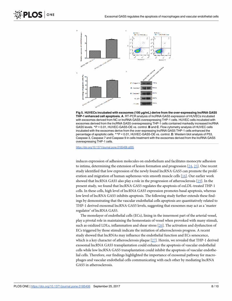
Fig 5. HUVECs incubated with exosomes (100 μg/mL) derive from the over-expressing lncRNA GAS5
THP-1 enhanced cell apoptosis. A. RT-PCR analysis of lncRNA GAS5 expression of HUVECs incubated
with exosomes derived from NC or lncRNA GAS5 overexpressing THP-1 cells. HUVEC cells incubated with
exosomes derived from the lncRNA GAS5 overexpressing THP-1 cells contained markedly increased lncRNA
GAS5 levels. *P < 0.01, HUVEC-GAS5-OE vs. control. B and C. Flow cytometry analysis of HUVEC cells
incubated with the exosomes derive from the over-expressing lncRNA GAS5 THP-1 cells enhanced the
percentage of apoptotic cells. **P < 0.01, HUVEC-GAS5-OE vs. control. D. Western blot analysis of P53,
Caspase 3, Caspase 7 and Caspase 9 in cells treatment with the exosomes derived from the lncRNA GAS5
overexpressing THP-1 cells.
https://doi.org/10.1371/journal.pone.0185406.g005
Exosomal GAS5 regulates the apoptosis of macrophages and vascular endothelial cells
PLOS ONE | https://doi.org/10.1371/journal.pone.0185406 September 25, 2017 8 / 10
Conclusions
As membrane vesicles, exosomes are crucial in the intercellular communications and may be a
key mediator of lncRNA GAS5, which provide the possibility of an alternative strategy for
treatment of atherosclerosis.
Author Contributions
Conceptualization: Lei Chen, Wusong Tong.
Data curation: Lei Chen.
Formal analysis: Lei Chen.
Funding acquisition: Lei Chen.
Investigation: Lei Chen, Wenjin Yang, Yijun Guo, Wei Chen, Ping Zheng, Jinsong Zeng,
Wusong Tong.
Methodology: Lei Chen.
Project administration: Wusong Tong.
Resources: Lei Chen, Wusong Tong.
Software: Lei Chen, Wusong Tong.
Supervision: Wusong Tong.
Validation: Lei Chen, Wusong Tong.
Visualization: Lei Chen, Wusong Tong.
Writing – original draft: Lei Chen, Wusong Tong.
Writing – review & editing: Lei Chen, Wusong Tong.
References1. Rafieian-Kopaei M, Setorki M, Doudi M, Baradaran A, Nasri H. Atherosclerosis: process, indicators, risk
factors and new hopes. Int. J. Prev. Med. 2014; 5:927e946.
2. Novak J, Bienertova-Vasku J, Kara T, Novak M. MicroRNAs involved in the lipid metabolism and their
possible implications for atherosclerosis development and treatment. Mediat. Inflamm. 2014; 275867.
3. Toba H, Cortez D, Lindsey ML, Chilton RJ. Applications of miRNA technology for atherosclerosis. Curr.
Atheroscler. Rep. 2014; 16:386. https://doi.org/10.1007/s11883-013-0386-9 PMID: 24395388
4. Back M, Hansson GK. Anti-inflammatory therapies for atherosclerosis. Nat. Rev. Cardiol. 2015;
12:199e211.
5. Aryal B, Rotllan N, Fernandez-Hernando C. Noncoding RNAs and atherosclerosis. Curr. Atheroscler.
Rep. 2014; 16:407. https://doi.org/10.1007/s11883-014-0407-3 PMID: 24623179
6. Lander ES, Linton LM, Birren B, Nusbaum C, Zody MC, Baldwin J, et al. Initial sequencing and analysis
of the human genome. Nature. 2001; 409 (6822):860–921. https://doi.org/10.1038/35057062 PMID:
11237011
7. Mattick JS. The functional genomics of noncoding RNA. Science. 2005; 309:1527–1528. https://doi.
org/10.1126/science.1117806 PMID: 16141063
8. Ma H, Hao Y, Dong X, Gong Q, Chen J, Zhang J, et al. Molecular mechanisms and function prediction
of long noncoding RNA. Scientific World Journal. 2012;541786. https://doi.org/10.1100/2012/541786
PMID: 23319885
9. Chen X, Yan GY. Novel human lncRNA-disease association inference based on lncRNA expression
profiles. Bioinformatics. 2013; 29 (20): 2617–2624. https://doi.org/10.1093/bioinformatics/btt426 PMID:
24002109
Exosomal GAS5 regulates the apoptosis of macrophages and vascular endothelial cells
PLOS ONE | https://doi.org/10.1371/journal.pone.0185406 September 25, 2017 9 / 10

10. Arase M, Horiguchi K, Ehata S, Morikawa M, Tsutsumi S, Aburatani H, et al. Transforming growth fac-
tor-beta-induced lncRNA-Smad7 inhibits apoptosis of mouse breast cancer JygMC(A) cells. Cancer
Sci. 2014; 105 (8):974–982. https://doi.org/10.1111/cas.12454 PMID: 24863656
11. Liu Q, Huang J, Zhou N, Zhang Z, Zhang A, Lu Z, et al. LncRNA loc285194 is a p53-regulated tumor
suppressor. Nucleic Acids Res. 2013; 41 (9):4976–4987. https://doi.org/10.1093/nar/gkt182 PMID:
23558749
12. Zhou T, Ding JW, Wang XA. Long noncoding RNAs and atherosclerosis. Atherosclerosis. 2016;
248:51–61. https://doi.org/10.1016/j.atherosclerosis.2016.02.025 PMID: 26987066
13. Krell J, Frampton AE, Mirnezami R, Harding V, De Giorgio A, Roca Alonso L, et al. Growth arrest-spe-
cific transcript 5 associated snoRNA levels are related to p53 expression and DNA damage in colorectal
cancer. PLoS One. 2014 Jun 13; 9(6):e98561. https://doi.org/10.1371/journal.pone.0098561 eCollec-
tion 2014. PMID: 24926850
14. Amaral PP, Clark MB, Gascoigne DK, Dinger ME, Mattick JS. lncRNAdb: a reference database for long
noncoding RNAs. Nucleic Acids Res. 2011;39.
15. Williams GT, Mourtada-Maarabouni M, Farzaneh F. A critical role for non-coding RNA GAS5 in growth
arrest and rapamycin inhibition in human T-lymphocytes. Biochem. Soc. Trans. 2011; 39 (2):482–486.
https://doi.org/10.1042/BST0390482 PMID: 21428924
16. Li L, Li X, The E, Wang LJ, Yuan TY, Wang SY, et al. Low expression of lncRNA-GAS5 is implicated in
human primary varicose great saphenous veins. PLoS One.2015; 10: e0120550. https://doi.org/10.
1371/journal.pone.0120550 PMID: 25806802
17. De Jong OG, Van Balkom BW, Schiffelers RM, Bouten CV, Verhaar MC. Extracellular vesicles: Poten-
tial roles in regenerative medicine. Frontiers in Immunology. 2014; 5:608. https://doi.org/10.3389/
fimmu.2014.00608 PMID: 25520717
18. Ahadi A, Khoury S, Losseva M, Tran N. A comparative analysis of lncRNAs in prostate cancer exo-
somes and their parental cell lines. Genomics. 2016; 9:7–9.
19. Chen L, Yao H, Hui JY, Ding SH, Fan YL, Pan YH, et al. Global transcriptomic study of atherosclerosis
development in rats. Gene. 2016; 592(1):43–48. https://doi.org/10.1016/j.gene.2016.07.023 PMID:
27425867
20. Gezer U, Ozgur E, Cetinkaya M, Isin M, Dalay N. Long non-coding RNAs with low expression levels in
cells are enriched in secreted exosomes. Cell Biol Int. 2014;Sep; 38(9):1076–9. https://doi.org/10.1002/
cbin.10301 Epub 2014 May 13. PMID: 24798520
21. Cardilo-Reis L, Gruber S, Schreier SM, Drechsler M, Papac-Milicevic N, Weber C, et al. Interleukin-13
protects from Interleukin-13 protects from atherosclerosis and modulates plaque composition by skew-
ing the macrophage phenotype. EMBO Molecular Medicine. 2012; 4(10):1072–1086. https://doi.org/10.
1002/emmm.201201374 PMID: 23027612
22. Khan R, Spagnoli V, Tardif JC, L’Allier PL. Novel anti-inflammatory therapies for the treatment of athero-
sclerosis. Atherosclerosis. 2015; 240(2):497–509. https://doi.org/10.1016/j.atherosclerosis.2015.04.
783 PMID: 25917947
23. Saha P, Modarai B, Humphries J, Mattock K, Waltham M, Burnand KG, et al. The monocyte/macro-
phage as a therapeutic target in atherosclerosis. Current Opinion on Pharmacology. 2009; 9(2):109–
118.
24. Mehta A, Yang B, Khan S, Hendricks JB, Stephen C, Mehta JL. Oxidized low-density lipoproteins facili-
tate leukocyte adhesion to aortic intima without affecting endothelium-dependent relaxation. Role of P-
selectin. Arteriosclerosis, Thrombosis, and Vascular Biology. 1995; 15(11):2076–2083. PMID: 7583592
25. Ayari H. Respective roles of cortisol, aldosterone and angiotensin II during pathophysiology of athero-
sclerosis. Ann. Biol. Clin. Paris. 2013; 71:381–388. https://doi.org/10.1684/abc.2013.0868 PMID:
23906564
26. Sun X, Belkin N, Feinberg MW. Endothelial microRNAs and atherosclerosis. Curr. Atheroscler. Rep.
2013; 15:372. https://doi.org/10.1007/s11883-013-0372-2 PMID: 24158362
27. Bianchessi V, Badi I, Bertolotti M, Nigro P, D’Alessander Y, Capogrossi MC, et al. The mitochondrial
lncRNA ASncmtRNA-2 is induced in aging and replicative senescence in endothelial cells, J. Mol. Cell
Cardiol. 2015; 81:62–70. https://doi.org/10.1016/j.yjmcc.2015.01.012 PMID: 25640160
Exosomal GAS5 regulates the apoptosis of macrophages and vascular endothelial cells
PLOS ONE | https://doi.org/10.1371/journal.pone.0185406 September 25, 2017 10 / 10


![July/August 1994 Gear Technology · 2014. 7. 17. · r,heGLD20 sliding head automatic, the 'GAC65. the Hes app nVH25 verticalmrmng machine, the Kapp VA 61 and GAS5] grinding machines.](https://static.fdocuments.us/doc/165x107/60c48e4083e139320d4a7d64/julyaugust-1994-gear-technology-2014-7-17-rhegld20-sliding-head-automatic.jpg)


