Exome sequencing in amyotrophic lateral sclerosis ... sequencing in amyotrophic lateral sclerosis...
7
RESEARCH ARTICLES ◥ ALS GENES Exome sequencing in amyotrophic lateral sclerosis identifies risk genes and pathways Elizabeth T. Cirulli, 1 * Brittany N. Lasseigne, 2 * Slavé Petrovski, 3 Peter C. Sapp, 4 Patrick A. Dion, 5 Claire S. Leblond, 5 Julien Couthouis, 6 Yi-Fan Lu, 3 Quanli Wang, 3 Brian J. Krueger, 3 Zhong Ren, 3 Jonathan Keebler, 7 Yujun Han, 7 Shawn E. Levy, 2 Braden E. Boone, 2 Jack R. Wimbish, 2 Lindsay L. Waite, 2 Angela L. Jones, 2 John P. Carulli, 8 Aaron G. Day-Williams, 8 John F. Staropoli, 8 Winnie W. Xin, 9 Alessandra Chesi, 6 Alya R. Raphael, 6 Diane McKenna-Yasek, 4 Janet Cady, 10 J. M. B. Vianney de Jong, 11 Kevin P. Kenna, 12 Bradley N. Smith, 13 Simon Topp, 13 Jack Miller, 13 Athina Gkazi, 13 FALS Sequencing Consortium,† Ammar Al-Chalabi, 13 Leonard H. van den Berg, 14 Jan Veldink, 14 Vincenzo Silani, 15 Nicola Ticozzi, 15 Christopher E. Shaw, 13 Robert H. Baloh, 16 Stanley Appel, 17 Ericka Simpson, 17 Clotilde Lagier-Tourenne, 18 Stefan M. Pulst, 19 Summer Gibson, 19 John Q. Trojanowski, 20 Lauren Elman, 21 Leo McCluskey, 21 Murray Grossman, 22 Neil A. Shneider, 23 Wendy K. Chung, 24 John M. Ravits, 25 Jonathan D. Glass, 26 Katherine B. Sims, 9 Vivianna M. Van Deerlin, 20 Tom Maniatis, 27 Sebastian D. Hayes, 8,28 Alban Ordureau, 28 Sharan Swarup, 28 John Landers, 4 Frank Baas, 11 Andrew S. Allen, 29 Richard S. Bedlack, 30 J. Wade Harper, 28 Aaron D. Gitler, 6 Guy A. Rouleau, 5 Robert Brown, 4 Matthew B. Harms, 10 Gregory M. Cooper, 2 Tim Harris, 8 ‡ Richard M. Myers, 2 § David B. Goldstein 3 § Amyotrophic lateral sclerosis (ALS) is a devastating neurological disease with no effective treatment.We report the results of a moderate-scale sequencing study aimed at increasing the number of genes known to contribute to predisposition for ALS. We performed whole-exome sequencing of 2869 ALS patients and 6405 controls. Several known ALS genes were found to be associated, and TBK1 (the gene encoding TANK-binding kinase 1) was identified as an ALS gene. TBK1 is known to bind to and phosphorylate a number of proteins involved in innate immunity and autophagy, including optineurin (OPTN) and p62 (SQSTM1/sequestosome), both of which have also been implicated in ALS.These observations reveal a key role of the autophagic pathway in ALS and suggest specific targets for therapeutic intervention. A myotrophic lateral sclerosis (ALS) is a fatal, progressive neurodegenerative disease char- acterized by loss of motor neuron function for which there is no effective treatment or definitive diagnostic test (most cases are diagnosed clinically) (1). Approximately 10% of ALS cases are familial and inherited in an auto- somal dominant, autosomal recessive, or X-linked mode; the remaining cases are apparently spo- radic (2, 3). Approximately 20 genes collectively explain a majority of familial cases, but these genes can explain only a minority (about 10%) of sporadic cases (2, 3) (Table 1). Protein and protein-RNA aggregates are a com- mon feature of ALS pathology. These aggre- gates often include proteins encoded by genes that cause ALS when mutated, including those encoding SOD1, TARDBP (TDP-43), and FUS (4). Multiple genes (e.g., C9orf72, GRN, VCP, UBQLN2, OPTN, NIPA1, SQSTM1) in addition to TARDBP harbor variants pathogenic for TARDBP protein- opathy manifesting as ALS. This pathological TARDBP is part of a common pathway linked to neurodegeneration caused by diverse genetic abnormalities ( 5). Although murine models of ALS are limited, silencing certain ALS genes can cause regression of the disease phenotypes and clearance of the protein aggregates (6). Identifying ALS genes To identify genetic variants associated with ALS, we sequenced the exomes of 2869 patients with ALS and 6405 controls. We ran a standard col- lapsing analysis in which the gene was the unit of analysis, and we coded individuals according to the presence or absence of “qualifying” var- iants in each sequenced gene, where qualifying was defined according to one of six different ge- netic models: dominant coding, recessive coding, dominant not benign, recessive not benign, dom- inant loss of function (LoF), and recessive LoF (7). A total of 17,249 genes had more than one case or control sample with a genetic variant meeting the inclusion criteria for at least one of the genetic models tested (Fig. 1 and figs. S1 and S2). After correcting for multiple tests, the known ALS gene SOD1 (P = 7.05 × 10 -8 ; dominant coding model) was found to have a study-wide signifi- cant enrichment of rare variants in ALS cases relative to controls, with qualifying variants in 0.871% of cases and 0.078% of controls. The genes HLA-B, ZNF729, SIRPA, and TP53 were found to have a significant enrichment of variants in controls; however, these associations appear to be due to sequencing differences and to subsets of the controls having been ascertained on the basis of relevant phenotypes. On the basis of their associations with ALS in a preliminary discovery-phase analysis that used 2843 cases and 4310 controls, we chose 51 genes (table S4) for analysis in an additional 1318 cases RESEARCH 1436 27 MARCH 2015 • VOL 347 ISSUE 6229 sciencemag.org SCIENCE 1 Center for Applied Genomics and Precision Medicine, Duke University School of Medicine, Durham, NC 27708, USA. 2 HudsonAlpha Institute for Biotechnology, Huntsville, AL 35806, USA. 3 Institute for Genomic Medicine, Columbia University, New York, NY 10032, USA. 4 Department of Neurology, University of Massachusetts Medical School, Worcester, MA 01655, USA. 5 Montreal Neurological Institute, Department of Neurology and Neurosurgery, McGill University, Montreal, Quebec H3A 2B4, Canada. 6 Department of Genetics, Stanford University School of Medicine, Stanford, CA 94305, USA. 7 Duke University School of Medicine, Durham, NC 27708, USA. 8 Biogen Idec, Cambridge, MA 02142, USA. 9 Neurogenetics DNA Diagnostic Laboratory, Center for Human Genetics Research, Department of Neurology, Massachusetts General Hospital, Boston, MA 02114, USA. 10 Neurology, Washington University School of Medicine, St. Louis, MO 63110, USA. 11 Department of Genome Analysis, Academic Medical Center, Meibergdreef 9, 1105AZ Amsterdam, Netherlands. 12 Academic Unit of Neurology, Trinity Biomedical Sciences Institute, Trinity College Dublin, Dublin, Republic of Ireland. 13 Department of Basic and Clinical Neuroscience, King’s College London, Institute of Psychiatry, Psychology and Neuroscience, London SE5 8AF, UK. 14 Department of Neurology, Brain Center Rudolf Magnus, University Medical Centre Utrecht, 3508 GA Utrecht, Netherlands. 15 Department of Neurology and Laboratory of Neuroscience, IRCCS Istituto Auxologico Italiano, Milan 20149, Italy, and Department of Pathophysiology and Transplantation, Dino Ferrari Center, Università degli Studi di Milano, Milan 20122, Italy. 16 Cedars Sinai Medical Center, Los Angeles, CA 90048, USA. 17 Houston Methodist Hospital, Houston, TX 77030, USA, and Weill Cornell Medical College of Cornell University, New York, NY 10065, USA. 18 Ludwig Institute for Cancer Research and Department of Neurosciences, University of California, San Diego, La Jolla, CA 92093, USA. 19 Department of Neurology, University of Utah School of Medicine, Salt Lake City, UT 84112, USA. 20 Department of Pathology and Laboratory Medicine, Perelman School of Medicine, University of Pennsylvania, Philadelphia, PA 19104, USA. 21 Department of Neurology, Penn ALS Center, Perelman School of Medicine, University of Pennsylvania, Philadelphia, PA 19104, USA. 22 Department of Neurology, Penn Frontotemporal Degeneration Center, Perelman School of Medicine at the University of Pennsylvania, Philadelphia, PA 19104, USA. 23 Department of Neurology, Center for Motor Neuron Biology and Disease, Columbia University, New York, NY 10032, USA. 24 Department of Pediatrics and Medicine, Columbia University, New York, NY 10032, USA. 25 Department of Neurosciences, University of California, San Diego, La Jolla, CA 92093, USA. 26 Department of Neurology, Emory University, Atlanta, GA 30322, USA. 27 Department of Biochemistry & Molecular Biophysics, Columbia University, New York, NY 10027, USA. 28 Department of Cell Biology, Harvard Medical School, Boston, MA 02115, USA. 29 Department of Biostatistics and Bioinformatics, Duke University School of Medicine, Durham, NC 27708, USA. 30 Duke ALS Clinic and Durham VA Medical Center, Durham, NC 27708, USA. *These authors contributed equally to this work. †The full author list is included at the end of the manuscript. ‡Corresponding author. E-mail: [email protected] §These authors contributed equally to this work. on May 27, 2015 www.sciencemag.org Downloaded from on May 27, 2015 www.sciencemag.org Downloaded from on May 27, 2015 www.sciencemag.org Downloaded from on May 27, 2015 www.sciencemag.org Downloaded from on May 27, 2015 www.sciencemag.org Downloaded from on May 27, 2015 www.sciencemag.org Downloaded from
Transcript of Exome sequencing in amyotrophic lateral sclerosis ... sequencing in amyotrophic lateral sclerosis...

RESEARCH ARTICLES◥
ALS GENES
Exome sequencing in amyotrophiclateral sclerosis identifies risk genesand pathwaysElizabeth T. Cirulli,1* Brittany N. Lasseigne,2* Slavé Petrovski,3 Peter C. Sapp,4
Patrick A. Dion,5 Claire S. Leblond,5 Julien Couthouis,6 Yi-Fan Lu,3 Quanli Wang,3
Brian J. Krueger,3 Zhong Ren,3 Jonathan Keebler,7 Yujun Han,7 Shawn E. Levy,2
Braden E. Boone,2 Jack R. Wimbish,2 Lindsay L. Waite,2 Angela L. Jones,2
John P. Carulli,8 Aaron G. Day-Williams,8 John F. Staropoli,8 Winnie W. Xin,9
Alessandra Chesi,6 Alya R. Raphael,6 Diane McKenna-Yasek,4 Janet Cady,10
J. M. B. Vianney de Jong,11 Kevin P. Kenna,12 Bradley N. Smith,13 Simon Topp,13
Jack Miller,13 Athina Gkazi,13 FALS Sequencing Consortium,† Ammar Al-Chalabi,13
Leonard H. van den Berg,14 Jan Veldink,14 Vincenzo Silani,15 Nicola Ticozzi,15
Christopher E. Shaw,13 Robert H. Baloh,16 Stanley Appel,17 Ericka Simpson,17
Clotilde Lagier-Tourenne,18 Stefan M. Pulst,19 Summer Gibson,19 John Q. Trojanowski,20
Lauren Elman,21 Leo McCluskey,21 Murray Grossman,22 Neil A. Shneider,23
Wendy K. Chung,24 John M. Ravits,25 Jonathan D. Glass,26 Katherine B. Sims,9
Vivianna M. Van Deerlin,20 Tom Maniatis,27 Sebastian D. Hayes,8,28 Alban Ordureau,28
Sharan Swarup,28 John Landers,4 Frank Baas,11 Andrew S. Allen,29 Richard S. Bedlack,30
J. Wade Harper,28 Aaron D. Gitler,6 Guy A. Rouleau,5 Robert Brown,4 Matthew B. Harms,10
Gregory M. Cooper,2 Tim Harris,8‡ Richard M. Myers,2§ David B. Goldstein3§
Amyotrophic lateral sclerosis (ALS) is a devastating neurological disease with no effectivetreatment.We report the results of a moderate-scale sequencing study aimed at increasingthe number of genes known to contribute to predisposition for ALS. We performedwhole-exome sequencing of 2869 ALS patients and 6405 controls. Several known ALSgenes were found to be associated, and TBK1 (the gene encoding TANK-binding kinase 1)was identified as an ALS gene. TBK1 is known to bind to and phosphorylate a numberof proteins involved in innate immunity and autophagy, including optineurin (OPTN)and p62 (SQSTM1/sequestosome), both of which have also been implicated in ALS. Theseobservations reveal a key role of the autophagic pathway in ALS and suggest specifictargets for therapeutic intervention.
Amyotrophic lateral sclerosis (ALS) is a fatal,progressive neurodegenerative disease char-acterized by loss of motor neuron functionfor which there is no effective treatment ordefinitive diagnostic test (most cases are
diagnosed clinically) (1). Approximately 10% ofALS cases are familial and inherited in an auto-somal dominant, autosomal recessive, or X-linkedmode; the remaining cases are apparently spo-radic (2, 3). Approximately 20 genes collectivelyexplain a majority of familial cases, but thesegenes can explain only a minority (about 10%) ofsporadic cases (2, 3) (Table 1).Protein and protein-RNA aggregates are a com-
mon feature of ALS pathology. These aggre-gates often include proteins encoded by genesthat cause ALS when mutated, including thoseencoding SOD1, TARDBP (TDP-43), and FUS (4).Multiple genes (e.g., C9orf72, GRN, VCP, UBQLN2,OPTN, NIPA1, SQSTM1) in addition to TARDBPharbor variants pathogenic for TARDBP protein-opathy manifesting as ALS. This pathologicalTARDBP is part of a common pathway linked
to neurodegeneration caused by diverse geneticabnormalities (5). Althoughmurine models of ALSare limited, silencing certain ALS genes cancause regression of the disease phenotypes andclearance of the protein aggregates (6).
Identifying ALS genes
To identify genetic variants associated with ALS,we sequenced the exomes of 2869 patients withALS and 6405 controls. We ran a standard col-lapsing analysis in which the gene was the unitof analysis, and we coded individuals accordingto the presence or absence of “qualifying” var-iants in each sequenced gene, where qualifyingwas defined according to one of six different ge-neticmodels: dominant coding, recessive coding,dominant not benign, recessive not benign, dom-inant loss of function (LoF), and recessive LoF(7). A total of 17,249 genes had more than onecase or control sample with a genetic variantmeeting the inclusion criteria for at least one ofthe genetic models tested (Fig. 1 and figs. S1 andS2). After correcting for multiple tests, the known
ALS gene SOD1 (P = 7.05 × 10⁻⁸; dominant codingmodel) was found to have a study-wide signifi-cant enrichment of rare variants in ALS casesrelative to controls, with qualifying variants in0.871% of cases and 0.078% of controls. The genesHLA-B, ZNF729, SIRPA, and TP53 were found tohave a significant enrichment of variants incontrols; however, these associations appear tobe due to sequencing differences and to subsetsof the controls having been ascertained on thebasis of relevant phenotypes.On the basis of their associations with ALS in a
preliminary discovery-phase analysis that used2843 cases and 4310 controls, we chose 51 genes(table S4) for analysis in an additional 1318 cases
RESEARCH
1436 27 MARCH 2015 • VOL 347 ISSUE 6229 sciencemag.org SCIENCE
1Center for Applied Genomics and Precision Medicine, DukeUniversity School of Medicine, Durham, NC 27708, USA.2HudsonAlpha Institute for Biotechnology, Huntsville, AL35806, USA. 3Institute for Genomic Medicine, ColumbiaUniversity, New York, NY 10032, USA. 4Department ofNeurology, University of Massachusetts Medical School,Worcester, MA 01655, USA. 5Montreal Neurological Institute,Department of Neurology and Neurosurgery, McGillUniversity, Montreal, Quebec H3A 2B4, Canada. 6Departmentof Genetics, Stanford University School of Medicine,Stanford, CA 94305, USA. 7Duke University School ofMedicine, Durham, NC 27708, USA. 8Biogen Idec,Cambridge, MA 02142, USA. 9Neurogenetics DNA DiagnosticLaboratory, Center for Human Genetics Research,Department of Neurology, Massachusetts General Hospital,Boston, MA 02114, USA. 10Neurology, Washington UniversitySchool of Medicine, St. Louis, MO 63110, USA. 11Departmentof Genome Analysis, Academic Medical Center, Meibergdreef9, 1105AZ Amsterdam, Netherlands. 12Academic Unit ofNeurology, Trinity Biomedical Sciences Institute, TrinityCollege Dublin, Dublin, Republic of Ireland. 13Department ofBasic and Clinical Neuroscience, King’s College London,Institute of Psychiatry, Psychology and Neuroscience,London SE5 8AF, UK. 14Department of Neurology, BrainCenter Rudolf Magnus, University Medical Centre Utrecht,3508 GA Utrecht, Netherlands. 15Department of Neurologyand Laboratory of Neuroscience, IRCCS Istituto AuxologicoItaliano, Milan 20149, Italy, and Department ofPathophysiology and Transplantation, Dino Ferrari Center,Università degli Studi di Milano, Milan 20122, Italy. 16CedarsSinai Medical Center, Los Angeles, CA 90048, USA.17Houston Methodist Hospital, Houston, TX 77030, USA, andWeill Cornell Medical College of Cornell University, New York,NY 10065, USA. 18Ludwig Institute for Cancer Research andDepartment of Neurosciences, University of California, SanDiego, La Jolla, CA 92093, USA. 19Department of Neurology,University of Utah School of Medicine, Salt Lake City, UT84112, USA. 20Department of Pathology and LaboratoryMedicine, Perelman School of Medicine, University ofPennsylvania, Philadelphia, PA 19104, USA. 21Department ofNeurology, Penn ALS Center, Perelman School of Medicine,University of Pennsylvania, Philadelphia, PA 19104, USA.22Department of Neurology, Penn FrontotemporalDegeneration Center, Perelman School of Medicine at theUniversity of Pennsylvania, Philadelphia, PA 19104, USA.23Department of Neurology, Center for Motor Neuron Biologyand Disease, Columbia University, New York, NY 10032, USA.24Department of Pediatrics and Medicine, ColumbiaUniversity, New York, NY 10032, USA. 25Department ofNeurosciences, University of California, San Diego, La Jolla,CA 92093, USA. 26Department of Neurology, EmoryUniversity, Atlanta, GA 30322, USA. 27Department ofBiochemistry & Molecular Biophysics, Columbia University,New York, NY 10027, USA. 28Department of Cell Biology,Harvard Medical School, Boston, MA 02115, USA.29Department of Biostatistics and Bioinformatics, DukeUniversity School of Medicine, Durham, NC 27708, USA.30Duke ALS Clinic and Durham VA Medical Center, Durham,NC 27708, USA.*These authors contributed equally to this work. †The full authorlist is included at the end of the manuscript. ‡Correspondingauthor. E-mail: [email protected] §These authorscontributed equally to this work.
on
May
27,
201
5w
ww
.sci
ence
mag
.org
Dow
nloa
ded
from
o
n M
ay 2
7, 2
015
ww
w.s
cien
cem
ag.o
rgD
ownl
oade
d fr
om
on
May
27,
201
5w
ww
.sci
ence
mag
.org
Dow
nloa
ded
from
o
n M
ay 2
7, 2
015
ww
w.s
cien
cem
ag.o
rgD
ownl
oade
d fr
om
on
May
27,
201
5w
ww
.sci
ence
mag
.org
Dow
nloa
ded
from
o
n M
ay 2
7, 2
015
ww
w.s
cien
cem
ag.o
rgD
ownl
oade
d fr
om

SCIENCE sciencemag.org 27 MARCH 2015 • VOL 347 ISSUE 6229 1437
Table 1. Variants in previously described and currently reported ALS genes. Entries for reported inheritance model, reported FALS explained, andreported SALS explained are adapted from (3, 4, 51) with additional information from (17–21, 52–54). AD, autosomal dominant; AR, autosomal recessive;XD, X-linked. Best-model data are based on discovery data set for genes not included in the replication data set, and otherwise D = discovery, R =replication, and C = combined. Potential ALS cases explained are calculated as [(cases with variant in best model) – (controls with variant in bestmodel)]; as case variants are risk factors for disease and may not be causal, this represents the potential percentage of cases for which this gene plays arole in disease.
GeneReported
inheritancemodel
ReportedFALS
explained
ReportedSALS
explained
Best model withcase enrichment in
present study (P value)
Cases withvariant inbest model
Controls withvariant inbest model
PotentialALS casesexplained
TBK1 N/A N/A N/A Dom not benign(D = 1.12 × 10⁻⁵;R = 5.78 × 10⁻⁷;C = 3.60 × 10⁻¹¹)
D = 23 (0.802%);R = 23 (1.745%);C = 46 (1.099%)
D = 12 (0.187%);R = 5 (0.211%);C = 17 (0.194%)
0.905%
NEK1 N/A N/A N/A Dom LoF(D = 1.06 × 10⁻⁶;R = 0.001;C = 3.15 × 10⁻⁹)
D = 25 (0.871%);R = 10 (0.759%);C = 35 (0.836%)
D = 6 (0.094%);R = 2 (0.084%);C = 8 (0.091%)
0.745%
SOD1 AR/AD 12% 1.50% Dom coding(7.05 × 10⁻⁸)
25 (0.871%) 5 (0.078%) 0.793%
TARDBP AD 4% 1% Dom coding(2.93 × 10⁻⁶)
19 (0.662%) 6 (0.094%) 0.569%
OPTN AR/AD <1% <1% Dom not benign(D = 0.023;R = 0.002;C = 0.002)
D = 18 (0.627%);R = 8 (0.607%);C = 26 (0.621%)
D = 16 (0.25%);R = 4 (0.169%);C = 20 (0.228%)
0.393%
SPG11 AR <1% <1% Dom LoF(D = 0.022;R = 0.183;C = 0.023)
D = 20 (0.697%);R = 5 (0.379%);C = 25 (0.597%)
D = 20 (0.312%);R = 7 (0.295%);C = 27 (0.308%)
0.289%
VCP AD 1% 1% Dom coding (0.022) 8 (0.279%) 4 (0.062%) 0.216%HNRNPA1 AD <1% <1% Dom coding (0.103) 6 (0.209%) 5 (0.078%) 0.131%ATXN2* AD <1% <1% Rec coding (0.205) 4 (0.139%) 2 (0.031%) 0.108%ANG AD <1% <1% Dom LoF (0.217) 2 (0.070%) 1 (0.016%) 0.054%CHCHD10 AD <1% <1% Dom coding (0.225) 2 (0.070%) 0 (0%) 0.070%SIGMAR1 AR <1% <1% Dom LoF (0.226) 1 (0.035%) 0 (0%) 0.035%FIG4 AR/AD <1% <1% Dom LoF (0.232) 9 (0.314%) 12 (0.187%) 0.126%SS18L1 AD <1% <1% Dom LoF (0.241) 1 (0.035%) 0 (0%) 0.035%GRN AD <1% <1% Dom not benign (0.357) 14 (0.488%) 24 (0.375%) 0.113%SETX AD <1% <1% Rec not benign (0.379) 3 (0.105%) 4 (0.062%) 0.042%HNRNPA2B1 AD <1% <1% Dom not benign (0.423) 3 (0.105%) 4 (0.062%) 0.042%SQSTM1 AD 1% <1% Dom LoF (0.546) 1 (0.035%) 2 (0.031%) 0.004%TAF15 AR/AD <1% <1% Rec not benign (0.555) 2 (0.070%) 1 (0.016%) 0.054%FUS AR/AD 4% 1% Dom LoF (0.612) 2 (0.070%) 3 (0.047%) 0.023%ALS2 AR <1% <1% Rec coding (0.655) 2 (0.070%) 4 (0.062%) 0.007%VAPB AD <1% <1% Dom not benign (0.688) 3 (0.105%) 5 (0.078%) 0.027%NEFH AD <1% <1% Dom coding (0.673) 22 (0.767%) 37 (0.578%) 0.189%C9orf72* AD 40% 7% Dom not benign (1.000) 4 (0.139%) 7 (0.109%) 0.030%CHMP2B AD <1% <1% Rec coding (1.000) 1 (0.035%) 1 (0.016%) 0.019%MATR3 AD <1% <1% Dom coding (1.000) 19 (0.662%) 35 (0.546%) 0.116%PFN1 AD <1% <1% Rec coding (1.000) 9 (0.314%) 15 (0.234%) 0.080%PRPH AD <1% <1% Dom LoF (1.000) 1 (0.035%) 2 (0.031%) 0.004%SPAST AD <1% <1% Dom coding (1.000) 6 (0.209%) 12 (0.187%) 0.022%TUBA4A AD 1% <1% Dom not benign (1.000) 2 (0.070%) 3 (0.047%) 0.023%ELP3† Allelic <1% <1% Rec coding (1.000) 0 (0%) 0 (0%) 0%DAO† AD <1% <1% Rec coding (1.000) 0 (0%) 0 (0%) 0%DCTN1† AD <1% <1% Dom coding (0.668) 32 (1.115%) 76 (1.187%) 0%EWSR1† AD <1% <1% Dom coding (0.375) 10 (0.349%) 28 (0.437%) 0%GLE1† AD <1% <1% Rec LoF (1.000) 0 (0%) 0 (0%) 0%UBQLN2† XD <1% <1% Dom LoF (1.000) 0 (0%) 0 (0%) 0%
*Because the known causal variants are repeat expansions that are not generally captured by next-generation sequencing, no case enrichment is expected. †Nomodel showed case enrichment.
RESEARCH | RESEARCH ARTICLES

and 2371 controls (sequenced using either wholeexome or custom capture) (7). This analysis de-finitively identified TANK-binding kinase 1 (TBK1)as an ALS gene, with a discovery association P =1.12 × 10⁻⁵, a replication P = 5.78 × 10⁻⁷, and acombined P = 3.60 × 10⁻¹¹ (dominant not benignmodel). In the combined data set, dominant notbenign variants in this gene were found in 1.099%of cases and 0.194% of controls, with LoF var-iants occurring in 0.382% of cases and 0.034%of controls.
Analysis of clinical features
We also performed gene-based collapsing analy-ses to identify genes associatedwith patients’ ageof onset, site of onset, and survival time. No genesshowed genome-wide significant association withany of these features. When applying multiple-test correction to only knownALS predispositiongenes and TBK1, we found that D-amino acid oxi-dase (DAO) significantly correlated with survivaltimes, with variant carriers showing shorter sur-vival times (P = 5.5 × 10⁻⁷, dominant coding mod-el). In mice, DAO is required for the clearance ofD-serine. Indeed, D-serine levels are increasedin SOD1 mutant mice and in spinal cords frompeople with familial ALS (FALS) or sporadicALS (SALS) (8, 9). Known FALS mutationsseem to reduce DAO activity, leading to neuro-toxicity (10).ALS patients withmutations inmore than one
known ALS gene are reported to have a youngerage of onset (11).We did not replicate this findingin our data set. Without sequence data for knownC9orf72 carriers (by far the most common ALSvariant) and without information about ATXN2expansions, we cannot adequately assess such anassociation.
Associations with other ALS genes
Although SOD1 was the only previously knownALS gene to attain a genome-wide significantassociation in our data, many other known ALSgenes showed strong associations. For example,rare coding variants in TARDBP occurred in0.662% of the ALS cases and 0.094% of controlsin our study, ranking this gene second to SOD1genome-wide under the dominant coding model(discovery data set, P = 2.93 × 10⁻⁶; Fig. 1). Consist-ent with previous reports and the ALS pathogenicTARDBP “DM” variants in the Human GenomeMutation Database (HGMD) (3, 12), we observedthat the implicated nonsynonymous variants weregenerally predicted to have a benign effect onprotein structure and function by PolyPhen-2(13) and were clearly concentrated in the 3′ protein-coding portion of the gene in the ALS cases rel-ative to controls (Fig. 2).In the case of OPTN, we observed rare dam-
aging variants in 0.621% of ALS cases and 0.228%of controls (combineddominant not benignmod-el, P = 0.002). The greatest enrichment was forLoF variants, which occur in 0.334% of cases and0.114%of controls (combined dominant LoFmod-el, P = 0.013). Whereas the initial studies ofOPTNin ALS found a role in only a few families with arecessive genetic model, subsequent studies iden-
tified dominantmutations (14, 15). Here, dominantvariants appeared to make a substantial contri-bution to sporadic disease.Finally, we also observed a modest excess of
qualifying variants in VCP (discovery dominantcoding model, P = 0.022) and of LoF variants inSPG11 (combined dominant LoFmodel,P= 0.023).The former was driven by variants near the celldivision protein 48 domain 2 region, where var-iants were found in 71% of case variants ascompared to 25% of control variants (Fig. 2).Similar to OPTN, SPG11 has previously been re-ported as a cause of recessive juvenile ALS, butour data indicate that it could play a broader rolebecause these cases did not have early onset (16).We did not identify even a nominal associa-
tion with other previously reported ALS genes inour data set, including the recently reportedTUBA4A,MATR3,GLE1, SS18L1, and CHCHD10(Table 1) (17–21). A fraction of our samples weregenetically screened for some of the known genesand positive cases had been removed before se-quencing, whichmay partially explain the lack ofsignal (7). Additionally, a comparison with genesimplicated in a recent assessment of the role of169 previously reported and candidate ALS genesin 242 sporadic ALS cases and 129 controls showedno overlap beyond signals for SOD1 and SPG11(22). Some of these previously studied genesare mutated so rarely that even the sample size
presented here is not sufficient to detect causalvariant enrichment, while others simply showcomparable proportions of rare variants amongcases and controls. Finally, certain genes did notshowassociations owing to thenature of the causalvariation: Most known pathogenic variants inATXN2 and C9orf72 are repeats that cannot beidentified in our sequence data.
TBK1, autophagy, and neuroinflammation
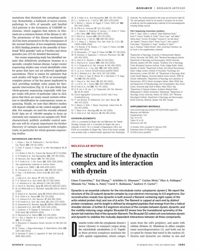
Previous studies have implicated both OPTN(optineurin) (23) and SQSTM1 (p62) (24) inALS. The current study implicates TBK1 andsuggests that OPTN is a more important diseasegene than previously recognized. These genesplay important and interconnected roles in bothautophagy and inflammation, emerging areas ofinterest in ALS research (Fig. 3) (25–27). Muta-tions in SOD1, TARDBP, and FUS result in theformation of protein aggregates that stain withantibodies to SQSTM1 and OPTN (28). These ag-gregates are thought to lead to a cargo-specificsubtype of autophagy involved in the degradationof ubiquitinated proteins through the lysosome(29). The SQSTM1 and OPTN proteins function ascargo receptors, recruiting ubiquitinated proteinsto the autophagosome via their LC3 interaction re-gion (LIR) motifs. TBK1 binds and phosphorylatesboth OPTN and SQSTM1 (30–32) and enhances thebinding of OPTN to the essential autophagosome
1438 27 MARCH 2015 • VOL 347 ISSUE 6229 sciencemag.org SCIENCE
Fig. 1. Quantile-quantile plot of discovery results for dominant codingmodel.Results for the analysisof 2869 case and 6405 control exomes are shown; 16,491 covered genes passed quality control with morethan one case or control carrier for this test. The genes with the top 10 associations are labeled. Thegenomic inflation factor l is 1.060.The association with SOD1 passed correction for multiple tests.
RESEARCH | RESEARCH ARTICLES

protein LC3, thereby facilitating the autopha-gic turnover of infectious bacteria coated withubiquitinated proteins, a specific cargo of theOPTN adaptor (33). Considering that TBK1 colo-calizes with OPTN and SQSTM1 in autophago-
somes, it is possible that all three proteinsassociate with protein aggregates in ALS (33).Indeed, TBK1 appears to play a role in the deg-radation of protein aggregates by autophagy(34). Additionally, OPTN also functions in the
autophagic turnover of damaged mitochondriavia the Parkin ubiquitin ligase pathway (35). Fi-nally, VCP, encoded by another gene with muta-tions that cause ALS, also binds to ubiquitinatedprotein aggregates. VCP and autophagy are re-quired for the removal of stress granules (densecytoplasmic protein-RNA aggregates), which area common feature of ALS pathology (36). Thus,OPTN, SQSTM1, VCP, and TBK1 may be criticalcomponents of the aggresome pathway requiredfor the removal of pathological ribonucleoproteininclusions (37). It appears that defects in this path-way can be selective for motor neuron death, insome cases apparently sparing other neuronalcell types.In addition to their roles in autophagy, OPTN,
SQSTM1, and TBK1 all function in the NF-kBpathway (Fig. 3) (27, 38). For example, IkB ki-nases (IKKa and IKKb) phosphorylate the IKK-related kinase TBK1, which in turn phosphorylatesthe IkB kinases, suppressing their activity in anegative autoregulatory feedback loop (39). TBK1also phosphorylates and activates the transcrip-tion factor IRF3 (40–42) and the critical innateimmunity signaling componentsMAVSandSTING(43). The coordinate activation ofNF-kB and IRF3turns on the transcription of many inflammato-ry genes, including interferon-b (44). The innateimmune pathway and neuroinflammation ingeneral are thought to be important aspectsof neurodegenerative disease progression (45).Thus, pathogenic variants in OPTN, SQSTM1, orTBK1 would be expected to lead to defects inautophagy and in key innate immunity signaling
SCIENCE sciencemag.org 27 MARCH 2015 • VOL 347 ISSUE 6229 1439
TARDBP
1 2 3 4 5Exons (CCDS122.1)
mRNA
V>AS
N>SS
S>LS
K>RS
G>S S
G>A S
G>V S
G>R S
G>SS
M>VS
P>SS
M>V S
M>IS
M>IS
N>KS
S>PS
G>DS
S>GS
I>VS
N>SS
G>DS
5'UTR 3'UTR
Protein
cd12321
VCP
1716 15 14 13 12 10 9 7 6 5 4 3 2 1Exons(CCDS6573.1)
mRNA
T>IS
F>LS
N>TS
M>TS
R>HS
I>VS
V>IS
K>RS
R>CS
G>AS
5'UTR 3'UTR
Protein
pfam00004
smart01072
pfam09336
pfam02359
Fig. 2. Variants in TARDBP and VCP. Dominant coding variants are shown in TARDBP and VCP (dis-covery data set). Case variants are enriched at the 3′ end of the gene in TARDBP and near the cell divisionprotein 48 domain 2 region in VCP. LoF variants are filled in red, and nonsynonymous variants are filled inblue. Case variants are shown with red lines, control variants are shown with blue lines, and variants foundin both cases and controls are shown with dashed lines.
Fig. 3. Genes and path-ways implicated in ALSdisease progression.Genes known to havesequence variants thatcause or are associated withALS are indicated in red.These mutations can lead tothe formation of protein orprotein-RNA aggregatesthat appear as inclusionbodies in post mortemsamples from both familialand sporadic ALS patients.Some of the mutant pro-teins adopt “prion-like”structures (see text formore detail). The misfoldedproteins activate theubiquitin-proteasomeautophagy pathways toremove the misfoldedproteins. Ubiquilin2(UBQLN2) functions in boththe ubiquitin-proteasomeand autophagy pathways.TBK-1 (boxed) lies at theinterface between autophagyand inflammation andassociates with and phos-phorylates both optineurinand p62, which can in turn enhance inflammation. ISG15 is induced by type I interferons (a and b) and interacts with p62 and HDAC6 in the autophagosome.
TBK-1
IFN
ISG15LRLRGG
LC3
SQSTM1/p62
Autophagy
HDAC6
LC3
Optineurin
Lysosome
Lysosomal Fusion
Inteferon Stimulated Genes HDAC6
VCPUBQLN2
ProteasomeSOD1/TDP43/FUShnRNPA1/2
Prion-like Proteinor Protein/RNA Aggregates
Neuroinflammation
IRF3/7
IKKα/β
NF-κB
Degradation
UBQLN2
Ubiquitin Proteasome Pathway
Autophagosome
Ubiquitin
RESEARCH | RESEARCH ARTICLES

pathways. Mutations in these genes might there-fore interfere with the normal function of thesepathways in maintaining normal cellular ribo-proteostasis (37).The simple observation of enrichment of qual-
ifying variants in patients shows that some of thevariants we have identified influence risk of dis-ease. We cannot determine, however, the extentto which they may interact with any other var-iants or other risk factors in determining risk.We therefore focus on estimating the proportionof patients in which variants in the relevant geneseither cause or contribute to disease by subtract-ing the proportion of controls with qualifyingvariants in a gene from the proportion of caseswith such variants. Although we saw no enrich-ment of case variants in SQSTM1, variants inOPTN and TBK1were estimated to explain or con-tribute to 1.30% of cases in our data set whentaken together (combined data set), suggesting animportant subgroup of patients that may have acommon biological etiology. No individual ALScases had qualifying variants in more than one ofthese three genes.The case variants found in OPTN and TBK1
were largely heterozygous and LoF, which sug-gests that a reduction in trafficking of cargothrough the autophagosomal pathway or disrup-tion of autophagosomalmaturationmay promotedisease. Although the most obvious enrichmentof case variants in TBK1 was seen for LoF, therewas also a signal for nonsynonymous variants,which were found in 1.027% of cases and 0.365%of controls (combined data set). If any of thesenonsynonymous variants are selective LoF forspecific TBK1 functions as opposed to completeLoF variants, they may help elucidate whichTBK1 function is most relevant to disease. We
did not observe any clear concentration of qual-ifying variants in any part of the TBK1 gene (Fig. 4).
NEK1 associates with ALS2 and VAPBAlthough no additional genes showed sufficient-ly strong evidence to be definitively declareddisease genes at this point, some of the stronglyassociated genes identified here may be securelyimplicated as sample sizes increase. One gene ofparticular interest isNEK1 (NIMA-related kinase 1).This gene just reached experiment-wide signif-icance in the combined discovery and replicationdata sets (discoveryP= 1.06 × 10⁻⁶, replication P=0.001, combined P = 3.15 × 10⁻⁹; dominant LoFmodel). In the combined data set, dominant LoFvariants in this gene were found in 0.836% ofcases and 0.091% of controls (fig. S3). Additionalstudies are needed to confirm this suggestive as-sociation. Even if LoF variants in this gene do pre-dispose to ALS, their relatively high prevalence inour controls and in public databases indicates thatsuch variants have quite low penetrance, giventhat the lifetime prevalence of ALS is approxi-mately 0.2%.NEK1 is a widely expressed multifunctional
kinase linked to multiple cellular processes, butit has not been linked to ALS. In an unbiasedproteomic search for NEK1-interacting proteinsin human embryonic kidney (HEK) 293T cells,we discovered an interaction between NEK1 andtwo widely expressed proteins previously foundto be mutated in familial ALS: (i) the RAB gua-nine nucleotide exchange factor ALS2 (also calledAlsin) involved in endosomal trafficking, and (ii)the endoplasmic reticulum protein VAPB involvedin lipid trafficking to the plasma membrane(fig. S4, A and B, and table S5) (46). ALS2 re-ciprocally associated with NEK1 in HEK293T
cells, and both ALS2 and VAPB associated withNEK1 reciprocally in mouse neuronal cell lineNSC-34 (fig. S4, C to E).Other top genes showing interesting associa-
tion patterns but not obtaining genome-wide sig-nificance includedENAH, with variants in 0.263%of cases and 0.011% of controls (combined dataset) (discoveryP= 1.82× 10⁻⁵, replicationP=0.133,combined P = 9.58 × 10⁻⁶; recessive not benignmodel); CRLF3, with variants in 0.453% of casesand 0.094% of controls (discovery P = 0.0002;dominant codingmodel);DNMT3A, with variantsin 1.003% of cases and 0.456% of controls (com-bined data set) (discovery P = 0.0002, replicationP = 0.261, combined P = 0.0002; dominant notbenign model); and LGALSL, with variants in0.382% of cases and 0.068% of controls (com-bined data set) (discovery P = 0.0002, replicationP = 0.356, combined P = 0.0002; dominant cod-ing model).
Conclusions
Our results implicate TBK1 as an ALS gene, there-by providing insight into disease biology andsuggesting possible directions for drug screeningprograms. We have also provided evidence thatOPTN plays a broader role in ALS than pre-viously recognized. Both TBK1 and OPTN are in-volved in autophagy, with TBK1 possibly playinga crucial role in autophagosome maturation aswell as the clearance of pathological aggregates(31, 34). These observations highlight a criticalrole of autophagy and/or inflammation in diseasepredisposition. It is also noteworthy that manydrugs have been developed that act on TBK1-mediated pathways owing to their role in tumorcell survival (47) and can therefore be used toinvestigate the effects of drug-dependent loss offunction of the kinase.We also provide a large genetic data set for
ALS, which suggests other possible ALS genes andprovides a substantial collection of pathogenicvariants across ALS genes (for genotype countsfor all genes for all cases from this study, seealsdb.org). After removing the number of var-iants expected to be seen on the basis of fre-quencies of rare variants in controls, we identifymore than 70 distinct pathogenic mutationsacross SOD1, OPTN, TARDBP, VCP, SPG11, andTBK1 that can be used in future efforts to func-tionally characterize the role of these ALS genes.The identification of TBK1 and the expandedrole for OPTN as ALS genes reinforce the grow-ing recognition of the central role of autophagyand neuroinflammation in the pathophysiologyof ALS (Fig. 3). These pathways appear to beactivated in response to the formation of varioustypes of cellular inclusions, the most prominentof which appear to be ribonucleoprotein com-plexes; this has led to the proposal that thecontrol of protein misfolding (proteostasis) orribonucleoprotein/RNA misfolding (“ribostasis”)plays a key role in neurodegenerative diseases(37). Cellular ribonucleoprotein inclusions canbe caused by mutations in low-complexity se-quence domains or “prion” domains of RNA bind-ing proteins (37, 48) and can be exacerbated by
1440 27 MARCH 2015 • VOL 347 ISSUE 6229 sciencemag.org SCIENCE
OPTN
1 2 3 4 5 6 7 8 9 10 11 12 13Exons (CCDS7094.1)
mRNA
D
D
K>NS
S>LS
I
Q>*S
K>NS
S>*S
I
C>WS
Q>*S
L>PS
M>IS
D
S
D
K>RS
D
A>VS
L>MS
L>PS
D
W>*S
R>WS
Q>*S
K>TS
P>LS
T>RS
M>IS
C>YS
SS
SS
5'UTR 3'UTR
Protein
pfam11577
cd09803
TBK1
1 2 3 4 5 6 7 8 910 11 12 1315 16 18 20Exons(CCDS8968.1)
mRNA
Q>*S
L>SS
N>HS
N>DS
R>HS
I>RS
R>*S
N>DS
V>ES
R>HS
S>CS
S>FS
G>AS
G>RS
R>HS
I>TS
L>VS
D
H>RS
H>DS
T>IS
T>SS
R>*S
Y>DS
Y>CS
D
R>*S
S
S
R>*S
D
C>YS
S>*S
I
I>MS
I
Q>PS
R>CS
R>HS
Q>HS
D
D
IE>ID
D
I>NS
SS
S
5'UTR 3'UTR
Protein
cd12219
cd00180
L>P
R>QG>R
Fig. 4. Variants in TBK1 andOPTN. Dominant not benign variants are shown in TBK1 andOPTN (combineddata sets). LoFvariants are filled in red, nonsynonymous variants are filled in blue, and splice variants are filledin purple and shown below the protein line. Case variants are shown with red lines, control variants are shownwith blue lines, and variants found in both cases and controls are shown with dashed lines.
RESEARCH | RESEARCH ARTICLES
mutations that diminish the autophagy path-way. Remarkably, a hallmark of motor neuronpathology in >95% of sporadic and familialALS patients is the formation of TARDBP in-clusions, which suggests that defects in ribo-stasis are a common feature of the disease (5, 49).The prominence of this disease mechanism inALS has been proposed to be the consequence ofthe normal function of low-complexity domainsin RNA binding proteins in the assembly of func-tional “RNA granules” such as P-bodies and stressgranules [see (37) for detailed discussion].Our exome sequencing study has identified var-
iants that definitively predispose humans to asporadic, complex human disease. Larger exomesequencing studies may reveal identifiable rolesfor genes that have not yet achieved significantassociations. There is reason for optimism thatsuch studies will begin to fill in an increasinglycomplete picture of the key genes implicated inALS, providing multiple entry points for ther-apeutic intervention (Fig. 3). It is also likely thatwhole-genome sequencing (especially with lon-ger reads) will prove of particular value in ALS,given that there are many causal variants refrac-tory to identification by contemporary exome se-quencing. Finally, we note that effective studieswill depend critically on the control samples avail-able. For example, we used the recently releasedExAC data set of >60,000 samples to focus onextremely rare variants in our samples (50).Well-characterized, publicly available control sam-ple sets will be of great importance for furtherdiscovery of variants associated with complextraits, in particular for whole-genome sequenc-ing studies.
REFERENCES AND NOTES
1. L. Poppe, L. Rué, W. Robberecht, L. Van Den Bosch,Exp. Neurol. 262, 138–151 (2014).
2. S. Chen, P. Sayana, X. Zhang, W. Le, Mol. Neurodegener. 8, 28(2013).
3. A. E. Renton, A. Chiò, B. J. Traynor, Nat. Neurosci. 17, 17–23 (2014).4. A. Al-Chalabi et al., Acta Neuropathol. 124, 339–352 (2012).5. M. Neumann, Rev. Neurol. 169, 793–798 (2013).6. R. A. Smith et al., J. Clin. Invest. 116, 2290–2296 (2006).7. See supplementary materials on Science Online.8. J. Sasabe et al., EMBO J. 26, 4149–4159 (2007).9. M. Thompson et al., J. Neurochem. 120, 598–610 (2012).10. P. Paul, J. de Belleroche, Amino Acids 43, 1823–1831
(2012).11. J. Cady et al., Ann. Neurol. 77, 100–113 (2015).12. P. D. Stenson et al., Hum. Mutat. 21, 577–581 (2003).13. I. A. Adzhubei et al., Nat. Methods 7, 248–249 (2010).14. A. Iida et al., Neurobiol. Aging 33, 1843.e19–1843.e24 (2012).15. M. van Blitterswijk et al., Neurobiol. Aging 33, 1016.e1–1016.e7
(2012).16. H. Daoud et al., Neurobiol. Aging 33, 839.e5–839.e9 (2012).17. B. N. Smith et al., Neuron 84, 324–331 (2014).18. J. O. Johnson et al., Nat. Neurosci. 17, 664–666 (2014).19. H. M. Kaneb et al., Hum. Mol. Genet. 24, 1363–1373 (2015).20. A. Chaussenot et al., Neurobiol. Aging 35, 2884.e1–2884.e4 (2014).21. S. Bannwarth et al., Brain 137, 2329–2345 (2014).22. J. Couthouis, A. R. Raphael, R. Daneshjou, A. D. Gitler,
PLOS Genet. 10, e1004704 (2014).23. H. Maruyama et al., Nature 465, 223–226 (2010).24. S. L. Rea, V. Majcher, M. S. Searle, R. Layfield, Exp. Cell Res.
325, 27–37 (2014).25. H. Maruyama, H. Kawakami, Geriatr. Gerontol. Int. 13, 528–532
(2013).26. M. Thomas, J. Alegre-Abarrategui, R. Wade-Martins, Brain 136,
1345–1360 (2013).27. D. Kachaner, P. Génin, E. Laplantine, R. Weil, Cell Cycle 11,
2808–2818 (2012).
28. B. A. Keller et al., Acta Neuropathol. 124, 733–747 (2012).29. E. L. Scotter et al., J. Cell Sci. 127, 1263–1278 (2014).30. S. Morton, L. Hesson, M. Peggie, P. Cohen, FEBS Lett. 582,
997–1002 (2008).31. M. Pilli et al., Immunity 37, 223–234 (2012).32. C. E. Gleason, A. Ordureau, R. Gourlay, J. S. Arthur, P. Cohen,
J. Biol. Chem. 286, 35663–35674 (2011).33. P. Wild et al., Science 333, 228–233 (2011).34. J. Korac et al., J. Cell Sci. 126, 580–592 (2013).35. Y. C. Wong, E. L. Holzbaur, Proc. Natl. Acad. Sci. U.S.A. 111,
E4439–E4448 (2014).36. J. R. Buchan, R. M. Kolaitis, J. P. Taylor, R. Parker, Cell 153,
1461–1474 (2013).37. M. Ramaswami, J. P. Taylor, R. Parker, Cell 154, 727–736 (2013).38. M. Komatsu, S. Kageyama, Y. Ichimura, Pharmacol. Res. 66,
457–462 (2012).39. K. Clark et al., Biochem. J. 434, 93–104 (2011).40. S. Sharma et al., Science 300, 1148–1151 (2003).41. K. A. Fitzgerald et al., Nat. Immunol. 4, 491–496 (2003).42. H. Hemmi et al., J. Exp. Med. 199, 1641–1650 (2004).43. S. Liu et al., Science 10.1126/science.aaa2630 (2015).44. M. G. Wathelet et al., Mol. Cell 1, 507–518 (1998).45. C. K. Glass, K. Saijo, B. Winner, M. C. Marchetto, F. H. Gage,
Cell 140, 918–934 (2010).46. Y. Yang et al., Nat. Genet. 29, 160–165 (2001).47. J. Li et al., Int. J. Cancer 134, 1972–1980 (2014).48. M. Kato et al., Cell 149, 753–767 (2012).49. E. B. Lee, V. M. Lee, J. Q. Trojanowski, Nat. Rev. Neurosci.
13, 38–50 (2012).50. Exome Aggregation Consortium (ExAC); http://exac.
broadinstitute.org.51. P. Van Damme, W. Robberecht, Curr. Opin. Neurol. 26,
466–472 (2013).52. C. Münch, A. Rolfs, T. Meyer, Amyotroph. Lateral Scler. 9,
251–253 (2008).53. T. Meyer et al., Neurology 65, 141–143 (2005).54. A. Chesi et al., Nat. Neurosci. 16, 851–855 (2013).
ACKNOWLEDGMENTS
J.W.H. is a consultant for Biogen Idec and Millennium: the TakedaOncology Company. R.B. is a consultant for Biogen Idec and acofounder of AviTx. F.B. is a founder of Regenesance. D.B.G. andR.M.M. are consultants for Biogen Idec. Some of the human sampleswere provided under a material transfer agreement from Washington
University. The results presented in this study can be found in table S6.The case genotype counts for all variants in all genes can be foundat alsdb.org. See the supplementary materials for full acknowledgmentsincluding funding sources.
FALS Sequencing Consortium members:Peter C. Sapp,1 Claire S. Leblond,2 Diane McKenna-Yasek,1
Kevin P. Kenna,3 Bradley N. Smith,4 Simon Topp,4 Jack Miller,4
Athina Gkazi,4 Ammar Al-Chalabi,4 Leonard H. van den Berg,5
Jan Veldink,5 Vincenzo Silani,6 Nicola Ticozzi,6 John Landers,1
Frank Baas,7 Christopher E. Shaw,4 Jonathan D. Glass,8
Guy A. Rouleau,9 Robert Brown1; other consortium members canbe found in the supplementary materials.
1Department of Neurology, University of Massachusetts MedicalSchool, Worcester, MA 01655, USA. 2Montreal Neurological Institute,Department of Neurology and Neurosurgery, McGill University,Montreal, Quebec H3A 2B4, Canada. 3Academic Unit of Neurology,Trinity Biomedical Sciences Institute, Trinity College Dublin, Dublin,Republic of Ireland. 4Department of Basic and Clinical Neuroscience,King’s College London, Institute of Psychiatry, Psychology andNeuroscience, London SE5 8AF, UK. 5Department of Neurology, BrainCenter Rudolf Magnus, University Medical Centre Utrecht, 3508 GAUtrecht, Netherlands. 6Department of Neurology and Laboratory ofNeuroscience, IRCCS Istituto Auxologico Italiano, Milan 20149, Italy,and Department of Pathophysiology and Transplantation, Dino FerrariCenter, Università degli Studi di Milano, Milan 20122, Italy.7Department of Genome Analysis, Academic Medical Center.Meibergdreef 9, 1105AZ Amsterdam, Netherlands. 8Department ofNeurology, Emory University, Atlanta, GA 30322, USA. 9MontrealNeurological Institute, Department of Neurology and Neurosurgery,McGill University, Montreal, Quebec H3A 2B4, Canada.
SUPPLEMENTARY MATERIALS
www.sciencemag.org/content/347/6229/1436/suppl/DC1Materials and MethodsSupplementary TextFigs. S1 to S4Tables S1 to S6References (55–65)
11 October 2014; accepted 11 February 2015Published online 19 February 2015;10.1126/science.aaa3650
MOLECULAR MOTORS
The structure of the dynactincomplex and its interactionwith dyneinLinas Urnavicius,1* Kai Zhang,1* Aristides G. Diamant,1* Carina Motz,1 Max A. Schlager,1
Minmin Yu,1 Nisha A. Patel,2 Carol V. Robinson,2 Andrew P. Carter1†
Dynactin is an essential cofactor for the microtubule motor cytoplasmic dynein-1.We report thestructure of the 23-subunit dynactin complex by cryo-electronmicroscopy to 4.0 angstroms.Ourreconstruction reveals how dynactin is built around a filament containing eight copies of theactin-related protein Arp1 and one of b-actin.The filament is capped at each end by distinctprotein complexes, and its length is defined by elongated peptides that emerge from thea-helicalshoulder domain. A further 8.2 angstrom structure of the complex between dynein, dynactin, andthe motility-inducing cargo adaptor Bicaudal-D2 shows how the translational symmetry of thedynein tailmatches that of thedynactin filament.TheBicaudal-D2 coiled coil runs betweendyneinand dynactin to stabilize the mutually dependent interactions between all three components.
Dynactin works with the cytoplasmic dynein-1motor (dynein) to transport cargos alongthe microtubule cytoskeleton (1–3). Togeth-er, these protein complexes maintain thecell’s spatial organization, return compo-
nents from the cell’s periphery, and assist withcellular division (4). Mutations in either complexcause neurodegeneration (5), and both can beco-opted by viruses that travel to the nucleus (6).Dynein and dynactin are similar in size and
SCIENCE sciencemag.org 27 MARCH 2015 • VOL 347 ISSUE 6229 1441
RESEARCH | RESEARCH ARTICLES

DOI: 10.1126/science.aaa3650, 1436 (2015);347 Science
et al.Elizabeth T. Cirulligenes and pathwaysExome sequencing in amyotrophic lateral sclerosis identifies risk
This copy is for your personal, non-commercial use only.
clicking here.colleagues, clients, or customers by , you can order high-quality copies for yourIf you wish to distribute this article to others
here.following the guidelines
can be obtained byPermission to republish or repurpose articles or portions of articles
): May 27, 2015 www.sciencemag.org (this information is current as of
The following resources related to this article are available online at
http://www.sciencemag.org/content/347/6229/1436.full.htmlversion of this article at:
including high-resolution figures, can be found in the onlineUpdated information and services,
http://www.sciencemag.org/content/suppl/2015/02/18/science.aaa3650.DC1.html can be found at: Supporting Online Material
http://www.sciencemag.org/content/347/6229/1436.full.html#relatedfound at:
can berelated to this article A list of selected additional articles on the Science Web sites
http://www.sciencemag.org/content/347/6229/1436.full.html#ref-list-1, 16 of which can be accessed free:cites 61 articlesThis article
http://www.sciencemag.org/content/347/6229/1436.full.html#related-urls1 articles hosted by HighWire Press; see:cited by This article has been
http://www.sciencemag.org/cgi/collection/geneticsGenetics
subject collections:This article appears in the following
registered trademark of AAAS. is aScience2015 by the American Association for the Advancement of Science; all rights reserved. The title
CopyrightAmerican Association for the Advancement of Science, 1200 New York Avenue NW, Washington, DC 20005. (print ISSN 0036-8075; online ISSN 1095-9203) is published weekly, except the last week in December, by theScience
on
May
27,
201
5w
ww
.sci
ence
mag
.org
Dow
nloa
ded
from