EXERCISE 9 The Axial Skeleton - Pearson · EXERCISE9 The Axial Skeleton 121 ... Name the three...
28
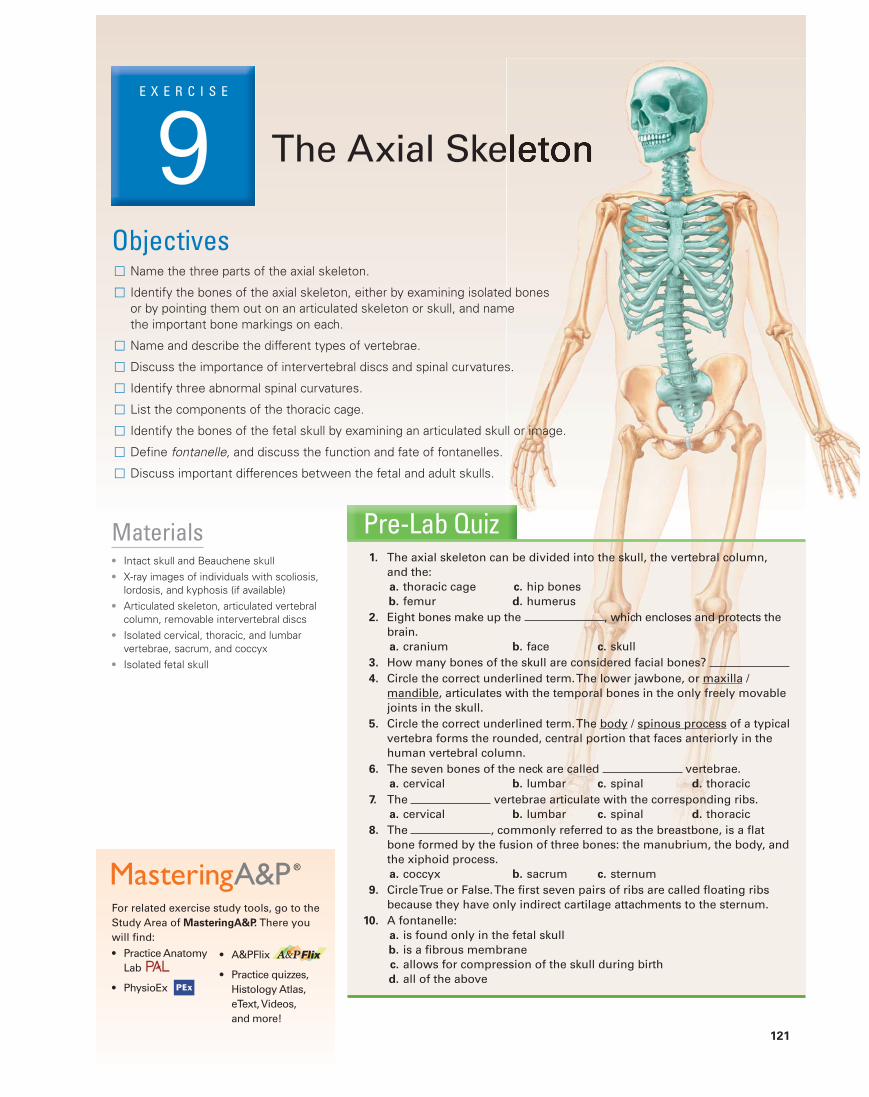
EXERCISE 9 The Axial Skeleton 121 Materials ● Intact skull and Beauchene skull ● X-ray images of individuals with scoliosis, lordosis, and kyphosis (if available) ● Articulated skeleton, articulated vertebral column, removable intervertebral discs ● Isolated cervical, thoracic, and lumbar vertebrae, sacrum, and coccyx ● Isolated fetal skull For related exercise study tools, go to the Study Area of MasteringA&P. There you will find: ● Practice Anatomy Lab ● PhysioEx ● A&PFlix ● Practice quizzes, Histology Atlas, eText, Videos, and more! Pre-Lab Quiz 1. The axial skeleton can be divided into the skull, the vertebral column, and the: a. thoracic cage c. hip bones b. femur d. humerus 2. Eight bones make up the , which encloses and protects the brain. a. cranium b. face c. skull 3. How many bones of the skull are considered facial bones? 4. Circle the correct underlined term. The lower jawbone, or maxilla / mandible, articulates with the temporal bones in the only freely movable joints in the skull. 5. Circle the correct underlined term. The body / spinous process of a typical vertebra forms the rounded, central portion that faces anteriorly in the human vertebral column. 6. The seven bones of the neck are called vertebrae. a. cervical b. lumbar c. spinal d. thoracic 7. The vertebrae articulate with the corresponding ribs. a. cervical b. lumbar c. spinal d. thoracic 8. The , commonly referred to as the breastbone, is a flat bone formed by the fusion of three bones: the manubrium, the body, and the xiphoid process. a. coccyx b. sacrum c. sternum 9. Circle True or False. The first seven pairs of ribs are called floating ribs because they have only indirect cartilage attachments to the sternum. 10. A fontanelle: a. is found only in the fetal skull b. is a fibrous membrane c. allows for compression of the skull during birth d. all of the above Objectives □ Name the three parts of the axial skeleton. □ Identify the bones of the axial skeleton, either by examining isolated bones or by pointing them out on an articulated skeleton or skull, and name the important bone markings on each. □ Name and describe the different types of vertebrae. □ Discuss the importance of intervertebral discs and spinal curvatures. □ Identify three abnormal spinal curvatures. □ List the components of the thoracic cage. □ Identify the bones of the fetal skull by examining an articulated skull or image. □ Define fontanelle, and discuss the function and fate of fontanelles. □ Discuss important differences between the fetal and adult skulls.
Transcript of EXERCISE 9 The Axial Skeleton - Pearson · EXERCISE9 The Axial Skeleton 121 ... Name the three...
E X E R C I S E
9 The Axial Skeleton
121
Materials● Intact skull and Beauchene skull● X-ray images of individuals with scoliosis,
lordosis, and kyphosis (if available)● Articulated skeleton, articulated vertebral
column, removable intervertebral discs● Isolated cervical, thoracic, and lumbar
vertebrae, sacrum, and coccyx● Isolated fetal skull
For related exercise study tools, go to the
Study Area of MasteringA&P. There you
will find:● Practice Anatomy
Lab
● PhysioEx
● A&PFlix
● Practice quizzes,
Histology Atlas,
eText, Videos,
and more!
Pre-Lab Quiz1. The axial skeleton can be divided into the skull, the vertebral column,
and the:a. thoracic cage c. hip bones
b. femur d. humerus
2. Eight bones make up the , which encloses and protects the brain.
a. cranium b. face c. skull
3. How many bones of the skull are considered facial bones?
4. Circle the correct underlined term. The lower jawbone, or maxilla / mandible, articulates with the temporal bones in the only freely movable joints in the skull.
5. Circle the correct underlined term. The body / spinous process of a typical vertebra forms the rounded, central portion that faces anteriorly in the human vertebral column.
6. The seven bones of the neck are called vertebrae. a. cervical b. lumbar c. spinal d. thoracic
7. The vertebrae articulate with the corresponding ribs. a. cervical b. lumbar c. spinal d. thoracic
8. The , commonly referred to as the breastbone, is a flat bone formed by the fusion of three bones: the manubrium, the body, and the xiphoid process.
a. coccyx b. sacrum c. sternum
9. Circle True or False. The first seven pairs of ribs are called floating ribs because they have only indirect cartilage attachments to the sternum.
10. A fontanelle:a. is found only in the fetal skullb. is a fibrous membranec. allows for compression of the skull during birthd. all of the above
Objectives□ Name the three parts of the axial skeleton.
□ Identify the bones of the axial skeleton, either by examining isolated bones or by pointing them out on an articulated skeleton or skull, and name the important bone markings on each.
□ Name and describe the different types of vertebrae.
□ Discuss the importance of intervertebral discs and spinal curvatures.
□ Identify three abnormal spinal curvatures.
□ List the components of the thoracic cage.
□ Identify the bones of the fetal skull by examining an articulated skull or image.
□ Define fontanelle, and discuss the function and fate of fontanelles.
□ Discuss important differences between the fetal and adult skulls.
122 Exercise 9
9
The axial skeleton (the green portion of Figure 8.1 on p. 108) can be divided into three parts: the skull, the ver-tebral column, and the thoracic cage. This division of the
skeleton forms the longitudinal axis of the body and protects the brain, spinal cord, heart, and lungs.
The Skull
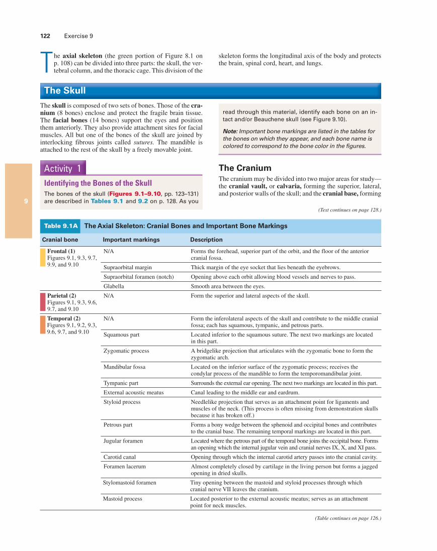
The skull is composed of two sets of bones. Those of the cra-nium (8 bones) enclose and protect the fragile brain tissue. The facial bones (14 bones) support the eyes and position them anteriorly. They also provide attachment sites for facial muscles. All but one of the bones of the skull are joined by interlocking fibrous joints called sutures. The mandible is attached to the rest of the skull by a freely movable joint.
Activity 1Identifying the Bones of the SkullThe bones of the skull (Figures 9.1–9.10, pp. 123–131) are described in Tables 9.1 and 9.2 on p. 128. As you
read through this material, identify each bone on an in-tact and/or Beauchene skull (see Figure 9.10).
Note: Important bone markings are listed in the tables for the bones on which they appear, and each bone name is colored to correspond to the bone color in the figures.
The CraniumThe cranium may be divided into two major areas for study—the cranial vault, or calvaria, forming the superior, lateral, and posterior walls of the skull; and the cranial base, forming
(Text continues on page 128.)
(Table continues on page 126.)
The Axial Skeleton: Cranial Bones and Important Bone MarkingsTable 9.1A
Cranial bone Important markings Description
Frontal (1) Figures 9.1, 9.3, 9.7, 9.9, and 9.10
N/A Forms the forehead, superior part of the orbit, and the floor of the anterior cranial fossa.
Supraorbital margin Thick margin of the eye socket that lies beneath the eyebrows.
Supraorbital foramen (notch) Opening above each orbit allowing blood vessels and nerves to pass.
Glabella Smooth area between the eyes.
Parietal (2) Figures 9.1, 9.3, 9.6, 9.7, and 9.10
N/A Form the superior and lateral aspects of the skull.
Temporal (2) Figures 9.1, 9.2, 9.3, 9.6, 9.7, and 9.10
N/A Form the inferolateral aspects of the skull and contribute to the middle cranial fossa; each has squamous, tympanic, and petrous parts.
Squamous part Located inferior to the squamous suture. The next two markings are located in this part.
Zygomatic process A bridgelike projection that articulates with the zygomatic bone to form the zygomatic arch.
Mandibular fossa Located on the inferior surface of the zygomatic process; receives the condylar process of the mandible to form the temporomandibular joint.
Tympanic part Surrounds the external ear opening. The next two markings are located in this part.
External acoustic meatus Canal leading to the middle ear and eardrum.
Styloid process Needlelike projection that serves as an attachment point for ligaments and muscles of the neck. (This process is often missing from demonstration skulls because it has broken off.)
Petrous part Forms a bony wedge between the sphenoid and occipital bones and contributes to the cranial base. The remaining temporal markings are located in this part.
Jugular foramen Located where the petrous part of the temporal bone joins the occipital bone. Forms an opening which the internal jugular vein and cranial nerves IX, X, and XI pass.
Carotid canal Opening through which the internal carotid artery passes into the cranial cavity.
Foramen lacerum Almost completely closed by cartilage in the living person but forms a jagged opening in dried skulls.
Stylomastoid foramen Tiny opening between the mastoid and styloid processes through which cranial nerve VII leaves the cranium.
Mastoid process Located posterior to the external acoustic meatus; serves as an attachment point for neck muscles.
The Axial Skeleton 123
9
Coronal suture Frontal bone
Sphenoid bone(greater wing)
Ethmoid bone
Lacrimal bone
Lacrimal fossa
Nasal bone
Zygomatic bone
Maxilla
Alveolar processes
Mandible
Mental foramen
Parietal bone
Lambdoidsuture
Squamoussuture
Occipitalbone
Occipitomastoidsuture
External acousticmeatus
Mastoid process
Styloid process
Condylar process
Mandibular notch
Mandibular ramus
Mandibular angle Coronoid process
Zygomaticprocess
Temporal bone
(a)
Condylarprocess
Frontal bone
Sphenoid bone(greater wing)
Ethmoid bone
Lacrimal bone
Lacrimal fossa
Nasal bone
Zygomatic bone
Maxilla
Alveolarprocesses
Mandible
Mental foramen
Mandibular notch
Mandibular ramus
Coronoid process
Coronal suture
Parietal bone
Lambdoidsuture
Squamoussuture
Occipitalbone
Occipitomastoidsuture
External acousticmeatus
Mastoid process
Styloid process
Mandibular angle
Zygomaticprocess
Temporal bone
(b)
Figure 9.1 External anatomy of the right lateral aspect of the skull. (a) Diagram. (b) Photograph.
Watch videos of the Cranium and Temporal Bone
>Study Area>Pre-Lab Videos
124 Exercise 9
9
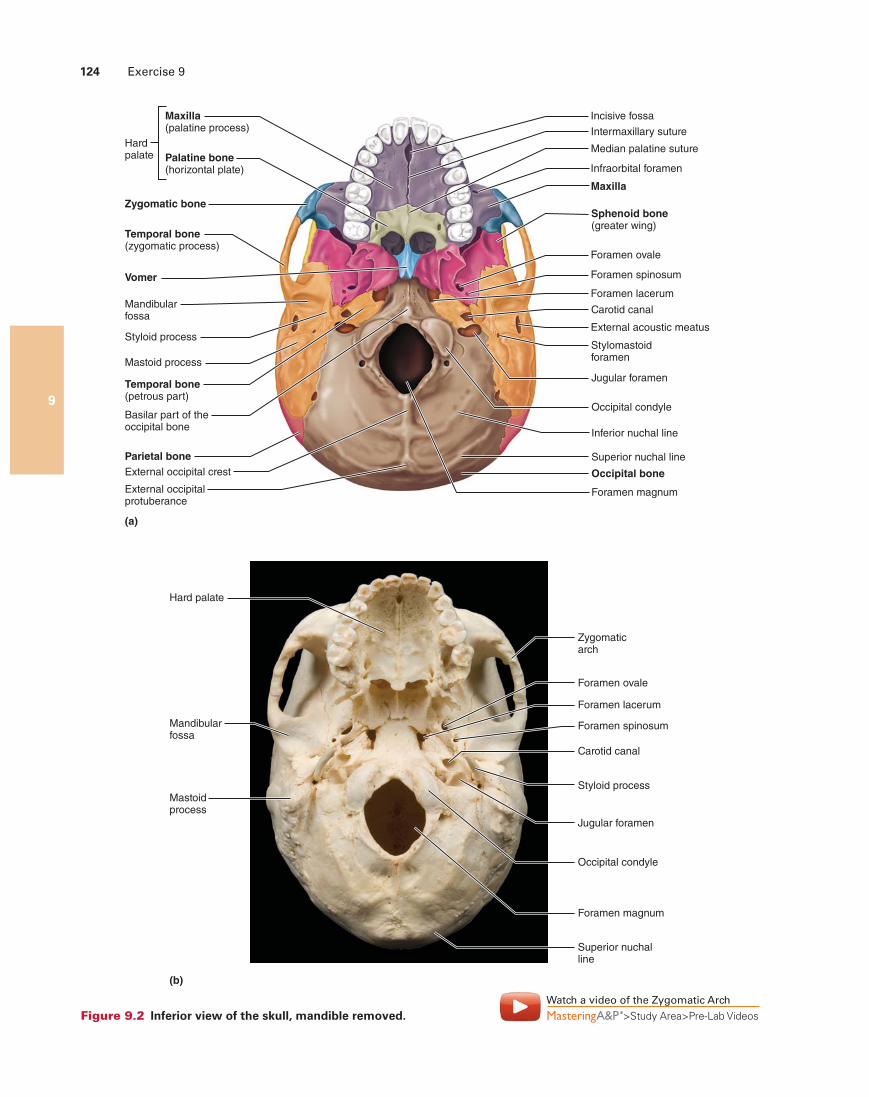
Maxilla(palatine process)
Hardpalate
Zygomatic bone
Incisive fossa
Median palatine suture
Intermaxillary suture
Infraorbital foramen
Maxilla
Sphenoid bone(greater wing)
Foramen ovale
Foramen lacerum
Carotid canal
External acoustic meatus
Stylomastoidforamen
Jugular foramen
Foramen magnum
Occipital condyle
Inferior nuchal line
Superior nuchal line
Occipital bone
Temporal bone(zygomatic process)
Mandibularfossa
Vomer
Styloid process
External occipital crest
External occipitalprotuberance
Mastoid process
Temporal bone(petrous part)
Basilar part of theoccipital bone
Parietal bone
Palatine bone(horizontal plate)
Foramen spinosum
(a)
Hard palate
Mandibularfossa
Mastoidprocess
Zygomaticarch
Foramen ovale
Foramen lacerum
Carotid canal
Styloid process
Jugular foramen
Occipital condyle
Foramen magnum
Superior nuchalline
(b)
Foramen spinosum
Figure 9.2 Inferior view of the skull, mandible removed.Watch a video of the Zygomatic Arch
>Study Area>Pre-Lab Videos
The Axial Skeleton 125
9
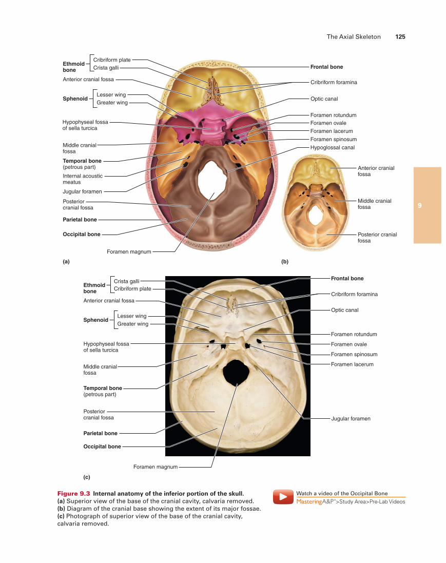
Frontal bone
Cribriform foramina
Lesser wingSphenoid
Anterior cranial fossa
Hypophyseal fossaof sella turcica
Middle cranialfossa
Temporal bone(petrous part)
Posteriorcranial fossa
Jugular foramen
Internal acousticmeatus
Parietal bone
Occipital bone
Foramen magnum
Greater wing
Cribriform plateEthmoidbone Crista galli
Optic canal
Foramen rotundum
Foramen ovale
Foramen spinosum
Hypoglossal canal
Foramen lacerum
(a)
Anterior cranialfossa
Middle cranialfossa
Posterior cranialfossa
(b)
Frontal bone
Cribriform foramina
Lesser wingSphenoid
Anterior cranial fossa
Middle cranialfossa
Temporal bone(petrous part)
Posteriorcranial fossa
Parietal bone
Occipital bone
Foramen magnum
(c)
Greater wing
Cribriform plateEthmoidbone
Crista galli
Optic canal
Foramen rotundum
Foramen ovale
Foramen spinosum
Jugular foramen
Foramen lacerum
Hypophyseal fossaof sella turcica
Figure 9.3 Internal anatomy of the inferior portion of the skull. (a) Superior view of the base of the cranial cavity, calvaria removed. (b) Diagram of the cranial base showing the extent of its major fossae. (c) Photograph of superior view of the base of the cranial cavity, calvaria removed.
Watch a video of the Occipital Bone
>Study Area>Pre-Lab Videos
126 Exercise 9
9
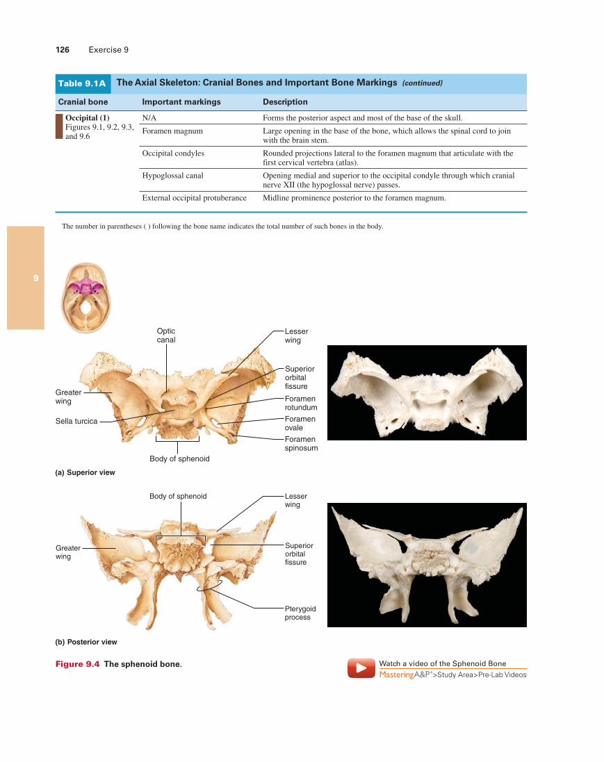
Opticcanal
Greaterwing
Sella turcica
Lesserwing
Foramenrotundum
Foramenovale
Foramenspinosum
Body of sphenoid
Superiororbitalfissure
(a) Superior view
Body of sphenoid
Greaterwing
Superior orbitalfissure
Lesser wing
Pterygoidprocess
(b) Posterior view
Figure 9.4 The sphenoid bone. Watch a video of the Sphenoid Bone
>Study Area>Pre-Lab Videos
Cranial bone Important markings Description
Occipital (1) Figures 9.1, 9.2, 9.3, and 9.6
N/A Forms the posterior aspect and most of the base of the skull.
Foramen magnum Large opening in the base of the bone, which allows the spinal cord to join with the brain stem.
Occipital condyles Rounded projections lateral to the foramen magnum that articulate with the first cervical vertebra (atlas).
Hypoglossal canal Opening medial and superior to the occipital condyle through which cranial nerve XII (the hypoglossal nerve) passes.
External occipital protuberance Midline prominence posterior to the foramen magnum.
The number in parentheses ( ) following the bone name indicates the total number of such bones in the body.
The Axial Skeleton: Cranial Bones and Important Bone Markings (continued)Table 9.1A
The Axial Skeleton 127
9
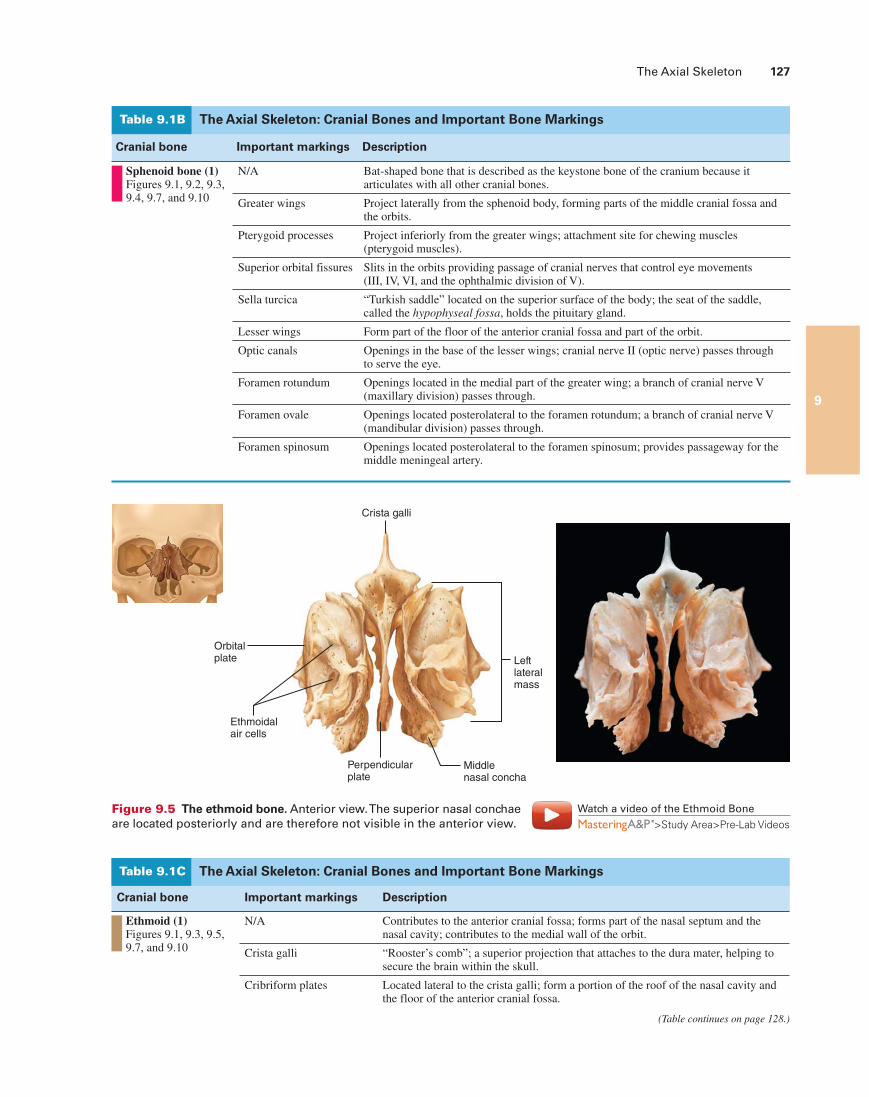
Orbitalplate
Ethmoidal air cells
Perpendicularplate
Middlenasal concha
Crista galli
Leftlateralmass
Figure 9.5 The ethmoid bone. Anterior view. The superior nasal conchae are located posteriorly and are therefore not visible in the anterior view.
The Axial Skeleton: Cranial Bones and Important Bone MarkingsTable 9.1C
Cranial bone Important markings Description
Ethmoid (1) Figures 9.1, 9.3, 9.5, 9.7, and 9.10
N/A Contributes to the anterior cranial fossa; forms part of the nasal septum and the nasal cavity; contributes to the medial wall of the orbit.
Crista galli “Rooster’s comb”; a superior projection that attaches to the dura mater, helping to secure the brain within the skull.
Cribriform plates Located lateral to the crista galli; form a portion of the roof of the nasal cavity and the floor of the anterior cranial fossa.
(Table continues on page 128.)
Watch a video of the Ethmoid Bone
>Study Area>Pre-Lab Videos
The Axial Skeleton: Cranial Bones and Important Bone MarkingsTable 9.1B
Cranial bone Important markings Description
Sphenoid bone (1) Figures 9.1, 9.2, 9.3, 9.4, 9.7, and 9.10
N/A Bat-shaped bone that is described as the keystone bone of the cranium because it articulates with all other cranial bones.
Greater wings Project laterally from the sphenoid body, forming parts of the middle cranial fossa and the orbits.
Pterygoid processes Project inferiorly from the greater wings; attachment site for chewing muscles (pterygoid muscles).
Superior orbital fissures Slits in the orbits providing passage of cranial nerves that control eye movements (III, IV, VI, and the ophthalmic division of V).
Sella turcica “Turkish saddle” located on the superior surface of the body; the seat of the saddle, called the hypophyseal fossa, holds the pituitary gland.
Lesser wings Form part of the floor of the anterior cranial fossa and part of the orbit.
Optic canals Openings in the base of the lesser wings; cranial nerve II (optic nerve) passes through to serve the eye.
Foramen rotundum Openings located in the medial part of the greater wing; a branch of cranial nerve V (maxillary division) passes through.
Foramen ovale Openings located posterolateral to the foramen rotundum; a branch of cranial nerve V (mandibular division) passes through.
Foramen spinosum Openings located posterolateral to the foramen spinosum; provides passageway for the middle meningeal artery.
9
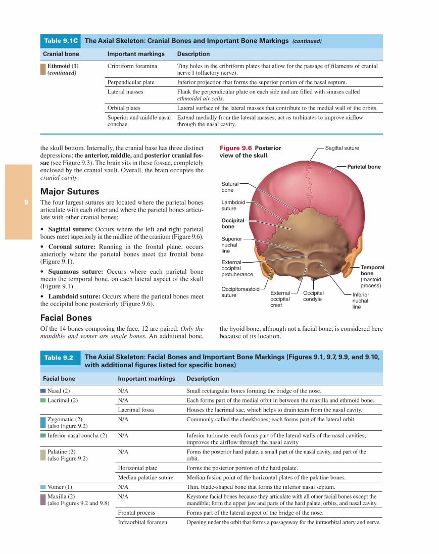
the skull bottom. Internally, the cranial base has three distinct depressions: the anterior, middle, and posterior cranial fos-sae (see Figure 9.3). The brain sits in these fossae, completely enclosed by the cranial vault. Overall, the brain occupies the cranial cavity.
Major SuturesThe four largest sutures are located where the parietal bones articulate with each other and where the parietal bones articu-late with other cranial bones:
Sagittal suture: Occurs where the left and right parietal bones meet superiorly in the midline of the cranium (Figure 9.6).
Coronal suture: Running in the frontal plane, occurs anteriorly where the parietal bones meet the frontal bone (Figure 9.1).
Squamous suture: Occurs where each parietal bone meets the temporal bone, on each lateral aspect of the skull (Figure 9.1).
Lambdoid suture: Occurs where the parietal bones meet the occipital bone posteriorly (Figure 9.6).
Facial BonesOf the 14 bones composing the face, 12 are paired. Only the mandible and vomer are single bones. An additional bone,
The Axial Skeleton: Cranial Bones and Important Bone Markings (continued)Table 9.1C
Cranial bone Important markings Description
Ethmoid (1) (continued)
Cribriform foramina Tiny holes in the cribriform plates that allow for the passage of filaments of cranial nerve I (olfactory nerve).
Perpendicular plate Inferior projection that forms the superior portion of the nasal septum.
Lateral masses Flank the perpendicular plate on each side and are filled with sinuses called ethmoidal air cells.
Orbital plates Lateral surface of the lateral masses that contribute to the medial wall of the orbits.
Superior and middle nasal conchae
Extend medially from the lateral masses; act as turbinates to improve airflow through the nasal cavity.
The Axial Skeleton: Facial Bones and Important Bone Markings (Figures 9.1, 9.7, 9.9, and 9.10, with additional figures listed for specific bones)
Table 9.2
Facial bone Important markings Description
Nasal (2) N/A Small rectangular bones forming the bridge of the nose.
Lacrimal (2)
N/A Each forms part of the medial orbit in between the maxilla and ethmoid bone.
Lacrimal fossa Houses the lacrimal sac, which helps to drain tears from the nasal cavity.
Zygomatic (2) (also Figure 9.2)
N/A Commonly called the cheekbones; each forms part of the lateral orbit
Inferior nasal concha (2) N/A Inferior turbinate; each forms part of the lateral walls of the nasal cavities; improves the airflow through the nasal cavity
Palatine (2) (also Figure 9.2)
N/A Forms the posterior hard palate, a small part of the nasal cavity, and part of the orbit.
Horizontal plate Forms the posterior portion of the hard palate.
Median palatine suture Median fusion point of the horizontal plates of the palatine bones.
Vomer (1) N/A Thin, blade-shaped bone that forms the inferior nasal septum.
Maxilla (2) (also Figures 9.2 and 9.8)
N/A Keystone facial bones because they articulate with all other facial bones except the mandible; form the upper jaw and parts of the hard palate, orbits, and nasal cavity.
Frontal process Forms part of the lateral aspect of the bridge of the nose.
Infraorbital foramen Opening under the orbit that forms a passageway for the infraorbital artery and nerve.
Lambdoidsuture
Occipitalbone
Superiornuchal line
Externaloccipitalprotuberance
Suturalbone
Occipitomastoidsuture Occipital
condyleExternaloccipitalcrest
Inferiornuchalline
Temporalbone(mastoidprocess)
Parietal bone
Sagittal suture Figure 9.6 Posterior view of the skull.
the hyoid bone, although not a facial bone, is considered here because of its location.
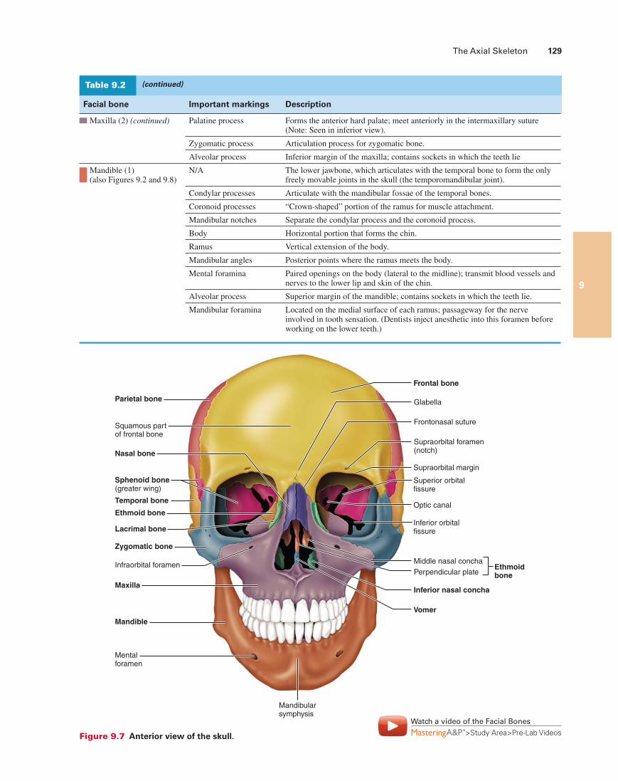
The Axial Skeleton 129
9
Parietal bone
Squamous part of frontal bone
Nasal bone
Sphenoid bone(greater wing)
Temporal bone
Ethmoid bone
Lacrimal bone
Zygomatic bone
Maxilla
Mandible
Infraorbital foramen
Mentalforamen
Mandibularsymphysis
Frontal bone
Glabella
Frontonasal suture
Supraorbital foramen(notch)
Supraorbital margin
Superior orbitalfissure
Inferior orbitalfissure
Middle nasal concha
Inferior nasal concha
Vomer
Optic canal
Perpendicular plateEthmoidbone
Figure 9.7 Anterior view of the skull.
Watch a video of the Facial Bones
>Study Area>Pre-Lab Videos
(continued)Table 9.2
Facial bone Important markings Description
Maxilla (2) (continued)
Palatine process Forms the anterior hard palate; meet anteriorly in the intermaxillary suture (Note: Seen in inferior view).
Zygomatic process Articulation process for zygomatic bone.
Alveolar process Inferior margin of the maxilla; contains sockets in which the teeth lie
Mandible (1) (also Figures 9.2 and 9.8)
N/A The lower jawbone, which articulates with the temporal bone to form the only freely movable joints in the skull (the temporomandibular joint).
Condylar processes Articulate with the mandibular fossae of the temporal bones.
Coronoid processes “Crown-shaped” portion of the ramus for muscle attachment.
Mandibular notches Separate the condylar process and the coronoid process.
Body Horizontal portion that forms the chin.
Ramus Vertical extension of the body.
Mandibular angles Posterior points where the ramus meets the body.
Mental foramina Paired openings on the body (lateral to the midline); transmit blood vessels and nerves to the lower lip and skin of the chin.
Alveolar process Superior margin of the mandible; contains sockets in which the teeth lie.
Mandibular foramina Located on the medial surface of each ramus; passageway for the nerve involved in tooth sensation. (Dentists inject anesthetic into this foramen before working on the lower teeth.)
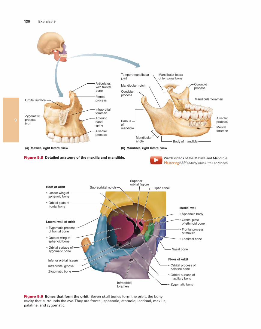
130 Exercise 9
9
Coronoidprocess
Mandibular foramen
Mentalforamen
Mandibularangle
Ramusofmandible
Condylarprocess
Mandibular notch
Mandibular fossaof temporal bone
Body of mandible
Alveolarprocess
(b) Mandible, right lateral view
Temporomandibularjoint
Frontalprocess
Articulateswith frontalbone
Anteriornasalspine
Infraorbitalforamen
Alveolarprocess
(a) Maxilla, right lateral view
Orbital surface
Zygomaticprocess(cut)
Figure 9.8 Detailed anatomy of the maxilla and mandible.
Roof of orbit
Medial wall
Orbital plateof ethmoid bone
Sphenoid body
Supraorbital notch Optic canal
Floor of orbit
Orbital process ofpalatine bone
Orbital surface ofmaxillary bone
Lacrimal bone
Nasal bone
Frontal processof maxilla
Lateral wall of orbit
Zygomatic processof frontal bone
Greater wing ofsphenoid bone
Orbital surface ofzygomatic bone
Zygomatic bone
Zygomatic bone
Inferior orbital fissure
Infraorbital groove
Infraorbitalforamen
Superiororbital fissure
Lesser wing ofsphenoid bone
Orbital plate offrontal bone
Figure 9.9 Bones that form the orbit. Seven skull bones form the orbit, the bony cavity that surrounds the eye. They are frontal, sphenoid, ethmoid, lacrimal, maxilla, palatine, and zygomatic.
Watch videos of the Maxilla and Mandible
>Study Area>Pre-Lab Videos
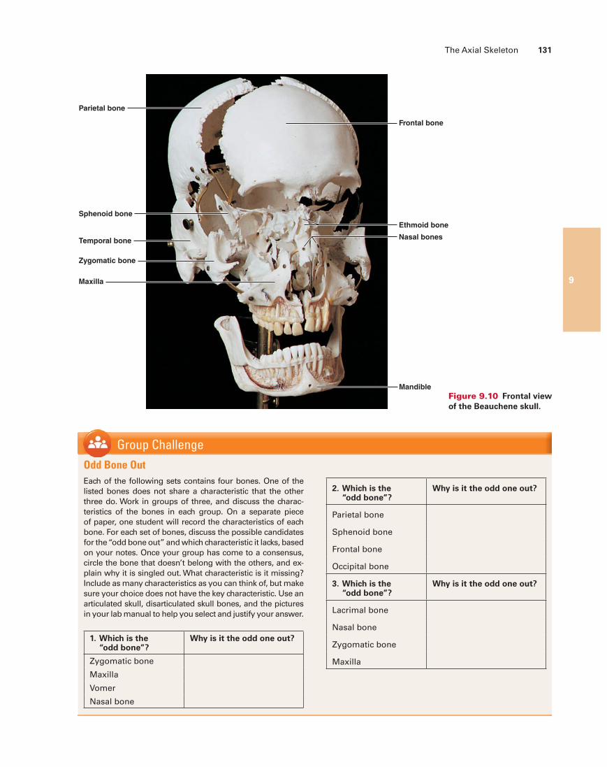
The Axial Skeleton 131
9
Parietal bone
Frontal bone
Temporal bone
Sphenoid bone
Ethmoid bone
Nasal bones
Zygomatic bone
Maxilla
Mandible
Figure 9.10 Frontal view of the Beauchene skull.
Group ChallengeOdd Bone OutEach of the following sets contains four bones. One of the listed bones does not share a characteristic that the other three do. Work in groups of three, and discuss the charac-teristics of the bones in each group. On a separate piece of paper, one student will record the characteristics of each bone. For each set of bones, discuss the possible candidates for the “odd bone out” and which characteristic it lacks, based on your notes. Once your group has come to a consensus, circle the bone that doesn’t belong with the others, and ex-plain why it is singled out. What characteristic is it missing? Include as many characteristics as you can think of, but make sure your choice does not have the key characteristic. Use an articulated skull, disarticulated skull bones, and the pictures in your lab manual to help you select and justify your answer.
2. Which is the “odd bone”?
Why is it the odd one out?
Parietal bone
Sphenoid bone
Frontal bone
Occipital bone
3. Which is the “odd bone”?
Why is it the odd one out?
Lacrimal bone
Nasal bone
Zygomatic bone
Maxilla
1. Which is the “odd bone”?
Why is it the odd one out?
Zygomatic bone
Maxilla
Vomer
Nasal bone
132 Exercise 9
9
Frontal sinus
Ethmoidal air cells
Maxillary sinus
Sphenoidal sinus
Frontalsinus
Ethmoidal air cells
Maxillarysinus
Sphenoidal sinus
(a)
(b)
Frontalsinus
Sphenoidalsinus
Maxillarysinus
Ethmoidalair cell
(c)
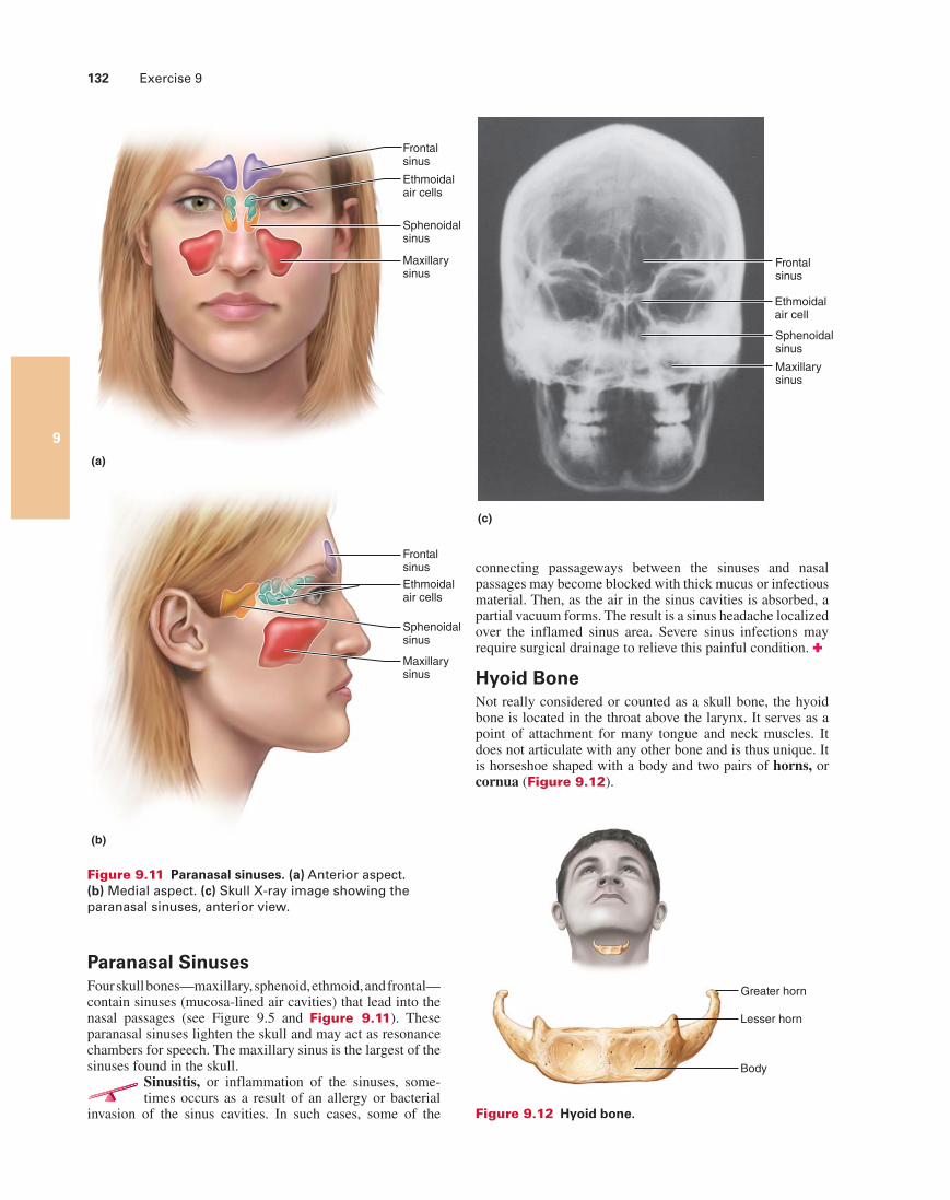
Figure 9.11 Paranasal sinuses. (a) Anterior aspect. (b) Medial aspect. (c) Skull X-ray image showing the paranasal sinuses, anterior view.
Paranasal SinusesFour skull bones—maxillary, sphenoid, ethmoid, and frontal— contain sinuses (mucosa-lined air cavities) that lead into the nasal passages (see Figure 9.5 and Figure 9.11). These paranasal sinuses lighten the skull and may act as resonance chambers for speech. The maxillary sinus is the largest of the sinuses found in the skull.
Sinusitis, or inflammation of the sinuses, some-times occurs as a result of an allergy or bacterial
invasion of the sinus cavities. In such cases, some of the
Greater horn
Lesser horn
Body
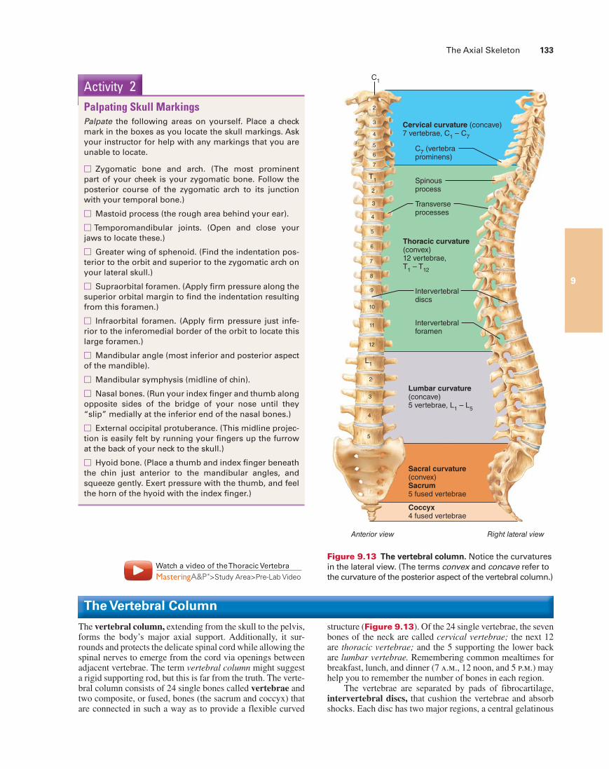
Figure 9.12 Hyoid bone.
connecting passageways between the sinuses and nasal passages may become blocked with thick mucus or infectious material. Then, as the air in the sinus cavities is absorbed, a partial vacuum forms. The result is a sinus headache localized over the inflamed sinus area. Severe sinus infections may require surgical drainage to relieve this painful condition. ✚
Hyoid BoneNot really considered or counted as a skull bone, the hyoid bone is located in the throat above the larynx. It serves as a point of attachment for many tongue and neck muscles. It does not articulate with any other bone and is thus unique. It is horseshoe shaped with a body and two pairs of horns, or cornua (Figure 9.12).
The Axial Skeleton 133
9
The Vertebral Column
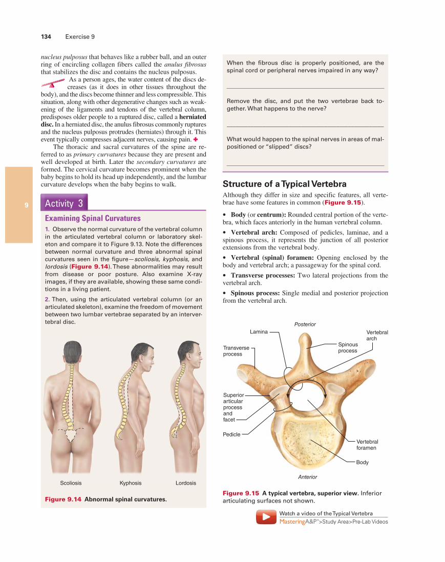
The vertebral column, extending from the skull to the pelvis, forms the body’s major axial support. Additionally, it sur-rounds and protects the delicate spinal cord while allowing the spinal nerves to emerge from the cord via openings between adjacent vertebrae. The term vertebral column might suggest a rigid supporting rod, but this is far from the truth. The verte-bral column consists of 24 single bones called vertebrae and two composite, or fused, bones (the sacrum and coccyx) that are connected in such a way as to provide a flexible curved
Activity 2Palpating Skull MarkingsPalpate the following areas on yourself. Place a check mark in the boxes as you locate the skull markings. Ask your instructor for help with any markings that you are unable to locate.
□ Zygomatic bone and arch. (The most prominent part of your cheek is your zygomatic bone. Follow the posterior course of the zygomatic arch to its junction with your temporal bone.)
□ Mastoid process (the rough area behind your ear).
□ Temporomandibular joints. (Open and close your jaws to locate these.)
□ Greater wing of sphenoid. (Find the indentation pos-terior to the orbit and superior to the zygomatic arch on your lateral skull.)
□ Supraorbital foramen. (Apply firm pressure along the superior orbital margin to find the indentation resulting from this foramen.)
□ Infraorbital foramen. (Apply firm pressure just infe-rior to the inferomedial border of the orbit to locate this large foramen.)
□ Mandibular angle (most inferior and posterior aspect of the mandible).
□ Mandibular symphysis (midline of chin).
□ Nasal bones. (Run your index finger and thumb along opposite sides of the bridge of your nose until they “slip” medially at the inferior end of the nasal bones.)
□ External occipital protuberance. (This midline projec-tion is easily felt by running your fingers up the furrow at the back of your neck to the skull.)
□ Hyoid bone. (Place a thumb and index finger beneath the chin just anterior to the mandibular angles, and squeeze gently. Exert pressure with the thumb, and feel the horn of the hyoid with the index finger.)
Cervical curvature (concave)7 vertebrae, C
1 – C
7
Thoracic curvature(convex)12 vertebrae,T
1 – T
12
Lumbar curvature(concave)5 vertebrae, L
1 – L
5
Sacral curvature(convex)Sacrum5 fused vertebrae
Coccyx4 fused vertebrae
Anterior view Right lateral view
C1
T1
2
3
4
5
6
7
8
9
10
11
12
L1
2
3
4
5
2
3
4
5
6
7
Spinousprocess
Transverseprocesses
Intervertebraldiscs
Intervertebralforamen
C7 (vertebra
prominens)
Figure 9.13 The vertebral column. Notice the curvatures in the lateral view. (The terms convex and concave refer to the curvature of the posterior aspect of the vertebral column.)
Watch a video of the Thoracic Vertebra
>Study Area>Pre-Lab Video
structure (Figure 9.13). Of the 24 single vertebrae, the seven bones of the neck are called cervical vertebrae; the next 12 are thoracic vertebrae; and the 5 supporting the lower back are lumbar vertebrae. Remembering common mealtimes for breakfast, lunch, and dinner (7 a.m., 12 noon, and 5 p.m.) may help you to remember the number of bones in each region.
The vertebrae are separated by pads of fibrocartilage, intervertebral discs, that cushion the vertebrae and absorb shocks. Each disc has two major regions, a central gelatinous
134 Exercise 9
9
When the fibrous disc is properly positioned, are the spinal cord or peripheral nerves impaired in any way?
Remove the disc, and put the two vertebrae back to-gether. What happens to the nerve?
_____________________________________________________
What would happen to the spinal nerves in areas of mal-positioned or “slipped” discs?
Posterior
Anterior
Lamina
Superior articularprocessandfacet
Transverseprocess
Pedicle
Spinousprocess
Vertebralarch
Vertebralforamen
Body
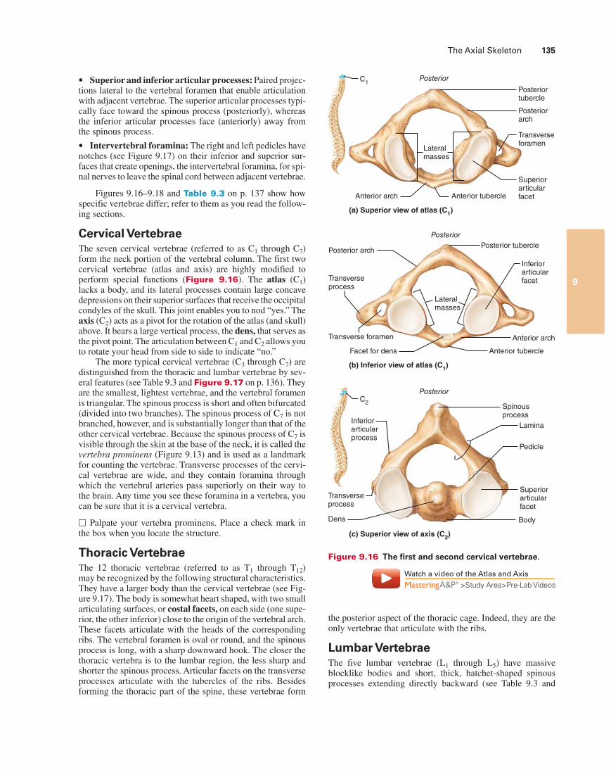
Figure 9.15 A typical vertebra, superior view. Inferior articulating surfaces not shown.
Watch a video of the Typical Vertebra
>Study Area>Pre-Lab Videos
nucleus pulposus that behaves like a rubber ball, and an outer ring of encircling collagen fibers called the anulus fibrosus that stabilizes the disc and contains the nucleus pulposus.
As a person ages, the water content of the discs de-creases (as it does in other tissues throughout the
body), and the discs become thinner and less compressible. This situation, along with other degenerative changes such as weak-ening of the ligaments and tendons of the vertebral column, predisposes older people to a ruptured disc, called a herniated disc. In a herniated disc, the anulus fibrosus commonly ruptures and the nucleus pulposus protrudes (herniates) through it. This event typically compresses adjacent nerves, causing pain. ✚
The thoracic and sacral curvatures of the spine are re-ferred to as primary curvatures because they are present and well developed at birth. Later the secondary curvatures are formed. The cervical curvature becomes prominent when the baby begins to hold its head up independently, and the lumbar curvature develops when the baby begins to walk.
Activity 3Examining Spinal Curvatures1. Observe the normal curvature of the vertebral column in the articulated vertebral column or laboratory skel-eton and compare it to Figure 9.13. Note the differences between normal curvature and three abnormal spinal curvatures seen in the figure—scoliosis, kyphosis, and lordosis (Figure 9.14). These abnormalities may result from disease or poor posture. Also examine X-ray images, if they are available, showing these same condi-tions in a living patient.
2. Then, using the articulated vertebral column (or an articulated skeleton), examine the freedom of movement between two lumbar vertebrae separated by an interver-tebral disc.
Kyphosis LordosisScoliosis
Figure 9.14 Abnormal spinal curvatures.
Structure of a Typical VertebraAlthough they differ in size and specific features, all verte-brae have some features in common (Figure 9.15).
Body (or centrum): Rounded central portion of the verte-bra, which faces anteriorly in the human vertebral column.
Vertebral arch: Composed of pedicles, laminae, and a spinous process, it represents the junction of all posterior extensions from the vertebral body.
Vertebral (spinal) foramen: Opening enclosed by the body and vertebral arch; a passageway for the spinal cord.
Transverse processes: Two lateral projections from the vertebral arch.
Spinous process: Single medial and posterior projection from the vertebral arch.
The Axial Skeleton 135
9
Superior and inferior articular processes: Paired projec-tions lateral to the vertebral foramen that enable articulation with adjacent vertebrae. The superior articular processes typi-cally face toward the spinous process (posteriorly), whereas the inferior articular processes face (anteriorly) away from the spinous process.
Intervertebral foramina: The right and left pedicles have notches (see Figure 9.17) on their inferior and superior sur-faces that create openings, the intervertebral foramina, for spi-nal nerves to leave the spinal cord between adjacent vertebrae.
Figures 9.16–9.18 and Table 9.3 on p. 137 show how specific vertebrae differ; refer to them as you read the follow-ing sections.
Cervical VertebraeThe seven cervical vertebrae (referred to as C1 through C7) form the neck portion of the vertebral column. The first two cervical vertebrae (atlas and axis) are highly modified to perform special functions (Figure 9.16). The atlas (C1) lacks a body, and its lateral processes contain large concave depressions on their superior surfaces that receive the occipital condyles of the skull. This joint enables you to nod “yes.” The axis (C2) acts as a pivot for the rotation of the atlas (and skull) above. It bears a large vertical process, the dens, that serves as the pivot point. The articulation between C1 and C2 allows you to rotate your head from side to side to indicate “no.”
The more typical cervical vertebrae (C3 through C7) are distinguished from the thoracic and lumbar vertebrae by sev-eral features (see Table 9.3 and Figure 9.17 on p. 136). They are the smallest, lightest vertebrae, and the vertebral foramen is triangular. The spinous process is short and often bifurcated (divided into two branches). The spinous process of C7 is not branched, however, and is substantially longer than that of the other cervical vertebrae. Because the spinous process of C7 is visible through the skin at the base of the neck, it is called the vertebra prominens (Figure 9.13) and is used as a landmark for counting the vertebrae. Transverse processes of the cervi-cal vertebrae are wide, and they contain foramina through which the vertebral arteries pass superiorly on their way to the brain. Any time you see these foramina in a vertebra, you can be sure that it is a cervical vertebra.
□ Palpate your vertebra prominens. Place a check mark in the box when you locate the structure.
Thoracic VertebraeThe 12 thoracic vertebrae (referred to as T1 through T12) may be recognized by the following structural characteristics. They have a larger body than the cervical vertebrae (see Fig-ure 9.17). The body is somewhat heart shaped, with two small articulating surfaces, or costal facets, on each side (one supe-rior, the other inferior) close to the origin of the vertebral arch. These facets articulate with the heads of the corresponding ribs. The vertebral foramen is oval or round, and the spinous process is long, with a sharp downward hook. The closer the thoracic vertebra is to the lumbar region, the less sharp and shorter the spinous process. Articular facets on the transverse processes articulate with the tubercles of the ribs. Besides forming the thoracic part of the spine, these vertebrae form
Figure 9.16 The first and second cervical vertebrae.
Watch a video of the Atlas and Axis
>Study Area>Pre-Lab Videos
Anterior arch
Superiorarticularfacet
Transverseforamen
Posteriorarch
Posteriortubercle
Anterior tubercle
Posterior
Lateralmasses
(a) Superior view of atlas (C1)
C1
C2
Facet for dens
Transverseprocess
Lateralmasses
Transverse foramen
Posterior archPosterior tubercle
Posterior
Anterior tubercle
Anterior arch
(b) Inferior view of atlas (C1)
Inferiorarticularfacet
Posterior
Dens
(c) Superior view of axis (C2)
Inferiorarticularprocess
Body
Superiorarticularfacet
Transverseprocess
Pedicle
Lamina
Spinousprocess
the posterior aspect of the thoracic cage. Indeed, they are the only vertebrae that articulate with the ribs.
Lumbar VertebraeThe five lumbar vertebrae (L1 through L5) have massive blocklike bodies and short, thick, hatchet-shaped spinous processes extending directly backward (see Table 9.3 and

136 Exercise 9
9
Inferiorarticularprocess
Superior articularprocess and facet Spinous
process
Vertebralforamen
Transverseprocess
Transverseforamen Body
Superior View Right Lateral View
Transverseprocess
Superiorarticularprocessand facet
Vertebralforamen
Body
Transversecostal facet (for tubercleof rib)
Superiorcostal facet (for head of rib)
Spinousprocess
Body
Inferiorarticularprocess and facet
Transverseprocess
Superior articularprocess
Body
Superior articularprocess and facet
Transversecostal facet(for tubercleof rib)
Superior costalfacet (for headof rib)
Inferiornotch
Inferior costalfacet (for headof rib)
Spinousprocess
Transverseprocess
Spinous process
Spinous process
Transverseprocess
Superiorarticularprocessand facet
Vertebralforamen
Body
Superiorarticularprocess
Inferior articular process and facet
Inferior vertebralnotch
Transverseprocess
Body
Spinousprocess
C1
C2
(c) Lumbar
(b) Thoracic
(a) Cervical
Figure 9.17 Superior and right lateral views of typical vertebrae.
Figure 9.17). The superior articular facets face posterome-dially; the inferior ones are directed anterolaterally. These structural features reduce the mobility of the lumbar region of the spine. Since most stress on the vertebral column occurs in the lumbar region, these are also the sturdiest of the vertebrae.
The spinal cord ends at the superior edge of L2, but the outer covering of the cord, filled with cerebrospinal fluid,
extends an appreciable distance beyond. Thus a lumbar punc-ture (for examination of the cerebrospinal fluid) or the ad-ministration of “saddle block” anesthesia for childbirth is normally done between L3 and L4 or L4 and L5, where there is little or no chance of injuring the delicate spinal cord.
Watch a video of the Lumbar Vertebra
>Study Area>Pre-Lab Videos
The Axial Skeleton 137
9
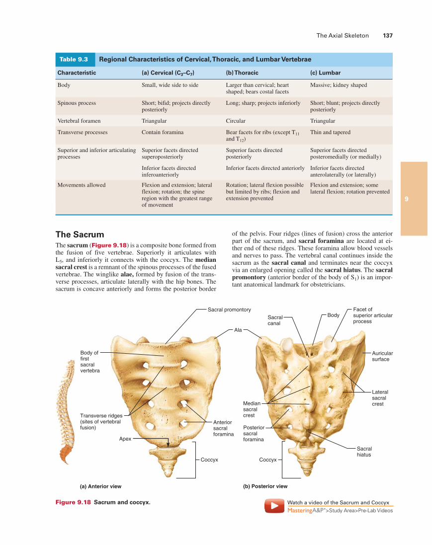
The SacrumThe sacrum (Figure 9.18) is a composite bone formed from the fusion of five vertebrae. Superiorly it articulates with L5, and inferiorly it connects with the coccyx. The median sacral crest is a remnant of the spinous processes of the fused vertebrae. The winglike alae, formed by fusion of the trans-verse processes, articulate laterally with the hip bones. The sacrum is concave anteriorly and forms the posterior border
of the pelvis. Four ridges (lines of fusion) cross the anterior part of the sacrum, and sacral foramina are located at ei-ther end of these ridges. These foramina allow blood vessels and nerves to pass. The vertebral canal continues inside the sacrum as the sacral canal and terminates near the coccyx via an enlarged opening called the sacral hiatus. The sacral promontory (anterior border of the body of S1) is an impor-tant anatomical landmark for obstetricians.
Body offirstsacralvertebra
Transverse ridges(sites of vertebralfusion)
Coccyx Coccyx
Anteriorsacralforamina
Apex
Posteriorsacralforamina
Mediansacralcrest
Sacral promontory
Sacralcanal
Sacralhiatus
BodyFacet ofsuperior articularprocess
Lateralsacralcrest
Auricularsurface
Ala
(a) Anterior view (b) Posterior view
Figure 9.18 Sacrum and coccyx. Watch a video of the Sacrum and Coccyx
>Study Area>Pre-Lab Videos
Regional Characteristics of Cervical, Thoracic, and Lumbar VertebraeTable 9.3
Characteristic (a) Cervical (C3–C7) (b) Thoracic (c) Lumbar
Body Small, wide side to side Larger than cervical; heart shaped; bears costal facets
Massive; kidney shaped
Spinous process Short; bifid; projects directly posteriorly
Long; sharp; projects inferiorly Short; blunt; projects directly posteriorly
Vertebral foramen Triangular Circular Triangular
Transverse processes Contain foramina Bear facets for ribs (except T11 and T12)
Thin and tapered
Superior and inferior articulating processes
Superior facets directed superoposteriorly
Superior facets directed posteriorly
Superior facets directed posteromedially (or medially)
Inferior facets directed inferoanteriorly
Inferior facets directed anteriorly Inferior facets directed anterolaterally (or laterally)
Movements allowed Flexion and extension; lateral flexion; rotation; the spine region with the greatest range of movement
Rotation; lateral flexion possible but limited by ribs; flexion and extension prevented
Flexion and extension; some lateral flexion; rotation prevented
138 Exercise 9
9
Intercostalspaces
Xiphisternaljoint
Heart
Sternalangle
Jugularnotch
True ribs(1–7)
Falseribs(8–12)
Jugular notch
Clavicular notch
Manubrium
Sternal angle
Body
Xiphisternaljoint
Xiphoidprocess
L1
VertebraFloatingribs (11, 12)
(b) (a)
T2
T4
T5
T3
T9
Sternum
Costal cartilage
Costal margin
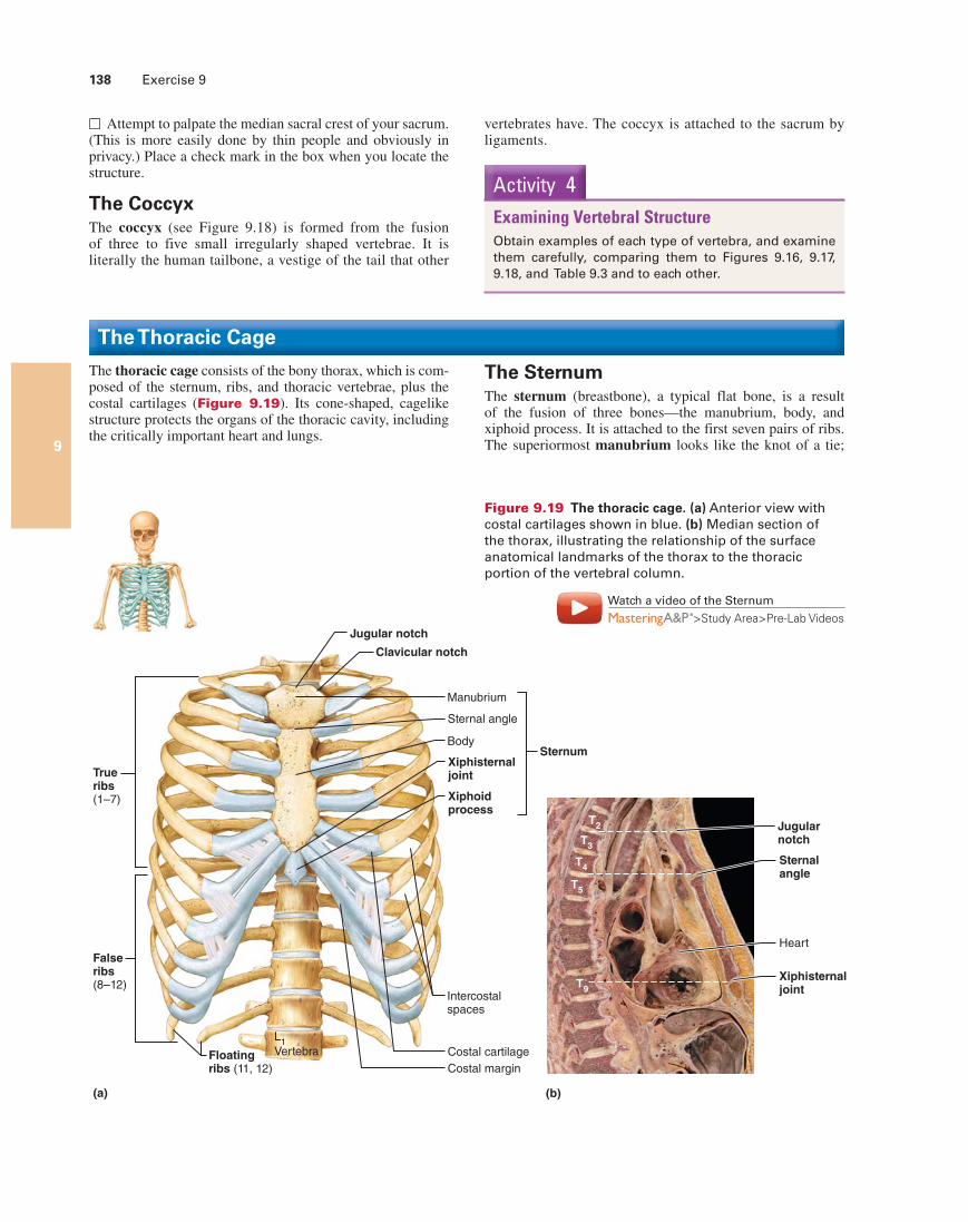
Figure 9.19 The thoracic cage. (a) Anterior view with costal cartilages shown in blue. (b) Median section of the thorax, illustrating the relationship of the surface anatomical landmarks of the thorax to the thoracic portion of the vertebral column.
Watch a video of the Sternum
>Study Area>Pre-Lab Videos
□ Attempt to palpate the median sacral crest of your sacrum. (This is more easily done by thin people and obviously in privacy.) Place a check mark in the box when you locate the structure.
The CoccyxThe coccyx (see Figure 9.18) is formed from the fusion of three to five small irregularly shaped vertebrae. It is literally the human tailbone, a vestige of the tail that other
Activity 4Examining Vertebral StructureObtain examples of each type of vertebra, and examine them carefully, comparing them to Figures 9.16, 9.17, 9.18, and Table 9.3 and to each other.
The Thoracic Cage
The thoracic cage consists of the bony thorax, which is com-posed of the sternum, ribs, and thoracic vertebrae, plus the costal cartilages (Figure 9.19). Its cone-shaped, cagelike structure protects the organs of the thoracic cavity, including the critically important heart and lungs.
The SternumThe sternum (breastbone), a typical flat bone, is a result of the fusion of three bones—the manubrium, body, and xiphoid process. It is attached to the first seven pairs of ribs. The superiormost manubrium looks like the knot of a tie;
vertebrates have. The coccyx is attached to the sacrum by ligaments.
The Axial Skeleton 139
9
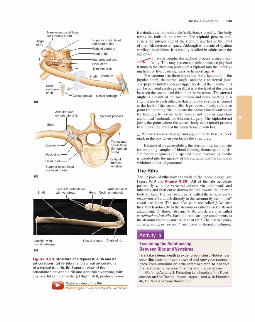
it articulates with the clavicle (collarbone) laterally. The body forms the bulk of the sternum. The xiphoid process con-structs the inferior end of the sternum and lies at the level of the fifth intercostal space. Although it is made of hyaline cartilage in children, it is usually ossified in adults over the age of 40.
In some people, the xiphoid process projects dor-sally. This may present a problem because physical
trauma to the chest can push such a xiphoid into the underly-ing heart or liver, causing massive hemorrhage. ✚
The sternum has three important bony landmarks—the jugular notch, the sternal angle, and the xiphisternal joint. The jugular notch (concave upper border of the manubrium) can be palpated easily; generally it is at the level of the disc in between the second and third thoracic vertebrae. The sternal angle is a result of the manubrium and body meeting at a slight angle to each other, so that a transverse ridge is formed at the level of the second ribs. It provides a handy reference point for counting ribs to locate the second intercostal space for listening to certain heart valves, and it is an important anatomical landmark for thoracic surgery. The xiphisternal joint, the point where the sternal body and xiphoid process fuse, lies at the level of the ninth thoracic vertebra.
□ Palpate your sternal angle and jugular notch. Place a check mark in the box when you locate the structures.
Because of its accessibility, the sternum is a favored site for obtaining samples of blood-forming (hematopoietic) tis-sue for the diagnosis of suspected blood diseases. A needle is inserted into the marrow of the sternum, and the sample is withdrawn (sternal puncture).
The RibsThe 12 pairs of ribs form the walls of the thoracic cage (see Figure 9.19 and Figure 9.20). All of the ribs articulate posteriorly with the vertebral column via their heads and tubercles and then curve downward and toward the anterior body surface. The first seven pairs, called the true, or verte-brosternal, ribs, attach directly to the sternum by their “own” costal cartilages. The next five pairs are called false ribs; they attach indirectly to the sternum or entirely lack a sternal attachment. Of these, rib pairs 8–10, which are also called vertebrochondral ribs, have indirect cartilage attachments to the sternum via the costal cartilage of rib 7. The last two pairs, called floating, or vertebral, ribs, have no sternal attachment.
Transverse costal facet (for tubercle of rib)
Superior costal facet (for head of rib)
Body of vertebra
Head of rib
Intervertebral disc
Tubercle of rib
Neck of rib
Shaft Sternum
Angleof rib
Crosssectionof rib
Costal groove Costal cartilage
(a)
Spinous process
Articular faceton tubercle of rib
Shaft
Ligaments
Neck of rib
Head of ribBody ofthoracicvertebra
Transversecostal facet(for tubercleof rib)
Superior costal facet(for head of rib)
(b)
Junction withcostal cartilage
Shaft Head NeckArticular faceton tubercle
Angle of ribCostal groove
Facets for articulationwith vertebrae
(c)
Figure 9.20 Structure of a typical true rib and its articulations. (a) Vertebral and sternal articulations of a typical true rib. (b) Superior view of the articulation between a rib and a thoracic vertebra, with costovertebral ligaments. (c) Right rib 6, posterior view.
Watch a video of the Rib
>Study Area>Pre-Lab Videos
Activity 5Examining the Relationship Between Ribs and VertebraeFirst take a deep breath to expand your chest. Notice how your ribs seem to move outward and how your sternum rises. Then examine an articulated skeleton to observe the relationship between the ribs and the vertebrae.
(Refer to Activity 3, Palpating Landmarks of the Trunk, section on The Thorax: Bones, steps 1 and 3, in Exercise 46, Surface Anatomy Roundup.)
140 Exercise 9
9
The Fetal Skull
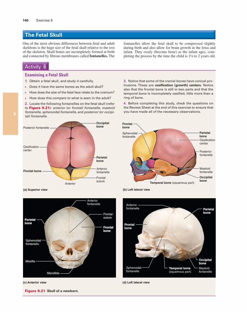
One of the most obvious differences between fetal and adult skeletons is the huge size of the fetal skull relative to the rest of the skeleton. Skull bones are incompletely formed at birth and connected by fibrous membranes called fontanelles. The
fontanelles allow the fetal skull to be compressed slightly during birth and also allow for brain growth in the fetus and infant. They ossify (become bone) as the infant ages, com-pleting the process by the time the child is 1½ to 2 years old.
1. Obtain a fetal skull, and study it carefully.
Does it have the same bones as the adult skull?
How does the size of the fetal face relate to the cranium?
How does this compare to what is seen in the adult?
2. Locate the following fontanelles on the fetal skull (refer to Figure 9.21): anterior (or frontal) fontanelle, mastoid fontanelle, sphenoidal fontanelle, and posterior (or occipi-tal) fontanelle.
Activity 6Examining a Fetal Skull
3. Notice that some of the cranial bones have conical pro-trusions. These are ossification (growth) centers. Notice also that the frontal bone is still in two parts and that the temporal bone is incompletely ossified, little more than a ring of bone.
4. Before completing this study, check the questions on the Review Sheet at the end of this exercise to ensure that you have made all of the necessary observations.
Mandible
Maxilla
Sphenoidalfontanel
Frontalsuture
Frontalbone
Frontal bone
Ossificationcenter
Anterior
Occipitalbone
(a) Superior view
Posterior fontanelle
Parietalbone
Anteriorfontanelle
Frontalsuture
(b) Left lateral view
Posteriorfontanelle
Mastoidfontanelle
Parietalbone
Ossificationcenter
Occipitalbone
Temporal bone (squamous part)
Frontalbone
Sphenoidalfontanelle
Frontalbone
Anteriorfontanelle Parietal
bone
Occipitalbone
Mastoidfontanelle
Sphenoidalfontanelle
Temporal bone(squamous part)
Mandible
Maxilla
Sphenoidalfontanelle
ParietalboneParietalbone
Anteriorfontanelle
Frontalsuture
Frontalbone
(d) Left lateral view(c) Anterior view
Figure 9.21 Skull of a newborn.
141141
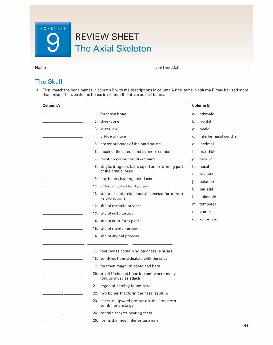
The Skull1. First, match the bone names in column B with the descriptions in column A (the items in column B may be used more
than once). Then, circle the bones in column B that are cranial bones.
Column A Column B
1. forehead bone
2. cheekbone
3. lower jaw
4. bridge of nose
5. posterior bones of the hard palate
6. much of the lateral and superior cranium
7. most posterior part of cranium
8. single, irregular, bat-shaped bone forming part of the cranial base
9. tiny bones bearing tear ducts
10. anterior part of hard palate
11. superior and middle nasal conchae form from its projections
12. site of mastoid process
13. site of sella turcica
14. site of cribriform plate
15. site of mental foramen
16. site of styloid process
, ,
17. four bones containing paranasal sinuses
18. condyles here articulate with the atlas
19. foramen magnum contained here
20. small U-shaped bone in neck, where many tongue muscles attach
21. organ of hearing found here
, 22. two bones that form the nasal septum
23. bears an upward protrusion, the “rooster’s comb,” or crista galli
, 24. contain sockets bearing teeth
25. forms the most inferior turbinate
a. ethmoid
b. frontal
c. hyoid
d. inferior nasal concha
e. lacrimal
f. mandible
g. maxilla
h. nasal
i. occipital
j. palatine
k. parietal
l. sphenoid
m. temporal
n. vomer
o. zygomatic
Name _____________________________________________________________ Lab Time/Date ___________________________________
REVIEW SHEETThe Axial Skeleton
E X E R C I S E
9
142 Review Sheet 9
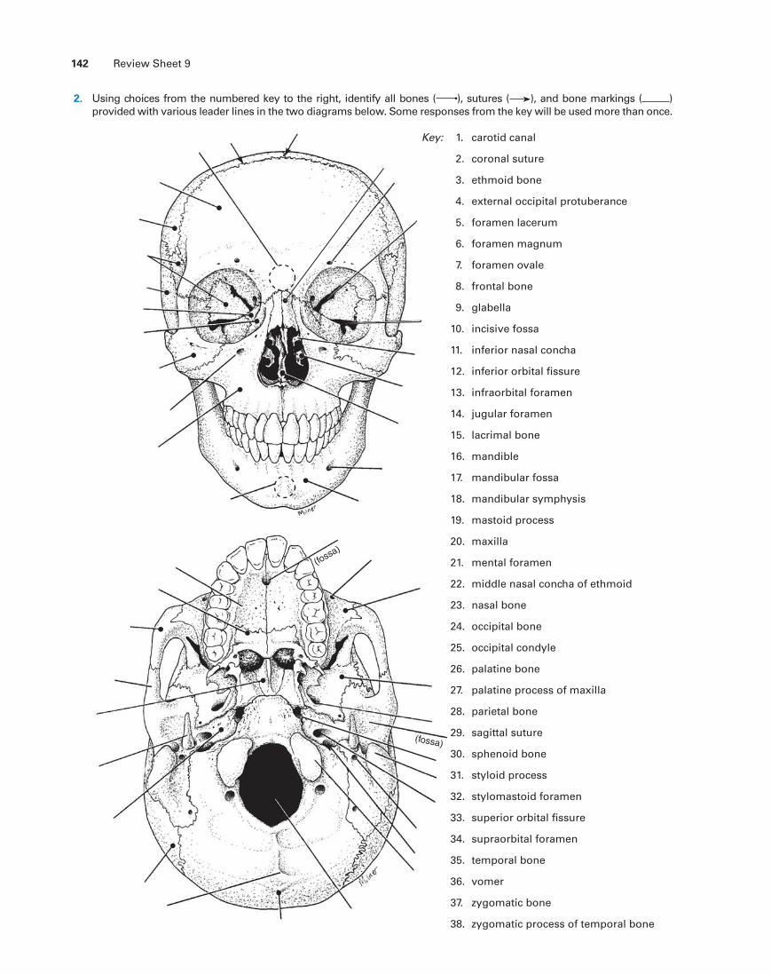
2. Using choices from the numbered key to the right, identify all bones ( ), sutures ( ), and bone markings ( ) provided with various leader lines in the two diagrams below. Some responses from the key will be used more than once.
(fossa)
(fossa)
1. carotid canal
2. coronal suture
3. ethmoid bone
4. external occipital protuberance
5. foramen lacerum
6. foramen magnum
7. foramen ovale
8. frontal bone
9. glabella
10. incisive fossa
11. inferior nasal concha
12. inferior orbital fissure
13. infraorbital foramen
14. jugular foramen
15. lacrimal bone
16. mandible
17. mandibular fossa
18. mandibular symphysis
19. mastoid process
20. maxilla
21. mental foramen
22. middle nasal concha of ethmoid
23. nasal bone
24. occipital bone
25. occipital condyle
26. palatine bone
27. palatine process of maxilla
28. parietal bone
29. sagittal suture
30. sphenoid bone
31. styloid process
32. stylomastoid foramen
33. superior orbital fissure
34. supraorbital foramen
35. temporal bone
36. vomer
37. zygomatic bone
38. zygomatic process of temporal bone
Key:
Review Sheet 9 143
3. Define suture.
4. With one exception, the skull bones are joined by sutures. Name the exception.
5. What bones are connected by the lambdoid suture?
What bones are connected by the squamous suture?
6. Name the eight bones of the cranium. (Remember to include left and right.)
7. Give two possible functions of the sinuses.
8. What is the orbit?
What bones contribute to the formation of the orbit?
9. Why can the sphenoid bone be called the keystone of the cranium?
The Vertebral Column 10. The distinguishing characteristics of the vertebrae composing the vertebral column are noted below. Correctly iden-
tify each described structure by choosing a response from the key.
a. atlasb. axisc. cervical vertebra—typical
d. coccyxe. lumbar vertebra
f. sacrumg. thoracic vertebra
Key:
1. vertebra type containing foramina in the transverse processes, through which the vertebral arteries ascend to reach the brain
2. dens here provides a pivot for rotation of the first cervical vertebra (C1)
3. transverse processes faceted for articulation with ribs; spinous process pointing sharply downward
4. composite bone; articulates with the hip bone laterally
5. massive vertebra; weight-sustaining
144 Review Sheet 9
6. “tail bone”; vestigial fused vertebrae
7. supports the head; allows a rocking motion in conjunction with the occipital condyles
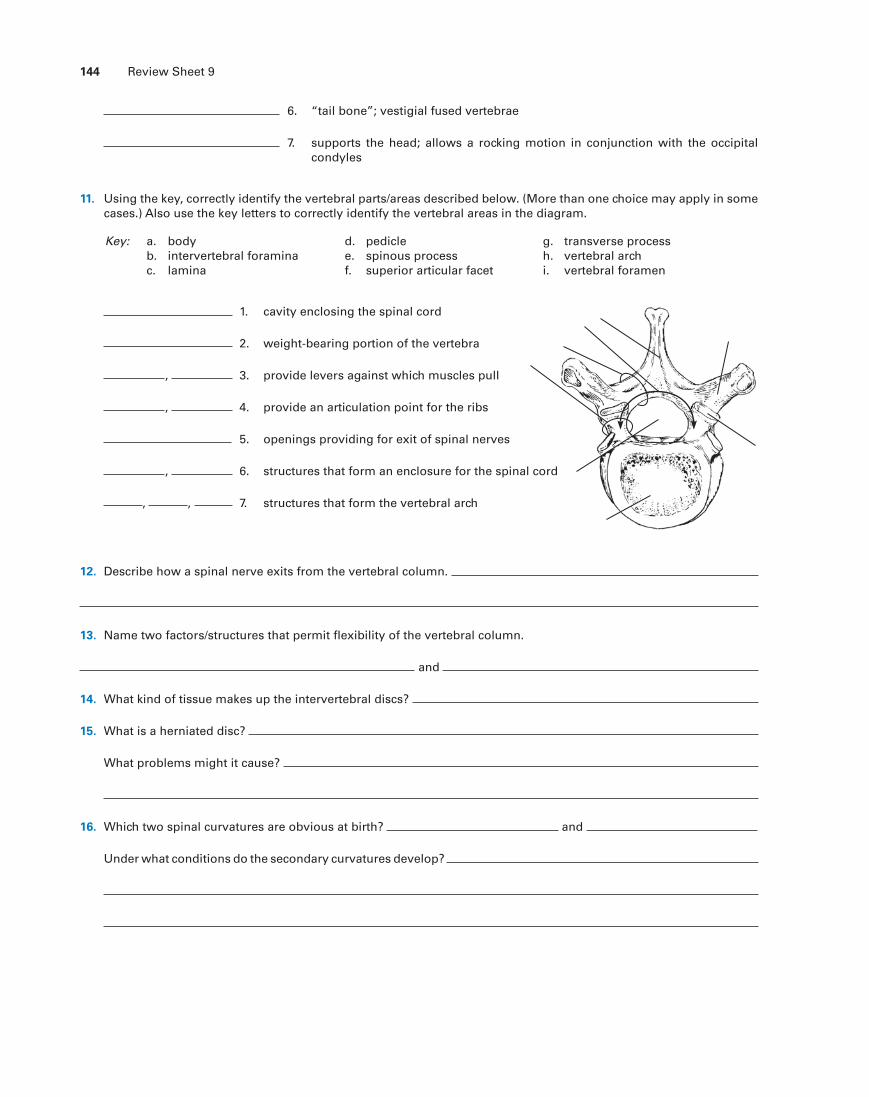
11. Using the key, correctly identify the vertebral parts/areas described below. (More than one choice may apply in some cases.) Also use the key letters to correctly identify the vertebral areas in the diagram.
a. bodyb. intervertebral foraminac. lamina
d. pediclee. spinous processf. superior articular facet
g. transverse processh. vertebral archi. vertebral foramen
Key:
1. cavity enclosing the spinal cord
2. weight-bearing portion of the vertebra
, 3. provide levers against which muscles pull
, 4. provide an articulation point for the ribs
5. openings providing for exit of spinal nerves
, 6. structures that form an enclosure for the spinal cord
, , 7. structures that form the vertebral arch
12. Describe how a spinal nerve exits from the vertebral column.
13. Name two factors/structures that permit flexibility of the vertebral column.
and
14. What kind of tissue makes up the intervertebral discs?
15. What is a herniated disc?
What problems might it cause?
16. Which two spinal curvatures are obvious at birth? and
Under what conditions do the secondary curvatures develop?
Review Sheet 9 145
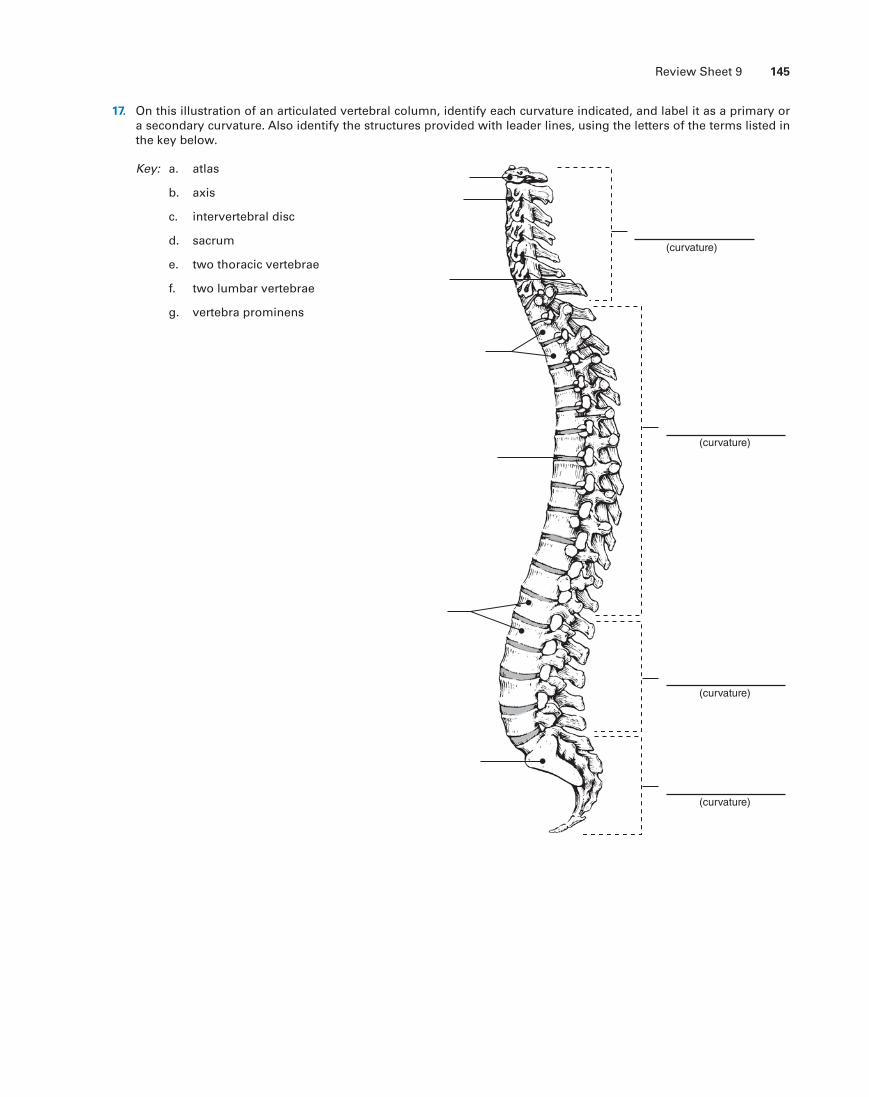
17. On this illustration of an articulated vertebral column, identify each curvature indicated, and label it as a primary or a secondary curvature. Also identify the structures provided with leader lines, using the letters of the terms listed in the key below.
Key: a. atlas
b. axis
c. intervertebral disc
d. sacrum
e. two thoracic vertebrae
f. two lumbar vertebrae
g. vertebra prominens
(curvature)
(curvature)
(curvature)
(curvature)
146 Review Sheet 9
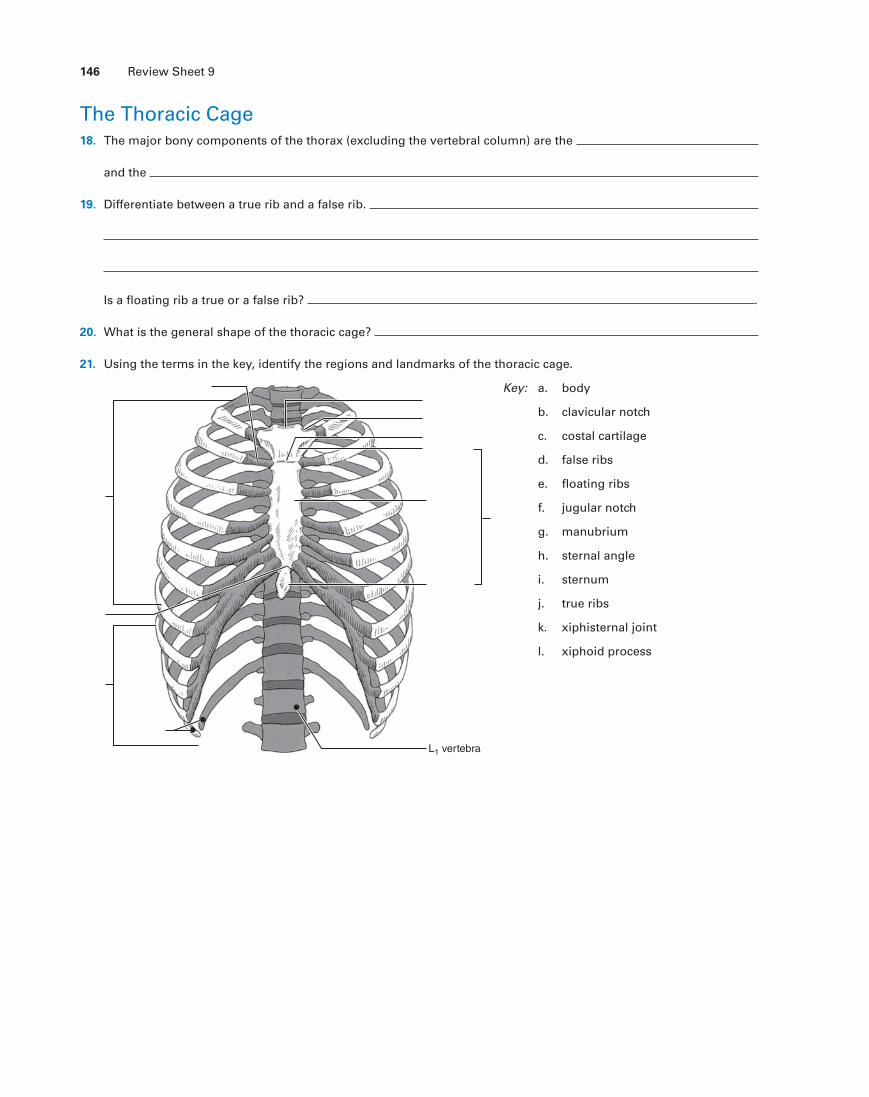
The Thoracic Cage 18. The major bony components of the thorax (excluding the vertebral column) are the
and the
19. Differentiate between a true rib and a false rib.
Is a floating rib a true or a false rib? .
20. What is the general shape of the thoracic cage?
21. Using the terms in the key, identify the regions and landmarks of the thoracic cage.
Key:
L1 vertebra
a. body
b. clavicular notch
c. costal cartilage
d. false ribs
e. floating ribs
f. jugular notch
g. manubrium
h. sternal angle
i. sternum
j. true ribs
k. xiphisternal joint
l. xiphoid process
Review Sheet 9 147
The Fetal Skull 22. Are the same skull bones seen in the adult also found in the fetal skull?
23. How does the size of the fetal face compare to its cranium?
How does this compare to the adult skull?
24. What are the outward conical projections on some of the fetal cranial bones?
25. What is a fontanelle?
What is its fate?
What is the function of the fontanelles in the fetal skull?
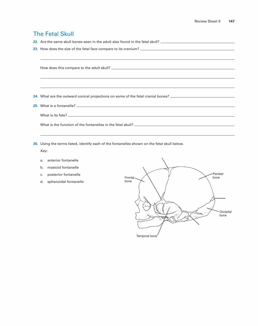
26. Using the terms listed, identify each of the fontanelles shown on the fetal skull below.
Key:
a. anterior fontanelle
b. mastoid fontanelle
c. posterior fontanelle
d. sphenoidal fontanelle
Parietalbone
Occipitalbone
Temporal bone
Frontalbone