Excitation-Contraction Coupling Once generated, the action potential: Is propagated along the...
14
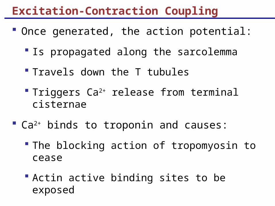
Excitation-Contraction Coupling Once generated, the action potential: Is propagated along the sarcolemma Travels down the T tubules Triggers Ca 2+ release from terminal cisternae Ca 2+ binds to troponin and causes: The blocking action of tropomyosin to cease Actin active binding sites to be exposed
-
date post
20-Dec-2015 -
Category
Documents
-
view
216 -
download
1
Transcript of Excitation-Contraction Coupling Once generated, the action potential: Is propagated along the...
Excitation-Contraction Coupling
Once generated, the action potential:
Is propagated along the sarcolemma
Travels down the T tubules
Triggers Ca2+ release from terminal cisternae
Ca2+ binds to troponin and causes:
The blocking action of tropomyosin to cease
Actin active binding sites to be exposed
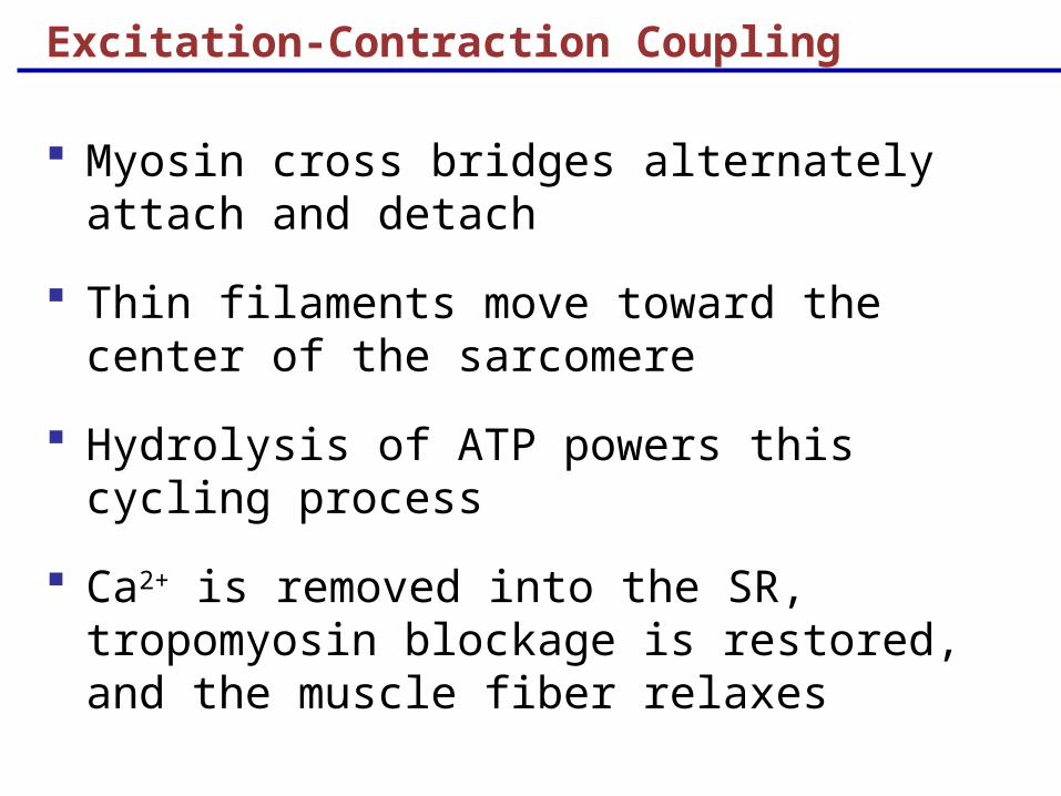
Excitation-Contraction Coupling
Myosin cross bridges alternately attach and detach
Thin filaments move toward the center of the sarcomere
Hydrolysis of ATP powers this cycling process
Ca2+ is removed into the SR, tropomyosin blockage is restored, and the muscle fiber relaxes
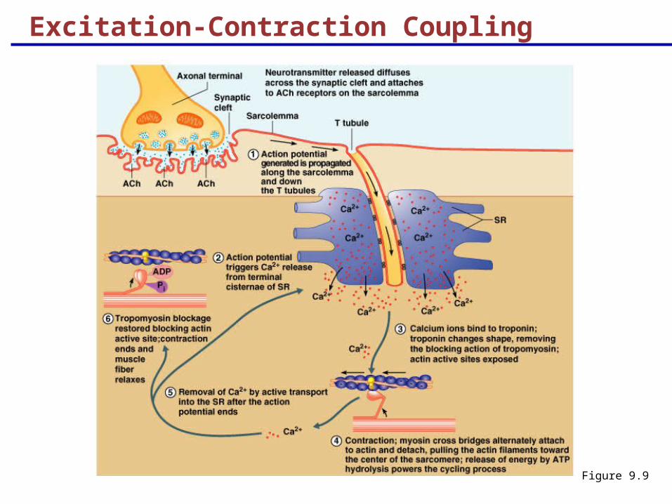
Excitation-Contraction Coupling
Figure 9.9
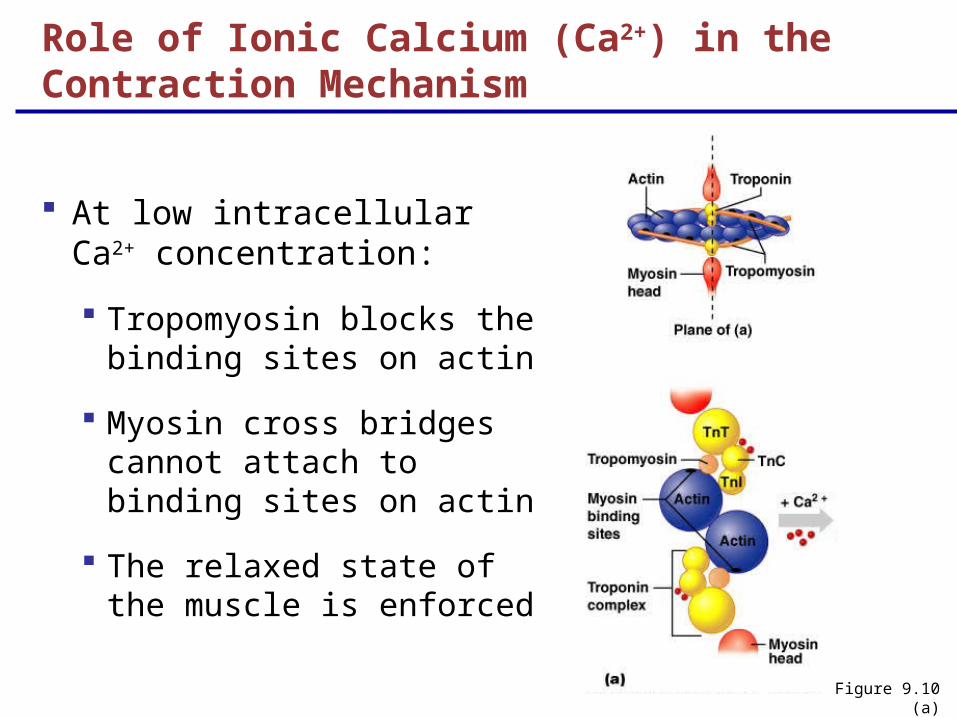
At low intracellular Ca2+ concentration:
Tropomyosin blocks the binding sites on actin
Myosin cross bridges cannot attach to binding sites on actin
The relaxed state of the muscle is enforced
Role of Ionic Calcium (Ca2+) in the Contraction Mechanism
Figure 9.10 (a)
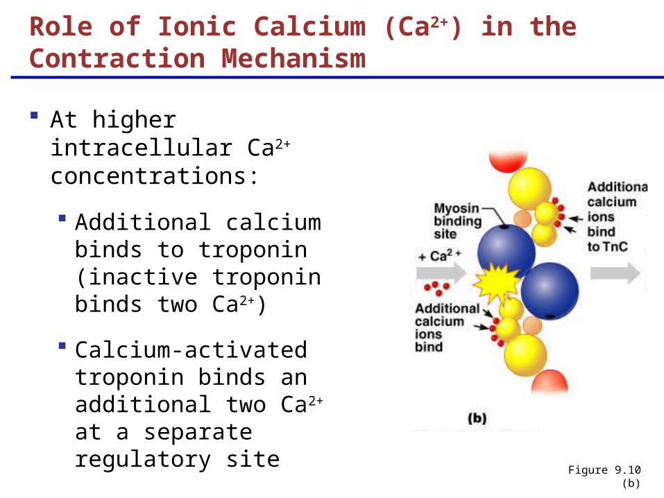
Figure 9.10 (b)
At higher intracellular Ca2+ concentrations:
Additional calcium binds to troponin (inactive troponin binds two Ca2+)
Calcium-activated troponin binds an additional two Ca2+ at a separate regulatory site
Role of Ionic Calcium (Ca2+) in the Contraction Mechanism
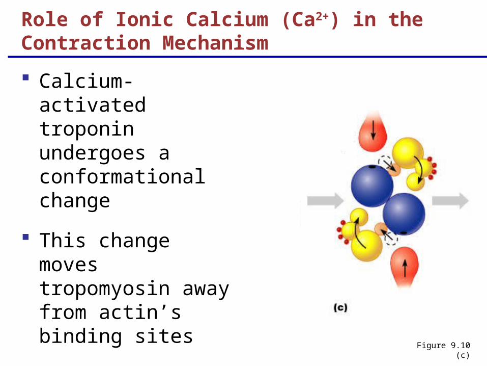
Calcium-activated troponin undergoes a conformational change
This change moves tropomyosin away from actin’s binding sites
Figure 9.10 (c)
Role of Ionic Calcium (Ca2+) in the Contraction Mechanism
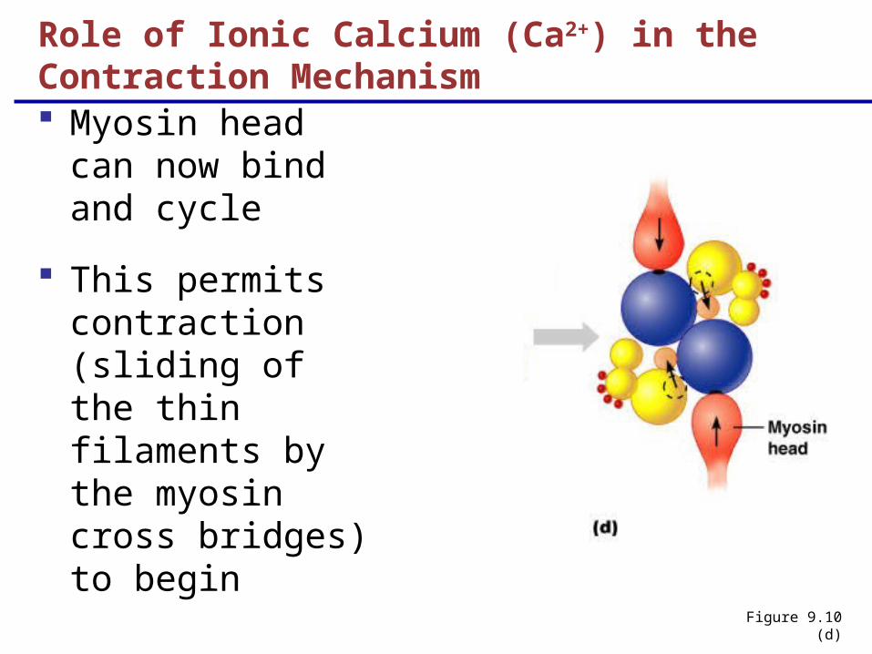
Myosin head can now bind and cycle
This permits contraction (sliding of the thin filaments by the myosin cross bridges) to begin
Figure 9.10 (d)
Role of Ionic Calcium (Ca2+) in the Contraction Mechanism
Sequential Events of Contraction
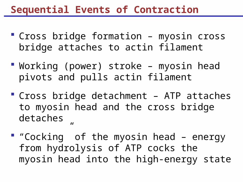
Cross bridge formation – myosin cross bridge attaches to actin filament
Working (power) stroke – myosin head pivots and pulls actin filament
Cross bridge detachment – ATP attaches to myosin head and the cross bridge detaches
“Cocking” of the myosin head – energy from hydrolysis of ATP cocks the myosin head into the high-energy state
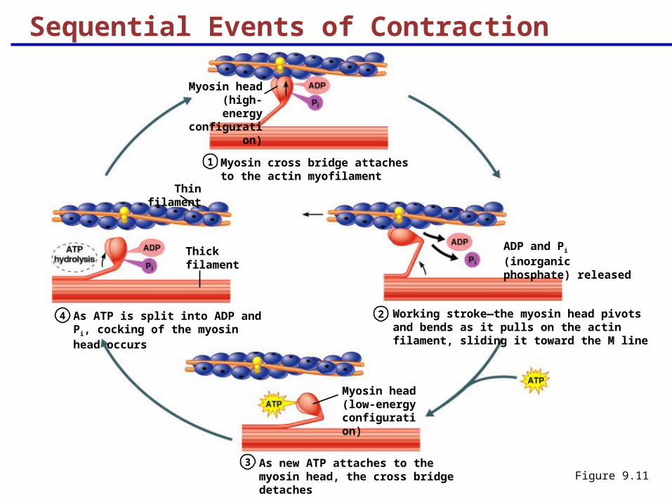
Myosin cross bridge attaches to the actin myofilament
1
2
3
4 Working stroke—the myosin head pivots and bends as it pulls on the actin filament, sliding it toward the M line
As new ATP attaches to the myosin head, the cross bridge detaches
As ATP is split into ADP and Pi, cocking of the myosin head occurs
Myosin head (high-energy
configuration)
Thick filament
Myosin head (low-energy configuration)
ADP and Pi (inorganic phosphate) released
Sequential Events of Contraction
Figure 9.11
Thin filament
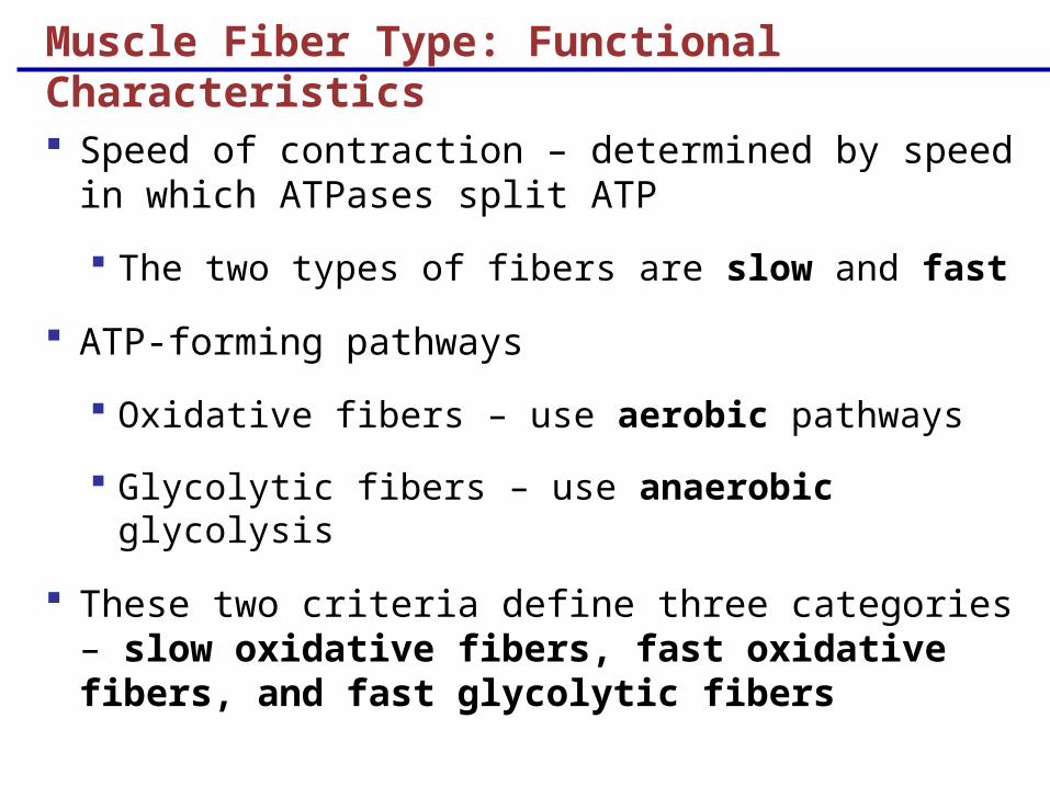
Muscle Fiber Type: Functional Characteristics
Speed of contraction – determined by speed in which ATPases split ATP
The two types of fibers are slow and fast
ATP-forming pathways
Oxidative fibers – use aerobic pathways
Glycolytic fibers – use anaerobic glycolysis
These two criteria define three categories – slow oxidative fibers, fast oxidative fibers, and fast glycolytic fibers
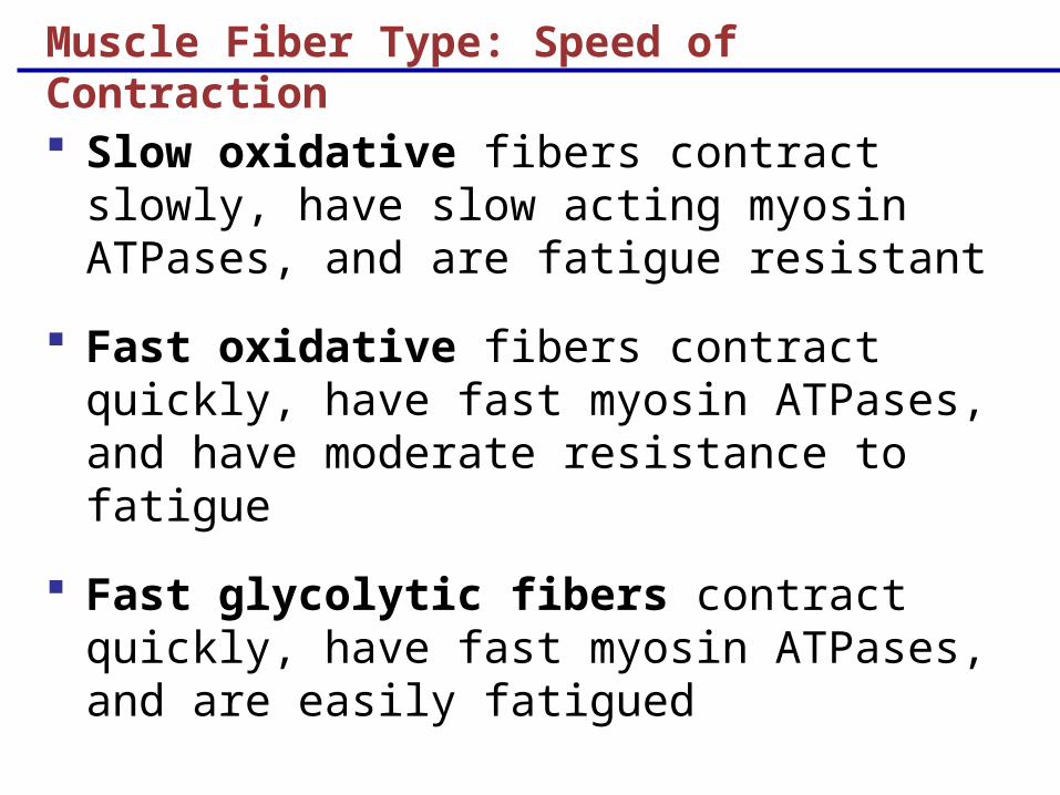
Muscle Fiber Type: Speed of Contraction
Slow oxidative fibers contract slowly, have slow acting myosin ATPases, and are fatigue resistant
Fast oxidative fibers contract quickly, have fast myosin ATPases, and have moderate resistance to fatigue
Fast glycolytic fibers contract quickly, have fast myosin ATPases, and are easily fatigued
Developmental Aspects: Regeneration
Cardiac and skeletal muscle become amitotic, but can lengthen and thicken
Smooth muscle has good regenerative ability
Developmental Aspects: Male and Female
There is a biological basis for greater strength in men than in women
Women’s skeletal muscle makes up 36% of their body mass
Men’s skeletal muscle makes up 42% of their body mass
Developmental Aspects: Age Related
With age, connective tissue increases and muscle fibers decrease
Muscles become stringier and more sinewy
By age 80, 50% of muscle mass is lost (sarcopenia)
Regular exercise can dramatically slow sarcopenia