Exanthematous drug eruptions - Confex · Exanthematous drug eruptions Werner J. Pichler, M.D. Clinc...
47
Exanthematous drug eruptions Werner J. Pichler, M.D. Clinc for Rheumatology and clinical Immunology/Allergology, Inselspital, University of Bern Switzerland E-mail: [email protected]
Transcript of Exanthematous drug eruptions - Confex · Exanthematous drug eruptions Werner J. Pichler, M.D. Clinc...

Exanthematous drug eruptions
Werner J. Pichler, M.D.
Clinc for Rheumatology
and clinical Immunology/Allergology,
Inselspital, University of Bern
Switzerland
E-mail: [email protected]

Disclosure slide
• Owner of adr-ac GmbH, a company devoted to drug hypersensitivity analysis and research
• Consultant for Pfizer, Hoffmann LaRoche, Böhringer- Ingelheim, Novartis, Menarini, Aicuris, Swatch
• Research support by Swiss National Science foundation, Swiss Center of Applied Toxicology, Ulrich Müller Gierok Foundation, Pfizer, Hoffmann-LaRoche, Menarini

Exanthematous drug eruptions
• «rashes»
• Urticaria immediate reactions
• Delayed appearing exanthems
with cell infiltration
it is frequent - antibiotics (0.5 - 8% of treated)
- antiepileptics
- allopurinol, diuretics,
antivirals, ........

7d
indapamid

Drug allergy
1) What has happened ?
2) Mechanism & how severe is the drug induced illness ?
3) Which drug is responsible ?
history
drug exposure
cofactors
viral infection
previous drug allergies
Danger signs:
timing
clinical
laboratory
Skin testing
lymphocyte stimulation tests
provocation tests (?)

Delayed reaction: it is T-cell mediated
1 2 3 4 5 6 7 8 9 10 days
T
Few precursor … Expansion….
cells
Symptoms arise if a certain
amount of specific T-cells
is homing to the tissue and
exerts effector function
No symptoms symptoms

Maculopapular drug exanthem CD4+ CD8 +

Delayed reaction
1 2 3 4 5 6 7 8 9 10 days
Few precursor … Expansion….
cells
Symptoms arise if a certain
amount of specific T-cells
is homing to the tissue and
exerts effector function
No symptoms symptoms
0
5
10
15
20
25
30
35
40
45
1 2 3 4 5 6 7 8 9 10 11 12 13 14 15 16 17
Symptoms (MPE)
days

maculopapular exanthema
mild
moderate
massive, confluent
25% have liver involvement
Am
ount
of
prec
ursor
&
effe
ctor
cells

1 2 3 4 5 6 7 8 9 10 days
4
4
4
4
4
4
8 4 8
4
4
4
4
4 4
4
4 8 4
precursor cells effector cells
8 4 4
4 8
No symptoms symptoms
1 2 3 4 5 6 7 8 9 10 days
4 8 4
8
8 4
8
8 4
8
4 8
8
8 4
No symptoms symptoms
4 4
8
cytotoxic
IFNg
IL-13
IL-5
IL-8/GM-CSF
4
8 8 8
8
granzymeB+
perforin+
cytotoxic
The function of effector T cells
determines clincal phenotype:
MPE
pustular
bullous
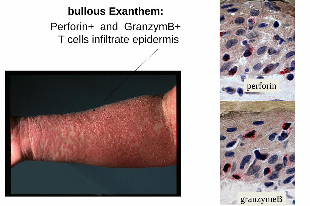
bullous Exanthem:
Perforin+ and GranzymB+
T cells infiltrate epidermis
granzymeB
perforin
granzymeB
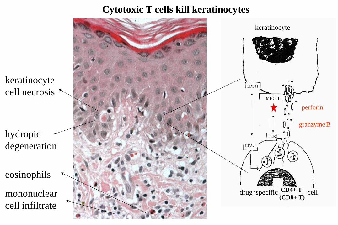
hydropic
degeneration
eosinophils
keratinocyte
cell necrosis
mononuclear
cell infiltrate
ICD541
LFA-1
keratinocyte
MHC II
TCR
drug - specific CD4+ T
(CD8+ T) cell
granzyme B
perforin
Cytotoxic T cells kill keratinocytes

Acute generalized exanthematous
pustulosis
(AGEP)

AGEP
a T cell reaction recruiting PMN
CD8 NEUTROPHIL
ELASTASE CD4
FIRST T cells
T cell infiltration into epidermis (cytotoxic and IL-
8/GM-CSF): vesicle
NEUTROPHIL ELASTASE
SECOND
PMN accumulation
pustule
later PMN

T-cells react with a drug, are stimulated and expand: they organize a certain pathology
Drug, e.g. amoxicillin
bullous E.
MHC-I (+ MHC-II)
CD8+ > CD4+
cytotoxicity (CD8+)
IFNg; IL-5
MPE
MHC-II
CD4+
cytotoxicity (CD4+)
IL-5; IFNg
AGEP
MHC-II + I
CD4+ & CD8+
cytotoxicity
IL-8; IL-5

Classification of drug-hypersensitivity reactions
TYPE IV a TYPE IV b TYPE IV c TYPE IV d
Th1 Th2 Cytotoxic T cells T cells
IFN-γ, TNF-α IL-5, IL-4, IL-13,
eotaxin
Perforin, granzyme B, FasL CXCL-8, GM-CFS
IL-17 (?)
Monocyte,
Macrophage
Eosinophilic
inflammation Cytotoxic T cells Neutrophils
Tuberkulin skin test,
(Contact dermatitis)
Maculopapular
exanthem with
eosinophilia
Contact dermatitis
Maculopapular, Bullous
exanthema
Pustular exanthema

Time of appearance of delayed skin reactions
0
5
10
15
20
25
30
35
40
45
1 2 3 4 5 6 7 8 9 10 11 12 13 14 15 16 17
SJS/TEN
day ~10 - >24
(allopurinol, SMX)
DRESS
day ~12 - >50
(antiepileptics)
AGEP
amoxicillin
~day 2-5
MPE day ~7-11

Severitiy? Danger signs – delayed reactions
Clinic • widely spread exanthema
• induration, bullae, pustules
• erythrodermia
• pain in skin
• Nikolsky sign
• mucosal involvement
• lymphadenopathy
• fever
• general symptoms / malaise (liver, kidney, lung, pancreas)
Laboratory
• differential blood count (eosinophilia,
activated lymphocytes)
• ALAT, ASAT, gGT, AP
• (CRP ~; Creatinine)
delayed reactions: certain laboratory examinations are
helpful and necessary

DRESS DRESS and
haematophagocytic
syndrome
TEN
Danger sign: facial edema, flash

Stomatitis
(SJS, TEN, DRESS)
Conjunctival
involvement
SJS, TEN
Danger sign: mucosal involvement
Pictures by A Bircher, Basel

Severity? Danger signs – delayed reactions
Clinic • widely spread exanthema
• induration, bullae, pustules
• erythrodermia
• pain in skin
• Nikolsky sign
• mucosal involvement
• lymphadenopathy
• fever
• general symptoms / malaise (liver, kidney, lung, pancreas)
Laboratory
• differential blood count (eosinophilia,
activated lymphocytes)
• ALAT, ASAT, gGT, AP
• (CRP ~; Creatinine)
delayed reactions: certain laboratory examinations are
helpful and necessary
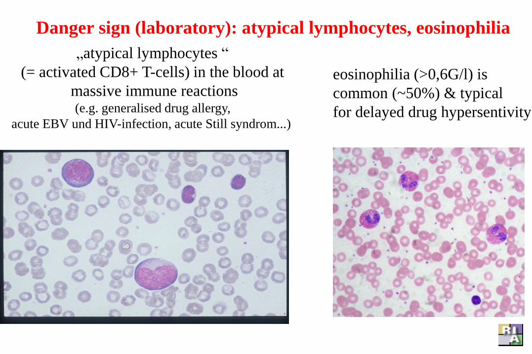
„atypical lymphocytes “
(= activated CD8+ T-cells) in the blood at
massive immune reactions (e.g. generalised drug allergy,
acute EBV und HIV-infection, acute Still syndrom...)
eosinophilia (>0,6G/l) is
common (~50%) & typical
for delayed drug hypersentivity
Danger sign (laboratory): atypical lymphocytes, eosinophilia

SUMMARY: Exanthematous drug eruptions
1. Are T cells reactions
2. Timing: Appear between 2 d (AGEP) and >50 d (DRESS) of drug exposure
3. One differentiates papular [MPE], pustular [AGEP], bullous [SJS] (and macular / urticarial ....) exanthems
4. determine the severity of MPE by clinical and laboratory signs

HOW ARE DRUGS STIMULATING T CELLS ?

Maculopapular drug eruption (MPE) - Immunohistology
T-cell infiltration into dermis, epidermis;
cytotoxicity (killing of keratinocytes)
recruitment of inflammatory cells
Cytotoxic
CD4+ T cell
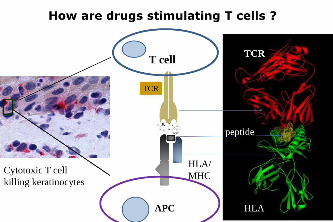
How are drugs stimulating T cells ?
T cell
HLA/
MHC
TCR
APC
TCR
HLA
peptide
Cytotoxic T cell
killing keratinocytes

pharmacological interaction with
immune receptors (p-i) concept:
a) the drug binds to the
TCR (by non-covalent bonds; not
restricted to a HLA-allele)
or
b) the drug binds to the
HLA molecule (NOT to the
presented peptide);
the {HLA-drug + peptide complex} is
recognized by the TCR
HLA-peptide-TCR complex
TCR
HLA

Pharmacological interaction with
immune receptors = p-i concept
It is a non-covalent binding of drugs to proteins functioning as immune receptors (TCR, HLA);
It explains an immune stimulation by a drug without postulating antigen-features of a drug !

p-i HLA: binding of drug to HLA molecule
Not the peptide (the «antigen»),
but the HLA molecule itself is modified

p-i concept: a drug fits into a particular HLA molecule
HLA-peptide-TCR complex
the drug binds to an allele-typic
region in the HLA by van der
Waals forces;
the {HLA-peptide-drug} complex
is then recognized by the TCR
HLA-B*5701:
binding groove
For abacavir
TCR
HLA
Illing et al, Nature 2012

p-i TCR: binding of drug to T cell receptor (TCR)

p-i TCR:
T cell clones specific for
sulfamethoxazole
(SMX):
- cross-reactivity
- inhibition of SMX
stimulation by other
sulfanilamides (n = 11)
- docking &
- dynamic modelling

1.3: only SMX; 11 other
sulfanilamides (SA)
not stimulatory
H13: SMX and 5 other SA
stimulatory
Two SMX specific T-cell clones «H13» & «1.3»
St Watkins & WJ Pichler, OJI, 2013
TCC «1.3»
TCC «H13»

SMX-specific Clone 1.3:
SMX binds to a unique site on the CDR3-a loop of the SMX specific TCR 1.3
TCR
HLA
TCR 1.3
1011
TCR
St. Watkins & WJ Pichler, OJI, 2013

75% inhibition of SMX induced
proliferation by the sulfanilamide
SMT (sulfamethazole)
35% inhibition of SMX induced
Ca++ influx by sulfanilamides
St Watkins & WJ Pichler, OJI, 2013
TCC 1.3

Clone 1.3
- The TCR 1.3 showed CDR3α recognition of SMX.
The NH2 of SMX
may contact the
peptide.
This may explain the
cross-reactivity of some
TCC reactive with
hapten (SMX-NO) and via
p-i (SMX)*
*Schnyder B et al. J Immunol. 2000 peptide

SMX is bound to TCRVb2 of TCR «H13»,
outside the HLA-peptide interaction site!
St Watkins & WJ Pichler: Sulfamethoxazole Induces a Switch Mechanism in T-Cell Receptors
Containing TCRVβ20-1, by Altering pHLA Recognition; PLOS One, 2013
TCC 1.3
TCC H13

Only SMX and 5 of
11 other sulfanilamides
fit into the pocket
formed by the
CDR2 region
(TCR H13)
Drug (SMX) binding to
the TCR-Vb CDR2 loop.
Stephan Watkins & Werner J. Pichler:
Activating Interactions of
Sulfanilamides with T Cell Receptors,
Open J Immunology, 2013
:
Clone H 13

Visualizing the H13 Binding Process
MD simulations of TCR H13 and HLA-DR*10:01 with or without SMX binding
SMX No
SMX
Humphrey W, Dalke A, Schulten K (1996) VMD: visual molecular dynamics. J Mol Graph 14: 33-38, 27-38.

H13 Summary of Motions
With SMX:
The analysis of motions reveals a “switch”, where the TCR constant domain either sits on the TCRVβ (above), changing to the TCRVα and a change from mostly Vβ recognition of the HLA and peptide, to a Vα recognition of the HLA.
No SMX
With SMX
b a
a b

Gibbs Free Energy, ΔG
- Free energy change is the most straightforward of the parameters
- For H13 it was shown SMX caused a 7 fold increase in affinity, from -24 to -140 kcal/mol.
- This translates from a 2 μmol to a 0.79 μmol affinity.
* Normal TCR affinities are in the range of 5-1 μmol, however we know the H13 T cell only proliferates with SMX present.

Allosteric effect of SMX binding to CDR2-Vb pocket of TCR H13
Stephan Watkins & Werner J. Pichler: Sulfamethoxazole Induces a Switch Mechanism
in T cell Receptors Containing TCRVb-20-1 Altering pHLA Recognition, PLOS ONE, in press

understanding “p-i TCR”
Two types of p-i TCR A) A small molecule binds to a region on the
TCR free from contact with the pHLA or other proteins-protein interfaces and the resulting complex can bind the pHLA through induced TCR conformations (Watkins
S & Pichler WJ, Plos One 2013).
B) The CDR3α or β recognizes a small molecule, and the resulting complex can then bind the pHLA, with the small molecule
acting as part of the TCR (Watkins S & Pichler
WJ, Open J Immunol, 2013).
-In either, there is a dependence on a particular pHLA, but the effect is mediated by the TCR binding the small molecule.
H13
1.3

SUMMARY II: p-i concept
1. p-i: pharmacological interaction of drugs with immune receptors
2. one differentiates between p-i TCR and p-i HLA
3. It explains T-cell reactivity to drugs without implying antigenic features of the drug
4. most severe reactions appear to be due to p-i, which is an off target activity of the drug on (selected) immune receptors (TCR, HLA)
5. In contrast to previous beliefs, an interaction of small molecules with the immune system is common, and needs to be better investigated

6th DRUG HYPERSENSITIVITY MEETING (DHM6)
in
BERN, SWITZERLAND
APRIL 9th – 13th, 2014
www.eaaci.org

Thanks Stephen Watkins (SMX), Natascha Wuillemin (FLUX), James Yun (ALL/OXY), Daniel Yerly,
Klara Ericsson, Karin Schnyder, Heidi Jamin (Insel/Univ.Bern)
Jacqueline Adam (ABC), Tatjana Petkovic, Oliver Hausmann, Antonia Bünter, Dario Doerig (ADR-AC)
Tom Kawabata (Pfizer) & Antonio Iglesias (Roche)
Collaborators: Stephan Krähenbühl; The Liverpool group (Dean Naisbitt, Kevin Park, Munir Pirmohamed)
Research supported by
Swiss National Science Foundation
Swiss Center for human Toxicology (SCAHT)
Ulrich Müller Gierok Foundation
ADR-AC & Roche, Switzerland

Literature to drug eruptions Pichler W.J. Pharmacological interaction of drugs with antigen-specific immune receptors: the p-i concept.
Curr Opin Allergy Clin Immunol 2002;2:301-305
Pichler W.J. Delayed Drug Hypersensitivity Reactions. Annals Int Med 2003;139:683-693
Pichler WJ, Dauber B, Kawabata T. Drug hypersensitivity: Flare-up reactions, cross-reactivity and multiple
drug hypersensitivity. Journal of Dermatology 2011; 38: 216–221
Adam J, Pichler WJ, Yerly D. Delayed drug hypersensitivity: Models of T-cell stimulation. Br J Clin
Pharmacol. 2011;71(5):701-7
Adam J., Eriksson K.K., Schnyder B., Fontana St, Pichler W. J., Yerly D. Avidity Determines T-cell
Reactivity in Abacavir Hypersensitivity, Eur. J. Immunol.2012;42:1-11
Adam J., Eriksson K.K., Schnyder B., Fontana St, Pichler W. J., Yerly D. Avidity Determines T-cell
Reactivity in Abacavir Hypersensitivity, Eur. J. Immunol.2012;42:1-11
Pichler WJ. Direct T-cell stimulations by drugs-bypassing the innate immune system. Toxicology. 2005; 209(2):95-
100
Wuillemin N, Adam J, Fontana S, Krähenbühl St, Pichler WJ, Yerly D HLA haplotype determines hapten
or p-i T cell reactivity to flucloxacillin, J Immunol 2013; 190: 4956-64.
Porebski G, Pecaric-Petkovic T, Groux-Keller M, Bosak M, Kawabata TT, Pichler WJ. In vitro drug
causality assessment in Stevens-Johnson syndrome – alternatives for lymphocyte transformation test. Clin exp.
Allergy. 2013;43(9):1027-1037
Schnyder B, Adam J, Rauch A, Thurnheer M, Pichler WJ. HLA-B*57:011 abacavir-naive individuals have
specific T cells but no patch test reactivity J Allergy Clin Immunol. 2013 Sep;132(3):756-8
Watkins S, Pichler WJ. Activating interactions of sulfanilamides with T cell receptors. Open J Immunol,
Vol.3, No.3, 139-157 (2013)
Watkins S, Pichler WJ. Sulfamethoxazole Induces a Switch Mechanism in T cell Receptors Containing
TCRVß20-1, Altering pHLA. PLoS one, 2013:(43) 1246–1255.
Yun J, Mattsson J, Schnyder K, Fontana S, Largiadèr CR, Yerly D, Pichler WJ. Allopurinol hypersensitivity
is mediated by dose dependent oxypurinol-specific T cell response, Clin exp. Allergy, 2013:(43) 1246–1255.
Yun J, Marcaida MJ, Eriksson KK, Jamin H, Fontana S, Pichler WJ, Yerly D. Oxypurinol directly and
immediately activates the drug-specific T cells via the preferential use of HLA-B*58:01. J. Immunol., under revision