EXAMPLES OF APPLICATIONS · and the following slide demonstrate the importance of tracking...
27
1 EXAMPLES OF APPLICATIONS 1 This presentation demonstrated the power of the Kinetworks™ approach to uncover significant research results in a cost effective and efficient manner.
-
Upload
nguyenminh -
Category
Documents
-
view
212 -
download
0
Transcript of EXAMPLES OF APPLICATIONS · and the following slide demonstrate the importance of tracking...

1
EXAMPLES OF APPLICATIONS
1
This presentation demonstrated the power of the Kinetworks™ approach to uncoversignificant research results in a cost effective and efficient manner.
20405
CosmoLogo_E

2
A major bottleneck in drug discovery is theidentification and validation of drug targets
Human genome project has identified over 5,000 cellsignalling proteins as potential drug targets
Less than 500 proteins have been investigated as drugtargets by pharmaceutical industry in its entire history
$5 billion spent annually screening non-ideal targets
Kinexus is dedicated to identifying and validating drugtargets and leads for our clients through offer of ourunique Kinetworks™ proteomics services
Drug Target Discovery
2
Drug discovery is a very risky and expensive proposition. Only one in a hundred thousandcompounds may become a successful drug, after the investment of over US $600 million onaverage and frequently more than 10 years in development.
To discover a drug, the right target must be identified first. This target can be used as the baitto fish for small molecule compounds that are typically inhibitors of the chosen target by highthroughput screening. Lead compounds are put sequentially through cell-based, then animal-based and finally human trials. Each subsequent step of the drug discovery and validationprocess is associated with dramatic increases in cost. Only about 1 in 10 lead compoundsthat look promising at the end of the animal trials make it successfully through Phase IIIhuman trials and receive FDA approval. It is this high rate of failure at the late stages ofclinical trials that has made drug discovery so expensive. If the failures could be identifiedearlier in the drug discovery process, this would markedly reduce costs and lost opportunity.
A common cause of failure in human trials is adverse drug reactions. The KinexusKinetworks™ screening platform for cell cycle, stress and apoptosis proteins can help toidentify at an early stage possible toxic drug reactions and prevent the investment of millions ofdollars on the testing of non-ideal targets.
To have the greatest impact on drug discovery, it is critical to identify the most appropriatedrug targets. This is a significant issue, because over $5 billion is spent annually by thepharmaceutical industry screening against targets that even if specific, potent and cellpermeable inhibitors were uncovered , the side effects associated with knocking out thesetargets would mitigate their utility.
With the identification of over 30,000 genes in the human genome, about 5000 cellsignalling proteins have been designated as potential drug targets. However, thepharmaceutical industry does not have the capacity to evaluate all of these proteins for usein high throughput drug screening. In fact, less than 500 proteins have been examinedextensively as potential drug targets by the pharmaceutical industry over the last century.
20405
CosmoLogo_E

3
Drug Target Discovery
Drug Discovery andCharacterization
Medicinal Chemistry
Preclinical Studies
Clinical Studies
Drug ScreeningToxicology
Validation ofAnimal Models
Kinetworks™immunoblots
Patient Profiling
DiseaseMarker/Drug
TargetDetection
Kinetworks™ Services MultipleSteps in Drug Development
3
An important outcome of the Kinetworks™ proteomics services offered by Kinexus isthe generation of vast amounts of data about the regulation of specific proteins. Thisunique immunoblotting service is only one of many novel services that Kinexus plansto offer its clients.
The data generated from the Kinetworks™ services is being merged into ourKinformatics™ functional proteomics database, which we are mining to establish thecomposition and architecture of protein kinase signalling networks in hundreds ofdifferent normal and pathological tissues and cells from diverse species. Our clientswill also have Internet access to this database by subscription to KiNET™bioinformatics services. The central objective of our endeavors is to identify proteinkinase signalling pathways that link to specific diseases to identify diagnostic markersand protein kinase targets for drug discovery. Clients are able to query KiNET on-lineabout the expression patterns and phosphorylation states of hundreds of signallingproteins in hundreds of different model systems.
20405
CosmoLogo_E

4
Provides quantitation ofprotein expression andphosphorylation levels withhighly validated antibodies
Highly sensitive, accurate,and reproducible
Rapid turnaround
Affordable pricing
Kinetworks™ Analysis
4
The Kinetworks™ analysis utilizes commercial antibodies that have been highlyvalidated by Kinexus to ensure accuracy. Enhanced chemoluminescence anddensitiometric analysis with a FluorS Max Imager from Bio-Rad permits permitssensitive detection with as little as 350 µg of lysate cell/tissue protein. Data is linearover a 2000-fold range with standard deviations within 20% for high reproducibility.Results are returned to clients within 4 weeks. Substantial discounts are available withfull disclosure of sample information or bulk orders.
20405
CosmoLogo_E

5
Signal transductionpathway mapping
Protein-protein interactions Novel protein discovery Disease markers Drug targets Drug mechanisms of
action Drug toxicity Animal model validation
Kinetworks™ Applications
5
Kinetworks™ has a broad range of applications. This presentation provides a samplingofhow Kinetworks™ has already been successfully used for many of these purposes.
20405
CosmoLogo_E

6
Cells differ markedly in their protein kinase expression patterns
KPKS 1.2 Kinase Analysis
6
Kinexus has performed over 10,000 immunoblot analyses. These studies haverevealed the vast differences between cell types in the expression patterns of proteinkinases and other signalling proteins.
20405
CosmoLogo_E

7
Tissue Differences in ProteinKinase Expression
0 2000 4000 6000 8000
BMXBTK
CaMK1CaMK4CaMKK
CDK1CDK2CDK4CDK5CDK6CDK7CDK9CK1dCK1eCK2aCK2a'CK2a"
COTCSK
DAPKDNAPK
ERK1ERK2ERK3ERK6
FAKFYNGCK
GRK2GSK3aGSK3b
HPK1IKKa
JAK1JAK2KSR1
LCKLYN
MEK1MEK2MEK4MEK6MEK7MNK2MOS1MST1
0 2000 4000 6000 8000
BMXBTK
CaMK1CaMK4CaMKK
CDK1CDK2CDK4CDK5CDK6CDK7CDK9CK1dCK1eCK2aCK2a'CK2a"
COTCSK
DAPKDNAPK
ERK1ERK2ERK3ERK6
FAKFYNGCK
GRK2GSK3aGSK3b
HPK1IKKaJAK1JAK2KSR1
LCKLYN
MEK1MEK2MEK4MEK6MEK7MNK2MOS1MST1
0 2000 4000 6000 8000
BMXBTK
CaMK1CaMK4CaMKK
CDK1CDK2CDK4CDK5CDK6CDK7CDK9CK1dCK1eCK2aCK2a'CK2a"
COTCSK
DAPKDNAPK
ERK1ERK2ERK3ERK6
FAKFYNGCK
GRK2GSK3aGSK3b
HPK1IKKa
JAK1JAK2KSR1
LCKLYN
MEK1MEK2MEK4MEK6MEK7MNK2MOS1MST1
Brain Heart Sk. Muscle Spleen TestesMonkeyRat
7
In the Science Magazine’s Signal Transduction Knowledge Environment (STKE) study,Kinetworks™ analysis(www.stke.org/cgi/content/full/sigtrans;2002/162/pe50), rThisand the following slide demonstrate the importance of tracking potential drug targets indiverse organs, since the expression patterns of protein kinases are not readilypredictable. Furthermore, we have recently observed large differences in proteinkinase expression patterns between male and female rats for the same somatictissues. More than a third of the protein kinases showed expression changes ofgreater than 5-fold in the same types of organs. These unexpected differencesbetween species may account in part for why drugs that perform well in pre-clinicalanimal trials fail in human clinical studies.
20405
CosmoLogo_E

8
Tissue Differences in ProteinKinase Expression
0 2000 4000 6000 8000
NEK2
p38 MAPK
PAKa
PAKb
PDK1
Pim1
PKA
PKBa
PKCa
PKCb1
PKCd
PKCe
PKCg
PKCl
PKCm
PKCq
PKCz
PKG1
PKR
PYK2
RAF1
RAFB
ROKa
RSK1
RSK2
S6K p70
SAPKb
SAPKb
SRC
SYK
YES1
ZAP70
ZIPK
ZIPK
0 2000 4000 6000 8000
NEK2p38 MAPK
PAKaPAKbPDK1
Pim1PKA
PKBaPKCa
PKCb1PKCd
PKCePKCgPKCl
PKCmPKCq
PKCzPKG1
PKRPYK2
RAF1RAFBROKa
RSK1RSK2
S6K p70SAPKb
SAPKbSRC
SYKYES1
ZAP70
ZIPKZIPK
0 2000 4000 6000 8000
NEK2p38 MAPK
PAKaPAKbPDK1Pim1PKA
PKBaPKCa
PKCb1PKCdPKCePKCgPKCl
PKCmPKCqPKCzPKG1
PKRPYK2RAF1RAFBROKaRSK1RSK2
S6K p70SAPKbSAPKb
SRCSYK
YES1ZAP70
ZIPKZIPK
0 2000 4000 6000 8000
NEK2p38 MAPK
PAKaPAKbPDK1Pim1PKA
PKBaPKCa
PKCb1PKCdPKCePKCgPKCl
PKCmPKCqPKCzPKG1
PKRPYK2RAF1RAFBROKaRSK1RSK2
S6K p70SAPKbSAPKb
SRCSYK
YES1ZAP70
ZIPKZIPK
0 2000 4000 6000 8000
NEK2p38 MAPK
PAKaPAKbPDK1Pim1PKA
PKBaPKCa
PKCb1PKCdPKCePKCgPKCl
PKCmPKCqPKCzPKG1
PKRPYK2RAF1RAFBROKaRSK1RSK2
S6K p70SAPKbSAPKb
SRCSYK
YES1ZAP70
ZIPKZIPK
Brain Heart Sk. Muscle Spleen TestesMonkeyRat
8
20405
CosmoLogo_E

9
Gender Differences in ProteinKinase Expression
9
0 5000 10000 15000 20000 25000
CDK7PKCdGRK2
p38 MAPKJAK1
PKCmCaMK4SAPKb
RAF1SAPKb
ERK3PKCmCDK5
CK2a"IKKbeta
PKBaRSK1PKCaPKG1PYK2PKCzJAK2RAF1DAPKKSR1
BMX (Etk)ZIP
COTCSKIKKa
FemaleMale
0 5000 10000 15000 20000 25000 30000 35000 40000
GSK3aGSK3b
PKCeCDK9ERK2ERK1PKCbMEK4PKCgPAK1ROKaCK2a
S6K p70MEK6PKCl
MOS1CK2a'
CaMKKERK6
FAKPKA
CK1eMEK1MEK2RAFB
FemaleMale
MouseBrain
We have recently explored the effect of gender on patterns of protein kinaseexpression in somatic tissues from reproductive-competent male and female rats usingthe Kinetworks KPKS 1.3 Protein Kinase Screen. This results shown in slide formouse brain and the following slide for mouse submadibular gland demonstrate thatmore than half of the protein kinases detected in each tissue displayed greater than 2-fold differences in expression for age-matched mice. The implications for this areprofound, as it reveals vast differences in the composition and architecture of proteinkinase networks between males and females, and this could result in differentialresponses to drugs.
20405
CosmoLogo_E

10
0 2000 4000 6000 8000 10000 12000 14000 16000
CK2a"RAFBMEK1PKCbCOT
CK2aERK1ERK2
p38 MAPKIKKa
RAF1MEK2JAK1
SAPKbRSK2CDK5
SAPKbMEK6PKG1CDK9
PKAMOS1PKCaERK6PYK2PKCzCDK7PKBa
PKCmPKCe
GSK3aGSK3bMEK4PKCdRSK1
CK2 a'ERK3
FAKGRK2JAK2
Female
Male
MouseSubmandibular
Gland
Gender Differences in ProteinKinase Expression
10
20405
CosmoLogo_E

11
Qualitative analysis of thephosphorylation of over 33known phosphorylation sites
KPSS Phospho-Site Analysis ForTrue Functional Proteomics
11
Untreated
CDK1
ERK1/2
MEK1
RB
GSK3b
EGF treated for 5 minCaCo cells
KPSS 1.1 Phospho-Site Screen
CaCo cells
KPSS 1.1 Phospho-Site Screen
Overlay
The Kinetworks™ KPSS 1.3 Phospho-Site Screen permits the tracking of 33 knownphosphorylation sites with specific antibodies. We also provide 7 other phospho-sitescreening services for different phosphoproteins in the KPSS series. In this example,as expected, EGF treatment of the human colon carcinoma CaCo cell line leads torapid phosphorylation of MEK1 and ERK1/2 at their activation sites. However, in thesame experiment, the KPSS 1.3 screen revealed an unexpected reduction in thephosphorylation of the Thr-161 activation site of CDK1 and enhanced phosphorylationof Tyr-216 activation site of GSK3-beta. RB serine phosphorylation was also reducedby EGF treatment. Kinexus has the capability to track over 200 different knownphosphorylation sites, but over 500 phosphoproteins can be detected with the KPSS1.3 and 10.1 to 12.1 screens.
20405
CosmoLogo_E

12
12
KPSS Phospho-site AnalysisTrue Functional Genomics
This slide shows with the KPSS 12.0 Phospho-Site screen enhanced epidermal growthfactor phosphorylation of a wide range of proteins in the human cervical carcinoma cellline A431, in which the EGF-receptor is over-expressed. EGF-treated A431 cell blotsshown in red are overlaid with blots from untreated A431 cells shown in blue.Approximately half of the changes detected novel proteins that could be easilyidentified. The availability of phospho-site antibodies for their enrichment and detectionpermits their purification for mass spectrometry identification.
20405
CosmoLogo_E

13
13
KPSS Phospho-site Analysis
No other company in the world provides commercial phosphorylation site assays thatcan compete with our Kinetworks™ KPSS Phospho-Site screening services forbreadth, speed, sensitivity and price. In the KPSS 11.0 analysis shown above, wetracked the phosphorylation states of a wide range of phosphoproteins in NIH-3T3cells treated with the tumour promoter phorbol myristate acetate for10 min. PMA-treated NIH-3T3 cell blot shown in red are overlaid with blots from untreated cellsshown in blue.
20405
CosmoLogo_E

14
14
0 2000 4000 6000 8000 10000 12000 14000Adducin aS724
AMP-PK T172CDK1 Y15
CDK1 T14/Y15CDK1 T161
eIF2a S52eIF4 S209
ERK1 T202/Y204ERK2 T185/Y187
FAK Y576FAK Y577FAK S722FAK S910
GRK2 S670GSK3a S21
GSK3a Y279GSK3b Y216
IKKa S180IKKb S181
InsR/IGF1R InsulinR Y972
JNK p39JNK p47
Lyn Y507Lyn Y507
MEK1 S217/S221MEK1 Y292MEK1 S298MEK1 T386MEK2 T394
MKK3/6 S189/S207MKK6 S207
MLK3 T277/S281MSK1/2 p79 S376MSK1/2 p80 S376
mTOR S2448NR1 S896
0 1000 2000 3000 4000 5000 6000 7000 8000 9000 10000p38 MAPK T180/Y182
PAK p54 S141PAK p56 S141
PDK1 S241PKBa S473PKBa T308PKCa S657
PKCa/b T638PKCd T505PKCe S719
PKCq T538PKCz T410PKR T451
PP1a T320PRK1 T778PRK2 T816
Raf1 p61 S259Raf1 p69 S259
RB T356RB S612RB S780
RB S807/S811RB T821RB T826
Rsk1 S380Rsk1 T573
S6K p70 T421/T424S6K p70 T389
S6K p73Shc1 p46 Y239/Y240Shc1 p66 Y239/Y240
Src Y418Src Y529
STAT1 S727STAT3 S727
MCF7Human BreastEpithelial Cells
Untreated5 µM DimethylVaracin for 2 h
KPSS Drug Characterization
This slide demonstrates the powerof the Kinetworks™ KPSS Phospho-Site screens touncover the mechanism of actions of drugs. In this case, dimethyl varacin, a cell cycleinhibitor, was found to markedly enhance the phosphorylation of the MAP kinasesERK1, ERK1, p38 and JNK, without increasing the phosphorylation of the MAP kinasekinases (MEK’s) that phosphorylate these MAP kinases. These results support thehypothesis that the site of action of dimethyl varacin is inhibition of the dual specificityphosphatases that target MAP kinases. This is currently being followed up by ourcollaborators.
20405
CosmoLogo_E

15
Detected major tyrosinedephosphorylated proteinduring seastar oocytematuration with a phospho-site antibody
Purified and identified theprotein by sequencing as p38MAP kinase
Over 12 companies nowinvolved with p38 inhibitors inclinical trials
Drug Target Discovery -Proof of Concept of Strategy
Morrison et al. (2000) J. Biol. Chem. 275, 34236
15
There are several examples already for how the Kinexus strategy with ourKinetworks™ proteomics services can be used successful to identify drug targets.
In one example, we detected an unknown protein that underwent tyrosinedephosphorylation when oocytes from starfish underwent conversion into fertilizableeggs. We discovered that this protein could also be detected with an antibody in ourpanel that corresponded to a protein kinase. Encouraged by this observation, weused this antibody to purify the protein sufficiently so that we could successfullyidentify it by mass spectrometry. It turned about to be a very important protein kinasethat amongst other things controls inflammation. There are already at least 12companies that now have inhibitors of this protein kinase in clinical studies. Althoughthis kinase was identified previously as a drug target, our findings validate the use ofthe Kinetworks™ approach for protein kinase drug target discovery.
20405
CosmoLogo_E
16
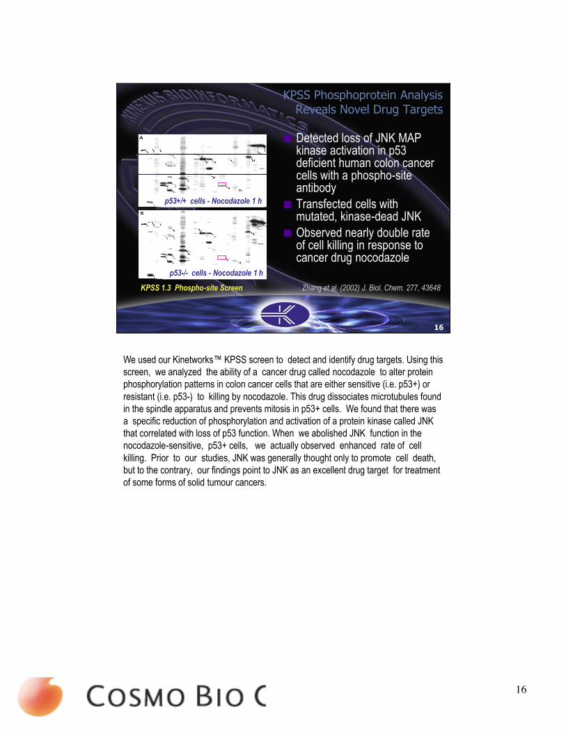
Detected loss of JNK MAPkinase activation in p53deficient human colon cancercells with a phospho-siteantibody
Transfected cells withmutated, kinase-dead JNK
Observed nearly double rateof cell killing in response tocancer drug nocodazole
p53+/+ cells - Nocodazole 1 h
p53-/- cells - Nocodazole 1 h
Zhang et al. (2002) J. Biol. Chem. 277, 43648
KPSS Phosphoprotein AnalysisReveals Novel Drug Targets
16
KPSS 1.3 Phospho-site Screen
We used our Kinetworks™ KPSS screen to detect and identify drug targets. Using thisscreen, we analyzed the ability of a cancer drug called nocodazole to alter proteinphosphorylation patterns in colon cancer cells that are either sensitive (i.e. p53+) orresistant (i.e. p53-) to killing by nocodazole. This drug dissociates microtubules foundin the spindle apparatus and prevents mitosis in p53+ cells. We found that there wasa specific reduction of phosphorylation and activation of a protein kinase called JNKthat correlated with loss of p53 function. When we abolished JNK function in thenocodazole-sensitive, p53+ cells, we actually observed enhanced rate of cellkilling. Prior to our studies, JNK was generally thought only to promote cell death,but to the contrary, our findings point to JNK as an excellent drug target for treatmentof some forms of solid tumour cancers.
20405
CosmoLogo_E

17
Mapping Protein KinaseNetworks
Kinetworks™ analysis has provided importantinsights into the architecture of MAP kinaseand CDK signalling networks. These include:
A. p38 MAPK binds and inhibits ERK1/ERK2B. p38 MAPK binds and activates CK2C. CK2 phosphorylates p53 indirectlyD. p53 mediates activation of JNK MAPKE. CDK1 phosphorylates B23 indirectly via PLK1F. CDK1 phosphorylates and activates STAT3
17
Over the last five years, we have applied the Kinetworks™ approach to uncover manynovel connections between different signalling pathways for MAP kinases and cyclin-dependent kinases. The next few slides provide several examples of where severalimportant insights have been generated and described in scientific publications(reprints are available on this CD) or patent applications.
20405
CosmoLogo_E

18
Mapping Protein KinaseNetworks
18
This slide summarizes the important connections that we have recently establishedbetween protein kinases involved in mitotic control.
20405
CosmoLogo_E

19
19
Kinetworks™ custom analysis has revealed the overproduction of the protein kinasesCK2, p38-alpha MAP kinase and Mek6 in solid human tumours from the breast, lung,liver and colon. While some companies have considered one or more of these proteinkinases as drug targets, our findings tend to invalidate these as suitable candidates.Our data shows that these kinases are integrated into a pathway that becomes upregulated in cancer cells to compensate for a loss of p53 function. Inhibition of thistumour suppressor pathway by drugs would probably serve to worsen the cancerprognosis.
20405
CosmoLogo_E

20
Detected increased phospho-rylation of STAT3 at Ser-727in response to nocodazoletreatment of HeLa cells
Normal
Nocodazole-treated for 24 h
Drug Target Discovery
STAT3
20
KPSS 1.3 Phospho-site Screen Shi et al. (2006) Biochemistry 45, 5857
In another example of the usefulness of the Kinetworks™ analyses to uncover novelphosphorylation events, we used our KPSS 1.3 Phosphoprotein Screen to detectproteins that underwent enhanced phosphorylation in response to treatment of anestablished human cervical cancer cell line to a cancer drug. One of the proteinsthat showed increased phosphorylation in response to nocodazole treatment for 24hours in HeLa cells was STAT3 at Ser-727.
20405
CosmoLogo_E

21
Microtubule disruptingagents causeenhancedphosphorylation ofSer-727 of STAT3
Phosphorylation ofSTAT3 at this siteinhibits itstranscriptional activity
Drug Target Discovery
Untrea
ted
Nocod
azole
Taxol
Vinblas
tine
Colchic
ine
-STAT3
21
Shi et al. (2006) Biochemistry 45, 5857
The effect of nocodazole and other disruptors of microtubule dynamics to enhanceSTAT3 Ser-727 phosphorylation in HeLa cells could be reproduced in Western blots ofcell lysates probed with the STAT3 Ser-727 phospho-site antibody. We have recentlydemonstrated that this appears to be partly mediated through direct phosphorylationby CDK1. Previous to these studies, there have been no reports in the literature thatSTAT proteins are phosphorylated on serine during mitosis.
20405
CosmoLogo_E

22
Detected increased phospho-rylation of unknown proteinin human colon carcinomaHCT-116 tumour cells inresponse to a cancer drugwith a phospho-site antibody
Purified and identified proteinby mass spectrometry as B23(also known asnucleophosmin)
Normal
Nocodazole-treated for 24 h
Drug Target Discovery
B23
STAT3
Zhang et al. (2004) J. Biol. Chem. 279, 35726
22
With nocodazole arrest of many cell lines, as shown above with HCT-116 cells, wealso detected the enhanced phosphorylation of a previously unidentified 40 kDaphosphoprotein (circled in yellow) using our KPSS phospho-site screens. This proteincross-reactived with a phospho-site antibody developed to recognize Mek1 at S217and S221, and it was identified as B23 using mass spectrometry. This protein waspreviously demonstrated to be highly produced in the nuclei of proliferating cells.
20405
CosmoLogo_E

23
Transfected HeLa cervicalcancer cells with mutatedB23 at phosphorylation site
Observed increased nucleisize and increasedcentrosome number
Patent filed for assay forinhibitors of B23 kinase
23
Flag-B23S4E Flag-B23S4A
Flag Tag=Green; Tubulin=Red; DAPI=Blue
DAPI=Red
KPSS Drug Target Discovery
Zhang et al. (2004) J. Biol. Chem. 279, 35726
Since we knew the epitope of the phospho-site antibody that cross-reacted with theB23 protein, we were immediately able to assign the amino acid residue that wasphosphorylated as Ser-4. When HeLa cells were transfected with a mutant B23 thatcould not be phosphorylation [Ser-4 to Ala-4], there was only one or no centromeresdetectable per cell. By contrast, transfection with a mutant B23 to mimic constitutivephosphorylation at this site [Ser-4 to Glu-4] resulted in up to six centrosomes per cell.
20405
CosmoLogo_E

24
24
Untreate
d
Plk1-si
RNA
IB: B23 -
KPSS Drug Target Discovery
IB: B23 pS4 -
IB: Plk1 -
IB: Plk1 -
Autorad32P-B23 -
IB: GST-B23 -
GST-B23
-WT
GST-B23
-S4A
IB: B23 pS4 -
IN VITRO IN VIVO
Plk1 phosphorylates B23 Ser-4
Zhang et al. (2004) J. Biol. Chem. 279, 35726
The Ser-4 site in B23 fitted the consensus phosphorylation site recognition sequencefor the polo-like kinase (Plk). We were able to demonstrate that Plk1 wasphosphorylated and activated in response to nocodazole in HeLa cells, and that thiswas mediated by cyclin-dependent kinase 1. In summary, our study identified a novelnon-radioactive assay for Plk1 and validated it as an appropriate target for drugdiscovery.
20405
CosmoLogo_E

25
KPKS 1.0 Protein KinaseScreen detection ofchanges in expressionlevels of protein kinases inspinal cord tissue fromhuman ALS patients andtransgenic SOD gain-of-function mice revealsstriking differences.
This indicates that theSOD mouse is not anappropriate animal modelof human ALS
Protein Kinase
• CaMKK• CDK5• Erk1• Erk2• Grk2• PKBα• PKCα• PKCβ• PKCζ• PKG• Rsk1• S6K
Human ALS
111% 90% 111% 109% 163% 400% 122% 825% 209% 91% 820% 540%
Mouse SOD
23% 9% 11% 14% 27% 17% 24% 3% 16% 12% 20% 6%
Animal Model Validation
Hu et al. (2003) J. Neurochem. 85:422Hu et al. (2003) J. Neurochem. 85:432
25
More than four out of five drug leads that look promising in animal studies fail in clinicaltrials. This is because the animal models of the disease are not reflective of thehuman situation. We recently completed a Kinetworks™ study in which wediscovered striking changes in the levels of protein kinases and the phosphorylationstates of targets for these kinases in spinal cord samples from patients who havedied from amyotrophic lateral sclerosis (ALS), a devastating neurodegenerativedisease. When we examined the leading transgenic mouse model for this disease,which is used extensively in preclinical studies of lead drugs to treat ALS, weshowed profound differences in the molecular changes in cell signalling in theestablished mouse model. Our findings question the appropriateness of this animalmodel, and our Kinetworks™ analysis could be a useful tool for the identification ofmore relevant animal models for this and other diseases.
20405
CosmoLogo_E

26
Phosphoproteins in HumanALS Spinal Cord
Control ALS Mean± S.Dev. Mean±S.Dev.
(n=5) (n=7)
Adducin alpha p120 (S724) 77 ± 85 12637 ± 13983*
Adducin gamma p80 (S662) 38 ± 26 5481 ± 6598*
GSK3 alpha p44 (Y279) 762 ± 364 1247 ± 464*GSK3 beta p40 (Y216) 177 ± 87 575 ± 288**
PKC alpha/beta p83 (T638) 264 ± 153 1927 ± 1455*
PKR p68 (T451) 14 ± 13 381 ± 299**
S6K p64 (T389) 7 ± 10 78 ± 91*
C1 C2 C3 C4 C5 A1 A2 A3 A4 A5 A6 A7
Control ALS
Hu et al. (2003) J. Neurochem. 85:422
26
When the Kinetworks™ KPSS 1.3 Phospho-Site screen was used to trackphosphoprotein changes in spinal cord sections of patients that died from ALS, therewere several changes in ALS (A) samples as compared to spinal cord samples fromcontrols (C). In particular, there was a more than 100-fold increase in thephosphorylation of adducin at the site targeted by protein kinase C. This was one ofthe protein kinases that were elevated about 9-fold in expression in ALS. Our findingspoint to protein kinase C as a potential drug target for treatent of ALS.
20405
CosmoLogo_E

27
THE SYSTEMSPROTEOMICS
COMPANY
27
We hope that these slides offer a sense of the power of our proteomics services. Wefeel that our services can position our clients at the cusp of a profound paradigm shiftin the way that research is conducted today and ultimately we hope to contribute to therealization of the goal of the delivery of personalized medicine. We believe that byeavesdropping on the molecular communications systems that operate in all of theliving cells of the human body, we can learn more about what has gone awry inthese cells during disease and assist in the healing process in a more rational waybased on deeper understanding. This will revolutionize medicine and improve humanwell being in ways that have been unprecedented and not previously feasible.
20405
CosmoLogo_E


![Receptor-Like Kinases Sustain Symbiotic Scrutiny1[OPEN]...Update on Receptor-Like Kinases in Symbiosis Receptor-Like Kinases Sustain Symbiotic Scrutiny1[OPEN] Chai Hao Chiu,2 and Uta](https://static.fdocuments.us/doc/165x107/60aa214268722c0ce00ae5e7/receptor-like-kinases-sustain-symbiotic-scrutiny1open-update-on-receptor-like.jpg)