Examining the Fingernails When Evaluating Presenting Symptoms in Elderly Patients
32
Examining the Fingernails When Evaluating Presenting Symptoms in Elderly Patients Mark E. Williams, MD Authors and Disclosures Posted: 03/26/2008 Introduction Human fingernails, located on t he dorsal aspect of the terminal 40% o f the distal phalanx of each finger, are complex structures involving 3 d ifferent layers: y The nail plate (the nail). This is the keratinized structure, which grows throughout life; y The nail bed (ventral matrix, sterile matrix). This is the vascular bed that is responsible for nail growth and support. It lies protected between the lunula (the "half moon" seen through the nail) and the hyponychium (the posterior part of the nail bed epithelium); and y The eponychium (cuticle). The epidermal layer between the proximal nail fold and the dorsal aspect of the nail plate. The primary purpose of the nail is protection. Abnormalities of the nail are often caused by skin disease and infection (most often fungal) but may also indicate more general medical conditions. This discussion does not address localized trauma o r nail infections but offers examples of nail abnormalities that may occur with systemic disease. Check to see whether the nails are normal by looking at the following (Figure 1): y Softness and flexibility of free edge; y Shape and color; y Q uality of paronychial tissue; and y Growth rate (about 6 months from cuticle to free edge). Time of e vents can be estimated from location.
-
Upload
annajosephineyunita -
Category
Documents
-
view
244 -
download
1
Transcript of Examining the Fingernails When Evaluating Presenting Symptoms in Elderly Patients

8/9/2019 Examining the Fingernails When Evaluating Presenting Symptoms in Elderly Patients
http://slidepdf.com/reader/full/examining-the-fingernails-when-evaluating-presenting-symptoms-in-elderly-patients 1/32
Examining the Fingernails When Evaluating
Presenting Symptoms in Elderly Patients
Mark E. Williams, MD
Authors and Disclosures
Posted: 03/26/2008
Introduction
Human fingernails, located on the dorsal aspect of the terminal 40% of the distal phalanx of eachfinger, are complex structures involving 3 different layers:
y The nail plate (the nail). This is the keratinized structure, which grows throughout life;
y The nail bed (ventral matrix, sterile matrix). This is the vascular bed that is responsible for nail
growth and support. It lies protected between the lunula (the "half moon" seen through the
nail) and the hyponychium (the posterior part of the nail bed epithelium); and
y The eponychium (cuticle). The epidermal layer between the proximal nail fold and the dorsal
aspect of the nail plate.
The primary purpose of the nail is protection. Abnormalities of the nail are often caused by skin
disease and infection (most often fungal) but may also indicate more general medical conditions.This discussion does not address localized trauma or nail infections but offers examples of nail
abnormalities that may occur with systemic disease.
Check to see whether the nails are normal by looking at the following (Figure 1):
y Softness and flexibility of free edge;
y Shape and color;
y Q uality of paronychial tissue; and
y Growth rate (about 6 months from cuticle to free edge). Time of events can be estimated from
location.

8/9/2019 Examining the Fingernails When Evaluating Presenting Symptoms in Elderly Patients
http://slidepdf.com/reader/full/examining-the-fingernails-when-evaluating-presenting-symptoms-in-elderly-patients 2/32
Figure 1. The normal nail.
Examining the Nails
Elderly people carry the last 6 months of their medical record on the approximately 10 squarecentimeters of keratin comprising the fingernails. Examining the fingernails can help the
clinician detect a number of general and specific factors, including the following:
y Overall vitality;
y Inner emotional state;
y Cerebral dominance;
y Occupations and hobbies;
y Medical history;
y Nutritional status;
y Cardiovascular function;
y Rheumatic conditions; and
y Dermatologic problems.

8/9/2019 Examining the Fingernails When Evaluating Presenting Symptoms in Elderly Patients
http://slidepdf.com/reader/full/examining-the-fingernails-when-evaluating-presenting-symptoms-in-elderly-patients 3/32
The patient's manicure can reveal state of health, nutritional status, past events, personality,occupation, and one's inner state. Systemic illness should show the nail changes in each of the
nails on one hand. The thumb may reveal more extensive changes given its increased size.
It is useful to follow the following sequence when examining the nails:
y Check the nail shape;
y Examine the nail color;
y Survey processes around the nails;
y Compare hands; and
y Note skin conditions.
It is critical to examine the nails in adequate light. Gently rotate the nail in the light so that the
reflection highlights all aspects of the nail. Notice the lunula, the pale crescent moonlikecoloration at the base of the nail. Leukonychia stria and a pointed tent-like lunula suggest anexcessive manicure and pushing on the cuticle. Paronychias suggest stress and poor attention to
hygiene. This can reflect depression, dementia, or psychiatric illness.
Nail Growth
Nail growth is continuous. It takes about 6 months for a fingernail in an elderly person to
completely grow out. Cold temperature can slow growth rates but not to any clinically significant
degree (pun intended). The middle finger nail grows the fastest, followed by the forefinger andring finger. Aging slows the growth rate from approximately 3 months in childhood to 6 months
in 70-year-olds. Nails in elderly people are also thicker than in younger people. Thin nails in a postmenopausal woman raise the possibility of metabolic bone disease. The nails of the
dominant hand grow slightly more quickly than the nondominant nails, probably because minor trauma accelerates nail growth. Conversely, immobility slows the growth rate of fingernails.
Understanding the growth rate is important because the time interval from a critical event can beestimated from the location of a nail lesion. For example, a white line appearing transversely
halfway up the nail suggests an acute illness 3 months earlier. Regular observation willdemonstrate its progression to the end of the nail edge.
Nail Polish
Distance from base and line of polish gives approximate date of application (nails grow 0.1mm/day). Picking at polish reflects nervousness and agitation. Toenail polish suggests unusualflexibility or a friendly helper.

8/9/2019 Examining the Fingernails When Evaluating Presenting Symptoms in Elderly Patients
http://slidepdf.com/reader/full/examining-the-fingernails-when-evaluating-presenting-symptoms-in-elderly-patients 4/32

8/9/2019 Examining the Fingernails When Evaluating Presenting Symptoms in Elderly Patients
http://slidepdf.com/reader/full/examining-the-fingernails-when-evaluating-presenting-symptoms-in-elderly-patients 5/32
Figure 3. Example of clubbed fingernails.
K oilonychia
K oilonychia are spoon-shaped concave nails (Figures 4A, 4B). This occurs normally in children
and usually resolves with aging. To determine whether a nail is spooned, perform the water droptest. Place a drop of water on the nail. If the drop does not slide off, then the nail is flattened
from early spooning. An experienced clinician can look at the nail and perform a "mental" water drop test. Causes include the following:
y Iron deficiency;
y Diabetes mellitus;
y Protein deficiency, especially in sulfur-containing amino acids (cysteine or methionine);
y Exposure to petroleum-based solvents;
y Systemic lupus erythematosus; and
y Raynaud's disease.

8/9/2019 Examining the Fingernails When Evaluating Presenting Symptoms in Elderly Patients
http://slidepdf.com/reader/full/examining-the-fingernails-when-evaluating-presenting-symptoms-in-elderly-patients 6/32
Figure 4A. Spooned nail.
Figure 4B. Spooned nail.
In 1846, Joseph Honoré Simon Beau described transverse lines in the substance of the nail as
signs of previous acute illness. The lines look as if a little furrow had been plowed across thenail. Illnesses producing Beau's lines include the following:
y Severe infection;
y Myocardial infarction;
y Hypotension, shock;
8/9/2019 Examining the Fingernails When Evaluating Presenting Symptoms in Elderly Patients
http://slidepdf.com/reader/full/examining-the-fingernails-when-evaluating-presenting-symptoms-in-elderly-patients 7/32
y Hypocalcemia; and
y Surgery.
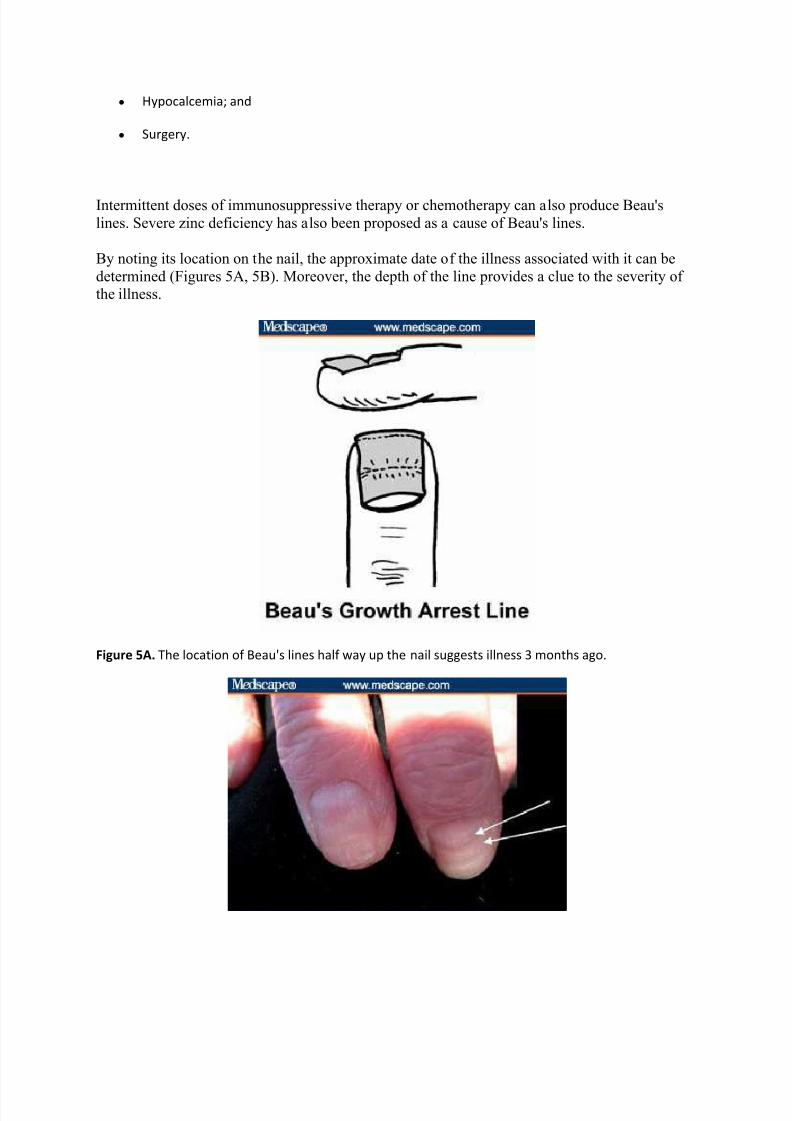
Intermittent doses of immunosuppressive therapy or chemotherapy can also produce Beau's
lines. Severe zinc deficiency has also been proposed as a cause of Beau's lines.
By noting its location on the nail, the approximate date of the illness associated with it can be
determined (Figures 5A, 5B). Moreover, the depth of the line provides a clue to the severity of the illness.
Figure 5A. The location of Beau's lines half way up the nail suggests illness 3 months ago.
8/9/2019 Examining the Fingernails When Evaluating Presenting Symptoms in Elderly Patients
http://slidepdf.com/reader/full/examining-the-fingernails-when-evaluating-presenting-symptoms-in-elderly-patients 8/32

8/9/2019 Examining the Fingernails When Evaluating Presenting Symptoms in Elderly Patients
http://slidepdf.com/reader/full/examining-the-fingernails-when-evaluating-presenting-symptoms-in-elderly-patients 9/32
y Folic acid deficiency; and
y Protein deficiency.
Figure 7. Example of a central nail ridge.
C entral Nail C anal (Median Nail Dystrophy)
When a central nail canal is present, the cuticle is usually normal (Figure 8A). Central nail canalis associated with:
y Severe arterial disease ("Heller's fir tree deformity" -- a central canal with a fir tree appearance -
- may occur with peripheral artery disease (Figure 8B);
y Severe malnutrition; and
y Repetitive trauma.

8/9/2019 Examining the Fingernails When Evaluating Presenting Symptoms in Elderly Patients
http://slidepdf.com/reader/full/examining-the-fingernails-when-evaluating-presenting-symptoms-in-elderly-patients 10/32
Figure 8A. Example of central nail canal.
Figure 8B. Central nail canal with Heller's fir tree deformity.
Nail Pitting
Nail pitting -- small punctate depressions -- are caused by nail matrix inflammation, which can
be the result of:
y Psoriasis (random appearance of pits) (Figure 9);
y Alopecia areata (geometric rippled grid) (Figure 10);
y Eczema; and

8/9/2019 Examining the Fingernails When Evaluating Presenting Symptoms in Elderly Patients
http://slidepdf.com/reader/full/examining-the-fingernails-when-evaluating-presenting-symptoms-in-elderly-patients 11/32
y Lichen planus.
Figure 9. Indication of psoriasis.

8/9/2019 Examining the Fingernails When Evaluating Presenting Symptoms in Elderly Patients
http://slidepdf.com/reader/full/examining-the-fingernails-when-evaluating-presenting-symptoms-in-elderly-patients 12/32
Figure 10. Indication of alopecia areata.
Nail Beading
With nail beading, the beads seem to drip down the nail like wax (Figure 11). It is associated
with endocrine conditions, including the following:
y Diabetes mellitus;
y Thyroid disorders;
y Addison's disease; and
y Vitamin B deficiency.
Figure 11. Nail beading.
Rough Nail Surface
When nails look sandpapered and dull, consider (Figure 12):
y Autoimmune disease;
y Psoriasis;
y Chemical exposure; and
y Lichen planus.

8/9/2019 Examining the Fingernails When Evaluating Presenting Symptoms in Elderly Patients
http://slidepdf.com/reader/full/examining-the-fingernails-when-evaluating-presenting-symptoms-in-elderly-patients 13/32
Figure 12. Example of a rough nail surface.
Nail T hickening
Slow nail growth produces thickness (Figure 13). In such cases, the following should be
considered:
y Onychomycosis;
y Chronic eczema;
y Peripheral vascular disease;
y Yellow nail syndrome; and
y Psoriasis.

8/9/2019 Examining the Fingernails When Evaluating Presenting Symptoms in Elderly Patients
http://slidepdf.com/reader/full/examining-the-fingernails-when-evaluating-presenting-symptoms-in-elderly-patients 14/32
Figure 13. Example of a nail thickening.
Onycholysis
Onycholysis is distal separation of the nail plate from the underlying nail bed (Figure 14). It is
associated with the following:
y Thyrotoxicosis;
y Psoriasis;
y Trauma;
y Contact dermatitis;
y Tetracycline;
y Eczema;
y Toxic exposures (solvents);
y Blistering from autoimmune disease; and
y Porphyria cutanea tarda (onycholysis and skin blistering from sun exposure).

8/9/2019 Examining the Fingernails When Evaluating Presenting Symptoms in Elderly Patients
http://slidepdf.com/reader/full/examining-the-fingernails-when-evaluating-presenting-symptoms-in-elderly-patients 15/32
Figure 14. Traumatic onycholysis (involving only 1 nail).
Severe Nail C urvature (Beaked Nails)
Curved or beaked nails are caused by resorption of distal digit (Figure 15). Consider the
following:
y Hyperparathyroidism
y Renal failure
y Psoriasis
y Systemic sclerosis

8/9/2019 Examining the Fingernails When Evaluating Presenting Symptoms in Elderly Patients
http://slidepdf.com/reader/full/examining-the-fingernails-when-evaluating-presenting-symptoms-in-elderly-patients 16/32
Figure 15. Example of severe nail curvature.
C omplete Nail Destruction
Complete local nail destruction can be caused by local mechanisms, including trauma and
paronychia. Generalized conditions that might cause complete nail destruction include thefollowing:
y Toxic epidermal necrolysis;
y Chemotherapy;
y Bullous diseases; and
y Vasculitis.
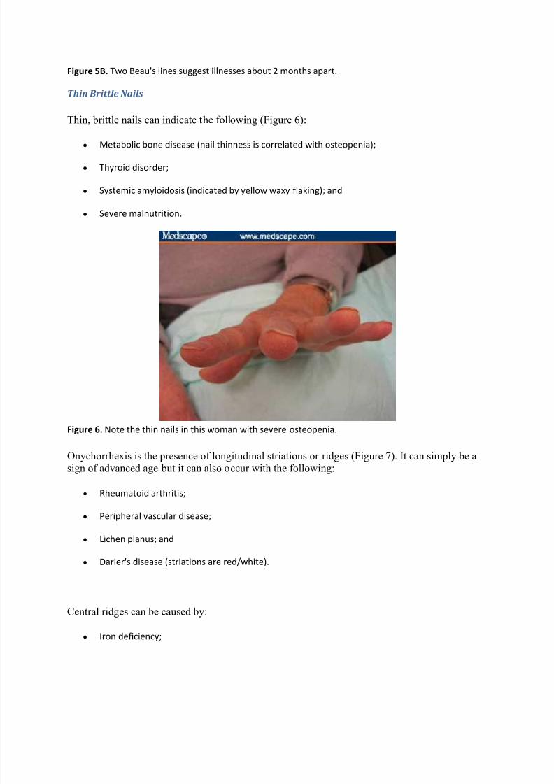
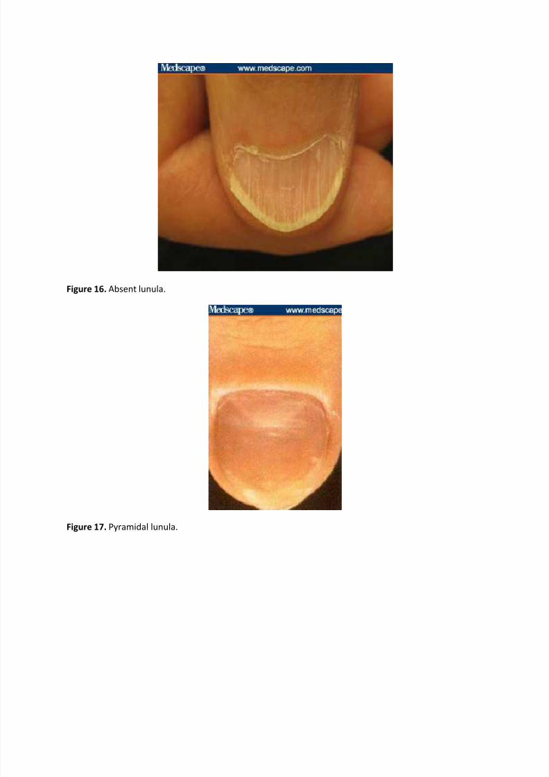
Observing Nail Color
Abnormalities of the Lunula
If the lunula is absent, consider anemia or malnutrition (Figure 16). A pyramidal lunula mightindicate excessive manicure or trauma (Figure 17). A pale blue lunula suggests diabetes mellitus.
If the lunula has red discoloration, consider the following causes among others (Figure 18):
y Cardiovascular disease;
y Collagen vascular disease; and
y Hematologic malignancy.
8/9/2019 Examining the Fingernails When Evaluating Presenting Symptoms in Elderly Patients
http://slidepdf.com/reader/full/examining-the-fingernails-when-evaluating-presenting-symptoms-in-elderly-patients 17/32
Figure 16. Absent lunula.
Figure 17. Pyramidal lunula.

8/9/2019 Examining the Fingernails When Evaluating Presenting Symptoms in Elderly Patients
http://slidepdf.com/reader/full/examining-the-fingernails-when-evaluating-presenting-symptoms-in-elderly-patients 18/32
Figure 18. Lunula with red discoloration.
T ransverse White Lines (Mee's lines)
Any acute illness can produce transverse milky white lines. In addition, they might be caused by
heavy metal toxicity (classically arsenic) or chemotherapy. The time of event may be determinedfrom the location of the lines on nail (Figure 19).
Figure 19. Note the Mee's line approximately one third of the way up the nail, suggesting a significant
illness 2 months previously.
Leukonychia Striae
Leukonychia striae are white splotches caused by minor trauma to the nail matrix (Figure 20).
The timing can be determined by the location of the splotches on the nail.

8/9/2019 Examining the Fingernails When Evaluating Presenting Symptoms in Elderly Patients
http://slidepdf.com/reader/full/examining-the-fingernails-when-evaluating-presenting-symptoms-in-elderly-patients 19/32
Figure 20. Example of leukonychia striae.Note location of white splotches, which can indicate timing of the traumatic event.
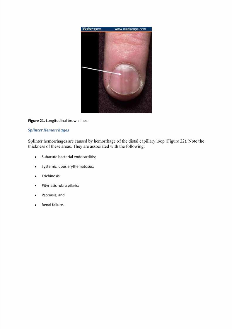
Longitudinal Brown Lines
Longitudinal brown lines form because of increased melanin produced by nail matrix
melanocytes (Figure 21). They are associated with:
y Addison's disease;
y Nevus at the nail base;
y Breast cancer;
y Melanoma (check for periungal pigmentation); and
y Trauma.
8/9/2019 Examining the Fingernails When Evaluating Presenting Symptoms in Elderly Patients
http://slidepdf.com/reader/full/examining-the-fingernails-when-evaluating-presenting-symptoms-in-elderly-patients 20/32

8/9/2019 Examining the Fingernails When Evaluating Presenting Symptoms in Elderly Patients
http://slidepdf.com/reader/full/examining-the-fingernails-when-evaluating-presenting-symptoms-in-elderly-patients 21/32
Figure 22. Splinter hemorrhages tend to be fat.
T erry's Half and Half Nails
With Terry's half and half nails, the proximal portion is white (edema and anemia) and the distal
portion is dark. These nails imply either renal or liver disease (Figures 23A, 23B).
Figure 23A. This example of Terry's half and half nails suggests liver disease (no brown lines).

8/9/2019 Examining the Fingernails When Evaluating Presenting Symptoms in Elderly Patients
http://slidepdf.com/reader/full/examining-the-fingernails-when-evaluating-presenting-symptoms-in-elderly-patients 22/32
Figure 23B. Half and half nails imply renal disease when there is a brown band at the junction of the
erythema and the free edge. Image courtesy of www.dermnet.com Used with permission.
Generalized Discolorations of the Nail Plat e
Nail discoloration is a useful method for identifying potential problems.
White Nails
White nails can be caused by anemia, edema, or vascular conditions (Figure 24). Consider thefollowing:
y Anemia;
y Renal failure;
y Cirrhosis;
y Diabetes mellitus;
y Chemotherapy; and
y Hereditary (rare).

8/9/2019 Examining the Fingernails When Evaluating Presenting Symptoms in Elderly Patients
http://slidepdf.com/reader/full/examining-the-fingernails-when-evaluating-presenting-symptoms-in-elderly-patients 23/32

8/9/2019 Examining the Fingernails When Evaluating Presenting Symptoms in Elderly Patients
http://slidepdf.com/reader/full/examining-the-fingernails-when-evaluating-presenting-symptoms-in-elderly-patients 24/32
Figure 25. Example of pink and red nails.
Brown-Gray Nails
Brown-gray nails may suggest the following (Figure 26):
y Cardiovascular disease;
y Diabetes mellitus;
y Vitamin B12 deficiency;
y Breast cancer;
y Malignant melanoma;
y Lichen planus;
y Syphilis; and
y Topical agents, including hair dyes, solvents for false nails, varnish, and formaldehyde (among
many others)

8/9/2019 Examining the Fingernails When Evaluating Presenting Symptoms in Elderly Patients
http://slidepdf.com/reader/full/examining-the-fingernails-when-evaluating-presenting-symptoms-in-elderly-patients 25/32
Figure 26. Example of brown-gray nails.
Y ellow Nails
Yellow nails suggest the following (Figure 27):
y Diabetes mellitus;
y Amyloidosis;
y Median/ulnar nerve injury;
y Thermal injury; and
y
aundice.
Consider yellow nail syndrome if a patient has lymphedema and bronchiectasis.

8/9/2019 Examining the Fingernails When Evaluating Presenting Symptoms in Elderly Patients
http://slidepdf.com/reader/full/examining-the-fingernails-when-evaluating-presenting-symptoms-in-elderly-patients 26/32
Figure 27. Example of yellow nails. Image courtesy of www.dermnet.com Used with permission.
Green or Black Nails
Green or black nails indicate the following (Figure 28):
y Topical preparations, including chlorophyll derivations, methyl green, and silver nitrate (among
others);
y Chronic Pseudomonas spp infection; and
y Trauma.
Figure 28. Example of black nails.

8/9/2019 Examining the Fingernails When Evaluating Presenting Symptoms in Elderly Patients
http://slidepdf.com/reader/full/examining-the-fingernails-when-evaluating-presenting-symptoms-in-elderly-patients 27/32
Processes Around the Nail
Paronychial Inflammation
Paronychia is associated with separation of the seal between the proximal nail fold and the nail
plate that provides entry for bacteria and leads to a localized infection of the paronychial tissues
of the hands (Figure 29). Symptoms may include inflammation, swelling, and/or scaling.
Figure 29. Example chronic paronychial inflammation.
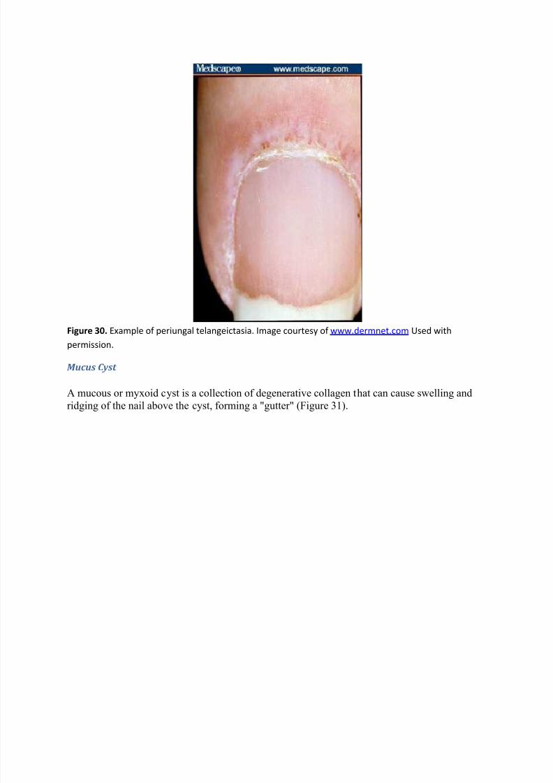
Periungal T elangeictasia
Periungal telangeictasia is caused by dilated capillary loops and results in atrophy of the cuticle(Figure 30). It is strongly associated with collagen vascular disease, including the following:
y Systemic lupus erythematosus;
y Dermatomyositis (especially withGotton's papules over knuckles); and
y Scleroderma.
8/9/2019 Examining the Fingernails When Evaluating Presenting Symptoms in Elderly Patients
http://slidepdf.com/reader/full/examining-the-fingernails-when-evaluating-presenting-symptoms-in-elderly-patients 28/32
Figure 30. Example of periungal telangeictasia. Image courtesy of www.dermnet.com Used with
permission.
Mucus C yst
A mucous or myxoid cyst is a collection of degenerative collagen that can cause swelling and
ridging of the nail above the cyst, forming a "gutter" (Figure 31).

8/9/2019 Examining the Fingernails When Evaluating Presenting Symptoms in Elderly Patients
http://slidepdf.com/reader/full/examining-the-fingernails-when-evaluating-presenting-symptoms-in-elderly-patients 29/32
Figure 31. Example of a mucus cyst.
Cases
The following are examples of patients in whom examining the fingernails may help identify
their conditions.

8/9/2019 Examining the Fingernails When Evaluating Presenting Symptoms in Elderly Patients
http://slidepdf.com/reader/full/examining-the-fingernails-when-evaluating-presenting-symptoms-in-elderly-patients 30/32
Slide 1. 78-year-old with multiple conditions.
Slide 2. 84-year-old man with a painful ankle.

8/9/2019 Examining the Fingernails When Evaluating Presenting Symptoms in Elderly Patients
http://slidepdf.com/reader/full/examining-the-fingernails-when-evaluating-presenting-symptoms-in-elderly-patients 31/32
Slide 3. 68-year-old man with esophageal cancer.
Slide 4. 62-year-old woman with dermatomyositis.

8/9/2019 Examining the Fingernails When Evaluating Presenting Symptoms in Elderly Patients
http://slidepdf.com/reader/full/examining-the-fingernails-when-evaluating-presenting-symptoms-in-elderly-patients 32/32
Acknowledgments
The author would like to thank the University of Virginia GME Office for funding support; Jim
Thomas, MD, of www.Dermnet.com for permission to use images from their extensivedermatologic atlas; and the internal medicine residents at the University of Virginia for pre-
testing and their helpful feedback.